
Table of Contents
ToggleAsphyxia neonatorum
Asphyxia neonatorum is one of the pediatric emergencies and is the leading cause of neonatal mortality and morbidity. It is also an important cause of developmental delay and neurological problems in both term and preterm infants. It is crucial for midwives and nurses to have the knowledge and skills to care for babies with this condition.
Definition of asphyxia neonatorum
This is a failure of the baby to initiate and sustain normal respiration at birth.
A normal baby has good muscle tone at birth and moves their arms and legs actively, while asphyxia neonatorum infants are completely limp and unable to move their limbs.
This condition is a neonatal emergency as it may lead to hypoxia (lowering of oxygen supply to the brain and tissues), and possible brain damage or death if not correctly managed.
Types of Asphyxia
Asphyxia livida (Blue asphyxia) or stage of cyanosis:
- Primarily due to respiratory failure with Apgar score 4-6.
- The most common cause is the blockage of the airway.
Asphyxia pallida or stage of shock:
- This is due to combined respiratory and vasomotor failure with Apgar score 0-3.
- Depending on the Apgar scoring system, a score of 0-3 indicates severe depression, 4-6 indicates moderate depression, and 7-10 indicates no depression.
Pathophysiology of asphyxia
Birth asphyxia is related to a reduction in arterial oxygen tension, accumulation of carbon dioxide, and a fall in pH. Acidosis occurs due to the anaerobic utilization of glucose, production of lactic acid, and accumulation of carbon dioxide.
These biochemical changes result in constriction of muscular pulmonary arterioles and raised pulmonary arterial pressure, leading to reduced filling of the left heart.
Hypoglycemia occurs due to glucose utilization and depletion of glycogen stores. Petechial hemorrhage occurs due to anorexic capillary changes. Cerebral edema develops due to the intracellular collection of sodium and inappropriate release of ADH.
In prolonged asphyxia, myocardial function and cardiac output deteriorate. Blood flow to all organs is reduced, and progressive organ damage results.
Initial deprivation of oxygen results in rapid breathing. If asphyxia continues, respiratory movements stop, and the heart rate begins to fall with the gradual diminution of neuromuscular tone. Then, the baby enters a period of apnea known as primary apnea. In this stage, stimulation and exposure to oxygen may induce respiration.
But if asphyxia continues, the neonate develops deep gasping respiration, blood pressure falls, the baby becomes flaccid, respiration becomes weaker, and weaker until the neonate takes a last gasp and enters a period of secondary apnea.
The baby becomes unresponsive to stimulation and will not spontaneously resume respiratory efforts unless resuscitation with assisted ventilation and oxygenation is initiated promptly.
Primary and secondary apnea are difficult to distinguish, and all apnea at birth should be considered as secondary apnea, requiring immediate resuscitation to prevent brain damage and multi-organ system dysfunction.
Signs & Symptoms
- The baby does not breathe but may make an attempt to breathe or gasp.
- The period of apnea is usually short (less than 30 seconds) but cries vigorously.
- The color is blue.
- Muscle tone is good.
- The cord is pulsating strongly and feels firm.
- The heartbeat is strong but rather slow.
- Apgar score is 4 – 6.
Aetiology
Approximately 90% of asphyxia events occur as a result of placental insufficiency due to ante partum and intra partum factors. Postnatal factors account for the remaining.
Ante partum factors include:
- Placental insufficiency due to conditions like pre-eclampsia, hypertension, anemia, diabetes mellitus, and post-maturity.
Other factors like;
- Antepartum hemorrhage,
- Malpresentation,
- Multiple pregnancies,
- Poor fetal growth,
- Rhesus immunization, bad obstetrical history, maternal systemic diseases (e.g., asthma, heart disease),
- Polyhydramnios or oligohydramnios,
- Maternal drug therapy (e.g., lithium) or maternal drug abuse,
- Vascular anomalies of the cord, and congenital anomalies of the fetus.
Intrapartum factors include:
- Fetal distress,
- Preterm labor,
- Antepartum hemorrhage (placenta previa, abruptio placentae),
- Cord prolapse, tight umbilical cord around the fetal neck,
- Prolonged labor exceeding 24 hours, prolonged second stage lasting more than 2 hours, maternal distress (dehydration, hypotension, and acidosis),
- The use of anesthesia and narcotics during labor,
- Birth trauma resulting in increased intracranial pressure due to hemorrhage, and difficult deliveries in malpresentation.
Postnatal factors include;
- They are mainly related to pulmonary, cardiovascular, and neurological abnormalities of the neonate,
- Including aspiration causing airway obstruction,
- Circulatory collapse due to blood loss and shock, preterm birth resulting in weak respiratory muscles,
- Poor pulmonary expansion, low alveolar surfactant, and an inefficient respiratory center.
Management
Maternity center:
- A baby of this type of Asphyxia responds to treatment promptly.
- As soon as the baby’s head is delivered, clear the airway and suck out the mucus from the nose with a mucus extractor.
- When a baby is completely delivered, put it over the mother’s abdomen and continue sucking out mucus.
- Clamp and cut the cord and separate the baby from the mother. Apgar score is assessed within one minute.
- In the absence of any respiratory effort, resuscitation measures are commenced.
- Put the baby on the resuscitation table, position the baby with the head slightly extended and the baby lying flat (NEUTRAL position), continue to clear the airway.
- Maintain warmth throughout the procedure.
- Give 0₂, 1 liter per minute (PRN).
In hospital management:
- The management is the same as that in a maternity center, except in the hospital, the doctor has to be informed, and oxygen must be administered.
- If necessary, place the baby in a cot with the head turned to one side.
- In summary:
- Put the baby in a neutral position.
- Clear the airway.
- Give oxygen.
Updated Evidence-Based Newborn Resuscitation and Nursing Care
Important: failure to breathe at birth requires immediate neonatal resuscitation. A low Apgar score alone does not diagnose birth asphyxia. Effective lung ventilation is the most important lifesaving intervention.
Preparation before every birth
- Identify maternal and intrapartum risks, assign trained roles and call additional skilled help for high-risk birth.
- Check warmth, dry towels, timer, correctly sized masks, bag/T-piece, air/oxygen source, suction for obstruction only, pulse oximeter, advanced-airway equipment and emergency medicines.
- Apply hand hygiene and test equipment before delivery.
Rapid assessment and first minute
- Ask: Is the baby term? Is there good tone? Is the baby breathing or crying? A vigorous baby receives routine warmth, skin-to-skin contact and breastfeeding support.
- For a non-vigorous baby: provide warmth, position the head neutrally/slightly extended, dry the term baby, remove wet linen and stimulate briefly.
- Clear secretions only when obstructing spontaneous breathing or effective ventilation. Routine suction causes delay, bradycardia and trauma.
- Assess breathing and heart rate. Start positive-pressure ventilation (PPV) promptly for apnoea, gasping or heart rate below 100/min.
Positive-pressure ventilation
- Use a correctly sized mask covering chin, mouth and nose without the eyes. Begin with room air for most term/late-preterm infants and titrate oxygen by gestation, response and protocol.
- Observe chest movement and heart-rate rise. If the chest does not move, correct mask seal, reposition the airway, suction only if obstructed, open the mouth, adjust pressure and consider an alternative airway.
- After about 30 seconds of effective PPV: if heart rate is at least 100/min and breathing is adequate, gradually stop support; at 60–99/min continue and correct ventilation; below 60/min begin coordinated chest compressions while ensuring ventilation is effective.
Chest compressions and advanced care
- Use the two-thumb encircling-hands technique on the lower third of sternum, compressing about one-third of chest depth.
- Coordinate 3 compressions to 1 ventilation—approximately 90 compressions and 30 breaths per minute.
- Reassess after about 60 seconds. Stop compressions when heart rate reaches 60/min or more, but continue ventilation until breathing and heart rate recover.
- If heart rate remains below 60/min despite verified effective ventilation and compressions, the advanced team may give adrenaline/epinephrine by neonatal protocol. Consider isotonic volume only when blood loss or shock is suspected.
Oxygen, glucose and medicines
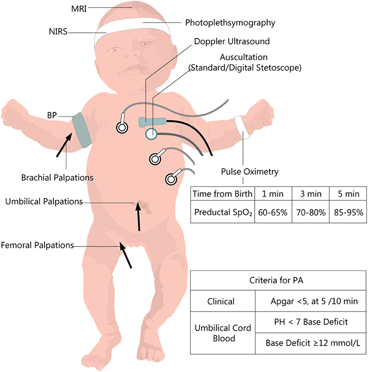
- Use preductal pulse oximetry on the right hand/wrist when prolonged resuscitation, oxygen or CPAP is required; titrate toward minute-specific newborn targets.
- Do not give routine sodium bicarbonate during brief resuscitation.
- Dexamethasone does not initiate breathing and is not a resuscitation drug.
- Assess and treat hypoglycaemia after stabilization using neonatal protocol; concentrated dextrose is not routine initial resuscitation.
Post-resuscitation Care
| Priority | Nursing actions | Escalation signs |
|---|---|---|
| Breathing | Continuous respiratory, heart-rate and SpO2 assessment; prescribed oxygen/CPAP/ventilation; blood gas where available. | Apnoea, grunting, recession, cyanosis, poor effort or rising oxygen need. |
| Temperature | Dry/warm, use controlled warmer/incubator when unstable and monitor temperature. | Hypothermia, hyperthermia, lethargy or hypoglycaemia. |
| Circulation | Perfusion, pulses, BP where indicated, capillary refill, urine and cautious prescribed fluid. | Weak pulses, prolonged refill, hypotension, oliguria or acidosis. |
| Neurology | Assess consciousness, tone, suck and seizures; minimize handling and refer urgently for encephalopathy. | Seizures, reduced consciousness, abnormal tone or loss of feeding reflexes. |
| Feeding/glucose | Check serial glucose; assess swallowing and stability before oral feeds; support expressed breast milk by safe route. | Choking, apnoea, severe distress, low glucose or feed intolerance. |
Hypoxic-ischaemic encephalopathy
Altered consciousness, abnormal tone/reflexes, weak suck, seizures, disordered breathing and multiorgan injury suggest HIE. Moderate–severe HIE needs urgent specialist care. Therapeutic hypothermia is a time-critical specialist treatment for selected near-term/term infants under a controlled protocol; uncontrolled cooling is unsafe.
Nursing Care Plan
| No. | Intervention | Rationale/evaluation |
|---|---|---|
| 1 | Maintain neutral airway and verify visible chest movement with PPV. | Ventilation failure is the commonest reason for persistent bradycardia. |
| 2 | Monitor heart rate, breathing, SpO2, temperature, glucose, perfusion and urine at severity-based intervals. | Detects recurrence and multiorgan injury early. |
| 3 | Use exact dose/concentration checks for all emergency medicines and infusions. | Newborn dosing errors cause severe harm. |
| 4 | Explain events honestly to the family, avoid blame and support safe contact. | Reduces anxiety and supports bonding and informed follow-up. |
Prevention and Follow-up
- Quality antenatal care, maternal infection treatment, skilled birth attendance, fetal/labour monitoring and timely obstetric referral.
- Resuscitation-skilled personnel and checked equipment at every birth, with regular simulation and audit.
- After significant resuscitation, follow growth, feeding, hearing, vision, tone and development; refer early for rehabilitation.
- Document time of birth, condition, every intervention and response, Apgar scores, family communication and transfer handover.
References
Note: The updated resuscitation guidance above replaces older routine suction and drug practices.
The notes are well summerised and nice to read