Nurses Revision

Uncategorized

NSAIDs & Prostanoids

NSAIDS & Prostanoids

NSAIDs & Prostanoids Pharmacology

Module Overview

This master guide covers the pharmacology of Non-Steroidal Anti-Inflammatory Drugs (NSAIDs) and Prostanoids. We will explore the Arachidonic Acid pathway, the profound differences between COX-1 and COX-2, specific drug classifications, and the synthetic prostanoids used to manipulate everything from childbirth to glaucoma. Enhanced with clinical scenarios and deep-dive explanations to guarantee exam success.


1. The Foundation: Prostanoids and the MOA of NSAIDs

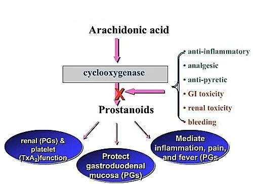
Before understanding the drugs, you must understand the assembly line that makes the molecules these drugs block. This is the Arachidonic Acid Pathway. Think of this pathway as a factory that takes raw materials from the cell wall and turns them into highly active chemical messengers.

MEMBRANE PHOSPHOLIPIDS


Enzyme: Phospholipase A2 (BLOCKED by Corticosteroids)

ARACHIDONIC ACID

Lipoxygenase

Leukotrienes
(Cause Bronchospasm/Asthma)

Cyclooxygenase (COX)
(BLOCKED by NSAIDs)

PGG2 → PGH2
(Endoperoxides)

PROSTANOIDS:
Prostaglandins (PGE2, PGF2α, PGD2)
Thromboxane (TXA2)
Prostacyclin (PGI2)

Clinical Pearl

Steroids vs. NSAIDs & The "Shunt" Phenomenon

Notice that Corticosteroids block the pathway at the very top (Phospholipase). Therefore, steroids stop BOTH Leukotrienes (which cause asthma) and Prostanoids. NSAIDs only block the COX enzyme lower down.

This means NSAIDs stop pain and fever (Prostanoids) but do nothing to stop Leukotrienes. In fact, in some asthma patients, giving an NSAID creates a "Leukotriene Shunt". Because the COX pathway is blocked, all the built-up Arachidonic acid is violently pushed down the Lipoxygenase pathway, causing a massive overproduction of Leukotrienes. This triggers a severe, life-threatening asthma attack (a condition clinically known as Aspirin-Exacerbated Respiratory Disease or AERD).

The Cyclooxygenase (COX) Isozymes: The "Housekeeper" vs. The "Fire Alarm"

The COX enzyme comes in different versions (isoforms). Knowing the difference is the absolute key to understanding NSAID side effects and why pharmaceutical companies spent billions inventing specific COX-2 inhibitors.

COX-1 (The Housekeeper) COX-2 (The Fire Alarm) COX-3 (The Mystery)
  • Constitutive: Always active, working in the background 24/7.
  • Responsible for physiologic production of prostanoids to regulate normal cellular processes.
  • Gastric Cytoprotection: Makes protective stomach mucus and neutralizes stomach acid.
  • Vascular Homeostasis & Platelet Aggregation: Balances blood flow and clotting.
  • Kidney Function: Regulates and maintains renal blood flow.
  • Inducible: Normally absent, but ramps up massively during emergencies (trauma, infection).
  • Responsible for elevated production of prostanoids in disease states.
  • Expression at sites greatly increases to cause Pain, Inflammation, and Fever.
  • (Also expressed normally in brain, kidney, and bone).
  • Predominantly has effects in the Central Nervous System (CNS).
  • Often theorized to be the exact target of Acetaminophen (Paracetamol) which beautifully explains why it reduces fever/pain centrally in the brain but has absolutely no anti-inflammatory effect in the body's tissues.

2. Classification of NSAIDs

NSAIDs are classified either by their chemical structure/efficacy or by how selectively they block the COX enzymes.

Classification by COX Selectivity (The Slide 4 Breakdown)

Note on exam preparation: Some drugs straddle the line of selectivity based on dose. For example, Aspirin is selective for COX-1 at low doses, but non-selective at high doses.

  • Selective COX-1 Inhibitors (Usually low doses): Low dose Aspirin, Ketoprofen, Flurbiprofen, Indomethacin, and Ketorolac (sometimes spelled 'Ketoloid' on older slides).
  • Non-Selective COX Inhibitors (Traditional NSAIDs): Piroxicam, Tenoxicam, Ibuprofen, Naproxen, Diclofenac. These hit both COX-1 and COX-2 equally, killing pain but ruining the stomach.
  • Selective COX-2 Inhibitors (The "-coxibs" & friends): Celecoxib, Etoricoxib, Meloxicam (preferential), Nimesulide. Designed to kill pain without giving you a stomach ulcer.

Classification by Efficacy and Chemical Class (The Slide 5 Breakdown)

Why do we care about chemical classes? Because if a patient is highly allergic or fails to respond to an NSAID from the "Propionic Acid" class, a wise doctor will switch them to a completely different chemical class, like an "Oxicam".

1. Analgesic & Marked Anti-inflammatory

Non-Selective COX Inhibitors (Traditional)

  • Salicylic Acid Derivatives: Aspirin
  • Propionic Acid Derivatives: Naproxen, Ibuprofen, Ketoprofen
  • Pyrazolon Derivatives: Phenylbutazone
  • Acetic Acid Derivatives: Diclofenac, Aceclofenac, Nebumetone, Sulindac
  • Pyrrolo-pyrrole Derivatives: Ketorolac
  • Indole Derivatives: Indomethacin
  • Oxicams: Piroxicam, Tenoxicam
2. Analgesic & Moderate Anti-inflammatory
  • Fenamates: Meclofenamic acid, Tolfenamic acid, Flufenamic acid
  • Anthranilic acid: Mefenamic acid

3. Preferential & Selective COX-2
  • Preferential COX-2 Inhibitors: Meloxicam, Nimesulide.
  • Selective COX-2 Inhibitors: Celecoxib, Etoricoxib.

4. Analgesics with POOR/NO Anti-inflammatory
  • Para-aminophenol Derivatives: Acetaminophen/Paracetamol.

3. Mechanism of Action (MOA) and General Adverse Effects

Primary MOA: NSAIDs inhibit the cyclooxygenase (COX) enzyme, resulting in the reduced biosynthesis of Prostanoids (Prostaglandins, Prostacyclin, and Thromboxane A2).

Why do Traditional NSAIDs cause side effects?

Aspirin and older, non-selective NSAIDs block BOTH COX-1 and COX-2. By blocking COX-2, they brilliantly stop inflammation, pain, and fever. BUT, by blocking COX-1, the release of PGs required for homeostatic (housekeeping) function is totally disrupted.

The Mechanisms of Toxicity

  • The Stomach: PGE2 and PGI2 normally stimulate the production of thick, protective gastric mucus and bicarbonate. They also maintain rich blood flow to the stomach wall. NSAIDs stop this synthesis.
    Result: The stomach acid literally burns through the unprotected stomach wall, causing Gastric and Duodenal Ulcers, and severe GI Bleeding.
  • The Kidneys: PGE2 and PGI2 are responsible for actively dilating the afferent renal arteriole (the blood vessel bringing blood INTO the kidney filter), which maintains the Glomerular Filtration Rate (GFR). If you block this (especially in elderly patients with already impaired kidneys or low blood volume), blood flow to the kidney drops sharply.
    Result: Serious kidney damage, acute renal failure, and severe fluid retention.

General Adverse Reactions of NSAIDs (System by System)

  • Gastrointestinal Tract (Most Common): Nausea, vomiting, diarrhea, constipation, epigastric pain, indigestion, abdominal distress, intestinal ulceration, stomatitis, jaundice, bloating, anorexia, and dry mouth.
  • Central Nervous System (CNS): Dizziness, headache, drowsiness, insomnia.
  • Cardiovascular: Decrease or increase in blood pressure (often increasing it due to fluid retention), and cardiac arrhythmias.
  • Renal: Hematuria (blood in urine) and acute renal failure (in those with pre-existing impaired function).
  • Special Senses: Visual disturbances, blurred or diminished vision.
  • Hematologic: Anemia (often secondary to chronic microscopic GI bleeding over months of daily NSAID use).

4. Deep Dive: Aspirin, Acetaminophen, and Selective COX-2s

A. ASPIRIN (Acetylsalicylic Acid)

Aspirin is completely unique among all NSAIDs. It irreversibly acetylates both isoforms of the COX enzyme. This means it covalently binds to the enzyme and kills it permanently. The cell must synthesize brand new enzymes from scratch to recover function. For a normal cell, this takes hours to days. But for platelets (which have no nucleus and cannot make new proteins!), the enzyme is dead for the entire 7-10 day lifespan of the platelet.

  1. As an Anti-inflammatory: Inhibits PG biosynthesis to modulate inflammation. Used in Rheumatoid Arthritis (RA), but note: it only helps the symptoms, it neither arrests nor cures the progress of the disease.
  2. As an Analgesic (Painkiller): Reduces production of PGE2. PGE2 normally sensitizes nerve endings to pain. By blocking it, Aspirin represses pain sensation. Used for toothache, dysmenorrhea (menstrual pain), and post-operative pain (often used alongside opioids to reduce the opioid dose). It also inhibits pain stimuli at subcortical sites (Thalamus & Hypothalamus).
  3. As an Antipyretic (Fever Reducer): Aspirin lowers raised body temperature by acting on the hypothalamus (resetting the brain's thermostat). It has no effect on normal body temperature.
  4. As an Antiplatelet (Blood Thinner): In low doses (e.g., 75mg - Ecorin-75), it permanently inhibits platelet aggregation because it stops the production of TXA2 (which normally promotes clotting). Used globally to prevent heart attacks and strokes.

Aspirin: Adverse Effects & Contraindications

Adverse Effects:

  • GI disturbances (Can be prevented if given with Misoprostol or as enteric-coated tablets).
  • Impaired hemostasis (prolonged bleeding—a small cut might bleed for a long time).
  • Allergy / Hypersensitivity reactions.
  • Hyperuricemia: At low doses, aspirin retains uric acid in the kidneys. (Clinical Trap: Giving low-dose aspirin to a patient with a history of Gout can trigger a massive gout attack!).
  • Decreased renal function.
  • Salicylism: A specific mild toxicity syndrome characterized by Vomiting, Tinnitus (severe ringing in ears), and Vertigo.
  • Respiratory depression in toxic doses (due to CNS effects and acid-base disturbances).
  • Reye's Syndrome: A fatal condition causing rapid brain and liver swelling in children recovering from viral illness (like chickenpox or the flu). Clinical Rule: Never give Aspirin to a child with a fever! Use Acetaminophen or Ibuprofen instead.

Contraindications:

  • Peptic ulcer disease.
  • Hemophilia or bleeding disorders.
  • Hypersensitivity.
  • Children with a viral illness.
  • Chronic liver disease.
  • Surgical Note: Aspirin must be stopped one week before elective surgery (because platelets take 7 days to regenerate).
  • Avoid high doses in G-6-PD deficient patients.
  • Pregnancy & Lactation: Avoid! Can cause rare but serious kidney problems in unborn babies and premature closure of the ductus arteriosus.

Note: There is NO specific chemical antidote for Aspirin overdose till date (treatment is supportive, largely involving alkalinizing the urine with sodium bicarbonate to trap the acid in the urine and force excretion).

B. ACETAMINOPHEN (Paracetamol)

MOA: Rapid absorption from GIT. Significant first-pass metabolism in gut wall and liver. It works mainly centrally (CNS) on COX-3.

Uses: Used for mild to moderate pain and fever.

Exam Trap: Acetaminophen has NO anti-inflammatory activity. It is NOT an NSAID. It will not reduce swelling in a sprained ankle or an arthritic knee.

Acetaminophen Toxicity & Overdose

At therapeutic doses, it is incredibly safe (may cause rare drug fever or mild increase in hepatic enzymes). However, in overdose (above 10-15g), the liver's normal metabolic pathways are totally overwhelmed. A minor pathway takes over, producing a highly toxic, tissue-destroying metabolite called NAPQI.

Overdose Symptoms: Hepatic necrosis (fatal liver failure), Renal tubular necrosis, Hypoglycemic coma.

The Antidote: N-acetyl Cysteine (NAC). Normally, the liver uses a substance called Glutathione to neutralize NAPQI. In overdose, glutathione runs out. NAC works by rapidly replenishing the liver's glutathione stores, neutralizing the toxic metabolite and saving the patient's liver.

C. SELECTIVE COX-2 INHIBITORS (The "Coxibs")

These drugs were engineered to be 10-20 times more selective for COX-2 and bind reversibly. The goal? Kill the pain/inflammation (by blocking COX-2) without hurting the stomach (by leaving COX-1 alone).

  • Celecoxib: Chemically a sulphonamide (watch for sulfa allergies!). Half-life of 11 hours.
  • Meloxicam: Related to Piroxicam. Preferentially selective.
  • Etoricoxib: Long half-life (22 hours). Requires strict monitoring of hepatic functions.
  • Nimesulide: Newer compound, less gastric irritation.

The "Coxib" Double-Edged Sword

The Advantages: Excellent analgesic, antipyretic, and anti-inflammatory effects. No inhibition of protective gastric PGs (No gastric irritation/ulcers). No inhibition of platelets (Does not prolong bleeding time).

The Disadvantages (The Fatal Flaw): High COX-2 selectivity ruins the delicate balance in the blood vessels. Normally, there is a "tug-of-war" between COX-2 (makes Prostacyclin, which dilates vessels and stops clots) and COX-1 (makes Thromboxane A2, which constricts vessels and makes platelets stick together).

By wiping out COX-2 completely, you leave COX-1 completely unopposed. The blood vessels clamp down and platelets clump together. Result: High risk of severe Cardiovascular thrombotic events (Myocardial Infarction / Strokes).

Historical Note: Drugs like Valdecoxib and Rofecoxib (Vioxx) were completely withdrawn from the market due to causing deadly heart attacks.

Other Adverse Effects: Renal toxicities (similar to non-selective NSAIDs) and Skin Rashes (specifically with Celecoxib due to its sulfa structure).


5. Master Clinical Uses Table (By Drug)

Memorize these specific associations based on your slides.

Generic Name Trade Name Specific Clinical Uses Specific Adverse Reactions
Celecoxib Zycel Rheumatoid arthritis (RA), Osteoarthritis (OA). Ophthalmic changes, Skin rashes, CV risk.
Diclofenac Sodium Voltaren, Olfen RA, OA, Ankylosing spondylitis. Gastric and duodenal ulcers formation, GI bleeding.
Fenoprofen Nalfon Long term management for mild to moderate pain. Visual disturbances, Jaundice, Peptic ulcers.
Ibuprofen Advil, Ibumex Mild to moderate pain, Painful dysmenorrhea, RA. GI Disturbances, Nausea, Dizziness, GI Bleeding.
Indomethacin Indocin RA, Ankylosing spondylitis, Acute gouty arthritis. Hematologic changes, Nausea, Constipation, Duodenal Ulcers.
Meflofenamate Meftal Mild to moderate pain, Painful dysmenorrhea. Rash, Bleeding, Headache, Dizziness, Nausea, Dyspepsia.
Naproxen Aleve, Anaprox Management of inflammatory disorders, Mild/mod pain, Dysmenorrhea. Visual changes, Nausea, Vomiting, GI bleeding.
Rofecoxib Vioxx Signs/symptoms of OA, Acute pain, Primary dysmenorrhea. (Withdrawn) Visual Disturbances, CV events.
Sulindac Clinoril Mild to moderate pain, RA, Ankylosing spondylitis, Gouty arthritis. Nausea, Vomiting, Diarrhea, Constipation, GI bleeding, Ulcers.
Valdecoxib Bextra OA, RA. (Withdrawn) Anemia, Headache, Dyspepsia, CV events.

Choosing an NSAID (Advantages vs Disadvantages)

  • Salicylates (Aspirin):
    Advantage: Low cost, long history of safety.
    Disadvantage: Upper GI disturbances are very common.
  • Indoleacetic acids (Indomethacin/Sulindac) & Oxicams (Piroxicam):
    Advantage: Long half-life permits convenient daily or twice daily dosing.
    Disadvantage: Very potent; should only be used after less toxic agents fail. CNS disturbances are common.
  • Propionic acids (Ibuprofen, Naproxen, Ketoprofen):
    Advantage: Lower toxicity and better acceptance in some patients. Less GI irritation than Aspirin.

6. Crucial NSAID Contraindications & Drug Interactions

  • Absolute Contraindications: Known hypersensitivity, Third trimester of pregnancy (causes premature closure of fetal heart vessels - the ductus arteriosus), and during lactation.
  • Cross Sensitivity: If a patient is allergic to ONE NSAID, there is a high increased risk of an allergic reaction with ANY OTHER NSAID.
  • Use Cautiously In: Patients with bleeding disorders, renal disease, cardiovascular disease, or hepatic impairment.
  • The Elderly: Highly increased risk of severe Ulcers and fatal GI bleeds in patients age 65 and above.

Drug-Drug Interactions:

  • Anticoagulants (Warfarin): NSAIDs prolong bleeding time and drastically increase the bleeding effects of anticoagulants. (Clinical Scenario: An elderly man on Warfarin for atrial fibrillation takes over-the-counter Ibuprofen for knee pain. A week later, he presents to the ER vomiting blood due to a massive, uncontrollable GI bleed).
  • Diuretics & Antihypertensives: NSAIDs decrease the efficacy of blood pressure medications. (Clinical scenario: A patient on BP meds starts taking Ibuprofen daily for arthritis, and suddenly their blood pressure spikes out of control because the NSAID is retaining water and constricting renal vessels).
  • The "Triple Whammy" (Kidney Death): A classic fatal interaction is a patient taking an ACE Inhibitor + a Diuretic + an NSAID simultaneously. The diuretic drops blood volume, the ACEi dilates the efferent arteriole, and the NSAID clamps the afferent arteriole. The kidney's filtration pressure drops to absolute zero, causing sudden Acute Renal Failure.

7. Therapeutic Uses of Prostanoids and Analogues

While NSAIDs block prostanoids, sometimes in medicine, we actually want to give the patient synthetic prostanoids to achieve a specific physiological effect.

A. Obstetrics and Gynecology

PGE2 and PGF2α cause powerful uterine contractions.

  • First Trimester Abortion: Misoprostol (PGE1) given orally alongside Mifepristone or Methotrexate in the first few weeks. It causes softening of the cervix and uterine contraction leading to expulsion of uterine contents.
  • Second Trimester (Mid-Term) Abortion: Dinoprost (PGF2α) or Carboprost (given via intra-amniotic injection). Note: Carboprost is least used for this now due to severe side effects like anaphylactic shock and cardiovascular (CVS) collapse.
  • Facilitation of Labour & Cervical Priming: Dinoprostone (PGE2) is used vaginally for ripening the cervix and inducing labor at full term. Gemeprost / Demeprost / Denoproste are used vaginally for cervical priming in early pregnancy.
  • Postpartum Haemorrhage (PPH): Carboprost (IM) is powerfully effective at violently contracting the uterus to clamp down on bleeding vessels and control hemorrhage after birth.

Exam Trap: Oxytocin is the Drug of Choice (DOC) for labor induction. Prostaglandins are ONLY used when Oxytocin is contraindicated (e.g., renal failure, pre-eclampsia, eclampsia). The major advantage of PGs is that they do not cause Na+ and water retention (unlike oxytocin). Side effect of PGs here: prolonged bleeding.

B. Gastrointestinal System
  • Healing of Peptic Ulcers (PGE2, PGI2): Misoprostol (Oral, 200μg QD) binds to PG receptors on the parietal cell, decreasing intracellular cAMP, which decreases the activity of the proton pump (↓ Acid secretion - anti-ulcerogenic). It also ↑ Mucous & bicarbonate production to protect stomach lining, and ↑ Mucosal blood flow.
  • Enoprostil is specifically used for NSAID-induced ulcers and ulcers in chronic smokers. (Side effect of Misoprostol/Enoprostil: Severe GIT discomfort and profound diarrhoea due to increased gut motility).
  • Chronic Constipation: Lubiprostone. It works by activating type 2 chloride channels in the intestinal epithelial cells. This promotes secretion of Cl-, followed by passive secretion of Na+ and water, increasing stomach content liquidity. It also stimulates smooth muscle contraction to facilitate stool passage.
C. Cardiovascular System & Blood
  • To Prevent Platelet Aggregation: Epoprostenol (PGI2) is used in renal dialysis machines to prevent blood from clotting in the tubes.
  • Pulmonary Arterial Hypertension: Epoprostenol and Treprostinil (IV infusion). PGI2 lowers peripheral pulmonary and coronary resistance. They increase production of cAMP → decreases levels of intracellular Ca++ → causes vascular smooth muscle to relax (vessel dilation).
  • Peripheral Vascular Disease: Beraprost (Oral PGI2 given thrice a day).
  • Myocardial Infarction: Iloprost (IM) decreases infarct size when given after an MI.
  • Patency of Ductus Arteriosus (PDA): In neonates born with a fatal congenital heart disease (like Transposition of the Great Arteries), the ductus arteriosus must be kept open until emergency surgery can be performed to allow blood to mix. Alprostadil (PGE1) or Epoprostenol (PGI2) IV infusion is used. Side effect: maintaining patency for a long time leads to ductus fragility and rupture.
D. Other Specific Uses
  • Treating Open Angle Glaucoma: Latanoprost (PGF2α analog), Bimatoprost, Travoprost, Unoprostone (Topical drops). They physically increase the outflow of aqueous fluid from the eye via the uveoscleral pathway, relieving intraocular pressure.
  • Key Side Effect: Bimatoprost causes elongation of eye lashes (hypertrichosis - excessive hair growth anywhere on the body). (Clinical Fun Fact: This "side effect" is now sold commercially as the cosmetic drug Latisse to grow long eyelashes!).
  • Male Impotence (Erectile Dysfunction): Alprostadil (PGE1) via intra-cavernosal injection. Increases cAMP → ↓Ca++ → relaxes the trabecular smooth muscle and dilates cavernosal arteries, allowing blood to rush in and improving erection.
  • Bronchial Asthma: Prostanoids can cause bronchodilation, but they carry a prominent cough side effect, so they are rarely preferred over standard beta-agonists.

Summary: Side Effects of Prostanoids

Prostaglandins exhibit highly dose-related adverse effects because they are intense, natural inflammatory mediators. Giving them systemically effectively gives the patient full-body inflammation symptoms:

  • General: Bronchoconstriction, Hypotension, Vomiting, Diarrhoea, Fever, Dizziness, and Flushing.
  • Carboprost (Intra-amniotic): Can cause extreme anaphylactic shock and CVS collapse.
  • Alprostadil: Ductus fragility and rupture (if used too long in neonates).
  • Misoprostol / Enoprostil: Severe GIT discomfort and diarrhea.
  • PGE (Acting on EP4 receptors): Stimulates osteoclast and osteoblast activity, breaking down bone and inducing hypercalciuria (excess calcium in urine).

Quick Quiz

NSAIDs & Prostanoids Quiz

Pharmacology - mobile-friendly and focused practice.

Privacy: Your details are used only for quiz tracking and certificates.

NSAIDS & Prostanoids Read More »

Eicosanoids Pharmacology

Autocoids — Eicosanoids

Eicosanoids Pharmacology


1. Introduction to Eicosanoids

Definition: Eicosanoids are biological signaling molecules (local hormones/autacoids) that are products of polyunsaturated long-chain fatty acids. The prefix "Eicosa-" means 20 in Greek, because these molecules are almost entirely derived from 20-carbon essential fatty acids, most commonly Arachidonic Acid.

Hormones vs. Eicosanoids (The "Global Email" vs. "Sticky Note" Analogy)

Unlike regular hormones (like insulin) which are stored in glands and travel globally through the blood, eicosanoids are not stored. They are highly unstable and have a half-life of seconds to minutes. Therefore, they are synthesized on demand from cell membrane lipids and act locally right where they are made (paracrine action on neighbors, or autocrine action on themselves).

Major Classifications

Eicosanoids are divided into families based on the specific enzyme that creates them from the raw material:

  • a) Cyclooxygenase (COX) derivatives: These include the Prostaglandins (PGs) and Thromboxane (TXA2).
  • b) Lipoxygenase (LOX) products: These include the Leukotrienes (LTs) and Lipoxins.
  • c) Cytochrome P450 (CYP) Epoxyoxygenase pathway: Produces EETs (Epoxyeicosatrienoic acids).

2. The Synthesis Cascade (The Arachidonic Acid Pathway)

To understand the drugs, you MUST understand how eicosanoids are made. Picture a cell membrane. The lipids in that membrane hold the raw material (Arachidonic Acid) locked away safely.

STEP 1: THE RELEASE

Cell Membrane Phospholipids (Diacylglycerol or Phospholipid)
↓ Enzyme: Phospholipase A2 (PLA2) (or Phospholipase C)
Arachidonic Acid (Free and active)

Exam Gold: The Corticosteroid Blockade

Exam Note: Corticosteroids (like Prednisone or Dexamethasone) stimulate the production of a protein called Annexin A1 (also known as Lipocortin-1), which completely blocks Phospholipase A2. This shuts down the ENTIRE cascade right at the top. No Arachidonic Acid means no prostaglandins and no leukotrienes. This is exactly why steroids are such incredibly powerful, broad-spectrum anti-inflammatories compared to NSAIDs!

Once Arachidonic Acid is free, it acts as a crossroads and can go down one of three enzymatic paths:

Path A: The COX Pathway

Arachidonic Acid + COX-1 or COX-2 (PGH2 Synthase / Peroxidase) → PGG2Prostaglandin H2 (PGH2).

PGH2 is the unstable "parent" molecule. Depending on the specific tissue enzymes present, PGH2 becomes:

  • Prostaglandins: PGE2, PGF, PGD2.
  • Prostacyclin (PGI2): Synthesized via Prostacyclin synthase (primarily in vascular endothelium).
  • Thromboxane (TXA2): Synthesized via Thromboxane synthase (primarily in platelets).
Path B: The LOX Pathway

Arachidonic Acid + 5-LOX (Lipooxygenase + FLAP protein) → 5-HPETE.

5-HPETE becomes:

  • Leukotrienes: LTA4 → LTB4, LTC4, LTD4, LTE4.
  • HETEs: (e.g., 8-HETE, 12-HETE, 15-HETE) - play crucial roles in inflammation and immune cell recruitment.
Path C: Cytochrome P450 Pathway

Arachidonic Acid + CYP EpoxygenasesEETs.

These play a role in maintaining vascular tone (vasodilation), renal function, and overall cardiovascular protection.


3. Mechanism of Action and Receptors

Eicosanoids do not enter cells. They bind to cell surface receptors that are all coupled to G-proteins (GPCRs).

Crucial Second Messenger Mechanisms

You must know whether they cause relaxation or contraction at the cellular level (tying back to your signaling lectures!):

  • Relaxers (PGI2 and PGE2): Link to Gs proteins. Increase Adenylyl Cyclase → Increases cAMP → Decreases intracellular Calcium (Ca++). Result: Smooth muscle relaxation and Vasodilation.
  • Contractors (TXA2, PGF): Link to Gq proteins. Activate Phospholipase C → Increases IP3 → Increases intracellular Calcium (Ca++). Result: Smooth muscle contraction, Vasoconstriction, and Platelet Aggregation.

4. Physiological & Pharmacologic Effects by System

This is where the exam will test your clinical application. Memorize these specific receptor actions:

A. The Vasculature (Blood Vessels)

  • PGEs (PGE1, PGE2): Potent vasodilators.
  • Prostacyclin (PGI2): Potent vasodilator. Can produce profound hypotension (low blood pressure).
  • Thromboxane A2 (TXA2): Potent vasoconstrictor.
  • Leukotrienes (LTC4, LTD4): Cause massive capillary leakiness (vascular permeability), contributing heavily to the swelling (edema) seen in severe inflammation.
  • **Alprostadil (PGE1): Specifically dilates the ductus arteriosus in neonates.

B. Platelets (The Blood Clotting Tug-of-War)

There is a constant balance (a "see-saw") in your blood between two eicosanoids to prevent you from bleeding out or forming fatal clots:

  • Prostacyclin (PGI2): Produced by healthy blood vessel walls. It INHIBITS platelet aggregation. (Mnemonic: Prostacyclin keeps blood CYCLING smoothly).
  • Thromboxane A2 (TXA2): Produced by platelets. It is a massive platelet activator/aggregator. (Mnemonic: Thromboxane causes THROMBI / clots).

Inflammation (Leukocytes): LTB4 is a powerful chemotactic agent (it acts as a chemical beacon, attracting eosinophils, monocytes, and neutrophils to the site of injury). Conversely, prostaglandins generally inhibit cellular and humoral immunity to keep the immune system from overreacting.

C. The Lungs (Bronchial Tone)

  • Prostaglandins: Have mixed effects on bronchial muscle (PGE1/PGE2 cause bronchodilation, PGD2/PGF cause constriction).
  • TXA2: Causes bronchoconstriction. Inhibitors of thromboxane will therefore reduce the bronchoconstrictive response.
  • Leukotrienes (LTC4, LTD4): Extremely potent bronchoconstrictors. These are the main culprits in deadly asthma attacks!

D. The Uterus (Obstetrics)

  • PGE2 and PGF: Cause powerful uterine contractions, especially in a pregnant uterus.
  • Clinical Tie-In (Dysmenorrhea): Overproduction of PGE2 and PGF during menstruation causes severe uterine cramping (primary dysmenorrhea). This is why taking an NSAID (which blocks these prostaglandins) cures menstrual cramps!
  • Clinically, synthetic versions are used as abortifacients (to induce medical abortions) or to induce labor at term.

E. Gastrointestinal Tract (GIT)

  • PGEs and PGI2: Inhibit gastric acid secretion (which is normally stimulated by feeding, histamine, or gastrin).
  • They act as a shield, promoting the maintenance of the gastric mucosa by stimulating heavy mucus and bicarbonate secretion.
  • Clinical Tie-In: This is exactly why taking NSAIDs (which block PGE production) causes stomach ulcers! You strip away the stomach's protective mucus shield.

F. The Kidneys

  • PGE2 and PGI2: Cause renal vasodilation (specifically of the afferent arteriole), increase Renal Blood Flow (RBF), increase GFR, and promote diuresis (water excretion). (If a patient takes too many NSAIDs, they lose this vasodilation, the kidney starves of blood, leading to Acute Kidney Injury).
  • TXA2: Causes renal vasoconstriction and has an ADH-like action (retains water).

G. Central Nervous System (CNS) & Eye

  • CNS: PGE2 is the primary mediator of Fever, Pain perception, and Sleep. When a virus attacks you, the brain generates PGE2 to reset the hypothalamus thermostat, causing fever.
  • Eye: PGF regulates the outflow of aqueous humor.

5. Clinical Pharmacology: Uses of Prostanoids and Analogues

In pharmacology, we create synthetic versions (analogs) of these molecules to treat diseases.
Mnemonic trick: If a drug name ends in "-prost" or has "prost" in the middle, it is a prostaglandin analog!

Group 1: Prostaglandin E1 (PGE1) Analogs

Drug Name Clinical Application & Mechanism
Alprostadil
(IV infusion, IV inj, Intracavernosal)
1. Patency of Ductus Arteriosus: Given to neonates born with severe congenital heart disease (e.g., Transposition of the Great Arteries) to keep the ductus arteriosus open, allowing oxygenated blood to mix until surgery can be performed. Side effect: Long-term use leads to ductus fragility and rupture.

2. Male Impotence: Injected directly into the penis. Increases cAMP → decreases Ca++ → relaxes trabecular smooth muscle and dilates cavernosal arteries, enhancing penile erection.
Misoprostol
(Oral)
1. Peptic Ulcers: Binds to PG receptors on parietal cells → decreases cAMP → inhibits proton pump → decreases acid secretion. It also increases mucous/bicarbonate and mucosal blood flow. Used specifically for NSAID-induced ulcers. Dose: 200μg QD.

2. Obstetrics (1st Trimester Abortion): Given orally with Mifepristone or Methotrexate in the first few weeks to soften the cervix and cause uterine contractions, expelling contents.

*Side Effects: Severe GIT discomfort and diarrhea.
Lubiprostone
(Oral)
Chronic Constipation: Activates Type 2 Chloride (Cl-) channels in intestinal epithelial cells. Cl- is secreted into the gut, followed passively by Na+ and water. This increases stomach content liquidity and stimulates smooth muscle passage of stool.

*Note: Enoprostil is another PGE1 analog used similarly to Misoprostol for NSAID ulcers/chronic smokers.

Group 2: Prostaglandin F (PGF) Analogs

Drug Name Clinical Application & Mechanism
Latanoprost, Bimatoprost, Travoprost, Unoprostone
(Topical Eye Drops)
Treating Open-Angle Glaucoma: These agents increase the outflow of aqueous fluid via the uveoscleral pathway, drastically lowering intraocular pressure.

*Key Side Effect (Exam Gold): Bimatoprost causes dramatic elongation, thickening, and darkening of eyelashes (hypertrichosis). This "side effect" is now used commercially (as the drug Latisse) to treat eyelash thinning!
Carboprost
(IM, Intra-amniotic)
1. Post-partum Hemorrhage (PPH): Highly effective at violently contracting the uterus to clamp down on bleeding vessels after birth.

2. Mid-Trimester Abortion: Intra-amniotic injection. Least used for this now due to severe side effects.

*Key Side Effect: Can cause severe anaphylactic shock and CVS (cardiovascular) collapse.
Dinoprost
(Intra-amniotic inj)
Mid-trimester (2nd Trimester) Abortion.

Group 3: Prostaglandin E2 (PGE2) Analogs

Drug Name Clinical Application & Mechanism
Dinoprostone
(Vaginal tab/gel/pessary)
Induction of Labour & Cervical Ripening: Used vaginally at full term to induce labor (improves the "Bishop score" by physically softening the cervix).
*Note: Oxytocin is usually the Drug of Choice (DOC) for labor induction. PGs are only used when Oxytocin is contraindicated (e.g., Renal failure, Pre-eclampsia, Eclampsia) because PGs do not cause Na+/water retention like oxytocin does.

Also used for Mid-Term Abortion.
*Side Effect: Prolonged bleeding.
Gemeprost / Demeprost / Denoproste
(Vaginal pessary)
Used vaginally for cervical priming in early pregnancy.

Group 4: Prostacyclin (PGI2) Analogs

Drug Name Clinical Application & Mechanism
Epoprostenol & Treprostinil
(IV Infusion)
1. Pulmonary Arterial Hypertension: Lowers peripheral pulmonary and coronary resistance. They increase cAMP → decrease Ca++ → cause profound pulmonary vessel dilation, taking the strain off the right side of the heart.

2. Renal Dialysis: Used to inhibit platelet aggregation so blood doesn't clot in the dialysis machine.
Beraprost
(Oral)
Used for Peripheral Vascular Disease (given orally, thrice a day) to dilate vessels in the legs.
Iloprost
(IM)
Decreases infarct size when given IM after a Myocardial Infarction (MI).

6. Clinical Uses of Eicosanoid Blockers

By blocking the synthesis pathways, we can treat various inflammatory and allergic conditions.

A. Asthma Management
  • Leukotriene Receptor Antagonists: Zafirlukast, Montelukast. They block the LTD4 receptors in the lungs, preventing bronchoconstriction.
  • Lipoxygenase (LOX) Inhibitors: Zileuton. Stops the synthesis of leukotrienes entirely.

Clinical Scenario: If you give an asthmatic patient Aspirin, it blocks the COX pathway. The built-up Arachidonic acid has nowhere to go, so it is all "shunted" down the LOX pathway, creating massive amounts of Leukotrienes. This triggers a deadly asthma attack known as Aspirin-Exacerbated Respiratory Disease (AERD).

B. Anti-inflammatory & Analgesia
  • NSAIDs (Non-Steroidal Anti-Inflammatory Drugs): Block Cyclooxygenase (COX-1 and COX-2), preventing the creation of pain/fever-inducing prostaglandins. Used for Rheumatoid arthritis and Dysmenorrhea (menstrual cramps).
C. Antiplatelet Action
  • Aspirin (Low Dose): Aspirin irreversibly inhibits COX. At low doses (e.g., 81mg), it is highly selective for blocking TXA2 in platelets (stopping clots) without totally destroying the protective PGI2 in blood vessels. Because platelets do not have a nucleus, they cannot make new COX enzymes. The anti-clotting effect lasts for the entire lifespan of the platelet (7-10 days)!

7. Selective COX-2 Inhibitors (The "Coxibs")

Traditional NSAIDs (like Ibuprofen) block both COX-1 (which makes stomach-protecting mucus) and COX-2 (which makes inflammatory pain molecules). This causes stomach ulcers. Selective COX-2 Inhibitors were developed to be 10-20 times more selective for COX-2, aiming to stop pain without hurting the stomach. They are reversible inhibitors.

  • Celecoxib: Chemically a sulfonamide. Half-life of 11 hours.
  • Meloxicam: Related to Piroxicam. Preferentially selective COX-2 inhibitor.
  • Etoricoxib: Long half-life (22 hours). Requires strict monitoring of hepatic (liver) functions.
  • Nimesulide: A newer compound causing less gastric irritation.

Advantages of COX-2 Inhibitors:

  • Excellent Analgesic, Antipyretic (reduces fever), and Anti-inflammatory effects.
  • NO inhibition of protective gastric PGs = No gastric irritation/ulcers!
  • NO inhibition of platelet aggregation = Does NOT prolong bleeding time (making them safer before surgeries).

The Massive Disadvantage / Adverse Effects (The Vioxx Disaster)

Drugs like Valdecoxib and Rofecoxib (Vioxx) were completely WITHDRAWN from the market. Why?

Because COX-2 usually makes Prostacyclin (PGI2) which stops clots, while COX-1 makes Thromboxane (TXA2) which causes clots. If you selectively block ONLY COX-2, you eliminate the anti-clotting mechanism, leaving TXA2 completely unopposed. This led to a massively higher risk of Cardiovascular thrombotic events (Myocardial Infarction / Heart Attacks and Strokes) in patients taking these drugs.


Other Side Effects: Renal toxicities (kidney damage) are exactly similar to non-selective NSAIDs. Celecoxib specifically can cause Skin Rashes (because it contains a sulfa group, triggering sulfa allergies).


8. Summary: Side Effects of Prostanoids

When giving synthetic prostanoids to a patient, you are basically causing a systemic inflammatory response. Effects are highly dose-related:

  • Systemic: Hypotension, fever, dizziness, flushing.
  • Respiratory: Bronchoconstriction (Cough is a notable side effect when using bronchodilators for asthma).
  • GI tract: Vomiting, severe diarrhea (especially Misoprostol and Enoprostil).
  • Severe reactions: Carboprost (anaphylactic shock, CVS collapse).
  • Neonatal: Alprostadil over-usage causes ductus fragility and rupture.
  • Bone/Kidney: PGE acting on EP4 receptors can increase osteoclast/osteoblast activity, inducing hypercalciuria (excess calcium in urine).

Quick Quiz

Eicosanoids Quiz

Pharmacology - mobile-friendly and focused practice.

Privacy: Your details are used only for quiz tracking and certificates.

Autocoids — Eicosanoids Read More »

Serotonin

Autocoids — Serotonin

Serotonin & Migraine Pharmacology


1. Brief Recap: What are Autacoids?

Before diving into Serotonin, remember the baseline definition from the start of the lecture. Autacoids are the body's local communication network.

  • Definition: Endogenous substances (made in the body) that act as biological factors or "local hormones". (Greek: Autos = self, Akos = remedy).
  • Characteristics: Present in very small amounts, have distinct biological activity, are short-living with a short duration of action, and act at or very close to their site of release.
  • Systemic Effect: Although they are "local", if produced in massive amounts, they can enter the circulation and cause whole-body (systemic) effects.
  • Functions: They regulate physiological baselines, mediate pathophysiological reactions to injuries (like inflammation), and modulate nerve transmission.
Analogy

Endocrine Hormones vs. Autacoids

Think of standard Endocrine Hormones (like insulin or thyroid hormone) as a company-wide email broadcast. They travel through the main server (the bloodstream) to reach every department in the body. In contrast, Autacoids are like sticky notes left on a coworker's desk. They are meant only for the immediate neighbor (local action) and are thrown away quickly (short duration of action).

Chemical Classification of Autacoids

Autacoids are classified into four main families based on their chemical structure:

1. Amines

Histamine, Serotonin (5-HT).

2. Polypeptides

Kinins, Oxytocin, Angiotensin, Vasopressin, Endothelins.

3. Fatty Acids

Prostaglandins, Leukotrienes, Thromboxanes, PAF (Platelet Activating Factor).

4. Others

Nitric Oxide (NO), Cytokines.


2. Serotonin (5-HT): Synthesis and Metabolism

Serotonin, chemically known as 5-hydroxytryptamine (5-HT), is an indoleethylamine. It is widely distributed in nature—found in plants (like bananas and pineapples), animal tissues, venoms, and insect stings.

A. The Synthesis Pathway

Serotonin is built from the amino acid L-tryptophan. This is a critical two-step process:

  1. L-Tryptophan
    ↓ (Enzyme: Tryptophan Hydroxylase) — *Rate Limiting Step*
  2. 5-Hydroxytryptophan (5-HTP)
    ↓ (Enzyme: Decarboxylase)
  3. 5-Hydroxytryptamine (Serotonin / 5-HT)
  • The Rate-Limiting Step: Hydroxylation at the C5 position is the bottleneck of the whole process. The body can only make Serotonin as fast as Tryptophan Hydroxylase works.
  • Experimental Blockers: You can chemically block this rate-limiting step using drugs like p-chlorophenylalanine (PCPA / fenclonine) and p-chloroamphetamine. Experimentally, these were used to reduce serotonin in carcinoid syndrome, but they are too toxic for clinical human use.

B. Inactivation and Metabolism

Once Serotonin does its job, it must be rapidly inactivated so it doesn't continuously overstimulate the body. It is metabolized primarily by the enzyme Monoamine Oxidase (MAO).

  • Serotonin (5-HT)
    ↓ (Enzyme: MAO)
  • 5-hydroxyindoleacetaldehyde
    ↓ (Enzyme: Aldehyde Dehydrogenase)
  • 5-HIAA (5-hydroxyindoleacetic acid)*The Principal Metabolite*
Exam Trap!

The Carcinoid Tumor Diagnostic Test

Clinical Scenario: A patient presents with severe flushing, severe diarrhea, and right-sided heart valve issues. You suspect a Carcinoid Tumor (a rare gut tumor that secretes massive amounts of serotonin).

The Test: You measure the 24-hour urinary excretion of 5-HIAA (the final breakdown product). High 5-HIAA confirms massive serotonin synthesis.

The Trap: Before the test, you MUST prohibit the patient from eating foods rich in serotonin or tryptophan (e.g., Bananas, Pineapples, Plums). If they eat a bunch of bananas before the test, their body will metabolize that dietary serotonin, their urine 5-HIAA will skyrocket, giving a false positive for a tumor!

Clinical Scenario: MAO Inhibitors & Serotonin Syndrome

If a patient is taking a drug that blocks Monoamine Oxidase (an MAOI antidepressant like Phenelzine), the serotonin cannot be broken down. If this patient then takes another drug that increases serotonin (like an SSRI or MDMA/Ecstasy), serotonin builds up to lethal levels. This causes Serotonin Syndrome: hyperthermia, muscle rigidity, tremors, and potentially death.


3. Storage, Release, and Locations of 5-HT

Where is Serotonin found in Mammals?

  • The Gut (90%): Over 90% of all serotonin in the human body is located in the enterochromaffin cells of the gastrointestinal tract. (Deep Explanation: This is why SSRI antidepressants, which increase active serotonin everywhere, almost always cause GI upset, nausea, and diarrhea in the first week of use! The gut has far more serotonin receptors than the brain).
  • The Blood (Platelets): Serotonin floats in the blood stored safely inside platelets. Platelets don't make serotonin; they suck it up from the plasma using an active Serotonin Transporter (SERT). (Why? When you get cut, platelets clump together and release serotonin to cause local vasoconstriction, stopping the bleeding!).
  • The Central Nervous System (Nerve Endings): Found heavily in the raphe nuclei of the brainstem. These neurons synthesize, store, and release 5-HT as a true neurotransmitter controlling mood and sleep.
  • The Pineal Gland: Here, serotonin serves as a precursor. An enzyme (Hydroxyindole-O-methyltransferase) converts serotonin into Melatonin, the hormone that induces sleep.

How is it Stored?

Whether in a nerve ending or a platelet, serotonin is pumped into protective storage vesicles by a pump called the Vesicle-Associated Transporter (VAT).

Pharmacological Blockade: The drug Reserpine completely blocks VAT. If serotonin cannot get into the protective vesicle, it is left out in the open and is destroyed by MAO in the cytoplasm. Therefore, Reserpine severely depletes stored serotonin (just like it depletes catecholamines), which historically caused severe, suicidal depression in patients taking it for high blood pressure.


4. Physiological Actions of Serotonin

System Specific Actions of 5-HT
Central Nervous System (CNS) Affects mood, sleep, appetite, temperature regulation, pain perception, blood pressure, and vomiting.
Deficiency: Causes depression, anxiety, migraines.
Neuroendocrine: Controls hypothalamic cells releasing anterior pituitary hormones.
Gastrointestinal (GI) Causes intense rhythmic contractions of the small intestines (via 5-HT4). Stimulates vomiting via the 5-HT3 receptors on vagal nerves.
Cardiovascular System Potent contraction of smooth muscle (via 5-HT2), causing constriction of veins. Exception: It does not contract skeletal muscle or heart muscle. Triggers Platelet aggregation (clotting) via 5-HT2.
Respiratory System Causes mild stimulation in healthy lungs, but triggers severe bronchoconstriction in asthmatics (via 5-HT2 in smooth muscles). (Explanation: Asthmatic airways are hyper-reactive to autacoids. Even a tiny bit of serotonin can trigger an asthma attack).

5. Serotonin Receptors (The Pharmacology Targets)

There are at least 15 types and subtypes of serotonin receptors. You must memorize the mechanisms of the main ones:

Crucial Mechanism Trap

Receptors 1 through 6 are all G-protein coupled receptors (GPCRs).
Receptor 5-HT3 is the ONLY exception! It is a Ligand-gated Na+/K+ ion channel. If an exam asks which receptor acts the fastest or doesn't use second messengers, the answer is always 5-HT3.

  • 5-HT1 (A-H): Found in CNS (usually inhibitory) and smooth muscles.
    • 5-HT1A: Role in Anxiety/Depression.
    • 5-HT1D / 1B: Role in Migraine (causes vasoconstriction when activated).
  • 5-HT2 (A-C): Found in CNS (usually excitatory). In the periphery, activation leads to vasodilation, contraction of bronchioles, GIT, uterine smooth muscle, and platelet aggregation.
  • 5-HT3: Found in the Area Postrema (the vomit center in the brain) and peripheral sensory/enteric nerves. Primary role: Nausea and Vomiting (especially from chemotherapy).
  • 5-HT4: Role in the management of irritable bowel syndrome (IBS) and constipation (stimulates GI motility).
  • 5-HT5 to 5-HT7: Novel targets for antidepressants and antipsychotics.

6. Serotonin Agonists & Migraine Management

Migraines are characterized by a variable duration involving nausea, vomiting, visual disturbances (auras), speech abnormalities, followed by a severe, throbbing headache.

Pathophysiology of a Migraine

  1. Involves the trigeminal nerve distribution to intracranial arteries.
  2. These nerves inappropriately release peptide neurotransmitters—especially Calcitonin Gene-Related Peptide (CGRP), which is an extremely powerful vasodilator. (Substance P and Neurokinin A are also involved).
  3. This causes massive vasodilation. Plasma and proteins leak out of the vessels, causing perivascular edema.
  4. This sudden swelling/edema stretches and activates pain nerve endings in the dura mater, causing the severe headache. (Deep Explanation: The headache is "throbbing" because the hyper-dilated blood vessels are physically pulsing against the stretched, sensitive nerves with every single heartbeat).

A. Acute Migraine Therapy: The Triptans (5-HT1D/1B Agonists)

Mechanism of Action: They have two hypothetical mechanisms:

  1. They activate 5-HT1D/1B receptors on presynaptic trigeminal nerve endings, which inhibits the release of vasodilating peptides (like CGRP).
  2. They act as direct vasoconstrictors, preventing the vasodilation and stretching of pain endings. By shrinking the blood vessel back down, it stops throbbing against the nerve.

Triptan Contraindications & Side Effects

Use: Acute severe migraine attacks (First-line therapy is Sumatriptan).

Side Effects: Tingling, warmth, dizziness, muscle weakness, neck pain. They can cause chest or throat pressure due to bronchospasms.

ABSOLUTE CONTRAINDICATION: Because Triptans heavily constrict blood vessels, they are strictly contraindicated in patients with Coronary Artery Disease (Angina) or previous heart attacks. Giving a triptan to someone with bad, clogged heart arteries can trigger a fatal myocardial infarction (heart attack)!

Pharmacokinetics of Triptans (Table 16-5)

You must know the basic routes and half-lives:

Drug Routes Time to Onset (h) Half-Life (h)
Almotriptan Oral 2.6 3.3
Eletriptan Oral 2 4
Frovatriptan Oral 3 27 (Longest half-life by far!)
Naratriptan Oral 2 5.5
Rizatriptan Oral 1 - 2.5 2
Sumatriptan Oral, nasal, Subcutaneous 1.5 (0.2 for SubQ) 2
Zolmitriptan Oral, nasal 1.5 - 3 2.8

B. Other Acute Migraine Drugs

  • Anti-inflammatory analgesics: Aspirin and Ibuprofen are helpful in controlling mild/moderate pain.
  • Antiemetics: For severe nausea and vomiting accompanying the migraine, parenteral Metoclopramide is highly helpful.
  • Ergot Alkaloids: (e.g., Ergotamine, Ergonovine). Act as partial agonists at 5-HT2, alpha, and other receptors. Cause severe vasoconstriction.
    Historical & Clinical Note

    Side effects of Ergots: Abortions (never give to pregnant women, it violently contracts the uterus), severe ischemia, and gangrene from prolonged vasoconstriction, GI distress. (Historically, consuming moldy rye bread infected with the ergot fungus caused "St. Anthony's Fire" — mass epidemics of people losing limbs to gangrene and hallucinating. This is suspected to have played a role in the Salem Witch Trials!)

C. Migraine Prophylaxis (Prevention)

These drugs do NOT stop an acute attack; they are taken daily to prevent recurrences:

  • Propranolol: Beta-blocker.
  • Amitriptyline: A Tricyclic Antidepressant (TCA) that blocks the reuptake of serotonin, used for neuropathic pain.
  • Valproic Acid & Topiramate: Anticonvulsants with good prophylactic efficacy.
  • Calcium Channel Blockers: Flunarizine is highly effective in trials. Verapamil has modest efficacy.

D. Other Serotonin Agonists

  • Buspirone: A partial 5-HT1A agonist used to treat Anxiety.
  • Fluoxetine (SSRI): A Selective Serotonin Reuptake Inhibitor. Keeps 5-HT in the synapse longer. Used for Depression.
  • LSD (Lysergic Acid Diethylamide): A 5-HT1A agonist. Used as an illicit drug of abuse; acts as a powerful hallucinogen.

7. Serotonin Antagonists (Blockers)


1. Methysergide and Cyproheptadine

Mechanism: Both are 5-HT1 and 5-HT2 antagonists.

  • Cyproheptadine is unique. It structurally resembles phenothiazine antihistamines. Therefore, it is a potent H1-receptor blocker AND a 5-HT2 blocker.
  • Actions: Prevents smooth muscle effects of both histamine and 5-HT. Has significant antimuscarinic effects (causes dry mouth) and causes strong sedation.
  • Clinical Use: Carcinoid tumor syndrome, other GI tumors, and cold-induced urticaria (hives).
    (Clinical Scenario: If a patient presents with Serotonin Syndrome from an antidepressant overdose, Cyproheptadine is the literal antidote because it aggressively blocks the 5-HT2 receptors!)

2. Atypical Antipsychotics (Receptors are in the CNS)

  • Olanzapine: A 5-HT2A antagonist with presynaptic effects. Used to decrease symptoms of psychosis and schizophrenia.
  • Clozapine: A 5-HT2A / 2C antagonist. Used for severe schizophrenia and psychosis.

3. Cardiovascular & Antiemetic Antagonists

  • Ketanserin: A 5-HT2 AND Alpha-1 antagonist. The alpha-blocking effect makes it a potent antihypertensive and useful for treating vasospasms.
  • Ondansetron: A pure 5-HT3 antagonist.
    • Mechanism: Blocks the activation of the 5-HT3 ion channel in the Area Postrema (Chemoreceptor Trigger Zone).
    • Clinical Use: The absolute gold standard for treating nausea and vomiting induced by Chemotherapy and Radiation, as well as post-operative nausea. (Deep Explanation: Chemotherapy drugs often damage the gut lining, causing enterochromaffin cells to dump massive amounts of serotonin. This serotonin hits the 5-HT3 receptors on the vagus nerve, sending a "vomit" signal to the brain. Ondansetron blocks this signal, revolutionizing cancer care by allowing patients to tolerate chemo!).

Quick Quiz

Serotonin Quiz

Pharmacology - mobile-friendly and focused practice.

Privacy: Your details are used only for quiz tracking and certificates.

Autocoids — Serotonin Read More »

Autocoids -- Histamine

Autocoids — Histamine

Histamine Pharmacology


1. Introduction to Autacoids


What is an Autacoid?

The term comes from the Greek words Autos (meaning "self") and Akos (meaning "medicinal agent" or "remedy"). Therefore, an autacoid is literally a "self-remedy."

By definition, Autacoids are endogenous substances (made naturally inside the body) that act as biological factors or "local hormones".

Exam Trap: Autacoids vs. Classic Hormones

A classic hormone (like insulin or thyroid hormone) is produced in a specific, centralized gland, dumped into the systemic bloodstream, and travels a long distance to reach its target organ.

Autacoids are DIFFERENT:

  • They are produced by widely distributed tissues all over the body, not a single gland.
  • They act locally (at or very close to their exact site of synthesis and release).
  • They are present in very small amounts.
  • They have a short lifespan with a very short duration of action (they are rapidly destroyed to prevent them from causing systemic chaos).

Note: However, if produced in massive, pathological amounts (like during severe anaphylactic shock), they can overcome local destruction, enter the systemic circulation, and have life-threatening systemic effects.

Classification & Examples of Autacoids

You must know the chemical classification of the different autacoids. Exam questions frequently mix these up:

Chemical Class Examples
Amines Histamine, Serotonin (5-HT)
Polypeptides (Proteins) Kinins (Bradykinin), Oxytocin, Angiotensin, Vasopressin, Endothelins
Fatty Acids (Eicosanoids) Prostaglandins, Leukotrienes, Thromboxanes, Platelet Activating Factor (PAF)
Others Nitric Oxide (NO - Endothelium-derived relaxing factor), Cytokines

2. Histamine: Synthesis, Storage, and Metabolism

Histamine is a ubiquitous molecule. It is present everywhere: in bacteria, plants, animals, and notably in venoms and stinging fluids (like bee stings, wasp venom, or stinging nettle plants).

Chemistry & Synthesis

  • Chemistry: It is a basic amine, specifically a β-aminoethylimidazole.
  • Synthesis: The amino acid L-Histidine undergoes decarboxylation (the chemical removal of a CO2 molecule) to become Histamine. The specific enzyme that performs this action is L-Histidine decarboxylase.

Inactivation & Metabolism

Because histamine is so incredibly potent, it must be deactivated rapidly if it isn't safely stored away. There are two major metabolic pathways the body uses to break it down and excrete it in the urine:

  1. Pathway 1 (Methylation): Conversion to N-methylhistamine (via the enzyme N-methyl transferase), which is then oxidized by MAO (Monoamine Oxidase) / DAO into methylimidazoleacetic acid.
  2. Pathway 2 (Oxidation): Direct conversion by the enzyme Diamine Oxidase (DAO) into imidazoleacetic acid (IAA).

3. Histamine Storage and Release Mechanisms

Where is histamine kept? In humans, it is mostly stored inside Mast Cells (found abundantly in tissues interfacing with the outside world like Skin, Lungs, and GI tract) and Basophils (circulating in the blood). Inside these cells, histamine is locked up in granules, tightly bound to a heparin-protein complex so it doesn't leak out.

Histamine can be released in two distinct ways: Immunologic (Antigen-mediated) and Non-Immunologic.

A. Immunologic Release (Antigen-Mediated)

This is the classic Type I Hypersensitivity (Immediate Allergic Reaction).

  • The Process: A person is exposed to an allergen (e.g., pollen, peanuts). Their immune system mistakenly creates IgE antibodies against it. These IgE antibodies attach to the surface of mast cells (a process called sensitizing the cell). Upon a second exposure to the same pollen, the allergen physically bridges and cross-links the IgE antibodies on the mast cell surface.
  • The Result: The mast cell degranulates "explosively", dumping massive amounts of histamine into the tissue. This specific process is energy-dependent (requires ATP) and requires calcium.
Crucial Physiological Concept

Negative Feedback & The Lung Exception

In skin mast cells and blood basophils, the released histamine eventually binds back onto its own H2 receptors located on the mast cell's own surface. This acts as a biological "brakes" system, inhibiting further histamine release (Negative Feedback).

EXAM EXCEPTION: This feedback inhibition does NOT occur in lung mast cells! This is exactly why allergic asthma attacks in the lungs can spiral out of control so rapidly and become fatal; there are no built-in brakes to stop the continuous histamine release in the bronchioles.

B. Non-Antigen Mediated Release

This release mechanism does not require the immune system to be sensitized with IgE. It happens through direct physical or chemical interaction.

  1. Chemical Release: Certain drugs and chemicals can physically enter the mast cell and displace histamine from its heparin complex, forcing it out.
    • Examples: Morphine, Tubocurarine (neuromuscular blocker), radiocontrast media (used in CT scans), amides, alkaloids, and basic polypeptides (like wasp/bee venoms).
  2. Mechanical Release: Physical trauma forces the mast cells to burst open. Examples: Vigorous scratching of the skin, severe burns, or crushing injuries.
  3. Cellular Proliferation: Pathological overgrowth of cells naturally increases total body histamine levels simply because there are more cells making it. Examples: Leukemia, Gastric Carcinoid Tumors.
  4. Physical Stimuli: Extreme cold, excessive heat, or exposure to bacterial toxins.
Clinical Scenario

"Red Man Syndrome" & IV Morphine

The Event: If a nurse pushes an intravenous dose of Morphine too fast, the patient may suddenly flush bright red, feel intensely hot, become incredibly itchy, and their blood pressure might drop precipitously.

The Mechanism: This is frequently mistaken for an allergy. It is not a true allergy (no IgE is involved). The rapid bolus of morphine chemically displaced histamine from the patient's mast cells all at once, causing sudden, massive vasodilation. This is a classic example of Non-Antigen Mediated Chemical Release.

The Fix: Stop the infusion, administer an antihistamine (like Diphenhydramine), and when restarting, push the morphine much slower.


4. Sites of Histamine Action

Histamine regulates multiple physiological systems beyond just making you sneeze:

  • Mast Cells & Basophils: Triggers standard inflammation and allergy symptoms (Skin itching, Lung wheezing, GIT cramping).
  • Central Nervous System (CNS): Acts as a critical neurotransmitter, keeping the brain awake and alert.
  • Neuroendocrine: Regulates hormones. It stimulates the release of ACTH, Prolactin (PRL), Vasopressin (VP), Oxytocin, and LH. It inhibits the release of GH and TSH.
  • Thermal & Cardio: Causes hyperthermia (feverish feeling) via H1/H3 receptors located in the preoptic nucleus of the hypothalamus.
  • Body Weight & Sleep: Acts as a powerful appetite suppressant (via H1), potentiates the hormone leptin (causing weight loss signaling), accelerates lipolysis (fat breakdown), and regulates sleep/arousal (keeps you awake).
  • Stomach: Released from entero-chromaffin-like (ECL) cells in the stomach wall. It is one of the primary secretagogues that activate parietal cells to pump out massive amounts of gastric acid.

5. Histamine Receptors & Their Effects

Histamine acts on four distinct receptors (H1, H2, H3, H4). ALL of them are G-Protein Coupled Receptors (GPCRs). Currently, clinical pharmacology heavily targets H1 and H2.

Receptor Location / Distribution Post-Receptor Mechanism Selective Antagonists (Blockers)
H1 Smooth muscle (bronchi, gut), Endothelium, Brain Gq → ↑ IP3, DAG → ↑ Intracellular Ca2+ Mepyramine, Cetirizine, Loratadine
H2 Gastric mucosa (parietal cells), Cardiac muscle, Mast cells, Brain Gs → ↑ cAMP Ranitidine, Cimetidine, Famotidine
H3 Presynaptic neurons (Brain, myenteric plexus) Gi → ↓ cAMP, ↓ Ca2+ Thioperamide

A. H1-Receptor Stimulation (The Allergy Receptor)

When histamine hits H1 receptors, it causes severe, rapid inflammatory changes:

  • Endothelial Contraction: The endothelial cells lining venules actually shrink and pull apart, widening the gaps between them. This drastically increases vascular permeability, allowing protein-rich fluid to leak out into the tissues (this causes edema/swelling and a runny nose).
  • Smooth Muscle Contraction: Causes severe bronchoconstriction (asthma attack), intestinal cramps (diarrhea), and uterine contractions.
  • Vasodilation: Despite contracting the venules, it heavily dilates the arterioles. This causes the classic red flushing, severe headaches (vessels in the brain swelling), and a dangerous drop in blood pressure.
  • Nerve Endings: Stimulates superficial sensory nerves to cause Pain and intense Itching (Pruritus).
Exam Must-Know

The Triple Response of Lewis

If you take a dull instrument and firmly scratch a person's skin, histamine is released locally. This causes three distinct, highly predictable visual phases to appear on the skin:

  1. Flush (Red Spot): A localized red spot appears instantly along the scratch line due to direct capillary vasodilation.
  2. Weal (Swelling/Bump): The scratched area raises up and becomes puffy due to vascular leakage (edema) caused by endothelial contraction.
  3. Flare (Red Halo): A much wider, brighter red area spreads outwards surrounding the scratch. This is caused by indirect vasodilation (an axon reflex triggering nearby vessels to also dilate).

B. H2-Receptor Stimulation (The Stomach Receptor)

  • Stomach: Activates Parietal Cells to massively secrete H+ (stomach acid). This is the major target for ulcer-healing drugs.
  • Heart: Increases the force of contraction (positive inotropy) and increases Heart Rate (positive chronotropy).
  • Blood Vessels: Causes vasodilation.

C. H3-Receptor Stimulation (The Brain/Nerve Receptor)

H3 receptors are mostly presynaptic (they sit on the nerve terminal that is releasing the chemical, acting as volume control knobs).

  • Autoreceptors: When histamine binds to an H3 autoreceptor on a histamine-releasing neuron, it provides negative feedback, stopping the synthesis and release of more histamine.
  • Heteroreceptors: When histamine binds to H3 receptors on *other* nerve types, it inhibits the release of other major neurotransmitters like GABA, Norepinephrine, Dopamine, Serotonin, and Acetylcholine.

Future Pharmacology: H3 Agonists

Because H3 receptors regulate brain chemistry so heavily, they are massive potential therapeutic targets for cognitive and psychiatric disorders such as Sleep disorders (Narcolepsy), Parkinson's disease, ADHD, and Schizophrenia.

Examples of H3 Agonists:

  • α-methylhistamine
  • Cipralisant
  • Imbutamine (also an H4 agonist)
  • Immepip
  • Imetit
  • Immethridine
  • Methimepip
  • Proxyfan

6. Pathological Reactions & Clinical Uses of Histamine

Pathology Mediated by Histamine

  • Type I Hypersensitivity: Hay fever, allergic rhinitis (itchy/watery eyes, sneezing), urticaria (hives from nettles or insect stings).
  • Anaphylactic Shock: Massive systemic histamine release causing severe hypotension (shock from vasodilation) and suffocation (from severe bronchoconstriction).
  • Emesis: Histamine mediates motion sickness pathways in the brain.
  • Peptic Ulcer Disease (PUD): Excessive H2 stimulation causes an acid overload, eating through the protective stomach lining.

Clinical Uses of Pure Histamine

Doctors rarely give pure histamine as a treatment because it is highly uncomfortable and dangerous (it causes shock and asthma). However, it has one specific diagnostic use:

Diagnostic Positive Control: It is used as a positive control injection during allergy skin testing. If a doctor is trying to see what you are allergic to, they will prick your back with 20 different allergens. They will also prick you with pure histamine. If the pure histamine prick doesn't produce a Weal and Flare, it means either your immune system is completely unresponsive, or you cheated and took an antihistamine pill before the test, rendering the entire allergy test invalid.


7. Antagonists (The "Antihistamines")

A. H1 Antagonists (Allergy & Cold Meds)

These drugs competitively block histamine from binding to H1 receptors. They reliably relieve sneezing, itchy eyes, runny nose, and hives. They are also used for allergies, motion sickness, vertigo, and insomnia.

They are divided into two distinct generations based heavily on their ability to cross the Blood-Brain Barrier (BBB).

1st Generation

The Sedating Ones

These are lipophilic, cross the BBB easily, block H1 in the brain (causing profound sleepiness), and often lack specificity (they also block muscarinic receptors, causing dry mouth, blurred vision, and urinary retention).

  • Highly Sedative & Potent: Promethazine, Hydroxyzine, Diphenhydramine, Dimenhydrinate (great for motion sickness).
  • Moderately Sedative: Pheniramine, Cinnarizine, Meclizine, Buclizine, Cyproheptadine (unique because it also stimulates appetite).
  • Mild/Less Sedative: Chlorpheniramine, Dexchlorpheniramine, Clemastine, Mebhydroline, Dimethindone.
2nd Generation

The Non-Sedating Ones

These are bulky or ionized molecules that do not cross the BBB well. They are mainly pure anti-allergics with little to no sleepiness and fewer muscarinic side effects.

  • Examples: Cetirizine, Levocetirizine, Loratadine, Desloratadine, Fexofenadine, Azelastine, Ebastine, Mizolastine, Rupatadine.

Clinical Application of H1 Blockers

The Truck Driver: If a commercial truck driver has bad seasonal allergies, you MUST NOT prescribe Diphenhydramine (1st gen), or he will fall asleep at the wheel and crash. You must prescribe Loratadine or Fexofenadine (2nd gen).

The Itchy Sleepless Patient: Conversely, if a patient cannot sleep because they are covered in an incredibly itchy poison ivy rash, Diphenhydramine is the absolutely perfect drug because it cures the itch *and* utilizes its sedative side effect to help them sleep.

For Vertigo/Migraines: Flunarizine and Cinnarizine are specifically noted for having excellent antivertigo and antimigraine properties by regulating inner ear fluid and blood flow.

B. H2 Antagonists (The Acid Blockers)

H2 blockers profoundly reduce stomach acid production by competitively blocking histamine at the H2 receptors on the stomach's parietal lining. They are primarily used to treat heartburn, Gastroesophageal Reflux Disease (GERD), peptic ulcers, and indigestion.

Parietal Cell Mechanism (Why H2 blockers work so well)

  • ACh & Gastrin → bind to receptors → increase Intracellular Calcium (Ca2+)
  • Histamine → binds H2 Receptor → increases cAMP (via ATP)
  • Convergence: Both of these pathways ultimately converge to turn ON the Gastric K+/H+ Ion Pump (the Proton Pump), actively dumping severe acid (H+) into the stomach.
  • By taking an H2 blocker, you sever the cAMP pathway, heavily crippling the parietal cell's ability to produce acid, allowing the ulcer to heal.

The "Tidine" Family (Table 62-1 Comparison)

You must know the relative potencies and dosing strategies of these drugs:

Drug Relative Potency Typical Acute Ulcer Dose GERD Dose
Cimetidine 1 (Least Potent) 800 mg HS (at bedtime) or 400 mg bid (twice daily) 800 mg bid
Ranitidine 4 - 10x stronger 300 mg HS or 150 mg bid 150 mg bid
Nizatidine 4 - 10x stronger 300 mg HS or 150 mg bid 150 mg bid
Famotidine 20 - 50x stronger (Most Potent) 40 mg HS or 20 mg bid 20 mg bid
Exam Pearl

Cimetidine Side Effects

Although it is the historical prototype H2 blocker, Cimetidine is famous on pharmacology exams primarily for its negative side effects.

  • It heavily inhibits Cytochrome P450 enzymes in the liver, causing massive drug interactions by preventing the breakdown of other drugs (like Warfarin or Diazepam), leading to toxicity.
  • It has strong anti-androgenic effects (it blocks testosterone receptors). In men, chronic use can cause gynecomastia (breast tissue growth), decreased libido, and impotence.

Because of these issues, Ranitidine or Famotidine are usually preferred clinically, as they lack these severe side effects while being much more potent.

Quick Quiz

Histamine Quiz

Pharmacology - mobile-friendly and focused practice.

Privacy: Your details are used only for quiz tracking and certificates.

Autocoids — Histamine Read More »

Autocoids Neuropeptides & Ergot Alkaloids

Autocoids Neuropeptides & Ergot Alkaloids

Autacoids


1. Introduction to Autacoids

The word "Autacoid" comes from the Greek words Auto (meaning "self") and Coids (meaning "healing/remedy"). They are frequently referred to as Local Hormones.

Conceptual Check

Autacoids vs. Classic Hormones

Unlike classical hormones (like insulin or thyroid hormone) which are produced by a specific gland, secreted into the blood, and travel long distances to reach a target, Autacoids are produced locally by many different tissues, act locally near their site of synthesis, and have a very brief lifespan.

Analogy: Think of them as the body's "neighborhood watch" system. If a house is broken into (tissue trauma), you don't wait for the national army (classical hormones) to arrive; the local neighborhood watch (autacoids) acts immediately at the exact site of injury to raise the alarm (inflammation/pain) and start repairs.

Why are Autacoids Important? (Functions)

  • Physiological: Regulate normal baseline organ functions (e.g., gastric acid secretion, local blood flow).
  • Pathophysiological (Reaction to Injuries): They are the primary drivers of inflammation, pain, allergy, and the body's response to tissue trauma.
  • Transmission and Modulation: They act as mediators that fine-tune pain signals and nerve responses.
Everyday Clinical Example: When you take an NSAID like Ibuprofen for a sprained ankle, you are specifically blocking the production of a lipid autacoid called a Prostaglandin. By shutting down this local autacoid, you stop the localized pain and swelling!

Classification of Autacoids

Autacoids are categorized by their chemical structure:

Chemical Class Examples & Origin
A. Amine Derivatives
  • Histamine (derived from the amino acid Histidine)
  • Serotonin (derived from the amino acid Tryptophan)
B. Lipid Derivatives
  • Eicosanoids: Prostaglandins, Thromboxane, Leukotrienes.
  • Others: Interleukins, Platelet Activating Factor (PAF).
C. Peptide Derivatives
  • Kinins: Bradykinin.
  • Renin-Angiotensin system.
  • Neuropeptides.

2. Neuropeptides

Neuropeptides are small, protein-like molecules (short chains of amino acids) used by neurons to communicate with each other. They act in an autocrine (acting on the cell that released it) or paracrine (acting on immediate neighboring cells) manner.

Exam Trap: Neuropeptides vs. Classical Neurotransmitters

Classical neurotransmitters (like dopamine, serotonin, glutamate) are fired into the synapse and then quickly sucked back up by reuptake pumps to be recycled and used again.

NEUROPEPTIDES ARE NOT RECYCLED. Once they are secreted, they are broken down by specific enzymes (peptidases) and destroyed. The neuron must synthesize entirely new ones from the cell body (which takes time) and transport them down the axon. Do not forget this distinction!

General Functions of Neuropeptides

They are heavily responsible for higher-order brain functions and systemic regulation, including:

  • Analgesia (pain regulation)
  • Food intake (appetite stimulation/suppression)
  • Learning & Memory
  • Metabolism & Reproduction
  • Social Behaviors

Key Examples include: Neuropeptide Y (NPY), Cholecystokinin (CCK), Tachykinins (Substance P, Neurokinin), Arginine Vasopressin (AVP), and Corticotropin-Releasing Factor (CRF).

Neuropeptide Y (NPY)

NPY is a 36-amino acid peptide that acts as a potent neurotransmitter in both the Brain and the Autonomic Nervous System (ANS).

Location Source Physiological Actions
Brain (Central NPY) Produced mainly by the Hypothalamus.
  • ↑ Food intake (Potent appetizer/orexigenic)
  • ↑ Storage of energy as fat
  • ↓ Anxiety and stress
  • ↓ Voluntary alcohol intake
  • ↓ Blood pressure and pain perception
  • Regulates circadian rhythm and controls epileptic seizures.
ANS (Peripheral NPY) Produced mainly by sympathetic neurons.
  • Strong Vasoconstrictor
  • Promotes the growth of fat tissue.

NPY Receptors & Mechanisms

NPY acts on G-Protein Coupled Receptors (GPCRs). Mammals have 5 types (Y1-Y5), but humans only express 4 functional types.

  • Y1 (NPY1R) & Y5 (NPY5R): These are the Feeding Stimulators (Appetizers). Activation leads to massive hunger.
  • Y2 (NPY2R) & Y4 (NPY4R): These act as Appetite Inhibitors (Anorectic).
  • Mechanism of Action: NPY receptors are Gi-coupled (Inhibitory G-protein). When NPY binds, the Gi subunit is released, which inhibits the enzyme adenylate cyclase. This stops the conversion of ATP into the 2nd messenger cAMP.
Clinical Scenario

Anti-Obesity Drugs and NPY

Because Y1 and Y5 receptors powerfully drive hunger and fat storage, pharmaceutical companies are actively researching Y1/Y5 Antagonists as therapeutic targets for obesity. Blocking these receptors could shut off the brain's unnatural drive to overeat. Conversely, chronic stress increases NPY release in the periphery, which promotes the growth of visceral fat (explaining why chronic stress often leads to weight gain!).


3. Tachykinins (TAC) & Substance P

Tachykinins form the largest family of neuropeptides. They get their name because they induce a rapid ("tachy") contraction of gut tissues.

  • Chemical Characteristic: All tachykinins share a common "C-terminal" sequence: "Phe-X-Gly-Leu-Met-NH2" (Where 'X' is either an aromatic or aliphatic amino acid, and COOH-terminus is the end of the protein chain).
  • Synthesis Pathway: Preprotachykinin → Protachykinin → Tachykinin.

Tachykinin Genes and Products

  • TAC-1 Gene produces: Neurokinin A, Neurokinin K, Neuropeptide γ, and Substance P (SP).
  • TAC-3 Gene produces: Neurokinin B.

Tachykinin Receptors (GPCRs)

Tachykinin receptors are Gq-coupled. Activation leads to the activation of Phospholipase C (PLC), which chops PIP2 into IP3 and DAG. This ultimately causes a massive release of intracellular Calcium. There are three main receptors, each with a preferred agonist:

  • NK1R: Prefers Substance P.
  • NK2R: Prefers Neurokinin A.
  • NK3R: Prefers Neurokinin B.

Substance P (SP)

Substance P is an Undecapeptide (a chain of 11 amino acids). It is a highly potent mediator of pain signaling and inflammation.

  • Receptor: Primarily binds to NK1R. The binding occurs via specific amino acid residues on the extracellular loops and transmembrane regions of the NK1 receptor.
  • Physiological Roles:
    • Promotes wound healing in humans (especially non-healing ulcers).
    • Acts as a potent vasodilator. This vasodilation is entirely dependent on the release of Nitric Oxide (NO) from the endothelium.
    • Transmits intense, burning pain signals to the brain (Neurogenic Inflammation).
Clinical Application

Substance P Antagonists (SPA)

By blocking or depleting Substance P, we can block pain and severe nausea.

  • Capsaicin: The active ingredient in chili peppers! Clinically used as a topical analgesic cream for arthritis and diabetic neuropathy.
    Mechanism: It initially causes a burning sensation (triggering SP release), but it eventually forces the nerve to release ALL of its Substance P. Because neuropeptides take a long time to synthesize (they aren't recycled), the nerve is left empty of Substance P, rendering it completely unable to transmit pain signals for weeks!
Oncology Magic

The "-pitant" Drugs

  • Aprepitant: Used heavily in oncology as an antiemetic drug to treat severe, delayed nausea and vomiting caused by cancer chemotherapy.
  • Fosaprepitant: An IV prodrug form of Aprepitant used for adult chemo patients.
  • Casopitant: Has dual antidepressant and antiemetic activities.
  • Vestipitant: Under trial for treating tinnitus (ringing in ears) and insomnia.
  • Maropitant: FDA-approved veterinary antiemetic for dog/cat motion sickness.

Exam Hint: If a drug ends in "-pitant", it is an NK1 Receptor Antagonist used to stop Puking (Emesis)!

Neurokinin A (Substance K)

Binds primarily to NK2R (Gq coupled → Inositol phosphate + Calcium 2nd messengers).

  • Oncology Role: High circulating levels of Neurokinin A serve as an independent indicator of poor prognosis in certain cancers, specifically carcinoid tumors.
  • Asthma Role: Neurokinin A is a powerful bronchoconstrictor. Therefore, selective NK2 receptor antagonists (like MEN 11420) are being studied to suppress bronchial constriction in asthmatics. They may also possess anti-inflammatory effects.

Note: Standard asthma drugs like fluticasone (corticosteroid) and montelukast (leukotriene antagonist) also happen to indirectly reduce NKA-induced bronchoconstriction.


4. Kinins & Bradykinin

Kinins are potent peptide autacoids involved in the inflammatory response. The most famous and clinically relevant is Bradykinin.

Synthesis and Metabolism

  • Synthesis: Bradykinin is not stored; it is created on-demand. An enzyme called Kallikrein acts as molecular scissors, cutting (proteolytic cleavage) a circulating protein called Kininogen to form active Bradykinin.
  • Metabolism (Breakdown): Because it is so potent, Bradykinin must be destroyed quickly. It is broken down by three "kininase" enzymes:
    1. Angiotensin-Converting Enzyme (ACE) - This is the most clinically important one!
    2. Aminopeptidase P (APP)
    3. Carboxypeptidase N (CPN)

Receptors and Actions

Kinins activate B1, B2, and B3 receptors, which are linked to Phospholipase C / A2 (PLC/A2). The B2 receptor mediates the majority of Bradykinin's classic effects:

  • Cardiovascular:
    • Potent Vasodilation: It forces the endothelium to release Prostacyclin (PGI2), Nitric Oxide (NO), and Endothelium-Derived Hyperpolarizing Factor (EDHF). This leads to a massive drop in blood pressure.
    • Cardiac Stimulation: The sudden drop in BP triggers a compensatory reflex tachycardia (fast heart rate) and increased cardiac output.
    • Coronary Vasodilation: Acts as a cardiac anti-ischemic agent (protects the heart from lack of oxygen).
  • Smooth Muscle: Causes contraction of NON-vascular smooth muscle, leading to bronchoconstriction (lungs) and gut cramps.
  • Inflammation & Pain: Radically increases vascular permeability (causing fluids to leak out into tissues = edema/swelling) and directly stimulates and sensitizes pain nerve endings (nociceptors).
  • Kidneys: Causes natriuresis (excretion of sodium in urine), further dropping BP.

Crucial Board Exam Concept: ACE Inhibitors and Bradykinin

Scenario: A 55-year-old patient with hypertension is prescribed Lisinopril (an ACE Inhibitor). Weeks later, they return complaining of a relentless, dry, hacking cough. In a worst-case scenario, they return with massive, life-threatening swelling of their lips, tongue, and throat. What happened?

The Science: The enzyme ACE has two jobs in the body. Job 1 is to create Angiotensin II (which raises BP). Job 2 is to destroy Bradykinin.

When you give a patient an ACE Inhibitor, you block the destruction of Bradykinin. Bradykinin levels skyrocket. This is actually good for blood pressure (because Bradykinin is a vasodilator), but it also causes fluid leakage and bronchoconstriction in the lungs, triggering a dry cough (affecting up to 20% of patients). In severe, rare cases, this excessive Bradykinin causes massive facial and airway swelling known as Angioedema, which is a medical emergency requiring immediate airway management.

Pharmacological Manipulation of Kinins

We can manipulate this system by either stopping Bradykinin from being made, or blocking its receptors.

1. Kallikrein Inhibitors (Stop the synthesis of Bradykinin)

  • Aprotinin: Used to treat acute pancreatitis, carcinoid syndrome (which dumps excessive peptides), and hyperfibrinolysis.
  • Ecallantide: A human plasma kallikrein inhibitor given via subcutaneous injection to treat severe inflammation (like hereditary angioedema).

2. Bradykinin Antagonists (Block the B2 Receptor)

  • Deltibant: A novel antagonist used for Severe Systemic Inflammatory Response Syndrome (SIRS) and Sepsis.
  • Icatibant: A synthetic decapeptide that acts as a potent, competitive antagonist of the B2 receptor. Used primarily for Hereditary Angioedema (a genetic condition causing severe, unprovoked swelling underneath the skin because the body overproduces bradykinin).
  • Pharmacokinetics of Antagonists: Usually given SubQ (30mg). Half-life is 1-2 hours. Rapid onset within an hour. Local injection site reactions are common but transient. Drug Interaction: ACE inhibitors block B2 receptor desensitization, potentiating bradykinin effects far beyond just blocking its hydrolysis!
Natural Note: Bromelain, an extract from pineapple stems/leaves, suppresses trauma-induced swelling by preventing the release of bradykinin into the bloodstream.

5. Ergot Alkaloids

Ergot alkaloids are a fascinating and dangerous class of compounds produced by Claviceps purpurea, a fungus that infects grains, particularly rye.

Historical & Toxicological Context

St. Anthony's Fire (Ergotism)

Accidental ingestion of grain contaminated with this fungus leads to a horrific disease known as Ergotism. In the Middle Ages, this was called "St. Anthony's Fire" because victims felt a burning pain in their limbs and sought help from St. Anthony's monks. Symptoms include:

  • Dementia and florid hallucinations (Ergot compounds mimic serotonin/LSD).
  • Prolonged, severe vasospasm which completely cuts off blood supply to the limbs, eventually resulting in dry gangrene and requiring amputation.
  • Uterine smooth muscle stimulation resulting in violent cramps and spontaneous abortion.

Epidemiology: Epidemics mandate continuous grain surveillance (e.g., the Karamoja incidence in Uganda). Poisoning of grazing animals is also common.

Chemistry and Major Families

All ergot alkaloids share a tetracyclic ergoline nucleus. The fungus naturally synthesizes acetylcholine, histamine, and tyramine alongside the unique alkaloids. There are two major families:

  • Amine Alkaloids: Lysergic acid diethylamide (LSD), Ergonovine, Methysergide, 6-methylergoline, Lysergic acid.
  • Peptide Alkaloids: Ergotamine, α-ergocryptine, Bromocriptine.

Pharmacokinetics: They are variably absorbed from the GI tract. Oral absorption of ergotamine is significantly improved by co-administering Caffeine (caffeine also acts as a cranial vasoconstrictor, helping with migraines). They are extensively metabolized in the liver.

Pharmacodynamics & Receptor Action

Ergots are considered "dirty drugs" because they lack specificity. They act as agonists, partial agonists, and antagonists across three major receptor families:

  1. Alpha-adrenoceptors: Causes massive vasoconstriction.
  2. Serotonin (5-HT) Receptors: Especially 5-HT1A, 5-HT1D, and 5-HT2.
  3. Dopamine (D2) Receptors: In the CNS, primarily acting as agonists.
Ergot Alkaloid α-Adrenoceptor Dopamine Receptor Serotonin (5-HT2) Uterine Stimulation
Bromocriptine - +++ (Strong Agonist) - 0
Ergonovine + + - (Partial Agonist) +++ (Very Strong)
Ergotamine -- (Partial Agonist) 0 + (Partial Agonist) +++
LSD 0 +++ -- (Peripheral Antagonist)
++ (CNS Agonist)
+

6. Clinical Uses of Ergot Alkaloids

1. Central Nervous System & Hyperprolactinemia

  • LSD: A powerful hallucinogen. Acts as a potent peripheral 5-HT2 antagonist, but behavioral effects are mediated by agonist effects at pre/postjunctional 5-HT2 receptors in the CNS.
  • Bromocriptine & Cabergoline: These are highly selective Dopamine (D2) Agonists. Dopamine naturally suppresses the pituitary gland from releasing Prolactin. Therefore, these drugs are given to treat Hyperprolactinemia (excess prolactin usually caused by pituitary secreting tumors or antipsychotic drugs).
    Clinical note: High prolactin causes amenorrhea (loss of periods) and infertility in women, and galactorrhea (milky discharge) in both sexes. Bromocriptine (2.5mg 2-3x daily) suppresses the secretion and can even shrink pituitary tumors.
Neurology

2. Migraine Treatment

Migraines involve massive, painful vasodilation of cranial blood vessels. Ergotamine potently constricts human blood vessels (partial agonist at alpha-receptors and 5-HT2 receptors). Its antimigraine action is also linked to action on prejunctional neuronal 5-HT receptors.

  • Ergotamine: Highly specific for migraine pain, but only effective if given early in the attack. It becomes progressively less effective if delayed. Often combined with caffeine to enhance GI absorption.
  • The Danger: Because ergotamine dissociates very slowly from the alpha-receptor, the vasoconstriction is long-lasting and cumulative. Max dose limits: No more than 6mg per attack, and NO MORE than 10mg per week, or the patient risks gangrene.
  • Dihydroergotamine: Given IV (0.5-1mg) or intranasally for intractable, severe migraines lasting >72 hours.
Obstetrics

3. Postpartum Hemorrhage

The uterus possesses alpha-1 and serotonin receptors. During pregnancy, the dominance of alpha-1 receptors increases dramatically, making the uterus at term extremely sensitive to ergot alkaloids.

  • Ergot derivatives induce a powerful, prolonged spasm of the uterine muscle (unlike natural, rhythmic labor contractions).
  • ABSOLUTE CONTRAINDICATION: Never give ergots before delivery, as the prolonged tetanic contraction will suffocate the fetus or rupture the uterus.
  • Use: Used strictly for the control of late uterine bleeding (Postpartum hemorrhage) after the placenta has been delivered. Note: Oxytocin is the 1st line drug, but if it fails, Ergonovine maleate (0.2 mg IM) is the Drug of Choice among ergots because it works within 1-5 minutes and is less toxic than ergotamine.

Toxicity & Contraindications of Ergots

  • GI Disturbances: Diarrhea, nausea, vomiting (due to activation of medullary vomiting center and GI serotonin receptors).
  • Prolonged Vasospasm: Overdose of ergotamine leads to ischemia, bowel infarction (requires surgical resection), and gangrene (requires amputation). Treatment: Reversible with massive peripheral vasodilators like Nitroprusside or Nitroglycerin.
  • Contraindications: Pregnant patients (causes abortion/fetal distress). Patients with obstructive vascular disease (Peripheral Artery Disease, Coronary Artery Disease) and collagen diseases. Crucial Note: Never combine Ergotamine with Triptans (modern migraine drugs) within 24 hours, as both cause massive vasoconstriction and will trigger a heart attack or stroke!

7. Bonus Section: Self-Study Autacoids Guide

Your lecture noted to read up on these. Here is a simplified summary to ensure your knowledge is 100% complete for the exam, complete with clinical context:

Renin-Angiotensin System

Renin (from kidney) converts Angiotensinogen (from liver) to Angiotensin I. ACE (from lungs) converts AT-I to Angiotensin II (AT-II). AT-II is a massive vasoconstrictor and triggers Aldosterone release (retains sodium/water), sharply raising Blood Pressure.

Clinical Context: We block this system with ACE Inhibitors (Lisinopril) or ARBs (Losartan) to treat hypertension and heart failure.

Nitric Oxide (NO)

A gas that acts as a localized autacoid. Synthesized by eNOS in blood vessels. It diffuses into smooth muscle, increases cGMP, and causes profound vasodilation.

Clinical Context: Sildenafil (Viagra) works by preventing the breakdown of cGMP, vastly prolonging the vasodilatory effects of Nitric Oxide to maintain an erection.

Oxytocin & Vasopressin

Peptides from the posterior pituitary. Oxytocin causes rhythmic uterine contractions and milk let-down. Vasopressin (ADH) retains water in the kidney and constricts blood vessels at high doses.

Clinical Context: Synthetic Oxytocin (Pitocin) is used to safely induce labor. Vasopressin is given during cardiac arrest to clamp blood vessels and force blood to the brain.

Endothelins

The exact opposite of NO. They are the most potent naturally occurring vasoconstrictors in the human body.

Clinical Context: Endothelin receptor antagonists (like Bosentan) are used specifically to treat Pulmonary Arterial Hypertension by stopping this massive vessel clamping in the lungs.

Cholecystokinin (CCK)

Found in the gut and brain. In the gut, it stimulates gallbladder contraction and pancreatic secretion (digestion). In the brain, it acts as a satiety signal (tells you to stop eating) and is heavily implicated in anxiety, panic disorders, and social behavior modulation.

Quick Quiz

Autocoids Quiz

Pharmacology - mobile-friendly and focused practice.

Privacy: Your details are used only for quiz tracking and certificates.

Autocoids Neuropeptides & Ergot Alkaloids Read More »

Autonomic Nervous System (ANS) DRUGS

Autonomic Nervous System (ANS)

Autonomic Nervous System (ANS): An Introduction to the Pharmacology

Module Learning Outcomes

This master guide is designed to make you deeply conversant with:

  • The 4 Classes of Autonomic drugs.
  • The role of Autonomic drugs in Clinical Practice (Cardiology, Respiratory, Psychiatry, etc.).
  • Receptor and Non-receptor mechanisms of ANS drugs.

Note on Adverse Effects (Type A-F) & ADME: While listed in the lecture's opening slide, the provided slides focus exclusively on physiological effects and receptor dynamics. We will provide an emergency overview of Type A-F adverse effects at the end just in case it appears on your exam, but the bulk of this guide will strictly master the core ANS physiology and receptor profiles provided in the slides!


1. The Foundation: Why Autonomic Pharmacology?

Before memorizing drugs, we must understand what we are treating. The nervous system (NS) is the ultimate communication system of the body. It acts as the critical LINK between the BODY and the ENVIRONMENT (both internal, like your sudden drop in blood pressure when you stand up, and external, like a lion chasing you).

If this communication fails, HOMEOSTASIS (the stable, balanced state of the body) is violently disrupted. By understanding Autonomic Pharmacology, we can use drugs to artificially restore this communication and fix homeostasis.

Autonomic pharmacology is highly LOGICAL (if you know the normal physiology, you know the drug's effect) and incredibly CLINICALLY RELEVANT. It applies to:

  • Psychiatric Medicine: Treating anxiety (e.g., using beta-blockers for stage fright).
  • Respiratory Medicine: Treating asthma and COPD (e.g., inhalers that dilate airways).
  • Cardiovascular Medicine: Treating hypertension, heart failure, and arrhythmias.
  • GIT Medicine: Treating diarrhea, constipation, and stomach ulcers.
  • Genitourinary Medicine: Treating overactive bladder or enlarged prostate issues.

What is the Autonomic Nervous System (ANS)?

The nervous system has two main outputs: Voluntary (Somatic - moving your arm to write a note) and Involuntary (Autonomic). The Autonomic Nervous System (ANS) is simply the "AUTOMATIC" part of the nervous system. It controls visceral organs (the "liquid-like" internal organs: heart, lungs, intestines, blood vessels) without you having to think about it.

The ANS is divided into two competing branches. They are physiological antagonists (they do the exact opposite of each other to keep the body balanced):

  • Sympathetic Nervous System (SNS): The "Accelerator." Controls organs during STRESS (Fight, Flight, Fright).
  • Parasympathetic Nervous System (PNS): The "Brakes." Controls organs during REST (Rest and Digest / Breed and Feed).

2. The Sympathetic Nervous System: "Fight, Flight, Fright"

The Scenario: You are walking in the bush and suddenly a lion jumps out at you. Your body instantly activates the Sympathetic Nervous System. Every single physiological change that happens next is designed to do one thing: Help you survive by fighting the lion or running away.

The Chemical Messengers (Neurotransmitters)

The sympathetic system communicates using three specific chemicals (Catecholamines). Because these are the messengers, drugs that mimic them are called Sympathomimetics (or Adrenergic drugs), and drugs that block them are called Sympatholytics.

  • Noradrenaline (Norepinephrine): The primary neurotransmitter released directly at the nerve endings.
  • Dopamine: A precursor and neurotransmitter, heavily involved in the kidneys and brain to maintain perfusion.
  • Adrenaline (Epinephrine): This is a hormone, not a neurotransmitter. It is released by the Adrenal Gland directly into the blood. The adrenal gland output is 80% Adrenaline and 20% Noradrenaline. (This massive dump of adrenaline is what gives you that sudden "rush" in your chest when terrified).

Sympathetic System Effects by Organ

(Think deeply: "How does this help me run from the lion?")

Organ System Sympathetic Effect Why? (The Logical Reason)
Cardiovascular (Heart) Heart Races: Increased Heart Rate (Chronotropy), increased Force of Contraction (Inotropy), and increased Conduction speed (Dromotropy). To rapidly pump massive amounts of oxygenated blood to the vital organs and legs for running. Increased force means a higher stroke volume per beat.
Cardiovascular (Vessels) Blood is Diverted: ALL non-essential blood vessels (like those in the skin and gut) CONSTRICT. Blood vessels specifically going to Skeletal Muscles and the Brain DILATE. You don't need blood in your stomach right now. You need maximum blood (oxygen) in your brain to think fast, and in your muscles to run. (This is why people turn "pale as a ghost" when terrified—skin blood vessels clamp shut!).
Respiratory Bronchial Smooth Muscle RELAXES (Bronchodilation). Bronchial secretions DECREASE. Respiratory rate INCREASES. Relaxes the airways to open them up as wide as possible. Clears out mucus. This maximizes Oxygen (O2) uptake to fuel the skeletal muscles for sprinting.
Gastrointestinal (GIT) Digestion Shuts Down: Motility DECREASES, Secretions DECREASE (causing Anorexia/lack of appetite), Sphincters TIGHTEN. Digesting food wastes massive amounts of energy and blood. Constipation and delayed gastric emptying occur to save energy for survival. You won't feel hungry while running for your life.
Genitourinary Urine Output DECREASES: The bladder wall (Detrusor muscle) relaxes, but the exit door (Sphincters/Trigone) TIGHTENS. Renin-Angiotensin System is ACTIVATED. Stopping to pee while running from a lion is a bad idea. It wastes energy and time. Activating Renin reabsorbs Sodium and Water in the kidneys, raising blood volume and blood pressure to sustain the "fight."
Reproductive Penile Erection INHIBITED. Uterine smooth muscle RELAXES. Genital secretions DECREASE. Blood is diverted to skeletal muscles. Reproduction is a waste of energy during a life-or-death crisis. (Sympathetic system specifically triggers ejaculation, but inhibits the erection phase).
Central Nervous System Alertness INCREASES (can cause anxiety). Concentration INCREASES. Memory INCREASES. You need ultimate focus on the threat (the lion) to survive, dodging obstacles instantly.
Skin Sweating INCREASES. Body temperature RISES (due to high metabolism). Body hairs ERECT (Piloerection). Sweating cools the rapidly overheating engine (your body). Raised hairs attempt to make you look larger and more intimidating to predators.
Metabolism (CATABOLIC) Glucose goes UP: Glycogenolysis & Gluconeogenesis increase. Fat breaks down: Lipolysis increases. Proteins break down. Catabolism means breaking things down for energy. Your muscles need massive amounts of instant glucose and fatty acids to fuel the sprint, so the liver dumps its sugar reserves into the blood.
Exocrine Glands DECREASE in salivation (causing a dry mouth and difficulty speaking). Decrease in tearing (dry eyes). Decrease in bronchial secretions. Conserving bodily fluids. (Exam note: Thick, viscous, protein-rich saliva is produced, which makes the mouth feel sticky and dry compared to the watery saliva of the rest state).
Ocular (Eyes) Pupil DILATES (Mydriasis). Accommodation is set for FAR vision. Aqueous humor outflow decreases. Eye secretions reduce. Dilated pupils let in maximum light to see the predator in the dark. Far vision lets you scan the horizon for an escape route.
Clinical Scenario 1

Asthma Attack & Sympathomimetics

The Problem: A patient arrives at the clinic wheezing and struggling to breathe. Their bronchial smooth muscles are tightly constricted (bronchospasm).

The Pharmacological Solution: Based on the table above, the sympathetic nervous system naturally relaxes bronchial muscles. Therefore, we give the patient a Sympathomimetic drug (like Salbutamol/Albuterol). This drug chemically "switches ON" the sympathetic receptors in the lungs, tricking the lungs into a "fight or flight" state. The bronchioles rapidly dilate, allowing the patient to breathe again!

Adverse Effect Logic: Because this drug mimics adrenaline, if too much is absorbed into the blood, it will also hit the heart. What does sympathetic stimulation do to the heart? It makes it race! Therefore, a common side effect of asthma inhalers is tachycardia (fast heart rate), tremors, and palpitations.

Clinical Scenario 2

Anaphylaxis & The EpiPen

The Problem: A patient eats a peanut and goes into anaphylactic shock. Their blood pressure crashes (severe vasodilation) and their throat swells shut (bronchoconstriction).

The Pharmacological Solution: We inject pure Adrenaline (Epinephrine). Adrenaline hits every sympathetic receptor at once. It forces the blood vessels to clamp shut (restoring blood pressure instantly) and forces the airways to rip open (restoring breathing). It is the ultimate life-saving "fight or flight" override button.


3. The Adrenergic Receptors (Alpha & Beta)

Noradrenaline and Adrenaline don't just magically tell a cell what to do. They must bind to specific "keyholes" on the cell surface called Receptors. The sympathetic system uses Adrenergic Receptors, which are all linked to G-proteins.

There are two main families: Alpha (α) and Beta (β).

Properties & Affinities

  • α1 & α2: Have a greater sensitivity and affinity for Noradrenaline.
  • β1: Has an equal affinity for both Adrenaline and Noradrenaline.
  • β2: Binds exclusively with Adrenaline.
  • Mechanisms: Activation of β1 & β2 activates the cAMP pathway. Activation of α1 activates the IP3 / Ca2+ pathway. Activation of α2 actually inhibits cAMP.

Alpha (α) Receptors

General Rule: Alpha 1 is EXCITATORY (it squeezes/contracts things). Alpha 2 is INHIBITORY.

  • α1 Location (Excitatory): Think "Constriction and Squeezing".
    • Arteries: Causes severe vasoconstriction (raises blood pressure).
    • Iris (Pupil): Contracts the radial muscle, causing pupil dilation (Mydriasis).
    • Sphincters: Tightens the bladder and GI sphincters to stop flow.
    • Skin, Nostrils, Penis: Causes ejaculation, and massive nasal decongestion (shrinks swollen nasal vessels).
    • Drug Example: Phenylephrine (an α1 agonist) is used in nasal sprays to clear a stuffy nose by squeezing the vessels shut.
  • α2 Location (Inhibitory): Think "The Off Switch".
    • Autoreceptors (Pre-synaptic neuron): When activated, they tell the nerve to stop releasing Noradrenaline. It's a negative feedback loop to prevent overstimulation.
    • GIT smooth muscles: Relaxes the gut.
    • Platelets & Pancreas: Inhibits insulin release.
    • Drug Example: Clonidine or Methyldopa (an α2 agonist) tricks the brain into thinking there is too much adrenaline, so the brain shuts down sympathetic output, safely lowering blood pressure (often used in pregnancy).

Beta (β) Receptors

Exam Hack: You have 1 Heart (β1) and 2 Lungs (β2).

  • β1 Location (Excitatory):
    • HEART (Nodes and muscles): Massively increases Heart Rate (HR), Force of Contraction (FC), and Conduction velocity.
    • KIDNEY (Juxtaglomerular apparatus): Triggers the release of Renin, activating the Renin-Angiotensin-Aldosterone system to raise blood pressure.
  • β2 Location (Inhibitory/Relaxing):
    • ALL Non-Vascular smooth muscles: Relaxes them!
    • Bronchial smooth muscles: Bronchodilation (Asthma relief).
    • Uterine smooth muscles: Stops premature labor contractions (Tocolysis).
    • Urinary bladder smooth muscles (Detrusor): Relaxes to hold more urine.
    • GIT (Liver & Pancreas): Stimulates glucose release to fuel muscles.
    • Skeletal Muscle Blood Vessels: Causes vasodilation to rush blood to the running muscles.
  • β3 Location (Stimulatory):
    • Adipocytes (Fat cells): Stimulates lipolysis (fat breakdown for energy).
    • Bladder Detrusor Muscle: Enhances relaxation. (Drug Example: Mirabegron is a β3 agonist used to treat overactive bladder by forcing it to relax and hold more urine).

Clinical Scenario: Hypertension & Sympatholytics (Beta-Blockers)

The Problem: A patient has dangerously high blood pressure and a racing heart. Their sympathetic system is overworking the heart.

The Pharmacological Solution: We want to "SWITCH OFF" the sympathetic effect on the heart. We look at our receptors: The heart is driven by β1 receptors. Therefore, we prescribe a Sympatholytic drug specifically called a Beta-1 Blocker (like Atenolol or Metoprolol). This drug sits in the β1 receptor keyhole, blocking adrenaline from binding. The heart rate and force drop, and blood pressure returns to normal!

Contraindication Alert: What if we gave a non-selective beta-blocker (a drug that blocks BOTH β1 and β2, like Propranolol) to a patient who also has Asthma? Blocking β1 fixes the heart, but blocking β2 in the lungs prevents bronchial relaxation, triggering a deadly asthma attack! This is why knowing exact receptor locations is vital.


Clinical Scenario: Benign Prostatic Hyperplasia (BPH)

The Problem: An older man has an enlarged prostate that is squeezing his urethra, making it impossible to urinate. The urinary sphincter is too tight.

The Solution: We know α1 receptors cause sphincters to squeeze shut. So, we give an Alpha-1 Blocker (like Tamsulosin/Flomax). This blocks the α1 receptors in the prostate and bladder neck, causing the smooth muscle to instantly relax, allowing the patient to urinate normally.


4. The Parasympathetic Nervous System: "Rest & Digest"

The Scenario: You successfully escaped the lion. You are now sitting safely on your couch, watching TV, and eating a massive burger. Your body switches to the Parasympathetic Nervous System. Every physiological change is designed to REST, DIGEST, CONSERVE ENERGY, and BREED.

The Chemical Messenger (Neurotransmitter)

The parasympathetic system is incredibly simple compared to the sympathetic. It relies on exactly ONE chemical messenger:

  • Acetylcholine (Ach): Released by Cholinergic neurons.
  • Drugs that mimic Ach are called Parasympathomimetics (or Cholinergic drugs). Drugs that block it are called Parasympatholytics (or Anticholinergics).

Parasympathetic System Effects by Organ

(Think deeply: "How does this help me rest and digest my food?")

Organ System Parasympathetic Effect Why? (The Logical Reason)
Cardiovascular (Heart) Heart Slows Down: Decreased heart rate and conduction. Note: No direct effect on the force of contraction in the ventricles. You are resting. Pumping hard wastes energy. The vagus nerve puts the brakes on the SA and AV nodes.
Cardiovascular (Vessels) ALL blood vessels DILATE. (Crucial Exam Note: There is NO direct parasympathetic nerve supply to most blood vessels! However, circulating drugs that stimulate M receptors on blood vessels cause the release of EDRF/Nitric Oxide, which causes massive vasodilation). Lowers blood pressure to a calm, resting state.
Respiratory Bronchial Smooth Muscle CONTRACTS (Bronchoconstriction). Bronchial secretions INCREASE. Respiratory rate DECREASES. You don't need massive oxygen intake on the couch. Airways narrow to normal resting size to protect the lungs from debris. (Adverse effect of cholinergic drugs: Can cause suffocation/worsen breathing in asthmatics!)
Gastrointestinal (GIT) Digestion Opens for Business! Motility INCREASES, Secretions INCREASE (stomach acid, enzymes), Sphincters LOOSEN. To rapidly process the burger you just ate, absorb nutrients, and defecate the waste. (Adverse effect of excessive cholinergic drugs: Severe diarrhea and stomach cramps).
Genitourinary Urine Output INCREASES: The bladder wall (Detrusor) CONTRACTS to push urine out. The exit doors (Sphincters/Trigone) RELAX. Renin-Angiotensin has NO EFFECT. Now is the safe time to dispose of bodily waste without worrying about predators.
Reproductive Penile Erection INCREASED. Uterine smooth muscle CONTRACTS. Genital secretions INCREASE (vaginal lubrication). "Breed and Feed." Erection is driven by increased blood flow via parasympathetic vasodilation.
Central Nervous System Alertness, Concentration, and Memory are DECREASED. Allows the brain to REST and transition to sleep.
Skin Sweating INCREASES (specifically common after a heavy meal - "meat sweats"). Body temperature DROPS. Cooling down to a resting metabolic rate.
Metabolism (ANABOLIC) Glucose, Fat, and Protein ANABOLISM. Anabolism means building up. The body takes the digested nutrients and stores them as fat and glycogen to conserve energy for the next emergency.
Exocrine Glands INCREASE in salivation. INCREASE in tearing (crying). INCREASE in bronchial secretions. Copious, watery saliva is required to chew and swallow food efficiently. Tears protect the resting eye.
Ocular (Eyes) Pupil CONSTRICTS (Miosis). Accommodation is set for NEAR vision (reading a book on the couch). Eye secretions INCREASE. Protects the retina from excess light while resting. Near vision allows for close-up tasks like eating or reading.
Toxicity Scenario

Organophosphate Poisoning & The "DUMBELS" / "SLUDGE" Mnemonics

The Problem: A farmer accidentally sprays himself with toxic agricultural pesticides (organophosphates) or a soldier is exposed to Sarin nerve gas. These chemicals permanently block Acetylcholinesterase, the enzyme that normally destroys Acetylcholine. Suddenly, the patient has a massive, uncontrollable flood of Acetylcholine in his body. His entire Parasympathetic nervous system goes into severe, lethal overdrive.

The Symptoms: Because parasympathetic is "Rest and Digest" to an extreme, he leaks from every orifice. You can remember this via two famous mnemonics:

  • DUMBELS: Diarrhea, Urination, Miosis (pinpoint pupils), Bronchospasm/Bradycardia, Emesis (vomiting), Lacrimation (tears), Salivation.
  • SLUDGE: Salivation, Lacrimation, Urination, Defecation, GI distress, Emesis.

The Pharmacological Solution: The patient will die of suffocation from massive bronchial secretions and bronchospasm (drowning in their own fluids). You must immediately administer a Parasympatholytic drug (like Atropine). Atropine acts as an impenetrable shield, blocking the Muscarinic receptors from the massive flood of Acetylcholine, "switching off" the lethal parasympathetic response, drying up the lungs, and saving the patient's life.


5. The Cholinergic Receptors (Nicotinic & Muscarinic)

Acetylcholine acts on two completely different families of receptors: Nicotinic (N) and Muscarinic (M). Nicotine and Muscarine are natural plant toxins that helped scientists discover these different "keyholes".

1. Nicotinic (N) Cholinoceptors

These are fast-acting ligand-gated receptors. Binding of Ach to these initiates the opening of Na+ (Sodium) ion channels, causing instant electrical depolarization (firing). Note: Small doses of nicotine stimulate these, but large toxic doses paralyze/inhibit them!

  • Nm Receptor (Nicotinic-Muscle): Located on the motor end plate of the Somatic Nervous System (Voluntary movement). Binds Ach to cause skeletal muscle contraction.
    Clinical Note 1: Surgical Muscle Relaxants (like Rocuronium or Curare) work by blocking this exact receptor, paralyzing the patient for surgery!
    Clinical Note 2: In the autoimmune disease Myasthenia Gravis, the body's immune system destroys these Nm receptors, leading to profound muscle weakness.
  • Nn Receptor (Nicotinic-Neuron): Located at the Autonomic Ganglia (the relay stations for both Sympathetic AND Parasympathetic nerves) and the Adrenal Medulla. It propagates the nerve impulse down the chain.

2. Muscarinic (M) Cholinoceptors

These are slower, G-protein linked receptors located on the actual visceral target tissues (Heart, GIT, pupil, bladder, etc.). There are 5 subtypes (M1 through M5):

  • M1: Located in the GIT and CNS. (Promotes gastric acid secretion. Blocking it with drugs like Scopolamine treats motion sickness/nausea).
  • M2: Located in the HEART. (Remember: 2 lungs for β2, but for Muscarinic, M2 is the heart! It slows the heart rate down).
  • M3: Located on Exocrine glands (Lacrimal/tears, salivary, bronchial, sweat) causing massive secretions. Also located on Smooth muscles (Bronchial, Urinary Bladder, Uterine) causing contraction. (Drug example: Pilocarpine stimulates M3 in the eye to constrict the pupil and drain fluid in Glaucoma).
  • M4 & M5: Located primarily in the CNS.
Exam Hack - Receptor Summary Tree:
Cholinoceptors branch into Muscarinic and Nicotinic.
-> Nicotinic: Nn (Autonomic Ganglia, Adrenal Medulla) and Nm (Neuromuscular junction / Somatic).
-> Muscarinic: M1 (CNS/GIT), M2 (Heart), M3 (Exocrine, Bladder, Uterus), M4/M5 (CNS).

6. Crucial Autonomic Rules and Exceptions


1. Dual Innervation

MOST organs in the human body have dual innervation. This means they receive nerve cables from BOTH the Sympathetic and Parasympathetic systems. They act as Reciprocal Physiological Antagonists (one increases the function, the other decreases it to maintain balance). The heart is the perfect example: Sympathetic pushes the accelerator, Parasympathetic pushes the brake.

2. The "Sympathetic ONLY" Exception

Some organs do NOT have dual innervation. They ONLY receive Sympathetic Innervation. These are:

  • Most Blood Vessels: (Constricted by sympathetic tone. To dilate them naturally, the body just turns down the sympathetic signal. There is no parasympathetic "reverse" cable for most vessels).
  • Sweat Glands: (Crucial for temperature regulation).
  • Piloerector Muscles: (The tiny muscles that make body hair stand up).
  • Spleen.

3. The "Complementary & Synergistic" Exceptions

While the two systems usually fight each other, there are three major exceptions where they work together or do the same thing:

  • Salivary Secretion: BOTH systems increase salivation! (However, the quality is different. Parasympathetic = copious, watery saliva for digestion. Sympathetic = thick, mucous saliva for stress).
  • Sweating: BOTH systems can cause sweating. Sympathetic causes stress/heat sweating. Parasympathetic causes post-meal "meat sweats".
  • The Penis (Complementary Effects): The two systems work in a beautiful sequence to achieve reproduction.
    • Parasympathetic = Points (Produces ERECTION via vasodilation and engorgement).
    • Sympathetic = Shoots (Produces EJACULATION and seminal emission).

7. Summary: The 4 Classes of ANS Drugs

Whenever you are given a clinical scenario, you have 4 major pharmacological tools to fix the patient. Think of them as "SWITCH ON" and "SWITCH OFF" buttons for the two systems.

1. Sympathomimetics (Adrenergic Agonists)

SWITCH ON the Sympathetic system. (Mimic Noradrenaline/Adrenaline).

  • Uses: Asthma (open airways - Salbutamol), Anaphylaxis (Epinephrine), Cardiac Arrest (restart heart), Nasal congestion.
2. Sympatholytics (Adrenergic Blockers)

SWITCH OFF the Sympathetic system.

  • Uses: Hypertension (lower heart rate - Beta Blockers), Anxiety, Angina, Benign Prostatic Hyperplasia (Alpha Blockers).
3. Parasympathomimetics (Cholinergic Agonists)

SWITCH ON the Parasympathetic system. (Mimic Acetylcholine).

  • Uses: Glaucoma (constrict pupil to drain fluid - Pilocarpine), Urinary retention (force bladder to contract - Bethanechol).
4. Parasympatholytics (Anticholinergics)

SWITCH OFF the Parasympathetic system.

  • Uses: Organophosphate poisoning (Atropine), Overactive bladder (stop bladder spasms), Pre-surgery (dry up saliva to prevent choking), Motion sickness (Scopolamine).

These drugs achieve these effects by targeting various stages of the neurotransmitter lifecycle, including: Synthesis, Storage, Release, Receptor Recognition (Binding), Reuptake, and Metabolism.


Emergency Exam Supplement: Adverse Drug Effects (ADRs) Types A-F

As noted, this was in the Learning Outcomes slide but omitted from the lecturer's core presentation. If you are tested on it, here is the simplified universal pharmacological standard for ADRs:

  • Type A (Augmented): Predictable, dose-related. An exaggeration of the drug's normal action. (e.g., A blood pressure drug causing blood pressure to drop too low, making the patient faint).
  • Type B (Bizarre): Unpredictable, NOT dose-related. Usually allergic, immunological, or genetic reactions. (e.g., Anaphylactic shock from Penicillin).
  • Type C (Chronic): Occurs only after prolonged, chronic, long-term use. (e.g., Long-term Steroid use causing osteoporosis and adrenal suppression over years).
  • Type D (Delayed): Occurs years after the drug was stopped. Often teratogenic (birth defects) or carcinogenic (causes cancer).
  • Type E (End of Use): Withdrawal symptoms that occur when a drug is stopped abruptly. (e.g., Rebound severe hypertension if you suddenly stop taking a beta-blocker cold turkey).
  • Type F (Failure of Efficacy): Unexpected failure of the therapy, often caused by drug interactions (e.g., taking an antibiotic with antacids prevents absorption, so the antibiotic fails to cure the infection).

Quick Quiz

ANS Drugs Intro Quiz

Pharmacology - mobile-friendly and focused practice.

Privacy: Your details are used only for quiz tracking and certificates.

Autonomic Nervous System (ANS) Read More »

liver cirrhosis

Liver Cirrhosis

Nursing Notes - Biliary System and Liver

LIVER CIRRHOSIS

Cirrhosis is a chronic, irreversible disease characterized by the replacement of normal liver tissue with diffuse fibrosis (scar tissue). This scarring disrupts the normal structure and function of the liver, leading to necrosis of liver cells, nodule formation, and distortion of the liver's vascular network.

Types of Liver Cirrhosis

  • Alcoholic Cirrhosis (Laennec's Cirrhosis): The most common type, resulting from chronic alcohol ingestion and associated malnutrition. The scar tissue characteristically surrounds the portal areas.
  • Post-necrotic Cirrhosis: Characterized by broad bands of scar tissue, this type is often a late result of a previous acute viral hepatitis infection (especially Hepatitis B and C).
  • Biliary Cirrhosis: Scarring occurs around the bile ducts due to chronic biliary obstruction and infection (cholangitis). It is much less common.
  • Cardiac Cirrhosis: Results from long-standing, severe, right-sided heart failure, which causes chronic congestion and damage to the liver.

Causes of Liver Cirrhosis

  • Infections: Chronic viral hepatitis B and C are major causes.
  • Intoxication: Chronic, excessive alcohol consumption is the leading cause. Other toxins and drugs (e.g., methotrexate, isoniazid) can also cause cirrhosis.
  • Metabolic and Infiltrative Disorders: Non-alcoholic fatty liver disease (NAFLD), Wilson's disease (copper overload), and hemochromatosis (iron overload).
  • Biliary Obstruction: Chronic congestion with bile (e.g., primary biliary cirrhosis - PBC).
  • Vascular Congestion: Chronic congestion with blood (e.g., Budd-Chiari syndrome, cardiac failure).
  • Idiopathic: In some cases, the cause is unknown.

Clinical Features of Liver Cirrhosis

Signs and symptoms increase in severity as the disease progresses. Cirrhosis is often categorized as compensated or decompensated.

Compensated Cirrhosis

In this early stage, the liver is still able to perform most of its functions. Symptoms are often vague and may be discovered incidentally.

  • Intermittent mild fever.
  • Vascular spiders (spider angiomas) on the skin.
  • Palmar erythema (reddened palms).
  • Unexplained epistaxis (nosebleeds).
  • Ankle edema.
  • Vague morning indigestion and flatulent dyspepsia.
  • Abdominal pain.
  • A firm, enlarged liver (hepatomegaly) and splenomegaly.
Decompensated Cirrhosis

This is the late stage, where the liver is failing and signs of portal hypertension and liver insufficiency are prominent.

  • Ascites: Accumulation of fluid in the peritoneal cavity.
  • Jaundice: Yellowing of the skin and eyes.
  • Weakness and Muscle Wasting.
  • Weight Loss.
  • Endocrine Changes:
    • Loss of libido, testicular atrophy, gynecomastia (in males).
    • Amenorrhea, irregular menses, breast atrophy (in females).
  • Bleeding Tendencies: Spontaneous bruising, purpura (due to low platelet count), and epistaxis.
  • Hepatic Encephalopathy: Confusion, altered mental state, and asterixis ("liver flap") due to the accumulation of ammonia.
  • Other signs: Hair loss, finger clubbing, edema of the legs, and pain in the right upper abdominal quadrant.

Investigations for Liver Cirrhosis

  • Liver Function Tests (LFTs): To assess liver functional abnormalities. Shows elevated liver enzymes (AST, ALT), alkaline phosphatase, and bilirubin. Serum albumin will be low.
  • Complete Blood Count (CBC): To detect anemia and thrombocytopenia (low platelet count).
  • Serological Tests: Blood tests to rule out viral hepatitis (B, C) and HIV.
  • Coagulation Studies: Prothrombin Time (PT) will be prolonged due to decreased synthesis of clotting factors.
  • Serum Electrolytes: To check for imbalances, especially hyponatremia.
  • Abdominal Ultrasound: To reveal the size of the liver (can be enlarged or shrunken), assess for nodules, ascites, and other hepatic abnormalities.
  • CT Scan: To assess for lobe enlargement, vascular changes, and nodules in more detail.
  • Endoscopy (EGD): Crucial for identifying and assessing esophageal varices, a major complication of portal hypertension.
  • Liver Biopsy: The definitive test to confirm the diagnosis by revealing the destruction and fibrosis of liver tissues.

Management of a Patient with Liver Cirrhosis

Liver cirrhosis is a late-stage liver disease where healthy liver tissue is replaced by scar tissue, leading to irreversible liver damage and impaired liver function. Management is complex and aims to prevent further progression, manage complications, and improve the patient's quality of life.

Aims of Management

  • To remove or alleviate the underlying cause of cirrhosis (e.g., abstinence from alcohol for alcoholic liver disease, antiviral therapy for chronic viral hepatitis).
  • To prevent further liver damage and, where possible, promote regeneration of remaining healthy liver tissue.
  • To prevent and effectively treat complications arising from portal hypertension and liver dysfunction (e.g., ascites, variceal bleeding, hepatic encephalopathy, spontaneous bacterial peritonitis).
  • To improve the patient's quality of life and functional status.

Nursing Care Plan for Patients with Liver Cirrhosis

Nursing care is pivotal in managing symptoms, preventing complications, educating patients and families, and providing comprehensive supportive care.

1. Admission and Initial Assessment
  • Placement: Admit the patient to a medical ward. Ensure a quiet, calm, well-ventilated environment conducive to rest.
  • Information Gathering: Obtain comprehensive patient particulars and medical history from the patient or their relatives. This quick history is crucial for establishing the likely cause of cirrhosis and guiding immediate medical management. Document all findings in the admission records.
  • Physician Notification: Immediately inform the attending physician of the patient's admission and preliminary findings to facilitate prompt medical assessment and orders.
  • Psychological Care:
    • Establish Therapeutic Rapport: Build trust and rapport with the patient and family.
    • Provide Counseling and Reassurance: Explain the condition, the management plan, and the importance of adherence to treatment in clear, understandable terms. Address anxieties and fears openly and empathetically. Encourage questions.
  • 2. Ongoing Monitoring and Observations
  • Positioning: Nurse the patient in a position of comfort that optimizes breathing and circulation. For patients with ascites and dyspnea, a semi-Fowler's or high-Fowler's position is often preferred to alleviate pressure on the diaphragm and improve lung expansion.
  • Vital Observations:
    • Monitor temperature, pulse, respiration, blood pressure, and oxygen saturation regularly (e.g., hourly, 2-hourly, or as ordered based on the patient's condition).
    • Maintain an accurate observation chart.
    • Report any abnormalities immediately (e.g., hypotension, tachycardia, fever, tachypnea), as these could indicate complications like bleeding, infection, or worsening liver failure.
  • Specific/Physical Observations: Continuously monitor for and document:
    • Skin: Jaundice (assess sclera, skin), severe pruritus, and skin integrity (assess for excoriations, pressure areas, edema, spider angiomas, palmar erythema).
    • Bleeding: Signs of internal or external bleeding (epistaxis, hematemesis, melena, hematochezia, petechiae, purpura, easy bruising, bleeding gums).
    • Neurological Status: Assess for signs of hepatic encephalopathy – confusion, disorientation, lethargy, slurred speech, asterixis (flapping tremors), changes in sleep-wake cycle, and ultimately coma. Use a grading scale (e.g., West Haven Criteria) if appropriate.
    • Abdominal Assessment: Abdominal girth measurements (daily, at the same level) and assessment for fluid wave to quantify ascites. Note any tenderness or guarding.
    • Edema: Peripheral edema (pitting vs. non-pitting, location, severity).
    • Gastrointestinal: Nausea, vomiting, indigestion, abdominal discomfort, changes in bowel habits.
    • Symptom Intensity: Note the intensity of all symptoms and report significant changes to the medical team.
  • 3. Diagnostic Investigations
  • Prepare the patient and assist with various investigations as ordered to confirm diagnosis, assess liver function, identify etiology, and monitor for complications:
    • Complete Blood Count (CBC): To check for anemia (due to chronic bleeding, malnutrition, or hemolysis), leukopenia, and thrombocytopenia (due to hypersplenism).
    • Liver Function Tests (LFTs): Bilirubin (total and direct), AST, ALT, ALP, GGT to monitor liver synthetic and excretory function.
    • Coagulation Profile: Prothrombin Time (PT), International Normalized Ratio (INR), Partial Thromboplastin Time (PTT) to assess clotting ability (impaired in liver dysfunction).
    • Kidney Function Tests: Urea, Creatinine, Electrolytes to monitor renal function, especially with diuretics or potential hepatorenal syndrome.
    • Serum Albumin: To assess liver synthetic function and risk of ascites/edema.
    • Serum Ammonia: To monitor for hepatic encephalopathy.
    • Serology: Blood tests for Hepatitis B (HBsAg, anti-HBc, HBeAg), Hepatitis C (anti-HCV, HCV RNA), Hepatitis D, and HIV to identify viral causes. Autoimmune markers if suspected.
    • Imaging Studies:
      • Abdominal Ultrasound: To assess liver size, texture, presence of ascites, portal vein patency, and rule out hepatocellular carcinoma.
      • CT Scan/MRI: Provides more detailed imaging of the liver and associated structures.
    • Liver Biopsy: The gold standard for confirming the diagnosis of cirrhosis, assessing its severity, and sometimes identifying the specific etiology (though often not required if clinical and imaging evidence is conclusive).
    • Esophagogastroduodenoscopy (EGD): To screen for and manage esophageal varices.
  • 4. Pharmacological Management
  • Administer all prescribed medications accurately and on time. Maintain an accurate treatment chart. Common medications include:
    • Diuretics: For ascites and edema. Spironolactone (a potassium-sparing diuretic) is often the first-line and is frequently combined with Furosemide (a loop diuretic) for synergistic effects. Monitor fluid balance and electrolytes carefully.
    • Antiviral Treatment: For chronic Hepatitis B or C to manage the underlying cause and prevent disease progression.
    • Lactulose: To reduce ammonia levels in patients with hepatic encephalopathy. It works as a laxative, promoting ammonia excretion in stool, and acidifies the colon, trapping ammonia.
    • Rifaximin: A non-absorbable antibiotic sometimes used in conjunction with lactulose to reduce ammonia-producing bacteria in the gut.
    • Vitamin Supplements:
      • Vitamin B complex (especially thiamine, folate, B12) for nutritional deficiencies and to prevent Wernicke-Korsakoff syndrome in alcoholic cirrhosis.
      • Vitamin K: May be given to correct clotting abnormalities due to impaired synthesis of clotting factors.
      • Fat-soluble vitamins (A, D, E) if cholestasis is significant.
    • Beta-blockers (e.g., Propranolol, Carvedilol): To reduce portal pressure and prevent variceal bleeding.
    • Proton Pump Inhibitors (PPIs) or H2 Blockers: To decrease gastric acid secretion and prevent stress ulcers.
    • Antibiotics: For infections (e.g., IV Ceftriaxone for spontaneous bacterial peritonitis).
    • Albumin: Intravenous albumin infusions may be given during large-volume paracentesis or for severe hypoalbuminemia.
    • Analgesics: Administer pain relief as prescribed (e.g., Tramadol). Avoid hepatotoxic drugs, especially NSAIDs and high doses of paracetamol, which can exacerbate liver damage or increase bleeding risk.
    • Antiemetics: (e.g., Metoclopramide) for nausea and vomiting.
  • 5. Non-Pharmacological Management & Lifestyle Modifications
  • Abstinence from Alcohol: Complete and lifelong avoidance of alcohol is the single most crucial intervention for alcoholic cirrhosis to halt disease progression and allow for potential liver recovery.
  • Appropriate Nutrition:
    • Provide a well-balanced diet adequate in calories and protein to promote liver regeneration and prevent malnutrition.
    • Protein Moderation/Restriction: While protein is essential, it must be restricted only if the patient shows signs of hepatic encephalopathy (as protein breakdown produces ammonia). Otherwise, adequate protein intake is encouraged.
    • Sodium Restriction: A strict low-sodium diet (< 2g/day) is essential to help manage and prevent ascites and peripheral edema.
  • Fluid Restriction: Usually only required if dilutional hyponatremia is present and severe.
  • Bed Rest and Moderate Exercise: During acute decompensation, bed rest reduces metabolic demands on the liver. As the patient improves, encourage and support active exercises to prevent deconditioning, respiratory complications, and deep vein thrombosis (DVT).
  • Avoidance of Hepatotoxic Agents: Educate the patient to avoid all known hepatotoxins, including certain over-the-counter medications (e.g., acetaminophen in high doses), herbal remedies, and illicit drugs, without consulting their physician.
  • 6. Surgical Treatment and Procedures
  • Paracentesis: A therapeutic procedure to remove excess ascitic fluid from the peritoneal cavity for symptom relief (dyspnea, abdominal discomfort). Often followed by infusions of salt-poor albumin, particularly after large volume paracentesis (>5L), to prevent post-paracentesis circulatory dysfunction.
  • Endoscopic Sclerotherapy or Band Ligation: Procedures performed via endoscopy to treat acute bleeding from esophageal varices or to prevent re-bleeding by obliterating the varices.
  • Transjugular Intrahepatic Portosystemic Shunt (TIPS): A radiological procedure that creates a shunt within the liver to relieve portal hypertension by diverting blood flow from the portal vein directly into the hepatic vein. Used for refractory ascites or recurrent variceal bleeding not controlled by endoscopic means.
  • Liver Transplantation: The definitive treatment for patients with advanced, end-stage liver disease who meet specific criteria. It offers the potential for long-term survival and improved quality of life.
  • 7. Elimination Management
  • Bladder Care: Offer a bedpan or urinal regularly. Monitor urine output meticulously for color and amount. Maintain an accurate fluid balance chart (strict intake and output) to assess hydration status and response to diuretics.
  • Bowel Care: Offer a bedpan or commode. Observe stool for color (checking for melena or hematochezia) and consistency. Administer medications like lactulose as prescribed to treat constipation and reduce ammonia levels by promoting regular bowel movements.
  • 8. Hygiene and Skin Care
  • Daily Hygiene: Provide daily bed baths or assist with showers to ensure comfort and cleanliness.
  • Meticulous Skin Care: Patients with cirrhosis are highly prone to skin breakdown due to edema, pruritus, malnutrition, and impaired clotting.
    • Inspect skin daily for signs of breakdown, excoriations, or infection.
    • Use mild soaps and moisturizers.
    • Implement 4-hourly repositioning and use pressure-relieving devices (e.g., special mattresses, cushions) to prevent pressure sores.
    • Manage pruritus effectively (see symptom management above).
  • Oral Care: Perform daily oral care to prevent oral complications (e.g., gum bleeding) and stimulate appetite.
  • 9. Activity and Mobility
  • Physiotherapy/Exercises: Provide passive range of motion exercises for patients on bed rest to prevent joint stiffness and muscle atrophy. As the patient's condition improves, encourage and support active exercises, deep breathing exercises, and progressive ambulation to prevent respiratory complications (e.g., hypostatic pneumonia) and circulatory complications (e.g., DVT).
  • Rest and Sleep: Ensure a quiet and restful environment by minimizing noise, dimming lights, and restricting non-essential visitors. Administer medications in a timely manner to manage symptoms (like pain, pruritus, or insomnia) that may interfere with sleep.
  • 10. Discharge Planning and Education

    When the patient's condition has stabilized and they are deemed fit for discharge, provide comprehensive education to the patient and their family to ensure continuity of care and prevent readmission:

  • Medication Adherence: Emphasize the importance of taking all prescribed medications exactly as ordered, understanding their purpose, and completing the full course. Educate on potential side effects.
  • Rest and Activity: Advise on the need for adequate rest at home to conserve energy and promote recovery, balanced with gentle, regular exercise as tolerated.
  • Dietary Guidelines: Reinforce adherence to a well-balanced, low-sodium diet. Reiterate the absolute avoidance of alcohol. Discuss protein intake guidelines based on whether hepatic encephalopathy is a concern.
  • Monitoring for Complications: Educate on signs and symptoms of worsening cirrhosis or complications (e.g., increased abdominal swelling, confusion, new bleeding, fever, worsening jaundice) and when to seek immediate medical attention.
  • Follow-up Appointments: Emphasize the importance of attending all scheduled follow-up appointments with physicians and other healthcare providers.
  • Medication Storage: Advise on proper storage of medications (dry, cool place, out of reach of children).
  • Lifestyle Adjustments: Discuss avoidance of illicit drugs, certain over-the-counter medications, and supplements without medical advice.
  • Complications of Liver Cirrhosis

    The major complications of liver cirrhosis primarily stem from two pathological processes: portal hypertension and progressive liver cell failure. These complications are often life-threatening and require prompt and aggressive management.

    • Portal Hypertension: This is a key complication resulting from increased resistance to blood flow through the cirrhotic liver. The scar tissue obstructs the normal flow of blood from the portal vein (which collects blood from the GI tract and spleen) into the hepatic veins. This leads to an increase in blood pressure within the portal venous system, which then causes a cascade of other complications.
    • Variceal Hemorrhage: Due to portal hypertension, blood is shunted into collateral vessels, particularly in the esophagus and stomach (esophageal and gastric varices). These vessels are thin-walled, fragile, and not designed for high pressure. They are prone to rupture, leading to life-threatening gastrointestinal bleeding. Bleeding can be triggered by muscular exertion (e.g., straining during defecation, severe coughing), irritation from food, or gastric reflux. This is a medical emergency.
    • Ascites: The accumulation of large amounts of fluid in the peritoneal (abdominal) cavity. It is caused by a combination of high pressure in the portal system (forcing fluid out of vessels), low levels of serum albumin (due to impaired liver synthesis, reducing oncotic pressure and leading to fluid leakage from vessels), and renal retention of sodium and water.
    • Hepatic Encephalopathy: A complex, reversible neuropsychiatric syndrome resulting from the accumulation of toxic substances in the blood, primarily ammonia, which the damaged liver can no longer effectively detoxify. These toxins bypass the liver via shunts and reach the brain, leading to altered mental status, confusion, disorientation, changes in personality, asterixis (flapping tremors), and can progress to stupor and coma. Precipitating factors include GI bleeding, infection, constipation, high protein intake, and electrolyte imbalances.
    • Spontaneous Bacterial Peritonitis (SBP): A severe infection of the ascitic fluid that occurs in the absence of an obvious source of infection. It is a common and life-threatening complication in patients with ascites, believed to occur due to bacterial translocation from the gut into the ascitic fluid. Signs include fever, abdominal pain, and worsening encephalopathy.
    • Hepatorenal Syndrome (HRS): A severe and often fatal complication characterized by progressive kidney failure in people with advanced liver disease, particularly cirrhosis. It is a functional renal failure, meaning there is no intrinsic kidney disease; rather, it results from severe vasoconstriction of renal arteries due to complex circulatory abnormalities in liver failure, leading to reduced blood flow to the kidneys.
    • Hepatopulmonary Syndrome (HPS): A triad of liver disease, intrapulmonary vascular dilations, and arterial hypoxemia. It results from abnormal vasodilation of the pulmonary capillaries, leading to impaired gas exchange.
    • Portopulmonary Hypertension: Pulmonary hypertension that develops in patients with portal hypertension, not directly related to HPS, but due to pulmonary arterial vasoconstriction.
    • Hepatocellular Carcinoma (HCC): Cirrhosis, regardless of its cause, is the strongest risk factor for the development of primary liver cancer. Regular screening for HCC is crucial.
    • Coagulopathy: Impaired synthesis of clotting factors by the diseased liver leads to increased bleeding tendencies.
    • Malnutrition and Muscle Wasting: Common due to anorexia, malabsorption, and altered metabolism.
    • Infections: Patients with cirrhosis are immunocompromised and highly susceptible to various infections (e.g., pneumonia, UTIs, skin infections, SBP).

    Nursing Diagnoses and Interventions for Liver Cirrhosis

    Below are common nursing diagnoses for patients with liver cirrhosis, along with their associated nursing interventions.

    1. Excess Fluid Volume

  • Related to: Impaired regulatory mechanisms (e.g., renal sodium and water retention), portal hypertension, decreased plasma albumin.
  • Evidenced by: Edema (peripheral, sacral), ascites, weight gain, dyspnea, increased abdominal girth, altered electrolyte levels.
  • Nursing Interventions:
    • Monitor Fluid Balance: Accurately measure and record daily weight, strict intake and output.
    • Assess Edema and Ascites: Measure abdominal girth daily at the same level. Assess for peripheral and sacral edema (pitting vs. non-pitting).
    • Administer Diuretics: Give prescribed diuretics (e.g., Spironolactone, Furosemide) and monitor their effectiveness.
    • Monitor Electrolytes: Closely monitor serum sodium, potassium, and creatinine levels, reporting abnormalities.
    • Restrict Sodium: Implement and educate patient/family on a strict low-sodium diet as ordered.
    • Fluid Restriction: Implement fluid restriction only if ordered and necessary (e.g., severe dilutional hyponatremia).
    • Positioning: Elevate edematous extremities. Elevate the head of the bed (semi-Fowler's) to improve breathing if ascites is causing dyspnea.
    • Skin Care: Provide meticulous skin care to edematous areas to prevent breakdown.
    • Patient Education: Educate on rationale for sodium/fluid restriction, medication regimen, and reporting increased swelling or weight gain.
  • 2. Inadquate protein energy intake

  • Related to: Anorexia, nausea, impaired metabolism and absorption, altered fat and protein digestion/absorption (due to reduced bile production or portal hypertension affecting gut).
  • Evidenced by: Weight loss, muscle wasting, decreased albumin, electrolyte imbalances, fatigue.
  • Nursing Interventions:
    • Assess Nutritional Status: Monitor weight, evaluate dietary intake, assess for signs of malnutrition (muscle wasting, skin turgor).
    • Provide Nutritional Support: Collaborate with a dietitian to develop an individualized meal plan.
    • Offer Small, Frequent Meals: To improve tolerance and increase overall intake.
    • Encourage Calorie-Dense Foods: Unless contraindicated.
    • Protein Management: Provide adequate protein unless signs of hepatic encephalopathy are present. If encephalopathy, moderate protein intake as directed.
    • Administer Vitamin Supplements: As prescribed (e.g., B vitamins, fat-soluble vitamins, Vitamin K).
    • Manage Nausea: Administer antiemetics before meals as prescribed.
    • Oral Hygiene: Provide meticulous oral care before meals to enhance appetite.
    • Create Pleasant Environment: Ensure a comfortable and appealing environment for meals.
    • Patient Education: Educate on dietary modifications, avoidance of alcohol, and importance of nutrition.
  • 3. Risk for Bleeding

  • Related to: Impaired liver synthesis of clotting factors, portal hypertension leading to esophageal/gastric varices, thrombocytopenia (hypersplenism).
  • Evidenced by: (Potential for) Hematemesis, melena, epistaxis, petechiae, purpura, easy bruising, prolonged PT/INR.
  • Nursing Interventions:
    • Monitor for Bleeding: Routinely assess for signs of bleeding (check stool for melena, emesis for coffee grounds/bright blood, urine for hematuria, skin for petechiae/ecchymosis).
    • Monitor Coagulation Profile: Review PT/INR, PTT, and platelet count.
    • Administer Vitamin K: As prescribed to improve clotting factor synthesis.
    • Avoid Trauma: Use soft toothbrushes, electric razors. Avoid IM injections if possible; if given, use smallest gauge needle and apply prolonged pressure.
    • Prevent Constipation/Straining: Encourage high-fiber diet, fluids, and administer stool softeners/laxatives (like lactulose) to prevent straining, which can increase variceal pressure.
    • Administer Medications to Reduce Portal Pressure: Beta-blockers as prescribed.
    • Prepare for Endoscopic Procedures: If varices are known, prepare patient for EGD and band ligation/sclerotherapy.
    • Emergency Preparedness: Have emergency equipment (e.g., Sengstaken-Blakemore tube, IV access) readily available if variceal hemorrhage is suspected.
    • Patient Education: Educate on bleeding precautions, signs of bleeding to report, and medication adherence.
  • 4. Altered Thought Processes / Risk for Acute Confusion

  • Related to: Accumulation of toxins (especially ammonia) due to impaired liver detoxification.
  • Evidenced by: Changes in LOC (lethargy, disorientation), confusion, asterixis, personality changes, slurred speech, impaired judgment.
  • Nursing Interventions:
    • Assess Neurological Status: Perform frequent neurological assessments, including LOC, orientation, presence of asterixis, and appropriateness of behavior/speech. Use a standardized scale if applicable.
    • Monitor Ammonia Levels: Review serum ammonia levels.
    • Administer Medications: Give lactulose as prescribed to reduce ammonia (monitor for desired number of soft stools per day). Administer rifaximin if ordered.
    • Protein Restriction: If severe encephalopathy, ensure adherence to prescribed protein restriction (usually temporary).
    • Ensure Bowel Regularity: Encourage regular bowel movements to excrete ammonia.
    • Safety Precautions: Implement fall precautions (side rails up, bed in low position, assist with ambulation). Supervise activities.
    • Maintain Calm Environment: Minimize sensory overload. Provide reorientation as needed (calendar, clock).
    • Communicate Clearly: Use simple, direct commands. Allow time for response.
    • Family Education: Educate family on signs of encephalopathy and rationale for treatment.
  • 5. Impaired Skin Integrity / Risk for Impaired Skin Integrity

  • Related to: Edema, pruritus (scratching), malnutrition, altered clotting factors, jaundice.
  • Evidenced by: Excoriations, dryness, bruising, pressure ulcers.
  • Nursing Interventions:
    • Assess Skin Daily: Inspect skin for signs of breakdown, dryness, excoriations, color changes, and bruising.
    • Pressure Area Care: Turn patient every 2 hours or use pressure-relieving devices (e.g., air mattress, foam cushions).
    • Moisturize Skin: Apply emollients and lotions to dry skin.
    • Manage Pruritus: Administer anti-itch medications (e.g., cholestyramine, antihistamines) as prescribed. Keep nails short, suggest wearing soft cotton clothing. Provide cool baths.
    • Gentle Skin Care: Use mild soaps and avoid harsh scrubbing. Pat skin dry gently.
    • Nutrition: Promote good nutrition to support skin healing and integrity.
    • Protect from Injury: Pad side rails if patient is agitated or confused.
  • 6. Risk for Infection

  • Related to: Immunosuppression (impaired Kupffer cell function), ascites (risk of SBP), invasive procedures (paracentesis, endoscopy).
  • Evidenced by: (Potential for) Fever, chills, increased WBC, signs of peritonitis, worsening encephalopathy.
  • Nursing Interventions:
    • Monitor for Signs of Infection: Monitor temperature, WBC count. Assess for new onset or worsening abdominal pain, fever, or changes in mental status (suggesting SBP).
    • Aseptic Technique: Use strict aseptic technique for all invasive procedures (IV insertion, paracentesis, Foley catheterization).
    • Promote Pulmonary Hygiene: Encourage deep breathing and coughing to prevent pneumonia.
    • Administer Antibiotics: As prescribed for diagnosed infections (e.g., SBP prophylaxis or treatment).
    • Good Hand Hygiene: Educate patient, family, and staff on proper hand hygiene.
    • Avoid Crowds: Advise patient to avoid large crowds and sick individuals.
    • Vaccinations: Educate on importance of influenza and pneumococcal vaccines.
  • 7. Activity Intolerance

  • Related to: Fatigue, generalized weakness, muscle wasting, dyspnea (due to ascites), malnutrition, anemia.
  • Evidenced by: Reports of fatigue, weakness, dyspnea on exertion, inability to perform ADLs.
  • Nursing Interventions:
    • Assess Activity Level: Determine current activity tolerance and level of fatigue.
    • Promote Rest: Provide undisturbed periods of rest. Organize care to allow for rest.
    • Gradual Increase in Activity: Encourage progressive activity as tolerated. Collaborate with physical therapy for mobility plan.
    • Assist with ADLs: Provide assistance with self-care activities as needed to conserve energy.
    • Positioning: Elevate head of bed to ease breathing during activity.
    • Nutrition: Promote optimal nutrition to improve energy levels.
    • Patient Education: Educate on energy conservation techniques and importance of balancing rest and activity.
  • Liver Cirrhosis Read More »

    Nutrition in Children

    Nutrition in Children

    Nursing Notes - Child Growth and Development

    Nutrition in Children

    Balanced and sufficient nutritional intake is paramount for children. It serves multiple critical functions: promoting optimal growth and development, protecting and maintaining health, preventing nutritional deficiency conditions and various illnesses, and building reserves for periods of starvation or dietary stress. The term 'nutrition' itself is derived from 'nutricus', meaning 'to suckle at the breast', highlighting its fundamental connection to early life sustenance.

    Defining Key Terms

    • Nutrition: More broadly, nutrition is the intricate process by which consumed food is utilized for the nourishment and structural and functional efficacy of every cell in the body. In essence, it is the science that explores the relationship between food and health.
    • Food: Refers to anything that nourishes the body, encompassing solids, liquids, and semi-solids. Food provides the essential components for growth, energy, and bodily functions.

    Classification of Foods and Nutrients

    • Food Classification: Foods are typically classified based on their primary macronutrient content: proteins, fats, and carbohydrates. They also contain essential micronutrients like vitamins and minerals. Foods can be categorized by their origin, such as animal (e.g., meat, dairy) or vegetable (e.g., fruits, vegetables, grains).
    • Nutrients: These are the organic and inorganic complexes derived from food that the body requires for proper functioning. There are approximately 50 different essential nutrients that are normally supplied through the foods we eat.
    • Macronutrients vs. Micronutrients:
      • Macronutrients: Needed in larger quantities, these provide energy and building blocks for the body. This category includes carbohydrates, proteins, and fats.
      • Micronutrients: Required in much smaller amounts, these are vital for various metabolic processes, enzyme functions, and overall health. This category includes vitamins and minerals.

    Nutritional Requirements in Children

    Nutritional requirements vary significantly among individuals, influenced by metabolic differences, genetic predisposition, age, sex, and activity levels. It's crucial to understand that no single food, except for mother's milk (for infants), meets all the essential nutritional requirements for a baby.

    The primary components of a child's nutritional needs include:

    1. Water

    Water is arguably the most critical nutrient for the maintenance of life. It constitutes a significant portion of a child's body weight (around 70%), underscoring its importance. Water is essential for:

    • Digestion: Facilitates the breakdown of food and absorption of nutrients.
    • Metabolism: Involved in countless biochemical reactions within cells.
    • Renal Excretion: Helps the kidneys filter waste products from the blood and excrete them as urine.
    • Temperature Regulation: Helps maintain a stable body temperature through mechanisms like sweating.
    • Transportation: Acts as a medium for transporting nutrients, oxygen, hormones, and waste products throughout the body.
    • Maintenance of Fluid Volume: Crucial for maintaining blood volume and cellular turgor.
    • Growth: Essential for the formation of new cells and tissues.

    Water is absorbed throughout the intestinal tract. A critical note: Lack of water (dehydration) can lead to death far more rapidly than starvation, emphasizing its immediate necessity.

    2. Calories (Energy)

    The energy value of foods is measured in terms of calories (or kilocalories). The amount of energy produced varies depending on the type of food and how it's metabolized. Children require more calories per kilogram of body weight than adults, primarily due to their rapid growth and higher metabolic rates. Calorie requirements gradually decrease as a child approaches adulthood.

    Factors influencing calorie requirements in children include:

    • Body size and surface area.
    • Rate of growth.
    • Level of physical activity.
    • Individual food habits.
    • Climate (e.g., more energy needed in colder environments).

    Consequences of imbalanced calorie intake:

    • Deficiency: Inadequate calorie intake leads to weight loss, growth failure, and can result in protein-energy malnutrition (PEM).
    • Excess: An excessive intake of calories results in increased weight gain and can lead to obesity, posing significant long-term health risks.

    The average energy expenditure in children is distributed as follows:

    • Basal Metabolism: 50% (energy needed for basic bodily functions at rest).
    • Growth: 12% (energy used for tissue synthesis and development).
    • Physical Activity: 25% (energy expended during movement and play).
    • Fecal Loss: 8% (energy lost in undigested food).
    • Specific Dynamic Action (Thermic Effect of Food): 5-10% (energy expended in the digestion, absorption, and metabolism of food).
    3. Proteins

    Proteins are fundamental macronutrients, essential for a myriad of bodily functions, particularly in growing children. They are crucial for:

    • Synthesis of Body Tissues: Vital for the rapid growth and development of new cells, muscles, organs, and other tissues.
    • Body Repair: Involved in the repair and maintenance of existing tissues.
    • Formation of Vital Compounds: Essential for the production of digestive juices, hormones, plasma proteins, enzymes, hemoglobin (Hb), and immunoglobulins (antibodies, which are critical for the immune system).
    • Maintenance of Osmotic Pressure and Acid-Base Equilibrium: Proteins in the blood help regulate fluid balance and maintain the body's pH.
    • Source of Energy: While primarily building blocks, proteins can be used as an energy source when carbohydrate and fat intake is inadequate.

    Excess proteins, if consumed, are converted by the liver into fat and stored in body tissues. The human body requires 20 different amino acids (of which 9 are essential and must be obtained from the diet) to synthesize its own proteins. Protein requirements depend on age, sex, and physiological factors, gradually decreasing as age increases. Deficiency of protein intake can lead to growth failure and specific forms of protein-energy malnutrition, such as Kwashiorkor.

    4. Carbohydrates

    Carbohydrates are the body's primary and most readily available source of energy. They are essential for providing fuel for all bodily functions, including brain activity, muscle contraction, and maintaining body temperature. Beyond energy, they are also:

    • Essential for Digestion and Absorption: Aid in the proper digestion and absorption of other foods.
    • Protein-Sparing Effect: When sufficient carbohydrates are available, proteins can be spared from being used for energy and thus fully utilized for their primary roles in growth and various repair processes.

    Excess carbohydrates are converted into glycogen and stored in the liver and muscles for later use, or converted into fat if stores are full. While essential, excessive intake of carbohydrates, particularly refined ones, can contribute to obesity, increase the risk of ischemic heart disease, cataracts, and dental caries.

    5. Fats

    Fats are a concentrated source of energy, supplying a significant portion (40-50%) of the energy needed for infants due to their high caloric density. Besides providing energy, fats serve several other crucial roles:

    • Protection and Support: Provide cushioning and support for vital organs.
    • Insulation: Help insulate the body, regulating temperature.
    • Absorption of Fat-Soluble Vitamins: Necessary for the absorption of vitamins A, D, E, and K.

    Deficiency of essential fatty acids can lead to growth retardation, skin disorders, and increased susceptibility to infections. Recommended daily intake for young children is approximately 25g/day, and for older children, around 22g/day, though these can vary based on individual needs and dietary recommendations.

    6. Vitamins

    Vitamins are organic compounds required in minimal amounts for various metabolic processes and overall health. They are categorized into:

    • Fat-soluble vitamins: A, D, E, K (stored in the body's fatty tissues).
    • Water-soluble vitamins: B-complex vitamins and Vitamin C (not stored in the body and need to be replenished daily).

    Since water-soluble vitamins are not stored, a consistent, adequate daily dietary intake is crucial to prevent deficiency diseases.

    7. Minerals

    Minerals are inorganic elements essential for a wide range of physiological functions. They are required by the human body for growth, repair of tissues, and regulation of vital body functions. Minerals often act as catalysts in biochemical reactions, facilitating enzyme activity. More than 50 different minerals are found in the human body, all of which must be derived from the foods we eat (e.g., calcium for bones, iron for blood, zinc for immunity).

    Breastfeeding: The Optimal Infant Nutrition

    Breastfeeding is widely recognized as the safest, cheapest, and best natural feeding method for infants. It comprehensively meets the nutritional, emotional, and psychological needs of the infant. Tragically, many infants in vulnerable populations die from preventable illnesses like diarrhea and acute respiratory infections partly due to insufficient breastfeeding practices. Breastfeeding offers numerous advantages:

    Advantages for the Infant:
    • Nutritive Value: Breast milk contains all the essential nutrients in the right proportions needed for optimal growth and development of a baby up to 6 months of age. Its composition dynamically changes to meet the baby's evolving needs.
    • Digestibility: Breast milk is easily digestible because it contains unique proteins that form soft curds, which are gentle on an infant's immature digestive system. It also contains the enzyme lipase, which aids in the digestion of fats and provides easily absorbable free fatty acids.
    • Protective Value (Immunological Benefits): It is rich in critical immune factors, including IgA, IgM antibodies, macrophages, lymphocytes, lysozyme, and interferon. These components provide passive immunity, making a breastfed baby significantly less likely to develop infections, especially gastrointestinal and respiratory tract infections.
    • Psychological Benefits: Breastfeeding promotes a profound close physical and emotional bond between mother and infant through frequent skin-to-skin contact, eye contact, and interaction, fostering security and attachment.
    Maternal Benefits of Breastfeeding:
    • Uterine Involution: Helps reduce the chance of postpartum hemorrhage by stimulating uterine contractions and aids in better uterine involution (the process by which the uterus returns to its pre-pregnancy size).
    • Iron Stores Recovery: Promotes the recovery of maternal iron stores, reducing the risk of postpartum anemia.
    • Natural Contraception: Provides a natural, though not foolproof, form of contraception, protecting the mother from pregnancy for the first 6 months, particularly when breastfeeding is carried out exclusively (Lactational Amenorrhea Method - LAM).
    • Sense of Fulfillment: Provides a deep sense of satisfaction and fulfillment for the mother, contributing to maternal well-being.
    • Weight Loss: Improves maternal slimming by consuming extra fat accumulated during pregnancy, as lactation requires significant energy expenditure.
    • Convenience: It is highly convenient and time-saving, requiring no preparation, sterilization, or specific temperatures.
    Family and Community Benefits:
    • Economical: Breastfeeding is economical, saving families significant money that would otherwise be spent on formula, bottles, and sterilization equipment.
    • Environmental: Reduces environmental waste associated with formula production and packaging.
    • Public Health: Contributes to healthier communities by reducing infant morbidity and mortality rates.

    Preparation for Breastfeeding

    Successful breastfeeding begins long before delivery:

    • Antenatal Period: Preparation must begin during the antenatal period (pregnancy).
    • Education on Benefits: Mothers should be thoroughly educated about the extensive benefits of breastfeeding for both themselves and their babies.
    • Breast Examination: Examination of the breasts to identify any potential problems (e.g., inverted nipples) that might affect latch and provide solutions.
    • Maternal Health: Prevention of micronutrient deficiencies in the mother, along with advice on rest, regular exercise, and hygienic measures, contributes to successful lactation.
    • Counseling and Support: Antenatal counseling and strong family support are crucial for building the mother's confidence and preparing her for the breastfeeding journey.

    Initiation of Breastfeeding

    Early and proper initiation of breastfeeding is critical:

    • Immediate Initiation: Breastfeeding should be initiated within the first half an hour to one hour of birth, or as soon as possible after delivery, known as "immediate" or "early" initiation.
    • Benefits of Early Suckling: Early suckling provides warmth and security for the newborn and ensures they receive colostrum, the "first milk."
    • Exclusive Breastfeeding: Mothers should be strongly advised for exclusive breastfeeding up to 6 months. This means giving no food or drink other than breast milk to neonates.
    • Avoidance of Supplements: This includes no water, glucose water, animal milk, gripe water, indigenous medicines, or routine vitamin and mineral drops/syrups unless medically indicated.

    Indicators of Adequate Breastfeeding (Signs of Sufficient Milk Intake)

    Parents can look for several signs to confirm their baby is getting enough breast milk:

    • Audible Swallowing: Hearing the baby swallow during feeds.
    • Let-down Sensation: The mother may feel a tingling or fullness as milk is released from the breast.
    • Wet Nappies: 6 or more wet nappies (diapers) in 24 hours.
    • Breast Changes: Breasts feeling full before a feed and noticeably softer afterwards.
    • Bowel Movements: Frequent, soft bowel movements, typically 3-8 times in 24 hours (can decrease after the first few weeks).
    • Average Weight Gain: Consistent and appropriate weight gain as monitored by a healthcare professional.
    • Baby's Demeanor: Baby sleeps well, does not cry excessively, has good muscle tone, and healthy skin.

    Composition of Breast Milk

    Breast milk composition dynamically changes at different stages in the postnatal period to precisely fulfill the evolving needs of the baby:

    • Colostrum:
      • Secreted during the first 3 days after delivery.
      • Characterized by its thick, yellow appearance and small quantities.
      • Extremely rich in antibodies and immune cells, along with higher amounts of proteins and fat-soluble vitamins, providing crucial early protection.
    • Transitional Milk:
      • Secreted during the first 2 weeks of the postnatal period, following colostrum.
      • Bridge between colostrum and mature milk, with increased fat and sugar content as the milk volume increases.
    • Mature Milk:
      • Secreted from 10-12 days after delivery onwards.
      • Appears more watery but contains all the necessary nutrients in balanced proportions for optimal growth and development of the baby.
    • Preterm Milk:
      • Produced by mothers who deliver prematurely.
      • Contains specific nutrients and higher protein content tailored to the unique developmental needs and increased vulnerability of premature infants.
    • Foremilk:
      • The milk obtained at the beginning of a feed.
      • It is more watery and contains more proteins, sugar (lactose), vitamins, and minerals, primarily quenching the baby's thirst.
    • Hindmilk:
      • The milk obtained towards the end of a feed, after the foremilk.
      • Provides significantly more fat and thus more energy, crucial for the baby's growth and satiety. It's important for babies to get enough hindmilk.

    Techniques of Breastfeeding

    Proper technique ensures comfortable and effective breastfeeding for both mother and baby:

    1. Maternal Comfort: The mother should be comfortable and relaxed, both physically and mentally, before initiating a breastfeed.
    2. Correct Positioning: Ensure correct positioning of both the mother and the baby. The baby should be tummy-to-tummy with the mother, ear, shoulder, and hip in a straight line, and the head and body supported.
    3. Latching: Proper latching is crucial. The baby's chin should touch the breast, the cheek should touch the nipple, and the baby should open their mouth wide (rooting reflex). The nipple and most of the areola (the dark area around the nipple) should go into the baby's mouth, not just the nipple. This ensures effective milk transfer and prevents nipple soreness.
    4. Feeding Frequency: Breastfeeding can be offered at 1-2 hour intervals initially, and then on self-demand by the baby (feeding whenever the baby shows hunger cues).
    5. Duration of Feeding: The duration of a feed should be continued until the baby is satisfied and releases the breast on their own.
    6. Burping: Gently burp the baby after feeding to release swallowed air. However, if the baby has a good latch that prevents air entry, burping may not always be necessary.
    7. Post-Feeding Position: After feeding, the baby should be placed on their right side. Babies often fall asleep after feeding.
    8. Exclusive Breastfeeding Duration: Breastfeeding should be continued exclusively up to 6 months, as frequent suckling helps maintain an adequate milk supply for the baby.
    9. Complementary Feeding: At 6 months, complementary foods should be introduced, gradually and progressively, alongside continued breastfeeding up to 2 years or beyond. This is the process of transitioning the baby from solely breast milk to a varied family diet.
    10. Maternal Hygiene: The mother should maintain good hygienic measures, including daily bathing and washing breasts during baths, and wearing clean clothing to prevent contamination of breast milk.

    Assessment of Nutritional Status

    The nutritional status of an individual is a complex interplay of the adequacy of food intake (both in quality and quantity) and the individual's physical health. The purpose of nutritional assessment is to detect nutritional problems early and to develop a tailored plan to meet the child's specific nutritional needs. Common methods include:

    1. Dietary History:

      Involves collecting detailed information about the child's food intake, including types and quantities of cereals, pulses (legumes), vegetables, fruits, milk, meat, fish, eggs, oils, and sugar. This provides insight into dietary patterns and potential deficiencies or excesses.

    2. Clinical Examination:

      A thorough head-to-toe physical examination is performed to detect clinical signs of nutritional deficiency states. These can include hair changes (e.g., sparse, discolored hair in protein deficiency), anemia (pale conjunctiva), edema (swelling, often in severe protein deficiency), bleeding gums (Vitamin C deficiency), dental caries (poor oral hygiene/sugar intake), and enlarged thyroid gland (iodine deficiency).

    3. Anthropometry:

      A very valuable and widely used index for evaluating nutritional status. It involves taking precise body measurements, which are then compared to standardized growth charts. Key anthropometric measurements include:

      • Height/Length: For assessing linear growth and identifying stunting.
      • Weight: For assessing overall nutritional status and identifying underweight or overweight.
      • Skinfold Thickness: Measures subcutaneous fat, indicating body fat reserves.
      • Arm Circumference: Mid-upper arm circumference (MUAC) is a quick screening tool for acute malnutrition.
      • Head Circumference: Important for infants and toddlers as an indicator of brain growth.
      • Chest Circumference: Less commonly used alone but can be part of overall body proportion assessment.

    4. Biochemical Evaluation and Lab Tests:

      These involve the estimation of nutrient levels and their concentration in body fluids (e.g., blood tests for iron, vitamins). They can also assess enzyme levels or detect abnormal amounts of metabolites that indicate nutritional imbalances. While highly accurate, these tests are often time-consuming and expensive, usually performed in more complicated or ambiguous conditions.

    5. Functional Assessment:

      Emerging as an important aspect of diagnostic tools, functional assessments evaluate how nutritional status impacts the body's physiological functions. Examples include tests for nerve function (e.g., in thiamine deficiency) or assessing the working capacity of the heart (e.g., in severe malnutrition affecting cardiac muscle).

    6. Radiology:

      Radiological imaging can detect physical signs of nutritional deficiencies affecting skeletal health. Examples include:

      • Retardation of Bone Age: Indicates chronic malnutrition affecting skeletal maturation.
      • Osteoporosis: Can be seen in prolonged calcium or Vitamin D deficiency.
      • Classical Signs of Scurvy or Rickets: Specific bone changes indicative of severe Vitamin C or Vitamin D deficiency, respectively.

    Nutrition in Children Read More »

    Natural Body Defense Mechanism

    Natural Body Defence Mechanism

    Nursing Notes - Asepsis & Investigations

    Topic 3.10 / 3.11: Natural Body Defence Mechanism

    The human body possesses a sophisticated array of natural defense mechanisms designed to protect against pathogens (e.g., bacteria, viruses, fungi) and foreign substances, as well as to repair damaged tissues. These defenses can be broadly categorized into non-specific (innate) defenses and specific (adaptive) defenses.

    I. Non-Specific (Innate) Defenses:

    These are the body's first line of defense, providing immediate, general protection against a wide range of threats without prior exposure. They do not differentiate between types of pathogens.

  • First Line of Defense (Physical & Chemical Barriers):
    • Skin: Intact skin acts as a formidable physical barrier, preventing pathogen entry. It also produces antimicrobial peptides and has a slightly acidic pH (acid mantle) which inhibits bacterial growth.
    • Mucous Membranes: Line all body cavities open to the exterior (respiratory, gastrointestinal, genitourinary tracts). They trap pathogens with sticky mucus and often contain antimicrobial substances.
    • Cilia: Hair-like projections in the respiratory tract that sweep mucus and trapped pathogens upwards for expulsion (e.g., coughing, sneezing).
    • Normal Flora (Microbiota): Non-pathogenic microorganisms colonizing various body surfaces (skin, gut, vagina) that compete with pathogens for nutrients and space, often producing inhibitory substances.
    • Body Secretions:
      • Tears & Saliva: Contain lysozyme, an enzyme that breaks down bacterial cell walls.
      • Gastric Acid: Highly acidic environment in the stomach destroys most ingested pathogens.
      • Urine: The acidic pH and flushing action help prevent bacterial colonization of the urinary tract.
      • Vaginal Secretions: Acidic pH inhibits the growth of many pathogens.
      • Cerumen (Earwax): Traps particles and contains antimicrobial properties.
  • Second Line of Defense (Internal Cellular & Chemical Defenses):

    If pathogens breach the first line, the second line of defense activates, involving cellular and chemical responses.

    • Phagocytes: Specialized white blood cells that engulf and digest foreign particles and pathogens.
      • Neutrophils: Abundant, early responders to infection, highly phagocytic.
      • Macrophages: Develop from monocytes, larger and longer-lived, act as "clean-up crews" and antigen-presenting cells.
    • Natural Killer (NK) Cells: Lymphocytes that non-specifically kill virus-infected cells and cancer cells by inducing apoptosis (programmed cell death).
    • Inflammatory Response: A localized tissue response to injury or infection, characterized by redness (rubor), heat (calor), swelling (tumor), and pain (dolor). Its purpose is to:
      1. Prevent spread of damaging agents.
      2. Dispose of cell debris and pathogens.
      3. Set the stage for repair.

      Key chemical mediators like histamine and prostaglandins cause vasodilation and increased capillary permeability.

    • Antimicrobial Proteins:
      • Interferons (IFNs): Proteins released by virus-infected cells that protect neighboring uninfected cells from viral replication.
      • Complement System: A group of plasma proteins that, when activated, enhances inflammation, promotes phagocytosis (opsonization), and directly lyses (bursts) bacterial cells.
    • Fever: Systemic response to infection, raising body temperature. Moderate fever can:
      • Inhibit the growth of some microorganisms.
      • Increase metabolic rate, speeding up repair processes.
      • Enhance phagocytic activity.
  • II. Specific (Adaptive) Defenses:

    This is the body's third line of defense, which is highly specific, systemic, and has memory. It targets specific pathogens and improves with each subsequent exposure.

  • Key Characteristics:
    • Specificity: Recognizes and targets specific antigens.
    • Memory: Remembers previous encounters with pathogens, allowing for a faster and stronger response upon re-exposure.
    • Systemic: Not restricted to the initial infection site.
  • Components:
    • Lymphocytes:
      • B Lymphocytes (B cells): Responsible for humoral immunity. They produce antibodies that circulate in bodily fluids and target extracellular pathogens (e.g., bacteria, toxins).
      • T Lymphocytes (T cells): Responsible for cellular immunity. They directly attack infected cells, cancer cells, or foreign cells. Types include:
        • Helper T cells (CD4+): Coordinate both humoral and cellular immunity.
        • Cytotoxic T cells (CD8+): Directly kill target cells.
        • Regulatory T cells: Suppress immune responses to prevent autoimmunity.
    • Antigen-Presenting Cells (APCs): Cells (e.g., macrophages, dendritic cells) that present antigens to T cells, initiating an adaptive immune response.
    • Antibodies (Immunoglobulins): Proteins produced by plasma cells (differentiated B cells) that bind specifically to antigens, marking them for destruction.
  • Types of Adaptive Immunity:
    • Active Immunity: Develops when the body produces its own antibodies or activated T cells in response to an antigen.
      • Naturally acquired active immunity: Infection (e.g., getting sick with measles).
      • Artificially acquired active immunity: Vaccination (e.g., MMR vaccine).
    • Passive Immunity: Occurs when antibodies are transferred from one individual to another. Provides immediate but temporary protection as the body does not produce its own memory cells.
      • Naturally acquired passive immunity: Antibodies passed from mother to fetus via placenta or to infant via breast milk.
      • Artificially acquired passive immunity: Injection of pre-formed antibodies (e.g., antivenom, rabies immunoglobulin).
  • Interrelationship of Defenses:

    It's crucial to understand that these defense mechanisms do not operate in isolation. Innate defenses provide immediate protection and also activate and guide the more specific adaptive immune responses. For example, inflammation helps to bring immune cells to the site of infection, and macrophages (innate) can act as APCs, linking to adaptive immunity. A healthy immune system relies on the coordinated action of all these components.

    INFLAMMATION

    Inflammation is part of the body's immune response to irritation, injury, or infection. Inflammation is a defensive mechanism in the body. Inflammation is a defensive reaction intended to neutralize, control or eliminate the offending agent and to prepare the site for repair.

    It can be beneficial when, for example, your knee sustains a blow and tissues need care and protection. However, sometimes, inflammation can persist longer than necessary, causing more harm than benefit.

    Cells or tissues of the body may be injured or killed by any of the agent (physical, chemical, infections) when this happens, an inflammatory response (inflammation) naturally occurs in healthy tissues adjacent to the site of injury.

    Note: inflammation is not the same as infection, an infectious agent is only one of several agents that may trigger an inflammatory response. An infection exist when the infectious agent is living, growing and multiplying in the tissues and is able to overcome the body’s normal defense.

    Inflammation differs from antibody mediated immunity and cell mediated immunity (AMI and CMI) in two important ways:

    • Inflammatory protection is immediate but short term. It does not provide true immunity on repeated exposure to the same organisms.
    • Inflammation is a non-specific body defense to invasion or injury and can be started quickly by almost any event, regardless of where it occurs or what causes it.
    Functions of inflammation
    • When something harmful or irritating affects a part of our body, there is a biological response to try to remove it. The signs and symptoms of inflammation can be uncomfortable but are a show that the body is trying to heal itself.
    • Cells of inflammation or tissues of the body may be injured or killed by any of the agents (physically chemical, infectious) when this happens an inflammatory response (inflammation) naturally occurs in the healthy tissues adjacent to the site of injury.
    • It provides immediate protection against the effects of tissue injury and invading foreign proteins.
    • Inflammation also helps start both antibodies mediated and cell mediated actions to activate full immunity.
    • It can be a barrier to prevent organisms from entering the body or can be an attacking force that eliminates organisms that have already entered the body.
    • This type of immunity cannot be transferred from one person to another and is not an adaptive response to exposure or invasion by foreign proteins.
    • The inflammatory response are part of innate immunity and other parts of innate immunity include;- This is the body’s ability to resist invading organisms and It is achieved through natural barriers, biologically functionally and chemically using:
      • The skin as a barrier,
      • Mucus to trap organisms,
      • mucus membranes as a barrier
      • Biological agents like normal flora
      • Functional like taking a lot of fluids to flash
      • Chemical secretions like tears to clear away
      • Cell mediated like lymphocytes or antibodies

    CELL TYPES INVOLVED IN INFLAMMATION

    The leukocytes (white blood cells) involved in inflammation are neutrophils, macrophages, eosinophil’s and basophils. An additional cell type important in inflammation is the tissue mast cell. Neutrophils and macrophages destroy and eliminate foreign invaders. Basophils, Eosinophil’s and mast cells release chemicals that act on blood vessels to cause tissue level responses that help neutrophil and microphage actions.

    NEUTROPHILS
    • Mature neutrophils make up between 55% and 70% of the normal total white blood cell count.
    • Neutrophils come from the stem cells and complete the maturation process in the bone marrow.
    • They are also called granulocytes because of the large number of granules present inside each cell; other names of neutrophils are based on their appearance and maturity.
    • Mature neutrophils are also called segmented neutrophils because of their nuclear shape.
    • Usually, growth of a stem cell into a mature neutrophil requires 12 to 14 days.
    • In a healthy person with full immunity, more than 100 billion fresh, mature neutrophils are released from the bone marrow into the circulation daily.
    • This huge production is needed because the life span of each neutrophil is short about 12 to 18 days.
    • Neutrophil function provides protection after invaders especially bacteria enter the body. This powerful army of small cells destroys invaders by phagocytosis and enzymatic digestion, although each cell is small and can take part in only one episode of phagocytosis.
    MACROPHAGE
    • Macrophage come from the committed myeloid stem cells in the marrow: and form the mono nuclear phagocyte system.
    • The stem cells first form monocytes which are released into the blood stream at this stage until they mature. Monocytes have limited activity.
    • Most monocytes move from the blood into the body tissues where they mature into macrophage.
    • Some macrophages become fixed in position within the tissues whereas others can move within and between tissues.
    • The liver, spleen and intestinal tract within large numbers of these cells.
    FUNCTIONS
    • Macrophage protects the body in several ways;-
    • These cells are important in immediate inflammatory responses and also stimulate the longer-lasting immune responses of antibody mediated immunity and cell mediated immunity.
    • Macrophage functions include phagocytosis, repair antigen presenting and secretion of cytokines for the immune system control.
    BASOPHILS
    • Basophils come from myeloid stem cells and make up only about 1% of the total circulating white blood cell count.
    • These cells cause the manifestation of inflammation.
    Functions
    • Basophils act on blood vessels and release chemicals which include;- heparin, histamine, serotonin, kinins and leukotriene’s.
    • Basophils have sites that bind the portion of immune-globulin E (IgE) molecules which binds to and is activated by allergens.
    • When allergens bind to the IgE on the basophils, the basophils membrane opens and releases the vaso-active amines into the blood, where most of them act on smooth muscle and blood vessel walls.
    • Heparin inhibits blood and proteins clotting.
    • Histamine constricts small veins inhibiting blood flow and decreasing venous return.
    • This effect causes blood to collect in capillaries and arterioles.
    • Kinins dilate arterioles and increase capillary permeability.
    • These actions cause blood plasma to leak into the interstial space.
    EOSINOPHILS
    • These come from the myeloid line and contain many vaso-active chemical.
    Functions
    • Eosinophil’s are very active against infestations on rurastic larvae and also limits inflammatory reaction.
    • The eosinophil granules contain many different substances; some are enzymes that degrade the vaso-active chemicals released by other leukocytes.
    TISSUE MAST CELLS
    • These cells have functions very similar to basophils and eosinophils. Although mast cells do originate in the bone marrow, they come from different parent cells than leukocytes and do not circulate as mature cells.
    • Instead they differentiate and mature in tissues especially those near blood vessels, nerves, lung tissues skin and mucous membranes.
    • Some mast cells also respond to the inflammatory products made and released by T. lymphocytes.

    The tissue mast cells have important roles in maintaining and prolonging inflammatory and hypersensitivity reactions.

    STAGES/ PHASES OF INFLAMMATION

    Injury
    • Any type of injury of exogenous (outside the body) or endogenous (inside the body) injury can initiate the inflammatory y response; heat cold, radiations, chemicals, trauma infections, immunological injuries, neoplasms etc.
    • Whatever the stimulus the response itself is the same but the degree of response varies with the type and severity of the injury.
    Vascular response
    • The vascular response consists of transitory vasoconstriction followed by immediate vasodilation. This reaction is due to chemical mediators such as histamine, serotonin or kinins being released at the site of infection or injury.
    • The mediator cause increase in blood flow to the area causing redness and heat.
    • They also cause increased permeability of the capillaries which increase blood flow to the interstitial space. The extra fluid dilutes toxins and microorganism the area and serves as a vehicle by which phagocytes and nutrients needed for healing to reach the injured site.
    Fluid exudation
    • Fluid exudation from the capillaries into the interstitial spaces begins immediately and is most active during the first 24hours after.
    • The exudate is serous fluid but the capillary walls become more permeable and proteins are lost into the interstitial spaces causing increased pressure in this space which encourages tissue swelling and oedema.
    Cellular exudation
    • It occurs when WBCS are summoned to the vessels in the affected area as a result of the release of chemostastic substance from injured cells and activation of complement.
    • WBCS adhere to the capillary walls and migrate through the walls. Neutrophils are the first to respond usually within first few hours.
    • Neutrophils ingest dead tissue cells and then die, releasing proteolytic enzyme that liquefy the dead neutrophils, dead bacteria and other dead cells forming pus.
    Healing

    The inflammatory response contains the spread of bacteria and prepares tissue for healing by two overlapping process: reconstruction and maturation. For repair to proceed, acute inflammation must subside and pus and dead tissue must be removed. Repair of wound involves three processes:

    • Filling in the wound
    • Sealing the wound
    • Shrinking the wound
    Reconstruction
    • Once the inflamed area is clean or debrided, reconstruction begins and new cells are produced to fill in the space left by the injury.
    • Fibroblast is attracted to the area which secret fibrin – a thread like structure that encircles the space.
    Maturation
    • Maturation follows reconstruction phase, during maturation which can last for months to years, scar is remodeled. Capillaries contract leaving a vascular scar and structure and function of damaged tissue are restored.

    Types of inflammation

    There are three main types of inflammation and its categorized by its duration and the type of exudate produced.

    • Acute inflammation
    • Chronic inflammation
    • Sub-acute inflammation
    ACUTE INFLAMMATION

    An acute inflammation is one that starts rapidly and becomes severe in a short space of time. Signs and symptoms are normally only present for a few days but may persist for a few weeks in some cases.

    The 5 Cardinal Signs (PRISH):
    • Pain: The inflamed area is likely to be painful, especially during and after touching.
    • Redness: This occurs because the capillaries in the area are filled with more blood than usual.
    • Immobility: There may be some loss of function in the region of the inflammation.
    • Swelling: This is caused by a buildup of fluid.
    • Heat: More blood flows to the affected area, and this makes it feel warm to the touch.
    Causes of Acute Inflammation
    • Burns
    • Chemical irritants
    • Frostbite
    • Toxins
    • Infection by pathogens
    • Physical injury, blunt or penetrating
    • Immune reactions due to hypersensitivity
    • Radiation
    • Foreign bodies, including splinters, dirt and debris
    • Trauma

    Examples of diseases, conditions, and situations that can result in acute inflammation include:

    • acute bronchitis
    • infected ingrown toenail
    • a sore throat from a cold or flu
    • a scratch or cut on the skin
    • high-intensity exercise
    • acute appendicitis
    • dermatitis
    • tonsillitis
    • infective meningitis
    • sinusitis
    • a physical trauma
    CHRONIC INFLAMMATION

    This refers to long-term inflammation and can last for several months and even years. It can result from:

    • Failure to eliminate whatever was causing an acute inflammation.
    • An autoimmune disorder that attacks normal healthy tissue.
    • Exposure to a low level of a particular irritant, such as an industrial chemical, over a long period.

    Examples of diseases and conditions that include chronic inflammation:

    • Rheumatoid arthritis
    • Asthma
    • Chronic peptic ulcer
    • Tuberculosis
    • Periodontitis
    • Ulcerative colitis and Crohn's disease
    • Sinusitis
    • Active hepatitis
    Acute Chronic
    Caused by Harmful bacteria or tissue injury Pathogens that the body cannot break down, including some types of virus, foreign bodies that remain in the system, or overactive immune responses
    Onset Rapid Slow
    Duration A few days From months to years
    Outcomes Inflammation improves, turns into an abscess, or becomes chronic Tissue death and the thickening and scarring of connective tissue

    Management of Inflammation

    Investigation
    • White blood cell count
    • Bacteriological examination of specimen got from the site of infection.
    • Serum tests for the presence of antibodies.
    Common treatments
    • Simple measures like fluid intake and rest can be considered to aid resolution.
    • Antibiotics may be given to combat infection.
    • Rest of the affected part.
    • Surgical interventions may be necessary if all fails, excision and removal of necrotic tissue can be done.
    • Incision and drainage may be done to drain pus.
    • Rehabilitation is done to restore the functions.
    Anti-inflammatory medications
    • Non-steroidal anti-inflammatory drugs (NSAIDs) can be taken to alleviate the pain caused by inflammation. Examples of NSAIDs include naproxen, ibuprofen, and aspirin.
    • Acetaminophen, such as paracetamol or Tylenol, can reduce pain without affecting the inflammation.
    • Corticosteroids, such as cortisol, are a class of steroid hormones that prevent a number of mechanisms involved in inflammation.
    Inflammation diet

    There are several foods that can have been shown to help reduce the risk of inflammation, including:

    • olive oil, tomatoes, nuts, leafy greens, fatty fish, fruit.

    Avoid eating foods that aggravate inflammation, including:

    • fried foods, white bread, soda and sugary drinks, red meat, Margarine.

    Natural Body Defence Mechanism Read More »

    Special investigations in surgical nursing (1)

    Aseptic technique & Special investigations in surgical nursing

    Nursing Notes - Asepsis & Investigations

    Topic 3.7: Aseptic Technique & Special Investigations

    Sub-topic 3.7.3: Aseptic Technique / Surgical Asepsis

    Introduction to Surgical Asepsis
    • It is defined as the absence of micro-organisms that can cause disease.
    • Surgical asepsis promotes tissue healing by determining pathogens from coming into contact with the surgical wound.
    • Practices that suppress, reduce and inhibit injection processes are known as aseptic technique.
    • Surgical asepsis prevents contamination of surgical wounds.
    • All members of the operating theatre (OR) team are responsible for strict adherence to aseptic techniques.
    • It is essential that OR nurses acquire a surgical conscience – vigilant adherence to aseptic technique throughout the entire peri-operative period.
    Operating Theatre Environment and Asepsis

    The purpose of maintaining asepsis in the operating theatre is paramount. The theatre environment should have the following:

    • Air conditioned ventilation.
    • Charnel enclosure for orthopedic work.
    • Easily cleanable fabric.
    • A one way traffic circulation from clean area to dirty area.
    • Adequate shower facilities for medical staff after finishing a day’s operation.

    Basic Rules of Surgical Asepsis in the OR

    1. Scrubbed persons function within sterile field

      Scrubbed personnel wear gloves and gowns at the surgical field. The gown of scrubbed team member is considered sterile in front, from the chest to the level of the sterile field and the sleeves are sterile front two inches above the elbow to the stockinette cuff. The non-sterile areas of the gowns include; stockinet cuff, neckline, shoulder, axillary region and back. Dressing in OR attire proceeds from head to toe.

    2. Sterile drapes are used to create a sterile field

      Sterile drapes are placed on the patient equipment and furniture used within the sterile field. Draped tables are sterile only at the table level; items extending over the table edge are contaminated. Handling of the drapes should be minimized.

    3. All items used in the sterile field are sterile

      If the sterility of an item is questioned, it must be considered unsterile. Packaging materials must guarantee that items will remain sterile until removed.

    4. Supplies introduced into the sterile field

      Are delivered in a manner that ensures the sterility of the item and maintains the integrity of the sterile field. The nurse opens a sterile package from the far side first and near side last and holds the wrapper tails when the item is presented to the sterile field. The nurse pours solutions carefully to avoid splashing liquids on to the field. After opening a bottle of a sterile solution, the nurse must present the entire contents to the sterile field or discard it.

    5. Maintenance and monitoring of sterile field

      The possibility for contamination increases with time, therefore the sterile field should be established as close to the time of use as possible. Un-attended sterile field is considered contaminated.

    6. The integrity of the sterile field must be maintained by individuals moving within or around the sterile field

      Only scrubbed personnel touch and reach over sterile areas. Sterile persons remain close to the sterile field and never turn their backs to it. Sterile individuals change positions by passing back to back or face to face. Un-scrubbed personnel only touch and reach over non-sterile areas, do not walk between sterile fields and approach sterile fields by facing them.

    SURGICAL ASEPSIS
    • It is defined as the absence of micro-organisms that can cause disease.
    • Surgical asepsis promotes tissue healing by determining pathogens from coming into contact with the surgical wound.
    • Practices that suppress, reduce and inhibit injection processes are known as aseptic technique.
    • Surgical asepsis prevents contamination of surgical wounds.
    • All members of the operating theatre (OR) team are responsible for strict adherence to aseptic techniques.
    • It is essential that OR nurses acquire a surgical conscience – vigilant adherence to aseptic technique throughout the entire peri-operative period.
    The purpose of maintaining asepsis, operating theatre. They should have the following;
    • Air conditioned ventilation.
    • Charnel enclosure for orthopedic work.
    • Easily cleanable fabric.
    • A one way traffic circulation from clean area to dirty area.
    • Adequate shower facilities for medical staff after finishing a day’s operation.

    Client Preparation for Surgery

    Although much preparation have taken place prior to clients transfer to the surgical department additional activities such as shaving and positioning may be performed.

    Skin preparation

    The goal of skin preparation is to reduce the risk of post-operative wound infection by;

    • Removing transient microbes from the skin
    • Reducing the resident microbes count to sub-pathogenic amounts
    • Inhibiting rapid rebound growth of microbes

    The skin is prepared by mechanically scrubbing or cleaning around the surgical site with anti-microbial agents. If the patient is very hairy or if the hair will interfere with the surgical procedure, the nurse removes it; usually either wet shaving, clippers or use of depilatory agent. The area is then scrubbed in a circular motion. The principal of scrubbing from the clean area (site of incision) to the dirty area (periphery) is observed at all times. A liberal area is cleansed to allow added protection and unexpected occurrences during the procedure. After preparation of the skin, the sterile members of the surgical team drape the area. Only the site to be incised is left exposed.

    Positioning the patient
    • It is a critical part of every procedure and usually follows administration of the anesthesia.
    • Anesthetist will indicate when to begin the positioning.
    • The circulating nurse ensures optimal positioning and continually assess the client.
    • The position of the patient should allow accessibility to the operative site, administration and monitoring of anesthetic agents and maintenance of the patient’s airway.
    • Improper positioning would potentially result into muscle strain, joint damage and other unwanted effects.
    • It is a nurse’s responsibility to secure the extremities provide adequate padding and support and obtain sufficient physical or mechanical help to avoid unnecessary straining of self or patient frequently.
    Positions used frequently include;
    • The surprise position: it is used for many abdominal surgeries, thoracic surgeries and some surgeries on the extremities
    • The semi-sitting up position: it is used for surgeries on the thyroid and neck areas
    • The prone position: it is used for spinal fusions and removal of hemorrhoids
    • The lateral chest position: it is used for gynecological, perinea or rectal surgeries
    • The jackknife: it is used for proctologic and some spinal surgeries
    • The Trendelenburg position: it is used for examinations and for performing abdominal surgeries
    • Lateral position: it is used for surgeries of the anal area

    NB: see positions in medical surgical nursing (patient centered collaborative care 8th edition)

    Anesthesia

    The term anesthesia is derived from the word anesthesis meaning “no sensation” therefore anesthesia is limited or total loss of feeling (normal sensation) with or without loss of consciousness. There are two broad classifications of anesthesia; general and local anesthesia.

    General Anesthesia

    Involves unconsciousness, complete insensitivity to pain, amnesia, motionless and muscle relaxation. It involves four overlapping stages i.e. induction (going to sleep), maintenance, emergence (waking up) and recovery.

    • Induction time period starting with pre-operative medication, initiation of appropriate IV access, application of monitors, initiation of sequence of medication that render the patient unconscious, securing airway, drugs used include; benzodiazepines, narcotics, hypnotics and volatile gases.
    • Maintenance-time period during which the surgical procedures is performed, patient remains in an unconscious state with appropriate measures to ensure safety of the airway. Drugs are the same as above.
    • Emergence-time – it is a period during which the surgical procedure is completed. Patient is prepared for return to return to consciousness and removal of airway assist devices. Drugs used; reversal agents – anticholinergic, sympathometics, narcotic, antagonists, benzodiazepines antagonist.
    • Recovery-time / period during which the patient regains his/her clear thinking ability. This often takes longer with some residual thinking difficulty persisting for several days or even weeks. Many anesthetic drugs are metabolized slowly. The speed of metabolism depends on amount of drug given, the length of surgery and how deeply the patient is breathing.
    Local Anesthesia

    Allows operative procedures to be performed on a particular part of the body without loss of consciousness or sedation. The duration of action of the local anesthetic frequently carries into the post-operative period providing continued analgesia.

    The disadvantages
    • Inadvertent IV administration producing hypotension and potential seizures
    • Inability to precisely match the duration of action of the agents administered to the duration of surgical procedure
    • Technique difficulty and discomfort that may be associated with infections
    Methods of administration
    • Topical application – application of the agent directly to the skin, mucous membranes or open surface
    • Local infiltration – injection of the agent into the tissues through which the surgical incision will pass
    • Regional nerve block – injection of the agent into or around a specific nerve or group of nerves. Examples of spinal anesthesia (injection of the agent into CSF found in the subarachnoid space, usually below L2 ) and epidural black (injection of agent epidural space via either a thoracic or lumber approach)
    Conscious sedation

    A minimally depressed level of consciousness with maintenance of patient’s protective airway, reflexes. Its primary goal is to reduce the patient’s anxiety and discomfort and to facilitate cooperation. Often a combination of sedative. The anesthetist determines the choice and method of administering the anesthesia according to;

    • Patient’s preferences, age, physical status and emotional status
    • Type and length of the surgical procedure
    • Patient’s positioning during surgery
    • Co-existing disease

    NB: operating theatre nurses do not administer anesthetic agents but they must understand the various anesthetics used in surgery and the potential side effects and complications (check pharmacology). This knowledge enables the nurse to plan intra-operative nursing care to assist the anesthesia team.


    Sub-topic 3.7.4: Special Investigations in Surgical Nursing

    Special investigations are diagnostic procedures used to confirm or rule out a surgical condition, determine the extent of disease, and plan for surgery. The nurse plays a vital role in patient preparation, education, and post-procedure care.

    X-ray & Contrast Studies

    The X-ray has been called one of the most significant advances in medical history. Routine X-rays involve exposing a body part to a small dose of radiation to produce an image of an internal organ. It is a fast and easy procedure. Patients will experience no discomfort or side effects from their examination and are allowed to leave immediately following their X-ray test.

    General Preparation of Patients for X-rays
    • Explain to the patient what is going to happen. This is especially necessary for x-rays which are done in a darkened room e.g. barium meal.
    • Remove jewellery e.g. necklaces for a chest X-rays.
    • Take the patient to the X-ray room, in a chair, or on a stretcher, or walking as ordered by the doctor, and bring with you the patient’s chart and previous x-rays, if any.
    • On arrival, remove the patient’s clothing and put on an X-rays gown.
    Contrast Studies
    • Esophagram (Barium Swallow): An examination of the pharynx and oesophagus using still and fluoroscopic X-ray images, after the patient drinks a barium solution.
    • Upper GI Series: A series of X-rays of the oesophagus, stomach, and small intestine taken after the patient drinks a barium solution.
    • Small Bowel or Small Intestine Series: A series of X-rays of the part of the digestive tract that extends from the stomach to the large intestine.
    • Barium Enema / Lower GI Series: A series of X-rays of the lower intestine (colon) and rectum taken after the patient is given an enema with a barium solution.
    • Intravenous Pyelogram (IVP): An X-ray examination of the kidneys, their drainage to the bladder, and the bladder, using an injected contrast dye.
    • Hysterosalpingogram: X-ray of the uterus and Fallopian tubes; usually done in diagnosing infertility.
    • Arthrogram: X-ray of a joint after the injection of a contrast medium.

    Advanced Imaging Techniques

    MRI (Magnetic Resonance Imaging)

    MRI is a method of obtaining detailed pictures of internal body structures without the use of radiation. It uses a magnetic field and radio waves. The patient will hear a repeated drum-like knocking sound as the scans are recorded. High quality images require the patient to lie still.

    How to Prepare For the MRI Exam
    • Patient wears loose, comfortable clothing without metal snaps or zippers.
    • Patient goes with a referral form from the doctor.
    • If the patient is having an MRI of the abdomen performed, advise the patient not to eat or drink anything after midnight the night before your procedure.
    CT (Computed Tomography)

    CT scanning is a rapid, painless diagnostic examination that combines X-rays and computers to see the location, nature, and extent of many different diseases or abnormalities.

    HOW to Prepare For the CT Exam
    • The meal prior to your CT examination should consist of CLEAR liquids ONLY.
    • If oral contrast (barium drink) is required, specific instructions on when to drink it will be given (e.g., TWO HOURS BEFORE and ONE HOUR BEFORE the appointment).

    Nuclear Imaging

    This provides information about both structure and function by using safe and painless techniques to image the body and treat disease. It involves introducing a small amount of a radioactive chemical (radionuclide or radiotracer) into the body.

    • PET/CT: Combines Positron Emission Tomography (PET) with CT to identify areas of abnormal metabolic activity, often used in cancer diagnosis and staging.
    • SPECT/CT: Combines Single-Photon Emission Computed Tomography (SPECT) with CT for similar purposes.
    Common Nuclear Scans
    • Bone Scan: A radionuclide collects in areas of high bone activity (fractures, infection, cancer), seen as 'hot spots'.
    • Cardiac Scan: Assesses blood flow to the heart muscle.
    • Renal Scan, Hepatobiliary Scan, etc.
    Preparation for Nuclear Medicine Exams

    Preparation varies. Some scans require no prep (Bone Scan), while others require fasting (Cardiac Scan, PET/CT). Patients must inform staff if they are diabetic or pregnant.

    Endoscopy

    Endoscopy means looking inside and typically refers to looking inside the body for medical reasons using an endoscope, an instrument used to examine the interior of a hollow organ or cavity of the body. Unlike most other medical imaging devices, endoscopes are inserted directly into the organ.

    Components of an Endoscope:
    • A rigid or flexible tube
    • A light delivery system
    • A lens system
    • An eyepiece
    • An additional channel to allow entry of medical instruments or manipulators
    Uses (Examples by Body System):
    • GI Tract: Esophagogastroduodenoscopy (EGD), Colonoscopy, ERCP.
    • Respiratory Tract: Rhinoscopy, Bronchoscopy.
    • Urinary Tract: Cystoscopy.
    • Joints: Arthroscopy.
    Preparation and Risks

    Preparation usually involves fasting to ensure the organ is empty. Risks, though infrequent, include infection, perforation (a tear) of the organ lining, and bleeding.

    Advanced Imaging Techniques

    MRI (Magnetic Resonance Imaging)

    Magnetic Resonance Imaging (MRI) is a method of obtaining detailed pictures of internal body structures without the use of radiation or radioactive substances of any kind.

    This is accomplished by placing the patient in a magnetic field while radio waves are turned on and off.

    This causes the body to emit its own weak radio signals which vary according to tissue characteristics.

    These signals are then picked up by a sensitive antenna and fed to a computer which produces detailed images of the body for interpretation by trained radiologists.

    During the examination the patient will not feel anything unusual. He/she will, however, hear a repeated drum-like knocking sound as the scans are recorded. The patient is free to bring a favourite CD or cassette tape to listen to during the scan to make her/himself comfortable. Hearing protection are provided to those patients who do not wish to listen to music.

    To produce high quality images, the patient has to lie still during the examination while breathing normally. The average scan takes 5 to 15 minutes—the complete examination about 30 to 45 minutes—during which time several dozen images will be produced.

    How to Prepare For the MRI Exam
    • Patient wears loose, comfortable clothing without metal snaps or zippers.
    • Patient goes with a referral form from the doctor.
    • If the patient is having an MRI of the abdomen performed, advise the patient not to eat or drink anything after midnight the night before your procedure.
    CT (Computed Tomography)

    Computed Tomography (CT) scanning is a rapid, painless diagnostic examination that combines X-rays and computers.

    A CT scan allows the radiologist to see the location, nature, and extent of many different diseases or abnormalities inside your body.

    HOW to Prepare For the CT Exam

    The meal prior to your CT examination should consist of CLEAR liquids ONLY. (You may have coffee/tea WITHOUT milk; broth; soda; and grape, cranberry or apple juice.)

    If you are having an out patient, provide the barium drink to the patient to take home. The patient SHOULD NOT REFRIGERATE the barium drink.

    TWO (2) HOURS BEFORE THE SCHEDULED APPOINTMENT

    • The patient removes cap and drinks the liquid within 30 minutes to the first designated line on the container.

    ONE (1) HOUR BEFORE THE SCHEDULED APPOINTMENT

    • Drink the liquid within 30 minutes to the 2nd designated line on the container.

    REMAINDER OF LIQUID

    • THE patient brings the remainder of the liquid to the hospital.
    • The patient will finish drinking the liquid when the study begins.
    • Prescription medications may be taken as usual.
    • EXCEPTION: Do not take Glucophage.

    Nuclear Imaging

    Nuclear Medicine provides doctors with information about both structure and function by using safe and painless techniques to image the body and treat disease. It is a superior way to gather medical information that would otherwise be unavailable or require surgery.

    Nuclear Imaging now offers two of the most advanced nuclear imaging modalities for the early detection of disease: PET/CT and SPECT/CT.

    PET/CT

    PET/CT is a state-of-the-art technique that combines Positron Emission Tomography (PET) with Computed Tomography (CT) to image tissue and organ function. This scan is designed to accurately identify even small areas of abnormal metabolic activity, which are associated with several disease processes. PET/CT’s major clinical impact to date is in cancer diagnosis and staging; however, PET/CT is also a useful modality for imaging the heart and brain. PET/CT can show more than just where tumours are located. PET/CT can reveal whether lesions are benign or malignant and can assess the effectiveness of treatment, whether surgery, chemotherapy, or radiation therapy.

    When the patient arrives at the Nuclear Imaging Suite, a technologist will discuss the PET/CT procedure with him/her and ask if s/he has any questions. When the patient is ready for the PET/CT scan, s/he will have the blood sugar tested. Next, most patients will receive an oral contrast (barium drink). An IV will then be started, and s/he will receive an injection of a small amount of safe, radioactive sugar (radiotracer). The patient will then be asked to wait very quietly in a seated area. Any activity, even talking or gum chewing, may affect the results of the test. Prior to the scan, the patient will be asked to empty his/her bladder.

    The patient will lie on a bed that passes slowly through the scanner. For scanning purposes, it is important that the patient lies quietly and remain still on the bed during the scan. The length of time between scans can vary depending on the body areas being studied, typically between 30 to 60 minutes. The patient should plan to spend approximately three hours total time at the Nuclear Imaging Suite for the entire PET/CT procedure.

    How to Prepare For the PET/CT Exam
    • Refrain from eating for at least six hours prior to the exam since the results of the test are affected by the blood sugar level.
    • It is important to be well hydrated for the test, so please make sure that the patient drinks plenty of water beginning the day before the exam up to the appointment time.
    • Do not perform any heavy lifting or exercising the day before or the day of the PET/CT scan.
    • If the patient is diabetic, please notify the technologist so that s/he may administer special instructions to you as necessary prior to the PET/CT scan.
    • It is also recommended that the patient wears comfortable clothing.
    SPECT/CT

    SPECT/CT is an advanced medical imaging technology that combines Single-Photon Emission Computed Tomography (SPECT) with Computed Tomography (CT) to enable physicians to detect heart disease, cancer and other diseases earlier and target treatments with greater precision.

    SPECT, like Positron Emission Tomography (PET), is a nuclear medicine exam that allows direct visualization of tissues, tumours and organs, such as the heart. SPECT/CT system allows physicians to obtain more detailed information and increased image clarity in a single, non-invasive procedure than is possible through separate procedures. The system detects changes in patients’ molecular activity – before structural changes become visible – and combines this information with precise anatomical detail obtained through CT technology to pinpoint the location of abnormal tissue.

    When the patient arrives at the Nuclear Imaging Suite, a technologist will discuss the SPECT/CT procedure with him/her and ask ifs/he has any questions. Then a small amount of radiopharmaceuticals will be introduced into the body by injection, swallowing or inhalation. The radiopharmaceuticals are attracted to specific organs, bones or tissues. The amount of radiopharmaceuticals used for the patient’s exam will be carefully determined to provide the least amount of radiation exposure and to ensure an accurate test.

    The scanner then creates images of the area being examined and identifies “hot spots” that indicate the location and extent of disease, such as the increased metabolic activity characteristic of cancer. The combination of high-resolution CT through the SPECT/CT allows physicians to accurately localize these hot spots and make a definitive diagnosis.

    How to Prepare For the Nuclear Medicine Exam
    • Bone Scan
      • The patient may eat and drink prior to his/her scan.
      • Please do not schedule an X-ray barium study on the same day as the patient’s Bone Scan.
      • You may schedule a CT exam on the day of the patient’s Bone Scan.
      • If the patient had a Barium Enema (BE) a day or two before the scheduled appointment time, an X-ray may be taken to make sure that the barium is all out of the system.
    • Cardiac Scan
      • Please do not eat or drink after midnight, the day before the Cardiac Scan.
      • At the time of scheduling your exam, the patient will be told whether or not s/he will receive Persantine during the exam. If the patient will be receiving Persantine, let him/her not ingest caffeine for 24 hours prior to the exam.
      • The doctor will advise the patient of which medications s/he may and may not take the morning of exam.
    • Hepatobiliary: Please do not eat or drink after midnight, the day before the scan.
    • Gastric Emptying: Please do not eat or drink after midnight, the day before your scan.
    • Brain: There is no preparation for this exam. The doctor will advise the patient of which medications s/he may and may not take the morning of exam.
    • Parathyroid: There is no preparation for this exam.
    • Renal Scan: There is no preparation for this exam.

    Ultrasound

    Ultrasound uses sound waves to obtain a medical image or picture of various organs and tissues in the body. It is a painless and safe procedure. Ultrasound produces very precise images of the soft tissues (heart, blood vessels, uterus, bladder, etc.) and reveals internal motion such as heart beat and blood flow. It can detect diseased or damaged tissues, locate abnormal growths and identify a wide variety of changing conditions, which enable the doctor to make a quick and accurate diagnosis.

    What will the exam be like?

    A technologist will assist the patient onto the examination table. At this time, a water-based transmission gel will be applied to the area of the body that will be examined. A transducer will be moved slowly over the body part being imaged. The transducer sends a signal to an on-board computer which processes the data and produces the ultrasound image. It is from this image that the diagnosis is made.

    The patient won’t feel a thing except for the slight pressure and movement of the transducer over the part of the body being imaged. It is important that the patient remains still and relaxed during the procedure. The ultrasound images will appear on a monitor similar to a TV screen and will be recorded either on paper or film for a detailed study.

    How to prepare for The Ultrasound exam of the pelvis

    Eat meals - DO NOT FAST! Drink 32 ounces of clear liquids (no soda) one hour and 15 minutes prior to the time of the appointment. (All of the liquid is to be in your system one hour before the appointment so that the bladder will be full.) DO NOT EMPTY the bladder until the study has been completed or the patient has spoken with a technologist.

    How to prepare for The Ultrasound exam for pregnancy, kidneys, and bladder
    • Eat meals - DO NOT FAST! Drink 20 ounces of water one hour and 15 minutes prior to the time of the appointment.
    • Continue as above
    How to Prepare For the Ultrasound Exam of the Abdomen
    • Do not eat or drink anything after midnight the night before the procedure.

    Bone Density (DEXA)

    Bone Densitometry is a fast, safe and painless test that uses advanced technology called DEXA (Dual Energy X-Ray Absorptiometry) to measure symptoms of osteoporosis -- such as low density and mineral content of bone -- that may have developed unnoticed over many years. Because osteoporosis can result in bone fractures that can cause chronic pain, disability and loss of independence, it is important to begin treating osteoporosis at an early stage. Bone densitometry can detect the early signs of osteoporosis so that patients can begin treating it before a debilitating fracture occurs.

    During a comprehensive DEXA bone evaluation, a patient lies comfortably on a padded table while the DEXA unit scans one or more areas of his/her body, usually the spine or hip because they are particularly prone to fracturing.

    When the exam is complete, the patient’s images are sent to a computer and analyzed. They are then given to a radiologist, a physician who specializes in the diagnostic interpretation of medical images. After the study has been reviewed, the doctor will receive a report of the findings. This report will include patient’s bone mineral density (BMD), along with the FRAX (Fracture Risk Assessment Tool) results. The radiologist will use the FRAX assessment tool, developed by the World Health Organization, to obtain two results, expressed as percentages. These numbers are a ten-year probability of hip fracture and ten-year probability of a major osteoporotic fracture (clinical spine, forearm, hip or shoulder fracture).

    Digital Mammography

    A mammogram is a safe low-dose X-ray procedure that takes pictures of the internal tissues of the breasts. This simple exam is performed as a screening or diagnostic study, to determine the possibility of irregularities within the breast. It can reveal areas too small or deep to feel, which may or may not require further investigation. Digital Mammography is the most advanced diagnostic technology available for the early detection of breast cancer.

    What are the benefits of Digital Mammography?

    There are numerous benefits to digital mammography. For the patient, digital mammograms are faster. The test is "filmless," so nothing has to be developed. Images are read on a monitor and stored electronically in the PACS (Picture Archiving and Communications System). For the radiologist, digital mammograms provide more comprehensive visibility. Calcifications can be enhanced or magnified on the screen to aid the radiologist in interpreting whether or not the calcifications are suspicious. That is good news for younger women and those who have dense breasts. Digital mammography units are also able to accommodate women with larger breasts. This means fewer images and less radiation for these patients.

    Radionuclide (Isotope) Scan

    A radionuclide scan is a way of imaging bones, organs and other parts of the body by using a small dose of a radioactive chemical. A radionuclide (sometimes called a radioisotope or isotope) is a chemical which emits a type of radioactivity called gamma rays. A tiny amount of radionuclide is put into the body, usually by an injection into a vein. (Sometimes it is breathed in, or swallowed, depending on the test.)

    Gamma rays are detected by a device called a gamma camera. The computer builds a picture by converting the differing intensities of radioactivity emitted into different colours or shades of grey. Areas of the target organ or tissue which emit lots of gamma rays may be shown as red spots ('hot spots'). Areas which emit low levels of gamma rays may be shown as blue ('cold spots').

    Are there any risks with radioisotope scans?

    The term 'radioactivity' may sound alarming. But, the radioactive chemicals used in radionuclide scans are considered to be safe, and they leave the body quickly in the urine. The dose of radiation that your body receives is very small. However:

    • As with any other types of radiation (such as X-ray), there is a small risk that the gamma rays may affect an unborn child. So, tell your doctor if you are pregnant or if you may be pregnant.
    • Rarely, some people have an allergic reaction to the injected chemical. Tell your doctor if you are allergic to iodine.

    Endoscopy

    Endoscopy means looking inside and typically refers to looking inside the body for medical reasons using an endoscope, an instrument used to examine the interior of a hollow organ or cavity of the body. Unlike most other medical imaging devices, endoscopes are inserted directly into the organ.

    Risks
    • Infection
    • Punctured organs
    • Over-sedation

    The main risks are perforation, or a tear, of the stomach or oesophagus lining and bleeding. Although perforation generally requires surgery, certain cases may be treated with antibiotics and intravenous fluids. Bleeding may occur at the site of a biopsy or polyp removal. Seldom does surgery become necessary.

    After the Endoscopy

    After the procedure the patient will be observed and monitored by a qualified nurse in the endoscopy room or a recovery area until a significant portion of the medication has worn off. Occasionally the patient is left with a mild sore throat, which may respond to saline gargles, or chamomile tea. The patient may have a feeling of distention from the insufflate air that was used during the procedure. Both problems are mild and fleeting. When fully recovered, the patient will be instructed when to resume their usual diet.

    Aseptic technique & Special investigations in surgical nursing Read More »

    Want notes in PDF? Join our classes!!

    Send us a message on WhatsApp
    0726113908

    Scroll to Top
    Enable Notifications OK No thanks