

Eye is the organ for sight. The globe-shaped eyeball occupies the anterior part of the orbit/eye socket. The eyeball is embedded in the orbital cavity.
The eye contains the receptors for vision and a refracting system that focuses light rays on the receptors in the retina.
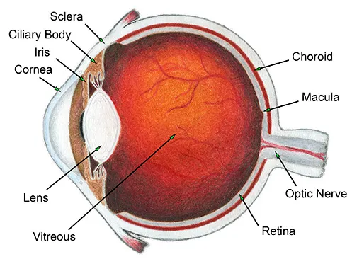
Diagram Showing the structure of the Eye.

The eye is spherical in shape and the diameter of an adult eye is approximately 2.5cm.
Internally, the eye is divided into 2 chambers. The lens, suspensory ligaments and ciliary body separate the 2 chambers; Anterior and posterior chamber.
- Anterior chamber: It is filled with a clear watery fluid called aqueous humour. This chamber is in front of the lens. It is further divided into 2 cavities ie anterior and posterior cavities.
- Posterior chamber: It is filled with a jelly like substance called vitreous humour (vitreous body). This chamber is behind the lens.
- The outer fibrous layer consisting of sclera and cornea.
- The middle vascular layer or uveal tract consisting of the choroid, ciliary body and iris.
- The inner nervous tissue layer consisting of the retina.
This consists of sclera and cornea.
- The sclera or white of the eye forms the outermost layer of the posterior and lateral aspects of the eyeball. It consists of a firm fibrous membrane that maintains the shape of the eye. This membrane gives attachment to the extrinsic muscles of the eye.
- The cornea: The sclera is continuous anteriorly with a clear transparent epithelial membrane, the cornea. The cornea is transparent due to its vascularity (avascularity) and the regular arrangement of its fibres. Its surface is lined by the conjunctiva. It is well supplied with nerve endings from the trigeminal nerve. Light rays pass through the cornea to reach the retina. The cornea is convex anteriorly. It is involved in refracting (bending) light rays to focus them on the retina.
The middle vascular layer is also known as the uveal tract. This layer consists of the choroid, ciliary body and iris.
- The choroid: lines sclera in the posterior compartment of the eye. The choroid is rich in blood supply and is chocolate brown in colour.
- The ciliary body: is an anterior continuation of the choroid which is inserted into suspensory ligaments. These ligaments extend to the lens and hold it in position. The ciliary body is supplied by the 3rd cranial nerve (Oculomotor). The ciliary body also consists of;
- Ciliary muscles: Contraction and relaxation of these smooth muscles determine the size and thickness of the lens.
- Secretory epithelial cells (Ciliary glands): These secrete aqueous humour which nourishes structures in the anterior chamber.
- The iris: is the visible coloured ring at the front of the eye. The iris extends anteriorly from the ciliary body lying behind the cornea and in front of the lens. It divides the anterior chamber of the eye into anterior and posterior cavities. It contains both circular and radiating muscle fibres which control the size of the pupil. The colour of iris is genetically determined and depends on the number of pigment cells present.
NB: The Oculomotor nerve supplies the muscles of the iris and ciliary body (intrinsic eye muscles).
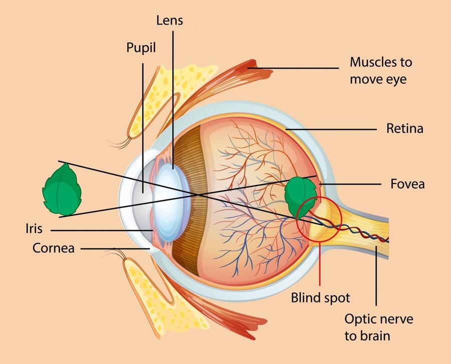
The inner layer of the eye ball is the retina. It is the light sensitive (photosensitive) part of the eye. It contains several millions of sensory photo receptor cells. These cells are responsible for converting light into nerve impulses.
The retina consists of two layers:
- The pigmented outer layer which lines choroid.
- The inner most neural layer which is in contact with the vitreous humour.
The light sensitive layer consists of sensory receptor cells ie rods and cones. These contain photosensitive pigments that convert light rays into nerve impulses.
- Rod cells pre-dominate in the periphery and function best in dim light. These cells are much more numerous.
- Cone cells pre-dominate near the centre of the retina. These are adapted for bright light and colour vision.
Near the posterior of the retina is a part called macula lutea or yellow spot. The greatest concentration of cone cells is at a small area in the yellow spot called the fovea centralis. It is the most vital part of retina for high definition or vision.
The optic disc or blind spot is a small area where the optic nerve leaves the eye. The blind spot does not have light sensitive cells.
- Eyebrows - protect eyeball from sweat, dust and other foreign bodies.
- Eyelids – movable folds acting as curtains, preventing injuries. Meet at palpebral fissure (both eyelids meet). It contains sebaceous glands, sweat glands and accessory lacrimal glands all aligned with conjuctival material.
- Conjunctiva - clear, delicate mucous membrane. it lines the eyelids and is highly vascularised. It protects the eye against infections. It also acts as a physical barrier, and produces mucin (goblet cells) which lubricates the eye ball.
- Sclera - is a fibrous tissue of the eye (white), is tough and contains collagen fibres, and covers 5/6 of the eyeball. It protects inner structures maintains the shape of the eyeball. It also acts as a passage of blood vessels and nerves.
- Cornea - covers 1/6 of the eyeball. Its clear, transparent and has 5 layers. Its functions include; protection of the eye as it’s very refractive media of the eye, and prevents aqueous from coming out of the eye.
- Anterior chamber - Is just behind the cornea and its functions include; Refractive media, maintains shape and structure of the eyeball, bathes/nourishes the eye, production and flow of aqueous humor.
- Aqueous Humor - is secreted by the epithelial cells of the ciliary body. it passes through suspensory ligaments into the posterior chamber, then flow through the pupil into the anterior chamber. From anterior chamber it drains through trabecular meshwork into the canal of schlemm (scleral venous sinus) then goes to the general circulation.
- Iris - is the thin visible, contractile, and coloured part of the eye, with a central aperture known as the pupil. It divides the anterior segment of the eye into anterior and posterior chambers. It controls amount of light entering the eye and plays a role in accommodation.
- Ciliary body - Is continuous with choroid (middle layer of eyeball). It suspends the lens which is important during accommodation, and produces aqueous.
- Choroid - Is the soft brown part behind the eye. Is most vascularized, and nourishes the retina.
- Lens - is the transparent, highly elastic biconvex body, that lies immediately behind the pupil/front of the vitreous body. Its thickness is controlled by ciliary muscle through the suspensory ligaments. Its functions include: Refractive media, absorbs ultra violet rays.
- Retina - Innermost layer of eyeball where images are formed. It has macula, optic disk, rods and cones. It consists of 2 layers: Epithelial and Nervous layer. It absorbs, stores and releases vitamin.
- Vitreous body - is transparent, jelly like media. It maintains the shape of eyeball and acts as refractive media.
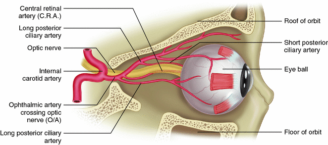
- Blood Supply: The blood supply of the eye is from the ciliary and central retinal arteries. These are branches of ophthalmic artery which is also a branch of the internal carotid artery.
- Venous Drainage: Venous drainage is by the central retinal vein. These vessels run alongside the optic nerve.
- Nerve Supply: Nerve supply is by the optic nerve which is the 2nd cranial nerve. The retinal nerve fibres originate in the retina. These fibres converge to form the optic nerve at the optic disc.
| Feature | Tears (Tear Fluid) | Aqueous Humor |
|---|---|---|
| Source of Production | Secreted by the lacrimal glands (accessory organs outside the eyeball). | Secreted by the epithelial cells of the ciliary body (inside the eyeball). |
| Location | Found on the external surface of the eye, covering the conjunctiva and cornea. | Found internally, filling the anterior and posterior cavities of the anterior chamber. |
| Primary Functions | Lubricates the eye, washes away foreign bodies/dust, provides oxygen to the avascular cornea, and prevents infection via bactericidal enzymes (lysozyme). | Maintains intraocular pressure, bathes/nourishes internal avascular structures (lens and cornea), and acts as a refractive medium. |
| Drainage Pathway | Drains via the lacrimal puncta → canaliculi → lacrimal sac → nasolacrimal duct into the nasal cavity. | Drains via the pupil → anterior chamber → trabecular meshwork → Canal of Schlemm into general circulation. |
| Composition | Water, mineral salts, antibodies, lysozyme, and mixed with meibum (oily secretion). | Clear, watery ultrafiltrate of blood plasma containing nutrients (glucose, amino acids) and minimal protein. |
To help you remember the complex process of vision, we have arranged the physiology of sight into four logical, memorable steps.
The "MAC" Reflex for Near Vision
When focusing on a near object, remember the autonomic reflex MAC:
- Miosis (constriction of pupil)
- Accommodation (lens convexity increases)
- Convergence (eyeballs move inward)
- Step 1: Refraction of Light Rays
- Light rays from objects are bent (refracted) as they pass through varying densities of the clear media of the eye to focus onto the retina.
- Before reaching the retina, the light rays pass through the cornea. The cornea also plays a role in the refractive power of the eyes.
- In the eye, the biconvex shape of the lens refracts and focuses light rays on the retina.
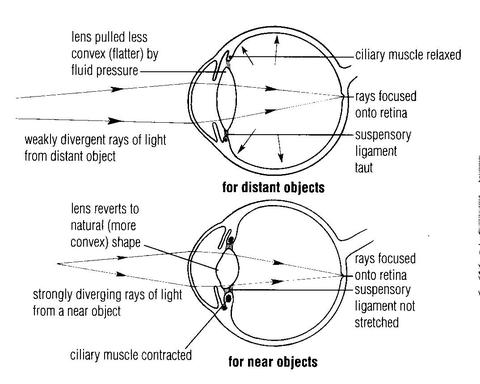
- Step 2: Focusing and The Accommodation Reflex
- The normal eye in its relaxed state brings rays of light from distant objects into sharp focus.
- The lens is elastic thus has ability to change shape. Change in shape varies the amount of refraction for clarity of focus. This is known as accommodation. Accommodation is necessary in order for objects at different distances to be visualized with equal clarity.
- However for clear focusing on near objects, an autonomic reflex comes into play. The reflex involves accommodation, miosis and convergence as follows:
- Accommodation: This refers to the increase in the refractive power of the lens in order to focus light rays from near objects on the retina. The ciliary muscle contracts and changes shape of the lens to bulge increasing its convexity and refractive power.
- Miosis: This is also known as constriction of pupils. It accompanies accommodation. It ensures that light rays are concentrated to pass through the centre of the lens and focus on the retina.
- Convergence (movement of the eyeballs): This refers to bilateral movement of the eyes at the same time in order to focus on a nearby object eg focusing the tip of one’s nose.
- Step 3: Transduction by Photoreceptors
- The light sensitive layer in the retina containing sensory photo receptor cells (rods and cones) convert light rays into nerve impulses.
- NB: The images refracted on the retina are upside down.
- Step 4: Transmission and Interpretation in the Brain
- These impulses are transmitted through the visual pathways to the visual area in occipital lobe of cerebrum.
- Here, they are interpreted as sensation of light form. They are processed into images of objects which are given meaning by other cerebral areas.
- This process involves interaction with information stored as memory in the association areas of the brain.
- The brain adapts to this early in life so that objects are perceived as upside/upright.
The eye is a delicate organ on the body and it is protected by several structures. These include;
- These are numerous hairs that project from the skin at the supra orbital margins of the frontal bone.
- These protect the eye from sweat, dust and other foreign bodies.
- These are two movable folds of tissue above and below the front of each eye.
- There are sebaceous glands, some open into the hair follicles of the eye lids.
- The eyelids contain two muscles. These include;
- Levator palpebrae superioris which raises the upper eyelid.
- Orbicularis oculi which closes the eyelids.
- The hair on the eye lid is called eye lashes.
- The eyelids have a lining (mucous membrane) of the conjunctiva. This lining is a fine transparent membrane that is on the inner surface of the eyelid. This layer also covers the eyeball.
- Where it lines the eyelids, there is a highly vascularized columnar epithelium.
- The corneal conjunctiva has avascular stratified epithelium. This means that the conjunctiva has epithelium without blood vessels at the cornea.
- The medial and lateral angles where the eyelids come together are called medial and lateral canthus respectively.
- At the edges of the eyelids, are eyelid margins that have numerous sebaceous glands. These are modified and secrete an oily material (meibum) spread over the conjunctiva by blinking. The material delays evaporation of the tears.
- Function of the eyelids and eyelashes: Protect the eye from injury. Blinking at about 3 to 7 seconds interval spreads tears and oily secretions over the cornea. This prevents drying of the eyeball.
The lacrimal apparatus consists of the structures that secrete tears and drain them from the front of the eyeball. These include;
- 1 lacrimal gland and its ducts
- 2 lacrimal canaliculi ie superior and inferior to the caruncle of the eye.
- 1 lacrimal sac
- 1 nasolacrimal duct
Each eye has a lacrimal gland behind the supra orbital margin. Lacrimal glands are exocrine glands. They secrete tears which are composed of water, mineral salts, antibodies and bactericidal enzymes. The tears leave the lacrimal glands by several small ducts.
They then pass over to the front of the eye under the eyelids towards the medial canthus where they drain into two lacrimal canaliculi. The opening of canaliculi on each side is called punctum. The canaliculi lie above one another separated by a red body called caruncle.
The tears then drain into the lacrimal sac which is the upper expanded part of the nasolacrimal duct. When foreign bodies or other irritants enter the eye, secretion of tears is greatly increased and the conjunctival blood vessels dilate. Secretion of tears is also increased in emotional states like crying and laughing. Excess tears are drained from the eye via the lacrimal apparatus into the lacrimal sac and then into the nasolacrimal duct.
- It has a fluid which is filled into the conjunctival sac.
- This fluid consists of tears and oily (meibum) secretions of meibomian/tarsal glands.
- The fluid is spread over the cornea by blinking.
- This mixture washes away irritants eg dust.
- It provides oxygen and nutrients to the avascular corneal conjunctiva and drains off wastes.
- Bactericidal enzyme lysozyme protects the eye by preventing microbial infection.
- The oiliness nature of the fluid delays its evaporation and prevents drying/friction of the conjunctiva.
- The fluid also prevents the eyelids from sticking together while sleeping.
- To lubricate the eye to facilitate oxygen and carbon dioxide exchange.
- To produce an optically smooth cornea surface.
- To cleanse the eye with a bactericidal enzyme lysozyme.
- To prevent the conjunctiva from drying.

These are also called extrinsic muscles. They are 6 in number and include the following;
- Medial rectus which rotates the eyeball inwards.
- Lateral rectus which rotates the eyeball outwards.
- Superior rectus which rotates the eyeball upwards.
- Inferior rectus which rotates the eyeball downwards.
- Superior oblique which rotates the eyeball downwards and outwards.
- Inferior oblique which rotates the eyeball upwards and outwards.
They protect the eye through the flexible movement of the types of muscles. These movements help us to see in all directions of the eyeball movement. Hence they also play a protective function ie protecting the eye and the whole body.
What is the management of an admitted patient with traumatic eye injury until discharge.
Thanks it’s a well highly digested notes for consumer
Thanks again
Thanks 👍👍👍😊😊😊
Tnx muchoo but why am I confused with the physiology of sight
I appreciate your technology in tackling nurses issues via revision guides
And keep it up