Obstetric Anatomy

Obstetric Anatomy Q & A

Obstetric Anatomy Q & A

Obstetric Anatomy, Obstetrics is the field of study concentrated on pregnancy, childbirth and the postpartum period
Question 1
 

(a) Describe the vagina.

(b) Outline indications of vaginal examination.

(c) What information must you note on vaginal examination?

(d) List contraindications of vaginal examination

SOLUTIONS.

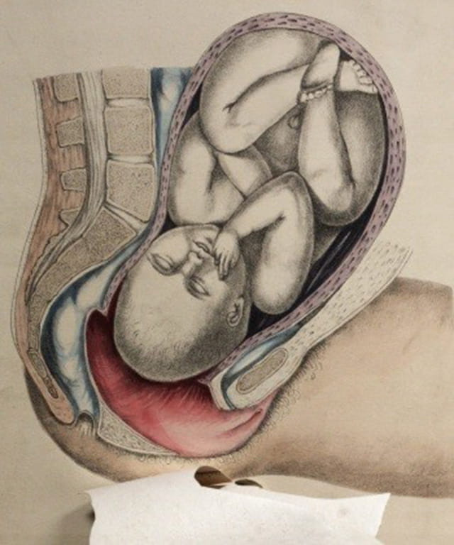
  1. A vagina is a muscular fibrous canal which forms the part of the internal female reproductive organs

Situation

It is a canal which extends from the vestibule below to the cervix above running in an upward and backward direction between the planes of the pelvic brim.

Shape

It is a potential tube which runs upwards and backwards with its walls in close contact but can be separated during coitus, menstruation, vaginal examination and child birth.

Size

The anterior wall measures 7.5cm

The posterior wall is longer and it measures 10cm.This is because the uterus enters the vagina at an angle of 90 degrees and bends forwards towards the anterior wall hence it encroaches on it

Structure 

Gross structure

Superiorly; the upper end of the vagina is known as the vault, where the cervix protrudes into the vault it forms circular recess known as fournices.

The vagina is made up of four fournices that is to say; The anterior fornix which is smaller and fairly deep The 2 lateral fournices which are shallow

The posterior fornix which is the longest and deepest

The lower end of the vagina is narrow and inferiorly we find the vulva_hymen enclosing the vaginal opening only present in virgins. If hymen is ruptured it leaves tags of membranes referred to as carunculae mytiformes. Vaginal orifice is also called introitus.

Microscopic structure

It is made up of four layers;

  1. Squamous epithelium arranged in folds known as rugae and makes the inner most layer of the vagina, the rugae increase the surface area and offer the vagina ability to stretch when need be for example during coitus and child
  2. Vascular connective tissue layer which is rich in blood vessels, nerves and lymphatics and is found just beneath the epithelium.
  3. Muscular layer. This is thin but a strong layer which is divided into two; the weak inner circular and strong outer longitudinal fibres.
  4. The pelvic fascial which is made up of loose connective It forms the outer protective coat and is continuous with the pelvic fascia.

Blood supply (arterial)

obstetric anatomy blood supply

The vagina is supplied by the branches of internal iliac artery which include vaginal artery and uterine artery

Venous drainage

By the corresponding veins ie branches of internal iliac veins which include vaginal veins and uterine veins.

Lymphatic drainage

Into the inguinal, the iliac and the sacro glands

Nerve supply

By the sympathetic and parasympathetic nerves which are branches from the lee Franken lanser plexus

Contents of the vagina

It doesn‘t contain any glands but its kept moist by cervical mucus and a transudation from the underlying blood vessels through the epithelium.

Its media is acidic (PH 3.8 to 4.5) and this is made possible by presence of lactic acid after action of doderleins bacilli on glycogen

Relationships of the vagina

obstetric anatomy relation

Anteriorly

Below, the base of the bladder rests on the upper ½ of the vagina and the urethra is embedded in the lower ½.

Posteriorly

Pouch of Douglas above, the rectum medial and perineal body below Laterally

Pubococcygeous muscles below and pubic fascial containing the uterus above Inferiorly

The structure of the vulva Superiorly

The cervix and the fournices

Functions of the vagina

  1. Exit from menstrual flow
  2. Entrance for spermatozoa
  3. Exit for products of conception
  4. Supports the uterus
  5. Prevents ascending infection due to acidic PH
  6. For assessing the pelvis
  7. Drug administration

PART B

Indications of vaginal examination

Indications can be divided into during pregnancy, labour and puerperium

During pregnancy

  • To confirm pregnancy using hegars, jacquemiers and osianders signs
  • To rule out abnormalities in genital organs g. polyps, cervical erosion and cancer of the cervix
  • To rule out causes of bleeding in early weeks

During labour

First stage of labour

  • To diagnose onset of labour
  • To determine progress of labour by finding out degree of cervical dilatation
  • To note state of membranes
  • To confirm presentation, position and engagement of head
  • To assess moulding
  • To exclude cord prolapse when membranes rupture
  • To note dilatation before giving a narcotic

Second stage of labour

  • To confirm second stage of labour
  • To note cause of delay in second stage of labour
  • To confirm presentation of second twin before rupturing membranes

Third stage of labour

  • To determine cause of postpartum haemorrhage
  • Incase of retained placenta, to detect cause of retained placenta and exclude construction ring
  • To detect condition of birth canal following child birth

During puerperium

  • To rule out cause of secondary PPH
  • At 6-8weeks after delivery, to detect if the reproductive organs have gone back to their pregravida state.
  1. Information to note on vaginal examination

On inspection

State of the vulva, note any abnormal discharges like pus, blood, abnormal growths like warts,  oedema and scars.

On examination

Note condition of the vagina

Normally the vaginal walls feel warm and moist and dilatable. If dry may be a sign of infection or obstruction.

State of the cervix

If thin, thick, whether soft or rigid and whether its well applied to the presenting part.

Note dilatation and cervical effacement.

State of the membranes

Whether intact or ruptured. If ruptured check colour and smell of liquor

Presentation and presenting part

Note level of presenting part in the pelvis

Confirm position by finding or palpating sutures and fontanelles and relate them to the maternal pelvis.

Note moulding.

Do internal pelvic assessment and note

  • -sacro promontary if protruding
  • -hollow of the sacrum if well curved
  • -sciatic notches if well rounded
  • -ischial spines if prominent
  • -sub pubic arch-if it accommodates 2 ½ to 3 fingers
  • -inter tuberous diameter if it accommodates 4 knuckles

Contraindications of vaginal examination

  • Ante partum haemorrhage
  • Threatened abortion
  • Elective caesarean section

a) Describe

b) Outline the formation of the

c) List variations of the placenta

SOLUTIONS

  1. Describe Fertilization

 Fertilization is the fusion of the male gamete (sperm) and female gamete (ovum)

Fertilization occurs when the female gamete fuse to form a zygote during the time of intercourse, about 300 million spermatozoa are deposited into the vagina.

Some sperms cannot survive the acidic media of the vaginal secretions so the weak ones die and only the strong ones survive.

The surviving sperms continue moving forwards this is made possible by the special arrangement of the mucus lining of the cervix (arborvitae) which prevents back flow of sperms.

Sperms continue their journey but still the weak ones continue to die off. Movement is slowed down by the presence of hair like projections called cilia and more eradication of weak sperms continues. And if ovulation had taken place within 48 hours and the ovum is still viable the two gametes will meet in the ampulla and fusion will take place hence fertilization.

b) Outline the Formation of the Placenta 

Placenta is a vital organ of communication between the mother and the fetus.

It‘s a maternal-fetal organ which begins developing at implantation of the blastocyst and is delivered with the fetus at birth.

Formations of the placenta

  • Maternal surface

This is the surface next to the uterus

Its dark red in color due to presence of maternal blood and it has 18-20 lobes which are collections of chronic villi each cotyledon is separated from the other by tissues.

  •       Fetal surface

This is the surface next to the fetus

It has a shiny surface due to presence of amniotic membrane

c). List variations of the placenta / abnormalities of the placenta

  • Succenturiate lobe

This occurs due to abnormalities in development and it is the most significant abnormality. The additional lobule separates from the main part of the placenta.

  • Circumvallated placenta

It is an opaque ring seen on the fetal surface of the placenta and this is due to doubling back of the chorion and amnion

  • Bi-partite placenta or bi-lobed placenta

This placenta has two complete and separate parts each with a branch of umbilical cord vessels which later join to form one cord.

  • Battledore insertion of the cord

In this the placentas cord is inserted at the edge/margin of the placenta and the placenta has an appearance of a table tennis bat in shape.

  1. Velamentous insertion of the cord

The cord is inserted into the fetal membranes some distance away from the edge of the placenta.

The diagram below shows the different variations of the placenta discussed above.

placenta variations

a) Describe the non-pregnant

b) What changes take place in this organ during pregnancy?


Description of the non pregnant uterus

  • This is a hollow muscular pear /ovacado shaped

Situation

  • It is situated in the true pelvis between the urinary bladder and the

Size

  • It is 5cm, long 5cm wide, and 2.5cm thick so each wall is 1.25cm thick.

Position

  • The uterus is anteverted (bends forward) and anteflexed (bends on itself)

Shape

  •  The uterus is pear shaped (avocado) with the upper part bigger than the lower part .

Description of the non pregnant uterus


Gross structure

This is made up of the following

  • The body.

 This forms the upper part of the whole uterus.

  • Fundus

 This is a raised area between the insertion of the uterine tubes.

  • Cornua

Upper outer angles of the uterus where the uterine tubes are inserted. This is made up of the following

  • Cavity

This is a potential space between the anterior and posterior uterine walls.

It is triangular in shape.

  • Isthmus

This is a narrow area between the body of the uterus and the cervix.

  • Cervix or neck

This forms other lower third of the whole uterus into the

Microscopic structure

Endometrium

  • this is a layer of ciliated mucus
  • It changes constantly with the menstrual cycle
  • It lines the uterine cavity and shades off during menstruation up to the basal
  • The cervical endometrium does not change during menstruations

Myometrium

  • Middle layer and is formed by different muscle fibres.
  • Longitudinal fibres ;mainly found in the upper part of the uterus .
  • Oblique muscle fibres ;mainly found in the fundus
  • Circular muscle fibres which are located at the cornua and they prevent contractions extending to the uterine tubes.
  • Some are found around the cervix which help in dilatation during labour.

Perimetrium

  • It is a serous membrane which is a continuation of the peritoneum
  • It covers the outer aspect of the uterus.
  • It bends upwards to form the uterovesco pouch anteriorly and the pouch of dauglous posterioly

Blood supply

  • By uterine arteries which are the branches of the internal iliac artery and they supply the lower ovarian arteries the branches of the abdominal aorta and supply the upper parts.

Nerve supply

  • By sympathetic and parasympathetic
  • Lymph drains into internal illiac glands

Supports

  • The uterus is supported by pelvic floor muscles and maintained in position by ligaments.
  • Transverse ligament
  • Utero sacral ligament
  • Pubo cervical ligament
  • Broad ligament
  • Round ligament and,
  • Ovarian ligament.

Relations of the Body of the Uterus


  • Anteriorly:The bladder and vesco-uterine pouch.
  • Posteriorly: The pouch of douglas
  • Laterally: The broad ligament on each side uterine tubes and ovaries.
  • Superiorly: intestines
  • Inferiorly :vagina

Functions of the uterus

  • It shades the endometrium during menstruation
  • To prepare a bed for the fertilized ovum
  • It shelters the fetus during pregnancy
  • It expels the products of conception at term
  • To involute following childbirth

(B) THE PHYSIOLOGICAL CHANGES THAT OCCUR IN UTERUS DURING PREGNANCY

Pregnancy

 Is the growth and the development of a fertilized ovum from the time of conception until its expulsion. In normal pregnancy expulsion of the fetus takes place at term. Thus, 38-42 weeks of gestation with an average of 40 weeks.

  • Physiological changes that take place during and pregnancy are associated with and caused by the effects of specific hormones.
  • These are temporary adaptations of the body that helps to meet the demands of the fetus.


CHANGE IN THE UTERINE SIZE DURING PREGNANCY.

The body of the uterus

 After conception, the body develops to provide a nutritive and a protective environment into which the fetus will grow and develop.

Size: 7.5x5x2.5- 30×23 x20 (cm) 

Weight: changes from 60 to 1000gm 

Uterine shape and situation.

  • The uterus changes to a globular shape to accommodate the growing fetus, increasing amounts of liquor and placental tissues.
  • The lowest part of the uterus elongates 3 times its original length during the first trimester, giving the appearance of a stalk below the globular segment. This is the beginning of the upper and lower uterine
  • By 12th week of pregnancy, the uterus rises out of the pelvis and becomes upright. It is no longer anteverted and ante flexed. It is about the size of a grape fruit and may be palpated abdominally above the symphysis pubis.
  • By 16th week, the conceptus has grown enough to put pressure on the isthmus, causing it to open out so that the uterus become more globular n shape.
  • By 20th week of pregnancy, the uterus becomes spherical in shape and has a thicker, and more rounded fundus. As the uterus continues to rise in the abdomen, the uterine tubes, being ristricted by attachments to the broad ligaments, become progressively more At 20th week the fundus of the uterus may be palpated at or just below the umblicus.
  • At 30th week, the lower uterine segment can be It is the portion of the uterus above the internal os of the cervix. The fundus can be palpated midway between the umblicus and the xiphisternum.
  • The uterus reaches the level of the xisphisternum by the 38th A reduction in fundal height , known as lightening, may occur at the end of pregnancy when the fetus sinks into the lower pole of the uterus.

Decidua

 The decidua is the name given to the endometrium during pregnancy. Estrogen and progesterone produced by the corpus luteum, causes decidua to become thicker and richer and more vascular at the fundus and in the upper body of the uterus, which are the usual site for implantation.

The decidua provides a glycogen rich environment for the blastocyst until the placenta is formed.


Myometrium.

  • It is made up of smooth muscle fibres, held together by connective These muscle fibres grow up to 15 to 20 times their non pregnant length.
  • The hypertrophy and hyperplasia of the uterine muscle is due to the effect of estrogen and
  • The uterus continues to grow in this way for the first three months, after which the growth is related to the distension by the growing
  • The wall of the myometrium become thicker during the first few months of pregnancy, and as gestation advances, the walls become thinner owing to the gross enlargement of the uterus being only 5 cm thick or less at term.
  • These prelabor contractions are associated with the ―ripening of the cervix‖ and eventually becomes the contractions of labor as the effects of estrogen supersede those of progesterone e. progesterone normally suppresses myometrial activity.
  • During pregnancy, the muscle layers become more differentiated and organized and organized for their part in expelling the fetus.
  • The thickness of the upper uterine acts as a piston to force the fetus into the receptive, passive lower uterine segment.

Contractions of these muscles fibres is necessary to entrap and enmesh bleeding vessels and ligate them after the placenta is delivered.

  • The inner circular layer is thin and forms sphincters around the openings at the cornua, and around the lower uterine segment and

Blood vessels


  • The uterine blood vessels increase in diameter and new vessels develop under the influence of estrogen. the blood supply through the uterine and ovarian arteries increase to about 750mls per minute at term to keep pace with its growth and also to meet its needs of the functioning placenta.

The cervix

  • The cervix remains tightly closed during pregnancy , providing protection to the fetus and resistance to the pressure from above when the woman is in a standing up
  • The mucus secreted by the endo cervical cells become thicker and more viscous during The thicken mucus then forms a plug called the operculum, which provides protection from ascending infections.
  • Cervical vascularity increases during pregnancy, giving the cervix a bluish color if viewed through a speculum.
  • The cervix remains 5cm long throughout pregnancy.
  • In late pregnancy, softening or ripening of the cervix occurs making it more distensible. The muscles of the fundus enhance tension in the outer longitudinal layer of muscles of the cervix contributing to the process of effacement.
Spread the love

Leave a Comment

Your email address will not be published. Required fields are marked *

Contact us to get permission to Copy

We encourage getting a pen and taking notes,

that way, the website will be useful.

Scroll to Top