Upper Respiratory Tract Infections (URTIs)
Upper Respiratory Tract Infections (URTIs)
1. Overview and Magnitude of the Problem
An Upper Respiratory Tract Infection (URTI), commonly referred to as "the common cold", is a symptom complex primarily caused by viruses, occasionally bacteria, and very rarely fungi.
The term "URTI" is actually considered a misnomer (inaccurate name). Why? Because it incorrectly implies that there are absolutely no lower respiratory tract symptoms (like deep chest coughs or bronchial irritation), which isn't always true. Viral URTIs often trigger lower respiratory reactivity, meaning a "head cold" frequently causes chest symptoms.
The Magnitude (How common is it?)
- Global/USA: The "Coryza syndrome" (common cold) is the most common condition seen in Outpatient Departments (OPD). Acute pharyngitis accounts for 7 million annual visits in adults (1-2% of all visits). Acute sinusitis hits 20 million people annually.
- Uganda : The prevalence of URTIs among children in rural Uganda was recorded at 37.4% (Mbonye, 2004), and 18.33% among under-fives (UDHS 2000/01).
- Regional Vulnerability: In Uganda, the highest percentage of cases were in the Northern region, followed by the Eastern region. Children aged 6-35 months are far more susceptible than infants <5 months (who still have maternal antibodies) or children >35 months (who have built their own immunity through repeated exposure).
- Socioeconomic Impact: URTIs carry a massive cost to society, causing missed work days, missed school classes, and unnecessary medical expenses (especially when parents demand unnecessary antibiotics).
Risk Factors for URTIs
Why do some people get sick while others don't? It comes down to environmental and host factors:
- Climate: Cold winter months in temperate zones; rainy seasons in the tropics. Elaboration: The cold weather itself doesn't cause the virus. Rather, bad weather forces people to stay indoors, keeping windows closed, breathing recycled air, and sharing germs in close proximity.
- Environment: Indoor overcrowding (homes, schools, daycare centers) and indoor air pollution (like wood-burning stoves). Overcrowding in crisis/refugee-affected areas is a massive risk due to poor ventilation and shared living spaces.
- Host Factors: Lack of immunization, congenital (birth) or acquired (e.g., HIV) immunodeficiency, and anatomical disorders (like a cleft palate or a severely deviated septum which impairs normal nasal drainage).
- Transmission: Spread via aerosols (fine mist that hangs in the air), droplets (heavy sneezes that fall on surfaces), or direct hand-to-hand contact with infected secretions, which are then passed to the nares (nose) or eyes. Example: Rubbing your eye after touching an infected doorknob is a primary route of infection!
2. Anatomy and Innate Immunity of the URT
Anatomical Relevance
The URT consists of the nasal cavity, paranasal sinuses, pharynx, and larynx. The critical exam concept here is anatomical continuity. The nasopharynx is directly connected to the middle ear via the Eustachian tube, and directly connected to the paranasal sinuses via small openings called ostia. Therefore, a simple nose infection can easily travel up the tubes into the ears or sinuses.
Innate Immunity (How the body protects itself)
The URT is not defenseless. It has a robust, multi-layered defense system:
- Pseudostratified Columnar Ciliated Epithelium: This is the dominant tissue lining the URT. It acts like an escalator. The cilia (tiny hairs) constantly beat in a coordinated manner to sweep trapped harmful agents downward towards the pharynx to be swallowed and destroyed by stomach acid. Clinical Note: Cigarette smoking literally paralyzes these cilia, which is why smokers get frequent chest and sinus infections!
- Mucosal Secretions: Goblet cells secrete mucus. Mucus is a sticky macromolecular polysaccharide. It is *not* nutritious for bacteria, meaning bacteria can't eat it to survive. It traps foreign particles, and as it sloughs off, the pathogens are removed with it.
- Saprophytic Microorganisms (Normal Flora): These are "good" bacteria living in your nose and throat. They offer protection via competitive inhibition—they eat up the local resources and take up physical space, preventing "bad" pathogenic bacteria from taking root.
- Lysozyme (Muramidase): A crucial hydrolytic enzyme found in secretions. Mechanism: It specifically breaks the bond between N-acetylglucosamine (GlcNac) and N-acetylmuramic acid (MurNac) in bacterial cell walls, essentially popping the bacteria like a balloon.
- Collectins (SP-A and SP-D): Surfactant Proteins. SP-A binds to the Lipopolysaccharide (LPS) of Gram-negative bacteria, acting as a flag (opsonization) to induce macrophages to eat them. SP-D acts in the humid phase of airways but does not induce phagocytosis directly.
- Other Factors: Complement system, Interferons (IFNs - fight viruses), lactoferrin (steals iron from bacteria to starve them), and Acute Phase Proteins (LBP).
Non-specific immune cells jump into action:
- Airway epithelial cells.
- Phagocytes: Neutrophils/PMNs, eosinophils, monocytes, macrophages.
- Natural Killer (NK) cells: Seek out and destroy your own cells that have been hijacked by viruses.
- Basophils/Mast cells: Release histamine to trigger beneficial inflammation.
- Dendritic Cells: Antigen Presenting Cells (APCs) that show the virus to the T-cells.
3. Specific URTI Syndromes
A. The Common Cold (Coryza)
A self-limiting viral infection of the upper respiratory tract, lasting about 7-10 days.
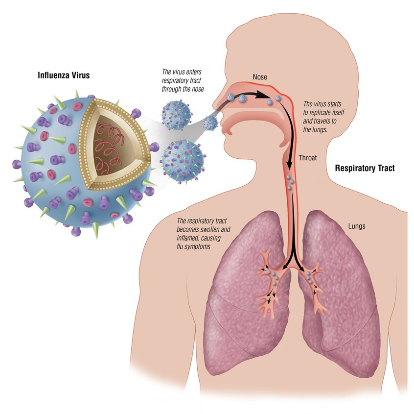
- Aetiology (Causes): Rhinovirus is the undisputed king (up to 60% of cases). Others include Coronavirus, Parainfluenza, RSV (Respiratory Syncytial Virus), Adenovirus, Influenza, and Enterovirus/Coxsackievirus. Exam Note: These viruses evade the immune system by constantly undergoing antigenic variation (mutating their surface proteins so your memory cells don't recognize them next time).
- Pathogenesis: Virus invades the epithelium → triggers massive inflammation → sloughing off of columnar epithelial cells. Symptoms are driven by chemical mediators (Bradykinins, Prostaglandins, Histamine, Interleukins IL-1, IL-6, IL-8) and parasympathetic/alpha-adrenergic nerve reflexes.
- Clinical Features: Incubation is short (12-72 hrs). Cardinal signs: Nasal discharge, nasal obstruction, sneezing, scratchy/sore throat, cough. Mild fever (high fever is uncommon and suggests something worse, like the Flu or a bacterial infection). Can have facial pressure/ear fullness.
- Complications: Mucosal damage from the virus alters the normal flora. This, combined with aggressive nose blowing, physically pushes bacteria into sterile areas (sinuses/middle ear), causing secondary bacterial infections.
- Treatment: Purely symptomatic! Antihistamines, NSAIDs (for pain/fever), warm saline gargles. Antibiotics are useless against viruses and only cause harm by promoting resistant bacterial colonization. *Note: Even if nasal discharge becomes thick and greenish/yellowish, do NOT give antibiotics unless it persists for more than 10-14 days!*
- Prevention: Hand washing is #1. Cover coughs/sneezes, use disposable tissues. Interferon-alpha 2b is in trials.
B. Sinusitis (Rhinosinusitis)
Inflammation of the mucosal lining of one or more paranasal sinuses (Maxillary, Frontal, Sphenoid, Ethmoid). Under normal conditions, these sinuses are completely sterile.
Pathogenesis & "Double Sickening"
A viral cold causes mucosal inflammation → this swelling blocks the sinus ostia (drainage hole) → fluid is trapped inside the sinus → normal upper airway bacteria enter, get trapped, and proliferate rapidly in the dark, moist fluid.
The "Double Sickening" Phenomenon: A classic sign of bacterial sinusitis is a patient who gets a standard viral cold, starts to feel a bit better around day 5, and then suddenly gets drastically worse (spike in fever, severe facial pain) on day 7 or 8. This indicates the trapped fluid has become secondarily infected by bacteria.
- Aetiology:
- Viral: Most common (Rhinovirus, Influenza, etc.). 60% resolve spontaneously.
- Community-Acquired Bacterial (ACBS): Streptococcus pneumoniae, Haemophilus influenzae (the top two). Also Moraxella catarrhalis, S. aureus, and Group A Strep.
- Nosocomial (Hospital-Acquired): Major risk in ICU patients on ventilators or with nasogastric tubes. Caused by enteric Gram-negatives (P. aeruginosa, S. marcescens, K. pneumoniae, Enterobacter) and S. aureus. Often polymicrobial.
- Fungal: Seen in immunocompromised or diabetic patients (Aspergillus, Zygomycetes). Can be highly invasive.
- Clinical Presentation:
- Viral: Standard cold symptoms.
- Bacterial (ACBS): Suspect this if cold symptoms persist > 10-14 days, or if there is severe high fever (>39°C), severe facial/tooth pain (especially when bending over), purulent discharge, and hyposmia (loss of smell).
- Nosocomial: Presents as PUO (Pyrexia of Unknown Origin) in a ventilated patient.
- Fungal: Masses, proptosis (bulging eye), bony erosion.
- Diagnosis: Usually clinical. X-rays (showing air-fluid levels, opacification, mucosal thickening) only if complications are suspected. Gold Standard for microbial diagnosis: Paranasal puncture and aspiration for Culture & Sensitivity (must avoid nasal secretion contamination).
- Management:
- First-line: Amoxicillin (40 mg/kg/day) by doubling standard dose.
- If no response in 48 hrs: Assume the bacteria (like H. flu or M. catarrhalis) is producing beta-lactamase (destroying the amoxicillin). Switch to a beta-lactamase stable drug: Amoxicillin-clavulanate (Augmentin) or cephalexin. Treat for minimum 10 days.
- Symptomatic: Topical decongestants, NSAIDs, antihistamines.
- Complications: Intracranial (meningitis, brain abscess), Orbital (cellulitis), Respiratory. Chronic sinus disease happens due to no treatment, inadequate treatment, or anatomical defects.
C. Pharyngitis (Tonsillopharyngitis)
Inflammation of the mucous membranes of the throat. Subdivided into illness with nasal symptoms (nasopharyngitis - usually viral) and without nasal symptoms (tonsillopharyngitis - higher chance of bacterial).
Comes in with a sore throat, runny nose, sneezing, and a slight cough. Diagnosis: Likely Viral Nasopharyngitis (Adenovirus is most common). Treatment: Rest and fluids. (The presence of cough and runny nose strongly points AWAY from strep).
Comes in with a sudden severe sore throat, painful swallowing, high fever, swollen tonsils with white pus (exudate), swollen neck lymph nodes, but NO cough and NO runny nose. Diagnosis: Highly likely Group A Beta-Hemolytic Streptococcus (GAS / S. pyogenes). Treatment: Antibiotics.
- Bacterial Aetiology: Group A Beta-Hemolytic Streptococcus (GAS) is the most important bacterial cause (15-30% of cases in kids, 5-10% in adults). Other unusual causes: Group C/G strep (food outbreaks), mixed anaerobes (Vicent's angina), N. gonorrhoeae, C. diphtheriae.
- Why do we care so much about GAS? Because if left untreated in children, GAS can trigger a severe autoimmune complication called Acute Rheumatic Fever (which damages heart valves). Mechanism: The immune system makes antibodies to fight the Strep bacteria, but due to "molecular mimicry," those antibodies accidentally attack the child's own heart tissue. *Note: The risk of rheumatic fever is extremely low in adults.*
- Diagnosis: Clinical grounds are not enough.
- Throat Culture: Swab both tonsils and posterior pharyngeal wall (DO NOT touch teeth/tongue). Grow on Blood Agar at 35-37°C for 18-24 hrs (up to 48 hrs). GAS is identified because it is Bacitracin sensitive (0.04 U).
- RADT (Rapid Antigen Detection Test): Faster than culture. Uses EIA or chemiluminescent DNA probes. Allows kids to return to school faster and stops spread immediately.
- Management:
- First-line for GAS: Penicillin V (Oral, 10 days) or Benzathine Penicillin G (Single Intramuscular Dose: 1.2 million Units for adults/older kids).
- Penicillin Allergic: Erythromycin or first-generation cephalosporins (for 10 days).
- Vicent's Angina (mixed anaerobes): Amoxicillin + metronidazole or clindamycin.
- Symptomatic: Warm saline gargles, analgesics.
D. Acute Epiglottitis (Supraglottitis)
Inflammation of the epiglottis. THIS IS A TRUE MEDICAL EMERGENCY. The swelling can cause abrupt, complete airway obstruction, suffocating the patient.
- Aetiology: Haemophilus influenzae type b (Hib) used to cause ~100% of cases in kids before the Hib vaccine was introduced. Other causes: Pneumococcal, Staphylococcal. Non-infectious: chemical burns, physical trauma, severe allergy.
- Clinical Presentation: Classic patient is an unvaccinated child aged 2 to 4 years. Sudden onset (6-12 hours) of high fever, extreme irritability, dysphonia (muffled voice), and severe dysphagia (cannot swallow).
Classic Signs: The child sits leaning forward in a "tripod" position, drooling (because swallowing hurts too much), and has inspiratory stridor (high-pitched gasping sound when breathing in). - Diagnosis & CRITICAL PRECAUTION: Diagnosis is clinical, supported by a lateral neck X-ray showing the classic "Thumb Sign" (a swollen, thumb-shaped epiglottis).
WARNING: Never blindly swab or use a tongue depressor on a child suspected of epiglottitis! Disturbing the inflamed epiglottis can trigger a reflex laryngeal spasm, completely closing off the airway and killing the child instantly. Examination must only be done in an Operating Room with a surgeon ready to perform an emergency intubation or tracheostomy. - Management: Support the airway immediately! Give IV Antibiotics: Cephalosporins or Ampicillin-sulbactam. Vaccinate all unvaccinated household children with Hib vaccine.
E. Acute Laryngitis
Inflammation of the vocal cords.
- Aetiology: Mostly respiratory viruses. Can be GAS, C. diphtheriae, or TB/Fungi (uncommon). Non-infectious: Voice abuse (e.g., a teacher talking all day, or screaming at a concert), GERD (acid reflux burning the cords at night).
- Clinical Features: Recent onset of hoarseness or husky voice, often with a dry cough. Can progress to aphonia (complete loss of voice). Exam shows hyperemic (red) and edematous (swollen) vocal cords due to vascular engorgement.
- Diagnosis: Clinical. If swabbing is needed (to check for Diphtheria or TB), use a laryngeal mirror and an applicator bent at 120° to avoid blind contamination.
- Management: Voice rest and humidification (steam). Antibiotics have no objective benefits and are NOT routinely recommended.
F. Otitis Media (OM)
Inflammation of the middle ear with fluid presence. Huge burden in pediatrics.
Why do babies get it so much?
Peak incidence is between 6 and 24 months. Anatomy explains this: An infant's Eustachian tube is shorter, wider, and more horizontal than an adult's. When a baby gets a cold or cries while lying flat on their back ("bottle propping" is a major risk factor), nasopharyngeal secretions and milk pool straight into the middle ear. By age 5-6, the skull grows, making the tube angle steeply downward, draining fluid effectively.
- Epidemiology & Risk Factors: By age 3, over 2/3 of kids have had at least 1 episode. Males > Females. Breastfeeding >3 months protects (provides maternal IgA antibodies). Daycare centers and passive smoking heavily increase the risk. HIV+ children have high rates starting at 6 months.
- Aetiology:
- Bacteria: S. pneumoniae (most common), H. influenzae (mostly non-typeable), M. catarrhalis, Group A Strep.
- Viruses: RSV, Influenza, Rhinovirus.
- Pathogenesis: Eustachian tube dysfunction/obstruction → negative pressure → fluid accumulation → bacterial suppuration.
- Clinical Features: Otalgia (severe ear pain, baby tugging at ear; *pain is often worse when lying down*), ear drainage (if eardrum bursts, relieving the pressure), hearing loss, fever, irritability. Early exam shows a red, bulging tympanic membrane.
- Diagnosis: Pneumatic otoscopy (puffing air into the ear to see if the eardrum moves—if fluid is trapped behind it, it will be rigid and won't move). Tympanometry. Tympanocentesis (needle aspiration) only for severe/resistant cases.
- Complications: Chronic perforation, Cholesteatoma (destructive skin cyst in the ear), Adhesive OM, Hearing loss causing intellectual/speech impairment, and cranial complications (meningitis).
- Management: Amoxicillin is the initial choice (double dose). If fails, use Amoxicillin-clavulanate, Macrolides, or Cephalosporins. Symptomatic: decongestants/antihistamines. Surgical: Myringotomy (lancing eardrum to drain pus), Adenoidectomy, Tympanostomy tubes (grommets for chronic fluid).
G. Otitis Externa (OE)
Infection of the external auditory canal. A totally different beast from Otitis Media.
Clinical Distinction (The Pinna Pull Test): In OE, pulling on the outer ear (pinna) or pushing the tragus causes agonizing pain. In OM, pulling the outer ear does not cause extra pain, because the infection is deep behind the eardrum.
- Pathogenesis: The canal is narrow and tortuous. Water gets trapped (hence "swimmer's ear"), macerating (softening) the skin. The protective epithelium sheds, allowing bacteria to invade. Since skin here is tightly bound to cartilage, expansion causes severe pain.
- Classification & Aetiology:
- Acute Localized OE: A pustule/furuncle (pimple) on a hair follicle. Caused by S. aureus.
- Acute Diffuse OE (Swimmer's Ear): Hot/humid weather. Edematous, red, itchy canal. Caused mainly by Pseudomonas aeruginosa.
- Chronic OE: Caused by constant pus draining out of a perforated eardrum from Chronic OM.
- Fungal Otitis: Caused by Aspergillus or Candida albicans.
- Malignant (Invasive) OE: A severe, life-threatening necrotizing infection spreading to cartilage and temporal bone. Classic Patient: Elderly, Diabetic, or Immunocompromised. Caused almost exclusively by P. aeruginosa. Poor blood flow (diabetic microangiopathy) allows deep tissue invasion. Can cause permanent facial paralysis (Cranial Nerves 7, 9, 10, 12).
- Management:
- General: Gentle cleansing, irrigation with hypertonic saline/acetic acid/alcohol mixtures to dry the ear.
- Uncomplicated OE: Topical drops: Ciprofloxacin-dexamethasone or Neomycin/polymyxin + hydrocortisone (10 days).
- Malignant OE: Requires aggressive systemic (IV) therapy. Ceftazidime, Cefepime, or Piperacillin + Aminoglycoside, OR high-dose oral Ciprofloxacin for 4 to 6 weeks.
H. Mastoiditis
Inflammation of the mastoid air cells (the honeycomb-like bone right behind the ear). Almost always a complication of poorly treated Otitis Media.
- Pathogenesis: Middle ear infection pushes through the antrum into the mastoid air cells. Purulent exudate builds up under pressure → causes necrosis of the thin bony septa → creates a coalescent abscess cavity. Anatomical Danger: This bone borders the brain cavity; infection can easily erode through and cause meningitis.
- Clinical Features: Swelling, redness, and extreme tenderness over the mastoid bone (behind the ear). The pinna (outer ear) is visibly pushed outward and downward by the swelling behind it.
- Diagnosis: CT scan or X-ray showing loss of sharpness of cellular walls (demineralization) and cloudiness in the mastoid bone.
- Management: IV Antibiotics targeting S. pneumoniae and H. flu. If prolonged, must cover for S. aureus and Gram-negatives. If an abscess is fully formed, a surgical Mastoidectomy is required to drill out the infected bone and drain the pus.
4. Modern Challenges and Trends in URTIs
- Aetiological Diagnosis is Hard: Many sites (like sinuses or middle ear) are completely inaccessible for routine swabbing without invasive procedures (like sticking a needle through the eardrum). Furthermore, distinguishing between normal flora and actual pathogens on a throat swab is a constant clinical challenge.
- Viral vs. Bacterial Dilemma (Antibiotic Stewardship): Determining clinically if an infection is viral or bacterial is incredibly difficult. This leads to massive global over-prescription of antibiotics by anxious doctors and demanding patients, fueling dangerous antimicrobial resistance (e.g., rising rates of drug-resistant S. pneumoniae and MRSA).
- Vaccine Development: Still a challenging area for the sheer variety of URTI pathogens (especially the hundreds of different serotypes of Rhinovirus—you can catch a "cold" 100 times because it's a slightly different virus each time).
- HIV Staging: Chronic sinusitis and chronic otitis media are significant enough to be formally included in the WHO HIV Clinical Staging system as key markers of immune decline.
- Emerging Pathogens: Human Metapneumovirus is a relatively newly discovered viral agent now recognized as a significant cause of URTIs worldwide, reminding us that new viruses continue to emerge.
Quick Quiz
Upper Respiratory Tract Infections (URTIs)
Microbiology - mobile-friendly and focused practice.
Privacy: Your details are used only for quiz tracking and certificates.
Upper Respiratory Tract Infections (URTIs)
Microbiology
Preparing questions...
Choose your answer and keep your streak alive.
Great effort.
Here is your quick performance summary.
Upper Respiratory Tract Infections (URTIs) Read More »