Eicosanoids Pharmacology
Autocoids -- Eicosanoids
Eicosanoids Pharmacology
1. Introduction to Eicosanoids
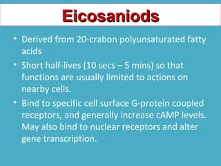
Definition: Eicosanoids are biological signaling molecules (local hormones/autacoids) that are products of polyunsaturated long-chain fatty acids. The prefix "Eicosa-" means 20 in Greek, because these molecules are almost entirely derived from 20-carbon essential fatty acids, most commonly Arachidonic Acid.
Unlike regular hormones (like insulin) which are stored in glands and travel globally through the blood, eicosanoids are not stored. They are highly unstable and have a half-life of seconds to minutes. Therefore, they are synthesized on demand from cell membrane lipids and act locally right where they are made (paracrine action on neighbors, or autocrine action on themselves).
Major Classifications
Eicosanoids are divided into families based on the specific enzyme that creates them from the raw material:
- a) Cyclooxygenase (COX) derivatives: These include the Prostaglandins (PGs) and Thromboxane (TXA2).
- b) Lipoxygenase (LOX) products: These include the Leukotrienes (LTs) and Lipoxins.
- c) Cytochrome P450 (CYP) Epoxyoxygenase pathway: Produces EETs (Epoxyeicosatrienoic acids).
2. The Synthesis Cascade (The Arachidonic Acid Pathway)
To understand the drugs, you MUST understand how eicosanoids are made. Picture a cell membrane. The lipids in that membrane hold the raw material (Arachidonic Acid) locked away safely.
STEP 1: THE RELEASE
Cell Membrane Phospholipids (Diacylglycerol or Phospholipid)
↓ Enzyme: Phospholipase A2 (PLA2) (or Phospholipase C)
Arachidonic Acid (Free and active)
Exam Note: Corticosteroids (like Prednisone or Dexamethasone) stimulate the production of a protein called Annexin A1 (also known as Lipocortin-1), which completely blocks Phospholipase A2. This shuts down the ENTIRE cascade right at the top. No Arachidonic Acid means no prostaglandins and no leukotrienes. This is exactly why steroids are such incredibly powerful, broad-spectrum anti-inflammatories compared to NSAIDs!
Once Arachidonic Acid is free, it acts as a crossroads and can go down one of three enzymatic paths:
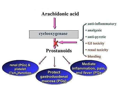
Arachidonic Acid + COX-1 or COX-2 (PGH2 Synthase / Peroxidase) → PGG2 → Prostaglandin H2 (PGH2).
PGH2 is the unstable "parent" molecule. Depending on the specific tissue enzymes present, PGH2 becomes:
- Prostaglandins: PGE2, PGF2α, PGD2.
- Prostacyclin (PGI2): Synthesized via Prostacyclin synthase (primarily in vascular endothelium).
- Thromboxane (TXA2): Synthesized via Thromboxane synthase (primarily in platelets).
Arachidonic Acid + 5-LOX (Lipooxygenase + FLAP protein) → 5-HPETE.
5-HPETE becomes:
- Leukotrienes: LTA4 → LTB4, LTC4, LTD4, LTE4.
- HETEs: (e.g., 8-HETE, 12-HETE, 15-HETE) - play crucial roles in inflammation and immune cell recruitment.
Arachidonic Acid + CYP Epoxygenases → EETs.
These play a role in maintaining vascular tone (vasodilation), renal function, and overall cardiovascular protection.
3. Mechanism of Action and Receptors
Eicosanoids do not enter cells. They bind to cell surface receptors that are all coupled to G-proteins (GPCRs).
Crucial Second Messenger Mechanisms
You must know whether they cause relaxation or contraction at the cellular level (tying back to your signaling lectures!):
- Relaxers (PGI2 and PGE2): Link to Gs proteins. Increase Adenylyl Cyclase → Increases cAMP → Decreases intracellular Calcium (Ca++). Result: Smooth muscle relaxation and Vasodilation.
- Contractors (TXA2, PGF2α): Link to Gq proteins. Activate Phospholipase C → Increases IP3 → Increases intracellular Calcium (Ca++). Result: Smooth muscle contraction, Vasoconstriction, and Platelet Aggregation.
4. Physiological & Pharmacologic Effects by System
This is where the exam will test your clinical application. Memorize these specific receptor actions:
A. The Vasculature (Blood Vessels)
- PGEs (PGE1, PGE2): Potent vasodilators.
- Prostacyclin (PGI2): Potent vasodilator. Can produce profound hypotension (low blood pressure).
- Thromboxane A2 (TXA2): Potent vasoconstrictor.
- Leukotrienes (LTC4, LTD4): Cause massive capillary leakiness (vascular permeability), contributing heavily to the swelling (edema) seen in severe inflammation.
- **Alprostadil (PGE1): Specifically dilates the ductus arteriosus in neonates.
B. Platelets (The Blood Clotting Tug-of-War)
There is a constant balance (a "see-saw") in your blood between two eicosanoids to prevent you from bleeding out or forming fatal clots:
- Prostacyclin (PGI2): Produced by healthy blood vessel walls. It INHIBITS platelet aggregation. (Mnemonic: Prostacyclin keeps blood CYCLING smoothly).
- Thromboxane A2 (TXA2): Produced by platelets. It is a massive platelet activator/aggregator. (Mnemonic: Thromboxane causes THROMBI / clots).
Inflammation (Leukocytes): LTB4 is a powerful chemotactic agent (it acts as a chemical beacon, attracting eosinophils, monocytes, and neutrophils to the site of injury). Conversely, prostaglandins generally inhibit cellular and humoral immunity to keep the immune system from overreacting.
C. The Lungs (Bronchial Tone)
- Prostaglandins: Have mixed effects on bronchial muscle (PGE1/PGE2 cause bronchodilation, PGD2/PGF2α cause constriction).
- TXA2: Causes bronchoconstriction. Inhibitors of thromboxane will therefore reduce the bronchoconstrictive response.
- Leukotrienes (LTC4, LTD4): Extremely potent bronchoconstrictors. These are the main culprits in deadly asthma attacks!
D. The Uterus (Obstetrics)
- PGE2 and PGF2α: Cause powerful uterine contractions, especially in a pregnant uterus.
- Clinical Tie-In (Dysmenorrhea): Overproduction of PGE2 and PGF2α during menstruation causes severe uterine cramping (primary dysmenorrhea). This is why taking an NSAID (which blocks these prostaglandins) cures menstrual cramps!
- Clinically, synthetic versions are used as abortifacients (to induce medical abortions) or to induce labor at term.
E. Gastrointestinal Tract (GIT)
- PGEs and PGI2: Inhibit gastric acid secretion (which is normally stimulated by feeding, histamine, or gastrin).
- They act as a shield, promoting the maintenance of the gastric mucosa by stimulating heavy mucus and bicarbonate secretion.
- Clinical Tie-In: This is exactly why taking NSAIDs (which block PGE production) causes stomach ulcers! You strip away the stomach's protective mucus shield.
F. The Kidneys
- PGE2 and PGI2: Cause renal vasodilation (specifically of the afferent arteriole), increase Renal Blood Flow (RBF), increase GFR, and promote diuresis (water excretion). (If a patient takes too many NSAIDs, they lose this vasodilation, the kidney starves of blood, leading to Acute Kidney Injury).
- TXA2: Causes renal vasoconstriction and has an ADH-like action (retains water).
G. Central Nervous System (CNS) & Eye
- CNS: PGE2 is the primary mediator of Fever, Pain perception, and Sleep. When a virus attacks you, the brain generates PGE2 to reset the hypothalamus thermostat, causing fever.
- Eye: PGF2α regulates the outflow of aqueous humor.
5. Clinical Pharmacology: Uses of Prostanoids and Analogues
In pharmacology, we create synthetic versions (analogs) of these molecules to treat diseases.
Mnemonic trick: If a drug name ends in "-prost" or has "prost" in the middle, it is a prostaglandin analog!
Group 1: Prostaglandin E1 (PGE1) Analogs
| Drug Name | Clinical Application & Mechanism |
|---|---|
| Alprostadil (IV infusion, IV inj, Intracavernosal) |
1. Patency of Ductus Arteriosus: Given to neonates born with severe congenital heart disease (e.g., Transposition of the Great Arteries) to keep the ductus arteriosus open, allowing oxygenated blood to mix until surgery can be performed. Side effect: Long-term use leads to ductus fragility and rupture. 2. Male Impotence: Injected directly into the penis. Increases cAMP → decreases Ca++ → relaxes trabecular smooth muscle and dilates cavernosal arteries, enhancing penile erection. |
| Misoprostol (Oral) |
1. Peptic Ulcers: Binds to PG receptors on parietal cells → decreases cAMP → inhibits proton pump → decreases acid secretion. It also increases mucous/bicarbonate and mucosal blood flow. Used specifically for NSAID-induced ulcers. Dose: 200μg QD. 2. Obstetrics (1st Trimester Abortion): Given orally with Mifepristone or Methotrexate in the first few weeks to soften the cervix and cause uterine contractions, expelling contents. *Side Effects: Severe GIT discomfort and diarrhea. |
| Lubiprostone (Oral) |
Chronic Constipation: Activates Type 2 Chloride (Cl-) channels in intestinal epithelial cells. Cl- is secreted into the gut, followed passively by Na+ and water. This increases stomach content liquidity and stimulates smooth muscle passage of stool. |
*Note: Enoprostil is another PGE1 analog used similarly to Misoprostol for NSAID ulcers/chronic smokers.
Group 2: Prostaglandin F2α (PGF2α) Analogs
| Drug Name | Clinical Application & Mechanism |
|---|---|
| Latanoprost, Bimatoprost, Travoprost, Unoprostone (Topical Eye Drops) |
Treating Open-Angle Glaucoma: These agents increase the outflow of aqueous fluid via the uveoscleral pathway, drastically lowering intraocular pressure. *Key Side Effect (Exam Gold): Bimatoprost causes dramatic elongation, thickening, and darkening of eyelashes (hypertrichosis). This "side effect" is now used commercially (as the drug Latisse) to treat eyelash thinning! |
| Carboprost (IM, Intra-amniotic) |
1. Post-partum Hemorrhage (PPH): Highly effective at violently contracting the uterus to clamp down on bleeding vessels after birth. 2. Mid-Trimester Abortion: Intra-amniotic injection. Least used for this now due to severe side effects. *Key Side Effect: Can cause severe anaphylactic shock and CVS (cardiovascular) collapse. |
| Dinoprost (Intra-amniotic inj) |
Mid-trimester (2nd Trimester) Abortion. |
Group 3: Prostaglandin E2 (PGE2) Analogs
| Drug Name | Clinical Application & Mechanism |
|---|---|
| Dinoprostone (Vaginal tab/gel/pessary) |
Induction of Labour & Cervical Ripening: Used vaginally at full term to induce labor (improves the "Bishop score" by physically softening the cervix). *Note: Oxytocin is usually the Drug of Choice (DOC) for labor induction. PGs are only used when Oxytocin is contraindicated (e.g., Renal failure, Pre-eclampsia, Eclampsia) because PGs do not cause Na+/water retention like oxytocin does. Also used for Mid-Term Abortion. *Side Effect: Prolonged bleeding. |
| Gemeprost / Demeprost / Denoproste (Vaginal pessary) |
Used vaginally for cervical priming in early pregnancy. |
Group 4: Prostacyclin (PGI2) Analogs
| Drug Name | Clinical Application & Mechanism |
|---|---|
| Epoprostenol & Treprostinil (IV Infusion) |
1. Pulmonary Arterial Hypertension: Lowers peripheral pulmonary and coronary resistance. They increase cAMP → decrease Ca++ → cause profound pulmonary vessel dilation, taking the strain off the right side of the heart. 2. Renal Dialysis: Used to inhibit platelet aggregation so blood doesn't clot in the dialysis machine. |
| Beraprost (Oral) |
Used for Peripheral Vascular Disease (given orally, thrice a day) to dilate vessels in the legs. |
| Iloprost (IM) |
Decreases infarct size when given IM after a Myocardial Infarction (MI). |
6. Clinical Uses of Eicosanoid Blockers
By blocking the synthesis pathways, we can treat various inflammatory and allergic conditions.
- Leukotriene Receptor Antagonists: Zafirlukast, Montelukast. They block the LTD4 receptors in the lungs, preventing bronchoconstriction.
- Lipoxygenase (LOX) Inhibitors: Zileuton. Stops the synthesis of leukotrienes entirely.
Clinical Scenario: If you give an asthmatic patient Aspirin, it blocks the COX pathway. The built-up Arachidonic acid has nowhere to go, so it is all "shunted" down the LOX pathway, creating massive amounts of Leukotrienes. This triggers a deadly asthma attack known as Aspirin-Exacerbated Respiratory Disease (AERD).
- NSAIDs (Non-Steroidal Anti-Inflammatory Drugs): Block Cyclooxygenase (COX-1 and COX-2), preventing the creation of pain/fever-inducing prostaglandins. Used for Rheumatoid arthritis and Dysmenorrhea (menstrual cramps).
- Aspirin (Low Dose): Aspirin irreversibly inhibits COX. At low doses (e.g., 81mg), it is highly selective for blocking TXA2 in platelets (stopping clots) without totally destroying the protective PGI2 in blood vessels. Because platelets do not have a nucleus, they cannot make new COX enzymes. The anti-clotting effect lasts for the entire lifespan of the platelet (7-10 days)!
7. Selective COX-2 Inhibitors (The "Coxibs")
Traditional NSAIDs (like Ibuprofen) block both COX-1 (which makes stomach-protecting mucus) and COX-2 (which makes inflammatory pain molecules). This causes stomach ulcers. Selective COX-2 Inhibitors were developed to be 10-20 times more selective for COX-2, aiming to stop pain without hurting the stomach. They are reversible inhibitors.
- Celecoxib: Chemically a sulfonamide. Half-life of 11 hours.
- Meloxicam: Related to Piroxicam. Preferentially selective COX-2 inhibitor.
- Etoricoxib: Long half-life (22 hours). Requires strict monitoring of hepatic (liver) functions.
- Nimesulide: A newer compound causing less gastric irritation.
Advantages of COX-2 Inhibitors:
- Excellent Analgesic, Antipyretic (reduces fever), and Anti-inflammatory effects.
- NO inhibition of protective gastric PGs = No gastric irritation/ulcers!
- NO inhibition of platelet aggregation = Does NOT prolong bleeding time (making them safer before surgeries).
The Massive Disadvantage / Adverse Effects (The Vioxx Disaster)
Drugs like Valdecoxib and Rofecoxib (Vioxx) were completely WITHDRAWN from the market. Why?
Because COX-2 usually makes Prostacyclin (PGI2) which stops clots, while COX-1 makes Thromboxane (TXA2) which causes clots. If you selectively block ONLY COX-2, you eliminate the anti-clotting mechanism, leaving TXA2 completely unopposed. This led to a massively higher risk of Cardiovascular thrombotic events (Myocardial Infarction / Heart Attacks and Strokes) in patients taking these drugs.
Other Side Effects: Renal toxicities (kidney damage) are exactly similar to non-selective NSAIDs. Celecoxib specifically can cause Skin Rashes (because it contains a sulfa group, triggering sulfa allergies).
8. Summary: Side Effects of Prostanoids
When giving synthetic prostanoids to a patient, you are basically causing a systemic inflammatory response. Effects are highly dose-related:
- Systemic: Hypotension, fever, dizziness, flushing.
- Respiratory: Bronchoconstriction (Cough is a notable side effect when using bronchodilators for asthma).
- GI tract: Vomiting, severe diarrhea (especially Misoprostol and Enoprostil).
- Severe reactions: Carboprost (anaphylactic shock, CVS collapse).
- Neonatal: Alprostadil over-usage causes ductus fragility and rupture.
- Bone/Kidney: PGE acting on EP4 receptors can increase osteoclast/osteoblast activity, inducing hypercalciuria (excess calcium in urine).
Quick Quiz
Paediatrics i Day 1 Quiz
Paediatrics - mobile-friendly and focused practice.
Privacy: Your details are used only for quiz tracking and certificates.
Paediatrics i Day 1 Quiz
Paediatrics
Preparing questions...
Choose your answer and keep your streak alive.
Great effort.
Here is your quick performance summary.
Eicosanoids Pharmacology Read More »