Batch 4: Biochemistry Bioenergetics Quiz
Batch 4: Biochemistry Bioenergetics Quiz Read More »
Test your knowledge with these 20 questions.
Question 1/20
Here are your results, .
Your Score
18/20
90%
Batch 4: Biochemistry Bioenergetics Quiz Read More »
Quick Quiz
Paediatrics - mobile-friendly and focused practice.
Privacy: Your details are used only for quiz tracking and certificates.
Paediatrics
Choose your answer and keep your streak alive.
Here is your quick performance summary.
Test your knowledge with these 25 questions.
Question 1/25
Here are your results, .
Your Score
27/30
90%
IMNCI Session 3 Identify Treatment Quiz Read More »
Test your knowledge with these 20 questions.
Question 1/20
Here are your results, .
Your Score
18/20
90%
Batch 3: Biochemistry Introduction Quiz Read More »
Test your knowledge with these 20 questions.
Question 1/20
Here are your results, .
Your Score
18/20
90%
Batch 2: Biochemistry Introduction Quiz Read More »
Test your knowledge with these 30 questions.
Question 1/30
Here are your results, .
Your Score
27/30
90%
IMNCI Session One Continuation QUIZ Read More »
Test your knowledge with these 20 questions.
Question 1/20
Here are your results, .
Your Score
18/20
90%
Batch 1: Biochemistry Introduction Quiz Read More »
Test your knowledge with these 30 questions.
Question 1/30
Here are your results, .
Your Score
27/30
90%
IMNCI Session One Asess and cLASSIFY QUIZ Read More »
By the conclusion of this exhaustive master guide, you will be deeply conversant with:
Nerve and muscle physiology is a highly specialized branch of medical physiology that specifically studies the function, architecture, and electrochemical mechanisms of nervous tissue (nerves) and muscle tissue (muscles). Collectively, nerves and muscles are uniquely classified as "excitable tissues."
This classification means they possess the extraordinary ability to undergo rapid, transient changes in their resting membrane potential. They explore how these tissues generate and transmit electrical signals (like action potentials) and how these sheer electrical impulses are seamlessly converted into specific, tangible cellular functions.

Nervous system excitability is defined as the inherent ability of nerve cells (neurons) to respond to an external or internal stimulus by generating and propagating an action potential—a massive, self-propagating electrical impulse that travels down the length of the cell.
This property is the absolute fundamental basis of the nervous system's function. It depends entirely on the neuron membrane's selective permeability, the precise opening and closing of voltage-gated ion channels, and the relentless work of active ion pumps. A sudden, sufficient change in membrane potential leads directly to this firing event, which is essential for transmitting information throughout the entire body. The physiology of the nervous system involves its main divisions—the Central Nervous System (CNS) and Peripheral Nervous System (PNS)—which utilize neurons and electrochemical signals to sense stimuli, integrate complex data, and produce highly coordinated, life-sustaining responses.
A motor neuron is a highly specialized, efferent nerve cell that transmits electrical command signals strictly away from the central nervous system (brain and spinal cord) to effector targets like muscles or glands, thereby initiating physical movement or chemical secretion.
Motor neurons are universally referred to in physiology as the final common pathway. This term emphasizes a profound physiological principle: all the impossibly complex neural computations happening in higher brain centers (e.g., motor planning in the cerebral cortex, coordination in the basal ganglia, and balance checking in the cerebellum) must ultimately converge onto these lower motor neurons.
It is only through the firing of a lower motor neuron that a skeletal muscle can be activated and a movement can physically occur. Regardless of whether a movement is voluntary (throwing a ball) or reflexive (pulling your hand away from a hot stove), the command signal ultimately travels down a lower motor neuron to its target muscle fibers. This makes the motor neuron a critical biological bottleneck and the ultimate determinant of muscle activity.
Clinical Example: Amyotrophic Lateral Sclerosis (ALS) is a devastating disease that specifically targets and destroys these motor neurons. Because they are the final common pathway, their destruction leads to complete, irreversible muscle paralysis, even though the patient's higher brain functions remain perfectly intact.

The metabolic and genetic center of the neuron. It contains the nucleus, extensive rough endoplasmic reticulum (Nissl bodies), and other organelles. It synthesizes vital neurotransmitters and structural proteins, and it receives synaptic inputs directly from thousands of other neurons.
Branching, tree-like extensions protruding from the soma. These are the primary receptive (input) regions of the cell. They are studded with ligand-gated ion channels that receive chemical signals (neurotransmitters) from adjacent cells and convert them into graded electrical potentials (EPSPs and IPSPs).
A specialized, cone-shaped anatomical region where the axon originates from the soma. This is the critical "trigger zone". It possesses the highest density of voltage-gated Na⁺ channels in the entire cell. It acts as an organic calculator, mathematically integrating all incoming graded potentials; if the sum reaches the critical threshold, an action potential is violently generated here.
A single, microscopic, tube-like projection that transmits the action potential (the output signal) away from the cell body toward the target. Some axons (like those reaching down to your toes) can exceed a full meter in length!
A thick, fatty, insulating layer wrapped around many axons. It is formed by Schwann cells in the PNS and Oligodendrocytes in the CNS. It is absolutely crucial for dramatically increasing the speed of action potential conduction and preventing electrical signal leakage.
Microscopic, unmyelinated gaps evenly spaced along the myelin sheath. These exposed patches contain a massive concentration of voltage-gated Na⁺ and K⁺ channels. The action potential is chemically regenerated at these nodes, allowing the signal to "jump" lightning-fast from node to node in a process called saltatory conduction.
The highly branched, club-like ends of the axon that form physical synapses with other cells. They contain thousands of synaptic vesicles packed with neurotransmitters and are specialized for converting the electrical action potential back into a chemical signal.
To understand the flow of neurological information, we map these anatomical structures into four distinct functional zones:
Synaptic transmission is the fundamental, microscopic process by which one neuron (the presynaptic neuron) communicates with another neuron (the postsynaptic neuron) or an effector cell (like a muscle). Most synapses in the human nervous system are chemical synapses, meaning they utilize chemical messengers known as neurotransmitters to physically bridge the gap between cells where electricity cannot cross.

This phase is the miracle of converting an electrical spark into a chemical flood:
The deadly Botulinum toxin (used cosmetically as Botox) and Tetanus toxin work by destroying the SNARE proteins mentioned in Step 4. Without SNARE proteins, synaptic vesicles cannot fuse with the membrane. The nerve fires, calcium rushes in, but the neurotransmitters are permanently trapped inside the terminal. This completely severs communication to the muscle, resulting in flaccid paralysis (in Botulism) or rigid, spastic paralysis (in Tetanus).
Once dumped into the cleft, neurotransmitters diffuse rapidly across the microscopic gap and bind reversibly to their specific, lock-and-key receptors on the postsynaptic membrane.
To ensure precise, crisp, and discrete signaling, the chemical message must be swiftly deleted from the cleft the moment the signal is delivered. If left unchecked, the muscle or nerve would seize up in a state of permanent, toxic overstimulation. This termination happens through three mechanisms:
The motor neuron is constantly bombarded with thousands of chemical signals (both excitatory and inhibitory) from thousands of other neurons every second. The neuron must mathematically integrate these signals to decide whether to fire a massive, "all-or-nothing" action potential.
A single tiny EPSP is overwhelmingly too weak to push a neuron to threshold. Therefore, the Axon Hillock adds them all up through summation:
If the absolute algebraic sum of all incoming excitatory (EPSPs) and inhibitory (IPSPs) signals reaches the critical Threshold Potential (typically around -55 mV), the Axon Hillock opens its gates and an action potential is irrevocably generated.
Once generated, the action potential propagates down the axon without losing a fraction of its strength. It follows a highly stereotyped, predictable voltage curve:
| Phase | Voltage Level | Cellular Event (Ion Channel Status) |
|---|---|---|
| 1. Resting State | -70 mV | The membrane is highly polarized. All voltage-gated Na⁺ and K⁺ channels are tightly closed. The Resting Membrane Potential (RMP) is maintained primarily by K⁺ leak channels (potassium constantly trickling out) and the relentless, energy-burning Na⁺/K⁺-ATPase pump. |
| 2. Depolarization to Threshold | -70 mV to -55 mV | The summed EPSPs cause a localized, sluggish depolarization. If the internal charge reaches the -55 mV threshold, it triggers a catastrophic, positive feedback loop. |
| 3. Rising Phase (Depolarization) | -55 mV shooting up to +30 mV | At threshold, thousands of voltage-gated Na⁺ channels violently snap open. A massive, sudden influx of Na⁺ rushes into the cell, erasing the negative charge and making the inside of the membrane highly positive. |
| 4. Falling Phase (Repolarization) | +30 mV dropping back toward -70 mV | At the exact peak (+30 mV), an inactivation gate swings shut on the Na⁺ channels, instantly stopping the Na⁺ influx. Simultaneously, the sluggish voltage-gated K⁺ channels finally fully open. A massive efflux of K⁺ rushes out of the cell, rapidly repolarizing the membrane back to negative. |
| 5. Undershoot (Hyperpolarization) | Dips below -70 mV (e.g., -80 mV) | The voltage-gated K⁺ channels are slow to close, causing an excessive, prolonged efflux of K⁺. The membrane becomes briefly more negative than its resting state. During this time, the neuron enters its Relative Refractory Period, where only an abnormally massive stimulus can trigger another firing. (Note: The Absolute Refractory Period occurs during the rising and falling phases, where it is physically impossible for the cell to fire again, ensuring the signal only travels in one forward direction). |
| 6. Restoration | Returns to -70 mV | All voltage-gated channels finally slam shut. The Na⁺/K⁺-ATPase pump works continuously in the background, burning ATP to shovel Na⁺ back out and K⁺ back in, fully restoring the long-term chemical gradients. |
When a dentist injects Lidocaine into your gums, the drug physically enters the nerve cell and plugs up the Voltage-Gated Na⁺ Channels from the inside. If Na⁺ cannot rush in during the Rising Phase, the action potential completely fails to generate. The pain signal is stopped dead in its tracks, and your brain never feels the drill.
While nerves specialize in communication, muscle tissue is purely specialized for mechanical contraction, generating kinetic force and bodily movement. We focus here on Skeletal Muscle.

The sarcomere is the fundamental, repeating contractile unit of a myofibril, extending from one Z-disc to the next Z-disc. Its highly organized geometry gives skeletal muscle its striated (striped) appearance.

The neuromuscular junction (NMJ) is arguably the most important chemical synapse in the human body. It is the highly specialized, mandatory interface where a motor neuron's axon terminal docks with a skeletal muscle fiber.
This is the explosive, step-by-step translation of a nerve thought into a muscle shock:
This is the awe-inspiring physiological bridge by which an invisible electrical signal (the muscle action potential) is instantly converted into a violent mechanical event (muscle contraction).

The Sliding Filament Theory universally proposes that muscle shortening (contraction) occurs strictly by the thick (myosin) and thin (actin) filaments sliding past one another, dragging the Z-discs closer together. The filaments themselves never actually shrink or change length; they merely increase their overlap.
Before the engines can engage, the track must be cleared. The Ca²⁺ ions released from the SR flood the sarcomere and bind immediately to the Troponin C subunit perched on the thin actin filaments. This binding causes a profound shape change in the entire troponin complex, which in turn violently tugs on the long, thread-like tropomyosin molecule. The movement of tropomyosin physically drags it away from the myosin-binding sites on the actin beads, exposing them completely to the waiting myosin heads.
The cross-bridge cycle is a blindingly fast, repetitive, four-step biochemical engine sequence that physically causes the thin filaments to slide over the thick filaments.
The energized, upright ("cocked") myosin head—which is already tightly holding onto a spent ADP and inorganic phosphate (Pi) molecule from the previous cycle—possesses a massive chemical attraction (affinity) for the exposed actin filament. The moment the tropomyosin shifts to uncover the binding sites (thanks to calcium), the myosin head violently snaps upward and forms a strong, unbreakable physical link with the actin. This physical connection between thick and thin filaments is the famous "cross-bridge."
The instant physical formation of the cross-bridge chemically triggers the immediate release of the trapped inorganic phosphate (Pi) from the myosin head. This release unleashes the stored mechanical energy, causing the myosin head to forcefully pivot on its hinge, jerking from its high-energy 90° angle to a low-energy, bent 45° angle. This brutal pivoting movement is the power stroke. Because it is firmly attached like a grappling hook, the pivoting myosin head violently drags the entire thin actin filament a short microscopic distance (~10 nm) toward the dead center (M-line) of the sarcomere. Immediately following the pivot, the remaining ADP molecule is ejected, leaving the myosin head in a rigid, low-energy state, still tightly locked onto the actin.
Following the power stroke, the spent myosin head remains rigidly "stuck" to the actin in a low-energy configuration (known as the "rigor" state). The absolute only biochemical way for the myosin head to let go of the actin is for a brand new, fresh molecule of ATP to physically bind to the empty ATP-binding site on the back of the myosin head. The instant this new ATP binds, it causes an allosteric (shape) change that severely weakens the molecular bond between the myosin and the actin, reducing their chemical affinity to zero, and forcing the myosin head to abruptly detach.
The completely detached myosin head, now carrying its fresh ATP, immediately acts as an aggressive enzyme (myosin ATPase). It instantly hydrolyzes (burns) the ATP, snapping it into ADP and inorganic phosphate (Pi). The explosive energy released from breaking this intense ATP chemical bond is captured entirely by the myosin hinge, physically forcing the head to snap backward, moving from its low-energy bent position back to its high-energy, upright, "cocked" position. It is now fully energized, reset, and coiled like a rat-trap, ready to violently repeat the entire cycle by latching onto another active site further down the actin filament (provided Ca²⁺ is still present in the sarcoplasm keeping the sites exposed).
Why do bodies become stiff after death? Upon death, cellular respiration halts, and ATP production completely drops to zero. Simultaneously, cell membranes degrade, allowing calcium to leak into the sarcoplasm, triggering Step 1 (Cross-Bridge Formation) and Step 2 (The Power Stroke). However, because there is absolutely no new ATP being produced by the dead body, Step 3 (Detachment) cannot occur. Millions of myosin heads remain permanently, rigidly locked onto the actin filaments in the low-energy rigor state. The entire muscular system locks into a solid, unbreakable spasm known as rigor mortis, which only subsides days later when the muscle proteins literally begin to rot and decompose.
As millions of myosin heads asynchronously paddle through this cycle thousands of times per second, the results are profound across the geometry of the cell:
When hundreds of thousands of sarcomeres lined up in series shorten simultaneously, the entire gross muscle belly shortens, pulling the tendon and generating massive skeletal force.
Muscle relaxation is not simply a passive fading out; it is a highly active, metabolically expensive, energy-requiring biochemical process. You must burn ATP to relax!
This is a rare, life-threatening genetic emergency triggered by the administration of certain general anesthetics (like halothane) or muscle relaxants (like succinylcholine) during surgery. The patient possesses a mutated, defective Ryanodine Receptor (RyR). When exposed to the anesthetic gas, the mutant RyR jams wide open, dumping an endless, unstoppable flood of calcium into the sarcoplasm. The muscles instantly go into a massive, full-body hyper-metabolic contraction. The SERCA pumps burn through the body's entire ATP supply trying to pump the calcium back in, generating highly lethal amounts of bodily heat (hyperthermia) and lactic acid. Without immediate administration of the antidote Dantrolene (which specifically acts to forcefully slam the RyR channels shut), the patient will rapidly die of a metabolic meltdown on the operating table.
This comprehensive synthesis of excitable tissue physiology is got from and aligns with the following gold-standard academic texts and resources universally utilized in medical and physiological education:
Quick Quiz
Physiology - mobile-friendly and focused practice.
Privacy: Your details are used only for quiz tracking and certificates.
Physiology
Choose your answer and keep your streak alive.
Here is your quick performance summary.
Nerve and Muscle Physiology Read More »
By the end of this exhaustive physiological guide, you will master:
Excitability refers to the fundamental ability of a living cell to respond to an environmental stimulus by generating a rapid, highly coordinated electrical signal known as an Action Potential. It is defined as a measurable physical and chemical change that occurs across the cell membrane when a stimulus is applied to a tissue. A stimulus is any external or internal agent (electrical, chemical, thermal, or mechanical) that produces this excitation.
The action potential is a transient, rapid, and self-propagating reversal of the electrical potential across the cell membrane. This electrical signal is the medium through which cells rapidly transmit information, either along the length of an individual cell (like a wire) or to other cells via specialized junctions (synapses). This property is crucial for rapid communication and coordination within the body, underpinning virtually every complex physiological function, from perception, thought, and memory to voluntary movement and visceral regulation.

Think of an excitable cell like a highly sensitive electrical tripwire or alarm system.

While all living cells exhibit some degree of responsiveness to their environment, only a select group possess the highly specialized membrane proteins and machinery required to generate and propagate rapid electrical action potentials. These are the "excitable cells."
Expanded Role: Neurons are the fundamental functional units of the nervous system. Their primary function is the rapid transmission of electrical and chemical signals for sensory input, integration, motor output, cognition, and emotion.
Unique Features: They possess specialized anatomical structures: dendrites (the "antennae" to receive signals), a cell body (soma) (the metabolic center), and a long axon (the "transmission cable" to send signals), which is often insulated by a lipid-rich myelin sheath to drastically speed up conduction velocity.
Muscle cells are specialized for contraction, which generates physical force and movement. Their electrical excitability is the absolute prerequisite for this mechanical action (Excitation-Contraction Coupling).
Expanded Role: Many glandular cells (e.g., in the adrenal medulla, pancreatic beta-cells) exhibit true excitability. They can respond to an electrical stimulus from a neuron or a chemical change (like high blood glucose) by generating their own electrical depolarization event.
Excitability Link: This electrical event is typically coupled directly to the release of their internal secretions (hormones, digestive enzymes). For example, adrenal medullary cells depolarize in response to a sympathetic neuronal signal, triggering massive Calcium (Ca²+) influx and the immediate exocytosis of epinephrine. This ensures precise, rapid control over systemic hormone release.

The capacity of these cells to generate electrical signals rests entirely on the foundational concept of the Membrane Potential. This is the voltage difference across the cell's outer boundary (the lipid bilayer). It represents stored, potential electrical energy created by an uneven distribution of ions (electrically charged particles) between the Intracellular Fluid (ICF) and the Extracellular Fluid (ECF).
When an excitable cell is quiet, it maintains a stable, baseline electrical charge called the Resting Membrane Potential (RMP). In this state, the inside of the cell consistently holds a negative charge relative to the outside. Standard RMP values include:
The RMP is not static; it is a highly dynamic state, constantly maintained by an active, energy-consuming interplay of three major factors:

The Equilibrium Potential for a specific single ion is the exact membrane voltage at which there is no net movement of that ion across the membrane. At this specific voltage, the electrical force pulling the ion in one direction is perfectly, equally balanced by the chemical (concentration) force pushing it in the other direction. The Nernst Equation calculates this theoretical value:
Eion = (RT / zF) * ln([ion]out / [ion]in)
Physiological Context: The equilibrium potential for Potassium (EK) is approximately -90 mV. The equilibrium potential for Sodium (ENa) is approximately +60 mV. Because the resting membrane is highly permeable to Potassium and mostly impermeable to Sodium, the overall RMP (-70 mV) sits very close to the Potassium equilibrium potential!
Ion channels are highly specialized, complex transmembrane proteins that form selective pores allowing specific ions to cross the otherwise impermeable lipid bilayer.
These channels are essentially always open. They are instrumental in establishing the RMP, particularly the widespread K+ leak channels that allow the continuous efflux of positive charge.
These channels have molecular "gates" that open or close only in response to a particular physiological trigger. They are the absolute essential machinery for generating action potentials.
| Type of Gated Channel | Trigger Mechanism | Physiological Example |
|---|---|---|
| Voltage-Gated Channels | Open or close in direct response to changes in the transmembrane voltage. They possess charged amino acids that physically move when the voltage changes. | Voltage-gated Na+ channels (driver of depolarization) and Voltage-gated K+ channels (driver of repolarization). |
| Ligand-Gated Channels (Chemically Gated) | Open or close only when a specific chemical messenger (a ligand), such as a neurotransmitter or hormone, binds to a receptor site on the channel. | The Nicotinic Acetylcholine Receptor at the neuromuscular junction. Binding of ACh opens the pore to let Na+ rush in. |
| Mechanically Gated Channels | Open or close when the membrane itself is physically deformed, stretched, or subjected to pressure. | Pacinian corpuscles in the skin (deep pressure receptors) or hair cells in the inner ear (responding to sound waves). |
A stimulus is any detectable change (electrical, chemical, or mechanical) in the cell's environment that has the potential to alter its resting membrane potential. Stimuli can cause two types of local changes:
Threshold is the critical, specific voltage level that a depolarization must reach for an action potential to fire (typically around -55 mV in neurons). It operates on an "all-or-none" principle: if a stimulus causes a local depolarization that reaches this threshold, a full, unstoppable action potential fires. If the stimulus is weak and only depolarizes the cell to -60 mV, nothing happens, and the potential simply dissipates.

The action potential is the primary electrical signal employed by excitable cells to swiftly transmit information across massive anatomical distances (like from the spinal cord to the toe). It stands as an "all-or-nothing" phenomenon: once initiated, it proceeds through its entire sequence with consistent strength, never diminishing over distance.
| Stage | Membrane Potential | Ionic Movement & Channel Status |
|---|---|---|
| 1. Resting State | -70 mV | All voltage-gated Na+ and K+ channels are CLOSED. The RMP is maintained by K+ leak channels and the active Na+/K+ pump. |
| 2. Depolarization to Threshold | -70 mV to -55 mV | A local stimulus causes a few voltage-gated Na+ channels to open. A small amount of Na+ trickles into the cell. If enough Na+ enters to push the voltage to the -55 mV threshold, the cell commits to firing. |
| 3. Rising Phase (Depolarization) | -55 mV to +30 mV | Threshold triggers a massive positive feedback loop. A vast number of voltage-gated Na+ channels rip open. A massive and swift surge of Na+ rushes into the cell, causing the inside to rapidly become positive (+30 mV). |
| 4. Repolarization Phase | +30 mV back down toward -70 mV | At the +30 mV peak, the voltage-gated Na+ channels inactivate (a physical "plug" blocks the pore), instantly stopping Na+ influx. Simultaneously, the slower voltage-gated K+ channels finally open fully. A massive outflow of K+ rapidly restores the membrane's negative internal charge. |
| 5. Afterhyperpolarization (Undershoot) | Dips below -70 mV (e.g., -80 mV) | The voltage-gated K+ channels are sluggish and close slowly. K+ continues to exit for a brief period, causing the membrane to become temporarily more negative than the baseline RMP. |
| 6. Return to Rest | Returns to -70 mV | The slow K+ channels finally close completely. The ever-active Na+/K+ pump and normal leak channels re-establish the original resting ion concentration gradients. |
A cell cannot fire continuously without resetting. This required downtime is called the refractory period.
The massive electrical shift at one point on the membrane triggers the opening of voltage-gated Na+ channels in the immediately adjacent area. This process repeats endlessly, moving the signal down the length of the nerve or muscle fiber like a burning fuse.
Many nerve fibers are thickly insulated by a fatty lipid layer called the Myelin Sheath (produced by Schwann cells in the PNS and Oligodendrocytes in the CNS). Action potentials cannot form where myelin exists. Therefore, the signal must "jump" from one microscopic uninsulated gap (a Node of Ranvier) to the next. This rapid, jumping propagation is termed Saltatory Conduction and dramatically increases the signal's speed while saving vast amounts of cellular energy.
Factors Influencing Conduction Speed:
Just as cells must generate signals to act, they desperately need ways to inhibit signals, ensuring precise motor control, emotional regulation, and preventing uncontrolled, chaotic firing (seizures).
Inhibitory neurotransmitters (like GABA in the brain or Glycine in the spinal cord) bind to ligand-gated channels that selectively allow Cl− (Chloride) to enter the cell or K+ to leave. The outcome is an immediate increase in the negative charge inside the cell (e.g., dropping from -70 mV to -80 mV). This creates an Inhibitory Postsynaptic Potential (IPSP), making it significantly harder for any excitatory stimulus to push the cell to threshold.
An inhibitory neuron releases a neurotransmitter (e.g., GABA) directly onto the axon terminal of an excitatory neuron. This slightly reduces the electrical charge of the terminal. Consequently, when an action potential arrives at that terminal, fewer voltage-gated Calcium channels open, meaning fewer excitatory neurotransmitters are released into the synaptic cleft. This allows for brilliant fine-tuning and selective dampening of specific signals without shutting down the whole system.
A vast array of modern drugs and natural toxins work by directly interfering with ion channels to deliberately shut down excitability.
An in-depth comprehension of cellular excitability is absolutely vital for understanding, diagnosing, and creating effective medical treatments for numerous devastating conditions affecting the nervous system and musculature.
Because the entire system of excitability relies on precise concentration gradients of ions, systemic electrolyte imbalances alter the very foundation of the resting membrane potential and threshold, leading to severe clinical emergencies.
| Electrolyte Imbalance | Pathophysiological Mechanism & Cellular Effect | Clinical Consequences |
|---|---|---|
| Hyperkalemia (Elevated K+) |
High extracellular K+ prevents K+ from leaking out of the cell. The resting membrane potential becomes less negative (e.g., moves from -90mV to -70mV in the heart), putting it dangerously close to threshold. While this initially causes twitchy hyperexcitability, prolonged depolarization permanently inactivates voltage-gated Na+ channels, ultimately rendering cells completely inexcitable. | Lethal cardiac arrhythmias, characteristic "peaked T-waves" on an ECG, and eventual flaccid paralysis leading to cardiac arrest. |
| Hypokalemia (Low K+) |
Low extracellular K+ creates a steeper gradient, forcing too much K+ to leak out of the cell. The RMP becomes more negative (hyperpolarized, e.g., dropping to -100mV). This moves the cell much further away from the threshold, making it significantly harder to fire an action potential. | Profound muscle weakness, fatigue, diminished reflexes, and dangerous heart arrhythmias (prominent U-waves on ECG). |
| Hyponatremia / Hypernatremia (Sodium Imbalances) |
Because the rapid, massive influx of Na+ is the absolute primary driver of the depolarization spike, severe imbalances in ECF Na+ levels disrupt the amplitude (height) and speed of action potentials. Furthermore, severe sodium imbalances cause massive cellular swelling or shrinking due to osmotic shifts. | Confusion, lethargy, severe muscle twitching, seizures, coma, and brain damage. |
| Hypocalcemia (Low Ca²+) |
Paradoxical Effect: Calcium ions normally bind to the outside of the cell membrane, physically "shielding" Na+ channels. When calcium is low, this shield is removed. Voltage-gated Na+ channels become highly unstable and rip open at much lower, more negative voltages. The threshold is dangerously lowered. | Severe, involuntary muscle spasms, cramps, and tetany (e.g., Trousseau's sign and Chvostek's sign). |
| Hypercalcemia (High Ca²+) |
High extracellular calcium heavily coats the membrane, stabilizing the Na+ channels and making them incredibly stiff and hard to open. The threshold is raised, drastically decreasing neuronal excitability. | "Bones, stones, groans, and psychiatric overtones." Severe muscle weakness, lethargy, depressed reflexes, and reduced overall neurological function. |
Quick Quiz
Physiology - mobile-friendly and focused practice.
Privacy: Your details are used only for quiz tracking and certificates.
Physiology
Choose your answer and keep your streak alive.
Here is your quick performance summary.
PHYSIOLOGY OF EXCITABLE TISSUES Read More »
By the conclusion of this exhaustive master guide, you will be deeply conversant with:
At the absolute heart of all physiological processes involving body fluids is the complex biochemical interaction between solutes and solvents, and their dynamic movement across semipermeable biological membranes.
The primary and overwhelmingly abundant solvent in all body fluids is WATER (H&sub2;O). Water possesses unique biochemical properties that make it the ideal medium for life:
Body fluids are not just water; they are highly complex "soups" containing a vast array of life-sustaining solutes:
The movement of these substances is primarily governed by passive, physical processes that strictly do not require the expenditure of cellular energy (ATP).
Rule 1: Solutes move by Diffusion (from high solute to low solute).
Rule 2: Water moves by Osmosis (from low solute to high solute). In clinical terms: "Water follows solutes."
These passive movements are absolutely essential for delivering oxygen and nutrients to tissues, removing metabolic wastes, and maintaining precise cell volume and shape.
To truly appreciate the clinical dynamics of body fluids, we must map exactly where this fluid resides. Imagine the human body as a complex system of interconnected containers. This meticulous compartmentalization is the ultimate key to maintaining cellular and systemic homeostasis.
TBW refers to the absolute sum of all water contained within the body. It represents a massively significant proportion of human body mass.
The total 42 Liters of body water is not uniformly distributed but is strictly divided into two primary compartments by the cell membrane:
The ICF is the fluid locked inside the trillions of cells in the body. It is the immediate, highly regulated aqueous environment where the vast majority of metabolic and biochemical activities occur.

The ECF is all the fluid found outside the cells. It acts as the body's vast "internal sea" that bathes, feeds, and cleanses all cells.
This is the literal "tissue fluid" filling the microscopic spaces between the cells. It is the largest component of the ECF, comprising about 80% of ECF volume (~10.5 Liters). Its ionic composition is nearly identical to blood plasma, but it lacks large proteins. The ISF is the critical "middle-man" medium for exchanging nutrients and gases between the blood capillaries and the cell membranes.
This is the pale-yellow fluid component of blood, circulating violently within the cardiovascular system. It accounts for 20% of ECF volume (~3.5 Liters). Its defining characteristic, compared to ISF, is a massive concentration of large plasma proteins (like Albumin). Plasma is the primary transport highway for red/white blood cells, nutrients, and waste.
A highly specialized, trapped component of the ECF, representing only 1-2% of body weight (approx. 1 to 2 Liters). It consists of fluids actively secreted by specific epithelial cells into distinct, enclosed body cavities. Examples include: Cerebrospinal Fluid (CSF), Intraocular Fluid (aqueous humor), Synovial Fluid in joints, Serous Fluids (pleural, pericardial, peritoneal fluid), and massive amounts of Gastrointestinal secretions.
Physicians and physiologists cannot simply drain a human to measure fluid volumes. Instead, they use a highly accurate mathematical principle called the Indicator-Dilution Method.
The Principle: You inject a known mass (amount) of a non-toxic marker substance (indicator) into the blood. You allow it time to distribute evenly through a specific compartment. You then draw a blood sample and measure the final concentration.
Formula: Volume = Mass of Indicator Injected / Concentration of Indicator in Sample.
The entire validity of the test relies on choosing an indicator that distributes only in the target compartment you wish to measure.
You cannot directly measure the Interstitial Fluid or the Intracellular Fluid because there is no chemical tracer that exclusively targets them without first going through plasma. Therefore, they are derived via simple subtraction:
The precise, uninterrupted movement of water and solutes between the body's fluid compartments is the absolute cornerstone of physiological survival. This dynamic equilibrium is meticulously regulated by intense physical forces and membrane properties.
The exchange of massive amounts of fluid, nutrients, and waste between the blood plasma and the tissue cells (via the ISF) occurs primarily across the microscopically thin, porous walls of the capillaries. This movement is governed by Starling Forces.

The absolute net movement of fluid is determined by a mathematical balance of these forces, expressed by the Starling equation:
NFP = (Pc - Pif) - (πc - πif)
Notice the math: +11 out, but only -9 back in. There is a continuous, slight physiological imbalance where filtration constantly exceeds reabsorption by about 2-3 Liters a day. This excess fluid, along with any escaped proteins, is swept up by the Lymphatic System. The lymphatics act as an emergency drainage sewer, returning this "lymph" fluid back into the venous circulation at the subclavian veins.
Clinical Pathology: If the lymph nodes are blocked (e.g., by a tumor, radiation, or parasitic elephantiasis), the fluid has nowhere to go, resulting in massive, disfiguring tissue swelling called Lymphedema.
While capillary walls are leaky, the cell membrane is highly restrictive. Exchange between the ISF and the ICF is driven exclusively by Osmosis. The cell membrane is highly permeable to water (via aquaporins) but highly impermeable to most solutes (like Sodium).
The extracellular fluid has the exact same concentration of non-penetrating solutes as the inside of the cell. There is zero net movement of water. Cell volume remains perfectly stable and healthy.
The extracellular fluid is diluted (has fewer trapped solutes than the inside of the cell). Water aggressively rushes INTO the hyper-concentrated cells to dilute them. Cells swell massively, and may rupture (lyse). Can cause deadly cerebral edema.
The extracellular fluid is highly concentrated (has too much sodium/solutes). The strong ECF aggressively sucks water OUT of the cells. Cells shrink and shrivel violently (crenation). Can cause severe neurological damage and coma.
While water movement is passive, maintaining these osmotic gradients requires massive energy. Cells are packed with highly concentrated, trapped negative proteins that constantly try to suck water into the cell. To prevent all human cells from swelling and exploding, the Na+/K+ ATPase pump runs continuously. By burning ATP to forcefully kick 3 Na+ ions OUT of the cell for every 2 K+ ions it brings in, it creates a net outward osmotic pull that exactly counters the inward pull of the proteins. If a cell loses oxygen (hypoxia) and runs out of ATP, the pump fails, sodium rushes in, water follows, and the cell undergoes fatal swelling (hydropic degeneration).
The human body uses incredibly sophisticated feedback loops involving the brain, heart, and kidneys to ensure fluid balance remains absolute.

The overall volume of the blood and ECF is dictated entirely by its total Sodium (Na+) content. The golden rule of physiology is: "Where Sodium goes, water is forced to follow."
ECF osmolarity is primarily determined by the concentration of solutes relative to water. The body alters osmolarity purely by holding onto or urinating out pure free water.
Disturbances in fluid regulation can have profound, rapidly life-threatening consequences.
| Pathology | Definition & Causes | Clinical Consequences |
|---|---|---|
| Hypovolemia (ECF Volume Deficit) |
A massive loss of isotonic fluid. Caused by acute traumatic hemorrhage, severe vomiting/diarrhea, or extensive third-degree burns (plasma weeping). | Decreased venous return, plummeting blood pressure, tachycardia, poor tissue perfusion, hypoxia, and progression to lethal Hypovolemic Shock. |
| Hypervolemia (ECF Volume Excess) |
Excessive accumulation of isotonic fluid. Caused by severe Heart Failure (failing pump), Renal Failure (can't excrete urine), or Cirrhosis. | Massive high blood pressure, distended jugular veins, severe peripheral pitting edema. If backed up into the lungs, it causes lethal Pulmonary Edema, destroying gas exchange. |
| Hyponatremia (Low Plasma Sodium) |
A disorder of water excess relative to sodium (Plasma Na+ < 135 mEq/L). Creates a dangerously hypotonic ECF. | Because the blood is dilute, water violently shifts INTO the concentrated brain cells. Causes lethal Cerebral Edema, leading to confusion, intractable seizures, brain herniation, and coma. |
| Hypernatremia (High Plasma Sodium) |
A disorder of absolute water deficit relative to sodium (Plasma Na+ > 145 mEq/L). Creates a highly hypertonic ECF. Caused by diabetes insipidus or extreme sweating without water replacement. | The salty blood violently sucks water OUT of the brain cells. Causes lethal Cellular Crenation (Shrinkage), leading to brain hemorrhage (tearing of bridging veins), intense lethargy, seizures, and death. |
| Edema (Third Spacing) |
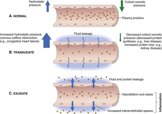
Abnormal accumulation of excess interstitial fluid in the tissues. | Caused by disruption of Starling Forces: 1) Increased Pc (Heart Failure), 2) Decreased πc (Liver failure/malnutrition causing low albumin), 3) Increased capillary permeability (Inflammation/Sepsis), 4) Blocked lymphatics. |

The administration of IV fluids is the most common invasive procedure in medicine. Safe administration requires an absolute mastery of fluid tonicity and exactly how fluids distribute upon entering the vein.
At the heart of all physiological processes involving fluids is the interaction between solutes and solvents, and their movement across various compartments.

The primary and overwhelmingly abundant solvent in all body fluids is WATER (H₂O).
Water's unique properties make it an ideal biological solvent:
Body fluids are complex solutions containing a vast array of solutes:
The movement of substances is primarily governed by passive processes that do not require cellular energy (ATP).
These passive movements are essential for:
Basic Principle: Water follows solutes. Specifically, water moves from an area of lower effective solute concentration (higher water concentration) to an area of higher effective solute concentration (lower water concentration) across a semipermeable membrane.
| IV Fluid Type | Effective Tonicity | Primary Distribution | Effect on ICF Cells |
|---|---|---|---|
| Isotonic (NS, LR) | Isotonic | ECF only (plasma & ISF) | No change |
| Hypotonic (0.45% NaCl, D5W) | Hypotonic | ECF & ICF | Swell |
| Hypertonic (3% NaCl) | Hypertonic | ECF (draws from ICF) | Shrink |
| Colloids (Albumin) | Effectively Hypertonic (oncotic) | Plasma only (draws from ISF) | No direct effect |
Quick Quiz
Physiology - mobile-friendly and focused practice.
Privacy: Your details are used only for quiz tracking and certificates.
Physiology
Choose your answer and keep your streak alive.
Here is your quick performance summary.
Body Fluids and Compartments Read More »
Javascript not detected. Javascript required for this site to function. Please enable it in your browser settings and refresh this page.