Nurses Revision

nursesrevision@gmail.com

homeostasis physiology

Homeostasis Physiology

Homeostasis: Maintaining the Internal Balance

Homeostasis

Imagine you're driving a car, aiming to maintain a constant speed of 60 mph. You press the gas going uphill and ease off going downhill. Your goal is to keep that speed constant despite external changes. That's essentially what your body does, constantly, for hundreds of variables.

Homeostasis (from Greek "homoios" meaning "similar" and "stasis" meaning "standing still") is the ability of an organism to maintain a relatively stable internal environment despite continuous changes in the external environment. It's not a static state, but a dynamic equilibrium where conditions fluctuate within narrow, acceptable limits around a set point.

Many physiologists translate this into the saying, “constantly changing to stay the same.” The ability of the human body to quickly adapt to any changes and to re-establish stability is the essence of homeostasis.

The Importance of Homeostasis

Survival itself depends on the body's ability to maintain this internal balance. Deviations outside the normal range can impair cell function, leading to disease or death.

Enzyme and Protein Function

Almost all biochemical reactions are catalyzed by enzymes (proteins), which are highly sensitive to their environment.

Impact of Imbalance: Deviations in temperature or pH can denature enzymes, altering their 3D shape and halting vital metabolic pathways.

Cellular Integrity and Volume

The cell membrane's selective permeability and active transport mechanisms are critical for maintaining appropriate solute concentrations.

Impact of Imbalance: Changes in extracellular fluid osmolarity can cause cells to swell and burst (lysis) or shrink and die (crenation). Disrupted ion gradients incapacitate nerve and muscle function.

Efficient Communication Systems

The nervous and endocrine systems require specific conditions to transmit signals effectively.

Impact of Imbalance: Improper electrolyte balance (Na⁺, K⁺, Ca²⁺) can lead to severe nerve and muscle dysfunction, including seizures, paralysis, and cardiac arrhythmias.

Energy Production (ATP)

Cells require a continuous supply of oxygen and nutrients, and efficient removal of waste, to produce ATP.

Impact of Imbalance: Oxygen deprivation (hypoxia) leads to a cellular energy crisis and buildup of lactic acid. Accumulation of wastes like CO₂ can become toxic and alter pH, leading to organ failure.

Immune System Function

Immune cells and proteins need stable conditions to effectively fight off pathogens without harming healthy tissues.

Impact of Imbalance: Uncontrolled fever can become detrimental to immune cells themselves. Chronic stress and elevated cortisol can suppress the immune system.

Examples of Homeostatically Regulated Variables

The body tightly regulates hundreds of variables to maintain this dynamic equilibrium. Key examples include:

  • Body temperature
  • Blood pressure
  • Blood glucose levels
  • Blood pH
  • Oxygen and carbon dioxide levels
  • Water balance
  • Ion concentrations (Na⁺, K⁺, Ca²⁺)

Homeostasis is Maintained by Feedback Loops

The primary way the human body maintains homeostasis is with the use of feedback loops. A feedback loop is a mechanism that allows for continual assessment of the body’s physiology and a way to correct various elements if they should go out of balance. There are two types of feedback loops: negative and positive.

Negative Feedback Loop

The response opposes (or negates) the original stimulus. This is by far the most common type in the human body.

Positive Feedback Loop

The response augments (or intensifies) the original stimulus. The cycle repeats until it is broken. This type is very rare but critically important.

Parameters and Set Points

For any feedback loop, there is a parameter that is being monitored, and it has a set point, or a ‘normal range’ in which it exists when the body is in balance. The stimulus that starts the feedback loop is a change in that parameter that pushes it above or below its normal set point range.

Table 1.1: Examples of Blood Parameters and Their Set Points
Osmolarity of Blood295-310 mOsM
pH of Blood7.35-7.45
Arterial PCO₂35-46 mmHg
Arterial PO₂80-100 mmHg
Glucose (fasting)70-100 mg/dL
Sodium (Na⁺)135-145 mM
Potassium (K⁺)3-5 mM

Example: Blood Glucose Regulation (Between Meals)

A person’s blood glucose (parameter) has a normal range (set point) of 70 to 100 mg/dL. If a person has not eaten in a while, their blood glucose decreases. If it goes below 70 mg/dL, the person will have hypoglycemia (low blood sugar). This decrease is the stimulus.

This decrease is detected by receptors in the pancreas, which responds by releasing the hormone glucagon into the bloodstream. Glucagon travels to the liver and stimulates hepatocytes (liver cells) to break down their glycogen stores and release glucose molecules into the blood. This increases blood glucose levels, opposing the original stimulus. Once glucose is restored to its normal range, the signal for glucagon release dissipates. This "off switch" is a key element of negative feedback.

The Nitty Gritty of the Feedback Loop

To describe feedback loops with consistent terms, we can identify seven general components that create the loop.

1. Stimulus: The change (above or below the set point) that starts the loop.
2. Receptor: The element or structure that detects this change.
3. Afferent Pathway: The incoming pathway used to convey information about this change.
4. Integration Center: The site where an evaluation is made about what to do.
5. Efferent Pathway: The outgoing pathway used to signal a tissue how to respond.
6. Effector Tissue: The structures acted upon to respond to the stimulus.
7. Response: The change created by the effector tissue in response to the original stimulus.

Homeostatic Control Mechanisms (The "Feedback Loops")

To maintain homeostasis, the body uses control systems, most of which involve feedback loops. These loops constantly monitor conditions, detect changes, and initiate responses to bring variables back to their set point.

Every feedback loop has three basic components:

1. Receptor (Sensor)

Function: Monitors the environment and responds to changes (stimuli). It detects the deviation from the set point.

Action: Sends information (input) along an afferent pathway (e.g., nerve impulses) to the control center.

Example: Thermoreceptors in the skin and hypothalamus detect changes in body temperature.

2. Control Center (Integrator)

Function: Receives and analyzes the input from the receptor. It compares the input to the set point (the ideal value) and determines the appropriate response.

Action: Sends commands (output) along an efferent pathway (e.g., nerve impulses, hormones) to the effector.

Example: The hypothalamus in the brain acts as the body's thermostat, comparing current body temperature to the set point of ~37°C (98.6°F).

3. Effector

Function: Carries out the control center's response. It provides the means for the control center's output to affect the stimulus.

Action: Its action either reduces the stimulus (negative feedback) or enhances it (positive feedback).

Example: Sweat glands, blood vessels in the skin, and skeletal muscles (shivering) are effectors that help regulate body temperature.

The Communication Pathway

RECEPTOR

Afferent Pathway

CONTROL CENTER

Efferent Pathway

EFFECTOR

Types of Feedback Loops

Negative Feedback Loops

(Most Common and Essential for Homeostasis)

Mechanism: The output of the system shuts off or reduces the intensity of the original stimulus, bringing the variable back toward the set point. It works to counteract the change.

Goal: To prevent severe changes and maintain stability.

Analogy: A thermostat controlling a furnace. When the temperature drops, the furnace turns on. Once the temperature reaches the set point, the furnace turns off (negative feedback).

Specific Example: Increased Body Temperature

If a person has been digging in the garden on a hot day, their body temperature rises above its set point of about 98.6°F. This is the stimulus. Thermoreceptors in the skin detect this change and send afferent information to the hypothalamus (the integration center). The hypothalamus then sends efferent signals to the effector tissues: sweat glands and cutaneous blood vessels. The response is diaphoresis (sweating) and cutaneous vasodilation (widening of blood vessels in the skin). Evaporation of sweat and increased blood flow to the skin dissipate heat, causing body temperature to decrease back to its set point.

Other Physiological Examples:

  • Blood Glucose Regulation: After a meal, high blood glucose stimulates the pancreas to release insulin. Insulin causes cells to take up glucose, lowering blood glucose levels.
  • Blood Pressure Regulation: Baroreceptors detect high blood pressure, signal the brain, which then slows heart rate and dilates blood vessels to lower pressure.

Positive Feedback Loops

(Rare, but Important for Specific Events)

Mechanism: The output of the system enhances or exaggerates the original stimulus, driving the variable further away from the initial set point. This is often part of a process that needs to be completed quickly.

Goal: To amplify a process until a specific event is completed.

Analogy: A microphone picking up sound, which is amplified and fed back into the microphone, creating a loop of increasing volume.

Specific Example: Childbirth

When a baby is ready to be born, its head pushes down upon the cervix, increasing pressure. This stretch (the stimulus) is detected by mechanoreceptors, which send an afferent signal to the brain. The brain (integration center) signals the posterior pituitary to release the hormone oxytocin. Oxytocin (efferent pathway) travels in the blood to the uterus (effector tissue), causing its smooth muscle to contract more forcefully. This pushes the baby’s head harder against the cervix, intensifying the original stimulus and triggering more oxytocin release. This cycle repeats, with contractions becoming stronger and more frequent, until the baby is born, which breaks the loop.

Other Physiological Examples:

  • Blood Clotting: Platelets at an injury site release chemicals that attract more platelets, rapidly forming a plug.
  • Generation of an Action Potential: An initial depolarization opens some Na⁺ channels, causing more Na⁺ to enter, which opens even more Na⁺ channels, leading to a rapid, all-or-nothing spike.

Diseases from Homeostatic Imbalance

The failure of homeostatic control mechanisms to maintain the body's stable internal environment leads directly to disease. Here are several examples:

Diabetes Mellitus

Imbalance: Chronic hyperglycemia (high blood glucose).

Mechanism: Insufficient insulin production (Type 1) or cellular resistance to insulin's effects (Type 2).

Consequences: Widespread damage to blood vessels, leading to heart attack, stroke, kidney failure, blindness, and nerve damage.

Hypo- and Hyperthyroidism

Imbalance: Disruption of thyroid hormone levels, which regulate metabolism.

Mechanism: Underproduction (Hypothyroidism) or overproduction (Hyperthyroidism) of thyroid hormones.

Consequences: Hypothyroidism leads to slowed metabolism, weight gain, and fatigue. Hyperthyroidism leads to accelerated metabolism, weight loss, anxiety, and rapid heart rate.

Kidney Failure (Renal Failure)

Imbalance: Inability to regulate fluid volume, electrolytes, pH, and excrete metabolic wastes.

Consequences: Fluid overload (edema), fatal cardiac arrhythmias from high potassium (hyperkalemia), toxic accumulation of urea (uremia), and dangerous drops in blood pH (acidosis).

Hypertension (High Blood Pressure)

Imbalance: Chronic elevation of systemic arterial blood pressure.

Mechanism: Multifactorial, often involving dysfunction in the nervous or endocrine systems' regulatory mechanisms (e.g., renin-angiotensin-aldosterone system).

Consequences: Increased risk of heart attack, stroke, kidney disease, and heart failure.

Dehydration and Overhydration

Imbalance: Disruption of fluid and electrolyte balance.

Consequences: Dehydration leads to low blood volume and pressure. Overhydration can dilute electrolytes (especially sodium), leading to brain cell swelling, seizures, and death (hyponatremia).

Sepsis

Imbalance: A life-threatening, dysregulated systemic response to infection.

Mechanism: The body's own immune response becomes overactive, leading to widespread inflammation and organ damage.

Consequences: Septic shock, multi-organ failure, and death.

Summary of Homeostasis

ConceptDescription
DefinitionMaintenance of a relatively stable internal environment (dynamic equilibrium).
ImportanceEssential for cell survival, optimal enzyme function, and overall health.
Control Loop ComponentsReceptor (detects change), Control Center (determines response), Effector (carries out response).
Negative FeedbackMost common. Output reduces/counteracts the original stimulus to restore the set point. Goal is stability. (e.g., Temperature, Blood Glucose).
Positive FeedbackRare. Output enhances/exaggerates the original stimulus to complete an event. Goal is amplification. (e.g., Childbirth, Blood Clotting).
Homeostatic ImbalanceOccurs when control mechanisms fail, leading to disease.

Test Your Knowledge

A quiz on the principles of Homeostasis.

1. Which of the following best defines homeostasis?

  • The process of responding to external stimuli.
  • The body's ability to maintain a relatively stable internal environment despite external changes.
  • The process by which an organism grows and develops.
  • The irreversible cessation of bodily functions.

Correct (b): This is the classic and most accurate definition of homeostasis. It emphasizes the "relatively stable" nature, acknowledging minor fluctuations.

Incorrect (a): Responding to stimuli is a broader biological characteristic, not exclusively homeostasis.

Incorrect (c): Growth and development are separate biological processes.

Incorrect (d): This describes death, the opposite of maintaining life.

2. A shivering response to cold, which raises body temperature, is an example of what feedback mechanism?

  • Positive feedback
  • Negative feedback
  • Feedforward control
  • Adaptation

Correct (b): The shivering response reverses the initial change (cold temperature) by generating heat. This counteraction is the hallmark of negative feedback.

Incorrect (a): Positive feedback would amplify the cold, making the body colder.

Incorrect (c): Feedforward control anticipates changes before they happen.

Incorrect (d): Adaptation refers to long-term adjustments, not acute responses.

3. Which component of a feedback loop detects changes in a regulated variable?

  • Effector
  • Control center
  • Receptor (sensor)
  • Set point

Correct (c): Receptors are specialized structures that detect changes (stimuli) in the environment.

Incorrect (a): The effector carries out the response.

Incorrect (b): The control center processes information.

Incorrect (d): The set point is the desired value, not a detection component.

4. In a negative feedback loop, the response of the effector:

  • Amplifies the original stimulus.
  • Counteracts or reverses the original stimulus.
  • Has no effect on the original stimulus.
  • Creates a new stimulus.

Correct (b): The defining characteristic of negative feedback is that the system's response works against the initial change to bring the variable back to its set point.

Incorrect (a): This describes positive feedback.

5. Childbirth labor contractions, which amplify in a cycle, are an example of what type of feedback?

  • Negative feedback
  • Positive feedback
  • Homeostatic imbalance
  • Allosteric regulation

Correct (b): The contractions stimulate more oxytocin, which causes even stronger contractions, creating a self-amplifying cycle. This amplification is characteristic of positive feedback.

Incorrect (a): Negative feedback would reduce contractions.

6. The "set point" in a homeostatic control system refers to the:

  • Actual value of the variable at any given moment.
  • Desired or ideal value around which the variable is maintained.
  • Range within which the variable is allowed to fluctuate.
  • Output generated by the effector.

Correct (b): The set point is the reference value for a regulated variable (e.g., 37°C for body temperature).

Incorrect (a): The actual value fluctuates around the set point.

Incorrect (c): This describes the "normal range" or "dynamic equilibrium."

7. Which of the following is typically regulated by negative feedback loops to maintain homeostasis?

  • Blood clotting
  • Blood glucose levels
  • Ovulation
  • Action potential generation

Correct (b): Blood glucose is tightly regulated by insulin and glucagon in a negative feedback loop.

Incorrect (a, c, d): Blood clotting, ovulation, and action potentials are all examples of processes involving positive feedback.

8. When homeostatic mechanisms are overwhelmed or fail, what condition can result?

  • Adaptation
  • Positive feedback
  • Homeostatic imbalance
  • Physiological resilience

Correct (c): When homeostatic mechanisms fail, the body enters a state of homeostatic imbalance, which can lead to disease.

9. What is the primary role of the control center in a homeostatic feedback loop?

  • To carry out the response.
  • To detect the stimulus.
  • To receive input, compare it to the set point, and send commands.
  • To amplify the deviation from the set point.

Correct (c): The control center (e.g., the brain) is the integration point that processes information and determines the response.

Incorrect (a): This is the role of the effector.

Incorrect (b): This is the role of the receptor.

10. A change in the external environment that causes a deviation from the set point is called a:

  • Response
  • Effector
  • Stimulus
  • Feedback

Correct (c): A stimulus is any detectable change in the internal or external environment that can initiate a response.

11. Which statement about positive feedback loops is generally TRUE?

  • They are more common than negative feedback loops.
  • They amplify the initial stimulus to complete a specific event.
  • They work to bring a variable back to its set point.
  • They are primarily involved in regulating body temperature.

Correct (b): Positive feedback loops are characterized by amplification, driving a process to a swift conclusion, such as childbirth or blood clotting.

Incorrect (a): Negative feedback is far more common for daily regulation.

Incorrect (c): Bringing a variable back to its set point is negative feedback.

12. The range of normal values around a set point is often referred to as:

  • Set point itself
  • Dynamic equilibrium
  • Control limit
  • Regulatory threshold

Correct (b): Homeostasis maintains a "dynamic equilibrium" because variables constantly fluctuate slightly around the set point, not held rigidly at a single value.

13. Maintaining internal body temperature within a narrow range is an example of:

  • Allostasis
  • Positive feedback
  • Homeostasis
  • Non-equilibrium thermodynamics

Correct (c): Maintaining a stable internal temperature is a classic example of homeostatic regulation.

Incorrect (a): Allostasis refers to achieving stability through change, a more complex adaptive process.

Incorrect (b): Positive feedback would lead to runaway heating or cooling.

14. Which body system is NOT considered a major regulator of homeostatic functions?

  • Nervous system
  • Endocrine system
  • Integumentary system
  • Respiratory system

Correct (c): While the skin (integumentary system) is a crucial effector in temperature regulation, it is not a primary regulatory system with control centers like the nervous and endocrine systems.

15. If blood pressure drops, the response of increased heart rate is primarily initiated by the:

  • Effector
  • Control center
  • Stimulus
  • Receptor

Correct (b): Receptors detect the drop, send info to the control center (brain), which then sends commands to the effectors (heart, vessels) to initiate the response.

16. A system that maintains a dynamic constancy of internal conditions is said to be in _________.

Rationale: This directly defines the term homeostasis as the dynamic maintenance of internal stability.

17. In a feedback loop, the component that receives commands and produces a change is the _________.

Rationale: The effector (e.g., a muscle or gland) is the part of the system that carries out the response dictated by the control center.

18. A negative feedback mechanism will act to _________ a deviation from the set point.

Rationale: The fundamental purpose of negative feedback is to oppose or counteract the initial change, bringing the variable back towards the set point.

19. The regulation of blood glucose by insulin and glucagon is a classic example of a _________ feedback loop.

Rationale: Both hormones work to counteract deviations from the blood glucose set point: insulin lowers high glucose, and glucagon raises low glucose.

20. A physiological state where conditions fluctuate within a narrow, healthy range is known as _________.

Rationale: This term emphasizes that homeostatic conditions are not rigidly fixed but are instead constantly adjusting within a tight, healthy range.

Homeostasis Physiology Read More »

Physiology and Cell Physiology

Physiology and Cell Physiology

Introduction to Basic Physiology & The Cell

Physiology is the rigorous scientific discipline dedicated to studying the functional activities and intricate mechanisms within a biological body. For example: why can the heart automatically beat, and how do electrical impulses coordinate this action? The word Physiology is derived from two ancient Greek words: physis, meaning "nature"; and logos, meaning "study". Thus, it is the study of the nature of living things.


1. Physiology Involves Process and Function

Words, names, and terms are of paramount importance in any scientific discipline because they carry highly precise meanings. Knowing and deeply understanding the relationships and etymological roots of these words will help tremendously in remembering and comprehending the vast amounts of information in a much deeper, more permanent way. This foundational knowledge will stay with you long after the course is over, enabling you to recognize important elements in other medical disciplines when you connect them to their deeper meanings.

Defining Physiology

The etymology (word origin) of the term Physiology traces back to 1560’s French, descending directly from the Latin physiologia, meaning “The study and description of natural objects, natural philosophy". This, in turn, is derived from the Greek physios meaning "nature, natural, physical"; and logia meaning "study". This gives us the fullest, most accurate meaning of Physiology as the "Science of the normal function of living things". When studying physiology, it is absolutely imperative that we also deeply understand the basic anatomy involved, as anatomy (structure) and physiology (function) are two sides of the same coin and go hand in hand.

Defining Anatomy

The etymology of the term Anatomy originates from Late 1300’s terminology in both Latin (anatomia) and Greek (anatome). These ancient words are derived from ana which means "up"; and tomos (or temnein) which means "to cut". Together this translates to "a cutting up", which is clearly referencing the act of physical dissection! In general clinical terms, anatomy is considered the “Study or knowledge of the structure (form) and organization of the human body“. While courses and textbooks for anatomy and physiology are often separated for convenience, they are inextricably connected to each other. A structure is designed specifically to support its function, and a function can only be carried out by an appropriately designed structure.


2. Etymology for the Language of Physiology

Another incredibly useful concept related to the importance of words in physiology (and anatomy) is mastering the etymology (origin of the word) of the vast array of scientific terms used in the healthcare field. Since the overwhelming majority of these words are derived from Latin and Greek, it is incredibly helpful to know the origins and ‘translations’ of these prefixes, roots, and suffixes. Becoming highly aware of the origins of words will greatly help students to:

  1. Understand exactly what a complex medical term means without memorizing it blindly.
  2. Assist you in instantly predicting what a brand-new, unseen term means when you first encounter it in a clinical setting.
Example 1: Hypertonic

The solution is hypertonic. Hyper means "above normal" or "excessive", and tonic refers to "strength" or "tension". Therefore, the solution is strong, excessively concentrated, and has a high osmotic pressure compared to another fluid.

Example 2: Hypoglycemia

The patient has hypoglycemia. Hypo is the direct opposite of hyper and means "below normal" or "deficient". The glyc portion refers to glucose (a type of sugar), and emia means "condition of the blood". Therefore, this statement translates perfectly to: the person has abnormally low blood sugar levels.

Example 3: Hyponatremia

A marathon runner presents with hyponatremia. Hypo still means "below normal". The natr portion comes from natrium, which is the Latin word for sodium (this is exactly why the chemical symbol for sodium on the periodic table is Na), and emia still means "blood". Therefore, this diagnosis means the person has dangerously low sodium levels in their blood.

Extra Example: Erythropoiesis

The bone marrow undergoes erythropoiesis. Erythro means "red", and poiesis means "to make" or "formation". Thus, this term describes the physiological process of producing new red blood cells.

Along the way in this physiology course, we will encounter many of these terms that, once we know the origin and meaning of, will help us figure out newer terms with absolute ease and familiarity. Anyone who has taken a medical terminology course will know the immense value of understanding the meaning of roots, prefixes, and suffixes.

Interactive Exercise: Decode the Term

Diagnosis: Pancytopenia. (Hint: there are 3 distinct root terms here: pan, cyto, and penia).

The Detailed Answer:
Pan means "all" or "every" (like a pandemic, which affects all people).
Cyto means "cell".
Penia means "deficiency", "lack of", or "poverty".
Translation: A deficiency of ALL cellular components in the blood. A patient with pancytopenia has dangerously low levels of red blood cells (anemia), white blood cells (leukopenia), and platelets (thrombocytopenia), usually due to complete bone marrow failure.


3. Compare Function and Process in Human Physiology

As we look to understand the central themes of physiology, an important concept is learning exactly how to ask questions about what’s occurring in the human body. In general, there are two basic, distinct approaches to physiology: 1) We can ask Functional Questions; and 2) We can ask Process Questions.

1. Functional Questions (The "Why")

These are strictly related to Why something occurs. They seek to understand the ultimate purpose or the evolutionary advantage of a mechanism. For example, what is the survival purpose of the heart beating? These can often be answered from a "big picture" perspective without requiring microscopic detail.

  • Q: Why does blood flow?
    A: To continuously transport vital nutrients, hormones, and gases (oxygen) around the body to sustain tissues, and to carry away toxic metabolic wastes (carbon dioxide, urea) to the excretory organs.
  • Q: Why do Red Blood Cells (RBCs) transport O₂?
    A: To efficiently deliver oxygen to all the body tissues that desperately need it to perform aerobic cellular respiration and generate ATP (energy).
  • Q: Why do we breathe?
    A: To extract essential oxygen (O₂) from the inhaled atmospheric air and to continuously release volatile carbon dioxide (CO₂) back out of the body, thus preventing fatal acid build-up in the blood.

2. Process Questions (The "How")

These are related to How something physically and chemically occurs. For example, how does the heart actually manage to beat? Often these issues must be answered in an extensively detailed, step-by-step, mechanistic manner.

  • Q: How does blood flow?
    A: The muscular ventricles of the heart contract (systole) to generate a massive high-pressure wave. Blood flows strictly down this pressure gradient, moving from the high-pressure aorta through the progressively lower-pressure arteries, capillaries, and veins, finally returning to the heart aided by skeletal muscle pumps and tissue fluid pressures.
  • Q: How do RBCs transport O₂?
    A: Inside the red blood cells, the iron-containing heme portion of the hemoglobin molecule undergoes conformational changes. It exhibits a high chemical affinity for O₂ when the surrounding partial pressure of O₂ is high (such as in the pulmonary capillaries of the lungs, forcing O₂ to bind). Conversely, it exhibits a low affinity for O₂ when the surrounding partial pressure for O₂ is low (such as in metabolically active, oxygen-depleted muscle tissue, forcing the hemoglobin to drop the O₂ where it is needed).
  • Q: How do we breathe?
    A: The diaphragm and external intercostal muscles (skeletal muscles of respiration) contract, pulling the rib cage up and the diaphragm down. This physical action dramatically increases the volume of the thoracic cavity. According to Boyle's Law, an increase in volume causes an inverse decrease in pressure. This creates a negative pressure gradient inside the lungs compared to the outside atmosphere, effectively vacuuming air down its pressure gradient into the lungs. Exhalation is the passive recoil of these same structures.

Things to Notice about Function and Process

Notice that the How part (the process) requires vastly more details and involves a strict ‘pathway’ approach. It is much more like sequential storytelling compared to the broader, less detailed functional aspects. The more arduous and demanding component of physiology is mastering these detailed processes. This is precisely the reason we need to take our time and fully, deeply understand the fundamentals before we delve into intricate, microscopic details.

What most students quickly recognize about physiology is that it is vastly more conceptual than anatomy because there is almost always a chemical or physical process to describe in a logical, step-by-step manner. There are usually two sides to the functions discussed in physiology. This is because at the very center of the human body is balance, which provides the equilibrium necessary to function properly. When we explain the mechanism of how we breathe in, we must simultaneously understand how we breathe out. When we explain how insulin lowers blood sugar, we must understand how glucagon raises it. Often, once you master one side of the physiological story, the other side falls into place much more easily.


4. Basic Functions of a Complex Organism

Holistically, we will examine Human Physiology as it relates to the foundational basics of how a multi-system, trillions-of-cells living organism functions as a single, perfectly coordinated entity. To be considered a living organism, the body must perform several critical basic functions. Below is an excessively detailed breakdown of these functions:

  1. Differentiation: The process by which unspecialized, generic cells (like stem cells) develop into highly specialized cells with distinct structures and functions. Extra Example: A single fertilized egg cell divides and its progeny differentiate into vastly different structures, such as conductive nerve cells, contractile muscle cells, and absorptive intestinal cells.
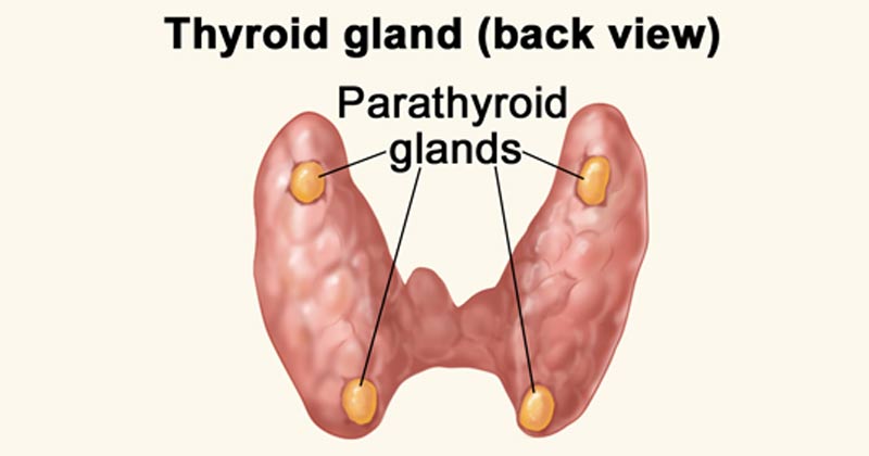
  2. Responsiveness (Excitability): The body's ability to detect and respond instantly to changes in its internal or external environment. Extra Example: If you touch a hot stove (external stimulus), sensory nerve endings detect the heat and trigger a withdrawal reflex. Internally, if blood calcium levels drop, the parathyroid glands detect this and release hormones to restore balance.
  3. Metabolism: The sum of all the intricate chemical processes that occur in the body. It consists of two phases: Catabolism (the breaking down of complex molecules into simpler ones to release energy, like digesting food) and Anabolism (the building up of complex molecules from simpler ones, requiring energy, like building new muscle protein).
  4. Growth and Repair: Growth refers to an increase in body size, either due to an increase in the size of existing cells (hypertrophy), an increase in the total number of cells (hyperplasia), or an increase in the amount of material surrounding cells. Repair is the ongoing process of replacing dead, damaged, or scraped-off cells (like skin cells constantly replacing themselves).
  5. Movement: Not just walking or running, but motion at every single level. This includes the coordinated action of the entire body, the movement of individual organs (like the stomach churning food or the gallbladder squeezing bile), single cells (like white blood cells physically crawling toward an infection), and even tiny organelles moving inside a cell.
  6. Excretion: The vital process of removing the toxic by-products of digestion and metabolism. If wastes like urea, carbon dioxide, or lactic acid are allowed to accumulate, they rapidly become fatal. The respiratory, digestive, and urinary systems all share this massive burden.
  7. Reproduction: The formation of new cells for tissue growth, repair, or replacement, OR the production of a completely new individual to ensure the continuation of the human species.

What we will find is that all of the diverse systems we will study in this course will contain many, if not all, of these basic functions deeply embedded within them.


5. Levels of Organization & Body Systems

A body system (also called an organ system) is a highly integrated, complex collection of organs in the body that work seamlessly together to perform a specific, vital function. The truth is that all systems are intimately connected, relying heavily on one another, but it is highly useful for educational purposes to study them separately, even though they are not separate at all. With all of our body systems operating constantly and simultaneously, it is absolutely necessary to have an overarching regulatory system in place to maintain stability, harmony, and equilibrium across all these integrated systems. This unifying, central element in all of physiology is called Homeostasis.

The Six Levels of Structural Organization:

  1. Chemical Level: The most basic level. Includes atoms (like Carbon, Hydrogen, Oxygen, Nitrogen) combining to form essential molecules (like DNA, glucose, water).
  2. Cellular Level: Molecules combine to form cells, the basic structural and functional units of an organism. (e.g., Muscle cells, nerve cells, blood cells).
  3. Tissue Level: Groups of similar cells and their surrounding materials working together to perform a specific function. (The four basic types: Epithelial, Connective, Muscular, Nervous).
  4. Organ Level: Structures composed of two or more different types of tissues. They have specific functions and recognizable shapes. (e.g., The stomach, heart, liver, brain).
  5. System Level: Consists of related organs with a common function. (e.g., The digestive system includes the mouth, esophagus, stomach, intestines, liver, and pancreas working to extract nutrients).
  6. Organismal Level: The highest level. All the parts of the human body functioning together to constitute the total living organism.

6. The Cell: The Fundamental Unit of Life

The cell is the absolute basic building block of all living things. Every single physiological process begins at the cellular level. To understand how the cell works, we can beautifully compare it to a bustling, highly organized city.

A. The Outer Boundary: The City Wall and Gates

The Cell (Plasma) Membrane

This is the outermost boundary of the cell, an incredibly thin, highly flexible, and selectively permeable (or semipermeable) barrier. It's primarily composed of a phospholipid bilayer, thickly populated with embedded proteins, carbohydrates, and cholesterol molecules.

  • Phospholipid Bilayer: Two opposing layers of phospholipids. Each lipid molecule has a hydrophilic ("water-loving") polar head that faces the watery environments inside and outside the cell, and two hydrophobic ("water-fearing") non-polar fatty acid tails that face inward toward each other, forming the water-repelling core of the membrane.
  • Proteins: These are crucial for nearly all membrane functions. Integral (transmembrane) proteins span the entire width of the membrane, forming open channels, carrier pumps, and signaling receptors. Peripheral proteins are loosely attached to the inner or outer surface, often involved in intracellular signaling cascades or anchoring the cytoskeleton.
  • Cholesterol: Wedged deeply within the hydrophobic core between the fatty acid tails, cholesterol helps stabilize the membrane's fluidity. It prevents the membrane from freezing solid at low temperatures and stops it from becoming too loose and falling apart at high body temperatures.
  • Glycocalyx (Carbohydrates): Chains of complex carbohydrates attached to proteins (forming glycoproteins) or attached to lipids (forming glycolipids) on the outer surface. This forms a unique, fuzzy "sugar coat" or cellular "ID tag." Extra Example: The ABO blood group markers on your red blood cells are actually part of the glycocalyx!

Physiological Functions of the Cell Membrane:

  • Selective Permeability: Strictly controls exactly what enters and leaves the cell, maintaining perfect homeostasis. (Functions like the city's border control).
  • Cell Recognition: The glycocalyx allows immune cells to recognize each other and identify foreign invaders.
  • Communication/Signaling: Receptor proteins bind to chemical messengers like hormones and neurotransmitters, translating outside signals into internal actions.
  • Cell Adhesion: Proteins allow cells to stick tightly together to form robust tissues (like skin).
  • Protection: Provides a flexible physical barrier shielding internal components.
Mnemonic: "People Call Me Protector" for Phospholipids, Cholesterol, Membrane Proteins.

B. The Cell's Internal Environment: The City Hall and Workers

The Cytoplasm

The cytoplasm is literally everything inside the cell membrane but entirely outside the nucleus. It consists of three main elements:

  • Cytosol: The jelly-like, semi-fluid, viscous portion where all organelles are suspended. It's mostly water heavily packed with dissolved solutes (ions, glucose, amino acids, ATP, fatty acids, etc.).
  • Organelles: The highly specialized "little organs" with very specific, indispensable functions (discussed next).
  • Inclusions: Temporary, non-functioning storage bodies. Extra Examples: massive glycogen granules in liver and muscle cells for energy storage, giant lipid droplets in fat cells (adipocytes), and melanin pigment granules in skin cells.

Physiological Functions of the Cytoplasm:

  • Site of Many Metabolic Reactions: Crucial biochemical pathways, such as glycolysis (the first, anaerobic step of glucose breakdown to extract energy), occur entirely free-floating in the cytosol.
  • Suspension of Organelles: Provides the perfect physical and chemical medium for all organelles to exist, interact, and function.

C. The Control Center: The City Hall/Mayor's Office

The Nucleus

Usually the largest and most prominent organelle, the nucleus is securely enclosed by a double-layered membrane called the nuclear envelope, which is punctured by nuclear pores that highly regulate the entry and exit of molecules. Inside, it contains:

  • Chromatin: The relaxed, uncondensed, thread-like form of DNA (our genetic material) carefully wrapped around specialized structural proteins called histones. When the cell prepares to divide, this messy chromatin tightly condenses into distinct, visible structures called chromosomes.
  • Nucleolus: A dense, dark, spherical body deeply embedded within the nucleus. It is the primary site of rapid ribosome synthesis.

Physiological Functions of the Nucleus:

  • Genetic Control: Contains the cell's entire genetic blueprint (DNA), directing absolutely all cell activities by strictly controlling which proteins are synthesized. (The "master architectural plan" for the city).
  • DNA Replication & Transcription: This is where DNA faithfully copies itself before cell division (mitosis) and where DNA's genetic code is safely transcribed into messenger RNA (mRNA), which then leaves the nucleus to give instructions to the factories.
  • Ribosome Production: The nucleolus continuously synthesizes and assembles ribosomal RNA (rRNA) subunits.

D. Protein Synthesis and Processing: The Factories and Delivery Services

Ribosomes

Tiny, granular, non-membranous organelles made of ribosomal RNA (rRNA) and various proteins. They are the ultimate "protein factories" of the cell. They physically read the mRNA code coming from the nucleus to perfectly assemble amino acids into long proteins (a process called translation). They can exist as free ribosomes floating in the cytosol (making proteins for immediate use within the cell) or bound ribosomes securely attached to the Endoplasmic Reticulum (making proteins destined for export out of the cell or for embedding into membranes).

Mnemonic: "Ribosomes Read RNA to make Really good pRotein."

Endoplasmic Reticulum (ER)

An extensive, labyrinth-like network of interconnected membranous tubes and flattened sacs that extends massively throughout the cytoplasm, and is directly continuous with the outer membrane of the nuclear envelope.

  • Rough Endoplasmic Reticulum (RER): Heavily studded with ribosomes, giving it a "rough" or sandpaper-like appearance under a microscope. Its primary function is to synthesize, fold, and modify proteins that are destined for secretion, insertion into cell membranes, or delivery to lysosomes. (e.g., adding sugar chains in a process called glycosylation). Extra Example: Pancreatic cells that produce massive amounts of digestive enzymes have incredibly large, active RERs.
  • Smooth Endoplasmic Reticulum (SER): Completely lacks ribosomes. Its highly specialized functions include massive lipid and steroid hormone synthesis, intense detoxification of harmful drugs and poisons (making it extremely abundant in liver cells), and the storage and release of calcium ions. Extra Example: In skeletal muscle cells, a specialized SER called the sarcoplasmic reticulum stores the calcium absolutely crucial for triggering muscle contraction.
Mnemonic: "Rough ER is Rough on Ribosomes & Really helps Really good pRotein; Smooth ER is Smoothly Synthesizing Steroids & Storing Salcium (calcium) and Speedily Solving Substance Spoilage (detox)."

Golgi Apparatus (Golgi Complex)

A distinct stack of flattened, slightly curved membranous sacs called cisternae. It acts as the ultimate "Post Office" or "Packaging and Shipping Center" of the cell.

Physiological Functions of the Golgi Apparatus:

  • Modification, Sorting, and Packaging: It receives vesicles containing raw proteins and lipids from the ER at its cis face, further modifies them (like adding or trimming sugar tags), sorts them based on their final destination, and packages them into new vesicles leaving from its trans face.
  • Vesicle Formation: It actively forms various types of vesicles, including secretory vesicles (for exocytosis/dumping out of the cell), lysosomes (cellular stomachs), and transport vesicles that seamlessly deliver new structural components to patch the plasma membrane.
Mnemonic: "Golgi Gathers, Grades, and Gets rid of Garbage (or packages good stuff!)."

E. Energy Production: The Power Plant


Mitochondria

Oval or bean-shaped organelles distinctly enclosed by a double membrane: a relatively smooth outer membrane and an inner membrane that is highly folded inward to form structures called cristae. These folds drastically increase the surface area available for chemical reactions. The dense, fluid-filled space within the inner membrane is called the matrix. Mitochondria also astonishingly possess their own unique, circular DNA (mtDNA) inherited exclusively from the mother.

Physiological Function (The "Powerhouses of the Cell"):

They are the primary site of aerobic cellular respiration. They utilize oxygen to meticulously break down and convert fuel molecules like glucose and fatty acids into ATP (adenosine triphosphate), the primary, usable energy currency of the entire cell. Extra Example: Cells that require massive, continuous amounts of energy, such as cardiac (heart) muscle cells and swimming sperm cells, have spectacularly high numbers of mitochondria compared to inactive cells.

Mnemonic: "Mighty Mitochondria Make Much More Money (ATP)."

F. Waste Management and Recycling: The Cleaning Crew

Lysosomes

Tiny, spherical membranous sacs packed tightly with incredibly powerful hydrolytic (digestive) enzymes that function best in an acidic environment. They act as the "Recycling Centers" and "Stomachs" of the cell.

  • They aggressively break down ingested foreign substances and bacteria (phagocytosis).
  • They digest worn-out, broken cellular organelles to recycle their raw materials (a process called autophagy).
  • In dying or damaged cells, they can burst open to digest the entire cell (autolysis).
  • Clinical Correlate: Tay-Sachs disease is a fatal genetic disorder where a specific lysosomal enzyme is missing. Toxic lipids build up uncontrollably in the brain because the lysosomes cannot break them down.
Mnemonic: "Lyso-some = "Lysol" – they lyse (break down) stuff."

Peroxisomes

Smaller membranous sacs containing potent oxidative enzymes, most notably catalase and oxidase. They act as the highly specialized "Detoxification Squad".

  • They aggressively neutralize highly reactive, harmful free radicals and detoxify toxins like alcohol (making them very abundant in kidney and liver cells).
  • They heavily assist in the breakdown (beta-oxidation) of very long-chain fatty acids for use in energy production.
  • During their reactions, they produce dangerous hydrogen peroxide (H₂O₂), but their built-in catalase enzyme instantly converts it safely into water and oxygen.
Mnemonic: "Peroxisomes Produce Peroxide to Purify."

G. The Cell's Internal Support and Movement: The Infrastructure


The Cytoskeleton

An intricate, dynamic network of protein filaments extending massively throughout the cytoplasm, providing cell shape, internal support, and physical highways for transport. It consists of three main types of filaments:

  • Microfilaments (Actin): The thinnest fibers; deeply involved in cell movement, gross shape changes, cell division (cleavage furrow), and forming the contractile machinery of muscle cells.
  • Intermediate Filaments: Tough, rope-like proteins that provide immense structural stability and fiercely resist mechanical pulling stress on the cell. Extra Example: Keratin in skin cells is an intermediate filament that makes skin tough and waterproof.
  • Microtubules: The largest elements; hollow tubes made of tubulin. They determine the overall cell shape, form the "railroad tracks" for motor proteins to drag organelles across the cell, and are the primary structural core of cilia, flagella, and the mitotic spindle.
Mnemonic: "Cytoskeleton Supports the Cell Shape and Ships things Swiftly."

Centrosomes, Cilia, and Flagella

  • Centrosomes and Centrioles: Located strategically near the nucleus, the centrosome contains two barrel-shaped centrioles positioned at right angles. It acts as the main Microtubule-Organizing Center (MTOC), generating and organizing the mitotic spindle required to pull chromosomes apart during cell division.
  • Cilia and Flagella: Hair-like, motile cellular projections entirely made of microtubules.
    • Cilia are short and numerous, beating in coordinated waves to move substances across the cell surface. Extra Example: Millions of cilia in your respiratory tract constantly sweep mucus and trapped dust up and out of your lungs.
    • Flagella are much longer and usually singular, acting like a whip to propel the entire cell forward. Extra Example: The only human cell with a flagellum is the male sperm cell.
Mnemonic: "Centrosomes and Centrioles Control Cell Civision Carefully."

Summary Table of Organelles

Organelle Key Physiological Functions
Plasma Membrane Selective barrier, cell recognition, intercellular communication, structural protection.
Nucleus Ultimate genetic control, houses DNA, site of DNA replication and mRNA transcription.
Ribosomes Direct protein synthesis via the translation of mRNA into amino acid chains.
Rough ER (RER) Synthesis, folding, and intense modification of proteins destined for export or membrane insertion.
Smooth ER (SER) Lipid and steroid synthesis, detoxification of drugs/poisons, massive intracellular Ca²⁺ storage.
Golgi Apparatus Modifies, sorts, meticulously packages proteins and lipids into transport/secretory vesicles.
Mitochondria Site of aerobic cellular respiration, massive ATP synthesis (the ultimate powerhouse).
Lysosomes Intracellular digestion of debris, bacteria, and old organelles; primary cellular waste removal.
Peroxisomes Detoxification (especially neutralizing free radicals and alcohol), extensive fatty acid breakdown.
Cytoskeleton Maintains cell shape, structural support, internal transport tracks, enables cell motility.
Centrosomes Organize the microtubule network and generate the mitotic spindle during cell division.
Cilia / Flagella Cilia move external substances across the cell surface; Flagella physically propel the entire cell.

7. Biological Membranes: The Fluid Mosaic Model

Biological membranes are highly dynamic, incredibly fluid structures that sharply define the boundaries of cells (plasma membrane) as well as the internal organelles. They are absolutely essential for maintaining cellular integrity, strictly regulating transport, facilitating communication, and housing vital enzymatic reactions. The most universally accepted scientific model describing membrane structure is the Fluid Mosaic Model.

The Fluid Mosaic Model

Proposed brilliantly by Singer and Nicolson in 1972, this model describes the cell membrane as a highly fluid lipid bilayer where an array of proteins are embedded or attached, looking much like a complex, ever-shifting mosaic artwork.

  • "Fluid": Refers to the constant, rapid movement of individual phospholipid molecules and proteins within the two-dimensional plane of the membrane. Lipids and many proteins are not locked in place; they constantly drift laterally, rotate on their axes, and flex their tails.
  • "Mosaic": Refers to the diverse, scattered "patchwork" of different functional proteins and other structural molecules (like cholesterol and carbohydrates) permanently or temporarily embedded within the vast ocean of the lipid bilayer.

A. Lipids of the Cell Membrane

The central, primary structural framework of the membrane is the fluid lipid bilayer, which is predominantly made of phospholipids and cholesterol.

1. Phospholipids

Phospholipids are by far the most abundant lipids in the membrane. They possess a crucial chemical property: they are amphipathic. This means a single molecule contains both a highly hydrophilic (water-loving) polar head and two highly hydrophobic (water-fearing) non-polar fatty acid tails. In the watery environment of the body, they spontaneously and instantly self-assemble to form a bilayer. The hydrophobic tails hide inward, desperately avoiding water, while the hydrophilic heads proudly face the watery environments inside (intracellular fluid) and outside (extracellular fluid) the cell.

2. Cholesterol

Cholesterol molecules are tough, rigid, ring-shaped lipids inserted tightly between the phospholipids. They act as the ultimate membrane temperature buffer, perfectly regulating fluidity. At normal, warm body temperatures, cholesterol restricts movement, reducing excessive fluidity and making the membrane stronger and less leaky. Conversely, at dangerously low temperatures, cholesterol prevents the phospholipid tails from packing too tightly together, thus increasing fluidity and stopping the cell membrane from freezing solid.

Lipid Functions in the Cell Membrane:

  • Forms the fundamental, self-healing bilayer structure.
  • Provides a robust selectively permeable barrier, naturally allowing only small, uncharged, fat-soluble substances (O₂, CO₂, steroid hormones) to easily pass through directly.
  • Acts as an impenetrable physical barrier for all water-soluble substances (glucose, salts, ions), which absolutely require physical assistance from embedded proteins to cross.

B. Membrane Proteins

While lipids form the barrier, proteins are the hardworking machines of the membrane, performing almost all of its specific, dynamic functions.

1. Integral (Transmembrane) Proteins

These are tightly bound proteins that fully span across the entire thickness of the membrane. They are deeply embedded in the hydrophobic core and can only be removed by completely destroying the bilayer. They primarily function as open channels, carrier transport proteins, active pumps, receptors, and enzymes.

2. Peripheral Proteins

These are loosely, temporarily bound to the membrane's inner or outer surface. They do not penetrate the hydrophobic core and are easily detached without harming the membrane. They often function as localized enzymes, signaling relays, or cytoskeletal anchors holding the membrane shape.

Functions of Membrane Proteins:

  • Transport: Facilitating the precise movement of specific, rejected substances across the barrier (channels, carriers, ATP pumps).
  • Enzymatic Activity: Catalyzing vital metabolic reactions directly at the membrane surface.
  • Signal Transduction: Acting as sophisticated receptors catching chemical messengers (like adrenaline) and transmitting the order inside.
  • Cell-Cell Recognition: Acting as strict identification tags (glycoproteins) so immune cells don't attack the body's own tissues.
  • Intercellular Joining: Forming tight junctions and desmosomes to physically link neighboring cells together.
  • Attachment to Cytoskeleton & ECM: Providing immense structural stability by tethering the cell's skeleton to the outside world.

C. Carbohydrates of the Cell Membrane

Carbohydrates are exclusively found on the external, outward-facing surface of the plasma membrane. They never face the inside. They are permanently attached to lipids (forming glycolipids) or to proteins (forming glycoproteins). This entire "sugar coat" covering the cell is called the glycocalyx, which serves as a highly specific, unique molecular signature for every different cell type in your body.

Functions of Membrane Carbohydrates (Glycocalyx):

  • Cell-Cell Recognition: Crucial for distinguishing "self" tissues from "non-self" invaders (e.g., orchestrating immune responses, dictating ABO blood types, and organ transplant rejection).
  • Cell Adhesion: The sticky sugars help cells firmly bind to one another in tissues.
  • Receptors: Can uniquely act as receptors for specific hormones, or tragically, as hijacking points for bacterial toxins and viruses (like the flu or COVID-19).
  • Protection: Provides a cushioning, protective physical barrier against mechanical damage and harsh enzymes.

Properties of the Cell Membrane

The specific composition and arrangement of lipids, proteins, and carbohydrates endow the cell membrane with its most essential, life-sustaining properties:

  1. Selectively Permeable (Semi-permeable): This is arguably its most important property. The membrane precisely and selfishly regulates exactly which substances can enter or leave the cell. The hydrophobic lipid core acts as the primary absolute barrier. Small, nonpolar molecules (O₂, CO₂) and highly lipid-soluble molecules pass directly and effortlessly. However, charged ions (Na⁺, K⁺) and large polar molecules (glucose) are utterly rejected and require specific, designated transport proteins to grant them entry.
  2. Fluidity: The membrane is absolutely not a rigid, solid shell; its molecular components are in constant, swirling motion. Fluidity is heavily influenced by body temperature, the amount of cholesterol (which acts as a stabilizing buffer), and the degree of saturation of the fatty acid tails (kinked, unsaturated tails increase fluidity). This property is absolutely essential for vesicles to fuse with the membrane, for the cell to divide, and for transport proteins to physically shift their shapes.
  3. Asymmetry: The two faces (the inner leaflet touching the cytoplasm, and the outer leaflet touching the blood/fluid) of the plasma membrane are completely structurally and functionally different. For example, carbohydrates are exclusively on the outer surface (glycocalyx), and specific lipids and signaling proteins are oriented strictly in one direction. This is vital for directional signaling, shape maintenance, and cell recognition.
  4. Self-Sealing Capability: Due to the powerful thermodynamic forces of hydrophobic interactions, if the membrane is punctured or torn, the lipids have a natural, spontaneous tendency to instantly re-seal themselves, completely preventing the fatal leakage of cytoplasmic contents. This is crucial for maintaining cell integrity during physical stress or when a needle enters a cell.

Summary of Key Membrane Functions:

  • Protective Barrier: Encloses the cell's delicate contents, safely separating the intracellular from the chaotic extracellular environment.
  • Selective Transport: Heavily regulates the passage of every single substance entering and exiting the cell.
  • Cell-Cell Communication: Contains specific receptors for circulating hormones, drugs, and neurotransmitters.
  • Cell Recognition & Adhesion: Facilitates cell identification by the immune system and the formation of solid tissues.
  • Enzymatic Activity: Houses membrane-bound enzymes that rapidly catalyze specific biochemical reactions.
  • Maintenance of Cell Shape: Provides profound structural support in conjunction with the inner cytoskeleton.
  • Generates Membrane Potential: Crucial for the electrical firing of nerve and muscle cells.
  • Endocytosis & Exocytosis: Manages the bulk, massive transport of large materials into and out of the cell.

8. Membrane Potential: The Electrical Voltage Across the Membrane

Before looking at exactly how things physically move across the membrane, it's absolutely essential to understand that there is a permanent electrical difference, or voltage, sitting directly across every single cell membrane in your body. This is called the membrane potential.

Membrane Potential is the exact measurable difference in electrical charge (or potential energy) between the inside and the outside of a cell. By strict scientific convention, the inside of the cell is always measured as being negative relative to the outside.

How is the Membrane Potential Established?

It is driven by three distinct, constantly working factors:

  1. Unequal Distribution of Ions: There are radically different concentrations of ions (charged particles) inside versus outside the cell.
    • Outside the cell (Extracellular Fluid - ECF): Features a massively high concentration of Na⁺ (sodium) and Cl⁻ (chloride).
    • Inside the cell (Intracellular Fluid - ICF): Features a massively high concentration of K⁺ (potassium) and huge, negatively charged proteins and phosphates (which are physically too large to ever leave the cell, trapping their negative charge inside).
  2. Selective Permeability of the Membrane: The cell membrane is not equally permeable to all ions. At rest, it is packed with specific "leak channels" that make it vastly more permeable to K⁺ than to Na⁺. Because K⁺ is highly concentrated inside, it constantly leaks out down its concentration gradient. Every time a positive K⁺ leaves, it leaves behind a negative charge, making the inside of the cell significantly more negative.
  3. Sodium-Potassium Pump (Na⁺/K⁺ ATPase): This relentless active transport pump burns massive amounts of ATP to constantly eject exactly 3 Na⁺ ions out of the cell for every 2 K⁺ ions it pumps back in. Because it pumps out more positive charges (3) than it brings in (2), this pump is inherently electrogenic and contributes directly and continuously to the negative charge inside the cell.

Resting Membrane Potential

In a resting (unstimulated) neuron or muscle cell, the steady-state electrical potential established and maintained perfectly by these factors is called the Resting Membrane Potential. It is typically measured at around -70 mV (millivolts), meaning the inside is 70 millivolts more negative than the outside.

Physiological Significance:

The resting membrane potential is absolutely not just a passive, resting state; it is a massive form of stored, coiled potential energy (like a stretched rubber band) crucial for:

  • Excitability: It allows excitable cells (like neurons and heart muscle cells) to instantly open gates and generate rapid, explosive electrical signals (action potentials) for instant communication, thought, and muscle contraction.
  • Secondary Active Transport: The immense energy stored in the steep Na⁺ and K⁺ ion gradients can be brilliantly harnessed by the cell to power the "uphill" transport of other essential substances (like glucose and amino acids) across the membrane.

Clinical Correlate: Hyperkalemia

If a patient's kidneys fail, potassium (K⁺) builds up in the blood outside the cell (a condition called hyperkalemia). This destroys the carefully maintained K⁺ gradient. The resting membrane potential is ruined, the heart muscle cells cannot reset their electrical charge, and the patient suffers a sudden, fatal cardiac arrhythmia (heart attack).


9. Membrane Transport

Membrane transport is the fundamental physiological process that meticulously governs the movement of all substances across biological membranes. It's essential for maintaining cellular homeostasis, acquiring vital nutrients, aggressively expelling toxic waste products, and facilitating cell-to-cell communication. Substances cross the membrane via two general, overarching mechanisms: Passive Transport and Active Transport.

1. Passive Transport: Moving Downhill

Passive transport is the movement of substances across a cell membrane completely without the direct expenditure of cellular metabolic energy (ATP). This movement is always "downhill," meaning down the electrochemical or concentration gradient of the substance. The energy driving this movement comes purely from the inherent, random kinetic energy of the molecules themselves and the potential energy stored in the concentration gradient.

1.1. Simple Diffusion: Through the Lipid Bilayer

In simple diffusion, substances move directly and effortlessly through the lipid bilayer without any help from membrane proteins whatsoever.

  • Highly Permeable: Small, nonpolar (lipophilic/fat-soluble) molecules like O₂, CO₂, and steroid hormones readily dissolve directly into the hydrophobic core and pass completely through to the other side.
  • Moderately Permeable: Small, uncharged polar molecules like water and ethanol can pass to a very limited degree because they are small enough to slip through the lipid gaps.
  • Impermeable: Large polar molecules (like glucose) and absolutely all charged ions (Na⁺, K⁺, Ca²⁺) cannot pass through the lipid core on their own under any circumstances.

The sole driving force is the concentration gradient. The random, chaotic molecular motion (kinetic energy) results in a net movement from an area of highly crowded concentration to an area of lower concentration, continuing unabated until perfect equilibrium is reached across the membrane.

Key Characteristics of Simple Diffusion:

  • Absolutely no membrane proteins are involved.
  • Does not exhibit saturation kinetics: The rate of diffusion increases linearly forever with the concentration gradient; there is no maximum transport rate (Vmax) because there are no proteins to get "full".
  • According to Fick's Law, the rate is directly proportional to the gradient magnitude, lipid solubility, and membrane surface area, and inversely proportional to molecular size and the physical thickness of the membrane.

1.2. Facilitated Diffusion: Protein-Assisted Passage

This process uses specialized integral membrane proteins (channels or carriers) to beautifully facilitate the movement of specific, impermeable substances down their electrochemical gradient. It is still completely passive, as absolutely no ATP is directly consumed.

A. Channel Proteins (Pores)

These proteins form a hollow, water-filled pore directly across the membrane, allowing the incredibly rapid, single-file passage of specific ions or water molecules. Most channels are tightly gated, meaning they act like doors that only open or close in response to specific, required stimuli:

  • Voltage-Gated Channels: Respond instantly to changes in electrical membrane potential (e.g., Na⁺ and K⁺ channels crucial in firing neurons).
  • Ligand-Gated Channels: Respond only when a specific chemical messenger binds to them (e.g., neurotransmitter receptors at muscle synapses).
  • Mechanically-Gated Channels: Respond directly to physical deformation or stretching of the membrane (e.g., touch receptors in your skin, or hearing receptors in the ear).
  • Leak Channels: Are generally always randomly fluttering open and closed, heavily contributing to the resting membrane potential.
  • Extra Examples: Ion channels (Na⁺, K⁺, Cl⁻, Ca²⁺) and aquaporins, which are highly specialized channels dedicated exclusively to letting massive amounts of water through instantly.

B. Carrier Proteins (Transporters)

These proteins work like revolving doors. They physically bind to a specific molecule on one side, undergo a massive conformational (shape) change, and then release the molecule safely on the other side. This physical shifting process makes it much slower than channel-mediated transport.

  • Saturation Kinetics: Because there are a strictly finite number of carrier proteins on a cell, the transport rate has an absolute maximum speed limit (Vmax) that hits when every single carrier is occupied and working as fast as it can.
  • Specificity: Carriers are incredibly highly specific for the exact molecule(s) they are designed to transport.
  • Competition: Structurally similar, "imposter" molecules can compete for the exact same binding site, blocking the real molecule.
  • Extra Examples: Glucose Transporters (GLUT proteins, which insulin commands to surface) and various amino acid transporters.

Key Characteristics of Facilitated Diffusion:

  • Strictly involves specific integral membrane proteins (channels or carriers).
  • Exhibits saturation kinetics (Vmax limit) due to the limited, physical number of transporters.
  • Can be easily subject to chemical competition.
  • Can be heavily regulated by the cell (e.g., by gating channels shut, or inserting/removing carriers from the membrane to control absorption).

1.3. Osmosis: The Grand Movement of Water

Osmosis is the incredibly powerful net movement of water across a selectively permeable membrane, strictly from an area of higher water concentration (which means lower solute concentration) directly to an area of lower water concentration (which means higher, saltier solute concentration). The driving force is the water potential gradient, determined entirely by the difference in non-penetrating solute concentration on either side of the membrane.

Osmotic pressure is the "pulling" or "sucking" force a solution with a higher solute concentration forcefully exerts to draw water toward it. Tonicity refers to the actual physical effect of a surrounding solution on the cell's total volume:

  • Isotonic: The concentration is perfectly equal inside and outside. There is no net water movement; cell volume remains perfectly normal.
  • Hypotonic: The outside fluid is dilute (watery). Water rushes rapidly into the cell, causing it to aggressively swell and potentially burst (a fatal event called lysis).
  • Hypertonic: The outside fluid is extremely concentrated (salty). Water is sucked rapidly out of the cell, causing the cell to drastically shrivel and shrink (a process called crenation).

Clinical Correlate: Intravenous (IV) Fluids

When a patient arrives at the hospital dehydrated, you cannot simply inject pure, hypotonic tap water into their veins. Doing so would cause their red blood cells to rapidly absorb the water, swell, and violently explode (hemolysis), killing the patient. Instead, nurses administer 0.9% Normal Saline, which is perfectly isotonic to human blood plasma, safely rehydrating the body without destroying the cells.

2. Active Transport: Against the Current, with Energy

Active transport is the strenuous, demanding process of actively moving substances across a cell membrane directly against their electrochemical or concentration gradient (i.e., dragging them from a region of lower concentration forcefully into a region of higher concentration). This distinctly "uphill" movement necessitates the direct or indirect expenditure of precious cellular metabolic energy, almost invariably derived from the violent hydrolysis of ATP.

2.1. Primary Active Transport: Direct ATP Expenditure

Primary active transporters are robust integral membrane proteins that function fundamentally as ATPases; they directly bind to and snap the phosphate off of ATP, utilizing that massive burst of released energy to physically power the forced movement of solutes. These heavy-duty transporters are often called "pumps."

  • Na⁺/K⁺ ATPase (Sodium-Potassium Pump): Found aggressively pumping in virtually all animal cells, this absolutely vital, life-sustaining pump forcibly moves 3 Na⁺ ions entirely out of the cell and drags 2 K⁺ ions back into the cell for every single ATP molecule hydrolyzed. It is strictly electrogenic (creates a negative charge imbalance) and is undeniably fundamental for maintaining steep Na⁺/K⁺ gradients, establishing the baseline resting membrane potential, regulating water balance and cell volume, and driving secondary active transport.
  • Ca²⁺ ATPases (e.g., SERCA, PMCA): These relentless pumps maintain the extremely, dangerously low intracellular Ca²⁺ concentration needed for survival. SERCA constantly pumps Ca²⁺ safely into the sarcoplasmic/endoplasmic reticulum for deep storage (which is absolutely crucial for allowing a muscle to relax after a contraction), while PMCA furiously pumps Ca²⁺ completely out of the cell membrane into the blood.
  • H⁺/K⁺ ATPase (Gastric Proton Pump): Located almost exclusively in the specialized parietal cells of the stomach lining, this pump aggressively secretes H⁺ (acid protons) directly into the stomach lumen, creating the highly destructive, acidic environment (pH 1-2) necessary for dissolving food and activating digestion. Extra Example: This exact pump is the sole target of Proton Pump Inhibitor (PPI) drugs, like Omeprazole, which shut the pump down to cure severe stomach ulcers and acid reflux.
  • ABC Transporters (ATP-Binding Cassette): A gigantic, ancient superfamily of transporters that move a vast, diverse array of substrates.
    Extra Example 1: MDR1 (P-glycoprotein) is an ABC transporter notorious in oncology; it causes multidrug resistance in cancer cells by acting as a biological bouncer, actively catching and pumping expensive chemotherapy drugs right back out of the tumor cell before they can work.
    Extra Example 2: The CFTR protein is a specialized Cl⁻ channel; a genetic mutation destroying this exact pump causes the thick, deadly mucus buildup characteristic of the disease Cystic Fibrosis.

2.2. Secondary Active Transport (Co-transport)

Secondary active transport is highly efficient because it does not directly hydrolyze ATP itself. Instead, it ingeniously acts like a waterwheel; it uses the immense potential kinetic energy stored in an existing, steep electrochemical gradient (typically the massive Na⁺ gradient that was previously created and paid for by the Na⁺/K⁺ pump) to power the transport of a completely second substance against its own gradient.

  1. Symporters (Cotransporters): Both the driving ion (e.g., Na⁺ rushing down its gradient) and the "freeloading" transported solute are physically dragged across the membrane in the exact same direction. Extra Example: The Na⁺-Glucose Symporter (SGLT) found heavily in the intestine and kidneys, which utilizes the rushing flow of Sodium to forcefully absorb essential Glucose from your food against a steep gradient, ensuring none is wasted in the feces or urine.
  2. Antiporters (Exchangers): The driving ion rushes in, providing energy to forcefully eject the transported solute in the exact opposite direction. Extra Example: The Na⁺-Ca²⁺ Exchanger (NCX), which is absolutely crucial for aggressively removing toxic excess Ca²⁺ from cardiac (heart) muscle cells after every single heartbeat, allowing the heart to relax. Also, the Na⁺-H⁺ Exchanger (NHE) which pumps acid out of the cell to regulate intracellular pH.

3. Vesicular Transport (Bulk Transport): For the Heavy Lifting

Vesicular transport is the macro-scale mechanism used exclusively for moving massively large molecules, whole macromolecules, and giant particulate matter (even whole bacteria) into or out of the cell. It involves the complex, energy-demanding physical formation, movement, and fusion of large membrane-bound sacs called vesicles. Because it physically reshapes the cell membrane, it absolutely always requires massive amounts of cellular energy (ATP).

3.1. Endocytosis: Bringing the Outside In

Endocytosis is the active process by which cells internalize large substances. The plasma membrane physically sinks inward (invaginates), wraps entirely around the material, and pinches off, forming a brand-new intracellular vesicle.

  • Phagocytosis ("Cell Eating"): The aggressive, targeted ingestion of massive, solid particles like invading bacteria, dead tissue, or cellular debris by highly specialized immune sentinels (e.g., macrophages and neutrophils). The cell physically extends long "arms" called pseudopods to totally engulf the target, forming a giant internal death-chamber called a phagosome, which then merges with a lysosome for total destruction.
  • Pinocytosis ("Cell Drinking"): The constant, non-specific, routine gulping of tiny droplets of extracellular fluid and whatever dissolved solutes happen to be floating in it. It is how cells sample their environment.
  • Receptor-Mediated Endocytosis: An exquisitely, highly specific, targeted process. Specific extracellular ligands (e.g., LDL-cholesterol particles, iron-carrying transferrin, or even sneaky viruses) must perfectly bind to complementary, matching receptors on the surface. These loaded receptors then rapidly slide together and cluster into specialized depressions called clathrin-coated pits, which fold inward and are cleanly brought into the cell as a customized vesicle.

3.2. Exocytosis: Releasing to the Outside

Exocytosis is the reverse process by which cells actively release large substances. Intracellular, membrane-bound vesicles are driven by motor proteins to the edge of the cell, where they seamlessly fuse with the plasma membrane, violently dumping their contents to the outside world.

  • Constitutive Secretion: A continuous, ongoing, unregulated baseline process operating in all cells. It is used to constantly deliver newly minted lipids and proteins to expand the plasma membrane, and to continuously secrete the structural components of the extracellular matrix (like collagen).
  • Regulated Secretion: A highly controlled process that occurs exclusively in specialized secretory cells (e.g., brain neurons, pancreatic endocrine cells). Secretory vesicles containing highly potent products like neurotransmitters, digestive enzymes, or hormones are stockpiled safely near the membrane. They are strictly held back and only released in a massive burst in response to a very specific, targeted signal (almost always a sudden, sharp rise in intracellular Ca²⁺).

10. The "Why": Importance and Functions of Membrane Transport

The precise, unrelenting control over exactly what enters and exits a cell underlies the success of virtually every single physiological process in the entire human body.

  • Maintenance of Cellular Homeostasis: Strict, life-or-death ion gradients (e.g., maintaining low intracellular Na⁺ and high K⁺) and exact internal pH are aggressively maintained at incredibly great energy cost to absolutely prevent cell death and ensure proper, flawless enzyme function.
  • Nutrient Acquisition: Transport systems enable cells to highly efficiently scavenge, absorb, and aggressively concentrate essential, life-sustaining molecules like glucose, vitamins, and amino acids from the blood.
  • Waste Removal: Active transporters continuously expel harmful, toxic metabolic waste products (like urea, lactic acid, and excess acid protons), totally preventing their lethal accumulation inside the cytoplasm.
  • Generation of Electrical Signals: In complex neurons and muscle cells, the rapid, highly controlled movement of ions surging through gated channels generates action potentials, which are the fundamental electrical basis of every single thought, heartbeat, and physical movement.
  • Cell-to-Cell Communication: Exocytosis violently releases neurotransmitters across synapses and dumps hormones into the blood, while endocytosis helps regulate receptor sensitivity by swallowing up overused receptors from the membrane.
  • Regulation of Cell Volume: Constant ion pumping, most especially the tireless Na⁺/K⁺ ATPase, strictly controls internal intracellular osmolarity, absolutely preventing cells from fatally swelling up and bursting or aggressively shrinking and collapsing.
  • Absorption and Reabsorption: Massive, highly coordinated transport processes in the Gastrointestinal (GI) tract and the nephrons of the kidneys are utterly essential for absorbing nutrients from food and perfectly regulating the entire body's water, total electrolyte, and systemic acid-base balance.
Summary of Membrane Transport Mechanisms
Process Energy Requirement Gradient Direction Transporter Requirement What Specifically Moves? Examples & Clinical Notes
Passive Processes (No ATP Burned)
Simple Diffusion None Downhill (High to Low) None (Straight through lipid) Small, highly lipid-soluble molecules O₂, CO₂, steroid hormones crossing tissues.
Facilitated Diffusion None Downhill (High to Low) Yes (Channel or Carrier protein) Ions, large glucose, amino acids Glucose transporters (GLUT), Na⁺/K⁺ voltage channels.
Osmosis None Downhill (High water to Low water) Yes (Aquaporins heavily aid) Water molecules exclusively Red blood cells expanding or shrinking in different tonic IV solutions.
Active Processes (Requires ATP)
Primary Active Transport Yes (Directly breaks ATP) Uphill (Low to High) Yes (A specialized Pump) Ions against steep gradients Na⁺/K⁺ pump, Ca²⁺ SERCA pump, H⁺ stomach acid pump.
Secondary Active Transport No (Uses kinetic energy of an existing ion gradient) Uphill (Low to High) Yes (Symporter or Antiporter) Ions, glucose, amino acids dragged along Na⁺-glucose co-transporter (SGLT) in the kidney.
Vesicular Transport Yes (Massive ATP used) N/A (Bulk physical movement) No (Uses membrane vesicles) Large particles, whole bacteria, macromolecules, bulk fluids Phagocytosis (macrophages eating bacteria), Exocytosis (releasing insulin).

List of Academic References

  • Hall, J. E., & Hall, M. E. (2020). Guyton and Hall Textbook of Medical Physiology (14th ed.). Elsevier. (Definitive source on membrane potentials, the Na⁺/K⁺ pump, and systemic physiology).
  • Boron, W. F., & Boulpaep, E. L. (2016). Medical Physiology (3rd ed.). Elsevier. (Excellent deep dive into cellular organelles, secondary active transport, and molecular physiology).
  • Alberts, B., Johnson, A., Lewis, J., et al. (2014). Molecular Biology of the Cell (6th ed.). Garland Science. (The absolute gold standard for the Fluid Mosaic Model, vesicular transport, and cytoskeletal structures).
  • Costanzo, L. S. (2017). Physiology (6th ed.). Elsevier. (Highly recommended for clear, mechanistic explanations of functional vs. process physiology and simple diffusion mathematics).
  • Chabner, L. (2020). The Language of Medicine (12th ed.). Saunders. (A premier resource for medical terminology, etymology, roots, and prefixes like pancytopenia).

Quick Quiz

Cell Physiology & Transport Quiz

Physiology - mobile-friendly and focused practice.

Privacy: Your details are used only for quiz tracking and certificates.

Physiology and Cell Physiology Read More »

Anatomy & Physiology 2023 Paper

Final Examination Paper

Anatomy & Physiology

Bachelors in Nursing • Semester 2, 2023

3 Hours

Duration

100 Marks

Total Marks

3 Sections

A, B, C

Instructions to Candidates

  • Answer ALL questions in Section A (Objectives & Fill-ins).
  • Answer any THREE questions from Section B.
  • Answer any TWO questions from Section C.
  • Write clearly and legibly.
  • Do not write anything in the margins.

SECTION A

40 Marks

Part I: Objectives (20 Marks)
Answer ALL questions in this part. Choose the most appropriate answer.

1. Which bone cell is responsible for resorbing (breaking down) bone matrix?

A. OsteocyteB. OsteoblastC. OsteoclastD. Osteogenic cell

2. The "Waiter's Tip" position is a classic sign of injury to which part of the brachial plexus?

A. Lower Trunk (C8, T1)B. Upper Trunk (C5, C6)C. Medial CordD. Posterior Cord

3. All muscles of facial expression are innervated by which cranial nerve?

A. Trigeminal Nerve (CN V)B. Facial Nerve (CN VII)C. Accessory Nerve (CN XI)D. Hypoglossal Nerve (CN XII)

4. During which stage of lung maturation does surfactant production begin?

A. Pseudoglandular StageB. Canalicular StageC. Saccular StageD. Alveolar Stage

5. Which muscle is the primary flexor of the forearm at the elbow?

A. Biceps BrachiiB. BrachialisC. Triceps BrachiiD. Brachioradialis

6. The sella turcica, which houses the pituitary gland, is a feature of which cranial bone?

A. Frontal BoneB. Ethmoid BoneC. Occipital BoneD. Sphenoid Bone

7. In oogenesis, meiosis I is completed just before ovulation, resulting in:

A. One ovum and three polar bodiesB. Four functional ovaC. Two secondary oocytesD. One secondary oocyte and one polar body

8. Which muscle is NOT part of the rotator cuff (SITS) group?

A. SupraspinatusB. Teres MajorC. InfraspinatusD. Subscapularis

9. The primary action of the muscles in the lateral compartment of the leg (Fibularis Longus and Brevis) is:

A. DorsiflexionB. InversionC. EversionD. Plantarflexion

10. An inability to abduct the thigh and a pelvic drop on the unsupported side (Trendelenburg sign) indicates damage to which nerve?

A. Femoral NerveB. Obturator NerveC. Inferior Gluteal NerveD. Superior Gluteal Nerve

11. The olecranon process is a prominent feature of which bone?

A. RadiusB. HumerusC. UlnaD. Scapula

12. All hamstring muscles are innervated by the tibial portion of the sciatic nerve EXCEPT:

A. Long head of Biceps FemorisB. Short head of Biceps FemorisC. SemitendinosusD. Semimembranosus

13. Which of the following is NOT part of the axial skeleton?

A. SternumB. RibsC. ClavicleD. Vertebrae

14. The "anatomical snuffbox" is formed by the tendons of all the following muscles EXCEPT:

A. Abductor Pollicis LongusB. Extensor Pollicis BrevisC. Abductor Pollicis BrevisD. Extensor Pollicis Longus

15. Referred pain to the shoulder tip is often a sign of irritation to the diaphragmatic pleura, carried by which nerve?

A. Vagus NerveB. Phrenic NerveC. Intercostal NerveD. Long Thoracic Nerve

16. The patella is classified as which type of bone?

A. Long BoneB. Irregular BoneC. Flat BoneD. Sesamoid Bone

17. Which muscle is responsible for the first 15 degrees of arm abduction?

A. DeltoidB. Pectoralis MajorC. SupraspinatusD. Latissimus Dorsi

18. The Adductor Pollicis muscle in the hand is innervated by the:

A. Median NerveB. Radial NerveC. Musculocutaneous NerveD. Ulnar Nerve

19. The microscopic, cylindrical unit of compact bone is called a(n):

A. TrabeculaB. LamellaC. OsteonD. Canaliculus

20. The "sit bones" are technically known as the:

A. Iliac CrestsB. Pubic TuberclesC. Ischial TuberositiesD. Sacral Promontory

Part II: Fill in the Blanks (20 Marks)
Answer ALL questions in this part.

21. The primary muscle of respiration that separates the thoracic and abdominal cavities is the [Click to reveal].

22. The nerve that innervates the muscles of facial expression is the [Click to reveal].

23. The final maturation stage where a round spermatid is remodeled into a spermatozoon is called [Click to reveal].

24. The mnemonic "PAD" helps to remember that the Palmar Interossei muscles [Click to reveal] the fingers.

25. The C1 vertebra is known as the [Click to reveal], while the C2 vertebra is the [Click to reveal].

26. The three muscles that insert at the pes anserinus on the medial side of the tibia are the Sartorius, Gracilis, and [Click to reveal].

27. "Winging of the scapula" is caused by paralysis of the Serratus Anterior muscle due to injury to the [Click to reveal].

28. The inorganic component that gives bone its hardness and resistance to compression is primarily [Click to reveal].

29. In a female, a secondary oocyte is arrested in [Click to reveal] of meiosis until fertilization occurs.

30. The longest muscle in the human body is the [Click to reveal].

SECTION B

30 Marks

Answer any THREE questions from this section.

1. Describe the structure of a long bone, identifying the diaphysis, epiphyses, metaphysis, periosteum, and medullary cavity.

2. List the four muscles of the Quadriceps Femoris group and state their common insertion and primary action.

3. Explain the clinical significance of the Long Thoracic Nerve, including the muscle it innervates and the resulting deficit if it is injured.

4. Differentiate between the visceral and parietal pleura in terms of location and nerve supply.

5. List the five major terminal nerves of the brachial plexus and state the primary motor compartment each one supplies.

SECTION C

30 Marks

Answer any TWO questions from this section.

1. Describe the five stages of endochondral ossification, from the formation of a hyaline cartilage model to the appearance of secondary ossification centers.

2. Compare and contrast the muscles of the anterior and posterior compartments of the leg. For each compartment, state the general innervation, primary actions, and list at least two major muscles.

3. Describe the anatomy of the skull. List the 8 bones of the cranium and the 14 bones of the face.

Anatomy & Physiology 2023 Paper Read More »

Muscles of the Lower Limb

Anatomy of the Lower Extremities: Muscles from the Pelvis to the Toe

Module Learning Outcomes

By the end of this highly detailed anatomical master guide, you will be deeply conversant with:

  • The Osteology (Bones) of the lower limb, including the intricate details of the bony pelvis, hip bone, and femur.
  • The vital Ligaments of the Hip Joint that provide unmatched stability and posture control.
  • The precise Origins, Insertions, Innervations, and Actions of every muscle group from the gluteal region down to the four layers of the foot.
  • Clinical Correlations and Real-World Examples illustrating what happens when these bones, nerves, and muscles are injured.

Part 1: Osteology — The Bony Pelvis & The Hip Bone

The lower limb is engineered for two massive responsibilities: bearing the entire weight of the upper body and locomotion (movement). The hip joint is one of the most important joints in the body for complex movements like walking, running, or dancing. It relies on a strong bony foundation.


The Bony Pelvis

The bony pelvis is a massive, basin-shaped ring of bones connecting the flexible vertebral column to the femurs (thigh bones). It is formed by the sacrum, the coccyx (tailbone), and the two large hip bones (Os coxae).

Functional Example: The pelvis acts like a structural funnel. When you stand, the weight of your head, arms, and torso travels down your spine, hits the sacrum, splits in half across the pelvic ring, and is driven straight down into the heads of your two femurs.

The Hip Bone (Os Coxa)

Each large, irregularly shaped hip bone is actually a fusion of three primary bones. In children, these are separate bones joined by cartilage, but they completely fuse into a single solid bone by the end of puberty (around age 20-25).

1. Ilium

The largest, most superior (top) part of the hip bone. It forms the prominent "wings" of the pelvis that you can feel when you put your hands on your hips.

  • Iliac Crest: The palpable superior border. It serves as a major attachment site for abdominal muscles.
  • Spines: It terminates anteriorly as the Anterior Superior Iliac Spine (ASIS) and posteriorly as the Posterior Superior Iliac Spine (PSIS). Below these are the AIIS and PIIS.
  • Surfaces: The large, concave internal Iliac Fossa (houses the iliacus muscle); the rough outer Gluteal Surface (where the gluteal muscles attach); and the medial Auricular Surface (which articulates with the sacrum to form the sacroiliac joint).
  • Notches: The Greater Sciatic Notch, a massive indentation allowing the thick sciatic nerve to exit the pelvis and enter the leg.
2. Ischium

Forms the posteroinferior (lower-back) part of the hip bone. This is the heavy, durable bone you sit on.

  • Ischial Tuberosity: The large, roughened "sitting bone." It supports your entire body weight when seated and is the origin point for the hamstring muscles.
  • Ischial Spine: A pointed projection posterior to the acetabulum. It physically separates the Greater and Lesser Sciatic Notches.
  • Ramus of the Ischium: A branch of bone that projects forward to join with the pubis.
3. Pubis (Pubic Bone)

Forms the anterior (front) part of the hip bone.

  • Body of Pubis: The central, flat part that meets the other pubic bone at the midline cartilaginous joint called the Pubic Symphysis.
  • Superior & Inferior Rami: Bars of bone that branch off the body to help form the acetabulum and the obturator foramen.
  • Key Markings: Includes the Pubic Tubercle (where the inguinal ligament attaches) and the Obturator Crest for muscle attachments.

Key Landmarks of the Fused Hip Bone

  • The Acetabulum: The deep, cup-shaped socket on the lateral surface of the hip bone, formed by the union of all three bones (Ilium, Ischium, Pubis). It articulates with the head of the femur.
    • Lunate Surface: The crescent-shaped, smooth articular cartilage region.
    • Acetabular Fossa: The rough, deep central depression.
    • Acetabular Labrum: A raised ring of fibrocartilage that deepens the socket, gripping the femoral head like a suction cup for increased stability.
  • Obturator Foramen: The massive opening created by the ischium and pubis. While it looks huge on a skeleton, in a living person it is almost completely closed by the tough obturator membrane, leaving only a tiny obturator canal for the obturator nerve and blood vessels to squeeze through into the thigh.

The Femur (Thigh Bone)

The femur is the longest, strongest, and heaviest bone in the human body. It is designed to transmit weight seamlessly from the hip bone down to the tibia.

  • Proximal End: Features the highly spherical Head (which fits into the acetabulum). The head contains a tiny pit called the Fovea Capitis (attachment for the ligament of the head of the femur). Below the head is the constricted Neck. At the base of the neck are the massive Greater and Lesser Trochanters (leverage points for massive hip muscles). The Intertrochanteric Line (anterior) and Intertrochanteric Crest (posterior) connect the two trochanters.
  • Shaft: A long, cylindrical body that bows slightly forward. It features a very prominent posterior ridge called the Linea Aspera, acting like a seam for the attachment of many powerful thigh muscles. Proximally, the linea aspera splits into the Pectineal Line and the Gluteal Tuberosity.
  • Distal End: Flares out to form the knee joint with the massive Medial and Lateral Condyles. The deep posterior notch between them is the Intercondylar Fossa. The sides feature the Medial and Lateral Epicondyles (for knee ligament attachment), and the front features the smooth Patellar Surface (where the kneecap glides).
Clinical Scenario: "Hip Fractures" in the Elderly

When an elderly patient with osteoporosis "breaks their hip," they usually have not broken the hip bone (os coxa). Instead, they have suffered a fracture across the Neck of the Femur. Because the blood vessels supplying the femoral head run exactly along this neck, a fracture here can sever the blood supply, leading to Avascular Necrosis (bone death) of the femoral head, often requiring a total hip replacement.


Part 2: Key Ligaments of the Hip Joint

Because the hip bears so much weight, its joint capsule is heavily reinforced by spiraling, thick ligaments. When you stand up straight, these ligaments twist tightly like wringing out a towel, pulling the femoral head deep into the socket and locking the joint in place. This allows you to stand for long periods using almost zero muscle energy!

  • Iliofemoral Ligament (Y-ligament of Bigelow): The strongest ligament in the entire human body! Located anteriorly, it looks like an inverted 'Y'. Action: It prevents hyperextension of the hip when standing upright.
  • Pubofemoral Ligament: Located anteroinferiorly (front and bottom). Action: It limits excessive abduction (spreading the legs too wide) and extension.
  • Ischiofemoral Ligament: Located posteriorly (the back of the joint capsule). Action: It limits excessive internal rotation and adduction.
  • Ligament of the Head of the Femur (Ligamentum Teres): Located entirely inside the joint cavity. It connects the fovea capitis of the femur to the acetabular fossa. It provides minimal physical stability but carries a tiny artery to feed the head of the femur in children.
  • Transverse Acetabular Ligament: A strong band that bridges the gap (notch) at the bottom of the acetabulum, completing the full 360-degree socket ring.

Part 3: Muscles of the Lower Limb

The powerful muscles of the lower limb are uniquely designed for stability, locomotion, and maintaining an upright posture. We will cover them regionally, starting from the hip and working our way down to the toes.

1. The Hip Muscles: The Iliopsoas Group

The Iliopsoas is not one muscle, but a composite of two muscles that merge. It is the strongest hip flexor in the body.

  • Psoas Major: Originates high up from the lumbar vertebrae of the spine.
  • Iliacus: Originates from the broad iliac fossa of the pelvis.
  • Insertion: The two muscle bellies merge and insert together onto the lesser trochanter of the femur.
  • Main Actions: Flexes the hip. This is the primary muscle you use when doing high-knee sprints, lifting your leg to climb a tall stair, or doing sit-ups (because it pulls the torso toward the thighs).
Clinical Scenario: Psoas Abscess

Because the Psoas Major originates from the lumbar spine and travels all the way down into the thigh, infections from the spine (like spinal tuberculosis) can drain into the sheath of this muscle. This creates a painful pocket of pus called a Psoas Abscess. A patient will present with severe groin pain and will refuse to extend their hip, keeping their leg pulled up in a flexed posture to reduce the stretch on the infected muscle.


2. Muscles of the Gluteal Region (Buttocks)

These muscles are absolutely essential for hip movement, pelvic stability, and keeping the trunk upright. They are divided into a superficial layer and a deep layer.

Superficial Gluteal Muscles

Gluteus Maximus

The largest, thickest, and most superficial gluteal muscle. It forms the primary mass of the buttocks.

  • Action: It is the main extensor of the thigh. It generates massive power.
  • Functional Example: You barely use this muscle when walking normally on flat ground. However, you use it heavily when climbing steep stairs, running, or performing a heavy barbell squat (standing up from a seated position). It is also a lateral rotator.
Gluteus Medius

Lies deep to the Maximus. A thick, fan-shaped muscle.

  • Action: The main abductor and medial rotator of the thigh.
  • Functional Example: Its most vital job is stabilizing the pelvis during walking. When you lift your right foot off the ground to take a step, your left Gluteus Medius contracts aggressively to keep your pelvis level, preventing your right hip from dropping towards the floor.
Gluteus Minimus

The smallest and deepest of the three gluteal muscles.

  • Action: It works as an exact synergist with the Gluteus Medius to abduct and medially rotate the thigh, helping to stabilize the pelvis during the gait cycle.
Tensor Fasciae Latae (TFL)

A small muscle situated anterolaterally (front and side) near the hip pocket.

  • Action: Flexes, abducts, and medially rotates the thigh.
  • Functional Example: Its main job is to pull tight (tense) the massive band of connective tissue on the side of your leg called the Iliotibial (IT) tract. By pulling this tight, it acts like a brace to lock and stabilize the knee in extension.
Clinical Scenario: Trendelenburg Gait

If the Superior Gluteal Nerve (which innervates the Gluteus Medius and Minimus) is injured—perhaps due to a poorly placed intramuscular injection in the buttocks—the patient loses the ability to stabilize their pelvis. When they stand on the affected leg, their pelvis will visibly drop on the unsupported, healthy side. To compensate while walking, they will lean their torso heavily over the injured side, producing a very distinctive waddling walk known as the Trendelenburg Gait.

Deep Gluteal Muscles (Short External Rotators)

This group of six smaller muscles lies completely hidden deep to the bulky gluteus maximus. They act much like the "rotator cuff" of the shoulder. They collectively function as powerful lateral rotators of the thigh and physically hug the head of the femur, stabilizing it tightly inside the acetabulum.

  • Piriformis: Pear-shaped muscle.
    • Origin: Anterior surface of sacrum. Insertion: Superior border of greater trochanter. Innervation: Nerve to Piriformis (S1, S2).
    • Actions: Laterally rotates, abducts (when hip is flexed), and extends the thigh.
  • Superior Gemellus:
    • Origin: Ischial spine. Insertion: Medial surface of greater trochanter. Innervation: Nerve to Obturator Internus.
    • Actions: Laterally rotates and abducts the thigh.
  • Obturator Internus:
    • Origin: Pelvic surface of obturator membrane. Insertion: Medial surface of greater trochanter. Innervation: Nerve to Obturator Internus.
    • Actions: Laterally rotates and abducts the thigh.
  • Inferior Gemellus:
    • Origin: Ischial tuberosity. Insertion: Medial surface of greater trochanter. Innervation: Nerve to Quadratus Femoris.
    • Actions: Laterally rotates and abducts the thigh.
  • Obturator Externus:
    • Origin: External surface of obturator membrane. Insertion: Trochanteric fossa of femur. Innervation: Obturator Nerve.
    • Actions: Laterally rotates and adducts the thigh.
  • Quadratus Femoris: A flat, square-shaped muscle.
    • Origin: Lateral border of ischial tuberosity. Insertion: Quadrate tubercle on intertrochanteric crest. Innervation: Nerve to Quadratus Femoris.
    • Actions: A powerful lateral rotator and adductor of the thigh.
Clinical Scenario: Piriformis Syndrome

The massive Sciatic Nerve (the thickest nerve in the body) exits the pelvis and usually passes directly inferior to (underneath) the piriformis muscle. If a person overworks their glutes, the piriformis muscle can become tight, inflamed, or undergo spasms. This swollen muscle physically crushes the sciatic nerve against the pelvic bone. The patient will suffer from excruciating shooting pain, tingling, and numbness radiating from the buttock all the way down the back of the leg to the foot. This is called Sciatica caused by Piriformis Syndrome.

Summary Table of Gluteal Muscles

Muscle Origin Insertion Innervation Main Actions
Gluteus Maximus Ilium, sacrum, coccyx IT tract, gluteal tuberosity Inferior Gluteal N. Extends & laterally rotates thigh
Gluteus Medius External surface of ilium Greater trochanter Superior Gluteal N. Abducts & medially rotates thigh; stabilizes pelvis
Gluteus Minimus External surface of ilium Greater trochanter Superior Gluteal N. Abducts & medially rotates thigh; stabilizes pelvis
Tensor Fasciae Latae ASIS, iliac crest IT tract Superior Gluteal N. Flexes, abducts, medially rotates thigh
Piriformis Anterior sacrum Greater trochanter N. to Piriformis Laterally rotates & abducts thigh
Obturator Internus Obturator membrane Greater trochanter N. to Obturator Internus Laterally rotates & abducts thigh
Gemelli (Sup & Inf) Ischial spine/tuberosity Greater trochanter Varies Laterally rotate & abduct thigh
Quadratus Femoris Ischial tuberosity Intertrochanteric crest N. to Quadratus Femoris Powerful lateral rotator of thigh

Part 4: Muscles of the Thigh

The powerful muscles of the thigh are tightly packed and separated by thick walls of fascia (connective tissue) into three distinct compartments: Anterior, Medial, and Posterior. Each compartment has a primary nerve and a primary action.

1. Anterior Compartment of the Thigh (The Extensors)

Innervation: Femoral Nerve (L2, L3, L4).
Main Actions: Primarily extension of the knee (kicking a ball); some flexion of the hip.

  • Quadriceps Femoris: The massive muscle on the front of your thigh. It is actually a group of four distinct muscle bellies that converge into a single, incredibly strong patellar tendon (which encases the kneecap). It is the powerful extensor of the knee (essential for standing up, jumping, and kicking).
    • Rectus Femoris: Runs straight down the middle. Unique because it originates on the pelvis (AIIS), meaning it crosses two joints. It extends the knee AND flexes the hip.
    • Vastus Lateralis: The large muscle on the outside of the thigh.
    • Vastus Medialis: The teardrop-shaped muscle on the inside of the knee. (Crucial for patellar tracking; keeps the kneecap from dislocating).
    • Vastus Intermedius: Hidden completely beneath the Rectus Femoris.
  • Sartorius: The longest muscle in the human body. It runs diagonally like a seatbelt from the outer hip (ASIS) to the inner knee. It flexes, abducts, and laterally rotates the thigh, and flexes the knee. Functional Example: It is known as the "tailor's muscle" because it performs the exact movement required to sit cross-legged on the floor.

2. Medial Compartment of the Thigh (The Adductors)

Innervation: Mostly Obturator Nerve (L2, L3, L4).
Main Actions: Primarily adduction of the thigh (squeezing the knees together).

This group includes five muscles. Functional Example: These are the muscles horseback riders use heavily to grip the horse, or soccer players use to pass the ball with the inside of their foot. Straining these muscles causes a "groin pull."

  • Pectineus
  • Adductor Longus
  • Adductor Brevis
  • Adductor Magnus: The largest and most powerful. It is a hybrid muscle. It has an "adductor part" (innervated by the obturator nerve) and a "hamstring part" (innervated by the sciatic nerve) that helps extend the hip.
  • Gracilis: A very long, delicate, strap-like muscle running down the extreme medial thigh.

3. Posterior Compartment of the Thigh (The Hamstrings)

Innervation: Sciatic Nerve (Tibial portion), except for the short head of Biceps Femoris.
Main Actions: Primarily flexion of the knee (bending the knee backward) and extension of the hip.

  • Biceps Femoris: The lateral (outer) hamstring muscle. As the name suggests, it has two heads (a long head from the ischial tuberosity and a short head from the linea aspera). It flexes the knee and laterally rotates the leg.
  • Semitendinosus: A medial hamstring muscle. About half its length is a long, cord-like tendon. It flexes the knee and medially rotates the leg.
  • Semimembranosus: A medial hamstring muscle lying directly deep to the Semitendinosus. It features a wide, flat, membranous tendon at its origin. It flexes the knee and medially rotates the leg.
Clinical Scenario: Hamstring Tears

Hamstring injuries are incredibly common in sprinters, hurdlers, and soccer players. These muscles cross two joints (hip and knee). During an explosive sprint, the hamstrings must eccentrically contract (brake) to stop the leg from violently snapping forward at the end of the swing phase. This massive, sudden tension often causes the muscle fibers to tear, usually near their origin at the Ischial Tuberosity, resulting in severe pain, bruising, and inability to extend the leg.

Summary Table of Thigh Muscles

Muscle Origin Insertion Innervation Main Actions
ANTERIOR COMPARTMENT
Rectus Femoris AIIS Patella & Tibial Tuberosity Femoral N. Extends knee, flexes hip
Vastus Lateralis Greater trochanter, linea aspera Patella & Tibial Tuberosity Femoral N. Extends knee
Vastus Medialis Intertrochanteric line, linea aspera Patella & Tibial Tuberosity Femoral N. Extends knee
Vastus Intermedius Femoral shaft Patella & Tibial Tuberosity Femoral N. Extends knee
Sartorius ASIS Medial tibia (Pes Anserinus) Femoral N. Flexes, abducts, lat. rotates thigh; flexes knee
MEDIAL COMPARTMENT
Adductor Longus/Brevis/Magnus Pubis, Ischial ramus Femur (linea aspera) Obturator N. (Magnus also Sciatic N.) Adduct thigh; Magnus also extends thigh
Gracilis Pubic symphysis Medial tibia (Pes Anserinus) Obturator N. Adducts thigh, flexes knee
POSTERIOR COMPARTMENT (HAMSTRINGS)
Biceps Femoris Long: Ischial tuberosity; Short: Linea aspera Head of fibula Sciatic N. (Tibial & Common Fibular) Flexes knee, extends hip, lat. rotates leg
Semitendinosus Ischial tuberosity Medial tibia (Pes Anserinus) Sciatic N. (Tibial) Flexes knee, extends hip, med. rotates leg
Semimembranosus Ischial tuberosity Medial condyle of tibia Sciatic N. (Tibial) Flexes knee, extends hip, med. rotates leg

Part 5: Muscles of the Leg

The portion of the lower limb between the knee and the ankle is anatomically called the "leg" (crus). The muscles here are divided into four tightly packed compartments bounded by the tough interosseous membrane and unyielding fascial septa.

1. Anterior Compartment of the Leg

Innervation: Deep Fibular (Peroneal) Nerve (L4, L5, S1).
Main Actions: Primarily dorsiflexion of the ankle (pulling the toes up toward the shin) and extension of the toes.

  • Tibialis Anterior: The thick muscle next to the shin bone. The main dorsiflexor and invertor of the foot. It prevents your toes from dragging on the ground when you swing your leg forward to walk.
  • Extensor Digitorum Longus: Extends the lateral four toes.
  • Extensor Hallucis Longus: Extends the great toe (hallux).
  • Fibularis (Peroneus) Tertius: A small muscle that dorsiflexes and everts the foot.
Clinical Scenario: Foot Drop

If a patient fractures the head of their fibula, the Deep Fibular Nerve wrapping around the bone can be crushed. This paralyzes the entire anterior compartment. The patient completely loses the ability to dorsiflex the ankle. As they walk, their foot flops down limply (Foot Drop), and their toes drag on the ground. To compensate, they must lift their knee abnormally high on every step, producing a "Steppage Gait."

2. Lateral Compartment of the Leg

Innervation: Superficial Fibular (Peroneal) Nerve (L5, S1, S2).
Main Actions: Primarily eversion of the foot (turning the sole outward) and weak plantarflexion.

This compartment contains two muscles: Fibularis (Peroneus) Longus and Fibularis (Peroneus) Brevis. Functional Example: When you accidentally step on a rock and your ankle starts to roll inward (inversion sprain), these muscles fire aggressively, everting the foot to try and save your ankle ligaments from tearing.

3. Posterior Compartment of the Leg

Innervation: Tibial Nerve (L4-S2).
Main Actions: Primarily plantarflexion of the ankle (pointing toes down, standing on tiptoes), inversion of the foot, and flexion of the toes.

Superficial Group (The Calf Muscles)

This group forms the massive bulk of the calf. They all merge to insert onto the heel bone via the massive Calcaneal (Achilles) tendon.

  • Gastrocnemius: The prominent, two-headed muscle that gives the calf its shape. Because it crosses the knee joint, it can both flex the knee and plantarflex the ankle. Highly active during explosive jumping and sprinting.
  • Soleus: A massive, flat, powerful muscle lying deep to the gastrocnemius. It is a pure plantarflexor. Functional Example: It is packed with slow-twitch endurance fibers because it fires continuously all day long to keep you from falling forward when standing still.
  • Plantaris: A tiny muscle belly with an incredibly long, thin tendon. Often mistaken for a nerve by medical students.

(Together, the Gastrocnemius and Soleus are known as the Triceps Surae).


Deep Group

These muscles lie buried beneath the soleus, plastered against the back of the tibia and fibula.

  • Popliteus: A small diagonal muscle behind the knee. Its crucial job is to "unlock" the fully extended knee by laterally rotating the femur slightly, allowing flexion to begin.
  • Flexor Digitorum Longus: Flexes the lateral four toes.
  • Flexor Hallucis Longus: Flexes the great toe (provides the powerful "push-off" force at the end of a walking stride).
  • Tibialis Posterior: The deepest muscle. It is the main invertor of the foot and provides massive dynamic support to the medial arch of the foot.
Mnemonic (The Medial Malleolus):
The tendons, artery, and nerve of the deep posterior compartment pass snugly behind the medial malleolus (inner ankle bone) on their way to the foot. You can remember their order from anterior to posterior using the classic phrase:
"Tom, Dick, And Very Nervous Harry"
Tibialis posterior, flexor Digitorum longus, posterior tibial Artery, Vein, tibial Nerve, flexor Hallucis longus.

Summary Table of Leg Muscles

Muscle Origin Insertion Innervation Main Actions
ANTERIOR COMPARTMENT
Tibialis Anterior Lateral tibia Medial cuneiform, 1st metatarsal Deep Fibular N. Main dorsiflexor; inverts foot
Extensor Digitorum Longus Tibia, fibula Distal phalanges of digits 2-5 Deep Fibular N. Extends lateral four toes
Extensor Hallucis Longus Fibula Distal phalanx of great toe Deep Fibular N. Extends great toe
LATERAL COMPARTMENT
Fibularis (Peroneus) Longus Head of fibula 1st metatarsal, medial cuneiform Superficial Fibular N. Everts foot; plantarflexes ankle
Fibularis (Peroneus) Brevis Lateral fibula Base of 5th metatarsal Superficial Fibular N. Everts foot; plantarflexes ankle
POSTERIOR COMPARTMENT (SUPERFICIAL)
Gastrocnemius Femoral condyles Calcaneus via Achilles tendon Tibial N. Plantarflexes ankle, flexes knee
Soleus Tibia, fibula Calcaneus via Achilles tendon Tibial N. Powerful plantarflexor
POSTERIOR COMPARTMENT (DEEP)
Tibialis Posterior Tibia, fibula Navicular, cuneiforms, etc. Tibial N. Main invertor of foot
Flexor Digitorum Longus Posterior tibia Distal phalanges of digits 2-5 Tibial N. Flexes lateral four toes
Flexor Hallucis Longus Posterior fibula Distal phalanx of great toe Tibial N. Flexes great toe

Part 6: Muscles of the Foot

The intrinsic muscles of the foot (meaning both origin and insertion are within the foot) are divided into dorsal (top) and plantar (sole) groups. They are responsible for fine motor control, spreading the toes, and providing dynamic, spring-like support to the arches of the foot when walking barefoot on uneven surfaces like sand.


1. Dorsal Muscles of the Foot

  • Extensor Digitorum Brevis: Originates from the calcaneus (heel bone) and helps extend toes 2-4.
  • Extensor Hallucis Brevis: Originates from the calcaneus and helps extend the great toe.

2. Plantar Muscles of the Foot (The Sole)

These muscles are incredibly complex and are organized into four distinct layers from superficial (closest to the skin) to deep (closest to the bones). They are covered by the thick, leathery Plantar Aponeurosis (fascia). They are primarily innervated by the Medial and Lateral Plantar Nerves (branches of the Tibial Nerve).

Layer 1 (Superficial)

The first layer you see after stripping away the thick plantar fascia.

  • Abductor Hallucis: Abducts and flexes the great toe. (Innervated by Medial Plantar N.)
  • Flexor Digitorum Brevis: Flexes the lateral four toes at the PIP joints. (Innervated by Medial Plantar N.)
  • Abductor Digiti Minimi: Abducts and flexes the little toe. (Innervated by Lateral Plantar N.)
Layer 2

Contains two muscle groups directly associated with the long tendons coming from the leg.

  • Quadratus Plantae: A square muscle that grabs onto the tendon of the Flexor Digitorum Longus (FDL). Because the FDL tendon comes in at a diagonal angle from the ankle, the Quadratus Plantae pulls on it to straighten its line of pull, ensuring the toes flex straight down. (Innervated by Lateral Plantar N.)
  • Lumbricals (4): Worm-like muscles originating off the FDL tendons. They flex the MTP joints and extend the IP joints of the lateral four toes. (Innervated by Medial and Lateral Plantar Nerves).
Layer 3

Focuses primarily on the great and little toes.

  • Flexor Hallucis Brevis: Flexes the great toe at the MTP joint. Its split tendon houses the two tiny sesamoid bones of the foot. (Innervated by Medial Plantar N.)
  • Adductor Hallucis: Has a large oblique head and a transverse head. It firmly adducts the great toe, helping maintain the transverse arch of the foot. (Innervated by Lateral Plantar N.)
  • Flexor Digiti Minimi Brevis: Flexes the little toe. (Innervated by Lateral Plantar N.)
Layer 4 (Deepest)

Buried deeply between the metatarsal bones.

  • Plantar Interossei (3): Adduct toes 3-5.
    Mnemonic: PAD (Plantar Adduct). (Innervated by Lateral Plantar N.)
  • Dorsal Interossei (4): Abduct toes 2-4.
    Mnemonic: DAB (Dorsal Abduct). (Innervated by Lateral Plantar N.)
Functional Note on Extrinsic Tendons

Although their fleshy muscle bellies are located way up in the leg, the massive tendons of the Tibialis Anterior and Fibularis Longus cross the ankle joint and insert deep into the bones of the foot. The Fibularis Longus tendon wraps completely under the sole of the foot to meet the Tibialis Anterior, creating a structural "stirrup" or "sling." Together, they provide immense dynamic support holding up the arches of your foot and preventing flat feet (pes planus).

Summary Table of Foot Muscles

Layer/Group Muscle Origin Insertion Innervation Main Actions
DORSAL MUSCLES
Dorsal Extensor Digitorum Brevis Calcaneus Extensor expansions 2-4 Deep Fibular N. Extends toes 2-4
Dorsal Extensor Hallucis Brevis Calcaneus Prox. phalanx of great toe Deep Fibular N. Extends great toe
PLANTAR MUSCLES
Layer 1 Abductor Hallucis Calcaneus Prox. phalanx of great toe Medial Plantar N. Abducts & flexes great toe
Layer 1 Flexor Digitorum Brevis Calcaneus Middle phalanges 2-5 Medial Plantar N. Flexes toes 2-5 (PIP)
Layer 1 Abductor Digiti Minimi Calcaneus Prox. phalanx of 5th toe Lateral Plantar N. Abducts & flexes 5th toe
Layer 2 Quadratus Plantae Calcaneus Tendon of FDL Lateral Plantar N. Assists FDL in flexing
Layer 2 Lumbricals (4) Tendons of FDL Extensor expansions 2-5 Medial & Lateral Plantar N. Flex MTPs, Extend IPs
Layer 3 Flexor Hallucis Brevis Cuboid, cuneiforms Prox. phalanx of great toe Medial Plantar N. Flexes great toe
Layer 3 Adductor Hallucis Metatarsals 2-4 Prox. phalanx of great toe Lateral Plantar N. Adducts great toe
Layer 3 Flexor Digiti Minimi Brevis Base of 5th metatarsal Prox. phalanx of 5th toe Lateral Plantar N. Flexes little toe
Layer 4 Plantar Interossei (3) Metatarsals 3-5 Prox. phalanges 3-5 Lateral Plantar N. Adduct toes (PAD)
Layer 4 Dorsal Interossei (4) Adjacent metatarsals Prox. phalanges 2-4 Lateral Plantar N. Abduct toes (DAB)

List of References

The anatomical details, functional biomechanics, and clinical correlations in this guide are synthesized based on universally accepted medical anatomy curricula and reference standards, including:

  • Moore, K. L., Dalley, A. F., & Agur, A. M. R. Clinically Oriented Anatomy. 8th Edition. Lippincott Williams & Wilkins. (Core standard for clinical and regional anatomy).
  • Standring, S. Gray's Anatomy: The Anatomical Basis of Clinical Practice. 42nd Edition. Elsevier. (Definitive reference for gross anatomical structures).
  • Netter, F. H. Atlas of Human Anatomy. 7th Edition. Elsevier. (For visual correlations of muscle layers, bony landmarks, and fascial compartments).
  • Snell, R. S. Clinical Anatomy by Regions. 9th Edition. Lippincott Williams & Wilkins.
  • Drake, R., Vogl, A. W., & Mitchell, A. W. M. Gray's Anatomy for Students. 4th Edition. Elsevier.

Quick Quiz

Muscles of Lower Limbs Quiz

Anatomy - mobile-friendly and focused practice.

Privacy: Your details are used only for quiz tracking and certificates.

Muscles of the Lower Limb Read More »

Muscles of the Upper Limb

Muscles of the Upper Limbs and The Brachial Plexus

Learning Objectives

The upper limb is a area designed for both power (like lifting heavy weights) and precision (like performing microsurgery or playing the violin). By the end of this guide, you will master:

  • The complex "wiring" of the upper limb via the Brachial Plexus.
  • The origins, insertions, and innervations of the Chest, Shoulder, Arm, Forearm, and Hand muscles.
  • Real-world biomechanical examples of how these muscles function in daily life.
  • The devastating clinical consequences (Pathology and Palsies) when these nerves or muscles are injured.


Part 1: The Brachial Plexus

Before we can study the muscles, we must understand how the brain communicates with them. The brachial plexus is a highly complex, interwoven network of nerves formed by the anterior rami (branches) of the lower four cervical nerves (C5, C6, C7, C8) and the first thoracic nerve (T1). It is exclusively responsible for the motor and sensory innervation of the entire upper limb.

Memory Hack: The Structure of the Plexus
To remember the anatomical progression of the plexus from the spine down to the arm, use the famous mnemonic:
"Real Texans Drink Cold Beer"
(Roots → Trunks → Divisions → Cords → Branches)

1. Roots (C5, C6, C7, C8, T1)

The five roots are the anterior primary rami of the spinal nerves. They emerge from the spinal cord and pass through a tight space in the neck between the anterior and middle scalene muscles. (Clinical tip: Tight neck muscles can compress these roots, causing numbness down the arm—a condition called Thoracic Outlet Syndrome).

  • Key Branches from Roots:
    • Dorsal Scapular Nerve (C5): Innervates the Rhomboids and Levator Scapulae (pulls the shoulder blades together).
    • Long Thoracic Nerve (C5, C6, C7): (Mnemonic: "C5, 6, 7 keep your wings from heaven"). Innervates the Serratus Anterior.

2. Trunks (Superior, Middle, Inferior)

As the roots travel laterally over the first rib, they merge to form three distinct trunks:

  • Upper (Superior) Trunk: Formed by the union of C5 and C6 roots.
  • Middle Trunk: A direct, straight continuation of the C7 root.
  • Lower (Inferior) Trunk: Formed by the union of C8 and T1 roots.
  • Key Branches from Trunks:
    • Suprascapular Nerve (C5, C6): Originates strictly from the Upper Trunk. It dives deep to innervate the Supraspinatus and Infraspinatus (key rotator cuff muscles).
    • Nerve to Subclavius (C5, C6): Innervates the subclavius muscle.

3. Divisions (Anterior and Posterior)

As the trunks pass under the clavicle (collarbone), each of the three trunks splits into an Anterior and a Posterior division. This is a crucial evolutionary separation!

  • The 3 Posterior Divisions: Combine to supply all the future extensors of the upper limb (triceps, wrist extensors).
  • The 3 Anterior Divisions: Combine to supply all the future flexors of the upper limb (biceps, wrist flexors).

4. Cords (Lateral, Posterior, Medial)

The six divisions regroup in the axilla (armpit) to form three cords. They are named strictly for their physical position relative to the massive Axillary Artery.

  • Lateral Cord (C5-C7): Formed by the anterior divisions of the upper and middle trunks. Gives off the Lateral Pectoral Nerve.
  • Posterior Cord (C5-T1): Formed by the posterior divisions of all three trunks. Gives off the Upper & Lower Subscapular Nerves and the Thoracodorsal Nerve (which powers the mighty Latissimus Dorsi).
  • Medial Cord (C8-T1): Formed by the anterior division of the lower trunk. Gives off the Medial Pectoral Nerve and Medial Cutaneous Nerves of the arm and forearm.

5. Branches (The 5 Major Terminal Nerves)

The cords finally split into the five major nerves that travel down the arm.
(Mnemonic: MARMU)

1. Musculocutaneous Nerve (C5-C7)

The "Popeye" Nerve.

  • Motor: Powers the entire anterior arm compartment (Biceps Brachii, Brachialis, Coracobrachialis). Responsible for elbow flexion.
  • Sensory: Pierces the muscle to become the lateral cutaneous nerve, supplying the skin of the lateral forearm.
2. Axillary Nerve (C5-C6)

The "Shoulder Pad" Nerve.

  • Motor: Wraps around the surgical neck of the humerus to power the Deltoid and Teres Minor.
  • Sensory: Skin over the lower deltoid (the "regimental badge area").
3. Radial Nerve (C5-T1)

The "Great Extensor" Nerve.

  • Motor: Powers EVERY single muscle in the posterior compartments of the arm and forearm (Triceps and all wrist/finger extensors).
  • Sensory: Posterior skin of arm/forearm, and the dorsal aspect of the lateral 2.5 digits (the anatomical snuffbox area).
4. Median Nerve (C5-T1)

The "Laborer's Nerve".

  • Motor: Powers most anterior forearm muscles (flexors/pronators), and the critical thenar (thumb) muscles for grasping.
  • Sensory: Skin of the lateral palm and palmar aspect of the lateral 3.5 digits.
5. Ulnar Nerve (C8-T1)

The "Musician's Fine-Tuning" Nerve.

  • Motor: Powers only 1.5 muscles in the forearm (Flexor Carpi Ulnaris, medial half of FDP), but powers almost ALL the intrinsic fine-motor muscles of the hand (interossei/lumbricals).
  • Sensory: Skin of the medial 1.5 digits (the pinky and half the ring finger).

Brachial Plexus Summary Table

Level Components Key Nerve Branches Clinical Notes
ROOTS Anterior Rami of C5, C6, C7, C8, T1 Dorsal Scapular N (C5): Rhomboids, Levator Scapulae
Long Thoracic N (C5-C7): Serratus Anterior
Emerge between Scalenes. Injury to Long Thoracic N. → Winged Scapula.
TRUNKS Upper: C5 + C6
Middle: C7
Lower: C8 + T1
Suprascapular N (C5, C6): Supraspinatus, Infraspinatus
N. to Subclavius (C5, C6): Subclavius
Pass over 1st rib. Erb-Duchenne palsy is an upper trunk injury.
DIVISIONS Each trunk divides into an Anterior & Posterior Division No direct named branches. Posterior divisions strictly supply extensors; Anterior supply flexors.
CORDS Lateral: Ant. divisions of Upper & Middle
Posterior: Post. divisions of all 3
Medial: Ant. division of Lower
Lateral Pectoral N.
Upper & Lower Subscapular N., Thoracodorsal N.
Medial Pectoral N., Medial Cutaneous Nerves
Named for physical position wrapping around the axillary artery.
BRANCHES Terminal Nerves Musculocutaneous N.
Axillary N.
Radial N.
Median N.
Ulnar N.
Major nerves of the upper limb. Injuries lead to highly distinct motor & sensory deficits.

Brachial Plexus Injuries and Clinical Correlates

Nerve injuries are devastating and present with highly specific, testable postures:

  • Upper Plexus Injury (Erb-Duchenne Palsy): Affects C5-C6 roots. Usually caused by traumatic tearing, such as shoulder dystocia during a difficult childbirth or a motorcycle accident where the head and shoulder are violently pushed apart.
    Result: The classic "Waiter's Tip" position. The arm hangs limply adducted (deltoid paralyzed), medially rotated (external rotators paralyzed), and the elbow is extended with the wrist flexed.
  • Lower Plexus Injury (Klumpke's Palsy): Affects C8-T1 roots. Caused by excessive, violent abduction of the arm (e.g., grabbing a tree branch to stop a fall from a height, or pulling a baby out by its arm).
    Result: Affects intrinsic hand muscles, leading to a total "Claw Hand" of all digits.
  • Radial Nerve Injury (Wrist Drop): Commonly caused by mid-shaft humeral fractures (the nerve spirals tightly around the bone) or compression in the axilla ("Saturday night palsy" from passing out with an arm over a chair, or improperly fitted crutches).
    Result: Absolute inability to extend the wrist and fingers. The hand hangs completely limp (Wrist Drop).
  • Median Nerve Injury (Carpal Tunnel Syndrome): Compression of the median nerve as it passes under the flexor retinaculum at the wrist (often due to repetitive typing or vibration).
    Result: Causes numbness and "pins and needles" in the lateral 3.5 digits, and severe weakness/wasting of the fleshy base of the thumb (thenar atrophy), producing an "Ape Hand" deformity.
  • Ulnar Nerve Injury ("Claw Hand"): Injury usually occurs where it wraps around the medial epicondyle of the elbow (hitting your "funny bone") or at the wrist.
    Result: Loss of intrinsic hand muscles leads to specific "clawing" (hyperextension of MCP and flexion of IP joints) of the 4th and 5th digits, and severe sensory loss over the medial pinky side of the hand.

Part 2: Muscles of the Chest (Pectoral Region)

The muscles of the pectoral region serve a dual purpose: the superficial layers anchor the heavy upper limb to the axial skeleton of the thorax, while the deeper layers are the biological bellows responsible for the mechanics of breathing.

1. Superficial Muscles of the Pectoral Region

  • a. Pectoralis Major: A massive, fan-shaped muscle covering the upper chest. It has two parts: a clavicular head and a larger sternocostal head.
    Biomechanical Example: It is the ultimate hugging muscle. It is a powerful adductor and medial rotator of the arm. The clavicular head flexes the arm (e.g., throwing a bowling ball underhand), while the sternocostal head extends it from a flexed position (e.g., the downward pull in a butterfly swimming stroke or performing a push-up).
  • b. Pectoralis Minor: A thin, triangular muscle lying completely hidden deep to the Pectoralis Major.
    Biomechanical Example: It depresses the shoulder and protracts the scapula (pulls it forward and downward). Think of the motion of reaching down to pick up a heavy suitcase.
  • c. Subclavius: A tiny, cylindrical muscle located directly inferior to the clavicle.
    Function: It actively anchors and depresses the clavicle. More importantly, it acts as a soft biological cushion to protect the underlying subclavian vessels and brachial plexus if the collarbone fractures.
  • d. Serratus Anterior: Visually distinct, saw-toothed slips of muscle on the lateral thoracic wall.
    Biomechanical Example: Known as the "Boxer's Muscle". It is the prime mover for aggressively protracting the scapula (the motion of throwing a punch or pushing a heavy stalled car). It is also essential for upwardly rotating the scapula, which is required to lift your arm entirely above your head. Paralysis (Long Thoracic N. injury) leads to the pathognomonic "winged scapula".

2. Deep Muscles of the Thorax (Associated with Respiration)

  • a. Intercostal Muscles (External, Internal, Innermost): Three distinct layers of muscles woven tightly in the intercostal spaces between the ribs.
    Function: The External Intercostals lift and elevate the ribs out and up, expanding chest volume for forced inspiration (taking a deep breath before diving into water). The Internal and Innermost Intercostals violently depress the ribs for forced expiration (blowing out all the candles on a birthday cake).
  • b. Transversus Thoracis: A thin, star-like muscle on the inner anterior thoracic wall (behind the sternum) that weakly depresses the ribs.

Summary Table of Chest Muscles

Muscle Origin Insertion Innervation Main Actions
SUPERFICIAL PECTORAL MUSCLES
Pectoralis Major Clavicle, Sternum, Costal Cartilages 1-6 Intertubercular groove of humerus (lateral lip) Lat & Med Pectoral N. Adducts & medially rotates arm; flexes & extends arm.
Pectoralis Minor Ribs 3-5 Coracoid process of scapula Medial Pectoral N. Depresses shoulder; protracts the scapula.
Subclavius 1st rib Inferior surface of clavicle N. to Subclavius Depresses & anchors the clavicle tightly.
Serratus Anterior Ribs 1-9 Medial border of scapula (anterior surface) Long Thoracic N. Protracts & upwardly rotates scapula (prevents winging).
DEEP THORACIC (RESPIRATORY) MUSCLES
External Intercostals Rib above Rib below Intercostal Nerves Elevate ribs (forced, deep inspiration).
Internal Intercostals Rib above Rib below Intercostal Nerves Depress ribs (forced, active expiration).

Part 3: Muscles of the Upper Limbs

The muscles of the upper limb enable a remarkable range of movements. The shoulder provides maximum 360-degree mobility, the elbow acts as a powerful hinge lever, the forearm provides rotational positioning, and the hand yields unparalleled dexterity. We divide them regionally.


1. Muscles of the Shoulder (Scapulohumeral Region)

These muscles act primarily on the incredibly shallow, highly mobile glenohumeral (shoulder) joint, providing both movement and vital dynamic stability.

  • a. Deltoid: The massive, thick, triangular muscle forming the rounded, armored contour of the shoulder.
    Biomechanical Example: Its three parts (anterior, middle, posterior) allow it to perform almost every action. The entire muscle is the absolute prime mover of arm abduction (lifting the arm out to the side like a bird flapping its wings) only after the first 15 degrees. The anterior fibers flex and medially rotate (reaching forward to shake a hand), while the posterior fibers extend and laterally rotate (pulling open a heavy vault door).
  • b. Rotator Cuff Muscles (SITS): A critical group of four muscles that tightly surround the shoulder joint like a biological cuff. Their tendons physically blend directly into the joint capsule, dynamically sucking the humerus into the shallow socket to prevent dislocation during movement. Remembered by the mnemonic SITS.
    • Supraspinatus: Action: Initiates the very first 15 degrees of arm abduction (before the deltoid takes over). Clinical: It is the most commonly torn rotator cuff muscle because its tendon gets aggressively pinched (impingement syndrome) under the acromion bone during repetitive overhead lifting.
    • Infraspinatus: Action: A powerful lateral rotator of the arm (e.g., the motion of winding up to pitch a baseball).
    • Teres Minor: Action: Synergist to the infraspinatus; also laterally rotates the arm.
    • Subscapularis: Action: The only rotator cuff muscle on the front of the scapula. It is a powerful medial rotator of the arm (e.g., the motion of tucking your shirt into the back of your pants).
  • c. Teres Major: A thick, rounded muscle inferior to Teres Minor.
    Biomechanical Example: Often colloquially called "Lat's Little Helper." It is NOT part of the rotator cuff. Its main actions are to powerfully extend, adduct, and medially rotate the arm. Think of the aggressive, downward pulling motion of chopping wood with an axe or performing a pull-up.

Summary Table of Shoulder Muscles

Muscle Origin Insertion Innervation Main Actions
Deltoid Clavicle, acromion, spine of scapula Deltoid tuberosity of humerus Axillary N. (C5, C6) Abducts arm (>15°); flexes & medially rotates; extends & laterally rotates.
Supraspinatus Supraspinous fossa of scapula Greater tubercle of humerus (superior facet) Suprascapular N. (C5, C6) Initiates arm abduction (first 15°); stabilizes joint.
Infraspinatus Infraspinous fossa of scapula Greater tubercle of humerus (middle facet) Suprascapular N. (C5, C6) Laterally rotates arm; stabilizes joint.
Teres Minor Lateral border of scapula Greater tubercle of humerus (inferior facet) Axillary N. (C5, C6) Laterally rotates arm; stabilizes joint.
Subscapularis Subscapular fossa (anterior scapula) Lesser tubercle of humerus Upper & Lower Subscapular N. Medially rotates arm; provides massive anterior stability.
Teres Major Inferior angle of scapula Intertubercular groove of humerus (medial lip) Lower Subscapular N. Extends, adducts, medially rotates arm (woodchopper).

2. Muscles of the Arm (The Brachium)

The true "arm" (the segment between the shoulder and elbow) is cleanly divided into an anterior (flexor) and posterior (extensor) compartment by tough, fibrous intermuscular septa.

Anterior (Flexor) Compartment

Innervation: Musculocutaneous Nerve (C5, C6, C7)
Arterial Supply: Brachial artery

  • Biceps Brachii: A prominent two-headed muscle (Long and Short head) that astonishingly doesn't attach to the humerus at all!
    Biomechanical Example: Because it attaches to the radius, it is not just a flexor, but the most powerful supinator of the forearm. Think of aggressively driving a corkscrew into a wine bottle (supination) and then pulling the cork out (flexion).
  • Brachialis: Lies deep to the biceps.
    Biomechanical Example: This is the absolute "workhorse" and primary flexor of the elbow. Because it attaches to the ulna (which doesn't rotate), it simply flexes the elbow powerfully regardless of hand position (e.g., doing heavy hammer curls or lifting a heavy mug of beer).
  • Coracobrachialis: The smallest of the three. It assists in weak flexion and adduction of the arm tightly to the body at the shoulder.

Posterior (Extensor) Compartment

Innervation: Radial Nerve (C6, C7, C8, T1)
Arterial Supply: Deep brachial artery (Profunda brachii)

  • Triceps Brachii: The massive, sole muscle occupying the entire posterior arm, possessing three heads (long, lateral, medial).
    Biomechanical Example: It is the powerful, absolute extensor of the forearm at the elbow. Think of the violent snapping motion of throwing a dart, hammering a nail, or doing a triceps push-down. The long head (crossing the shoulder joint) also assists in extending and pulling the arm backward.
  • Anconeus: A tiny, triangular muscle at the posterior elbow. It assists the triceps in locking out forearm extension and prevents the joint capsule from being pinched during movement.

Summary Table of Arm Muscles

Muscle Origin Insertion Innervation Main Actions
ANTERIOR COMPARTMENT
Biceps Brachii Long head: Supraglenoid tubercle
Short head: Coracoid process
Radial tuberosity & bicipital aponeurosis Musculocutaneous N. Powerfully supinates forearm; flexes forearm.
Brachialis Anterior, distal half of humerus Coronoid process & tuberosity of ulna Musculocutaneous N. Primary, pure flexor of forearm (the workhorse).
Coracobrachialis Coracoid process of scapula Middle medial surface of humerus Musculocutaneous N. Flexes and adducts arm at the shoulder.
POSTERIOR COMPARTMENT
Triceps Brachii Long: Infraglenoid tubercle
Lat/Med: Posterior surface of humerus
Olecranon process of ulna Radial N. Powerful, primary extensor of forearm.
Anconeus Lateral epicondyle of humerus Lateral surface of olecranon & superior ulna Radial N. Assists triceps in extension; stabilizes elbow capsule.


3. Muscles of the Forearm (The Antebrachium)

The numerous muscles of the forearm are complexly arranged in stacked layers. They are cleanly divided into an anterior (flexor/pronator) and posterior (extensor/supinator) compartment. Their long tendons pass through the wrist to control the hand like strings on a puppet.

Anterior (Flexor-Pronator) Compartment

Innervation: Mostly Median Nerve. Exception: Flexor Carpi Ulnaris & the medial half of FDP are powered by the Ulnar Nerve.
Main Actions: Flexion of the wrist and fingers; pronation (turning palm down) of the forearm.

  • Superficial Layer (4 muscles originating from the Medial Epicondyle):
    • Pronator Teres: Pronates (turns palm down) and weakly flexes the forearm.
    • Flexor Carpi Radialis (FCR): Flexes and abducts the wrist.
    • Palmaris Longus: Flexes the wrist. (Interesting fact: It is entirely absent in about 15% of the human population! When present, its long, useless tendon is frequently harvested by surgeons for tendon grafts).
    • Flexor Carpi Ulnaris (FCU): Flexes and heavily adducts the wrist (ulnar deviation).
  • Intermediate Layer (1 muscle):
    • Flexor Digitorum Superficialis (FDS): Its tendons split in half. It specifically flexes the middle phalanges (PIP joints) of digits 2-5 (e.g., the specific curling finger motion used when playing a piano).
  • Deep Layer (3 muscles):
    • Flexor Digitorum Profundus (FDP): The deepest flexor. It passes entirely through the split tendons of the FDS to flex the absolute distal tips (DIP joints) of digits 2-5. Think of the aggressive, crushing fingertip grip needed by professional rock climbers.
    • Flexor Pollicis Longus (FPL): Flexes the distal phalanx of the thumb (the primary muscle used when rapidly texting on a smartphone).
    • Pronator Quadratus: A square muscle at the wrist. The primary, deep pronator of the forearm.

Posterior (Extensor-Supinator) Compartment

Innervation: Radial Nerve and its deep branch (Posterior Interosseous Nerve).
Main Actions: Extension of the wrist and fingers; supination (turning palm up) of the forearm.

  • Superficial Layer: Includes the wrist extensors (ECRL, ECRB, ECU), finger extensors (Extensor Digitorum, Extensor Digiti Minimi), and the highly unique Brachioradialis.
    Biomechanical Example: The Brachioradialis is the "drinking muscle". It sits in the extensor compartment and is innervated by the radial nerve, but it actually flexes the elbow when the arm is in a mid-pronated position (like bringing a glass of water to your mouth).
  • Deep Layer: Includes the Supinator muscle, and the three "outcropping" muscles of the thumb: Abductor Pollicis Longus (APL), Extensor Pollicis Brevis (EPB), and Extensor Pollicis Longus (EPL). These three tendons form the triangular depression on the back of the hand known as the Anatomical Snuffbox. Also includes the Extensor Indicis, which allows for independent extension of the index finger (pointing at something).

Summary Table of Forearm Muscles

Muscle Origin Insertion Innervation Main Actions
ANTERIOR COMPARTMENT
Pronator Teres Medial epicondyle, coronoid process Lateral surface of radius Median N. Pronates & flexes forearm.
Flexor Carpi Radialis Medial epicondyle Base of 2nd & 3rd metacarpals Median N. Flexes & abducts wrist (radial deviation).
Palmaris Longus Medial epicondyle Palmar aponeurosis Median N. Tenses palmar fascia; flexes wrist.
Flexor Carpi Ulnaris Medial epicondyle, olecranon Pisiform, hook of hamate, 5th metacarpal Ulnar N. Flexes & adducts wrist (ulnar deviation).
Flexor Digitorum Superficialis Medial epicondyle, coronoid, anterior radius Middle phalanges of digits 2-5 (splits) Median N. Flexes PIP joints and wrist.
Flexor Digitorum Profundus Anterior ulna, interosseous membrane Distal phalanges of digits 2-5 Median N. (lat half), Ulnar N. (med half) Flexes DIP joints (crushing grip).
Flexor Pollicis Longus Anterior radius, interosseous membrane Distal phalanx of thumb Median N. (AIN) Flexes distal thumb joint (texting).
Pronator Quadratus Distal, anterior ulna Distal, anterior radius Median N. (AIN) Primary pronator of forearm.
POSTERIOR COMPARTMENT
Brachioradialis Lateral supracondylar ridge of humerus Styloid process of radius Radial N. Flexes forearm in mid-pronated position.
Extensor Carpi Radialis Longus Lateral supracondylar ridge of humerus Base of 2nd metacarpal Radial N. Extends & abducts wrist.
Extensor Carpi Ulnaris Lateral epicondyle, posterior ulna Base of 5th metacarpal Radial N. (PIN) Extends & adducts wrist.
Supinator Lateral epicondyle, ulna crest Proximal third of radius Radial N. (Deep br.) Primary supinator of forearm (driving a screw).

4. Muscles of the Hand (The Intrinsic Controllers)

While the heavy lifting is done by the forearm muscles acting via long tendons, the intrinsic muscles of the hand (contained entirely within the hand itself) are exclusively responsible for fine motor control, delicate pinching, and exquisite dexterity.

a. Thenar Muscles (Ball of the Thumb)

This fleshy group acts strictly on the thumb (pollux). All are innervated by the Recurrent Branch of the Median Nerve, EXCEPT for the massive Adductor Pollicis.

  • Abductor Pollicis Brevis (APB): Pulls the thumb away from the palm.
  • Flexor Pollicis Brevis (FPB): Flexes the proximal joint of the thumb.
  • Opponens Pollicis (OP): Swings the thumb across the palm to touch the other fingers (the evolutionary hallmark of human dexterity).
  • Adductor Pollicis: The heavy muscle in the web space. It violently adducts the thumb (innervated by the Ulnar Nerve). Think of clamping a piece of paper tightly between your thumb and index finger.

b. Hypothenar Muscles (Ball of the Little Finger)

This group acts on the little finger (digiti minimi). All are innervated by the Deep Branch of the Ulnar Nerve.

  • Abductor Digiti Minimi (ADM): Pulls the pinky away.
  • Flexor Digiti Minimi Brevis (FDMB): Flexes the pinky.
  • Opponens Digiti Minimi (ODM): Opposes the little finger (rotates it to meet the thumb, forming a "cup" in your palm to hold water).

c. Deep Intrinsic Muscles

  • Lumbricals (4 muscles): Tiny, worm-shaped muscles that magically originate entirely from the moving tendons of the FDP, rather than bone!
    Action: They flex the MCP joints and extend the IP joints. (This creates the "bye-bye" hand wave or the "tabletop" position). Lateral two = Median N., Medial two = Ulnar N.
  • Interossei (7 muscles): Muscles tightly packed between the metacarpal bones. All innervated by the Ulnar N.
    Mnemonic Action: The 4 Dorsal Interossei Abduct the fingers (DAB - spreading fingers wide). The 3 Palmar Interossei Adduct the fingers (PAD - squeezing fingers together).

Summary Table of Hand Muscles

Group Muscle Origin Insertion Innervation Action
Thenar Abductor Pollicis Brevis Flexor retinaculum, scaphoid, trapezium Proximal phalanx of thumb Median N. (Recurrent br.) Abducts thumb
Flexor Pollicis Brevis Flexor retinaculum, trapezium Proximal phalanx of thumb Median N. (Recurrent br.) Flexes thumb
Opponens Pollicis Flexor retinaculum, trapezium 1st metacarpal (radial side) Median N. (Recurrent br.) Opposes thumb
Adductor Pollicis Capitate, 2nd & 3rd metacarpals Proximal phalanx of thumb Ulnar N. (Deep br.) Adducts thumb (pinching paper)
Hypothenar Abductor Digiti Minimi Pisiform bone Proximal phalanx of digit 5 Ulnar N. (Deep br.) Abducts little finger
Flexor Digiti Minimi Brevis Hook of hamate, flexor retinaculum Proximal phalanx of digit 5 Ulnar N. (Deep br.) Flexes little finger
Opponens Digiti Minimi Hook of hamate, flexor retinaculum 5th metacarpal (ulnar border) Ulnar N. (Deep br.) Opposes little finger (cupping palm)
Deep Intrinsic Lumbricals (4) Tendons of Flexor Digitorum Profundus Extensor expansions of digits 2-5 Lat 2: Median N.
Med 2: Ulnar N.
Flex MCP joints, Extend IP joints (tabletop)
Dorsal Interossei (4) Adjacent sides of two metacarpals (bipennate) Proximal phalanges & extensor expansions Ulnar N. (Deep br.) Abduct fingers (DAB)
Palmar Interossei (3) Palmar surfaces of 2nd, 4th, 5th metacarpals Proximal phalanges & extensor expansions Ulnar N. (Deep br.) Adduct fingers (PAD)

References

The anatomical and biomechanical descriptions provided in this guide have been synthesized in accordance with standard international anatomical literature and surgical texts, including:

  • Moore, K. L., Dalley, A. F., & Agur, A. M. R. (2018). Clinically Oriented Anatomy (8th ed.). Wolters Kluwer.
  • Standring, J. (Ed.). (2015). Gray's Anatomy: The Anatomical Basis of Clinical Practice (41st ed.). Elsevier.
  • Netter, F. H. (2018). Atlas of Human Anatomy (7th ed.). Elsevier.
  • Drake, R., Vogl, A. W., & Mitchell, A. W. M. (2019). Gray's Anatomy for Students (4th ed.). Elsevier.
  • Snell, R. S. (2011). Clinical Anatomy by Regions (9th ed.). Lippincott Williams & Wilkins.

Quick Quiz

Muscles of Upper Limbs Quiz

Anatomy - mobile-friendly and focused practice.

Privacy: Your details are used only for quiz tracking and certificates.

Muscles of the Upper Limb Read More »

Muscles of the Head, Neck and Trunk

Muscles of the Head, Neck and Trunk

Muscles of the Axial Skeleton & The Cranial Nerves

Module Learning Objectives

The axial skeleton forms the central axis of the human body, consisting of the skull, vertebral column, and thoracic cage. The muscles attached to this axis are fundamental for life—they allow us to express emotion, eat, breathe, maintain our posture, and protect our vital organs. By the end of this exhaustive module, you will be able to:

  • Identify and categorize the muscles of the head, face, neck, and torso based on their anatomical location.
  • Understand the precise origin, insertion, and innervation of each major axial muscle.
  • Relate muscular actions to activities of daily living (ADLs) and recognize clinical pathologies associated with muscular dysfunction.
  • Master the 12 Cranial Nerves, including their functional types, assessment methods, and clinical relevance.

A. Muscles of the Head and Face

The muscles of the head are unique compared to the rest of the body. They can be broadly categorized into two highly specialized functional groups: the muscles of facial expression and the muscles of mastication (chewing). Understanding these groups is vital for neurological assessments and diagnosing conditions like strokes or nerve palsies.


1. Muscles of Facial Expression

Unlike most skeletal muscles that connect bone to bone, these unique muscles frequently originate from the skull and insert directly into the skin or other muscles. This anatomical quirk allows them to tug on the skin, enabling us to show a vast, nuanced range of non-verbal emotions. They are all innervated by the Facial Nerve (Cranial Nerve VII).

a. Occipitofrontalis (Epicranius)

A broad, continuous musculofibrous layer covering the top of the skull. It consists of two bellies connected by a broad tendinous sheet called the galea aponeurotica.

  • Action: The Frontal belly raises the eyebrows and horizontally wrinkles the forehead skin (the classic expression of surprise or fright). The Occipital belly anchors and pulls the scalp posteriorly.
  • Clinical/Daily Example: Raising your eyebrows when you hear shocking news. Tension in the galea aponeurotica is a common cause of tension headaches.
b. Orbicularis Oculi

A complex, sphincter-like ring muscle encircling the entire orbit (eye socket).

  • Action: Its primary action is to close the eye. The inner (palpebral) part closes it gently (blinking, sleeping), while the outer (orbital) part closes it forcefully (squinting, winking).
  • Clinical/Daily Example: Squeezing your eyes shut when walking into a dust storm or facing bright sunlight. In Bell's Palsy (CN VII damage), the patient cannot close the eye, leading to severe dry eye (exposure keratitis).
c. Orbicularis Oris

The complex, multi-layered sphincter muscle encircling the mouth. It is the "kissing muscle."

  • Action: It closes, compresses, and protrudes the lips. It shapes the lips during speech.
  • Clinical/Daily Example: Puckering up for a kiss, playing a brass instrument like a trumpet, or drinking from a straw. Paralysis here causes drooling and slurred speech (dysarthria).
d. Zygomaticus Major and Minor

These diagonal muscles extend from the zygomatic bone (cheekbone) down to the corner of the mouth.

  • Action: They are the primary "smiling" muscles, actively raising the lateral corners of the mouth upward and outward.
  • Clinical/Daily Example: A genuine, joyful smile (often called a Duchenne smile) strongly engages the Zygomaticus Major alongside the Orbicularis Oculi.
e. Buccinator

A thin, flat, deep muscle forming the muscular wall of the cheek. Its name derives from the Latin word for "trumpeter."

  • Action: It forcefully compresses the cheek against the teeth. It is vital for whistling, sucking, and holding food safely between the teeth during chewing so you don't bite your own cheek.
  • Clinical/Daily Example: Infants rely heavily on the buccinator to suckle milk. Glassblowers and musicians use it to expel air forcefully.
f. Platysma

A broad, superficial, paper-thin sheet of muscle in the anterior neck, originating from the chest fascia and inserting into the mandible.

  • Action: It tenses the skin of the neck, depresses the mandible (opens the jaw slightly), and pulls the lower lip down and back.
  • Clinical/Daily Example: The expression of sudden horror or extreme fright. It is also the muscle men instinctively tense when shaving the front of their neck to create a flat surface.

2. Muscles of Mastication (Chewing)

These four powerful pairs of muscles are responsible for moving the mandible (lower jaw) at the Temporomandibular Joint (TMJ) for chewing, grinding, and speaking. Unlike facial expression muscles, they are all heavily innervated by the Mandibular division of the Trigeminal Nerve (Cranial Nerve V3).

a. Masseter

A thick, extremely powerful, quadrilateral muscle covering the lateral side of the jaw.

  • Action: It is the prime elevator of the mandible (forcefully closes the jaw).
  • Clinical/Daily Example: Biting down hard on a tough piece of steak. Bruxism (nighttime teeth grinding) causes the masseter to become severely hypertrophied (enlarged) and painful.
b. Temporalis

A large, fan-shaped muscle filling the temporal fossa on the side of the skull, passing deep to the zygomatic arch.

  • Action: Elevates the mandible (closes jaw) and its posterior fibers actively retract the mandible (pull it backward).
  • Clinical/Daily Example: If you place your fingers on your temples and clench your teeth, you will feel this muscle bulge. Tension here is a massive contributor to stress headaches.
c. Medial Pterygoid

Located deep inside the jaw, mirroring the masseter on the internal surface of the mandible.

  • Action: Elevates the jaw synergistically with the masseter and assists in side-to-side (excursive) grinding movements.
  • Clinical/Daily Example: The circular, side-to-side grinding motion used when chewing tough plant fibers or gum.
d. Lateral Pterygoid

A short, thick, two-headed muscle located deep and horizontally in the jaw. It is the functional oddball of the group.

  • Action: It protracts the mandible (pulls it forward), moves it side-to-side, and is the ONLY muscle of mastication that helps OPEN (depress) the jaw.
  • Clinical/Daily Example: Thrusting your lower jaw forward to create an underbite, or opening your mouth wide to bite an apple. TMJ disorders often involve spasms of the lateral pterygoid.

Summary Table of Head & Face Muscles

Muscle Origin Insertion Action
FACIAL EXPRESSION (Innervation: CN VII - Facial Nerve)
Occipitofrontalis Galea aponeurotica (Frontal); Occipital bone (Occipital) Skin of eyebrows; Galea aponeurotica Raises eyebrows, wrinkles forehead, pulls scalp backward
Orbicularis Oculi Frontal and maxillary bones (medial orbit) Tissue of eyelid Closes eye forcefully or gently, squints, blinks
Orbicularis Oris Maxilla and mandible (indirectly via surrounding muscles) Skin and muscle at angles of mouth Closes and protrudes lips (puckering, kissing, whistling)
Zygomaticus Major/Minor Zygomatic bone (Cheekbone) Skin and muscle at angle of mouth Raises lateral corners of mouth (smiling, laughing)
Buccinator Molar region of maxilla and mandible Orbicularis oris Compresses cheek against teeth (whistling, sucking, chewing)
Platysma Fascia of chest (pectoralis and deltoid) Base of mandible; skin at corner of mouth Tenses skin of neck, depresses mandible, expresses horror
MASTICATION (Innervation: CN V3 - Mandibular Branch of Trigeminal)
Masseter Zygomatic arch Angle and lateral ramus of mandible Prime elevator of mandible (closes jaw forcefully)
Temporalis Temporal fossa Coronoid process of mandible Elevates and retracts mandible
Medial Pterygoid Sphenoid and palatine bones Medial surface of ramus of mandible Elevates mandible, assists in side-to-side grinding
Lateral Pterygoid Sphenoid bone (Greater wing and lateral plate) Condylar process of mandible; TMJ capsule Protracts and depresses (OPENS) jaw, side-to-side movement

B. Muscles of the Neck

The muscles of the neck are highly diverse. They are responsible for moving the heavy head, stabilizing the fragile cervical spine, assisting in breathing (respiration), and facilitating the complex internal mechanics of swallowing (deglutition) and speech (phonation). They are categorized here based on anatomical location and their functional primary actions.

1. Superficial Anterior Neck Muscles

  • Sternocleidomastoid (SCM)
    A prominent, large, two-headed muscle on each anterolateral side of the neck. It is a major anatomical landmark dividing the neck into anterior and posterior triangles.
    Action: When acting alone (unilaterally), it rotates the head to the opposite side and laterally flexes it to the same side. When both act together (bilaterally), they powerfully flex the neck (pulling the chin to the chest).
    Clinical Example: Torticollis (Wryneck) is a condition where the SCM is in chronic spasm, forcing the patient's head to be tilted and rotated painfully. It is also an accessory muscle of breathing during respiratory distress.

2. Suprahyoid Muscles (Above the Hyoid Bone)

These muscles are located superior to the hyoid bone, forming the physical floor of the mouth. They are primarily responsible for elevating the hyoid bone and widening the esophagus during the initial, voluntary phase of swallowing and speaking.

  • a. Digastric: A unique two-bellied muscle (anterior and posterior bellies) connected by a central tendon. It acts to elevate the hyoid during swallowing, or if the hyoid is fixed, it strongly depresses the mandible (opens the mouth wide, like yawning).
  • b. Mylohyoid: A flat, triangular muscle that physically forms the sling-like floor of the mouth. It elevates the hyoid and forces the tongue upward to push food back into the throat.
  • c. Geniohyoid: A narrow muscle located deep to the mylohyoid. It elevates and strongly protracts (pulls forward) the hyoid bone.
  • d. Stylohyoid: A slender muscle running parallel to the posterior digastric belly. It elevates and retracts (pulls backward) the hyoid bone, elongating the floor of the mouth.

3. Infrahyoid Muscles (Strap Muscles - Below the Hyoid)

These "strap-like" muscles are located inferior to the hyoid bone. Their main physiological job is to depress the hyoid bone and larynx, returning them to their resting positions *after* the act of swallowing or speaking is complete. They act like biological bungee cords.

  • a. Sternohyoid: The most superficial strap muscle. It depresses the hyoid bone and larynx after elevation.
  • b. Omohyoid: A two-bellied muscle (superior and inferior bellies) that originates all the way from the scapula. It acts to depress and retract the hyoid.
  • c. Sternothyroid: Located deep to the sternohyoid. It specifically depresses the larynx (voice box) and the hyoid bone, crucial for altering vocal pitch (e.g., singing low notes).
  • d. Thyrohyoid: Acts to depress the hyoid bone, but uniquely, if the hyoid is fixed, it actually elevates the larynx to help close the airway during swallowing to prevent choking.

4. Deep Lateral Neck Muscles (Scalenes)

  • Scalene Group (Anterior, Middle, and Posterior)
    Located deep on the sides of the neck, attached to the cervical vertebrae and the top ribs.
    Action: Their primary action is lateral flexion of the neck. However, because they attach to the first two ribs, they act as major accessory muscles of inspiration by elevating the rib cage.
    Clinical Example: In severe asthma or COPD, you will see a patient's neck muscles visibly straining (Scalene retractions) as they fight to pull the rib cage open for air. Furthermore, tightness here can compress the brachial plexus nerves, leading to Thoracic Outlet Syndrome (numbness/tingling in the arm).

Summary Table of Neck Muscles

Muscle Origin Insertion Innervation Action
Sternocleidomastoid Manubrium of sternum & medial Clavicle Mastoid process of temporal bone Spinal Accessory (CN XI), C2-C3 Unilateral: Rotates head to opposite side, flexes same side. Bilateral: Flexes neck.
Digastric Mandible (anterior) & Mastoid process (posterior) Hyoid bone (via fascial sling) CN V3 (anterior) & CN VII (posterior) Elevates hyoid during swallowing; depresses mandible to open mouth wide.
Mylohyoid Medial surface of Mandible Hyoid bone & median raphe CN V3 (Mandibular nerve) Forms mouth floor; elevates hyoid & floor of mouth during swallowing.
Sternohyoid Manubrium & medial Clavicle Inferior border of Hyoid bone Ansa cervicalis (C1-C3) Depresses hyoid and larynx after swallowing.
Omohyoid Superior border of Scapula Inferior border of Hyoid bone Ansa cervicalis (C1-C3) Depresses and retracts hyoid.
Sternothyroid Posterior surface of Manubrium Thyroid cartilage of Larynx Ansa cervicalis (C1-C3) Depresses larynx and hyoid (lowers vocal pitch).
Thyrohyoid Thyroid cartilage of Larynx Inferior border of Hyoid bone C1 spinal nerve (via CN XII) Depresses hyoid; if hyoid is fixed, elevates larynx.
Scalenes (Ant, Mid, Post) Transverse processes of Cervical vertebrae (C2-C7) First & Second ribs Cervical spinal nerves Laterally flexes neck; elevates ribs 1 & 2 for forced inspiration.

C. Muscles of the Torso (Trunk)

The muscles of the trunk form the vital core cylinder of the body. They are absolutely critical for maintaining bipedal posture, protecting soft internal organs without bone coverage (the abdomen), facilitating the pressure changes needed for respiration, and enabling a highly dynamic range of physical movements.

1. Muscles of the Back

These complex, heavily layered muscles move and stabilize the vertebral column, head, and the shoulder girdle.

  • a. Superficial Back Muscles (The Appendicular Movers):
    Though located on the back, these primarily act to move the upper limbs.
    • Trapezius: A massive kite-shaped muscle covering the upper back. It moves the scapula (shoulder blade). Example: Shrugging your shoulders (elevation), pulling shoulders back for posture (retraction).
    • Latissimus Dorsi: The widest muscle in the human body. Example: The prime mover for a swimmer pulling through the water, or doing a pull-up (extends, adducts, and medially rotates the arm).
    • Rhomboids & Levator Scapulae: Deeper muscles that retract and elevate the scapula, keeping the shoulders squared.
  • b. Intermediate Back Muscles (The Respiratory Assistants):
    Thin muscles that attach to the ribs to assist in the mechanics of breathing.
    • Serratus Posterior Superior: Elevates ribs to help with inspiration.
    • Serratus Posterior Inferior: Depresses ribs to assist in forced expiration.
  • c. Deep (Intrinsic) Back Muscles (The Postural Core):
    Responsible for the posture and movement of the vertebral column itself.
    • Erector Spinae Group: A massive column of muscle flanking the spine (consisting of the Iliocostalis, Longissimus, and Spinalis). It is the prime mover of back extension. Clinical Example: Spraining this muscle group during a heavy deadlift or improper lifting technique is the most common cause of lower back pain.
    • Transversospinalis Group: Deeper still, these short muscles connect individual vertebrae, stabilizing the spine and providing fine rotational movements.

2. Muscles of the Thorax (Chest Wall)

These muscles are primarily dedicated to altering the volume of the thoracic cavity, making the mechanics of breathing (ventilation) possible.

  • a. Intercostal Muscles: Located *between* the ribs.
    • External Intercostals: Their fibers run down and inward. They elevate the ribs like a bucket handle to expand the chest for inspiration.
    • Internal and Innermost Intercostals: Their fibers run down and outward. They forcefully depress the ribs, shrinking the chest cavity for forced expiration (like blowing out a candle).
  • b. Diaphragm:
    The absolute primary muscle of respiration. It is a large, dome-shaped muscular and tendinous sheet that perfectly separates the thoracic and abdominal cavities.
    Action: When it contracts, the dome flattens, dropping inferiorly. This dramatically increases thoracic volume and decreases thoracic pressure, sucking air into the lungs.
    Clinical Example: Hiccups are involuntary, spasmodic contractions of the diaphragm. A spinal cord injury at or above C3 can cause respiratory paralysis because the Phrenic nerve (C3, C4, C5 "keeps the diaphragm alive") is severed.

3. Muscles of the Abdominal Wall

Because the abdomen lacks skeletal protection (like ribs), it relies entirely on a muscular corset. These muscles protect viscera, flex the trunk, and perform the Valsalva maneuver (compressing the abdomen to increase intra-abdominal pressure for defecation, urination, vomiting, and childbirth).

a. Rectus Abdominis

The famous vertical "six-pack" muscle running straight down the front. It is segmented by connective tissue bands called tendinous intersections.

  • Action: It is the powerful primary flexor of the vertebral column.
  • Daily Example: Performing a sit-up or a crunch.
b. Obliques & Transversus

Three distinct layers of flat muscles wrapping the sides of the abdomen like plywood for immense structural integrity.

  • External Oblique: Outermost; fibers run down and in (like hands in pockets). Rotates the trunk.
  • Internal Oblique: Middle; fibers run up and in. Flexes and rotates the trunk.
  • Transversus Abdominis: Deepest; fibers run horizontally. Acts like a tight weightlifting belt to suck in the gut and compress abdominal contents.
c. Quadratus Lumborum

A deep, thick, square-shaped muscle forming the posterior abdominal wall next to the spine.

  • Action: Unilaterally, it laterally flexes the trunk (bends you sideways) and elevates the hip ("hip hiking").
  • Clinical Example: Extremely tight QL muscles are a frequent, hidden cause of severe chronic lower back pain and pelvic tilting.

4. Pelvic Floor Muscles (Pelvic Diaphragm)

These muscles close the inferior outlet (the bottom hole) of the bony pelvis. They act as a supportive hammock.

  • Levator Ani Group & Coccygeus:
    This broad, highly important, funnel-shaped muscle group forms the major part of the pelvic floor.
    Action: It physically supports all the heavy pelvic organs (bladder, uterus, rectum) and constantly resists increases in intra-abdominal pressure (when you cough or sneeze). It contains the sphincters that control urinary and fecal continence.
    Clinical Example: Weakness in the Levator Ani (often due to trauma from vaginal childbirth) leads to urinary incontinence (leaking urine when coughing) or pelvic organ prolapse (where the uterus drops down). Medical professionals prescribe "Kegel exercises" to specifically strengthen this muscle group.

Summary Table of Torso Muscles

Muscle Origin Insertion Innervation Main Actions
BACK MUSCLES
Trapezius Occipital bone, Ligamentum nuchae, C7-T12 spinous processes Clavicle, acromion, spine of scapula Spinal Accessory (CN XI), C3-C4 Elevates, retracts, depresses, and upwardly rotates the scapula
Latissimus Dorsi T7-L5 spinous processes, iliac crest, thoracolumbar fascia Intertubercular groove of humerus Thoracodorsal Nerve (C6-C8) Prime mover of arm extension; adducts and medially rotates arm
Erector Spinae Group Iliac crest, sacrum, lumbar/thoracic spinous processes Ribs, thoracic/cervical vertebrae, mastoid process Dorsal rami of spinal nerves Prime mover of back extension; laterally flexes vertebral column
THORACIC MUSCLES
External Intercostals Inferior border of rib above Superior border of rib below Intercostal nerves (T1-T11) Elevate ribs (widens chest for inspiration)
Internal Intercostals Superior border of rib below Inferior border of rib above Intercostal nerves (T1-T11) Depress ribs (shrinks chest for forced expiration)
Diaphragm Xiphoid process, costal cartilages, lumbar vertebrae Central tendon Phrenic Nerves (C3-C5) Primary muscle of inspiration; flattens to expand thoracic volume
ABDOMINAL & PELVIC MUSCLES
Rectus Abdominis Pubic crest and symphysis Xiphoid process, costal cartilages 5-7 Intercostal nerves (T7-T12) Powerfully flexes vertebral column, compresses abdomen
External Oblique Lower 8 ribs (Ribs 5-12) Linea alba, pubic tubercle, iliac crest Intercostal nerves (T7-T12) Flexes vertebral column, rotates trunk to opposite side
Internal Oblique Thoracolumbar fascia, iliac crest, inguinal ligament Linea alba, pubic crest, last 3 ribs (10-12) Intercostal (T7-T12), Iliohypo/inguinal (L1) Flexes vertebral column, rotates trunk to same side
Transversus Abdominis Thoracolumbar fascia, iliac crest, inner cartilages of ribs 7-12 Linea alba, pubic crest Intercostal (T7-T12), Iliohypo/inguinal (L1) Compresses abdominal contents (increases intra-abdominal pressure)
Quadratus Lumborum Iliac crest and iliolumbar ligament Last rib (Rib 12), transverse processes of L1-L4 Lumbar Plexus (T12-L4) Laterally flexes vertebral column; maintains upright posture
Levator Ani Group Posterior body of pubis, ischial spine Coccyx, walls of pelvic organs (prostate/vagina/rectum) Pudendal Nerve (S2-S4), Direct S3-S4 branches Supports pelvic organs, maintains fecal and urinary continence

Reference: The 12 Cranial Nerves

The cranial nerves are an absolute cornerstone of neurological anatomy. They are a set of 12 paired nerves that arise directly from the brain and brainstem, bypassing the spinal cord entirely. They are uniquely responsible for conveying special sensory (vision, hearing), general sensory, and complex motor information to and from the head and neck region, as well as controlling vast parasympathetic visceral functions.

Crucial Mnemonics for Memorization

Medical students rely heavily on mnemonics to rapidly recall the order and functional type of the 12 cranial nerves.

  • For Nerve Names (I through XII):
    "Oh Oh Oh To Touch And Feel A Girls Vagina Ah Heaven"
    (Olfactory, Optic, Oculomotor, Trochlear, Trigeminal, Abducens, Facial, Auditory/Vestibulocochlear, Glossopharyngeal, Vagus, Accessory, Hypoglossal).
  • For Functional Type (S=Sensory, M=Motor, B=Both):
    "Some Say Marry Money, But My Brother Says Big Brains Matter More"

Deep Dive: The 12 Cranial Nerves

  • I. Olfactory Nerve (Sensory)
    Function: Dedicated solely to the special sense of smell (olfaction). It bypasses the thalamus and goes directly to the olfactory cortex.
    Clinical Test & Pathology: Ask the patient to identify non-irritating, common scents (e.g., coffee, soap, vanilla) with their eyes closed and one nostril blocked at a time. Total loss of smell is called Anosmia, common in severe head trauma (shearing of olfactory bulbs) or early signs of neurodegenerative diseases like Parkinson's.
  • II. Optic Nerve (Sensory)
    Function: Transmits the special sense of vision from the retina of the eye to the visual cortex in the occipital lobe.
    Clinical Test & Pathology: Test visual acuity utilizing a Snellen chart. Test visual fields by confrontation (testing peripheral vision). Examine the optic disc using an ophthalmoscope. Damage can result in partial or total blindness (anopsia).
  • III. Oculomotor Nerve (Motor & Parasympathetic)
    Function: Controls the majority of extraocular eye muscles (superior, inferior, medial recti, and inferior oblique) to move the eye up, down, and medially. It raises the upper eyelid (levator palpebrae). It also provides parasympathetic control to constrict the pupil (sphincter pupillae).
    Clinical Test & Pathology: Ask patient to track a finger making a wide "H-pattern" in the air. Shine a light in the eye to check for the pupillary light reflex. Damage presents as a "down and out" deviated eye, severe eyelid drooping (ptosis), and a blown, unreactive dilated pupil.
  • IV. Trochlear Nerve (Motor)
    Function: Controls exactly one muscle: the superior oblique muscle, which utilizes a pulley-like tendon (trochlea) to move the eye downward and inward.
    Clinical Test & Pathology: Ask the patient to look down and toward their nose. Damage causes the eye to drift upward, resulting in severe vertical double vision (diplopia), causing the patient to chronically tilt their head to compensate.
  • V. Trigeminal Nerve (Both - Sensory & Motor)
    Function: The massive sensory nerve for the entire face (touch, pain, temperature) via three branches: Ophthalmic (V1), Maxillary (V2), and Mandibular (V3). It provides Motor innervation specifically to the muscles of mastication.
    Clinical Test & Pathology: Test facial sensation across all three zones lightly with a cotton wisp and a sharp pin. Ask the patient to forcefully clench their jaw and palpate the masseter and temporalis muscles for equal strength. Trigeminal Neuralgia (Tic Douloureux) is a pathology involving excruciating, lightning-shock pain across the face.
  • VI. Abducens Nerve (Motor)
    Function: Controls exactly one muscle: the lateral rectus muscle, which moves the eye laterally (it literally "abducts" the eye away from the midline).
    Clinical Test & Pathology: Ask the patient to look far to the side (laterally). Damage paralyzes the lateral rectus, so the eye gets pulled inward (medially) by the unopposed medial rectus, resulting in a cross-eyed appearance and horizontal double vision.
  • VII. Facial Nerve (Both - Sensory, Motor, & Parasympathetic)
    Function: Motor control for all muscles of facial expression. Sensory for the special sense of taste from the anterior two-thirds of the tongue. Parasympathetic control of tearing (lacrimal glands) and salivation (submandibular/sublingual glands).
    Clinical Test & Pathology: Ask the patient to smile broadly, frown, puff out their cheeks, and raise their eyebrows tightly. Bell's Palsy is a severe acute unilateral paralysis of this nerve, resulting in a completely sagging face on one side, inability to close the eye, and drooling.
  • VIII. Vestibulocochlear Nerve (Sensory)
    Function: Splits into two distinct parts: the cochlear nerve handles the special sense of hearing, and the vestibular nerve handles balance, equilibrium, and spatial orientation.
    Clinical Test & Pathology: Test hearing using the whisper test, and tuning forks (Rinne and Weber tests) to differentiate conductive vs. sensorineural hearing loss. Test balance with the Romberg test. Pathologies here cause profound deafness, ringing in the ears (tinnitus), or severe, debilitating dizziness (vertigo).
  • IX. Glossopharyngeal Nerve (Both - Sensory, Motor, & Parasympathetic)
    Function: Motor function for swallowing (stylopharyngeus muscle). Sensory for taste from the posterior one-third of the tongue, and general sensation from the pharynx, tonsils, and middle ear. It also monitors blood pressure via the carotid sinus. Parasympathetic control of the parotid salivary gland.
    Clinical Test & Pathology: Lightly touch the back of the throat with a cotton swab to check the gag reflex. Ask the patient to say "ahhh" and watch for symmetrical uvula elevation. Damage causes loss of gag reflex and difficulty swallowing (dysphagia).
  • X. Vagus Nerve (Both - Sensory, Motor, & Parasympathetic)
    Function: The name means "the wanderer." It is the massive parasympathetic superhighway of the body, providing motor innervation to the heart (slowing it down), lungs, and most of the abdominal viscera (stimulating digestion). It also provides motor control to the pharynx and larynx for swallowing and speaking.
    Clinical Test & Pathology: Assessed simultaneously with CN IX (gag reflex, swallowing). Listen to the patient's voice; a damaged Vagus nerve will paralyze the vocal cords, leading to profound hoarseness and severe difficulty swallowing.
  • XI. Accessory Nerve (Motor)
    Function: A unique nerve with both cranial and spinal roots. It strictly controls two massive neck/back muscles: the trapezius and the sternocleidomastoid (SCM).
    Clinical Test & Pathology: Ask the patient to forcefully shrug their shoulders upward against the examiner's hands (testing Trapezius). Ask them to turn their head forcefully to the side against resistance (testing SCM). Damage causes a dropped, sagging shoulder and weakness turning the head.
  • XII. Hypoglossal Nerve (Motor)
    Function: Controls almost all the intrinsic and extrinsic muscles of the tongue, allowing for complex maneuvers required for speech and manipulating food.
    Clinical Test & Pathology: Ask the patient to stick their tongue straight out. If the nerve is damaged on one side, the healthy side pushes unopposed, causing the tongue to visibly deviate toward the paralyzed side. Long-term damage will also show atrophy (wasting away) and fasciculations (twitching) of the tongue muscle.

List of References

  • Tortora, G. J., & Derrickson, B. H. (2017). Principles of Anatomy and Physiology (15th ed.). Wiley. (Core foundational anatomy and muscular attachments).
  • Moore, K. L., Dalley, A. F., & Agur, A. M. R. (2018). Clinically Oriented Anatomy (8th ed.). Lippincott Williams & Wilkins. (Clinical correlations, facial expression pathologies, and cranial nerve assessments).
  • Netter, F. H. (2018). Atlas of Human Anatomy (7th ed.). Elsevier. (Visual spatial relationships of the deep neck, back, and pelvic floor musculature).
  • Guyton, A. C., & Hall, J. E. (2020). Textbook of Medical Physiology (14th ed.). Elsevier. (Physiological mechanisms of mastication, deglutition, and respiration).
  • Bickley, L. S., Szilagyi, P. G., & Hoffman, R. M. (2020). Bates' Guide to Physical Examination and History Taking (13th ed.). Wolters Kluwer. (Standardized clinical testing protocols for the 12 Cranial Nerves).

Quick Quiz

Muscles of Axial Quiz

Anatomy - mobile-friendly and focused practice.

Privacy: Your details are used only for quiz tracking and certificates.

Muscles of the Head, Neck and Trunk Read More »

Axial and Appendicular Skeleton

Axial and Appendicular Systems

Module Overview

The human skeleton is a living tissue that goes far beyond simple scaffolding. It is divided into two major functional parts: the Axial Skeleton and the Appendicular Skeleton. Together, these two divisions provide the support, protection, mineral storage, blood cell production (hematopoiesis), and leverage necessary for movement.

  • Axial Skeleton: Focuses on protection and support of the vital organs (brain, spinal cord, heart, lungs).
  • Appendicular Skeleton: Focuses on locomotion and environmental manipulation (walking, grasping, lifting).

PART I: The Axial Skeleton (The Body's Central Axis)

The axial skeleton forms the longitudinal axis of the body. In brief, it comprises the head and trunk. Its primary purpose is to encase and protect the body's most vital, fragile organs.

Composition (Exactly 80 bones):

  • Skull (22 bones + 7 associated bones): Protects the brain and forms the face. (The 7 associated bones include the 6 auditory ossicles for hearing and the 1 hyoid bone in the neck).
  • Vertebral Column (26 bones): Protects the spinal cord and supports the weight of the head and trunk.
  • Thoracic Cage (25 bones): Includes the 24 ribs and 1 sternum to protect the heart and lungs.

A. The Skull

The skull is an incredibly complex bony structure that forms a protective cavity for the brain, provides the head with its shape, and anchors the facial muscles. It is formed by 22 major bones joined tightly by immovable fibrous joints called sutures (Coronal, Sagittal, Lambdoid, and Squamous sutures). It consists of two main parts: the Cranium and the Face.

1. The Cranium (8 Bones)

The cranium (neurocranium) is the bony vault that houses and protects the brain.

  • Frontal Bone (1): Forms the forehead, the roofs of the orbits (eye sockets), and contains the frontal sinuses.
  • Parietal Bones (2): Form the massive superior and lateral walls of the cranium. They meet at the top of the head at the sagittal suture.
  • Temporal Bones (2): Form the inferolateral aspects of the skull and parts of the cranial base.
    Clinical Detail: They house the middle/inner ear (organs of hearing and balance) and feature the Mastoid Process (attachment for neck muscles) and the Styloid Process.
  • Occipital Bone (1): Forms the posterior wall and most of the base of the skull.
    Key Feature: Contains the massive Foramen Magnum, the large hole through which the brainstem connects to the spinal cord. It also features Occipital Condyles that rest on the first vertebra.
  • Sphenoid Bone (1): The central "keystone" bone of the cranium; it physically articulates with all other cranial bones. Shaped like a bat or butterfly.
    Key Feature: Contains a saddle-shaped depression called the Sella Turcica, which securely holds the pituitary gland.
  • Ethmoid Bone (1): Forms the anterior part of the cranial floor, the medial wall of the orbits, and the roof of the nasal cavity.
    Key Feature: Contains the Cribriform Plate (full of tiny holes for olfactory nerves responsible for smell) and the Crista Galli.
2. The Face (14 Bones)

These bones form the framework of the face, contain cavities for sensory organs (eyes, mouth, nose), and provide attachment sites for facial expression muscles.

  • Mandible (1): The lower jawbone; the largest, strongest, and ONLY freely movable bone of the skull. Forms the Temporomandibular Joint (TMJ).
  • Maxillae (2): The upper jawbones; they form the anterior hard palate and hold the upper teeth in alveolar margins. They are the facial "keystone" bones.
  • Zygomatic Bones (2): The cheekbones; they form the prominences of the cheeks and the inferolateral margins of the orbits.
  • Nasal Bones (2): Form the bony bridge of the nose (the rest of the nose is cartilage).
  • Lacrimal Bones (2): Tiny, fingernail-sized bones forming part of the medial walls of the orbits; contain the lacrimal fossa which houses the tear ducts.
  • Palatine Bones (2): L-shaped bones that form the posterior part of the hard palate (roof of the mouth).
  • Vomer (1): A plow-shaped bone that forms the inferior part of the nasal septum (dividing the left and right nostrils).
  • Inferior Nasal Conchae (2): Scroll-like bones projecting from the lateral walls of the nasal cavity. They rapidly warm, moisten, and filter inhaled air.

Clinical & Nursing Considerations: The Skull

  • Cleft Palate: Occurs when the two Maxillae bones and/or Palatine bones fail to fuse embryologically, leaving an opening between the mouth and the nasal cavity. Causes severe feeding and speech difficulties in infants.
  • TMJ Dislocation: The mandible can easily dislocate anteriorly (forward) if the mouth is opened too wide (e.g., a massive yawn), leaving the patient unable to close their mouth.
  • Orbital Blowout Fracture: A punch to the eye pushes the eyeball backward, breaking the fragile floor of the orbit (made mostly of the maxilla), potentially trapping eye muscles.

B. The Vertebral Column (Spine)

The vertebral column serves as the main structural mast of the body, protects the delicate spinal cord, and provides attachment points for the ribs, pelvic girdle, and back muscles. It is a flexible, curved structure composed of 26 irregular bones in adults (formed from 33 vertebrae in infants, some of which fuse later).

Functions of the Vertebral Column:

  • Support: Transmits the immense weight of the head and trunk directly down to the lower limbs.
  • Protection: Completely surrounds and protects the delicate, irreplaceable spinal cord within the vertebral canal.
  • Movement: Provides attachment points for muscles, allowing extensive trunk bending and neck rotation.
  • Shock Absorption: Intervertebral discs act as hydraulic shock absorbers during running and jumping.

Regions and Curvatures

The spine is divided into five regions and features four natural, S-shaped curves that increase its resilience and flexibility. Without these curves, the spine would easily snap under pressure.

Vertebral Region (Bones) Curvature Type Clinical / Pathological Notes
Cervical (C1-C7): 7 vertebrae in the neck. Concave posteriorly (Secondary curve - develops when a baby lifts its head). Highly mobile, susceptible to "whiplash" injury in car accidents.
Thoracic (T1-T12): 12 vertebrae in the chest. Convex posteriorly (Primary curve - present at birth). Kyphosis (Hunchback) is an exaggerated thoracic curve, common in elderly women with osteoporosis.
Lumbar (L1-L5): 5 vertebrae in the lower back. Concave posteriorly (Secondary curve - develops when a baby starts walking). Lordosis (Swayback) is an exaggerated lumbar curve, common in pregnant women or severe obesity.
Sacrum (1 bone): 5 fused vertebrae. Convex posteriorly (Primary curve). Forms the sturdy posterior wall of the pelvis.
Coccyx (1 bone): 3-5 fused vertebrae. N/A The "tailbone." Highly painful if bruised or fractured by falling hard on the buttocks.

Note: Scoliosis is an abnormal lateral (side-to-side) curvature of the spine, usually occurring in the thoracic region.

General Structure of a Vertebra and Discs

Most vertebrae share a common structural plan:

  • Vertebral Body (Centrum): The anterior, massive, weight-bearing disc of bone.
  • Vertebral Arch: Encloses the posterior space to form the vertebral foramen. Successive vertebral foramina line up to form the long vertebral canal for the spinal cord.
  • Processes: Projections that serve as levers for muscle attachment (Spinous process, Transverse processes) and joint articulation (Superior/Inferior articular processes).
Intervertebral Discs & Herniation

Located between adjacent vertebral bodies, these act as shock absorbers. Each is composed of an inner gelatinous core (Nucleus Pulposus) providing elasticity, and an outer tough collar of fibrocartilage (Anulus Fibrosus) holding it together.

Pathology: A "Herniated" or "Slipped" Disc occurs when the tough outer Anulus Fibrosus tears, allowing the jelly-like Nucleus Pulposus to squeeze out. This jelly presses directly on the adjacent spinal nerves, causing agonizing pain, numbness, and sciatica shooting down the leg.

Regional Characteristics of Vertebrae

  • Cervical Vertebrae (C1-C7): The smallest, lightest vertebrae. Their unique, identifying feature is the presence of Transverse Foramina (holes in the transverse processes) which protect the vertebral arteries traveling up to the brain. Most have a bifid (split) spinous process.
    • C1 (Atlas): Lacks a body entirely. It is a ring of bone that holds up the skull. It allows you to nod your head "YES".
    • C2 (Axis): Has a vertical bony peg called the Dens (odontoid process) that sticks up into the Atlas. It acts as a pivot, allowing you to shake your head "NO".
  • Thoracic Vertebrae (T1-T12): Distinguished by their articulation with the ribs. They have Costal Facets on their bodies and transverse processes to connect to ribs. They have a heart-shaped body and a long, slender spinous process that points sharply downward like a giraffe's snout.
  • Lumbar Vertebrae (L1-L5): The largest, blockiest, and strongest vertebrae, designed to bear the immense weight of the upper body. They have a massive, kidney-shaped body and a short, thick, hatchet-like spinous process that projects straight backwards.
  • Sacrum and Coccyx: The Sacrum forms the posterior wall of the pelvis, articulating with the ilium (Sacroiliac joint). The Coccyx provides slight support for pelvic floor muscles.

C. The Thoracic Cage (Bony Thorax)

The thoracic cage forms the protective, flexible "rib cage" around the vital organs of the chest. It includes the sternum anteriorly, the ribs laterally, and the twelve thoracic vertebrae posteriorly.

Functions of the Thoracic Cage:

  • Protection: Encloses and fiercely protects the heart, lungs, and great major blood vessels (aorta/vena cava).
  • Support: Provides the only skeletal attachment points for the shoulder girdles and upper limbs.
  • Respiration: The flexible costal cartilages allow the rib cage to expand and compress like a bellows, acting as a biological vacuum pump for breathing.

1. The Sternum (Breastbone)

A flat, dagger-like bone in the anterior midline of the thorax, composed of three fused parts:

  • Manubrium: The superior "knot" of the tie. It articulates with the clavicles and the first two pairs of ribs. Features the palpable Jugular (Suprasternal) Notch at the top.
  • Body (Gladiolus): The middle and largest bulk of the sternum, articulating with ribs 2-7.
  • Xiphoid Process: The inferior-most, tiny sword-like projection. It is hyaline cartilage in youth but ossifies in adults. It serves as an attachment point for the diaphragm and some abdominal muscles.

Clinical Note (Sternal Angle / Angle of Louis): The palpable horizontal ridge where the manubrium meets the body. It is a critical clinical landmark for nurses and doctors because it marks exactly where the 2nd rib attaches, allowing clinicians to accurately count ribs downward to place stethoscopes for listening to heart valves or performing ECGs.

2. The Ribs (12 pairs)

All 24 ribs attach posteriorly to the thoracic vertebrae and curve inferiorly and anteriorly toward the front of the body.

Classification of Ribs
  • True Ribs (Pairs 1-7): Also called vertebrosternal ribs. They attach directly to the sternum via their own individual bars of hyaline costal cartilage.
  • False Ribs (Pairs 8-12):
    • Pairs 8-10 (Vertebrochondral ribs): Attach indirectly to the sternum by fusing their costal cartilage to the cartilage of the 7th rib above them.
    • Pairs 11-12 (Floating Ribs): They have no anterior attachment at all. Their anterior tips embed freely into the lateral body wall muscles.
Anatomy of a Single Rib
  • Head: The posterior wedge that articulates with the vertebral body.
  • Neck & Tubercle: The tubercle articulates with the transverse process of the vertebra.
  • Shaft (Body): The main, curved, flat portion of the rib.
  • Costal Groove: A highly important deep groove on the inferior (bottom) border of the inside of the rib. It houses and protects the delicate intercostal nerve, artery, and vein. (Nursing Note: During a thoracentesis / chest tube insertion, the needle is ALWAYS passed over the TOP of a rib to avoid severing these vessels hidden in the groove below).

PART II: The Appendicular Skeleton

The appendicular skeleton encompasses the limbs (appendages) and the girdles that physically anchor those limbs to the axial skeleton. It is designed for maximum mobility.


A. The Pectoral (Shoulder) Girdle

The pectoral girdle consists of two bones on each side of the body: the clavicle (collarbone) and the scapula (shoulder blade). These bones attach the upper limbs to the axial skeleton.

Functions and Biomechanics:

The pectoral girdle is exceptionally light and allows the upper limb a degree of mobility seen nowhere else in the body. This extreme flexibility comes at the cost of stability (which is why shoulder dislocations are incredibly common). Mobility is maximized because:

  1. The only bony attachment to the axial skeleton is the tiny sternoclavicular joint.
  2. The socket of the shoulder joint (glenoid cavity) is exceedingly shallow and poorly reinforced.

1. The Clavicle (Collarbone)

A slender, S-shaped bone lying horizontally across the superior thorax. It acts as a strut or brace, holding the heavy scapula and arm away from the narrow upper trunk to ensure the arm swings freely.

  • Sternal (medial) end: Blocky and round. Articulates with the manubrium of the sternum (Sternoclavicular joint).
  • Acromial (lateral) end: Flattened. Articulates with the acromion of the scapula (Acromioclavicular joint).

Clinical Note - FOOSH Injury: The clavicle is highly sensitive to force. When a person falls on an outstretched hand (FOOSH), the massive force travels up the arm and snaps the clavicle. It almost always fractures outward at its middle third, keeping the broken bone fragments from piercing the vital subclavian artery lurking just beneath it.

2. The Scapula (Shoulder Blade)

A thin, triangular flat bone resting on the posterior aspect of the rib cage (ribs 2 through 7). It provides immense surface area for muscle attachment.

  • Spine & Acromion: The Spine is a prominent ridge on the posterior surface that you can easily feel. It ends laterally in the enlarged, flattened Acromion (the bony tip of your shoulder).
  • Glenoid Cavity (Fossa): A shallow, pear-shaped depression on the lateral angle. It articulates with the head of the humerus to form the glenohumeral (shoulder) joint.
  • Coracoid Process: A hook-like process projecting anteriorly, resembling a bent finger. It serves as a vital anchor point for the biceps muscle and ligaments.
  • Fossae (Depressions): Feature three major depressions for the massive rotator cuff muscles: the Supraspinous Fossa (above the spine), Infraspinous Fossa (below the spine), and Subscapular Fossa (the entire anterior surface facing the ribs).

B. The Upper Limbs

Each upper limb consists of 30 exact bones, highly adapted for dexterity, reaching, and manipulation. They are divided into the arm, forearm, and hand.


1. The Arm (Brachium): The Humerus

The humerus is the single bone of the upper arm, extending from the shoulder to the elbow. It is the longest and largest bone of the upper limb.

  • Proximal End: Features the smooth, hemispherical Head which fits into the glenoid cavity. Just below it is the Anatomical Neck. Adjacent are the Greater and Lesser Tubercles (sites of rotator cuff attachment), separated by the intertubercular sulcus. Below the tubercles is the Surgical Neck, named because it is the most frequently fractured part of the humerus.
  • Shaft: Includes the roughened Deltoid Tuberosity midway down for deltoid muscle attachment, and the posterior Radial Groove where the radial nerve wraps around the bone.
  • Distal End: Forms the elbow joint. It features two specialized articular surfaces: the medial spool-shaped Trochlea (articulates with the ulna) and the lateral ball-like Capitulum (articulates with the radius). It also features prominent Medial and Lateral Epicondyles (the "funny bone" nerve runs behind the medial epicondyle) and fossae (Olecranon, Coronoid, Radial) that accommodate the forearm bones during flexion and extension.

2. The Forearm (Antebrachium): Radius and Ulna

The forearm contains two parallel bones connected lengthwise by a flexible, ligamentous Interosseous Membrane. In the anatomical position (palms facing forward), the Radius is lateral (thumb side) and the Ulna is medial (pinky side).

  • Ulna (Medial Bone): The longer bone, solely responsible for forming the hinge joint of the elbow.
    • Proximal end: Features the massive, hook-like Olecranon Process (the literal "point" of the elbow you rest on a table) and the Coronoid Process. Together they form the deep Trochlear Notch, which perfectly grips the trochlea of the humerus like a wrench.
    • Distal end: Small and narrow, featuring a Head and a pointed Styloid Process.
  • Radius (Lateral Bone): The primary bone responsible for the wrist joint and forearm rotation (pronation/supination).
    • Proximal end: Features a flat, nail-head shaped Head that swivels against the humerus and ulna. Below it is the Radial Tuberosity (anchors the biceps brachii).
    • Distal end: Broad and massive, featuring a pointed Styloid Process on the thumb side.

Clinical Note (Colles' Fracture): A common fracture of the distal end of the radius, often caused by trying to break a fall with hands flat on the ground. The wrist is forced upward, displacing the radius backwards into a "dinner fork" deformity.


3. The Hand (Manus)

Each hand contains 27 specialized bones, divided into three regions:

Region Bone Details Specific Bones & Features
Carpus (Wrist) 8 small Carpal bones arranged in two rows of 4. They slide over each other providing incredible wrist flexibility. Proximal Row (lateral to medial): Scaphoid, Lunate, Triquetrum, Pisiform.
Distal Row (lateral to medial): Trapezium, Trapezoid, Capitate, Hamate.
Mnemonic Some Lovers Try Positions That They Can't Handle.
Clinical: The Scaphoid is the most frequently fractured carpal bone (falling on the palm). It has poor blood supply and suffers avascular necrosis.
Metacarpus (Palm) 5 long bones numbered I to V starting from the thumb. Their bases articulate with the carpals. Their bulbous distal heads form your "knuckles" when you clench your fist.
Phalanges (Fingers) 14 miniature long bones forming the digits. Thumb (Pollex - Digit I): Has only two phalanges (Proximal and Distal).
Fingers (Digits II-V): Each has three phalanges (Proximal, Middle, and Distal).

C. The Pelvic Girdle (Hip Girdle)

The pelvic girdle is a massive, robust, heavy basin-shaped structure. It is formed by two Ossa Coxae (hip bones or innominate bones), which articulate strongly with the sacrum posteriorly and with each other anteriorly.

Functions of the Pelvic Girdle:

  • Support: Transmits the entire massive weight of the upper body directly down to the lower limbs.
  • Protection: A deep bony cradle that encloses and fiercely protects the pelvic organs (urinary bladder, reproductive organs, lower colon).
  • Stability over Mobility: Unlike the shoulder, the hip joint socket is extremely deep and heavily reinforced by ligaments, sacrificing mobility for absolute stability.

1. Bones of the Pelvic Girdle: The Os Coxa

Each os coxa is a large, irregularly shaped bone formed by the embryological fusion of three separate bones: the Ilium, Ischium, and Pubis. They fuse perfectly at the Acetabulum, the deep hemispherical socket that holds the head of the femur.

  • a. Ilium: The largest, flaring, superior bone forming the upper flank (the part you rest your hands on).
    • Iliac Crest: The thickened superior margin of the ilium.
    • ASIS & PSIS: Anterior and Posterior Superior Iliac Spines. Crucial anatomical landmarks for taking bone marrow biopsies or identifying muscle attachments.
    • Greater Sciatic Notch: A deep indentation through which the massive sciatic nerve passes to reach the thigh.
    • Auricular Surface: A roughened ear-shaped area that tightly articulates with the sacrum to form the Sacroiliac joint.
  • b. Ischium: Forms the posteroinferior, L-shaped part of the hip bone.
    • Ischial Tuberosity: The massive, roughened, lowest projection of the pelvis. These are your "sit bones." When you sit on a hard chair, this bone bears all your weight.
    • Ischial Spine: A sharp projection pointing medially into the pelvic cavity.
  • c. Pubis: Forms the V-shaped anteroinferior part of the hip bone.
    • Pubic Symphysis: The anterior joint where the two pubic bones meet, joined by a thick disc of fibrocartilage.
    • Pubic Arch: The inverted V-shape angle formed below the pubic symphysis.

2. Important Features of the Pelvis as a Whole

  • Obturator Foramen: A massive opening inferior to the acetabulum, formed by the ischium and pubis. While large in bone, in life it is almost entirely sealed shut by a fibrous membrane, allowing only a few small nerves and vessels to pass.
  • Pelvic Brim (Inlet): A continuous oval ridge that divides the pelvis into two halves.
    • Greater (False) Pelvis: The broad, shallow area superior to the brim. It acts as part of the abdominal cavity, supporting intestines.
    • Lesser (True) Pelvis: The deep bowl inferior to the brim. It surrounds the pelvic cavity proper and entirely defines the birth canal in women.
Male vs. Female Pelvis Comparison

The female pelvis is evolutionarily modified to strictly accommodate childbearing and fetal passage.

  • Overall Structure: Male pelvis is thick, heavy, and narrow. Female pelvis is light, thin, wider, and shallower.
  • Pelvic Inlet (Brim): Male is heart-shaped. Female is a wider, spacious oval.
  • Pubic Arch/Angle: Male is acute (V-shaped, 50-60 degrees). Female is broad and round (U-shaped, 80-90 degrees).
  • Coccyx: Male points rigidly forward. Female is straighter and more flexible, pointing inferiorly to open the birth canal.

D. The Lower Limbs

Each lower limb carries the entire weight of the erect body and is subjected to exceptional forces during running and jumping. Therefore, its 30 bones are significantly thicker, heavier, and stronger than upper limb bones.


1. The Thigh: Femur and Patella

  • a. Femur (Thigh Bone): The single bone of the thigh. It is the longest, thickest, strongest, and heaviest bone in the body, accounting for about 1/4 of a person's total height.
    • Proximal End: Features the perfectly spherical Head which drives deep into the acetabulum. The head contains a small central pit (Fovea Capitis) where a vital stabilizing ligament attaches. The head is carried on a constricted Neck. Just below the neck are the massive Greater and Lesser Trochanters for major thigh and gluteal muscle attachment.
    • Shaft: Bows slightly forward. Features a prominent posterior vertical ridge called the Linea Aspera (the "rough line") where massive hamstring/quadriceps tendons anchor.
    • Distal End: Widens into massive, wheel-like Medial and Lateral Condyles that articulate with the tibia to form the knee joint. Between them anteriorly is the smooth Patellar Surface where the kneecap glides.

Clinical Note - "Broken Hip": When elderly individuals (especially those with osteoporosis) are said to have a "broken hip," they have almost always actually fractured the Neck of the Femur, not the pelvic bone itself. The neck is the weakest point of the femur.

  • b. Patella (Kneecap): A triangular sesamoid bone (a bone embedded entirely within a tendon) enclosed in the quadriceps tendon. It protects the knee joint anteriorly and drastically improves the mechanical leverage of the thigh muscles pulling on the leg.

2. The Leg: Tibia and Fibula

The leg consists of two parallel bones connected by an interosseous membrane. However, unlike the forearm, these two bones cannot cross over each other (no rotation).

  • a. Tibia (Shin Bone): The massive, medial, primary weight-bearing bone of the lower leg. It receives the entire weight of the body from the femur and transmits it to the foot.
    • Proximal end: Features broad, flat Medial and Lateral Condyles. Just below them anteriorly is the massive Tibial Tuberosity, which anchors the patellar ligament. (Pathology: Inflammation here in growing teenagers is known as Osgood-Schlatter disease).
    • Shaft: Features a sharp anterior crest (your shin) which is unprotected by muscle and highly sensitive to kicks.
    • Distal end: Forms the inner bulge of the ankle, the Medial Malleolus.
  • b. Fibula (Lateral Bone): A thin, stick-like bone on the outside of the leg. It bears NO body weight whatsoever, but is vital for muscle attachment and lateral ankle stabilization.
    • Proximal end: The Head articulates with the side of the tibia.
    • Distal end: Expands to form the prominent outer bulge of the ankle, the Lateral Malleolus. (Pathology: A Pott's Fracture involves the severe breaking of both the medial and lateral malleoli, often from forceful ankle twisting).

3. The Foot

The 26 bones of the foot have two critical functions: to support the crushing weight of the entire body, and to act as a flexible lever to propel the body forward during walking/running.

Region Bone Details Specific Bones & Features
Tarsus (Ankle/Posterior Foot) 7 irregularly shaped bones forming the back half of the foot. Weight is carried heavily here. Talus: The uppermost bone. It articulates with the tibia and fibula to form the true ankle joint. It is the only foot bone that directly receives body weight from the leg.
Calcaneus: The massive heel bone. The talus rests upon it. It bears the brunt of walking and anchors the thick Achilles (calcaneal) tendon.
Others: Cuboid (lateral), Navicular (medial), and three Cuneiforms (Medial, Intermediate, Lateral).
Metatarsus (Midfoot) 5 long bones making up the sole of the foot. Numbered I to V from medial (big toe) to lateral (pinky toe). The enlarged head of Metatarsal I forms the "ball" of the foot.
Phalanges (Toes) 14 miniature bones forming the digits. Big Toe (Hallux - Digit I): Has two massive phalanges (Proximal and Distal).
Other Toes (Digits II-V): Each has three tiny phalanges (Proximal, Middle, Distal).

4. The Arches of the Foot

The foot is not flat on the ground. The bones are uniquely arranged into three interlocking, springy arches supported by heavily strained ligaments and muscle tendons. These arches allow the foot to bear weight while providing "give" (like a leaf spring in a truck suspension) to absorb shock and bounce back during walking.

  • Medial Longitudinal Arch: The highest arch, curving well above the ground on the inside of the foot. (When this arch collapses, the patient suffers from "flat feet" or pes planus).
  • Lateral Longitudinal Arch: A very low arch on the outside of the foot.
  • Transverse Arch: Runs obliquely across the midfoot from one side to the other.

List of References & Recommended Reading

  • Moore, K. L., Dalley, A. F., & Agur, A. M. (2018). Clinically Oriented Anatomy (8th ed.). Lippincott Williams & Wilkins.
  • Standring, S. (2020). Gray's Anatomy: The Anatomical Basis of Clinical Practice (42nd ed.). Elsevier.
  • Tortora, G. J., & Derrickson, B. (2017). Principles of Anatomy and Physiology (15th ed.). Wiley.
  • Marieb, E. N., & Hoehn, K. (2018). Human Anatomy & Physiology (11th ed.). Pearson.
  • Netter, F. H. (2018). Atlas of Human Anatomy (7th ed.). Elsevier.

Quick Quiz

Axial and Appendicular Quiz

Anatomy - mobile-friendly and focused practice.

Privacy: Your details are used only for quiz tracking and certificates.

Axial and Appendicular Skeleton Read More »

Introduction to Musculoskeletal System Anatomy

Introduction to the Musculoskeletal System

Module Learning Objectives

By the end of this comprehensive guide, you will be deeply conversant with:

  • The structural and functional components of the musculoskeletal system.
  • The microscopic and macroscopic anatomy of bone tissue (including cells, composition, and markings).
  • The intricate, step-by-step biological processes of bone formation (ossification), growth, and fracture healing.
  • A detailed clinical understanding of congenital bone malformations and dysplasias.

1. Overview of the Musculoskeletal System

The Human Skeletal system is the remarkable body system composed of bones, cartilage, tendons, ligaments, and other connective tissues that perform essential, life-sustaining functions for the human body. Altogether, the skeleton makes up about 20% of a person's total body weight.

Far from being a dry, static scaffold (like the skeletons seen in Halloween decorations), the musculoskeletal system is highly dynamic, richly vascularized, and constantly remodeling itself to adapt to the physical stresses placed upon it.

Components of the Musculoskeletal System

1. Bones

The rigid organs that form the body's structural framework. The human skeleton is composed of around 270 bones at birth (many of which fuse together during growth, such as the sacrum and cranial bones). The adult human skeleton is composed of exactly 206 bones, which are made of highly specialized connective tissue featuring a mineralized matrix.

2. Cartilage

A soft, gel-like, avascular connective tissue that protects joints, facilitates smooth movement, and provides flexible support. Detail: It comes in three main types:

  • Hyaline Cartilage: The most abundant (covers joint surfaces, nose, trachea).
  • Fibrocartilage: The toughest (intervertebral discs, pubic symphysis).
  • Elastic Cartilage: The most flexible (external ear, epiglottis).
3. Ligaments

Strong, tough bands of elastic connective tissue that connect BONE to BONE. They support and strengthen joints, limiting their movement to prevent dislocation and injury. The human body has approximately 900 ligaments.

Clinical Note: An injury where a ligament is overstretched or torn is medically termed a Sprain (e.g., an ankle sprain).

4. Tendons

Strong, fibrous bands of dense regular connective tissue that attach MUSCLE to BONE. They act as cables, transmitting the mechanical force generated by muscle contractions to the bones to produce movement. The body has approximately 4,000 tendons.

Clinical Note: An injury where a tendon or muscle is overstretched or torn is termed a Strain.

5. Muscles (Skeletal)

Specialized contractile tissue attached to bones via tendons. Their voluntary, ATP-driven contraction generates the mechanical force required for all conscious movement, posture maintenance, and heat generation (shivering). The body has about 650 distinct skeletal muscles.


2. Functions of the Musculoskeletal System

The coordinated, harmonious action of these components provides the body with several critical, life-preserving functions.

  • Support: The skeleton forms the rigid internal framework that supports the body's weight against gravity and provides its distinct shape. Without it, we would collapse into a shapeless mass of tissue.
  • Movement: Bones act as rigid levers, joints act as pivots, and muscles provide the motor force, allowing for locomotion, breathing, and complex manipulation of the environment.
  • Protection: The skeleton physically shields delicate, vital internal organs from traumatic injury.
    • Examples: The heavily fused cranial bones protect the brain; the rib cage protects the heart and lungs; the vertebrae protect the spinal cord.
  • Mineral Storage and Buffering: Bones act as the body's "bank vault" and critical reservoir for essential minerals, predominantly storing 99% of the body's Calcium and 85% of its Phosphate. Bones continuously deposit or release these minerals into the blood to maintain strict systemic electrolyte homeostasis.
  • Hematopoiesis (Blood Cell Production): Red bone marrow, found within the spongy bone of certain bones (like the sternum, pelvis, ribs, and the heads of the femur and humerus in adults), is the exclusive site responsible for producing all red blood cells, white blood cells, and platelets.
  • Fat Storage: Yellow bone marrow, found primarily in the central medullary cavity of the long bones, stores triglycerides (fat) acting as a significant reserve of chemical energy.
  • Endocrine Regulation (Extra Detail): Bone cells release a hormone called Osteocalcin, which helps regulate blood sugar and fat deposition, proving bone is also an endocrine organ!

3. The Microscopic Structure of Bone

Bones are the basic unit of the human skeleton. They are highly vascular, living tissues continuously remodeled throughout life to prevent micro-damage accumulation.

A. Composition of Bone

Bone tissue is a remarkably engineered "composite material." Just like reinforced concrete used in buildings, bone relies on two contrasting materials (organic and inorganic) working together to give it its unique, shatter-resistant properties.

1. Organic Components (~35% of bone mass)

Composed of living cells and Osteoid (the unmineralized, organic matrix). The osteoid is primarily made of highly organized Type I collagen fibers suspended in a ground substance of proteoglycans.

  • FUNCTION: Provides profound flexibility, resilience, and tensile strength (resistance to being twisted or pulled apart).
  • Analogy: The flexible steel rebar inside a concrete pillar.
  • Clinical Path: If a patient lacks normal collagen (as seen in Osteogenesis Imperfecta), the bone loses its flexibility and shatters like glass.

2. Inorganic Components (~65% of bone mass)

Primarily consisting of Hydroxyapatite (a heavily crystallized complex of calcium phosphate) and other tightly packed mineral salts like magnesium, sodium, and fluoride.

  • FUNCTION: Provides extreme hardness and compressional strength (ability to withstand heavy crushing weight).
  • Analogy: The hard, rock-solid concrete poured over the steel rebar.
  • Clinical Path: If a patient lacks calcium/Vitamin D (causing Rickets in children or Osteomalacia in adults), the bone lacks hardness and literally bows/bends under the weight of the body.

B. Types of Bone Tissue: Compact vs. Spongy

At the macroscopic and microscopic level, bone organizes itself into two main structural architectures to balance strength with weight.

  • Compact Bone (Cortical Bone): The dense, solid outer layer.
    • Structure: It is highly organized into repeating structural cylinders called Osteons (Haversian systems). Think of them as a bundle of microscopic straws packed tightly together.
    • Each osteon consists of concentric tree-ring-like layers called lamellae surrounding a central Haversian canal, which houses the blood vessels and nerves.
    • Function: Provides immense strength, protection, and bears the heavy weight of the body. It forms the hard outer shell of all bones and the thick shafts of long bones.
  • Spongy Bone (Cancellous / Trabecular Bone): The internal, lightweight tissue.
    • Structure: It lacks osteons entirely. Instead, it consists of an irregular, honeycomb-like latticework of thin, branching columns of bone called Trabeculae.
    • The open spaces between the trabeculae are not empty; they are packed with red bone marrow (the site of hematopoiesis).
    • Function: It allows the skeleton to be remarkably strong but light enough for us to move easily. It also diffuses stress forces. It is found in the bulbous ends of long bones and making up the core of flat bones (like the skull).

C. The Four Types of Bone Cells

Bone is maintained by a specialized workforce of four cell types acting as a continuous "Bone Remodeling Unit."

1. Osteogenic Cells

Function: The stem cells of the bone. These are unspecialized mesenchymal stem cells that undergo rapid cell division and differentiate (mature) into osteoblasts. They are crucial for bone growth and fracture repair.

2. Osteoblasts

Function: The "Bone-Builders" (Remember: OsteoBlasts Build). They synthesize and secrete the soft, organic osteoid matrix and then actively initiate its calcification by pulling calcium from the blood.

3. Osteocytes

Function: Mature, bone-maintaining cells. When an osteoblast gets completely trapped in its own hardened matrix (inside a tiny cave called a lacuna), it becomes an osteocyte. They act as the "managers" or mechanosensors, detecting stress on the bone and signaling osteoblasts or osteoclasts to remodel the area accordingly.

4. Osteoclasts

Function: The "Bone-Resorbers" or demolition crew (Remember: OsteoClasts Chew). Derived from white blood cell (macrophage) lineages, these are massive, multi-nucleated cells that secrete highly concentrated hydrochloric acid and powerful lysosomal enzymes to dissolve the bone matrix. This releases trapped calcium back into the bloodstream.


4. The Gross Anatomy of Bone

Now that we've explored bone at the microscopic level, let's examine its larger, more observable features, including its classification, overall structure, and the critical bone markings.

A. Classification of Bones by Shape

  • Long Bones: Longer than they are wide. They have a distinct shaft and two ends. They act as levers for gross movement. Examples: Femur (thigh), Humerus (arm), Tibia, Fibula, Phalanges (fingers).
  • Short Bones: Roughly cube-shaped; they contain mostly spongy bone and provide stability with limited motion. Examples: Carpals (wrist bones), Tarsals (ankle bones).
  • Flat Bones: Thin, flattened, and often slightly curved. They serve as expansive shields providing severe protection, and offer broad surfaces for muscle attachment. Examples: Cranial bones (protecting the brain), Sternum (breastbone), Ribs, Scapulae (shoulder blades).
  • Irregular Bones: Complex, bizarre, and varied shapes that don't fit the other categories. Examples: Vertebrae (spinal bones), Hip bones (Os coxae), Sphenoid bone.
  • Sesamoid Bones: Small, sesame-seed-shaped bones exclusively embedded within large tendons. They alter the angle of muscle pull and protect the tendon from excessive frictional stress. Examples: Patella (kneecap), Pisiform (in the wrist).

B. Structure of a Long Bone

A typical long bone (like the femur) is the classic model used to study bone anatomy. It consists of specific anatomical regions:

  • Diaphysis: The main, long, cylindrical shaft of the bone. It is composed of a thick collar of compact bone surrounding a central hollow space called the medullary cavity (which holds yellow fat marrow).
  • Epiphysis: The expanded, bulbous ends of a long bone (proximal and distal). They consist of a thin outer layer of compact bone hiding a massive interior of spongy bone filled with red marrow.
  • Metaphysis: The transitional region where the diaphysis and epiphysis meet. In growing children, this contains the epiphyseal plate (a layer of hyaline cartilage allowing the bone to grow longer). In adults, this cartilage ossifies and becomes the epiphyseal line.
  • Articular Cartilage: A slick, thin layer of hyaline cartilage covering the epiphysis precisely where it forms a joint with another bone. It absorbs shock and massively reduces friction.
  • Periosteum & Endosteum:
    • The Periosteum is the tough, double-layered outer membrane covering the entire bone (except at the joints). It is richly supplied with nerve fibers and blood vessels. It is physically anchored to the bone by extremely strong collagen fibers known as Sharpey's fibers.
    • The Endosteum is the delicate, thin inner membrane lining the hollow medullary cavity and the trabeculae of spongy bone. Both membranes contain osteoblasts and osteoclasts for remodeling.

C. Bone Markings (Surface Features)

Bones are rarely perfectly smooth. Bone markings are characteristic projections, depressions, and openings on bone surfaces that serve as points of articulation (joint formation), attachment anchors for muscles and ligaments, or protective passageways for nerves and blood vessels.

1. Projections (Features that Bulge Outward)

Marking Description Classic Example
Head Prominent, rounded, ball-like articular surface. Head of femur, Head of humerus
Condyle Rounded, knuckle-like articular projection. Occipital condyles, Femoral condyles
Epicondyle Raised area situated directly above a condyle. Medial epicondyle of the humerus
Process Any generic bony prominence. Mastoid process, Zygomatic process
Spine Sharp, slender, often pointed projection. Ischial spine, Spine of the scapula
Tubercle Small, rounded projection or nodule. Greater tubercle of the humerus
Tuberosity Large, rounded, often roughened projection. Tibial tuberosity, Deltoid tuberosity
Trochanter Very large, blunt, irregularly shaped process (only found on the femur). Greater and Lesser trochanter of femur
Crest Narrow, prominent, ridge-like border of bone. Iliac crest (the "hip bone" you feel on your side)
Line Slight, elongated ridge (less prominent than a crest). Superior nuchal line, Linea aspera
Ramus Arm-like bar or branch of bone. Ramus of the mandible (jawbone)

2. Depressions and Openings (Indentations or Holes)

Marking Description Classic Example
Fossa Shallow, basin-like depression, often serving as an articular surface. Mandibular fossa, Glenoid fossa
Fovea Small pit or tiny depression. Fovea capitis (on the head of the femur)
Sulcus (Groove) A furrow or channel-like depression for a tendon, nerve, or blood vessel. Intertubercular sulcus (bicipital groove)
Foramen Round or oval hole right through a bone for nerve/vessel passage. Foramen magnum (where the spinal cord exits the skull), Mental foramen
Meatus Canal-like, tube-shaped passageway. External auditory meatus (the ear canal)
Fissure Narrow, slit-like, cleft opening. Superior orbital fissure (in the back of the eye socket)
Sinus Hollow, air-filled cavity within a bone, lined with mucous membrane. Frontal, Maxillary, and Paranasal sinuses
Facet Smooth, nearly flat, slightly concave or convex articular surface. Articular facets of vertebrae

5. Bone Formation (Ossification / Osteogenesis)

Ossification, also known as osteogenesis, is the remarkable biological process of creating new bone tissue. All bone tissue inherently originates from mesenchyme, a specialized embryonic connective tissue derived from the mesoderm layer of the embryo. Mesenchymal stem cells are multipotent; they can differentiate into both chondroblasts (cartilage-formers) and osteoblasts (bone-builders).

The Two Strategies for Bone Formation

The human body employs two totally distinct methods to construct the skeleton, differing primarily in their initial steps.

  1. Intramembranous Ossification: The simpler, more direct method where bone is formed directly within a fibrous sheet or "membrane" of mesenchymal tissue. No cartilage template is used.
    • Forms: Primarily the flat bones of the skull and face (frontal, parietal, occipital, temporal bones), the mandible, and parts of the clavicle (collarbone).
  2. Endochondral Ossification: A much more complex, indirect method. A miniature model made entirely of hyaline cartilage is created first. This squishy cartilage model then serves as a scaffold that is systematically destroyed and replaced by hard bone tissue.
    • Forms: Almost all other bones from the base of the skull downward, including long bones (femur, humerus), vertebrae, and ribs.

A. Intramembranous Ossification: A Step-by-Step Guide

This process occurs during fetal development (around week 8) and continues into infancy, primarily forming the protective flat bones of the skull.

Step 1: Mesenchymal Cells Condense
In the precise location where a new bone is needed, mesenchymal stem cells begin to cluster closely together and multiply, signaling the start of bone formation.
Analogy: "First, all the mesenchymal stem cells get a text message: 'Party at the skull-in-progress! Be there!' So they all cluster together in one spot."

Step 2: Differentiation and Osteoid Secretion
These clustered mesenchymal cells transform (differentiate) into osteoblasts, officially forming an ossification center. They immediately begin secreting osteoid, the unmineralized, organic collagen matrix that acts as the soft framework for the bone.
Analogy: "These cells change jobs. They become our bone-builders, the Osteoblasts. And what do they do? They start secreting this gooey stuff called osteoid. Think of it as the rebar and mesh before you pour the concrete."

Step 3: Calcification and Trapping of Osteocytes
Calcium salts from the surrounding blood are deposited into the osteoid in a matter of days, making it hard and rigid (calcification). Some osteoblasts become completely surrounded by their own calcified matrix, getting trapped in small spaces called lacunae. Once trapped, they mature into osteocytes, whose new job is to maintain the bone tissue.
Analogy: "Now the concrete truck arrives! Calcium hardens that osteoid. Some of the osteoblast workers are a bit slow and get trapped in their own concrete! They just change jobs again and become Osteocytes—the site managers."

Step 4: Formation of Spongy Bone
The ossification process radiates outward haphazardly, forming tiny, interconnected rods of bone called trabeculae. This creates the characteristic honeycomb structure of spongy (cancellous) bone. Blood vessels weave through the spaces to provide nutrients, and the remaining mesenchymal cells trapped in these spaces differentiate into red bone marrow.
Analogy: "This process keeps spreading out, creating a network of tiny bone struts called trabeculae. It looks like a sponge, which is why we call it spongy bone. Blood vessels sneak into the gaps, and the leftover mesenchyme turns into red bone marrow."

Step 5: Formation of Compact Bone and Periosteum
The surrounding mesenchyme on the outside condenses to form the fibrous periosteum (a protective outer membrane). The spongy bone layer just deep to the periosteum is then heavily remodeled by osteoblasts into a dense, strong layer of compact bone, creating a "sandwich" structure with spongy bone locked in the middle (called diploë in the skull).
Analogy: "Finally, the mesenchyme on the outside forms a tough wrapper called the periosteum. The spongy bone right underneath gets remodeled into super-dense compact bone. So you end up with a bone sandwich: two layers of hard compact bone with a spongy, marrow-filled center."


B. Endochondral Ossification: Building on a Cartilage Model

This more intricate, multi-stage process is responsible for the formation and longitudinal growth of most bones in the body, particularly the long bones of the limbs. It crucially relies on a hyaline cartilage model as a precursor.

Step 1: The Hyaline Cartilage Model is Formed
Mesenchymal cells differentiate into chondroblasts, which actively produce a miniature, scaled-down model of the future bone made entirely of hyaline cartilage. This model is surrounded by a fibrous connective tissue membrane called the perichondrium.
Analogy: "First, the body makes a perfect, wobbly model of the bone out of hyaline cartilage. It’s the exact shape of the final bone, just… squishier."

Step 2: Hypertrophy and Calcification in the Center
In the exact center of the diaphysis (the shaft), the cartilage cells (chondrocytes) swell immensely (hypertrophy). This swelling alters their chemical environment and causes the surrounding cartilage matrix to calcify, making it rigid and impenetrable to nutrients.
Analogy: "The cartilage cells right in the middle of the shaft get big and swollen. They get so big they make the area around them hard and chalky. It calcifies."

Step 3: The Periosteal Bone Collar Forms (Primary Ossification Center)
The perichondrium is vascularized by a blood vessel, which triggers the cells to transform into osteoblasts. The perichondrium is now officially the periosteum. These new osteoblasts secrete a thin, rigid cylinder of bone completely around the diaphysis, called the subperiosteal bone collar. This marks the establishment of the Primary Ossification Center.
Analogy: "The outer wrapping sees what’s happening and turns into a periosteum. Its osteoblasts build a thin collar of bone around the middle of the shaft. This is our primary ossification center."

Step 4: Invasion of the Periosteal Bud
Because the calcified cartilage matrix blocks nutrient diffusion, the central, swollen chondrocytes starve to death. As they die, their matrix deteriorates, leaving massive empty cavities. A complex of blood vessels, nerves, red marrow elements, osteoprogenitor cells, and osteoclasts—collectively known as the periosteal bud—invades these central cavities.
Analogy: "The cartilage cells in the middle can't get any food, and they die. Then, the cavalry arrives! A blood vessel called the osteogenic bud drills its way in, bringing the Osteoclasts (demolition team) and more Osteoblasts (construction team)."

Step 5 & 6: Spongy Bone Formation and Medullary Cavity
The invading osteoclasts break down the dead, calcified cartilage. The trailing osteoblasts lay down true new bone matrix on the remaining cartilage remnants, forming early spongy bone. As this primary ossification center rapidly expands towards the ends (epiphyses) of the bone, osteoclasts strictly in the very center resorb the newly formed bone, hollowing it out to carve out the vast medullary (marrow) cavity.
Analogy: "The osteoclasts clear out the dead cartilage, and the osteoblasts build spongy bone. The demolition crew is very efficient, hollowing out the very center of the shaft to create the medullary cavity. It’s a constant cycle of building and carving."

Step 7: Secondary Ossification Centers Appear
Shortly before or after birth, a nearly identical process occurs in the epiphyses (the ends of the bone). Epiphyseal blood vessels invade the swollen, calcified cartilage ends, and spongy bone is formed, creating Secondary Ossification Centers. This transforms the cartilage ends into solid bone, though a layer of articular cartilage remains on the joint surface, and the epiphyseal plate remains between the diaphysis and epiphysis.
Analogy: "After the baby is born, this whole process starts all over again at the ends of the bone, the epiphyses. These are the secondary ossification centers."


6. Bone Growth and Remodeling


A. How Bones Grow in Length (Longitudinal Growth)

The continuous increase in the length of long bones during childhood and adolescence is driven exclusively by the Epiphyseal Growth Plate, a thin, highly active layer of hyaline cartilage caught between the diaphysis and each epiphysis.

This plate is organized into five distinct, microscopic zones of relentless activity (Mnemonic: Real People Have Career Options):

  1. Zone of Reserve (Resting) Cartilage: Inactive, small chondrocytes that anchor the growth plate securely to the bony epiphysis.
  2. Zone of Proliferation: Chondrocytes undergo rapid, furious mitosis, forming tall stacks of new cells (like stacks of coins) that literally physically push the epiphysis away from the diaphysis, adding length to the bone.
  3. Zone of Hypertrophy & Maturation: Older chondrocytes stop dividing and enlarge significantly, leaving large interconnecting spaces.
  4. Zone of Calcification: The surrounding matrix calcifies, choking off nutrients, and the hypertrophied chondrocytes die.
  5. Zone of Ossification (Osteogenic zone): Osteoclasts dissolve the dead, calcified cartilage spicules, and osteoblasts swarm in to lay down new spongy bone on the remaining scaffolding, permanently extending the bony diaphysis.

Hormonal Control: During childhood, this process is heavily stimulated by Growth Hormone (GH) and Thyroid Hormones. At the end of puberty, a massive surge of sex hormones (Estrogen and Testosterone) initially causes a rapid growth spurt, but ultimately induces "epiphyseal closure." The cartilage growth completely stops, the plate is entirely replaced by bone (leaving a faint epiphyseal line), and longitudinal growth permanently ceases.

B. How Bones Grow in Width (Appositional Growth)

Bones must also grow in width to become thicker and stronger to support the increasing weight of the growing body. This is called appositional growth, and it can occur throughout life (especially in response to heavy weightlifting). It is a carefully balanced process of addition and subtraction:

  • On the Outside (Addition): Osteoblasts in the inner layer of the periosteum secrete new bone matrix onto the external bone surface, increasing the bone's outer diameter.
  • On the Inside (Subtraction): Simultaneously, osteoclasts in the endosteum actively resorb (destroy) bone from the inner surface that lines the medullary cavity.

This beautifully coordinated action allows the bone to massively increase in diameter and overall strength without the walls becoming excessively thick, dense, and unmanageably heavy.


7. Bone Healing (Fracture Repair)

Bone healing is a remarkable biological process that follows a highly predictable, four-step sequence to restore the integrity of a broken bone. Unlike soft tissue repair (which often results in weaker scar tissue), bone healing has the unique ability to regenerate true, original bone tissue, often making the healed site stronger than before the break.

The Four Stages of Fracture Repair

Stage 1: Hematoma Formation

(Inflammatory Stage - Hours to Days)

Immediately after a fracture, torn blood vessels within the bone and periosteum hemorrhage violently, forming a massive, pooling clot of blood called a hematoma at the fracture site. The area becomes intensely swollen, painful, and inflamed. Bone cells deprived of blood nutrition quickly die. Inflammatory cells (macrophages) and osteoclasts flood the area to aggressively clean up the dead cellular debris.

Clinical Note: Giving NSAIDs (like Ibuprofen) too early can actually delay fracture healing, because this initial inflammatory surge is absolutely required to trigger the healing cascade!

Stage 2: Fibrocartilaginous Callus

(Soft Callus Formation - Days to Weeks)

Within a few days, new capillary blood vessels grow into the clotted hematoma. Fibroblasts from the periosteum invade and produce dense collagen fibers to physically bridge the gap between the broken ends. Simultaneously, chondroblasts secrete a thick, rubbery cartilage matrix. This entire mass of repair tissue—a mix of collagen and cartilage—is known as the fibrocartilaginous (soft) callus, which acts as an internal, natural splint to stabilize the bone ends.

Stage 3: Bony Callus Formation

(Hard Callus - Weeks to Months)

As the soft callus forms, osteoblasts migrate to the area and rapidly multiply. They systematically begin replacing the soft fibrocartilage with hard, spongy bone trabeculae. This gradually converts the soft callus into a rigid, bony callus. This process firmly and physically unites the two broken bone fragments, significantly increasing the mechanical strength of the repair site to the point where a clinical cast can often be removed.

Stage 4: Bone Remodeling

(Months to Years)

Over the next several months to years, the bulky, unrefined bony callus is meticulously remodeled by the Bone Remodeling Unit. Osteoclasts shave away excess, bulging material on the outside of the bone shaft and hollow out the interior to re-establish the medullary cavity. Osteoblasts lay down highly organized compact bone to reconstruct the shaft walls. This final phase completely restores the bone to its original shape and mechanical strength, often leaving little to no trace of the original traumatic injury on an X-ray.

Factors Influencing Bone Healing

The success, quality, and speed of fracture repair can be heavily influenced by a variety of local and systemic factors.

  • Fracture Severity and Type: Simple, clean, closed fractures heal much more quickly than complex, severely comminuted (shattered), or open (compound/bone sticking through skin) fractures, which have a high risk of bacterial infection.
  • Blood Supply: An adequate, robust blood supply is absolutely crucial for delivering the necessary macrophages, osteoblasts, oxygen, and nutrients to the fracture site. Avascular necrosis (bone death due to lack of blood) is a severe complication.
  • Immobilization: Proper alignment (reduction) and rigid stabilization (e.g., with a plaster cast, or surgical ORIF - Open Reduction Internal Fixation using plates and screws) are essential to prevent movement that could constantly tear apart the delicate soft callus. Poor immobilization leads to nonunion or malunion.
  • Nutrition: A diet exceptionally rich in calcium, vitamin D, vitamin C (required for collagen synthesis), and high-quality protein is vital for providing the building blocks of new bone.
  • Age: Children and adolescents generally heal at a dramatically faster rate than adults and the elderly due to higher basal metabolic and osteogenic activity.
  • Health Status: Chronic, systemic diseases (like uncontrolled diabetes mellitus), systemic infections, smoking (which causes severe vasoconstriction), and certain medications (e.g., high-dose corticosteroids) can significantly impair or permanently delay the healing process.
  • Hormones: Adequate levels of Growth hormone, thyroid hormones, and hormones that regulate calcium (Calcitonin, Parathyroid Hormone - PTH) all play vitally important, supporting roles in bone metabolism and repair.

8. Congenital Bone Malformations

Congenital bone malformations, also known clinically as skeletal dysplasias, are a diverse group of over 400 distinct, rare genetic disorders that severely affect the prenatal development of bones and cartilage. These conditions result in profound abnormalities in the size, density, and shape of the skeleton, affecting approximately 1 in every 5,000 live births.

I. Disorders of Bone Formation (Dysplasias)

These involve abnormal, generalized development of bone or cartilage tissue itself at a cellular level, leading to systemic skeletal defects throughout the entire body.

Achondroplasia

Description: The most common form of short-limbed dwarfism in humans. It is an autosomal dominant genetic disorder caused by a specific gain-of-function mutation in the FGFR3 gene. This mutation effectively acts as an "overactive brake pedal" that severely impairs chondrocyte proliferation in the epiphyseal plate during endochondral ossification. This leads to severely shortened long bones (arms and legs) while the trunk remains relatively normal size. Features also include a prominent forehead (frontal bossing) and a flattened nasal bridge.


Osteogenesis Imperfecta (Brittle Bone Disease)

Description: A group of severe genetic disorders characterized by extremely fragile, weak bones that break or shatter easily, often from mild trauma or even no apparent cause. It is most commonly caused by mutations in the COL1A1 or COL1A2 genes, resulting in defective or severely deficient production of Type I collagen (the "steel rebar" of the bone). Without collagen, the bone is highly brittle. Classic clinical features include frequent childhood fractures, a distinct blue tint to the sclera (the whites of the eyes, due to thin scleral collagen revealing the underlying veins), hearing loss, and dental imperfections.

II. Disorders of Bone Number or Fusion

These embryological errors involve having too many bones, too few bones, or bones that have improperly fused together prematurely.

  • Polydactyly & Syndactyly:
    • Polydactyly is the physical presence of extra supernumerary fingers or toes.
    • Syndactyly is the failure of apoptosis (programmed cell death) between the digits during fetal development, resulting in the fusion of two or more digits (commonly known as "webbed" fingers or toes).
  • Spina Bifida:

    Description: A severe neural tube defect where the posterior vertebral arches (the bony rings that normally enclose and protect the spinal cord) completely fail to fuse posteriorly during early embryonic development. Severity ranges immensely from mild, asymptomatic forms (spina bifida occulta, often marked only by a tuft of hair on the lower back) to the most severe form (myelomeningocele), where the spinal cord and its protective meninges dangerously protrude outside the baby's back in a fluid-filled sac, often causing lower-limb paralysis and bowel/bladder dysfunction.

  • Craniosynostosis:

    Description: The abnormal, premature fusion (ossification) of one or more cranial sutures (the fibrous joints between the skull bones) in an infant's skull. Because the skull cannot expand normally at the fused suture, the growing brain forces the skull to expand in other directions, leading to a highly asymmetrical, abnormally shaped head and, if untreated, restricted brain growth and increased dangerous intracranial pressure.

III. Disorders of Limb Development

These are profound embryological disruptions involving massive malformations of the entire limb or significant, missing portions of it.

  • Amelia:

    Description: The complete and total absence of one or more arms or legs, resulting from a severe, early disruption or failure of the embryonic limb bud development around the 4th week of gestation.

  • Phocomelia:

    Description: An extremely rare congenital deformity where the hands or feet are attached abnormally close to the main trunk of the body, with the proximal limbs (arms and thighs) being vastly reduced in size or entirely absent (resembling the flippers of a seal). This condition is historically and tragically most notably associated with maternal exposure to the drug Thalidomide during early pregnancy in the 1950s and 60s.

IV. Genetic Syndromes with Skeletal Manifestations

Many complex genetic syndromes inherently include skeletal abnormalities as merely one part of a much broader, multi-systemic clinical picture.

Marfan Syndrome

Description: An autosomal dominant connective tissue disorder caused by a deleterious mutation in the FBN1 gene, which encodes the essential glycoprotein fibrillin-1. Because fibrillin is crucial for the integrity of connective tissue everywhere, the disease affects multiple systems.

Skeletal features are prominent and classic: Affected individuals exhibit unusually tall stature, excessively long limbs, and abnormally long, spider-like fingers (arachnodactyly). They also frequently present with highly hypermobile, flexible joints, severe curvature of the spine (scoliosis), and distinct chest wall deformities (such as pectus excavatum - a sunken chest, or pectus carinatum - a pigeon chest). Furthermore, they face massive, potentially fatal cardiovascular complications, notably progressive dilation and sudden tearing (dissection) of the aorta.


List of References

  1. Standring, S. (2020). Gray's Anatomy: The Anatomical Basis of Clinical Practice (42nd ed.). Elsevier. (Definitive reference for gross bone anatomy, structural classifications, and embryological bone development).
  2. Hall, J. E., & Hall, M. E. (2020). Guyton and Hall Textbook of Medical Physiology (14th ed.). Elsevier. (Core reference for the physiology of bone remodeling, calcium homeostasis, and the endocrine functions of bone).
  3. Mescher, A. L. (2021). Junqueira's Basic Histology: Text and Atlas (16th ed.). McGraw-Hill Education. (Detailed microscopic structures of osteons, trabeculae, bone cellular lineage, and the precise steps of intramembranous and endochondral ossification).
  4. Kumar, V., Abbas, A. K., & Aster, J. C. (2020). Robbins & Cotran Pathologic Basis of Disease (10th ed.). Elsevier. (Comprehensive clinical pathology of fracture healing mechanisms, osteogenesis imperfecta, achondroplasia, and Marfan syndrome).
  5. Moore, K. L., Dalley, A. F., & Agur, A. M. R. (2017). Clinically Oriented Anatomy (8th ed.). Lippincott Williams & Wilkins. (Practical clinical correlations involving bone markings, sprains, strains, and common congenital malformations like spina bifida and craniosynostosis).
  6. Waugh, A., & Grant, A. (2018). Ross & Wilson Anatomy and Physiology in Health and Illness (13th ed.). Elsevier. (Foundational overview of the musculoskeletal system functions, components, and cellular actions).

Quick Quiz

Intro to Musculoskeletal Quiz

Anatomy - mobile-friendly and focused practice.

Privacy: Your details are used only for quiz tracking and certificates.

Introduction to Musculoskeletal System Anatomy Read More »

Respiratory System

Respiratory System Anatomy

Respiratory System Anatomy: Breathe In, Breathe Out!

Objective & Scope

To exhaustively describe the macroscopic and microscopic anatomy of the respiratory system and relate structural morphology to its fundamental functions in the processes of air conduction, gas exchange, and physiological protection. This comprehensive guide leaves no stone unturned, detailing everything from embryological development to complex clinical pathologies.


1. Introduction to the Respiratory System

The respiratory system is a highly intricate, dynamic network of organs, tissues, and muscular pumps that work in perfect harmony to move air into and out of the body, facilitating the ultimate goal: Gas Exchange. It is a vital biological system absolutely responsible for the exchange of gases between the body's internal circulation and the external atmosphere.

Its primary physiological function is to take in oxygen (O₂) from the atmosphere—required by every cell in the body to produce ATP (energy)—and expel carbon dioxide (CO₂), a toxic waste product of cellular metabolism. This continuous, rhythmic process, known as respiration, is not only essential for energy production but also serves as a primary mechanism for maintaining the body's delicate pH balance (acid-base homeostasis).

Functional Division vs. Anatomical Division
The respiratory system can be broadly divided in two distinct ways:
1. Functionally: The Conducting Zone (for transporting, warming, and cleaning air) and the Respiratory Zone (the actual site of gas exchange).
2. Anatomically: The Upper Respiratory Tract (everything above the vocal cords) and the Lower Respiratory Tract (everything below the vocal cords).


2. Upper Respiratory Tract (The Conducting Zone)

This upper portion of the system is primarily involved in conditioning the inspired air. Before air ever reaches the delicate lung tissue, it must be aggressively filtered, warmed, and humidified.

A. The Nose and Nasal Cavity

  • External Nose: The visible protruding part of the face, supported superiorly by the nasal bones and inferiorly by pliable hyaline cartilage.
  • Nasal Cavity: A large, hollow space extending from the anterior nostrils (nares) all the way back to the posterior nasal apertures (choanae), which open into the throat.
  • Vestibule: The anterior-most, dilated part of the nasal cavity just inside the nostrils. It is lined with skin and heavily populated with stiff, coarse hairs called vibrissae that act as the first line of defense, filtering out large particulate matter like dust and insects.
  • Nasal Conchae (Turbinates): Three distinct bony projections on the lateral walls of the nasal cavity—the superior, middle, and inferior conchae. They are highly vascularized and covered by thick mucous membranes.

The Essential Function of Turbinates & Mucosa

The conchae are not just anatomical bumps; they are brilliant bio-engineering. They dramatically increase the surface area of the nasal cavity and force inhaled air to swirl around in a turbulent flow rather than moving in a straight line. This turbulence forces inhaled air molecules to constantly bounce against the moist, warm mucous membranes, which effectively:

  1. Filters: Traps microscopic dust, pollen, bacteria, and other particulate matter in the sticky mucus.
  2. Warms: Heat radiates from the underlying capillary beds (a rich vascular network known as Kiesselbach's plexus), instantly warming cold environmental air to body temperature (37°C) to prevent shocking the lungs.
  3. Humidifies: Water vapor evaporating from the mucus completely saturates the inhaled air with moisture, preventing the drying out and cracking of the delicate alveolar tissues deep in the lungs.

Mucosal Types in the Nasal Cavity:

  • Olfactory Mucosa: Located strictly in the superior-most roof of the nasal cavity; it contains specialized olfactory receptors for the sense of smell. These nerve fibers pass directly through the cribriform plate of the ethmoid bone into the brain.
  • Respiratory Mucosa: Lines the vast majority of the nasal cavity. It is composed of pseudostratified ciliated columnar epithelium packed with abundant goblet cells.
    • Goblet Cells: Specialized glandular cells that continuously produce and secrete a thick sheet of sticky mucus.
    • Cilia: Microscopic hair-like projections that beat rapidly and rhythmically to move the contaminated mucus backwards towards the pharynx, where it is swallowed and destroyed by stomach acid. This continuous sweeping is known as the mucociliary escalator.

Paranasal Sinuses

These are four paired, air-filled cavities located within the frontal, sphenoid, ethmoid, and maxillary bones surrounding the nasal cavity. They serve several functions: they significantly lighten the weight of the skull, provide additional surface area to warm and humidify air, and act as resonance chambers to amplify and deepen the sound of your voice. They drain their mucus directly into the nasal cavity.

Clinical Correlate

Sinusitis & Epistaxis

Sinusitis: When a viral or bacterial infection inflames the respiratory mucosa, the tissue swells. This swelling easily blocks the tiny drainage ducts of the paranasal sinuses. Mucus becomes trapped inside the sinus cavities, creating a perfect breeding ground for bacteria and causing severe pressure headaches and facial pain.

Epistaxis (Nosebleed): To warm the air efficiently, the nasal cavity requires massive blood flow. The anterior nasal septum contains a highly vascularized area called Kiesselbach's plexus. Because these vessels are very superficial, dry air, trauma (nose-picking), or high blood pressure can easily rupture them, leading to profuse bleeding.

B. The Pharynx (Throat)

The pharynx is a muscular, funnel-shaped tube extending from the posterior nasal cavity all the way down to the esophagus and larynx. It serves as a vital dual-purpose passageway for both air (to the lungs) and food/liquids (to the stomach).

It is divided into three distinct anatomical regions:

  1. Nasopharynx: Located directly posterior to the nasal cavity. Because it only ever handles air, it remains lined with respiratory epithelium (pseudostratified ciliated columnar). It houses the pharyngeal tonsils (adenoids) on its posterior wall and the openings of the auditory (Eustachian) tubes, which connect to the middle ear to equalize pressure.
  2. Oropharynx: Located posterior to the oral cavity (mouth). Because both air and abrasive, rough food pass through here, the lining dramatically shifts to stratified squamous epithelium to resist friction and wear-and-tear. It contains the palatine tonsils and the lingual tonsils at the base of the tongue.
  3. Laryngopharynx: Extends downwards from the epiglottis to the point where the digestive and respiratory tracts diverge (the esophagus and the larynx). It is also lined with robust stratified squamous epithelium to handle the passage of food.

C. The Larynx (Voice Box)

The larynx is a complex, primarily cartilaginous structure that connects the pharynx to the trachea. It is the gateway to the lower respiratory tract.

Main Cartilages of the Larynx

  • Thyroid Cartilage: The largest, shield-shaped cartilage. Its anterior prominence forms the "Adam's apple" (laryngeal prominence), which is more pronounced in males due to testosterone.
  • Cricoid Cartilage: A complete, signet-ring-shaped cartilage located inferior to the thyroid cartilage. It forms the solid base of the larynx and connects it to the trachea.
  • Epiglottis: A highly flexible, leaf-shaped flap of elastic cartilage anchored to the inner rim of the thyroid cartilage. It acts as the guardian of the airways (the glottis). During the act of swallowing, the entire larynx is pulled upward, causing the epiglottis to tip backwards like a lid, completely sealing off the opening to the trachea and preventing food or liquid from being aspirated into the lungs.
  • Arytenoid, Corniculate, & Cuneiform Cartilages: Small, paired cartilages located posteriorly. The arytenoid cartilages are particularly important as they anchor the vocal cords and pivot to control vocal cord tension and positioning.

Vocal Folds & Function

Vocal Folds (True Vocal Cords): These are tough, pearly-white elastic ligaments covered by a mucous membrane, stretching horizontally across the interior of the larynx. They vibrate intensely to produce sound as exhaled air rushes over them. The tension, length, and position of these folds are controlled by intricate, tiny intrinsic laryngeal muscles.

Functions of the Larynx:

  • Air Passageway: Its rigid cartilage skeleton keeps the airway permanently patent (open).
  • Phonation: The primary organ for voice production.
  • Sphincter Action (Valsalva Maneuver): The vocal cords can forcefully clamp shut to trap air in the lungs. This raises intra-abdominal pressure to help empty the rectum (defecation) or stabilize the body trunk when lifting heavy objects.
  • Prevention of Aspiration: Through the action of the epiglottis and vocal cord closure during swallowing.

3. Lower Respiratory Tract (Conducting and Respiratory Zones)

This portion begins at the base of the neck, enters the thoracic cavity, and branches extensively deep into the lungs.

A. The Trachea (Windpipe)

The trachea is a tough, flexible, rigid tube extending from the inferior edge of the larynx (around cervical vertebra C6) down through the mediastinum to the point where it bifurcates (around thoracic vertebra T4/T5).

  • Structure: It is structurally supported by 16 to 20 stacked, C-shaped rings of hyaline cartilage.
  • Function of Cartilage Rings: These hard rings are absolutely crucial; they prevent the trachea from collapsing inward under the negative pressure generated during inhalation, ensuring a permanently patent (open) airway.
  • The Trachealis Muscle: The posterior aspect of the C-rings is entirely open (meaning the rings do not form a full circle). This gap is bridged by smooth muscle called the trachealis muscle. Because the esophagus sits directly behind the trachea, this soft posterior wall allows the esophagus to expand anteriorly into the tracheal space when you swallow large chunks of food.
  • Lining: Similar to the nasal cavity, it is lined with pseudostratified ciliated columnar epithelium rich in goblet cells, maintaining a robust mucociliary escalator that traps deep lung debris and aggressively sweeps it upwards towards the pharynx.
  • The Carina: The internal ridge situated exactly at the point where the trachea bifurcates into the left and right main bronchi. The mucosa here is incredibly sensitive; even the slightest touch from a foreign object or fluid triggers a violent, uncontrollable cough reflex to expel the hazard.

B. The Bronchi (The Bronchial Tree)

At the carina, the trachea divides into two main (primary) bronchi, one traveling into each lung.

Clinical Note: Aspiration Phenomenon

Anatomically, the Right Main Bronchus is significantly shorter, wider, and runs more vertically (steeper angle) than the left main bronchus. Because it represents a more direct, straight-down pathway from the trachea, accidentally inhaled (aspirated) foreign objects—such as coins, peanuts, or vomit—are much more likely to lodge in the right lung than the left.

Once inside the lungs, the branching continues rapidly, resembling an inverted tree:

  1. Main (Primary) bronchi enter the hilum of each lung and immediately divide into:
  2. Lobar (Secondary) bronchi: There are three on the right (supplying the superior, middle, and inferior lobes) and two on the left (supplying the superior and inferior lobes).
  3. Segmental (Tertiary) bronchi: These branch further to supply specific, surgically isolatable sectors of the lung called bronchopulmonary segments (typically 10 in the right lung and 8-10 in the left).

Structural Changes down the Tree: As the tubes get smaller, structural changes occur. The rigid cartilage rings of the trachea break up into irregular cartilage plates in the bronchi, eventually disappearing entirely. Simultaneously, the amount of smooth muscle in the walls heavily increases, allowing the nervous system to control the diameter of the airway.

C. The Bronchioles

When the conducting airways branch down to a diameter of less than 1 millimeter and completely lose all cartilage support, they are officially termed bronchioles.

  • Terminal Bronchioles: These represent the absolute final, smallest branches of the conducting zone. Air here is just flowing, not exchanging. The epithelium thins out to simple cuboidal.
  • Club Cells (Clara cells): Found extensively in terminal bronchioles. These are non-ciliated cells that secrete a component of surfactant to prevent the small airways from sticking shut. They also contain cytochrome P450 enzymes to detoxify airborne toxins, and they act as stem cells to regenerate damaged airway lining.
  • Function: Because bronchioles lack rigid cartilage and consist almost entirely of smooth muscle, they are the primary site of airway resistance control. The autonomic nervous system acts here: Sympathetic nerves cause massive bronchodilation (opening), while parasympathetic nerves cause bronchoconstriction (narrowing).

D. The Respiratory Zone (Respiratory Bronchioles & Alveolar Ducts)

This is where the magic of gas exchange finally begins.

  • Respiratory Bronchioles: The terminal bronchioles divide into these. They are distinguished by the sudden appearance of scattered, balloon-like alveoli budding directly off their walls. Because alveoli are present, minimal gas exchange can occur here.
  • Alveolar Ducts: The respiratory bronchioles divide into alveolar ducts, which are essentially straight tubes composed almost entirely of rings of alveoli.
  • Alveolar Sacs: The ducts terminate in blind-ended clusters of alveoli called alveolar sacs, visually resembling a dense bunch of grapes. These represent the primary site of maximum gas exchange.


4. Lung Parenchyma and The Alveoli

The functional, spongy tissue of the lungs is known as the parenchyma, heavily dominated by the alveoli.

A. The Alveoli (The Air Sacs)

Alveoli are microscopic, incredibly thin-walled air sacs. A healthy adult possesses an astonishing 300 to 500 million alveoli per lung. Collectively, they expand the internal surface area of the lungs to approximately 70 to 100 square meters (roughly the size of a tennis court), providing a massive surface for gas diffusion.

The alveolar wall is an active biological frontier comprised of specific cell types:

Type I Pneumocytes

Squamous Alveolar Cells

These are extremely thin, flattened epithelial cells (0.1 to 0.5 micrometers thick). Although they are fewer in absolute number, their vast, flattened shape covers 95% of the total alveolar surface area. They form the primary structural wall of the alveolus and are the main site of gas exchange. Their extreme thinness minimizes the diffusion distance for oxygen and carbon dioxide.

Type II Pneumocytes

Septal / Secretory Cells

These are chubby, cuboidal cells interspersed sporadically among the flat Type I cells. They have two vital functions:

  1. Surfactant Production: They constantly secrete pulmonary surfactant, a complex mixture of phospholipids and proteins that coats the inner surface of the alveolus.
  2. Cellular Repair: They act as progenitor (stem) cells. If Type I cells are damaged by toxins or infections, Type II cells multiply and differentiate into new Type I cells to patch the holes.
Alveolar Macrophages

"Dust Cells"

These are aggressive, wandering phagocytic white blood cells that patrol the alveolar surface. Because the mucociliary escalator does not reach down into the alveoli, these macrophages are the last line of defense. They literally crawl around, actively engulfing and destroying inhaled dust particles, pollen, bacteria, and dead cell debris. When full, they migrate up into the bronchioles to be swept away or enter the lymphatic system.

Physics of the Lungs: Surfactant and LaPlace's Law

Why is surfactant so critical? The inside of an alveolus is lined with a thin layer of water. Water molecules are highly attracted to each other (surface tension). Without surfactant, the surface tension of this water would be so strong that it would pull the delicate walls of the alveolus inward, collapsing the lung entirely every time you exhaled (atelectasis).

According to LaPlace's Law, smaller spheres have a higher collapsing pressure than larger ones. Surfactant uniquely intersperses between water molecules, destroying their attraction to each other. This drastically lowers surface tension, prevents alveolar collapse during expiration, and severely reduces the muscular effort (work of breathing) required to inflate the lungs on the next breath.

B. The Alveolar-Capillary Membrane (Respiratory Membrane)

This is the ultimate barrier that gases must cross to move between the inhaled air and the bloodstream. It is an evolutionary marvel, measuring a mere 0.2 to 0.6 micrometers in thickness to optimize the rapid diffusion rate defined by Fick's Law.

It consists of distinct layers (from the air side to the blood side):

  1. The very thin layer of alveolar fluid containing the vital surfactant.
  2. The extremely thin cytoplasm of the alveolar epithelial cell (Type I pneumocyte).
  3. A fused basement membrane shared by both the alveolar epithelium and the capillary endothelium.
  4. The cytoplasm of the capillary endothelial cell.

The Interstitium: In some areas, there is a tiny connective tissue space between the epithelial and endothelial basement membranes containing elastic and collagen fibers.
Clinical Note: In diseases like Pulmonary Edema (fluid in the lungs) or Pulmonary Fibrosis (scarring), this interstitium becomes thick and swollen. This massively increases the distance oxygen has to travel, leading to severe hypoxia (oxygen starvation in the blood).


5. The Pleura and its Nerve Supply

The pleura are double-layered serous membranes that envelop the lungs and line the walls of the thoracic cavity.

A. The Pleural Layers

  • Visceral Pleura: Directly covers the entire surface of the lungs, dipping deep into the fissures between the lobes. It is thin, transparent, and firmly adherent to the lung tissue.
    • Innervation & Sensitivity: Supplied by autonomic nerves from the pulmonary plexus (Vagus nerve for parasympathetic, and Sympathetic trunks). It is completely insensitive to pain, touch, and temperature, but contains stretch receptors. (Clinical significance: Lung tumors do not cause pain until they invade the parietal pleura).
  • Parietal Pleura: Lines the inner surface of the thoracic cavity. It is subdivided based on the region it lines:
    • Cervical Pleura (Cupola): Extends superiorly into the neck, covering the lung apex.
    • Costal Pleura: Lines the inner surface of the ribs and intercostal muscles.
    • Mediastinal Pleura: Covers the lateral aspect of the mediastinum.
    • Diaphragmatic Pleura: Covers the superior surface of the diaphragm.
    • Innervation & Sensitivity: Supplied by somatic sensory nerves. It is highly sensitive to pain, touch, temperature, and pressure.
      -> The Intercostal nerves supply the costal pleura.
      -> The Phrenic nerves supply the mediastinal and central diaphragmatic pleura. (Clinical significance: Inflammation here causes sharp pleuritic chest pain. Phrenic nerve irritation can cause referred pain to the shoulder tip).

The Pleura and Mechanics of Breathing

The lungs themselves contain absolutely no skeletal muscle; they cannot expand or contract on their own. They rely entirely on the volume changes of the thoracic cage, mediated by the pleura.

A. The Pleural Layers

The lungs are hermetically sealed within double-layered serous membranes called the pleura.

  • Visceral Pleura: The inner layer that firmly and inextricably attaches directly to the entire surface of the lung tissue, dipping deep into the fissures between the lobes. (Innervation: Autonomic nerves; completely insensitive to sharp pain, but sensitive to stretch).
  • Parietal Pleura: The outer layer that lines the inner surface of the thoracic cavity, the mediastinum, and the superior surface of the diaphragm. (Innervation: Somatic intercostal and phrenic nerves; highly sensitive to sharp pain, touch, and temperature).

B. The Pleural Cavity & Pleural Fluid

The pleural cavity is the microscopic potential space between the visceral and parietal layers. It is completely sealed and contains a very small amount (10-20 mL) of serous pleural fluid secreted by the membranes.

Functions of the Pleura and its Fluid:

  1. Lubrication: It drastically reduces friction, allowing the lungs to glide silently and effortlessly against the chest wall during the tens of thousands of breaths taken every day.
  2. Surface Tension (The Suction Cup Effect): Just like two wet panes of glass stuck together, the pleural fluid creates a massive cohesive surface tension. Because the chest wall naturally wants to spring outward and the elastic lungs naturally want to recoil inward, this suction creates a constant negative intrapleural pressure (around -4 mmHg). This suction physically binds the lung to the chest wall. When the chest expands, it forcibly pulls the lungs open with it.

Clinical Correlate: Pneumothorax & Pleurisy

Pneumothorax: If a stab wound (or a ruptured lung blister) punctures the pleura, air rushes into the pleural cavity. The vacuum seal is instantly broken, the negative pressure is lost, and the lung's natural elasticity causes it to instantly recoil and collapse into a small ball.

Pleurisy (Pleuritis): Inflammation of the pleural membranes. Because the parietal pleura is highly innervated with pain fibers, when the inflamed, roughened surfaces rub against each other during breathing, it causes agonizing, sharp, stabbing chest pain with every single breath.

C. Pleural Recesses

These are "empty" areas where the parietal pleura extends further down than the resting lung borders, creating deep pockets.

  • Costodiaphragmatic Recess: The largest, deepest recess located at the very bottom of the rib cage where it meets the diaphragm. Because it is the lowest anatomical point of the thoracic cavity when a person is standing upright, it is the primary site where abnormal fluid (blood, pus, or edema) pools in gravity-dependent conditions like pleural effusions. Physicians use a needle here (thoracentesis) to drain excess fluid safely without hitting the lung.

D. The Respiratory Muscles

Breathing relies on Boyle's Law: If you increase the volume of a closed container, the pressure inside drops. Muscles increase the volume of the chest, creating negative pressure, which sucks air into the lungs.

Primary Muscles of Inspiration (Active Process)

  • The Diaphragm: The primary engine of breathing. A large, dome-shaped parachute of skeletal muscle separating the chest from the abdomen. When stimulated by the Phrenic Nerves (C3-C5), it contracts, flattens out, and pushes down into the abdomen. This massively increases the vertical height of the thoracic cavity.
  • External Intercostal Muscles: Located between the ribs. When they contract, they pull the entire rib cage upwards and outwards (like a bucket handle), increasing the side-to-side and front-to-back dimensions of the chest.

Muscles of Expiration (Passive/Active)

  • Normal Expiration: A completely passive process. You simply stop flexing the inspiration muscles. The diaphragm relaxes and bows upward, and the elastic lungs naturally snap back to their original size, forcing air out.
  • Forced Expiration: When blowing out candles or during exercise, expiration becomes active. The Internal Intercostal Muscles contract to violently pull the ribs down and inward. Simultaneously, the abdominal muscles contract, forcefully pushing the abdominal organs up into the diaphragm to crush the air out of the lungs.

6. Key Functions of the Respiratory System Summarized

  • Ventilation (Breathing): The mechanical, muscular process of moving bulk air into (inhalation) and out of (exhalation) the lungs.
  • Gas Exchange: The diffusion of O₂ and CO₂ between the lungs and blood (External respiration) and between the blood and bodily tissues (Internal respiration).
  • Acid-Base Balance: The lungs are incredibly fast pH regulators. By altering the breathing rate, they control how much CO₂ is expelled. (High CO₂ in blood creates acid. Rapid breathing blows off CO₂, making blood more alkaline).
  • Speech (Phonation): Air passing forcefully over the vocal cords produces sound for communication.
  • Olfaction (Smell): Olfactory receptors in the superior nasal cavity detect airborne chemicals.
  • Protection & Defense: The physical barrier, the mucociliary escalator, and alveolar macrophages actively protect the body from airborne pathogens and irritants.


7. Anatomical Differences Between Right and Left Lungs

While both lungs perform identical gas exchange functions, they are distinctly asymmetrical, primarily molded by the position of the heart and massive blood vessels within the left side of the thoracic cavity.

Anatomical Feature Right Lung Left Lung
Size & Weight Significantly larger, wider, and heavier. (Because the liver pushes up on it from below, it is slightly shorter vertically). Smaller, narrower, and lighter (to make room for the heart).
Lobes 3 Lobes: Superior, Middle, and Inferior. 2 Lobes: Superior and Inferior.
Fissures 2 Fissures: Oblique Fissure (separates inferior from superior/middle) AND a Horizontal Fissure (separates superior from middle). 1 Fissure: Oblique Fissure only. (No horizontal fissure).
Cardiac Notch Absent. (Only bears minor impressions). Prominent: A large, deep indentation on the anterior border of the superior lobe where the apex of the heart rests.
Lingula Absent. Present: A small, tongue-like projection of lung tissue extending from the inferior tip of the superior lobe, just below the cardiac notch. (Developmentally equivalent to the right middle lobe).
Main Bronchus Shorter, wider, and steeper (more vertical). High risk for aspiration. Longer, narrower, and more horizontal. (Forced horizontal because the massive aortic arch pushes down on it).
Hilum (Root) Arrangement Bronchus is superior and posterior. The pulmonary artery is anterior. The Azygos vein arches over the top of the root. The pulmonary artery is the most superior structure. The bronchus lies posterior and inferior. The Aortic Arch passes prominently over the top.

8. Respiratory System Embryology & Development

The respiratory system begins its fascinating development early in embryonic life (around week 4) as a ventral outgrowth from the primitive foregut (which forms the digestive system). This shared origin explains why congenital fistulas (abnormal connections) between the trachea and esophagus are so common.


Formation of the Structures

  1. Laryngotracheal Diverticulum (Respiratory Bud): A groove forms in the ventral wall of the foregut, deepens, and pinches off outward. The tracheoesophageal septum fuses, physically separating the front tube (which becomes the trachea/lungs) from the back tube (which becomes the esophagus).
  2. Tissue Origins:
    • Endoderm: The innermost embryonic layer forms the vital epithelial lining of the entire tract (larynx, trachea, bronchi, alveoli) and its glands.
    • Splanchnic Mesenchyme: The surrounding middle layer forms all the supporting physical structures: cartilage rings, smooth muscle, connective tissue, and the dense pulmonary blood vessels.
  3. Branching Morphogenesis: Around week 5, the singular tube splits into two bronchial buds. These buds repeatedly branch, dividing over and over in a fractal pattern to form the entire complex bronchial tree down to the microscopic bronchioles.

The Five Stages of Lung Maturation

The development from a simple tube into an organ capable of sustaining human life outside the womb is a prolonged, highly structured process.

1. Embryonic Stage (Weeks 4-7)

The initial formation of the respiratory bud and its division into primary, secondary, and tertiary bronchi. The basic, gross architectural framework is established.

2. Pseudoglandular Stage (Weeks 5-16)

Extensive branching forms the terminal bronchioles. Under a microscope, the lung tissue at this stage looks remarkably like an exocrine gland (hence the name). Crucially, no alveoli exist yet, and vascularization is poor. A fetus born at this stage absolutely cannot survive, as gas exchange is impossible.

3. Canalicular Stage (Weeks 16-26)

The terminal bronchioles divide into respiratory bronchioles, and the lung tissue becomes heavily vascularized (capillaries grow wildly). Primitive alveolar sacs begin to form at the end of this period. Survival becomes possible but extraordinarily difficult if born at the very end of this stage (around 24-26 weeks).

4. Saccular Stage (Weeks 26-Birth)

Alveolar ducts terminate in massive amounts of thin-walled terminal sacs. Type I pneumocytes thin out severely to prepare for gas exchange. Crucially, Type II pneumocytes mature and begin the massive production of Surfactant. Premature babies born before 28 weeks often lack this surfactant, suffering from lethal Neonatal Respiratory Distress Syndrome (NRDS) because their lungs collapse with every breath.

5. Alveolar Stage (Late Fetal to ~8 Years)

True, mature alveoli develop. A newborn infant has only about 50 million alveoli, representing just one-sixth of the adult number. Over the first 8 years of life, the lungs continue to grow via the massive multiplication of alveoli, eventually reaching the adult number of roughly 300 million.


9. Complications and Common Clinical Disorders

The respiratory system, being constantly exposed to the outside environment, is incredibly susceptible to a wide range of destructive disorders.

A. Obstructive Lung Diseases

These diseases are defined by an increased resistance to airflow. The patient can breathe in easily, but airways collapse or are blocked during exhalation, making it incredibly difficult to fully exhale the air. This traps old air in the lungs.

  • Chronic Obstructive Pulmonary Disease (COPD): A progressive, irreversible disease encompassing two entities. Chronic Bronchitis features inflamed, narrow airways overflowing with excess mucus (chronic productive cough). Emphysema involves the destruction of alveolar walls and elastic tissue by protease enzymes (often triggered by smoking or a genetic Alpha-1 Antitrypsin deficiency), leading to massive, floppy lungs full of trapped air.
  • Asthma: A chronic inflammatory disease characterized by hypersensitive, twitchy airways. Upon exposure to a trigger (allergen, cold air), immune mast cells dump leukotrienes and histamines, causing violent, reversible smooth muscle spasms (bronchoconstriction), swelling, and wheezing.
  • Cystic Fibrosis (CF): A severe genetic disorder caused by a mutated CFTR chloride channel. The body produces incredibly thick, sticky, dehydrated mucus that totally clogs the airways, paralyzing the mucociliary escalator and leading to recurrent, deadly bacterial infections and permanent airway scarring (bronchiectasis).

B. Restrictive Lung Diseases

Characterized by reduced total lung volumes and decreased lung compliance (stiffness). The lung physically cannot expand fully, making it very difficult to take a deep breath in.

  • Pulmonary Fibrosis: Severe scarring and thickening of the delicate alveolar interstitium, turning the spongy lung into stiff leather. It massively impairs oxygen diffusion. Can be idiopathic (unknown cause) or secondary to autoimmune diseases (like Rheumatoid Arthritis).
  • Pneumoconiosis: A group of occupational lung diseases caused by the chronic inhalation of indestructible inorganic dusts. Examples include Asbestosis (asbestos fibers from old construction), Silicosis (sand/silica dust from mining), and Coal Worker's Pneumoconiosis (black lung). Macrophages try to eat the dust, die, and release chemicals that cause massive scarring.
  • Chest Wall & Neuromuscular Disorders: Structural deformities like severe scoliosis, or paralyzing nerve diseases like ALS (Lou Gehrig's disease) and Muscular Dystrophy, which weaken the diaphragm and intercostal muscles until the patient can no longer pull air in.

C. Infections of the Respiratory System

  • Pneumonia: Acute inflammation of the deep lung parenchyma usually caused by a bacterial (Streptococcus pneumoniae) or viral infection. The alveoli fill with pus, dead white blood cells, and fluid (exudate), essentially drowning the patient from the inside and completely shutting down gas exchange in the affected area.
  • Tuberculosis (TB): A highly contagious, chronic bacterial infection caused by Mycobacterium tuberculosis. The bacteria evade immune destruction, forcing the body to wall them off inside hard, calcified nodules called caseating granulomas. This causes a chronic bloody cough, night sweats, and massive lung destruction over years.

D. Vascular and Pleural Disorders

  • Pulmonary Embolism (PE): A sudden, life-threatening medical emergency where a massive blood clot (usually formed in the deep veins of the leg—DVT) breaks off, travels through the heart, and jams tightly into a pulmonary artery. It immediately blocks blood flow to a section of the lung, causing sudden, sharp chest pain, extreme shortness of breath, and potential sudden cardiac death.
  • Pulmonary Hypertension: Chronically high blood pressure specifically isolated in the arteries of the lungs. The right side of the heart has to work incredibly hard to push blood against this pressure, eventually leading to right-sided heart failure (cor pulmonale).
  • Pleural Effusion: An abnormal, massive accumulation of fluid in the pleural cavity. Can be a transudate (watery fluid pushed out by heart failure) or an exudate (protein-rich fluid leaked out by infections or lung cancer). It physically crushes the lung from the outside, preventing it from inflating.

10. Developmental Anomalies of the Respiratory System

Because the embryological formation of the lungs involves complex tubes splitting and merging, catastrophic errors can occur in the womb, leading to severe congenital birth defects.

Airway Anomalies
  • Tracheoesophageal Fistula (TEF) & Esophageal Atresia (EA): The most common severe anomaly. The septum fails to separate the airway from the gut properly. The esophagus ends in a blind pouch (EA), and the lower esophagus connects directly to the trachea (TEF). Clinical Presentation: The newborn swallows milk, it fills the blind pouch, and spills over into the lungs via the fistula, causing immediate choking, violent coughing, turning blue (cyanosis), and massive pneumonia on day one of life.
  • Tracheal Stenosis / Atresia: A narrowing (stenosis) or complete absence (atresia) of a segment of the trachea. Atresia is uniformly fatal at birth without immediate, complex surgical intervention.
  • Tracheomalacia: The cartilage rings of the trachea are severely underdeveloped and floppy. When the infant exhales or cries, the trachea collapses shut, creating a harsh, seal-like barking cough and stridor (high-pitched wheezing).
Lung Parenchyma Anomalies
  • Pulmonary Hypoplasia: Severe underdevelopment of the lung. It is incredibly small with vastly reduced alveoli. This almost always occurs secondarily to something compressing the fetal lungs in the womb, most famously a Congenital Diaphragmatic Hernia (CDH), where a hole in the diaphragm allows fetal intestines and the liver to push up into the chest cavity, physically crushing the growing lung.
  • Congenital Pulmonary Airway Malformation (CPAM): A chaotic, disorganized mass of abnormal, cystic lung tissue that replaces normal alveoli. It does not exchange gas and can act as a space-occupying tumor, shifting the heart and crushing normal lung tissue.
  • Bronchopulmonary Sequestration: A bizarre anomaly where a chunk of lung tissue develops completely disconnected from the normal bronchial airway tree. Furthermore, it steals its blood supply directly from a massive abnormal artery branching right off the descending aorta, bypassing the pulmonary circulation entirely. Often requires surgical removal due to recurrent chronic infections.
  • Congenital Lobar Emphysema (CLE): An anomaly where cartilage in a specific lobe bronchus acts like a one-way flap valve. Air can enter the lobe when the baby inhales, but the flap closes when they exhale. The lobe becomes massively, dangerously over-inflated, trapping air and crushing the healthy lobes around it.

Summary of Developmental Anomalies of the Respiratory System

A. Anomalies of the Trachea and Bronchi

  • Tracheoesophageal Fistula (TEF) & Esophageal Atresia (EA): An abnormal connection between the trachea and esophagus (TEF), often with the esophagus ending in a blind pouch (EA). Clinical Presentation: Neonates present with severe choking, coughing, and cyanosis during their first feed, and an inability to pass a nasogastric tube into the stomach.
  • Tracheal Stenosis / Atresia: A narrowing (stenosis) or complete absence (atresia) of a segment of the trachea, leading to severe respiratory distress or stridor at birth.
  • Tracheomalacia / Bronchomalacia: Severe weakness of the tracheal or bronchial cartilage, leading to airway collapse during exhalation. Causes a barking cough and stridor that worsens with crying.
  • Bronchial Atresia: A blind-ending bronchus leading to an over-inflated, air-trapping segment of the lung distally.

B. Anomalies of the Lungs and Lung Development

  • Pulmonary Agenesis / Aplasia / Hypoplasia: A spectrum ranging from complete absence of a lung (agenesis) to severe underdevelopment with reduced alveoli (hypoplasia). Hypoplasia is most commonly associated with conditions that physically restrict lung growth in the womb.
  • Congenital Diaphragmatic Hernia (CDH): A defect in the diaphragm allowing abdominal organs (intestines/liver) to herniate up into the chest cavity, physically crushing the growing lungs and leading to severe pulmonary hypoplasia and lethal pulmonary hypertension at birth. A surgical emergency.
  • Congenital Pulmonary Airway Malformation (CPAM): A non-cancerous, disorganized lesion of abnormal, cystic lung tissue that can compress healthy lung and cause respiratory distress in neonates.
  • Bronchopulmonary Sequestration: A mass of non-functional lung tissue completely disconnected from the normal bronchial tree, which bizarrely receives its blood supply from a systemic artery (like the aorta) instead of the pulmonary circulation.
  • Congenital Lobar Emphysema (CLE): Over-inflation of a single lung lobe due to a defective cartilage "check-valve" mechanism where air gets trapped on expiration. Causes progressive respiratory distress and shifts mediastinal structures.

References and Further Reading

  • Standring, S. (2020). Gray's Anatomy: The Anatomical Basis of Clinical Practice (42nd ed.). Elsevier.
  • Moore, K. L., Dalley, A. F., & Agur, A. M. R. (2017). Clinically Oriented Anatomy (8th ed.). Lippincott Williams & Wilkins.
  • Hall, J. E. (2015). Guyton and Hall Textbook of Medical Physiology (13th ed.). Saunders.
  • Schoenwolf, S. C., Bleyl, S. B., Brauer, P. R., & Francis-West, P. H. (2014). Larsen's Human Embryology (5th ed.). Churchill Livingstone.
  • West, J. B., & Luks, A. M. (2015). West's Respiratory Physiology: The Essentials (10th ed.). Wolters Kluwer.
  • Kumar, V., Abbas, A. K., & Aster, J. C. (2020). Robbins & Cotran Pathologic Basis of Disease (10th ed.). Elsevier.

Quick Quiz

Respiratory Anatomy Quiz

Anatomy - mobile-friendly and focused practice.

Privacy: Your details are used only for quiz tracking and certificates.

Respiratory System Anatomy Read More »

Cardiovascular System Anatomy

Cardiovascular System Anatomy

Cardiovascular System Anatomy: For the love of the Heart

Cardiovascular System Anatomy


Introduction to the Cardiovascular System

The cardiovascular system, also known as the circulatory system, is a vast network responsible for transporting blood throughout the entire body. This system is essential for maintaining life and ensuring that every cell receives what it needs to function properly.


Key Components

The cardiovascular system is primarily composed of three main parts, working in perfect concert.

1. The Heart

This muscular organ, roughly the size of a clenched fist, is the central pump of the system. It continuously contracts and relaxes, driving blood through the vast network of vessels.

2. Blood Vessels
  • Arteries: Carry oxygenated blood away from the heart. Their thick, muscular walls withstand high pressure.
  • Veins: Carry deoxygenated blood back to the heart. Their thinner walls and internal valves prevent backward blood flow.
  • Capillaries: The smallest vessels, forming vast networks that connect arteries and veins. Their ultra-thin walls allow for the efficient exchange of gases, nutrients, and waste products with the body's cells.
3. Blood
  • Plasma: The liquid matrix, mostly water, that carries dissolved nutrients, hormones, and waste.
  • Red Blood Cells (Erythrocytes): Contain hemoglobin to transport oxygen from the lungs to tissues and return carbon dioxide.
  • White Blood Cells (Leukocytes): Key components of the immune system, defending the body against pathogens.
  • Platelets (Thrombocytes): Small cell fragments essential for initiating the blood clotting process to stop bleeding.

Primary Functions

The cardiovascular system performs several indispensable functions to maintain homeostasis and sustain life.

  • Transport of O₂ & Nutrients: Delivers oxygen and nutrients to every cell for energy and metabolic processes.
  • Removal of Waste: Collects metabolic waste like CO₂ and urea and transports them to the lungs and kidneys for excretion.
  • Hormone Transport: Acts as a delivery system for hormones, carrying them from glands to their target organs.
  • Temperature Regulation: Distributes heat throughout the body and regulates its dissipation to maintain a stable internal temperature.
  • Protection Against Disease: Circulates white blood cells and antibodies as part of the immune system to fight infections.
  • Blood Clotting: Platelets and clotting factors in the blood prevent excessive blood loss at sites of injury.

Anatomy of the Heart and Great Vessels

The heart is a hollow, muscular organ located in the mediastinum, the central compartment of the thoracic cavity, nestled between the lungs. It sits slightly to the left of the midline, resting on the diaphragm. Its pointed end, the apex, points inferiorly and to the left, while the broader base points superiorly and to the right.

I. The Pericardium: The Heart's Protective Sac

The heart is encased in a double-walled sac called the pericardium. It serves to anchor the heart, prevent it from overfilling, and provide a frictionless environment for its constant beating.

  • Fibrous Pericardium: The tough, outermost layer made of dense connective tissue. It anchors the heart to the diaphragm and great vessels, preventing overfilling and providing a strong protective barrier.
  • Serous Pericardium: A thinner, delicate inner layer, itself composed of two sub-layers:
    • Parietal Layer: Lines the inside of the fibrous pericardium.
    • Visceral Layer (or Epicardium): Adheres directly to the surface of the heart muscle.
  • Pericardial Cavity: The potential space between the parietal and visceral layers, containing a thin film of serous fluid that acts as a lubricant to eliminate friction during heartbeats.

II. Layers of the Heart Wall

The wall of the heart itself is composed of three distinct layers, from superficial to deep.

  1. Epicardium: The outermost layer (and also the visceral layer of the serous pericardium). It is a protective layer that contains the coronary blood vessels and adipose tissue.
  2. Myocardium: The thick, muscular middle layer composed of cardiac muscle cells (cardiomyocytes). This is the contractile layer responsible for the heart's pumping action. Its thickness is greatest in the left ventricle.
  3. Endocardium: The innermost layer, a thin, smooth membrane that lines the heart's chambers and covers the valves. Its smooth surface minimizes friction and prevents clot formation.

III. Chambers of the Heart

The heart is a four-chambered organ, divided by a muscular septum into right and left sides. This separation is crucial for ensuring that oxygen-poor and oxygen-rich blood do not mix.

Right Atrium (RA)
  • Receives deoxygenated blood from the body via the Superior Vena Cava (SVC), Inferior Vena Cava (IVC), and Coronary Sinus.
  • Pumps blood to the right ventricle.
Left Atrium (LA)
  • Receives oxygenated blood from the lungs via the four pulmonary veins.
  • Pumps blood to the left ventricle.
Right Ventricle (RV)
  • Receives deoxygenated blood from the right atrium.
  • Pumps deoxygenated blood to the lungs via the pulmonary artery.
Left Ventricle (LV)
  • Receives oxygenated blood from the left atrium.
  • The strongest chamber; pumps oxygenated blood to the entire body via the aorta.

IV. Heart Valves: Ensuring Unidirectional Blood Flow

The heart contains four valves that act as one-way doors, preventing the backflow of blood (regurgitation). They open and close passively in response to pressure changes within the chambers.

  • Atrioventricular (AV) Valves: Located between the atria and ventricles.
    • Tricuspid Valve: Between the right atrium and right ventricle (has three cusps).
    • Mitral (Bicuspid) Valve: Between the left atrium and left ventricle (has two cusps).
    The AV valves are anchored by fibrous chordae tendineae ("heart strings") to papillary muscles in the ventricles. When the ventricles contract, these muscles pull on the cords, preventing the valve flaps from being pushed back up into the atria.
  • Semilunar (SL) Valves: Located at the exit of the ventricles, preventing blood from flowing back from the great arteries.
    • Pulmonary Valve: Between the right ventricle and the pulmonary artery.
    • Aortic Valve: Between the left ventricle and the aorta.

V. Great Vessels of the Heart

These are the major blood vessels that are directly connected to the heart, responsible for carrying blood to and from its chambers.

  • Superior & Inferior Vena Cava (SVC & IVC): Bring deoxygenated blood from the upper and lower body, respectively, to the right atrium.
  • Pulmonary Artery: Carries deoxygenated blood from the right ventricle to the lungs.
    Note: It's an artery because it carries blood AWAY from the heart.
  • Pulmonary Veins: Four veins that carry oxygenated blood from the lungs to the left atrium.
    Note: They are veins because they carry blood TOWARDS the heart.
  • Aorta: The largest artery in the body, carrying oxygenated blood from the left ventricle to the entire systemic circulation.

Formation and Structure of the Heart (Embryonic Development)

The development of the heart is an intricate process that begins very early in embryonic life, transforming a simple tube into a four-chambered pump. This process is critical, as even minor errors can lead to significant congenital heart defects.

I. Early Origins: The Cardiogenic Field and Tube Formation

Around day 18-19, specialized mesenchymal cells in the cardiogenic field (a horseshoe-shaped area in the cranial end of the embryo) begin to form two separate endocardial tubes.

II. Fusion of the Endocardial Tubes and Positional Changes

As the embryo undergoes lateral and cephalic (head) folding, the two endocardial tubes are brought to the midline and, by day 21-22, fuse to form a single, straight primitive heart tube.

III. Elongation and Cardiac Looping

Beginning around day 23, the rapidly elongating heart tube, fixed at both ends, begins to bend and fold upon itself. This crucial process, known as cardiac looping, establishes the basic left-right asymmetry and positions the future chambers into their correct anatomical relationships.

IV. Formation of Septa and Chambers (Septation)

Occurring roughly between weeks 4 and 5, the single tube undergoes a complex series of septation events to create the four-chambered heart.

  1. Atrial Septation: The primitive atrium is divided into right and left atria by the growth of the septum primum and septum secundum, leaving a critical fetal opening called the foramen ovale.
  2. Ventricular Septation: The primitive ventricle is divided into right and left ventricles by the growth of the muscular and membranous parts of the interventricular septum.
  3. Atrioventricular (AV) Septation: The common AV canal is divided into right and left openings by the fusion of endocardial cushions, which are also crucial for valve formation.
  4. Outflow Tract Septation: A spiral aorticopulmonary septum grows and divides the single outflow tract (truncus arteriosus) into the aorta and the pulmonary artery, establishing their correct anatomical relationship.

V. Valve Formation

The four heart valves (tricuspid, mitral, aortic, pulmonary) develop from specialized mesenchymal tissue (endocardial cushions and tubercles) around the AV canals and outflow tracts. This tissue is remodeled and hollowed out by blood flow to form the functional cusps and leaflets.

VI. Vascular Development: Building the Circulatory Network

Occurring concurrently with heart development, the formation of the body's vast network of blood vessels is essential for embryonic survival and growth.

  • Vasculogenesis: The de novo (new) formation of blood vessels from endothelial progenitor cells (angioblasts), which coalesce into a primary vascular network.
  • Angiogenesis: The formation of new blood vessels by sprouting or splitting from pre-existing ones. This process expands and remodels the initial network.

This entire process is tightly regulated by a sophisticated interplay of growth factors like VEGF and signaling pathways like Notch, ensuring vessels mature, stabilize, and acquire their distinct arterial or venous identities.


Congenital Heart Diseases (CHD)

Congenital Heart Disease refers to cardiac anomalies present at birth, arising from defects in the heart's structure or function, including the great vessels. These lesions can either obstruct blood flow or alter the normal pathway of blood circulating through the heart.

Etiological Factors

The development of CHD can be influenced by a combination of environmental and genetic factors.

Environmental Factors
  • Viral Infections: Rubella during the first trimester.
  • Medications: Lithium, Accutane, some anti-seizure drugs.
  • Substances: Alcohol (FAS), smoking, cocaine.
  • Maternal Illnesses: Diabetes, PKU, folic acid deficiency.
Genetic Factors
  • Heredity: Family history of heart defects.
  • Gene Mutations: Can disrupt normal heart development.
  • Associated Syndromes: High incidence with Down syndrome and Turner syndrome.

Classification of CHD

Acyanotic Heart Defects ("Pink Babies")

In these defects, blood flow is altered, but there is no significant decrease in blood oxygen saturation, so cyanosis (bluish skin) is not present at birth. They are divided into two main groups:

  • A. Left-to-Right Shunts:
    • Atrial Septal Defect (ASD)
    • Ventricular Septal Defect (VSD)
    • Patent Ductus Arteriosus (PDA)
    • AV Canal Defect
  • B. Obstructive Lesions:
    • Coarctation of the Aorta
    • Aortic Stenosis
    • Pulmonic Stenosis

Common Acyanotic Defects Explained

  1. Atrial Septal Defect (ASD): A hole in the wall (septum) separating the heart's two upper chambers (atria). This allows oxygen-rich blood to leak from the left atrium into the right atrium, leading to increased blood flow to the lungs. Small ASDs may close on their own, while larger ones might require intervention to prevent complications like pulmonary hypertension or heart failure.
  2. Ventricular Septal Defect (VSD): A hole in the wall (septum) separating the heart's two lower chambers (ventricles). This allows oxygen-rich blood to flow from the left ventricle into the right ventricle, causing the right side of the heart to work harder and increasing blood flow to the lungs. VSDs are among the most common congenital heart defects and can range from small, asymptomatic holes to large defects requiring surgical repair.
  3. Patent Ductus Arteriosus (PDA): The ductus arteriosus is a blood vessel connecting the aorta and pulmonary artery that is essential for fetal circulation. Normally, it closes shortly after birth. In PDA, this vessel remains open, allowing oxygen-rich blood from the aorta to flow back into the pulmonary artery and overload the lungs. This can lead to increased work for the heart and potential lung problems if not treated.
  4. AV Canal Defect (Atrioventricular Septal Defect): This is a complex heart defect involving a large hole in the center of the heart where the upper and lower chambers meet, often with a single, common valve instead of separate mitral and tricuspid valves. This allows oxygen-rich and oxygen-poor blood to mix and causes increased blood flow to the lungs. It is commonly associated with Down syndrome and usually requires surgical correction.
  5. Coarctation of the Aorta (CoA): A narrowing of the aorta, the body's main artery that carries oxygen-rich blood from the heart to the rest of the body. This narrowing typically occurs just beyond the arteries branching off to the upper body. The coarctation obstructs blood flow to the lower body, leading to high blood pressure in the upper body and head, and lower blood pressure in the legs and abdomen.
  6. Pulmonary Stenosis (PS): A narrowing of the pulmonary valve, the valve that controls blood flow from the heart's right ventricle to the pulmonary artery and then to the lungs. This narrowing makes the heart work harder to pump blood to the lungs, which can lead to thickening of the right ventricle wall and, in severe cases, right-sided heart failure.
  7. Aortic Stenosis (AS): A narrowing of the aortic valve, the valve that controls blood flow from the heart's left ventricle to the aorta and then to the rest of the body. This narrowing forces the left ventricle to pump harder to push blood through the constricted valve, leading to thickening of the left ventricle wall and potentially reducing the heart's ability to pump blood effectively.

Cyanotic Heart Defects ("Blue Babies")

These defects result in a mixing of oxygenated and deoxygenated blood within the heart or great vessels, leading to decreased blood oxygen saturation and a characteristic bluish discoloration of the skin and mucous membranes (cyanosis).

  • Decreased Pulmonary Blood Flow:
    • Tetralogy of Fallot
    • Tricuspid Atresia
    • Pulmonary Atresia with VSD
  • Mixed Blood Flow:
    • Transposition of the Great Arteries
    • Total Anomalous Pulmonary Venous Return
    • Truncus Arteriosus
    • Hypoplastic Left Heart Syndrome

Cyanotic Heart Defects - The "5 T's" Trick!

The 5 main cyanotic congenital heart defects are easy to remember because they all start with the letter "T".

  • Truncus Arteriosus
  • Transposition
  • Tricuspid Atresia
  • Tetralogy of Fallot
  • TAPVR

The 5 Main "T" Defects Explained

Truncus Arteriosus

Instead of two separate arteries leaving the heart, there is only one large artery (the truncus) that then divides to supply blood to both the lungs and the body. A VSD is almost always present.

Transposition of the Great Arteries

The aorta and pulmonary artery are switched. The aorta arises from the right ventricle and the pulmonary artery from the left, creating two separate, parallel circuits. A connection (PDA, ASD, or VSD) is essential for survival at birth.

Tricuspid Atresia

The tricuspid valve is missing, meaning blood cannot flow from the right atrium to the right ventricle. Survival depends on an ASD and VSD to allow blood to reach the lungs.

Tetralogy of Fallot

A combination of four defects: a large VSD, pulmonary stenosis (narrowing), an overriding aorta, and right ventricular hypertrophy. The stenosis restricts blood flow to the lungs, forcing deoxygenated blood through the VSD into the aorta.

Total Anomalous Pulmonary Venous Return (TAPVR)

The four pulmonary veins, which should carry oxygenated blood to the left atrium, instead connect abnormally to the right atrium or another systemic vein. This causes all oxygenated and deoxygenated blood to mix in the right heart.

Cyanotic Heart Defects - Recap!

Remember the 5 T's and count them on your fingers:

  • 1 finger: Truncus Arteriosus - 1 great vessel leaves the heart instead of 2.
  • 2 fingers (crossed): Transposition - The 2 great arteries are reversed.
  • 3 fingers: Tricuspid Atresia - The tricuspid valve (3 leaflets) fails to form.
  • 4 fingers: Tetralogy of Fallot - A tetrad of 4 cardiac defects.
  • 5 fingers: Total Anomalous Pulmonary Venous Return - 5 words in the name.

Test Your Knowledge

Check your understanding of the Respiratory System's development and function.

1. Which of the following is the primary function of the respiratory system?

  • Digestion of nutrients
  • Regulation of body temperature
  • Gas exchange (oxygen and carbon dioxide)
  • Blood filtration
Rationale: The fundamental role of the respiratory system is to facilitate the intake of oxygen into the body and the removal of carbon dioxide, a waste product of metabolism.

2. During fetal development, the respiratory system originates from which germ layer?

  • Ectoderm
  • Mesoderm
  • Endoderm
  • Neuroectoderm
Rationale: The epithelial lining of the respiratory tract, including the lungs, trachea, bronchi, and alveoli, develops from the endoderm, specifically from the laryngotracheal groove of the foregut.

3. The production of surfactant, crucial for preventing alveolar collapse, begins to significantly increase during which stage of lung maturation?

  • Pseudoglandular stage
  • Canalicular stage
  • Saccular stage
  • Alveolar stage
Rationale: While some surfactant production begins in the canalicular stage, it significantly increases in the saccular stage (weeks 24-36), preparing the lungs for extrauterine life by reducing surface tension in the alveoli.

4. Respiratory Distress Syndrome (RDS) in newborns is primarily caused by:

  • Bacterial infection
  • Incomplete development of the diaphragm
  • Insufficient production of pulmonary surfactant
  • Structural abnormalities of the trachea
Rationale: RDS, often seen in premature infants, is due to the immature lungs not producing enough surfactant, leading to widespread alveolar collapse and difficulty breathing.

5. Which of the following describes the condition where the trachea fails to properly separate from the esophagus during development?

  • Bronchial atresia
  • Tracheoesophageal fistula
  • Congenital diaphragmatic hernia
  • Pulmonary hypoplasia
Rationale: A tracheoesophageal fistula (TEF) is an abnormal connection between the trachea and the esophagus, often resulting from incomplete partitioning of the foregut during development. This can lead to aspiration and feeding difficulties.

6. Which part of the respiratory system is responsible for warming, humidifying, and filtering inhaled air?

  • Alveoli
  • Bronchioles
  • Upper respiratory tract (nasal cavity, pharynx, larynx)
  • Diaphragm
Rationale: The nasal cavity, in particular, with its rich vascular supply and mucous membranes, plays a vital role in conditioning the air before it reaches the lungs.

7. A congenital diaphragmatic hernia (CDH) is characterized by:

  • An abnormal opening in the chest wall.
  • A portion of the diaphragm being underdeveloped, allowing abdominal contents to enter the chest cavity.
  • Complete absence of lung tissue.
  • Narrowing of the bronchi.
Rationale: CDH occurs when the diaphragm fails to close completely during fetal development, leading to abdominal organs moving into the chest, which can impede lung development.

8. During the canalicular stage of lung development, what significant event occurs?

  • The formation of the laryngotracheal bud.
  • The branching of the bronchi and bronchioles is complete.
  • The respiratory bronchioles and alveolar ducts begin to form, and vascularization increases.
  • Mature alveoli with thin walls are established.
Rationale: The canalicular stage (weeks 16-26) is characterized by the widening of the lumen of the bronchi and bronchioles, the formation of respiratory bronchioles and alveolar ducts, and a significant increase in the vascular supply, bringing capillaries close to the developing airspaces.

9. Which disorder is characterized by chronic inflammation and narrowing of the airways, often triggered by allergens or irritants?

  • Emphysema
  • Cystic Fibrosis
  • Asthma
  • Bronchitis
Rationale: Asthma is a chronic respiratory condition characterized by airway hyperresponsiveness, inflammation, and reversible airflow obstruction, leading to symptoms like wheezing, shortness of breath, chest tightness, and coughing.

10. The main muscle responsible for normal, quiet inspiration is the:

  • External intercostals
  • Internal intercostals
  • Diaphragm
  • Abdominal muscles
Rationale: The diaphragm is the primary muscle of inspiration. When it contracts, it flattens and moves downward, increasing the volume of the thoracic cavity and drawing air into the lungs.

11. The smallest conducting airways in the lungs are called _____________.

Rationale: Bronchioles are the smaller branches of the bronchial airways that lead to the alveoli. They play a key role in controlling airflow distribution in the lungs.

12. The final stage of lung maturation, where mature alveoli with thin walls and close contact with capillaries are formed, is known as the _____________ stage.

Rationale: The alveolar stage, which continues after birth, is marked by the formation of mature alveoli, which dramatically increases the surface area available for gas exchange.

13. A genetic disorder that causes thick, sticky mucus to build up in the lungs and other organs is _____________.

Rationale: Cystic Fibrosis is an inherited disorder that severely affects the respiratory and digestive systems by disrupting the normal function of mucus-producing cells.

14. The vocal cords are located within the _____________.

Rationale: The larynx, or voice box, houses the vocal cords and is responsible for sound production (phonation) and protecting the trachea from food aspiration.

15. _____________ is a condition where the lungs are incompletely developed or abnormally small.

Rationale: Pulmonary hypoplasia is a serious developmental issue where the lungs fail to grow to a normal size, often associated with conditions that limit chest space, like a congenital diaphragmatic hernia.

Cardiovascular System Anatomy Read More »

Want notes in PDF? Join our classes!!

Send us a message on WhatsApp
0726113908

Scroll to Top
Enable Notifications OK No thanks