Dermatophytosis and Other Superficial Mycoses
Dermatophytosis and Other Superficial Mycoses
By the conclusion of this exhaustive guide, you will be deeply conversant with:
- The Pathogenesis, Immunology, and Ecology of superficial fungal infections, distinguishing between dermatophytes and non-dermatophyte molds/yeasts.
- The comprehensive Taxonomy and Genera of pathogenic dermatophytes (Trichophyton, Microsporum, Epidermophyton).
- The Clinical Nomenclature (Tinea variations), diagnostic presentations, and complex "Id" reactions.
- Advanced Laboratory Diagnostic Techniques including KOH wet preps, Wood's Light physics, and Sabouraud’s agar culture specificities.
- Deep-dive Pharmacological Mechanisms for antifungal therapies and exact clinical treatment regimens.
- Extensive details on Pityriasis Versicolor, Tinea Nigra, and Piedra.
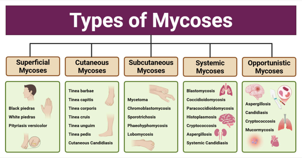
I. Introduction to Superficial Mycoses
Superficial mycoses are fungal infections strictly limited to the outermost, dead layers of the skin (the stratum corneum), the hair shafts, and the nail plates. Because these organisms do not invade living, vascularized tissue under normal circumstances, they typically do not elicit a massive systemic immune response, allowing them to persist. They are among the most ubiquitous infectious conditions globally and represent the absolute most common causes of dermatological disease in many tropical and subtropical countries, where heat and humidity foster fungal replication.
Common Clinical Examples Include:
- Dermatophytosis: Commonly and colloquially known in public spaces as "Ringworm." Caused by a specific group of molds.
- Pityriasis Versicolor: A highly prevalent yeast infection causing distinct skin discoloration (hypo- or hyperpigmentation) heavily linked to sun exposure.
- Rare/Endemic Disorders: Tinea nigra (black/brown palm lesions), White Piedra, and Black Piedra (fungal concretions on hair shafts).
II. Dermatophytosis & The Dermatophytes
Dermatophytes are highly specialized, pathogenic molds (filamentous, multicellular fungi). They are exclusively keratinophilic organisms, meaning they possess the unique enzymatic machinery required to break down and utilize keratin—the tough, fibrous structural protein that protects epithelial cells—as their sole source of carbon and nutrition for growth.
- Target Tissues: They strictly invade the non-living stratum corneum of the epidermis or the highly keratinized, dense appendages of the body (the hair and nails). Common sites of infection include the interdigital spaces of the feet, the moist folds of the groin, the scalp, and the nail beds.
- Evolution & Host Specificity: Dermatophytes originally arose millions of years ago as saprophytic soil fungi that adapted to break down keratin debris deposited in the environment (like shed animal hooves, horns, claws, or feathers). Over evolutionary time, most species have abandoned the soil and become exclusively parasitic to humans or animals. They are now highly adapted to a single or a very narrow range of host species.
The Three Genera of Pathogenic Dermatophytes
Dermatophytes are classified into three major genera based on their target tissues and microscopic spore structures:
- Trichophyton: The most aggressive genus. It possesses the enzymatic capacity to infect hair, skin, and nails. (Produces characteristic cylindrical, smooth-walled macroconidia and abundant microconidia).
- Microsporum: Infects the hair and skin (but almost never the nails). (Produces large, thick-walled, rough, spindle-shaped macroconidia).
- Epidermophyton: Infects the skin and nails (but is enzymatically incapable of invading hair). It contains only a single known pathogenic species globally: Epidermophyton floccosum. (Produces club-shaped macroconidia with absolutely no microconidia).
Dermatophyte Tissue Targets
To effortlessly remember which genus infects which tissues (Skin, Hair, Nails) on exams, look closely at the first letters of the genera!
- Trichophyton: Takes ALL (Skin, Hair, Nails).
- Microsporum: Misses Nails (Infects Skin, Hair).
- Epidermophyton: Excludes Hair (Infects Skin, Nails).
III. Classification & Taxonomy
Sexual vs. Asexual Forms (Teleomorph vs. Anamorph)
Fungi are fascinating organisms that can exist in different life phases depending on environmental stress.
- Anamorph State: Most clinical isolates recovered from human patients are "imperfect fungi" (existing exclusively in their asexual, reproducing state).
- Teleomorph State: The few known sexual forms of these fungi (which reproduce via spores in specialized sacs) have been assigned to two completely different genera names based on their asexual counterparts. This dual-naming system can be confusing but is standard in mycology:
- Trichophyton (asexual form) ➔ Arthroderma (sexual form).
- Microsporum (asexual form) ➔ Nannizzia (sexual form).
Basis for Modern Classification
- Modern taxonomy is based heavily on molecular tools (DNA sequencing, PCR, and molecular taxonomy), which remarkably, strictly agrees with historical, conventional morphological classification methods.
- Classification is also supported by mapping protein composition, the specific production of endogenous antibiotics (such as penicillins and fusidates used by the fungus to kill competing bacteria on the human skin), and the production of distinct enzymes (such as urease, which alters the local skin pH).
Pathogenicity Factors (High-Yield Clinical Application)
Both antibiotics and enzymes play a direct, measurable role in exactly how the fungus establishes disease and evades host defenses.
- Proteinases: These are powerful enzymes inducible by the presence of host amino acids. They aid in the rapid penetration of dense keratin. For example, Trichophyton rubrum secretes a highly specialized zinc-containing metalloprotease that specifically seeks out and degrades human keratin matrices.
- Elastase (Physiology Expansion): Why do some chronic fungal infections (like severe toenail fungus or chronic body ringworm) hardly itch or turn red, persisting for years? Dermatophytes actively secrete elastase. This enzyme directly inhibits the chemotaxis of neutrophils and the development of robust inflammatory responses in ringworm. By suppressing the immune system's alarm bells, the fungus can evade clearance and persist silently in the skin for decades!
IV. Epidemiology & Transmission Sources
The global distribution, transmission dynamics, and severity of the resulting human inflammatory response depend entirely on the primary ecological source of the fungal infection. Fungi are classified into three distinct ecological groups.
General Rule: Fungi that are NOT adapted to humans (Zoophilic/Geophilic) cause severe, angry, highly inflamed rashes in humans because our immune system attacks them violently. Fungi highly adapted to humans (Anthropophilic) cause mild, chronic, low-inflammation rashes because they know how to hide from our immune system.
| Ecological Source | Key Characteristics, Examples & Risk Groups |
|---|---|
| Zoophilic (Animal) | Animal pathogens highly capable of crossing over to cause human infection (zoonosis). They have a wide range of host specificities (infecting cats, dogs, horses, pigs, cattle, rodents). Key Pathogen: Microsporum canis. This is the most prevalent globally; it is carried mostly by household pets (cats and dogs) and is the absolute most common cause of human zoonotic fungal infections (especially pediatric scalp ringworm). Other Example: Trichophyton verrucosum (caught from cattle, highly inflammatory). |
| Geophilic (Soil) | Organisms that originate directly from the soil, breaking down shed animal debris. They are infrequent causes of human disease but are seen more commonly in tropical agricultural regions. At-Risk Groups: Exposed occupational groups (gardeners, landscapers, farm workers). Key Pathogen: Microsporum gypseum. Infection usually follows direct contact with contaminated, moist soil. |
| Anthropophilic (Human) | Natural, highly adapted pathogens of humans. They represent the single most common cause of human dermatophytosis globally. Transmitted strictly human-to-human via direct contact with infected desquamated (shed) skin scales. Key Pathogen: Trichophyton rubrum. The most prevalent dermatophyte throughout the world; the primary causative agent for tinea pedis (athlete's foot), tinea cruris (jock itch), and tinea corporis. At-Risk Groups: Individuals sharing common humid facilities where skin scales accumulate (public bathing areas, shower rooms, military barracks, boarding schools, wrestling mats, and public swimming pools). |
V. Pathogenesis & Host Immunity
The Infection Cycle
- Arthrospores: The infecting fungal organisms are not transmitted as active, growing hyphae. They are transferred by means of arthrospores. These are thick-walled, resilient vegetative cells formed by the fragmentation of dermatophyte hyphae. They are shed abundantly by the primary host along with dead skin scales or broken hair.
- Survival: Arthrospores are incredibly tough. They resist desiccation and can survive for considerable, extended periods outside the host (often surviving for more than 15 months on warm, damp locker room floors!).
- Invasion Sequence:
- Adherence of the fungal spore to human keratinocytes.
- Germination of the spore in a warm, moist environment.
- Invasion of the tissue (heavily aided by zinc-containing metalloproteinases drilling into the keratin).
- Timeline: Invasion induces a host inflammatory response which typically becomes maximal after 9 to 16 days. Following this peak, there is typically a spontaneous resolution of the infection (if the host is fully immunocompetent and the organism is highly inflammatory).
Immune Responses to Dermatophytes
- Innate Immunity:
- Epidermal Langerhans cells: These specialized dendritic cells reside in the skin and act as the primary Antigen Presenting Cells (APCs). They capture fungal antigens and travel to lymph nodes to present them to T-cells.
- Phagocytosis: Dermatophyte antigens attract human leukocytes (phagocytes like Polymorphonuclear neutrophils [PMNs] and Macrophages). These cells engulf and kill the fungi intracellularly and extracellularly mainly via oxidative pathways (creating deadly Reactive Oxygen Species / ROS).
- Complement: Fungal antigens directly activate the alternate complement pathway, amplifying inflammation.
- Desquamation: The skin mounts a brilliant mechanical defense! Increased epidermal cellular turnover occurs, leading to increased shedding of the stratum corneum to literally "push" the fungus off the body faster than it can burrow inward.
- Adaptive Immunity: The clearance of fungal infections is driven primarily by T lymphocytes (Th-1 and Th-17 cell-mediated responses). Antibodies (B-cells/Humoral immunity) play almost absolutely no role in clearing dermatophyte infections. If a patient has a T-cell deficiency (like HIV/AIDS), they will suffer intractable, massive fungal infections.
Age Incidence, Hormones & Sebum
Tinea capitis (scalp ringworm) is almost exclusively a childhood pediatric disease, with very rare cases occurring after puberty. Why does an infected 12-year-old often spontaneously cure themselves when they turn 13?
The Biochemical Mechanism: During puberty, dramatic surges in hormones (specifically androgens) trigger the sebaceous glands on the scalp to drastically increase production. Postpubertal individuals begin producing heavy sebum that is intensely enriched with medium-chain-length fatty acids (specifically C8 to C12 acids). These specific fatty acids are highly fungistatic and naturally, chemically inhibit the growth of dermatophytes. This effectively creates an invisible chemical shield, protecting adults from scalp ringworm!
(Note: Elderly women may sometimes contract it in association with scarring alopecia, as protective sebum production drops precipitously post-menopause).
VI. Clinical Features & Terminology
The Classic Dermatophyte Lesion (Ringworm)
The gross morphological presentation of dermatophytosis is unmistakable:
- Presents as an annular (ring-shaped), sharply demarcated scaling patch with a highly raised, active erythematous margin.
- Has a variable degree of inflammation depending on the ecological source (zoophilic = highly inflamed; anthropophilic = mildly inflamed).
- Pathological Central Clearing: The center of the ring is notably less inflamed than the edge. Why? This happens because the fungus rapidly metabolizes and depletes all the available keratin in the center of the lesion. To survive, it must continually spread outward in a circle to seek out fresh, uninfected keratin, leaving an empty, healing center behind!
Tinea Nomenclature (Clinical Terminology)
The clinical medical term "Tinea" refers universally to dermatophyte infections, followed directly by the Latin description of the precise anatomical site infected.
- Tinea Pedis: Feet (Athlete's foot; incredibly common, usually seen in adolescents/young adults wearing occlusive footwear).
- Tinea Cruris: Groin/Inner thighs (Jock itch; notably spares the scrotum and penis, distinguishing it from Candida).
- Tinea Corporis: Body (Trunk/limbs; classic ringworm presentation).
- Tinea Capitis: Scalp (Hair loss, broken hairs, severe scaling).
- Tinea Manuum: Hand (Often presents as "Two feet, one hand" syndrome due to auto-inoculation from scratching the feet).
- Tinea Faciei: Face (Often misdiagnosed as lupus or rosacea).
- Tinea Barbae: Beard region (Highly inflammatory, often contracted from farm animals).
- Tinea Unguium: Nails (Specifically dermatophyte onychomycosis, causing thick, brittle, yellowing nails).
- Tinea Imbricata: Unique, scaly polycyclic concentric rings spreading like ripples in water (caused exclusively by the pathogen T. concentricum, endemic to specific island populations).
Dermatophyte "Id" Reactions (Autoeczematization)
An "Id" reaction is a severe secondary allergic rash that develops at a site completely distant from the primary fungal infection.
Example: A patient has a severe fungal infection on their toes (Tinea pedis). Suddenly, their hands erupt in extremely itchy, sterile, fluid-filled vesicles. The "Id" rash on the hands itself is completely sterile and contains absolutely no fungus! It is a hypersensitivity reaction to circulating fungal antigens. If you treat the feet, the hands will spontaneously heal.
Tinea Incognito (The Clinical Trap)
Refers to dermatophyte infections that do not show the usual characteristic features (no raised red ring, no prominent scale, highly atypical borders).
Cause: This is an iatrogenic (doctor-caused) or patient-caused error due to the inappropriate application of strong topical corticosteroid creams! The steroid completely suppresses the local immune system (reducing the redness and intense itching), but acts as a massive fertilizer for the fungus. Without the immune system keeping it in check, the fungus invades deeply into the dermis and presents atypically, often confusing the physician and delaying proper antifungal treatment.
VII. Laboratory Diagnosis (Screening & Microscopy)
Visual diagnosis is prone to error; definitive diagnosis requires laboratory confirmation before prescribing long-term, potentially hepatotoxic oral antifungals.
1. Filtered Ultraviolet Light (Wood’s Light/Lamp)
A Wood's lamp emits long-wave UV radiation (peak at 365 nm).
- Indications: Used for rapid, non-invasive clinical screening of pediatric populations and for successfully selecting specific infected hairs for microscopy and culture in Tinea Capitis. Fluorescent hairs are infected.
- Species Differentiation:
- Microsporum spp. infections (like M. canis) fluoresce a brilliant, bright yellow-green.
- Trichophyton infections generally do not fluoresce (with the extremely rare exception of T. schoenleinii, which causes a severe crusting disease called favus and fluoresces dull blue).
❓ Applied Clinical Question: Wood's Lamp and Scalp Infections
Case: Two 8-year-old boys present to the clinic with patchy hair loss, broken hairs, and scaly scalps (Tinea capitis). The physician shines a Wood's Lamp on both scalps in a dark room. Boy A's scalp glows bright yellow-green. Boy B's scalp shows no fluorescence at all.
Question: What is the most likely genus causing the infection in each boy?
Answer: Boy A is infected with the Microsporum genus (highly likely M. canis contracted from a household pet). Boy B is infected with the Trichophyton genus (highly likely T. tonsurans), which is currently the most common non-fluorescent cause of scalp ringworm in humans in the US and Europe!
2. Specimen Collection & KOH Preparation
- Collection: Obtain aggressive scrapings or clippings from lesions. You must ALWAYS sample the active, raised, red edge of skin lesions (where the fungus is actively growing), not the dead, empty center. For infected hairs: use forceps to pluck broken stubs directly from the scalp.
- Potassium Hydroxide (KOH): The gathered material should be placed on a slide and allowed to soften in a 10% to 20% KOH solution. KOH is a strong alkali that powerfully dissolves human keratin cells, fats, and debris, but leaves the tough fungal chitin and beta-glucan cell walls completely intact and visible! Nails are incredibly dense and require up to 2 hours to soften (this process is hastened by gentle warming of the slide).
3. Microscopy Techniques
- KOH Wet Preparation: Fungal hyphae are seen as branching, septate chains of arthrospores under the light microscope.
- Ectothrix infection: Arthrospores are clustered densely on the outside of the hair shaft (physically destroys the hair cuticle).
- Endothrix infection: Arthrospores are completely contained within the hair shaft (the outer cuticle remains completely intact).
- Calcofluor White Stain: A highly advanced, special fluorescent chemo-fluorescent dye that binds strongly and specifically to the chitin and cellulose in fungal cell walls. Preparations are viewed using fluorescence microscopy, where the fungi glow bright apple-green or blue-white against a dark background. It significantly enhances the diagnostic yield, speed, and sensitivity of positive samples over plain traditional KOH.
VIII. Laboratory Diagnosis: Culture
While microscopy provides a rapid confirmation of a fungal infection (telling the doctor "Yes, it is a fungus"), it cannot identify the exact organism. A formal culture is absolutely required to identify the specific species of the dermatophyte to trace epidemiological outbreaks.
- Primary Isolation Media: Sabouraud’s Dextrose Agar (SDA). This is the absolute gold standard mycological medium (formulated with peptone, massive amounts of dextrose, and an acidic pH of 5.6 to favor fungi over bacteria).
- Crucial Additives: The agar must be enriched with specific drugs to prevent contamination. It must contain broad-spectrum antibiotics (like penicillin-streptomycin or chloramphenicol) to completely inhibit rapid bacterial overgrowth from skin flora, AND cycloheximide to inhibit the growth of rapidly growing saprophytic (environmental/airborne) molds that would otherwise outcompete the slow-growing dermatophyte.
- The Nail Exception: If isolating from damaged nails where the non-dermatophyte mold Scytalidium is suspected, you MUST use SDA media completely WITHOUT cycloheximide, because Scytalidium is highly sensitive to it and will not grow on routine fungal media!
Incubation & Identification Criteria
- Primary isolation is carried out at standard room temperature (25-30°C). Exception: Trichophyton verrucosum (a zoophilic cattle pathogen) grows optimally at human body temperature (37°C).
- Most dermatophytes grow slowly but can be successfully identified within 2 to 3 weeks.
- Identification Criteria: Depends on the gross colonial morphology (surface color, reverse pigment color, powdery/fluffy texture of the colony) and microscopic morphology (size, shape, and arrangement of the macroconidia and microconidia). Confirmatory tests include specific nutritional requirements (e.g., Trichophyton nutritional agar testing for thiamine/inositol) and in vitro hair penetration tests.
IX. Treatment of Dermatophytosis
The general pharmacological approach is to treat with topical therapy whenever possible to avoid systemic side effects (like liver toxicity). However, most nail (Onychomycosis) and all hair (Tinea capitis) infections, as well as widespread/intractable dermatophytosis, absolutely require oral systemic drugs. Topical creams physically cannot penetrate the dense, deep keratin beds of nails or hair follicles.
Mechanisms of Action of Systemic Antifungals
- Terbinafine (an Allylamine): Works by powerfully inhibiting the enzyme squalene epoxidase. This stops ergosterol synthesis early on, causing a massive, toxic buildup of squalene inside the fungal cell, leading directly to cell death (Fungicidal). It is highly concentrated in nail beds.
- Azoles (Itraconazole/Fluconazole): Work by inhibiting the CYP450-dependent enzyme 14-alpha-demethylase, preventing the downstream synthesis of ergosterol, which is essential for the structural integrity of the fungal cell membrane. The fungus stops growing (Fungistatic). Warning: Because they interact with CYP450, they have massive drug-drug interactions.
- Griseofulvin: An older drug that physically binds to fungal microtubules, disrupting the mitotic spindle and halting fungal cell division (mitosis). Must be taken with a fatty meal (like ice cream) for proper absorption.
Clinical Disease Patterns & Treatment Regimens
| Disease Pattern | Recommended Treatment |
|---|---|
| Tinea Pedis (Interdigital - between toes) | Topical cream/ointment (applied for 1-2 weeks): Terbinafine, imidazoles (miconazole, econazole, clotrimazole), undecenoic acid, tolnaftate. Keep feet dry. |
| Tinea Pedis ("Dry type" / Moccasin pattern) | Often requires Oral therapy due to thick heel skin: Terbinafine 250 mg/day for 2–4 weeks, Itraconazole 400 mg/day for 1 week per month (pulsed therapy), OR Fluconazole 200 mg weekly for 4–8 weeks. |
| Tinea Corporis (Small, localized) | Topical cream/ointment applied extending 2cm beyond the visible red margin: Terbinafine, imidazoles (miconazole, econazole, clotrimazole). |
| Tinea Corporis (Large, widespread) | Oral: Terbinafine 250 mg/day for 2 weeks, Itraconazole 200 mg/day for 1 week, OR Fluconazole 250 mg weekly for 2–4 weeks. |
| Tinea Capitis (Scalp) | Must be systemic. Griseofulvin: 10–20 mg/kg daily for a minimum of 6 to 8 weeks. Terbinafine (Weight-based pediatric dosing): <20 kg = 62.5 mg/day; 20–40 kg = 125 mg/day; >40 kg = 250 mg/day. Itraconazole: 4–6 mg/kg pulsed dose weekly. Fluconazole: 3–8 mg/kg pulsed dose weekly. |
| Onychomycosis (Fingernails) | Oral: Terbinafine 250 mg daily for 6 weeks, Itraconazole 400 mg/day for 1 week each month (repeated for 2–3 months), OR Fluconazole 200 mg weekly for 8–16 weeks. |
| Onychomycosis (Toenails) | Requires very long therapy due to slow nail growth: Oral Terbinafine 250 mg daily for 12 weeks, Itraconazole 400 mg/day for 1 week each month (repeated for 2–4 months), OR Fluconazole 200 mg weekly for 12–24 weeks. |
X. Other Superficial Mycoses
Not all superficial fungal infections are caused by classic dermatophytes (Trichophyton, Microsporum, Epidermophyton). Several other environmental molds and yeasts can opportunistically invade the skin, hair, and nails.
A. Scytalidium (Neoscytalidium) Infections
- Organisms: Two major species: Scytalidium hyalinum and Scytalidium dimidiatum (which was originally described as an agricultural plant pathogen).
- Pathogenesis & Epidemiology: The precise host-pathogen pathogenesis is not fully understood. It is highly endemic in the tropics and subtropics. Asymptomatic carriage on the soles of the feet is thought to result in active, clinical infection under appropriate, chronically humid conditions.
- Clinical Features: Physically and visually indistinguishable from classic dermatophyte infections (athlete's foot). Involves the interdigital spaces, thickened soles, palms, and chronic destruction of nails.
- Diagnosis: KOH wet preparation of scrapings/clippings reveals branching hyphae. Crucial Lab Step: It MUST be grown on Sabouraud’s agar entirely WITHOUT cycloheximide (because this common lab additive is highly inhibitory to this specific mold).
- Treatment: It is notoriously resistant to modern oral antifungals. Standard antifungal therapy is typically not satisfactory. Older keratolytic therapies like Whitfield’s ointment (salicylic acid + benzoic acid) may be used to physically peel off the infected skin.
B. Other Forms of Onychomycosis (Non-Dermatophyte Molds)
Environmental saprophytic fungi other than dermatophytes frequently cause highly destructive nail infections.
- Scopulariopsis brevicaulis: A ubiquitous soil mold notorious specifically for thick, yellow-brown Great Toe nail infections.
- Acremonium, Aspergillus, Fusarium species: Cause superficial white onychomycosis and severe proximal subungual disease.
- Systemic Risk Warning: While completely superficial and localized in healthy, immunocompetent people, systemic, fatal dissemination of these environmental molds (especially Fusarium and Aspergillus) can rapidly occur in severely neutropenic, immunocompromised patients (such as those undergoing bone marrow transplants or intensive leukemia chemotherapy) starting from a simple toenail infection!
- Diagnosis: Direct microscopy (visualization of massive fungal spores) and Culture (they are easy to isolate on routine media without cycloheximide).
- Treatment: There is almost no effective oral therapy for these tough molds in the nails. The absolute best treatment is physical or chemical nail avulsion (removal) combined with 40% urea cream (a potent, thick keratolytic agent that completely dissolves the remaining infected nail keratin).
XI. Pityriasis Versicolor (Tinea Versicolor)
A globally ubiquitous, very common superficial skin infection caused by a lipophilic yeast, completely unrelated to the dermatophyte molds.
The Organism: Genus Malassezia (Previously Pityrosporum)
- These are lipophilic yeasts (they strictly require external lipids/fats/oils to survive and replicate).
- They are normal, healthy skin commensals. Normal human skin is universally colonized in late childhood and adult life (precisely when puberty hits and sebum production increases).
- Seven pathogenic species: Malassezia furfur, M. pachydermatis (an exception, as it is not lipodependent and is mostly associated with canine/dog ear infections rather than human skin), M. sympodialis, M. globosa, M. restricta, M. obtusa, and M. slooffiae.
Pathogenesis
- Clinical disease occurs via the sudden morphological transformation of the friendly, commensal yeast-phase organisms into aggressive, invasive hyphal/mycelial forms.
- Triggers: The main trigger for this pathological transformation is likely excessive sun exposure, high ambient heat, and sweating (making these infections far more common in the tropics and during summer months). Other major risk factors include immunosuppression, systemic corticosteroid use, and Cushing’s syndrome.
- Malassezia yeasts specifically grow optimally in the presence of medium-chain-length fatty acids secreted in human sebum.
Clinical Presentation & Biochemical Mechanisms
- Infection is strictly confined to the upper trunk, chest, back, neck, or proximal aspects of the limbs.
- There is absolutely no invasion of hair shafts or nail plates.
- Characteristic Lesions: Hypopigmented (lighter/white) or hyperpigmented (darker/brown/pink) macules that amalgamate to cover the affected area with fine, powdery, scaling plaques.
Why does it cause depigmentation (white spots)?
The organism metabolizes skin lipids and produces a toxic carboxylic acid called azelaic acid in the stratum corneum. Azelaic acid acts as a direct, powerful competitive inhibitor of tyrosinase, the master enzyme that human melanocytes use to synthesize melanin. Halting melanin production leads directly to the striking hypopigmentation (white spots) seen in the lesions, which become extremely noticeable when the patient gets a suntan everywhere else!
Diagnosis & Treatment
"Spaghetti and Meatballs"
- Wood's Light: Patient screening in a dark room may reveal lesions that fluoresce a distinct, dull yellow-green or coppery-orange.
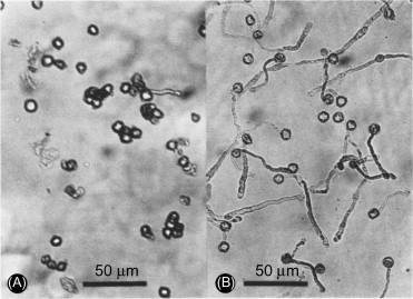
- KOH Wet Prep Microscopy: The absolute hallmark diagnostic sign on a skin scraping is the dense presence of clusters of round yeast forms / blastoconidia ("meatballs") mixed heavily with short, thick, fragmented hyphae ("spaghetti"). Adding a drop of Parker Quink blue ink to the KOH makes this classic appearance even easier to see.
- Culture: Skin scrapings are extremely difficult to culture on standard media. Because it is highly lipophilic, a thick overlay of sterile olive oil or Tween 80 must be added directly to the agar plate to encourage the yeast to grow.
- Topical (Most appropriate & First Line): Azole creams (ketoconazole, clotrimazole), terbinafine cream, 2% selenium sulfide lotion (Selsun Blue shampoo applied as a body wash), or 20% sodium thiosulfate.
- Relapse Prevention: Because the yeast lives naturally on everyone, relapse is almost guaranteed. Intermittent preventative applications of 50% propylene glycol in water, or washing with ketoconazole shampoo once a month.
- Severe Cases (Oral Therapy): Oral Ketoconazole, fluconazole, or itraconazole. Clinical visual recovery of scaling takes about 10 days, but complete mycologic recovery (total eradication of the fungus) takes 30 days.
Patient Counseling Note: It may take many months for the skin pigment/melanin to return to normal even after the fungus is completely dead.
XII. Other Malassezia Infections
Malassezia yeasts are not just responsible for Pityriasis Versicolor; they are heavily implicated in and associated with two other distinct dermatological conditions and one dangerous systemic hospital condition:
- Malassezia Folliculitis:
- Three main forms:
- Back/Upper Chest: Scattered, intensely itchy follicular papules or pustules. Very often appears suddenly after severe sun exposure or heavy sweating.
- Upper & Lower Portions of Back/Chest: Small follicular papules, surrounding erythema, and greasy perifollicular scales. Strongly associated with seborrheic dermatitis.
- Trunk and Face: Multiple florid pustules. Highly associated with profound immunosuppression (like advanced HIV infection/AIDS) and severe seborrheic dermatitis.
- Diagnosis: Scrapings or punch biopsies show numerous yeast cells physically occluding (plugging) the mouths of the hair follicles, triggering inflammation.
- Treatment: Topical azole antifungals. Oral ketoconazole/itraconazole is strictly reserved for extensive, severe, or immunocompromised cases.
- Three main forms:
- Seborrheic Dermatitis:
- A very common, chronic inflammatory condition causing flaky, greasy, yellow-white scales on the scalp (dandruff), eyebrows, nasolabial folds, and face.
- Malassezia yeasts are not the direct infectious "cause" per se, but their lipid metabolites and the host's exaggerated immune response to them are intrinsically involved in the pathogenesis and inflammatory triggering of the condition.
- Treatment: Main therapy is topical azole creams/shampoos (like Ketoconazole 2%) combined heavily with weak, low-potency topical corticosteroids (e.g., 1% hydrocortisone cream) to rapidly reduce the debilitating inflammation. Relapse is incredibly common and requires lifelong retreatment.
- Catheter-Acquired Sepsis (Systemic Emergency):
- In hospitalized patients (especially premature neonates in the NICU) receiving Total Parenteral Nutrition (TPN) that is heavily enriched with intravenous lipid emulsions, the lipophilic Malassezia living commensally on the patient's skin can track down the outside of the central IV line. Once in the bloodstream, they gorge on the lipid fluids and replicate explosively, causing severe, sometimes fatal systemic catheter-associated sepsis!
XIII. Tinea Nigra & Piedra (Hair & Skin Infections)
A. Tinea Nigra
- A completely superficial form of phaeohyphomycosis (the clinical term for infections caused exclusively by dematiaceous, darkly pigmented, melanin-producing fungi).
- Organism: Caused by the highly halophilic (salt-loving) black yeast, Hortaea (Exophiala) werneckii.
- Clinical Features: Mainly seen in the tropics/subtropics, usually in children or young adults. The infection is completely, painlessly confined to the thick stratum corneum of the palms of the hands or the soles of the feet. It presents uniquely as an irregular, large, non-scaly, completely flat brown or black superficial macule.
- Main Differential Diagnosis: Superficial acral lentiginous melanoma & pigmented junctional nevus. (It is absolutely crucial to diagnose this properly via scraping to prevent a patient from undergoing an unnecessary, disfiguring melanoma surgery for a simple, harmless fungal infection!).
- Diagnosis: Direct microscopy of KOH-treated scrapings reveals distinctly pigmented (brown/olive/dark green) thick hyphae. A positive culture on SDA confirms a black, yeast-like colony.
- Treatment: A simple, heavy keratolytic agent such as Whitfield’s ointment or 5% to 10% salicylic acid ointment physically peels the fungus off the skin in a matter of days. Antifungals are rarely needed.
B. White Piedra
- An uncommon superficial fungal infection targeting the hair shafts of the scalp, body, mustache, or pubic hair.
- Organism: Caused by yeasts of the genus Trichosporon (T. beigelii, T. inkin, T. mucoides, T. ovoides). Found universally in both humid tropics and temperate zones.
- Source & Transmission: Natural skin flora, the area around the anus, poor hygiene, and sexual transmission.
- Clinical Presentation: Usually entirely asymptomatic. Presents as small, circumscribed, relatively soft, easily detached nodular yellow/white concretions tightly adhered to the outer hair shafts.
- Systemic Risk: Like Fusarium, the Trichosporon species can cause deadly, widespread systemic dissemination in severely neutropenic patients (e.g., leukemia patients).
- Diagnosis & Treatment: Plucked (epilated) hair soaked in KOH shows masses of hyphae and arthrospores inside the nodules. Treatment is difficult because spores penetrate the hair cuticle; the absolute best and most definitive cure is simply shaving off all the affected hair. Topical econazole or oral ketoconazole can be used to treat the skin base, but relapse is incredibly common if hair is not removed.
C. Black Piedra
- A rare, visually distinct infection of the hair shafts, mainly confined strictly to isolated parts of the highly humid tropics (Central/South America, Southeast Asia).
- Organism: Caused by a slow-growing black yeast, Piedraia hortae.
- Clinical Presentation: Presents as small, extremely hard, gritty, stony black nodules very firmly attached and cemented to the hairs of the scalp. (Patients often report hearing a "metallic clicking" sound when combing their hair!).
- Main Differential Diagnosis: Pediculosis (Lice / nits).
Clinical Key: Severe itching is universally present with lice infestations, but itching and scalp inflammation are totally absent in black piedra. Also, lice nits slide easily along the hair shaft; black piedra nodules are cemented in place. - Diagnosis: Direct microscopy reveals dark nodules composed of organized hyphal elements and small ascospores encased within a very thick, dark, cement-containing stroma binding the hair shaft.
- Treatment: Shaving the head completely is curative. Alternatively, topical salicylic acid, 2% formaldehyde solutions, or a heavy azole cream can be utilized. Relapse is common due to poor hygiene or persistent environmental humidity.
❓ Final Module Review Question
Case: A 22-year-old medical student returning from a month-long medical mission trip in the humid tropics notices a dark, flat, entirely painless, brown patch on the palm of her left hand. Panicking, she schedules an immediate biopsy with a dermatologist, fearing it is an aggressive Acral Lentiginous Melanoma. The dermatologist gently scrapes the lesion with a scalpel blade and looks at the scales under a 20% KOH wet prep.
Question: What did the dermatologist see to cancel the surgical biopsy, what is the exact diagnosis, and what is the required treatment?
Answer: The dermatologist saw distinctively brown/pigmented (dematiaceous) septate hyphae under the microscope. The diagnosis is Tinea Nigra (caused by the halophilic yeast Hortaea werneckii). The treatment is completely non-surgical; simply applying a topical keratolytic like Whitfield's ointment or 10% Salicylic acid for a few weeks to physically peel the infected stratum corneum layer off.
Recommended References
- Mandell, Douglas, and Bennett's: Principles and Practice of Infectious Diseases - Sections on Dermatophytosis and Superficial Mycoses.
- Fitzpatrick's: Dermatology in General Medicine - Fungal Infections of the Skin, Hair, and Nails.
- Centers for Disease Control and Prevention (CDC): Guidelines for the management and treatment of ringworm and superficial fungal infections.
- World Health Organization (WHO): Bulletins on the epidemiology and management of Neglected Tropical Fungal Diseases.
- Clinical Microbiology Reviews: The Epidemiology and Pathogenesis of Malassezia Infections and Advances in Dermatophyte Taxonomy.
Quick Quiz
Intro to Mycology Quiz
Microbiology - mobile-friendly and focused practice.
Privacy: Your details are used only for quiz tracking and certificates.
Intro to Mycology Quiz
Microbiology
Preparing questions...
Choose your answer and keep your streak alive.
Great effort.
Here is your quick performance summary.
Dermatophytosis and Other Superficial Mycoses Read More »