Nurses Revision

nursesrevision@gmail.com

COMMON COLD/CORYZA

COMMON COLD/CORYZA

Introduction

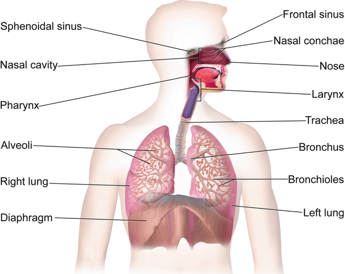
It is the acute inflammation of the upper respiratory tract; rhinitis (nasal mucosa) and rhinopharyngitis (nasal and pharyngitis).

Causes of common cold

  1. The most common virus is rhinovirus. Other viruses include the influenza virus, adenovirus, enterovirus, and respiratory syncytial virus.
  2. Bacteria may cause roughly 15% of sudden onset pharyngitis presentations. The most common is S. pyogenes, a Group A streptococcus.

Clinical manifestations

Manifestations of common cold infection typically appear after an incubation period of 12-72 hours and last 7-11 days, but may persist for longer.

Signs and symptoms include the following:
  1. Nasal dryness or irritation - May be first symptom
  2. Sore throat or throat irritation – Common and bothersome initial symptom
  3. Nasal discharge, nasal congestion, and sneezing – Intensify over 2-3 days
  4. Headache
  5. Facial and ear pressure
  6. Loss of sense of smell and taste
  7. Cough (30% of infected individuals)
  8. Hoarseness (20%)
  9. Irritability or restlessness
  10. Fever (unusual; when present, typically low grade)
  11. Tiredness with slight pyrexia
  12. General malaise
  13. Mild conjunctivitis
  14. Anorexia
  15. Loss of or swollen enlarged lymph nodes

Test and Diagnosis

History taking and physical examination – include the following:
  1. Inspection of the nose and ears to check for any other possible sites of infection.
  2. Inspection of the skin for any rash related to scarlet fever to rule out the condition.
  3. Palpation of the lymph nodes around the neck.
  4. Auscultation to listen to the patient’s breathing and heart sounds.
  5. In some cases, mononucleosis may be ruled out as it can also cause inflammation of the tonsils.
Other diagnostic tests may be performed as follow:
  1. Throat swab – a sterile swab rubbed over the throat will be sent to the lab to check for streptococcal bacteria and the need for antibiotics.
  2. Complete blood count – to show the presence of either a viral or bacterial infection depending on what blood cell is elevated.
  3. Because of the prolonged time to obtain positive culture findings, rhinovirus culture has rarely been found useful in clinical settings.
  4. PCR testing of respiratory specimens may be useful in evaluating severely immunocompromised patients.
DIFFERENTIAL DIAGNOSIS
  • Rhinitis
  • Early signs of measles
  • An allergy
  • Whooping cough

Management of Common Cold

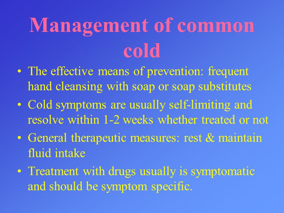
The common cold is primarily a self-limiting viral infection, and treatment is mainly supportive, focusing on relieving symptoms.

Aims of management
  • To promote quick recovery
  • To prevent further complication
  • Symptomatic Relief:
  • Rest: Adequate rest helps the body recover.
  • Hydration: Drink plenty of fluids like water, juice, clear broth, and warm lemon water with honey to prevent dehydration and soothe sore throats.
  • Pain relievers and fever reducers: Over-the-counter medications like acetaminophen (paracetamol) or ibuprofen can help relieve aches, pains, and fever.
  • Decongestants: Oral decongestants (e.g., pseudoephedrine, phenylephrine) or nasal sprays (e.g., oxymetazoline, xylometazoline) can help relieve nasal congestion. Nasal sprays should not be used for more than a few days to avoid rebound congestion.
  • Cough suppressants: For a dry cough, dextromethorphan may be used. For a cough with mucus, expectorants like guaifenesin can help loosen phlegm.
  • Antihistamines: First-generation antihistamines (e.g., diphenhydramine, chlorpheniramine) can help with sneezing, runny nose, and watery eyes, but may cause drowsiness.
  • Sore throat remedies: Warm salt water gargles, throat lozenges, and medicated sprays can provide relief for a sore throat.
  • Nasal saline sprays: Can help moisten nasal passages and loosen mucus.
  • When to Seek Medical Attention:

    While most common colds resolve on their own, it's important to seek medical advice if you experience any of the following:

    Advertisements

    • Symptoms that worsen or do not improve after 7-10 days.
    • High fever (above 103°F or 39.4°C).
    • Severe sore throat, especially if it's sudden and without other cold symptoms.
    • Swollen glands in the neck or jaw.
    • Significant sinus pain.
    • Shortness of breath, wheezing, or difficulty breathing.
    • Chest pain.
    • Earache.
    • New or worsening headache.
    • Symptoms in infants (e.g., difficulty breathing, unusual drowsiness, refusal to feed).
    • Weakened immune system due to other conditions (e.g., HIV, cancer treatment).
    Medical Management

    Common cold is a viral disease which needs only symptomatic treatment and no antibiotics are needed.

    Antibiotics:

    Antibiotics are ineffective against viral infections, including the common cold. They are only prescribed if a bacterial complication, such as a bacterial sinus infection or strep throat, is diagnosed.

    Drug therapy
    1. NSAIDS
    2. Antihistamines
    3. Corticosteroids
    4. Nasal decongestants
    Nursing interventions/management
    1. Assessment of the patient
    • a. Carrying out history of the presenting signs and symptoms e.g. fever, flue among others.
    • b. Taking vital observation e.g. TPR/BP and general examination to exclude other diseases.
    • c. Alerting the doctor who will order for investigations and admission, there the nurse will assist the patient throughout the process.
    2. Managing fever
    • a. Assess the patient’s vital signs at least every 4 hours.
    • b. Remove excessive clothing, blankets, and linens. Adjust the room temperature.
    • c. Administer the prescribed antibiotic and anti-pyretic.
    • d. Offer a tepid sponge bath.
    • e. Elevate the head of the bed.
    3. To relieve headache, joint pains, flue and cough
    • a. Assess the patient’s vital signs and characteristics of pain at least 30 minutes after administration of medication.
    • b. Elevate the head of the bed and position the patient in semi Fowler’s.
    • c. Encouraging patient to sneeze into the elbow not in the hand.
    • d. Must were a mask most time.
    • e. Should be isolated until he improves.
    • f. Encouraging patients to take soothing fluids like warm water and honey or lemon.
    • g. Administer cough suppressants, antibiotics and analgesics as prescribed.
    • h. Encourage patients to verbalise feeling of pain.
    • i. Measure the pain compliants of patients using a pain scale.
    • j. Encourage patients to take more fluids at least 3 liters.
    • Advertisements

    4. Prevention of complication
    • a. Assess the patient’s vital signs and characteristics of respirations at least every 4 hours. Assess for signs of hypoxia.
    • b. Place the patient on a side-lying or prone position.
    • c. Suction secretions.
    • d. Positioning the mattress at a 45° angle.
    • e. Discontinuing smoking or using alcohol.
    • f. Administer the prescribed medications (e.g. corticosteroids) and antibiotic medications.
    5. To prevent infection
    • a. Teach the patient
      • i. Self isolation
      • ii. Wearing masks while in public
      • iii. Maintain social distance
    • b. Assess vital signs and observe for any signs of infection as well as for any signs of respiratory distress.
    • c. Perform a focused assessment on the oropharyngeal region, particularly checking for any collection of abscess.
    • d. Teach the patient how to perform proper hand hygiene.
    • e. Administer antibiotics as prescribed.
    • f. Disinfecting the environment using phenol-alcohol–based compounds.
    • g. Washing hands.
    6. Health education of the patients
    • a. Educating the patient about wearing mask, maintaining hand hygiene.
    • b. Educating the patients about the disease.
    7. Discharge advice
    • a. Encourage proper hand hygiene, wearing masks.
    • b. Encourage proper adherence to drugs.
    • c. Inform the patient about the follow up date and encourage the patient to attend.

    Prevention of Common Cold

    While there is no vaccine for the common cold, certain measures can help prevent its spread:

    • Frequent handwashing: Wash hands thoroughly with soap and water for at least 20 seconds, especially after coughing, sneezing, or blowing your nose, and before eating. Hand sanitizers with at least 60% alcohol can be used when soap and water are not available.
    • Avoid touching face: Try to avoid touching your eyes, nose, and mouth, as viruses can enter the body through these routes.
    • Stay away from sick people: Maintain distance from individuals who are ill with a cold.
    • Clean and disinfect surfaces: Regularly clean and disinfect frequently touched surfaces, such as doorknobs, phones, and keyboards, especially when someone in the household is sick.
    • Boost your immune system: A healthy lifestyle, including a balanced diet, regular exercise, adequate sleep, and stress management, can help strengthen your immune system.
    • Use tissues: Cover your mouth and nose with a tissue when you cough or sneeze, then dispose of the tissue immediately. If a tissue isn't available, cough or sneeze into your elbow.

    COMPLICATION

    • Sinusitis
    • Lower respiratory tract infection (LRTI) e.g pneumonia
    • Deafness
    • Otitis media
    • Headache
    • Acute tonsillitis
    • Chronic bronchitis
    • Exacerbations of reactive airway disease (e.g. asthma)
    COMMON COLD/CORYZA Read More »

    COAGULATION DISORDERS

    COAGULATION DISORDERS

    COAGULATION DISORDERS

    A coagulation disorder is a medical condition characterised by excessive bleeding occurring as a result of deficiency of any of the essential clotting factors. Coagulations disorders are conditions that affect the blood’s clotting activities. Hemophilia, Von Willebrand disease, clotting factor deficiencies, hypercoagulable states and deep venous thrombosis are all coagulations disorders. Hemophilia and Von Willebrand disease are among the best known.

    Normal mechanism of blood clotting

    1. Damage or injury to the endothelium will initiate a cascade of events in an attempt to control bleeding.
    2. Disruption of the endothelium will first cause local vasoconstriction to occur, limiting blood flow to the area.
    3. Primary hemostasis initiates by platelets with the release of von Willebrand factor (vWF), a large plasma glycoprotein made and stored in endothelial cells and megakaryocytes.
    4. Platelets and vWF will combine to form a plug at the site of injury. Circulating vWF continues to bind with collagen and Factor VIII as well as other endothelial substances, allowing the platelet plug to adhere to the area of injury.
    5. Through activation of the clotting cascade and secondary hemostasis, this initial platelet plug will get reinforced to a sturdy fibrin clot.
    6. The clotting cascade operates through a dual process system in which the various clotting factors become activated with the result being the formation of a fibrin strand or clot at the site of tissue injury.

    NB: A deficiency of any of the essential clotting factors will result in difficulty forming a fibrin clot, and excessive bleeding can occur.

    Types of coagulation disorders

    Bleeding disorders fall into two main categories:

    1. Inherited coagulation disorders: Hereditary bleeding disorders are due to the absence or deficiency of specific clotting proteins which act as pro-coagulants through precise interactions in the clotting cascade. The three most common are:
    • a. Hemophilia A (Factor VIII deficiency): Hemophilia A is an X-linked recessive genetic disorder affecting 1 in 5000 males making it the most common congenital coagulopathy.
    • b. Hemophilia B (Factor IX deficiency): Hemophilia B is an X-linked genetic coagulopathy affecting 1 in 30000 male births.
    • c. Von Willebrand disease: It is characterised by excessive bleeding as a result of deficiency of von-Willebrand factor hence causing failure of platelet plug formation.
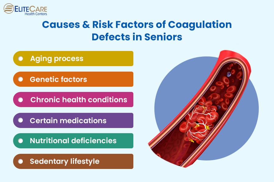
    2. Acquired coagulation disorders: Acquired bleeding disorders can be caused by conditions that an individual may develop at any point during their lifetime. These can be broader in range and dependent on comorbid conditions.
    • a. Liver disease
    • b. Vitamin k deficiency
    • c. Disseminated intravascular coagulation

    Causes of Coagulation disorders

    The major causes of acquired coagulation disorders are:

    1. Vitamin K deficiency
    2. Liver disease
    3. Disseminated intravascular coagulation (DIC)
    4. Development of circulating anticoagulants
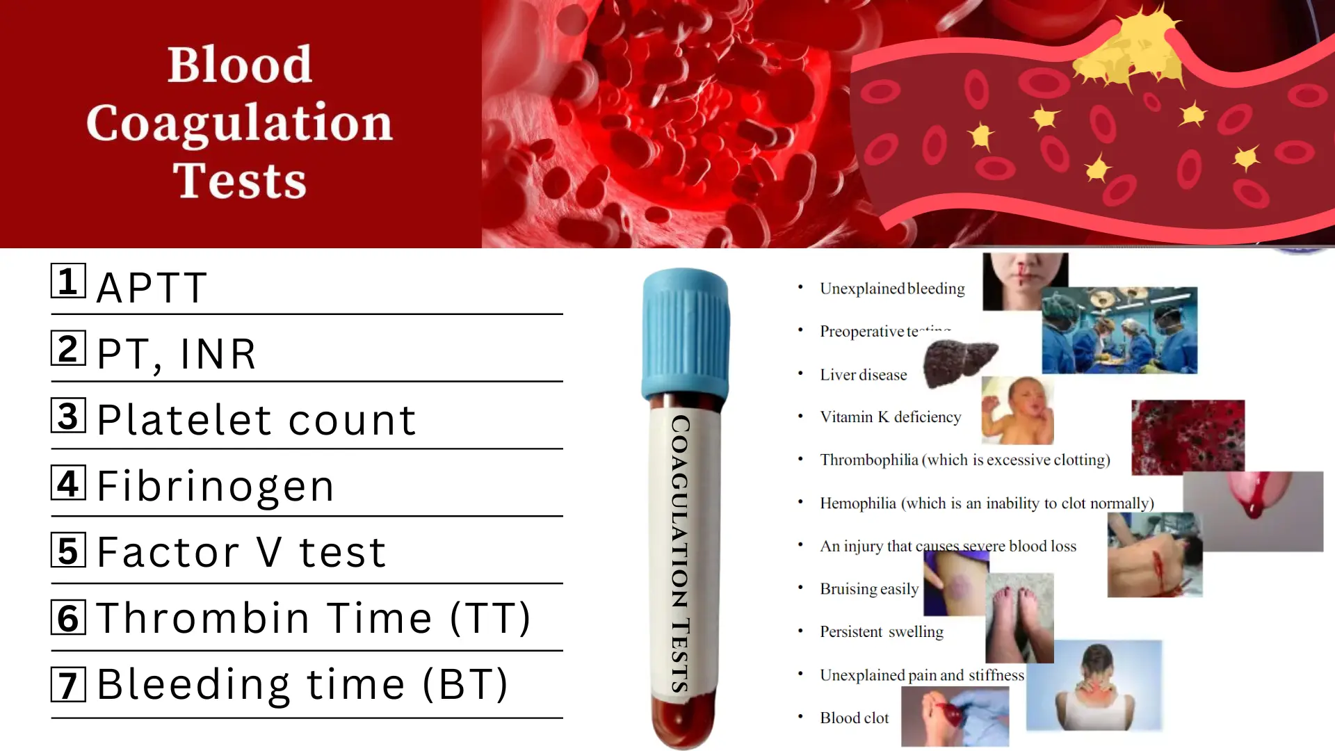
    5. Severe liver disease (e.g. cirrhosis, fulminant hepatitis, acute fatty liver of pregnancy) may disturb hemostasis by impairing clotting factor synthesis. Because all coagulation factors are made in the liver (by hepatocytes and endothelial cells), both the prothrombin time (PT) and partial thromboplastin time (PTT) are prolonged in severe liver disorders. (PT results are typically reported as INR [international normalized ratio].)

    The most common hereditary disorder of hemostasis is:

    • a. Von Willebrand disease (VWD)
    • b. The hemophilias

    Clinical manifestations

    Hemophilia:
    1. While mild hemophilia may only present after a traumatic injury or surgery.
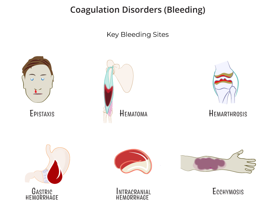
    2. Those with a moderate to severe form of the disease may exhibit hallmark characteristics such as:
      • a) Mucosal or gingival bleeding
      • b) Easy bruising
      • c) Hematoma formation
      • d) Hemarthrosis: is bleeding into joints, particularly in the ankles.
      • e) Bleeding into muscle tissue from minor traumas can result in anemia and
      • f) Compression of vital structures and nerves leading to compartment syndrome.
      • g) Intracranial bleeds
    3. Hemophilia can present in infancy with cephalohematoma formation after vaginal birth and with significant bleeding after circumcisions.
    4. Advertisements

    Von Willebrand Disease
    1. Von Willebrand disease can exhibit clinical signs and symptoms starting in childhood with a history of easy bruising and bleeding.
    2. While patients with a very mild version of the disease may not have clinical symptoms at all, patients with vWF that is qualitative or quantitatively low may present with a predisposition to mucosal bleeding and episodic epistaxis.
    3. Women with von Willebrand disease may have significant menorrhagia which is often a presenting sign of the illness, precipitating a workup and eventual diagnosis.
    4. These patients can also go unrecognized until undergoing major surgery or experiencing a traumatic injury.

    Test and Diagnosis for coagulation disorders

    Hemophilia
    1. Chromogenic assay: This assay is considered by some to be more accurate, as it measures the level of plasma factor VIII activity but it is less widely available in clinical laboratories in the United States.
    2. Laboratory studies: Laboratory studies for suspected hemophilia include a complete blood cell count, coagulation studies, and a factor VIII (FVIII) assay.
    3. CT scans: Head CT scans without contrast are used to assess for spontaneous or traumatic intracranial hemorrhage.
    4. MRI: Perform magnetic resonance imaging (MRI) on the head and spinal column for further assessment of spontaneous or traumatic hemorrhage; MRI is also useful in the evaluation of the cartilage, synovium, and joint space.
    5. Ultrasonography: Ultrasonography is useful in the evaluation of joints affected by acute or chronic effusions.
    6. Testing for inhibitors: Laboratory confirmation of a FVIII inhibitor is clinically important when a bleeding episode is not controlled despite infusion of adequate amounts of factor concentrate.
    7. Carrier testing: Screening for carrier status can be performed by measuring the ratio of FVIII coagulant activity to the concentration of von Willebrand factor (vWF) antigen; a ratio that is less than 0.7 suggests carrier status.
    8. Radiography: Radiography for joint assessment is of limited value in acute hemarthrosis; evidence of chronic degenerative joint disease may be visible on radiographs in patients who have been untreated or inadequately treated or in those with recurrent joint hemorrhages.

    Management

    Medical Management - Hemophilia

    The treatment of hemophilia may involve prophylaxis, management of bleeding episodes, treatment of factor VIII (FVIII) inhibitors, and treatment and rehabilitation of hemophilia synovitis.

    Pre-hospital care
    1. Rapid transport to definitive care is the mainstay of prehospital care; prehospital care providers should apply aggressive hemostatic techniques, assist patients capable of self-administered factor therapy, and gather focused historical data if the patient is unable to communicate.
    2. Emergency department care. Use aggressive hemostatic techniques; correct coagulopathy immediately; include a diagnostic workup for hemorrhage, but never delay indicated coagulation correction pending diagnostic testing; acute joint bleeding and expanding, large hematomas require adequate factor replacement for a prolonged period until the bleed begins to resolve, as evidenced by clinical and/or objective methods; life-threatening bleeding episodes are generally initially treated with FVIII levels of approximately 100%, until the clinical situation warrants a gradual reduction in dosage.
    3. Factor VIII and FIX concentrates. Various FVIII and FIX concentrates are available to treat hemophilia A and B; besides improved hemostasis, continuous infusion decreases the amount of factor used, which can result in significant savings; obtain factor level assays daily before each infusion to establish a stable pattern of replacement regarding the dose and frequency of administration.
    4. Desmopressin. Desmopressin vasopressin analog, or 1-deamino-8-D-arginine vasopressin (DDAVP), is considered the treatment of choice for mild and moderate hemophilia A; DDAVP stimulates a transient increase in plasma FVIII levels; DDAVP may result in sufficient hemostasis to stop a bleeding episode or to prepare patients for dental and minor surgical procedures.
    5. Management of bleeding. Immobilization of the affected limb and the application of ice packs are helpful in diminishing swelling and pain; early infusion upon the recognition of initial symptoms of a joint bleed may often eliminate the need for a second infusion by preventing the inflammatory reaction in the joint; prompt and adequate replacement therapy is the key to preventing long-term complications.
    6. Treatment of patients with inhibitors. Inhibitors are antibodies that neutralize factor VIII (FVIII) and can render replacement therapy ineffective; the treatment of patients with inhibitors of FVIII is difficult; assuming no anamnestic response, low-titer inhibitors (ie, concentrations below 5 Bethesda units [BU]) occasionally can be overcome with high doses of factor VIII; there is no established treatment for bleeding episodes in patients with high-titer inhibitors.
    7. Prophylactic factor infusions. The main goal of prophylactic treatment is to prevent bleeding symptoms and organ damage, in particular to joints; in December 2013, the US Food and Drug Administration (FDA) expanded the indication for anti-inhibitor coagulant complex (Feiba NF) to include routine prophylaxis in patients with hemophilia A or B who have developed inhibitors; approval was based on data from a pivotal phase III study in which a prophylactic regimen resulted in a 72% reduction in median annual bleed rate compared with on-demand treatment.
    8. Pain management. Hemophilic chronic arthropathy is associated with pain; narcotic agents have been used, but frequent use of these drugs may result in addiction; nonsteroidal anti-inflammatory drugs may be used instead because their effects on platelet function are reversible and because these drugs can be effective in managing acute and chronic arthritic pain; avoid aspirin because of its irreversible effect on platelet function.
    9. Activity. Generally, individuals with severe hemophilia should avoid high-impact contact sports and other activities with a significant risk of trauma; however, mounting evidence suggests that appropriate physical activity improves overall conditioning, reduces injury rate and severity, and improves psychosocial functioning.
    10. Gene therapy. With the cloning of FVIII and advances in molecular technologies, the possibility of a cure for hemophilia with gene therapy was conceived; ex vivo gene therapy, in which cells to be transplanted are genetically modified to secrete factor VIII and then are reimplanted into the recipient; in vivo gene therapy, in which a vector (typically a virus altered to include FVIII DNA) is directly injected into the patient; and nonautologous gene therapy, in which cells modified to secrete FVIII are packaged in immunoprotected devices and implanted into recipients.
    11. Radio-synovectomy. In patients who develop synovitis from joint bleeds, intra-articular injection of radioisotopes to ablate the synovium (radiosynovectomy) can be used to decrease bleeding, slow progression of cartilage and bone damage, and prevent arthropathy.
    12. Advertisements

    Pharmacologic Management - Hemophilia

    Medications of choice for patients with hemophilia are:

    1. Factor VIII. Factor VIII (FVIII) is the treatment of choice for acute or potential hemorrhage in hemophilia A; recombinant FVIII concentrate is generally the preferred source of factor VIII; prophylactic administration of FVIII is often recommended for pediatric patients with severe disease.
    2. Anti-fibrinolytic agents. Antifibrinolytic agents, such as aminocaproic acid and tranexamic acid, are especially useful for oral mucosal bleeds but are contraindicated as initial therapies for hemophilia-related hematuria originating from the upper urinary tract because they can cause obstructive uropathy or anuria.
    3. Factor IX. Factor IX is the treatment of choice for acute hemorrhage or presumed acute hemorrhage in hemophilia B. Recombinant factor IX is the preferred source for replacement therapy.
    4. Coagulation factor VIIa. These agents can activate coagulation factor X to factor Xa as well as coagulation factor IX to IXa.
    5. Coagulation factors. FVIII concentrates replace deficient FVIII in patients with hemophilia A, with the goal of achieving a normal hematologic response to hemorrhage or preventing hemorrhage; recombinant products should be used initially and subsequently in all newly diagnosed cases of hemophilia that require factor replacement; agents that bypass FVIII activity in the clotting cascade (eg, activated FVII) are used in patients with FVIII inhibitors.
    6. Anti-hemophilic agents. These agents are used to control bleeding in hemophilia B or FIX deficiency and to prevent and/or control bleeding in patients with hemophilia A and inhibitors to FVIII.
    7. Monoclonal antibodies. Monoclonal antibodies are used to bind to one specific substance in the body (eg, molecules, antigens); this binding is very versatile and can mimic, block, or cause changes to enact precise mechanisms (eg, bridging molecules, replacing or activating enzymes or cofactors, immune system stimulation).
    8. Vasopressin-related. Desmopressin transiently increases the FVIII plasma level in patients with mild hemophilia A.
    9. Advertisements

    Management - Von Willebrand disease

    Treatment depends on the type of VWD and should be decided by a hematologist. Options include the following:

    1. Hormonal treatments such as oral contraceptives and some intrauterine devices are highly effective in controlling menorrhagia. In fact, 88% of women with VWD report improvement in bleeding symptoms when treated only with oral contraceptives.
    2. Desmopressin (DDAVP) is effective in most patients with type 1 VWD and some patients with type 2. Recovery testing must be done to determine its effectiveness. During a recovery test, a blood sample is obtained before the medication is given and 30 to 60 minutes after administration. This test helps determine if the medication increases the patient’s factor levels enough to prevent or stop bleeding.
    3. Replacement factor made from plasma-derived concentrates can be used in any patient with VWD, but must be used in all patients with type 3 and in some patients with type 2. Replacement factor is also used when patients don’t respond to DDAVP.
    4. Anti-fibrinolytics such as aminocaproic acid and tranexamic acid are used in conjunction with factor or DDAVP to treat bleeding. Anti-fibrinolytics stabilize a clot by preventing it from breaking down too early, which would cause bleeding. Without anti-fibrinolytics, bleeding may occur several days or weeks after a procedure involving mucosal tissue. Antifibrinolytics are effective in treating mucosal bleeding such as with dental surgery, menstrual bleeding, nosebleeds, and gastrointestinal bleeding.
    Nursing intervention/management
    1. Relieve pain. Immobilize joints and apply elastic bandages to the affected joint if indicated; elevate affected and apply a cold compress to active bleeding sites, but must be used cautiously in young children to prevent skin breakdown.
    2. Maintain optimal physical mobility. Provide gentle, passive ROM exercise when the child’s condition is stable; educate on preventive measures, such as the application of protective gear and the administration of factor products; and refer for physical therapy, occupational therapy, and orthopedic consultations, as required.
    3. Assist in the coping of the family. Encourage family members to verbalize problem areas and develop solutions on their own; encourage family members to express feelings, such as how they deal with the chronic needs of a family member and coping patterns that help or hinder adjustment to the problems.
    4. Prevent bleeding. Monitor hemoglobin and hematocrit levels; assess for inhibitor antibody to factor VIII; anticipate or instruct in the need for prophylactic treatment before high-risk situations, such as invasive diagnostic or surgical procedures, or dental work; and provide replacement therapy of deficient clotting factors.
    5. Prevent injury. Utilize appropriate toys (soft, not pointed or small sharp objects); for infants, may need to use padded bed rail sides on crib; avoid rectal temperatures; provide appropriate oral hygiene (use of a water irrigating device; use of a soft toothbrush or softening the toothbrush with warm water before brushing; use of sponge-tipped toothbrush); and avoid contact sports such as football, soccer, ice hockey, karate.
    Complications
    1. Anemia
    2. Arthritis

    COAGULATION DISORDERS Read More »

    LEUKEMIA

    LEUKEMIA

    LEUKEMIA

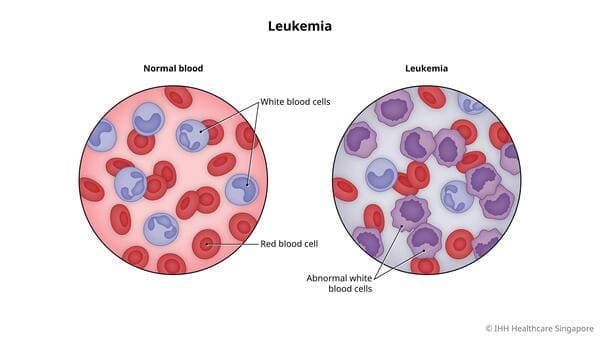
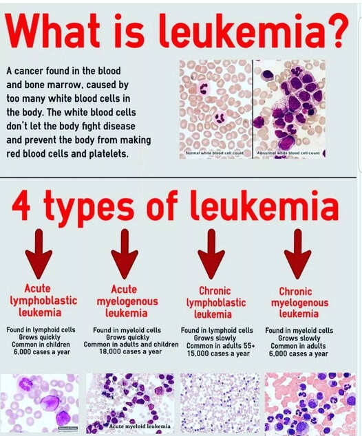
    Definition: Leukemias are a group of hematologic disorders characterized by the dysfunctional proliferation and development of leukocytes. Leukemias are cancers of white blood cells or of cells that develop into white blood cells.

    White blood cells develop from stem cells in the bone marrow. Sometimes the development goes awry, and pieces of chromosomes get rearranged. The resulting abnormal chromosomes interfere with normal control of cell division, so that affected cells multiply uncontrollably or are resistant to normal cell death, resulting in leukemia.

    Types of Leukemia

    As such, the four major subtypes of leukemia are:

    1. Acute lymphoblastic leukemia (ALL): ALL occurs when primitive white blood cells of lymphoid origin reproduce without developing into normal B and T cells. It is the most common leukemia in pediatrics, accounting for up to 80% of cases in this group vs. 20% of cases in adults.
    2. Acute myelogenous leukemia (AML): AML is also characterized by the hyperplasia of blasts, but in this case, of myeloid origin. It accounts for half of the leukemia cases diagnosed in teenagers and people in their 20s. It is the most common acute leukemia in adults.
    3. Chronic lymphocytic leukemia (CLL): CLL occurs when mature but abnormal white blood cells of lymphoid origin undergo hyperplasia, leading to a monoclonal population of dysfunctional lymphocytes. Most cases occur in people between ages 60 and 70.
    4. Chronic myelogenous leukemia (CML): A monoclonal population of self-renewing, dysfunctional myeloid cells (e.g., neutrophils, basophils, eosinophils, macrophages) characterizes CML. Most cases occur in people between ages 25 and 60.

    Note

    1. Acute vs. chronic: Acute leukemias are characterized by abnormal cells that are less mature, develop quickly, and leave the bone marrow as dysfunctional cells called “blasts.” These blasts crowd out healthy cells in the bone marrow, causing the rapid onset of symptoms. Blasts normally make up 1% to 5% of marrow cells, and having more than 20% blasts in the bone marrow is required for a diagnosis of acute leukemia. In contrast, chronic leukemias develop slowly and may take years to develop symptoms. They are composed primarily of more mature and functional cells, and there are generally not elevated numbers of blasts.
    2. Myeloid vs. lymphoid: Hematopoietic stem cells give rise to two types of blood cells: myeloid and lymphoid. Myeloid cells include monocytes, macrophages, neutrophils, basophils, eosinophils, erythrocytes, and megakaryocytes. Lymphoid cells include T cells, B cells, and natural killer cells. So myeloid leukemia affects myeloid cells and lymphoid leukemia affects lymphoid cells.

    Causes of Leukemia

    Several risk factors are associated with a higher risk of developing leukemia:

    1. Exposure to ionizing radiation is associated with an increased risk of multiple subtypes of leukemia.
    2. Exposure to benzene is a risk factor for leukemia in adults, particularly AML.
    3. Previous exposure to chemotherapy, especially alkylating agents and topoisomerase inhibitors, increases the risk for acute leukemia later in life.
    4. A history of any hematologic malignancy is a risk factor for subsequently developing another subtype of leukemia.
    5. Viral infections (e.g., human T-cell leukemia virus, Epstein Barr virus) are linked with subtypes of ALL.
    6. Several genetic syndromes (e.g., Down syndrome, Fanconi anemia, Bloom syndrome, Li-Fraumeni syndrome) are associated with an increased risk of AML and ALL.

    Clinical manifestations

    1. Fever
    2. Lethargy
    3. Bone pain or tenderness
    4. Myalgia
    5. Malaise or generalised body weakness
    6. Moderate to severe infections which may be recurrent
    7. Unexplained or unintentional weight loss
    8. Recurrent nosebleeds
    9. Tendency to bleed or bruise easily
    10. Petechiae – tiny red spots on the skin
    11. Excessive sweating, especially at night (nocturnal hyperhidrosis)
    12. Chronic Fatigue
    13. On palpation, you may feel lymph node swelling and enlargement of the liver and spleen i.e. Hepatosplenomegaly
    14. When you auscultate the patient’s lungs, you may hear decreased breath sounds, shallow and rapid respirations, a rapid heart rate, and a systolic ejection murmur.
    15. Musculoskeletal symptoms (especially in the spine and long bones) can also be clues to the diagnosis.
    16. Shortness of breath,
    17. Symptoms related to thrombocytopenia, such as excessive bruising or heavy menstrual cycles.

    NB: Chronic leukemia subtypes occur almost exclusively in adults. Many patients are asymptomatic at the time of diagnosis, identified only incidentally after:

    Advertisements

    • a) Marked leukocytosis is discovered on a CBC performed for another reason.
    • b) Hepatosplenomegaly and lymphadenopathy can be appreciated in some cases while bleeding and bruising are less common, presenting features relative to acute leukemia subtypes.

    Test and Diagnosis

    1. Medical history and physical exam,
    2. CBC and blood smear – peripheral WBC count varies widely from 1,000 to 100,000/mm3 and may include significant numbers of abnormal immature (blast) cells, anemia may be profound; platelet count may be abnormal and coagulopathies may exist.
    3. Bone marrow aspiration and biopsy – cells also studied for chromosomal abnormalities (cytogenetics) and immunologic markers to classify type of leukemia further.
    4. Lymph node biopsy – to detect the spread.
    5. Lumbar puncture and examination of cerebrospinal fluid for leukemic cells (especially ALL).

    Management

    Medical Management
    1. Chemotherapy – uses drugs to kill cancer cells. The most common chemotherapy protocols for leukemia may include combinations of anti-tumor antibiotics, vinca alkaloids, and other systemic anti-cancer therapy (SACT) medications.
    2. Targeted Therapy – uses drugs that attack specific abnormalities in the cancer cell
    3. Immunotherapy – utilizes the immune system to attack the leukemia cells; examples include immune system modulators and checkpoint inhibitors
    4. Radiotherapy. Radiotherapy uses radiation or high-powered energy beams such as protons and X-rays to kill the cancer cells. This can last from 3 days to 6 weeks.
    5. External beam radiation – aims the energy beams at the affected body area
    6. Brachytherapy – places radioactive material inside the body in order to perform radiation therapy
    7. Chimeric antigen receptor (CAR)-T Cell Therapy. This is a specialized treatment which involves the harvesting of the patient’s T-cells, engineering them to fight the leukemia cells, and infusing them back to the patient’s body.
    8. Bone Marrow Transplant. BMT is a procedure wherein the unhealthy bone marrow of the leukemia patient is removed and replaced by healthy stem cells which will cause a regeneration of healthy bone marrow to produce normal blood cells. It is also known as stem cell transplant.

    Nursing interventions/management

    1. Assessment of the patient
    • a. Carrying out history of the presenting signs and symptoms e.g. fever, chronic fatigue, bleeding disorders among others.
    • b. Taking vital observation e.g. TPR/BP and general examination to exclude other diseases
    • c. Alerting the doctor who will order for investigations and admission, there the nurse will assist the patient throughout the process.
    2. Managing fever (patient has 37.6 and above temperature, chills)
    • a. Assess the patient’s vital signs at least every 4 hours.
    • b. Remove excessive clothing, blankets, and linens. Adjust the room temperature.
    • c. Administer and monitor the prescribed antibiotics and anti-pyretics.
    • d. Assess the mental status of the patient because elevated temperatures can alter the function of the mind.
    • e. Offer a tepid sponge bath.
    • f. Elevate the head of the bed
    3. To relieve acute pain
    • a. Assess pain.
    • b. Place patient at complete rest pain episode.
    • c. Instruct patient to notify nurse immediately when pain occurs.
    • d. Assess and document patient response to medication to provides information about disease progression and also aids in evaluating effectiveness of interventions, and may indicate need for change in therapeutic regimen.
    • e. Identify precipitating event, if any: frequency, duration, intensity, and location of pain which will helps differentiate this chest pain, and aids in evaluating possible progression to unstable angina.
    • f. Stay with patient who is experiencing pain or appears anxious to allay anxiety
    • g. Maintain quiet, comfortable environment and also restrict visitors as necessary to prevent mental stress.
    4. To manage fatigue
    • a. Ask the patient to rate fatigue level (mild, moderate, or severe fatigue) to assess the patient’s activities of daily living, as well as actual and perceived limitations to physical activity inorder to create a baseline of activity levels, degree of fatigability, and mental status related to fatigue and activity intolerance.
    • b. For patients with grade 3 fatigue (severe fatigue), consider discussing having a treatment break with the oncology team because anti-cancer therapies such as chemotherapy treatments may increase the fatigue levels in a cancer patient, disabling them to perform even the most basic daily activities such as eating and bathing. Having a treatment break may be needed to allow the patient to recuperate before receiving further doses.
    • c. Encourage progressive activity through self-care and exercise as tolerated. Explain the need to reduce sedentary activities such as watching television and using social media in long periods. Alternate periods of physical activity with rest and sleep to gradually increase the patient’s tolerance to physical activity.
    • d. Teach deep breathing exercises and relaxation to allow the patient to relax while at rest. To allow enough
    • Advertisements

    5. To maintain healthy normal weight (patients complains of anorexia, unexplained weight loss)
    • a. Explore the patient’s daily nutritional intake and food habits (e.g., meal times, duration of each meal session, snacking, etc.) inorder to create a baseline of the patient’s nutritional status and preferences.
    • b. Create a daily weight chart and a food and fluid chart. Discuss with the patient the short term and long-term nutrition and weight goals.
    • c. Help the patient to select appropriate dietary choices to increase dietary fiber, caloric intake and alcohol and coffee intake inorder to promote nutrition and healthy food habits, as well as to boost the energy levels of the patient. Dietary fiber can help reduce stool transit time, thus promoting regular bowel movement.
    • d. Refer the patient to the hematology/oncology dietitian to provide a more specialized care for the patient in terms of nutrition and diet in relation to newly diagnosed leukemia.
    • e. Symptom control: Administer the prescribed medications for abdominal cramping and pain, such as anti spasmodics. Promote bowel emptying using laxatives as prescribed for constipation. On the other hand, provide advice on taking anti-diarrheal medications for diarrhea.
    6. Preventing and Managing bleeding:
    • a. Watch for signs of minor bleeding, such as petechiae, ecchymosis, conjunctival hemorrhage, epistaxis, bleeding gums, bleeding at puncture sites, vaginal spotting, and heavy menses.
    • b. Be alert for signs of serious bleeding, such as headache with change in responsiveness, blurred vision, hemoptysis, hematemesis, melena, hypotension, tachycardia, dizziness.
    • c. Test all urine, stool, emesis for gross and occult blood.
    • d. Monitor platelet counts daily.
    • e. Administer blood components as directed.
    • f. Keep patient on bed rest during bleeding episodes.
    7. Patient Education and Health Maintenance:
    • a. Teach signs and symptoms of infection and advise whom to notify.
    • b. Encourage adequate nutrition to prevent emaciation from chemotherapy.
    • c. Teach avoidance of constipation with increased fluid and fiber, and good perineal care.
    • d. Teach bleeding precautions.
    • e. Encourage regular dental visits to detect and treat dental infections and disease.
    8. Preventing infection: (due to lowered immunity)
    • a. Frequently monitor the client for pneumonia, pharyngitis, esophagitis, perianal cellulitis, urinary tract infection, and cellulitis, which are common in leukemia and which carry significant morbidity and mortality.
    • b. Monitor for fever, flushed appearance, chills, tachycardia; appearance of white patches in the mouth; redness, swelling, heat or pain in the eyes, ears, throat, skin, joints, abdomen, rectal and perineal areas; cough, changes in sputum; skin rash.
    • c. Check results of granulocyte counts. Concentrations less than 500/mm3 put the patient at serious risk for infection.
    • d. Avoid invasive procedures and trauma to skin or mucous membrane to prevent entry of microorganisms.
    • e. Use the following rectal precautions to prevent infections: Avoid diarrhea and constipation, which can irritate the rectal mucosa, avoid the use of rectal thermometers, and keep perineal are clean.
    • f. Care for the patient in private room with strict hand washing practice.
    • g. Encourage and assist patient with personal hygiene, bathing, and oral care.
    • h. Obtain cultures and administer antimicrobials promptly as directed.

    Complications

    Leukemia may cause several complications, which may include:

    1. Recurrent infections due to low levels of immunity
    2. Unintentional weight loss
    3. Anemia
    4. Bleeding problems
    5. Metabolic abnormalities – may lead to organ failure, particularly in the kidneys
    6. Central nervous system impairment
    7. Cataracts
    8. Infertility
    9. Increased risk of other types of cancer
    10. Mental health problems
    11. Poor quality of life
    12. Renal dysfunction
    13. Tumor lysis syndrome
    14. Nutritional depletion
    15. Mucositis

    Nursing Care Plan for a Patient with Leukemia

    This sample nursing care plan applies the five steps of the nursing process to a patient with leukemia using NANDA-I 2024–2026 diagnostic labels. Following the standard format, actual nursing diagnoses use three parts (Diagnosis + related to + as evidenced by), whereas potential (risk) nursing diagnoses use two parts (Diagnosis + related to).

    Key NANDA-I 2024–2026 Label Updates Reflected Below

    • Excessive fatigue burden: Replaces the older label Fatigue (alongside Decreased activity tolerance, which replaces Activity intolerance).
    • Inadequate nutritional intake: Replaces the older label Imbalanced nutrition: less than body requirements.
    • Inadequate health knowledge: Replaces the older label Deficient knowledge.
    • Unchanged priority labels: Risk for infection, Risk for bleeding, Hyperthermia, and Acute pain remain valid NANDA-I 2024–2026 diagnoses.
    AssessmentDiagnosis (NANDA-I 2024–2026)Planning (Goals or Expected Outcomes)Implementation / InterventionsRationaleEvaluation
    • Subjective: Patient reports recent diagnosis of leukemia and concern about susceptibility to illness.
    • Objective: Abnormal immature blast cells on blood smear/CBC; granulocyte count < 500/mm3; bone marrow suppression from leukemia or chemotherapy (no localized signs of infection at baseline).
    Risk for infection related to inadequate primary and secondary defenses (granulocytopenia < 500/mm3, dysfunctional leukocytes, bone marrow suppression, and invasive procedures).

    Prevent opportunistic and systemic infections (e.g., pneumonia, pharyngitis, esophagitis, perianal cellulitis, urinary tract infection) throughout hospitalization and treatment.

    Expected outcomes:

    • Patient will remain afebrile (< 37.6°C) without signs of localized or systemic infection.
    • Patient and caregivers will adhere to hand hygiene and protective precautions.
    1. Place the patient in a private room with strict handwashing practice for all staff and visitors, and screen visitors for contagious illness.
    2. Monitor granulocyte/WBC counts daily and assess regularly for fever, chills, tachycardia, cough or sputum changes, oral white patches, and perianal, respiratory, urinary, or skin inflammation.
    3. Avoid unnecessary invasive procedures and mucosal trauma: avoid rectal thermometers, enemas, and suppositories; prevent constipation and diarrhea; and maintain clean perineal care.
    4. Encourage and assist with daily personal hygiene, gentle bathing, and frequent non-irritating oral care.
    5. Obtain prescribed cultures promptly and administer prescribed antimicrobials on schedule if infection is suspected.
    1. Minimizes exposure to exogenous pathogens in a severely immunocompromised host.
    2. Granulocyte counts < 500/mm3 place the patient at grave risk of infection, and neutropenic patients may show subtle early signs before sepsis develops.
    3. Preserves intact skin and rectal/GI mucous membranes, which serve as primary physical barriers against bacterial entry.
    4. Reduces microbial colonization on the skin and oral cavity and helps prevent mucositis-associated sepsis.
    5. Allows accurate pathogen identification and rapid antimicrobial therapy before life-threatening sepsis progresses.
    Patient remained afebrile without signs of pneumonia, oral thrush, UTI, or perianal cellulitis, and strict hand hygiene was maintained throughout care.
    • Subjective: Patient reports history of easy bruising or recurrent nosebleeds.
    • Objective: Thrombocytopenia (low platelet count on daily CBC) and bone marrow crowding by blasts or coagulopathy (no severe active hemorrhage at baseline).
    Risk for bleeding related to thrombocytopenia, bone marrow suppression, and coagulopathy secondary to leukemia and chemotherapy.

    Prevent serious hemorrhage and detect minor bleeding early throughout hospitalization.

    Expected outcomes:

    • Patient will remain free from major bleeding (e.g., intracranial hemorrhage, hematemesis, melena, hemodynamic instability).
    • Patient will demonstrate bleeding-prevention measures.
    1. Monitor platelet counts daily and inspect frequently for minor bleeding (petechiae, ecchymosis, conjunctival hemorrhage, epistaxis, bleeding gums, puncture-site oozing, vaginal spotting, or heavy menses).
    2. Assess closely for signs of serious bleeding (severe headache with altered responsiveness, blurred vision, hemoptysis, hematemesis, melena, tachycardia, hypotension, dizziness) and test urine, stool, and emesis for gross or occult blood.
    3. Prevent trauma by avoiding intramuscular injections, aspirin/NSAIDs, forceful nose blowing, and sharp razors; use soft-bristled toothbrushes and apply firm direct pressure after venipuncture.
    4. Maintain bed rest during bleeding episodes and administer prescribed blood components (e.g., platelets or packed red blood cells) as ordered.
    1. Tracks hemostatic status and detects early mucocutaneous bleeding before severe hemorrhage occurs.
    2. Enables immediate medical intervention for life-threatening intracranial or gastrointestinal bleeding.
    3. Reduces mechanical tissue injury and avoids drug-induced platelet dysfunction.
    4. Minimizes fall and trauma risk during active bleeding while restoring platelets and oxygen-carrying capacity.
    Patient remained free from serious internal or intracranial hemorrhage, platelet counts were monitored daily, and patient correctly followed bleeding precautions.
    • Subjective: Patient complains of chills, feeling very hot, and generalized body weakness.
    • Objective: Elevated body temperature ≥ 37.6°C (e.g., 38.4°C), flushed skin, nocturnal hyperhidrosis, and tachycardia.
    Hyperthermia related to underlying leukemic hypermetabolism and suspected infectious process secondary to neutropenia as evidenced by body temperature of 38.4°C, chills, and flushed skin.

    Reduce body temperature to normal range (36.0°C–37.4°C) within 2–4 hours and identify any infectious cause.

    Expected outcomes:

    • Patient will verbalize relief from chills and feverishness.
    • Thermometer reading will return to 36.0°C–37.4°C with stable mental status.
    1. Assess vital signs (TPR/BP) at least every 4 hours and monitor mental status for confusion or drowsiness.
    2. Remove excessive clothing, blankets, and linens, adjust room temperature, and elevate the head of the bed.
    3. Offer a tepid sponge bath if temperature remains elevated without causing shivering.
    4. Ensure diagnostic cultures are obtained as ordered and administer prescribed antipyretics (avoiding aspirin/NSAIDs in thrombocytopenia) and prescribed antibiotics promptly.
    1. Evaluates fever progression and detects early hemodynamic instability or fever- and sepsis-related neurological changes.
    2. Promotes heat loss through convection and radiation while supporting comfortable breathing.
    3. Facilitates evaporative cooling safely; tepid water prevents shivering, which would increase metabolic heat production.
    4. Fever in a neutropenic leukemia patient is an urgent warning sign; antipyretics improve comfort while antibiotics treat potential bacteremia.
    Within 3 hours, body temperature decreased to 37.0°C, mental status remained alert, and patient verbalized relief from chills.
    • Subjective: Patient reports deep aching pain in the spine and long bones, myalgia, or oral discomfort (e.g., 6/10 on pain scale).
    • Objective: Facial grimacing, guarding tender bones/joints, restlessness, and reluctance to move.
    Acute pain related to bone marrow infiltration and expansion by leukemic cells, hepatosplenomegaly, or mucosal irritation as evidenced by patient verbalization of bone pain (6/10), guarding, and grimacing.

    Relieve acute bone and musculoskeletal pain within 1–2 hours.

    Expected outcomes:

    • Patient will report pain intensity reduced to ≤ 2/10.
    • Patient will rest comfortably without guarding or agitation.
    1. Assess pain location, frequency, duration, intensity (pain scale), and precipitating factors, and instruct the patient to report new or worsening pain immediately.
    2. Place the patient at rest in a supported, comfortable position during acute pain episodes, keeping the environment quiet and limiting visitors as needed.
    3. Administer prescribed analgesics (avoiding aspirin/NSAIDs if thrombocytopenic) and document the patient's response to medication.
    4. Remain with the patient during severe pain or anxiety episodes and use gentle repositioning and relaxation techniques.
    1. Establishes baseline pain characteristics, helps distinguish leukemic bone pain from acute complications (e.g., hemorrhage or infection), and allows timely relief.
    2. Decreases physical strain on tender bones and joints and reduces environmental stress that intensifies pain perception.
    3. Provides effective pharmacological relief safely without impairing clotting, while documentation guides regimen adjustments.
    4. Reassures the patient, lowers anxiety-driven muscle tension, and enhances pain tolerance.
    After 1 hour, patient rested comfortably in a quiet room and reported bone pain decreased from 6/10 to 2/10.
    • Subjective: Patient reports chronic tiredness, lethargy, and inability to finish bathing or eating without resting.
    • Objective: Generalized pallor, low hemoglobin on CBC (anemia), tachycardia or shallow rapid respirations on minimal exertion, and Grade 2–3 fatigue.
    Excessive fatigue burden related to profound anemia, hypermetabolic tumor burden, and chemotherapy effects as evidenced by verbalized exhaustion, pallor, and difficulty performing activities of daily living (ADLs).

    Reduce fatigue burden and support safe participation in essential ADLs within 3–5 days.

    Expected outcomes:

    • Patient will rate fatigue at a milder, manageable level.
    • Patient will balance light self-care activity with scheduled rest periods.
    1. Ask the patient to rate fatigue severity (mild, moderate, or severe) and assess its impact on ADLs, physical capacity, and mental status.
    2. Alternate periods of light self-care and progressive activity with planned rest and sleep, avoiding prolonged uninterrupted sedentary exhaustion.
    3. Teach deep-breathing and relaxation exercises during rest periods, and assist with strenuous ADLs when fatigue is severe.
    4. Monitor hemoglobin levels, administer prescribed packed RBC transfusions if indicated, and consult the oncology team regarding a treatment break if severe (Grade 3) chemotherapy-related fatigue prevents basic self-care.
    1. Creates an objective baseline of fatigability and functional limitations to guide safe activity planning.
    2. Prevents deconditioning from total inactivity while avoiding overexertion and oxygen depletion.
    3. Lowers oxygen consumption and muscular tension while preserving energy for essential tasks such as eating.
    4. Corrects symptomatic anemia and allows hematologic recovery before further myelosuppressive doses.
    After 4 days, patient performed basic grooming and meals with scheduled rest breaks and reported reduced fatigue severity.
    • Subjective: Patient complains of loss of appetite (anorexia), nausea, early satiety, or abdominal cramping.
    • Objective: Unexplained or unintentional weight loss, poor meal intake on food chart, or oral mucositis.
    Inadequate nutritional intake related to anorexia, nausea, oral mucositis, and hypermetabolic state secondary to leukemia and chemotherapy as evidenced by unintentional weight loss and oral intake below daily requirements.

    Stabilize body weight and increase daily caloric and protein intake within 1–2 weeks.

    Expected outcomes:

    • Patient will consume small, nutrient-dense meals and adequate fluids daily.
    • Patient will maintain stable weight or show gradual weight gain.
    1. Assess daily eating habits, food preferences, and barriers to eating, and maintain a daily weight, food, and fluid chart.
    2. Offer small, frequent, high-calorie, high-protein, soft or non-irritating meals with appropriate dietary fiber and fluids (avoiding alcohol and mucosal irritants).
    3. Administer prescribed antiemetics, antispasmodics, antidiarrheals, or oral laxatives as needed for symptom control (avoiding rectal suppositories/enemas).
    4. Refer the patient to a hematology/oncology dietitian for specialized meal planning.
    1. Establishes a nutritional baseline and tracks weight trends and intake deficits early.
    2. Meets elevated metabolic demands, supports tissue repair, and promotes regular bowel transit without irritating inflamed oral or GI mucosa.
    3. Controls nausea, cramping, diarrhea, or constipation that suppress appetite while protecting fragile rectal mucosa.
    4. Ensures individualized caloric, protein, and neutropenic food-safety guidance during cancer therapy.
    After 10 days, patient tolerated 5 small high-protein meals daily with controlled nausea and maintained stable body weight.
    • Subjective: Patient and family ask questions about leukemia treatment, infection signs, and home care precautions.
    • Objective: Uncertainty regarding bleeding precautions, oral/perineal hygiene, and red-flag symptoms requiring urgent medical attention.
    Inadequate health knowledge related to unfamiliarity with leukemia management, chemotherapy side effects, and infection/bleeding precautions as evidenced by patient questions and expressed need for guidance.

    Demonstrate accurate understanding of leukemia self-care and emergency warning signs prior to discharge or within 48 hours.

    Expected outcomes:

    • Patient/caregivers will list signs of infection and bleeding requiring immediate reporting.
    • Patient will describe safe oral, perineal, dental, and nutritional practices.
    1. Teach signs and symptoms of infection (fever ≥ 37.6°C, chills, cough, sore throat, painful urination, perineal pain) and serious bleeding, and clarify who to contact immediately.
    2. Educate on bleeding precautions (soft toothbrush, electric razor, avoiding aspirin/NSAIDs and trauma) and strict hand hygiene.
    3. Review constipation prevention using adequate fluids and dietary fiber, gentle perineal hygiene, and adequate nutrition to prevent chemotherapy-related emaciation.
    4. Encourage regular dental visits coordinated with the oncology team to prevent and treat dental infections.
    1. Enables rapid recognition and prompt emergency treatment of neutropenic sepsis or hemorrhage at home.
    2. Reduces avoidable bleeding episodes and opportunistic pathogen exposure between hospital visits.
    3. Prevents rectal mucosal tears, perianal cellulitis, and severe nutritional depletion during immunosuppression.
    4. Eliminates oral foci of infection that can trigger systemic bacteremia when white cell counts are low.
    Prior to discharge, patient and caregiver accurately stated infection and bleeding warning signs, emergency contact steps, and daily hygiene and nutrition precautions.

    Quick Quiz

    Leukemia ii Quiz

    Medical Nursing

    Privacy: Your details are used only for quiz tracking and certificates.

    LEUKEMIA Read More »

    Thrombus and Embolus

    Thrombus and Embolus

    THROMBUS, EMBOLUS, AND DEEP VEIN THROMBOSIS (DVT)

    Introduction: Thrombus vs. Embolus

    The circulatory system consists of the heart and blood vessels (arteries, veins, and capillaries), which maintain continuous blood flow to deliver oxygen and nutrients to tissues. Normal hemostasis prevents excessive bleeding after vessel injury, whereas pathological clotting inside intact vessels obstructs blood flow and causes tissue ischemia.

    Although thrombus and embolus are sometimes confused in casual speech, they are distinct clinical terms and must not be used interchangeably:

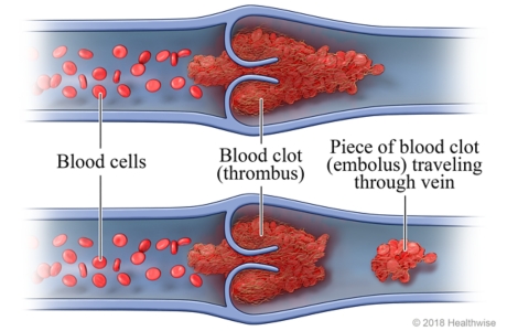
    • Thrombus: A solid mass of coagulated blood constituents (platelets, fibrin, red blood cells, and white blood cells) that forms inside a blood vessel or heart chamber and remains attached at its site of origin.
    • Embolus: Any detached intravascular solid, liquid, or gaseous mass carried by the bloodstream from its point of origin to a distant site, where it lodges in a vessel too narrow to pass. Over 90% of emboli are dislodged fragments of blood clots (thromboemboli), whereas non-thrombotic emboli consist of fat globules, air bubbles, amniotic fluid, tumour cells, infected vegetations, cholesterol crystals, or foreign debris (such as catheter fragments or talc)—which are not blood clots.

    Thrombus (Thrombosis)

    Definition, Pathophysiology, and Virchow's Triad

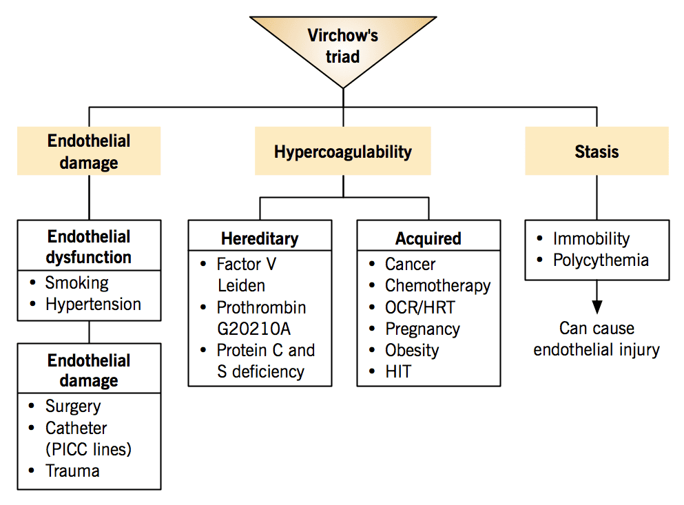
    A thrombus is a blood clot formed within the living circulatory system that adheres to the vessel wall or endocardium and partially or completely impedes blood flow. Pathological thrombus formation is governed by Virchow's Triad, which describes three interrelated predisposing factors:

    Diagram illustrating Virchow's Triad: endothelial injury, stasis of blood flow, and hypercoagulability
    1. Endothelial Injury: Damage to the smooth inner lining (endothelium) of a blood vessel or cardiac chamber. This is the dominant trigger in arterial and cardiac thrombosis, exposing subendothelial collagen and tissue factor that activate platelets and the coagulation cascade.
      • Examples: Atherosclerosis (most common arterial cause), hypertension, surgical or blunt trauma, indwelling intravenous or central venous catheters, vasculitis, and toxins from tobacco smoking.
    2. Abnormal Blood Flow (Stasis or Turbulence): Normal laminar blood flow keeps platelets central and dilutes activated clotting factors. Venous stasis allows clotting factors to accumulate locally without clearance, whereas turbulence damages the endothelium and forms pockets of stasis in arteries and the heart.
      • Examples of Stasis: Prolonged bed rest or immobility, long-distance travel, limb paralysis, heart failure, chronic venous insufficiency, varicose veins, and atrial fibrillation (blood pooling in the left atrial appendage).
      • Examples of Turbulence: Ulcerated atherosclerotic plaques, arterial aneurysms, stenotic or prosthetic heart valves, and vessel bifurcations.
    3. Hypercoagulability (Thrombophilia): An abnormal shift in the hemostatic balance favoring clotting due to excess pro-coagulant factors or reduced natural anticoagulants.
      • Inherited (Primary) Causes: Factor V Leiden mutation (most common), Prothrombin G20210A gene mutation, and deficiencies of Antithrombin III, Protein C, or Protein S.
      • Acquired (Secondary) Causes: Malignancy (cancer), pregnancy and the postpartum puerperium, estrogen-containing oral contraceptives and hormone replacement therapy, dehydration, severe sepsis, major surgery/trauma, nephrotic syndrome, and antiphospholipid syndrome.

    Causes and Risk Factors of a Thrombus

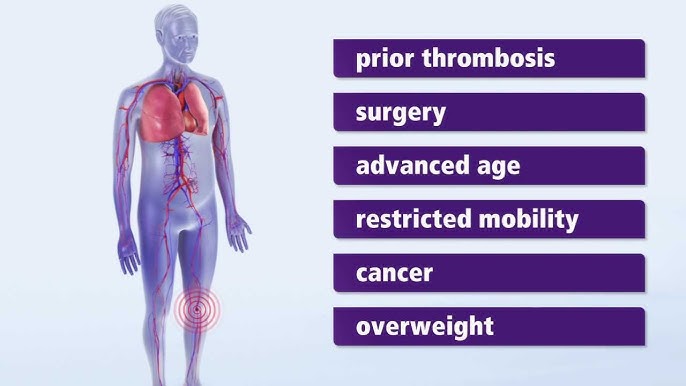
    Causes and risk factors contributing to thrombus formation

    Clinical risk factors act through one or more arms of Virchow's Triad:

    • Cardiovascular and Metabolic Factors: Atherosclerosis (plaque rupture), hyperlipidemia, hypertension, diabetes mellitus (micro- and macrovascular injury), atrial fibrillation, heart failure, and prior thromboembolism (DVT, PE, or stroke).
    • Lifestyle and Demographic Factors: Tobacco smoking, obesity (chronic inflammation and venous compression), sedentary lifestyle, advancing age, and dehydration (increased blood viscosity).
    • Clinical, Surgical, and Hormonal Triggers: Major surgery (especially orthopedic hip/knee and pelvic surgery), major trauma, prolonged immobility, malignancy (e.g., Trousseau's syndrome—migratory thrombophlebitis), pregnancy/postpartum period, combined oral contraceptives or hormone replacement therapy, autoimmune/inflammatory diseases (SLE, IBD), and inherited thrombophilias.

    Types and Fate of a Thrombus

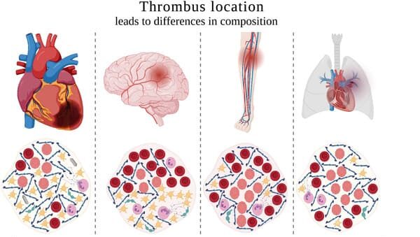
    Classification of thrombi by vascular location and composition
    • Arterial Thrombus ("White Thrombus"): Forms in high-pressure, fast-flowing arteries, usually over ruptured atherosclerotic plaques. Rich in platelets and fibrin (pale appearance); often occlusive (coronary, cerebral, mesenteric, or peripheral arteries), causing acute ischemia or infarction.
    • Venous Thrombus ("Red Thrombus"): Forms in low-pressure, slow-flowing veins due to stasis and hypercoagulability. Rich in red blood cells and fibrin (dark red appearance) and almost always occlusive; classically causes Deep Vein Thrombosis (DVT) and superficial thrombophlebitis.
    • Cardiac (Mural) Thrombus: Adheres to the inner wall of a heart chamber or valve—commonly in the left atrial appendage during atrial fibrillation or on an akinetic left ventricular wall after myocardial infarction; high risk of systemic arterial embolization.
    • Microvascular Thrombus: Tiny fibrin–platelet microthrombi forming widely in arterioles, capillaries, and venules during sepsis, severe trauma, or Disseminated Intravascular Coagulation (DIC), producing multi-organ dysfunction and consumptive bleeding.
    • Septic (Infective) Thrombus: A thrombus colonized by bacteria or fungi—seen on infected heart valves (infective endocarditis) or infected intravenous cannulae (suppurative thrombophlebitis)—which can shed septic emboli.

    Possible Outcomes (Fate) of a Thrombus

    Once formed, a thrombus follows one of four pathological pathways:

    1. Propagation: The thrombus accumulates additional platelets and fibrin and grows along the vessel, eventually obstructing critical branches.
    2. Embolization: Part or all of the thrombus detaches and travels downstream to occlude a distant vessel (e.g., DVT → pulmonary embolism).
    3. Dissolution (Fibrinolysis): The body's endogenous plasmin system (or therapeutic thrombolysis) enzymatically breaks down fresh fibrin, restoring patency before fibrosis occurs.
    4. Organization and Recanalization: Older thrombi become invaded by endothelial cells, fibroblasts, and smooth muscle cells (organization) and may form new capillary channels through the clot (recanalization), partially restoring flow.

    Clinical Manifestations of Thrombosis

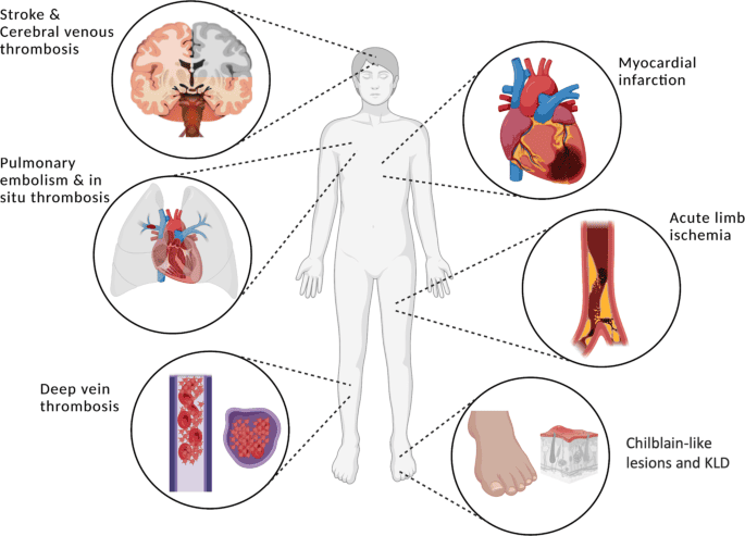
    Clinical manifestations of arterial and venous thrombosis

    Symptoms arise from reduced blood delivery (arterial ischemia) or impaired blood drainage (venous congestion and inflammation):

    A. Arterial Thrombosis

    • Coronary Artery Thrombosis (Myocardial Infarction): Crushing or tightening central chest pain radiating to the left arm, jaw, neck, or back; dyspnea, diaphoresis, nausea, vomiting, and lightheadedness.
    • Cerebral Artery Thrombosis (Ischemic Stroke): Sudden unilateral weakness or numbness (face, arm, leg), aphasia or dysarthria, visual field loss, severe dizziness, or loss of balance.
    • Peripheral Arterial Thrombosis (Acute Limb Ischemia): Presents with the classic "6 Ps": Pain, Pallor, Pulselessness, Paresthesia, Paralysis, and Poikilothermia (coldness) distal to the occlusion.
    • Mesenteric Artery Thrombosis: Sudden, severe abdominal pain disproportionate to early physical examination findings, accompanied by nausea, vomiting, distension, and later bloody stools.

    B. Venous Thrombosis

    • Deep Vein Thrombosis (DVT): Unilateral limb swelling, calf or thigh pain/tenderness, localized warmth, erythema or dusky discoloration, and distended superficial collateral veins (explored in detail below).
    • Superficial Thrombophlebitis: A palpable, tender, warm, erythematous cord along a superficial vein (often a varicose vein or an IV cannula site); carries lower PE risk than DVT unless it extends into deep junctions.

    Diagnosis of a Thrombus

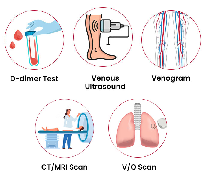
    Diagnostic workup for thrombosis including blood tests and imaging
    • Clinical Assessment: Detailed risk-factor history, validated probability scoring (e.g., Wells' score), and targeted cardiovascular, neurological, or peripheral vascular examination.
    • Laboratory Investigations:
      • D-dimer: Measures fibrin degradation products. Highly sensitive; a negative result helps exclude DVT/PE in low-probability patients, whereas a positive result is non-specific (elevated in infection, pregnancy, trauma, or malignancy) and does not diagnose thrombosis by itself.
      • Coagulation Profile & CBC: Baseline PT/INR, aPTT, and platelet count prior to anticoagulation; thrombophilia screening (Factor V Leiden, Protein C/S, antiphospholipid antibodies) only in selected unprovoked or familial cases.
    • Imaging Studies:
      • Compression Duplex Ultrasound: First-line, non-invasive gold standard for DVT and peripheral arterial occlusion.
      • CT Angiography (CTA) / MR Angiography (MRA): High-resolution imaging for arterial thrombi (cerebral, mesenteric, peripheral) and pulmonary embolism (CTPA).
      • Echocardiography (TTE/TOE): Detects mural or atrial appendage thrombi and valvular vegetations.
      • Conventional Angiography / Venography: Invasive contrast studies reserved mainly when catheter-directed intervention is planned.

    Advertisements

    Embolus and Embolism

    Comparison illustrating a stationary thrombus versus a traveling embolus

    Definition and Pathophysiology of Embolism

    An embolus (plural: emboli) is any detached intravascular mass (solid, liquid, or gas) carried by the bloodstream to a site distant from its origin. Embolism occurs when that mass lodges in a vessel too narrow to pass, abruptly obstructing blood flow, causing downstream tissue ischemia, and—unless collateral circulation compensates or perfusion is restored—leading to infarction.

    Types of Embolism

    1. Thromboembolism (Most Common): Detached fragments of thrombi:
      • Pulmonary Embolism (PE): Venous clot (usually from iliofemoral DVT) travels through the right heart and obstructs the pulmonary arterial tree, causing V/Q mismatch, hypoxemia, and right ventricular strain.
      • Systemic Arterial Embolism: Often originates from left-sided cardiac thrombi (atrial fibrillation, post-MI mural thrombus, prosthetic/diseased valves) or aortic plaques, causing cerebral embolism (ischemic stroke), acute limb ischemia, mesenteric embolism, renal/splenic infarction, or retinal artery occlusion (amaurosis fugax / sudden blindness).
      • Paradoxical Embolism: A venous thrombus crosses from the right atrium to the left atrium through a patent foramen ovale (PFO) or atrial septal defect, entering the systemic arterial circulation.
    2. Fat Embolism: Fat globules enter torn marrow sinusoids after long-bone fractures (femur/tibia), orthopedic arthroplasty, severe burns, or pancreatitis, causing Fat Embolism Syndrome (FES) 12–72 hours post-injury.
    3. Air (Gas) Embolism: Air enters the circulation via central venous line disconnection/insertion, neurosurgery/cardiac surgery, chest trauma, or decompression sickness ("the bends"). Venous air (> 100 mL rapidly) can cause an "air-lock" in the right ventricle; arterial air causes stroke or myocardial infarction.
    4. Septic Embolism: Infected fibrin/bacterial vegetations detach from endocarditis (e.g., tricuspid endocarditis in IV drug users causing septic pulmonary emboli; left-sided endocarditis causing brain/renal abscesses).
    5. Amniotic Fluid Embolism (AFE): Rare, catastrophic obstetric emergency during labour or immediate postpartum where amniotic fluid and fetal cells enter maternal circulation, triggering sudden hypoxia, cardiovascular collapse, and severe DIC.
    6. Cholesterol (Atheroembolic) Embolism: Microscopic cholesterol crystals shower from ulcerated aortic plaques (often after vascular catheterization), causing livedo reticularis, acute kidney injury, and "blue toe syndrome" despite palpable distal pulses.
    7. Tumour and Foreign Body Embolism: Malignant cell clusters spreading via blood/lymphatics (metastasis), or accidental intravascular catheter fragments, talc, or metallic pellets.

    Clinical Manifestations of Embolism

    Embolic events characteristically present with an abrupt, dramatic onset dictated by the target organ:

    • Pulmonary Embolism (PE): Sudden dyspnea, sharp pleuritic chest pain, tachypnea, tachycardia, hemoptysis, anxiety, syncope, and in massive PE, jugular venous distension, hypotension, and obstructive shock.
    • Cerebral, Peripheral, Mesenteric, and Retinal Embolism: Sudden focal neurological deficits (stroke), acute "6 Ps" in an extremity, severe pain out of proportion to abdominal exam (mesenteric), or sudden painless monocular vision loss (retinal).
    • Fat Embolism Syndrome (Classic Triad): Respiratory distress/hypoxemia + neurological confusion/agitation + non-blanching petechial rash across the upper chest, neck, axillae, and conjunctivae 12–72 hours after fracture.
    • Air and Septic Embolism: Venous air embolism causes sudden dyspnea, hypotension, cyanosis, and a churning "mill-wheel" murmur; septic embolism adds high fevers, chills, and focal abscesses.

    Diagnosis of an Embolism

    • Pulmonary Embolism: Clinical probability (Wells' PE score) + D-dimer (low/intermediate risk) + CT Pulmonary Angiography (CTPA) (gold standard) or V/Q scan; bedside ECG (sinus tachycardia, S1Q3T3, right heart strain), ABG, troponin/BNP, echocardiography, and leg Duplex ultrasound for DVT.
    • Cerebral Embolism: Urgent non-contrast CT head (to exclude hemorrhage) followed by CTA/MRA, echocardiography, and Holter/ECG monitoring for atrial fibrillation.
    • Peripheral, Fat, Air, and Septic Embolism: CTA/Duplex ultrasound for limb arteries; clinical diagnostic criteria + CXR/CT for Fat Embolism Syndrome; echocardiography/CT for intravascular air; blood cultures + echocardiography for septic emboli.

    Similarities and Differences Between Thrombus and Embolus

    Key Similarities: Both occur inside the circulatory system, both obstruct blood vessel lumina leading to downstream tissue ischemia or infarction, both are linked directly or indirectly to Virchow's Triad, and both can cause life-threatening emergencies such as stroke, myocardial infarction, or pulmonary embolism.

    FeatureThrombusEmbolus
    DefinitionA solid blood clot formed within a blood vessel or heart chamber that remains attached at its site of origin.Any detached intravascular mass (usually a fragment of a thrombus, or fat/air/amniotic fluid/tumour) that travels through the bloodstream and lodges distally.
    MobilityStationary and adherent to the vessel wall.Mobile until trapped in a narrower vessel.
    Site of ObstructionObstructs blood flow at the exact site where it formed.Obstructs blood flow at a distant site (commonly vessel bifurcations or narrow arterioles/capillaries).
    CompositionBlood elements only: platelets, fibrin, red cells, and leukocytes.Over 90% thrombotic debris; ~10% non-thrombotic (fat, gas/air, amniotic fluid, bacteria, tumour, cholesterol, foreign body).
    Onset of SymptomsMay develop gradually (e.g., expanding DVT, unstable angina) or acutely on plaque rupture.Almost always sudden and abrupt due to instant occlusion of an unconditioned vessel.
    Classic ExamplesDeep Vein Thrombosis (DVT), coronary artery thrombosis, left atrial appendage mural thrombus.Pulmonary Embolism (PE), embolic ischemic stroke, acute limb embolism, Fat Embolism Syndrome.

    Advertisements

    Detailed Focus: Deep Vein Thrombosis (DVT)

    Deep Vein Thrombosis (DVT)—formation of a thrombus within the deep venous system, most often in the lower extremities or pelvis—is the single most common preventable cause of in-hospital death due to its progression to Pulmonary Embolism (PE). Together, DVT and PE constitute Venous Thromboembolism (VTE).

    Anatomical and Clinical Classification of DVT

    • Proximal Lower-Extremity DVT: Involves the popliteal, femoral, or iliac veins (above the knee). Carries the highest risk of embolizing to the lungs (PE) and requires immediate therapeutic anticoagulation.
    • Distal (Calf) DVT: Confined to the infrapopliteal veins (anterior tibial, posterior tibial, or peroneal veins). Lower immediate PE risk, though ~15–20% can propagate proximally if untreated.
    • Upper-Extremity DVT: Involves the axillary, subclavian, or brachial veins; increasingly associated with central venous catheters, pacemakers, or thoracic outlet compression.
    • Provoked vs. Unprovoked DVT: Provoked DVT follows a transient or persistent identifiable trigger (surgery, trauma, immobility, pregnancy, estrogen therapy, active cancer), whereas unprovoked DVT occurs without an obvious trigger and carries a higher long-term recurrence rate.

    Pathophysiology and Risk Factors Specific to DVT

    Deep leg veins depend on calf-muscle pump contraction and intact one-way venous valves to return blood upward against gravity. Immobilization, venous valve pooling (especially in valve pockets of the soleal sinuses), post-operative tissue factor release, pregnancy-induced uterine compression of the inferior vena cava/left iliac vein, HIV-related chronic inflammation, malignancy, and trauma trigger fibrin-rich red thrombus growth.

    Clinical Features and Bedside Assessment (Including Wells' Criteria)

    • Typical Presentation: Unilateral pitting edema of the ankle, calf, or entire thigh; aching or cramping calf pain worsened by standing/walking; increased limb circumference (≥ 3 cm difference measured 10 cm below the tibial tuberosity); localized warmth; erythema or cyanosis; and engorged superficial collateral veins. Note: Many DVTs are clinically silent until PE occurs.
    • Severe Iliofemoral Syndromes (Limb-Threatening Emergencies):
      • Phlegmasia Alba Dolens ("Milk Leg"): Extensive iliofemoral occlusion causing a massively swollen, painful, pale/white leg (classically in late pregnancy/postpartum) due to arterial vasospasm while capillary flow persists.
      • Phlegmasia Cerulea Dolens: Near-total venous outflow occlusion causing extreme swelling, severe pain, deep cyanotic/bluish-purple discoloration, compartment pressure rise, absent distal pulses, and impending venous gangrene—a surgical/endovascular emergency.
    • Homans' Sign Precaution: Forceful dorsiflexion of the foot to elicit calf pain (Homans' sign) has poor sensitivity and specificity and should NOT be aggressively performed; structured probability scoring using the Wells' DVT Score is standard practice.
    • Key Differential Diagnoses in Uganda: Cellulitis, muscular tear/myositis, Baker's cyst rupture, trauma/contusion, superficial phlebitis, lymphedema, congestive cardiac failure (bilateral), and Kaposi sarcoma or soft-tissue/bone sarcoma of the leg.

    Wells' Clinical Prediction Score for Suspected DVT

    Clinical FeatureScore
    Active cancer (treatment ongoing, within previous 6 months, or palliative)+1
    Paralysis, paresis, or recent plaster immobilization of the lower extremities+1
    Recently bedridden > 3 days, or major surgery within 12 weeks requiring general/regional anesthesia+1
    Localized tenderness along the distribution of the deep venous system+1
    Entire leg swollen+1
    Calf swelling ≥ 3 cm larger than asymptomatic leg (measured 10 cm below tibial tuberosity)+1
    Pitting edema confined to the symptomatic leg+1
    Collateral superficial veins (non-varicose)+1
    Previously documented DVT+1
    Alternative diagnosis at least as likely as DVT (e.g., cellulitis, muscle tear)−2

    Interpretation (Two-Level Wells Score): Score ≥ 2 = DVT Likely (proceed directly to Compression Duplex Ultrasound). Score < 2 = DVT Unlikely (check quantitative D-dimer; if negative, DVT is safely ruled out; if positive, proceed to Duplex Ultrasound).

    Complications, Treatment, and Prophylaxis of DVT

    • Complications of DVT:
      • Pulmonary Embolism (PE): Acute life-threatening complication from proximal clot embolization.
      • Post-Thrombotic Syndrome (PTS): Chronic complication (20–50% of patients) caused by destroyed venous valves and persistent venous hypertension, leading to chronic aching leg swelling, varicosities, hemosiderin hyperpigmentation, lipodermatosclerosis, and venous stasis ulcers.
      • Recurrent DVT and Venous Gangrene: Especially if anticoagulation is stopped prematurely or underlying cancer/thrombophilia persists.
    • Specific DVT Management (UCG 2023 & International Standards):
      • Immediate Anticoagulation: Start subcutaneous Low Molecular Weight Heparin (Enoxaparin 1 mg/kg every 12 h or 1.5 mg/kg once daily) or IV Unfractionated Heparin (UFH), or an oral DOAC (e.g., rivaroxaban/apixaban where available).
      • Bridging to Warfarin: When using Warfarin (standard long-term oral agent in Uganda), start Warfarin concurrently with LMWH/UFH for at least 5 days and continue heparin until the INR is within therapeutic range (2.0–3.0) for two consecutive readings (never start Warfarin alone without heparin overlap due to transient Protein C/S depletion).
      • Duration & Special Populations: Typically 3 months for transient provoked DVT; 6 months or extended therapy for unprovoked DVT or active cancer. In pregnancy, Warfarin is teratogenic—use LMWH throughout pregnancy.
      • Mobilization & Limb Care: Encourage early ambulation once therapeutic anticoagulation is established (prolonged strict bed rest is no longer indicated for uncomplicated DVT); elevate the leg when resting, apply fitted graduated compression stockings to reduce edema/PTS symptoms, and NEVER massage the affected leg (risk of dislodging the thrombus into the lungs).

    General Medical, Pharmacological, and Interventional Management

    Treatment of Thrombosis and Embolism

    Overview of medical and interventional treatments for thrombosis and embolism
    • Anticoagulant Medications ("Blood Thinners"): Prevent thrombus propagation and new clot formation while allowing natural fibrinolysis to clear the existing clot.
      • Parenteral Heparins: Unfractionated Heparin (UFH, IV/SC) and Low Molecular Weight Heparin (LMWH—e.g., enoxaparin, dalteparin, SC).
      • Vitamin K Antagonist: Warfarin (oral; requires regular INR monitoring, target 2.0–3.0 for VTE/AF or 2.5–3.5 for mechanical valves).
      • Direct Oral Anticoagulants (DOACs): Direct Xa inhibitors (rivaroxaban, apixaban, edoxaban) and direct thrombin inhibitor (dabigatran).
    • Thrombolytic Medications ("Clot Busters"): (e.g., Alteplase, Tenecteplase, Streptokinase). Activate plasminogen to plasmin to actively dissolve fibrin clots. Reserved for life- or limb-threatening emergencies—such as massive PE with hypotension/shock, acute ischemic stroke within the thrombolytic window (after excluding hemorrhage on CT), STEMI, or phlegmasia cerulea dolens—due to high intracranial and systemic hemorrhage risk.
    • Antiplatelet Medications: (e.g., Aspirin, Clopidogrel, Ticagrelor, Prasugrel). Cornerstone for arterial thrombosis (coronary artery disease, non-cardioembolic ischemic stroke, peripheral arterial disease) and post-stent protection.
    • Mechanical Thrombectomy / Embolectomy: Catheter-directed aspiration/stent-retriever thrombectomy or open surgical embolectomy (e.g., Fogarty balloon catheter) for acute large-vessel ischemic stroke, acute limb ischemia, or massive PE when thrombolysis is contraindicated or fails.
    • Inferior Vena Cava (IVC) Filters: Retrievable filter placed in the IVC to trap leg emboli in patients with acute proximal DVT/PE who have an absolute contraindication to anticoagulation (e.g., active intracranial or GI bleeding) or recurrent PE despite adequate anticoagulation.
    • Specific Supportive & Non-Thrombotic Embolism Treatments:
      • Fat Embolism Syndrome: High-flow oxygen, early fracture stabilization, fluid balance, and mechanical ventilation if ARDS develops.
      • Venous Air Embolism: Clamp source immediately, place patient in left lateral decubitus Trendelenburg position (Durant's maneuver) to trap air in the right ventricular apex away from the pulmonary outflow tract, give 100% oxygen, and consider hyperbaric oxygen for arterial air embolism.
      • Septic and Amniotic Fluid Embolism: Targeted IV antibiotics and source control/valve surgery for septic emboli; immediate airway, vasopressor, and massive blood-product transfusion (for DIC) in amniotic fluid embolism.

    Pharmacokinetics and Pharmacodynamics of Antithrombotic Medications

    Drug / ClassPharmacodynamics (Mechanism & Reversal)Pharmacokinetics (ADME & Monitoring)Key Nursing & Safety Considerations
    Unfractionated Heparin (UFH)Binds Antithrombin III, accelerating inactivation of Factor IIa (thrombin) and Factor Xa ~1,000-fold. Antidote: Protamine sulfate (1 mg per ~100 units UFH).IV (immediate onset) or SC; does not cross placenta; reticuloendothelial/hepatic clearance (half-life ~1–2 h); safe in severe renal failure. Monitor aPTT (target 1.5–2.5× control) and platelets.Preferred in severe renal impairment or when rapid reversal is needed; monitor platelet count for Heparin-Induced Thrombocytopenia (HIT).
    LMWH (Enoxaparin, Dalteparin)Binds Antithrombin III with preferential inhibition of Factor Xa over IIa. Antidote: Protamine sulfate (only partially reverses ~60% of anti-Xa activity).Predictable SC bioavailability (> 90%); peak 3–5 h; half-life 4–5 h; renally excreted (accumulates in CKD/CrCl < 30 mL/min). Routine aPTT monitoring not required (anti-Xa used in severe CKD/pregnancy).Preferred initial VTE drug in Uganda; safe in pregnancy; rotate abdominal SC injection sites and do not expel the air bubble or rub the site after injection.
    Warfarin (Vitamin K Antagonist)Inhibits vitamin K epoxide reductase (VKORC1), blocking hepatic synthesis of clotting factors II, VII, IX, X and natural anticoagulants Proteins C & S. Antidote: Vitamin K (phytomenadione) + 4-factor PCC or FFP for life-threatening bleeding.Well absorbed orally; 99% albumin-bound; long half-life (~36–42 h); full antithrombotic effect takes 3–5 days; metabolized by hepatic CYP2C9/CYP3A4; crosses placenta (teratogenic). Monitor PT/INR (target 2.0–3.0).Bridge with heparin for ≥ 5 days until INR ≥ 2.0 for 2 days. Maintain consistent daily Vitamin K intake (dark leafy greens—do not drastically binge or eliminate); avoid NSAIDs/alcohol and check CYP drug interactions.
    DOACs
    (Rivaroxaban, Apixaban, Dabigatran)
    Directly and reversibly inhibit Factor Xa (rivaroxaban, apixaban) or Factor IIa/thrombin (dabigatran). Antidotes: Idarucizumab (dabigatran); Andexanet alfa or 4-factor PCC (Xa inhibitors).Oral; rapid onset (1–4 h); short half-lives (~8–15 h); variable renal clearance (dabigatran ~80% renal, rivaroxaban ~33% unchanged renal, apixaban ~27% renal); P-gp/CYP3A4 substrates. No routine INR/aPTT monitoring needed.Strict adherence is critical because short half-lives mean missed doses rapidly lose protection. Rivaroxaban 15–20 mg doses must be taken with food for full absorption; adjust doses in renal impairment.
    Thrombolytics
    (Alteplase, Tenecteplase, Streptokinase)
    Convert plasminogen into active plasmin, which directly cleaves cross-linked fibrin inside established thrombi.IV infusion/bolus or catheter-directed; very short plasma half-life (alteplase ~5 min) with hepatic clearance, though systemic fibrinolysis lasts hours.Screen rigorously for absolute contraindications (prior intracranial hemorrhage, active bleeding, recent major surgery/trauma, severe uncontrolled HTN). Avoid invasive punctures/IM injections during and after infusion.
    Antiplatelets
    (Aspirin, Clopidogrel)
    Aspirin: Irreversibly acetylates platelet COX-1, blocking thromboxane A2. Clopidogrel: Prodrug irreversibly blocking platelet P2Y12 ADP receptors.Oral; antiplatelet effect persists for the 7–10 day lifespan of platelets. Clopidogrel requires hepatic CYP2C19 bioactivation (reduced by omeprazole).Watch for GI bleeding/epistaxis; co-prescribe a PPI (prefer pantoprazole/rabeprazole with clopidogrel) in high GI-bleed-risk patients.

    Complications and Prevention of Thromboembolic Disorders

    • Major Complications: Fatal Pulmonary Embolism, Chronic Thromboembolic Pulmonary Hypertension (CTEPH), Post-Thrombotic Syndrome (PTS), permanent neurological disability from stroke, myocardial failure/arrhythmias, limb amputation from arterial/venous gangrene, and major bleeding or HIT from anticoagulation.
    • Prevention and Prophylaxis:
      • Hospital VTE Prophylaxis: Assess every admitted medical, surgical, and obstetric patient for VTE risk; administer prophylactic-dose LMWH (e.g., enoxaparin 40 mg SC daily) or UFH plus mechanical prophylaxis (graduated compression stockings or intermittent pneumatic compression) when bleeding risk is high.
      • Early Mobilization & Hydration: Encourage post-operative leg exercises and ambulation, adequate hydration, and calf/ankle pumps during prolonged travel.
      • Cardiovascular Risk Control: Treat hypertension, diabetes, hyperlipidemia, smoking, and anticoagulate high-risk atrial fibrillation.

    Advertisements

    Nursing Management of Thromboembolic Disorders and DVT

    Nursing Care Plan for a Patient with Thromboembolism / DVT (NANDA-I 2024–2026)

    This care plan applies current NANDA-I 2024–2026 terminology (Ineffective peripheral tissue perfusion, Impaired gas exchange [for PE], Inadequate health knowledge, Acute pain, Risk for bleeding, and Impaired physical mobility) prior to the detailed nursing interventions below.

    AssessmentDiagnosis (NANDA-I 2024–2026)Planning (Goals & Expected Outcomes)Implementation / InterventionsRationaleEvaluation
    • Subjective: Patient reports unilateral leg swelling, heaviness, and throbbing calf pain (or sudden dyspnea in PE).
    • Objective: Left calf circumference +4 cm larger than right, warmth, pitting edema, intact distal pulses (or SpO2 89% and tachypnea in PE).
    Ineffective peripheral tissue perfusion (or Impaired gas exchange in PE) related to interruption of venous or arterial blood flow by thrombus/embolus as evidenced by unilateral calf swelling (+4 cm), warmth, and pitting edema.

    Restore adequate tissue perfusion and oxygenation within 24–72 hours.

    Expected outcomes:

    • Affected limb maintains strong distal pulses, warm skin, and reduced swelling.
    • Patient remains free from signs of PE (SpO2 ≥ 94%, no dyspnea/chest pain).
    1. Perform neurovascular checks (6 Ps), measure daily bilateral calf circumferences, and monitor vital signs and SpO2 every 4 hours.
    2. Administer prescribed LMWH/UFH and bridge Warfarin promptly; monitor INR/aPTT and platelet counts.
    3. Elevate affected leg on pillows above heart level while resting; apply fitted compression stockings as ordered; never massage the leg.
    4. For PE, position in high-Fowler's and administer prescribed oxygen.
    1. Detects worsening ischemia (phlegmasia/arterial occlusion) or early embolization to the lungs immediately.
    2. Prevents clot extension and pulmonary embolization while keeping coagulation within safe therapeutic limits.
    3. Promotes gravity-assisted venous return and reduces edema without risking clot dislodgement from massage.
    4. Maximizes diaphragmatic excursion and corrects V/Q mismatch hypoxemia.
    Within 48 hours, calf circumference decreased by 2 cm, distal pulses remained strong, and SpO2 stayed at 97% with no signs of PE.
    • Subjective: Patient rates aching calf/thigh pain at 6/10, worse when standing.
    • Objective: Guarding of affected limb, facial grimacing, HR 102 bpm.
    Acute pain related to vascular occlusion, venous congestion, and local perivascular inflammation as evidenced by pain score 6/10 and limb guarding.

    Reduce pain to a tolerable level within 2 hours of intervention.

    Expected outcomes:

    • Patient will report pain ≤ 2/10.
    • Patient will rest comfortably without guarding.
    1. Assess pain score (0–10), location, and response to therapy regularly.
    2. Administer prescribed paracetamol or opioid analgesia; avoid NSAIDs due to bleeding risk with anticoagulants.
    3. Elevate the affected limb comfortably on soft pillows and support without pressure behind the knee.
    4. Avoid vigorous massage or direct hot/cold packs over the anticoagulated/ischemic limb.
    1. Monitors analgesic response and flags sudden pain spikes indicating arterial compromise or compartment rise.
    2. Controls pain safely without compounding platelet dysfunction or GI hemorrhage risk.
    3. Reduces venous hydrostatic pressure and inflammatory throbbing without compressing popliteal veins.
    4. Prevents mechanical clot dislodgement and thermal skin injury.
    Pain decreased from 6/10 to 2/10 within 2 hours; patient rested comfortably with limb elevated.
    • Subjective: Patient receiving therapeutic enoxaparin and warfarin (or thrombolytic therapy).
    • Objective: Active systemic anticoagulation; monitor INR, aPTT, hemoglobin, and platelet count.
    Risk for bleeding related to therapeutic anticoagulation (LMWH, warfarin, DOACs) or thrombolytic therapy.

    Prevent major hemorrhagic complications throughout hospitalization.

    Expected outcomes:

    • Patient will remain free from overt bleeding (epistaxis, hematuria, melena, CNS bleed).
    • INR/aPTT and hemoglobin will remain within safe target ranges.
    1. Monitor PT/INR, aPTT, CBC, and platelets as ordered; hold drug and notify clinician if INR is supratherapeutic or platelets fall sharply.
    2. Inspect skin, gums, urine, stool, vomitus, and injection sites daily for bruising, petechiae, or frank blood; monitor neurological status.
    3. Implement bleeding precautions: avoid IM injections, use soft toothbrush/electric razor, apply firm prolonged pressure (≥ 5 min) after venipuncture, and prevent falls.
    4. Keep antidotes (protamine sulfate, Vitamin K) readily accessible.
    1. Detects excessive anticoagulation or HIT before life-threatening hemorrhage occurs.
    2. Identifies occult internal or intracranial bleeding early while intervention is effective.
    3. Minimizes mechanical trauma to skin, mucosa, and vessels during reduced clotting capacity.
    4. Enables rapid pharmacologic reversal if severe bleeding develops.
    Patient remained free from bruising, mucosal bleeding, or internal hemorrhage with INR stabilized at 2.4.
    • Subjective: Patient fears moving leg or walking because of pain and clot worry.
    • Objective: Reluctance to ambulate; stiffness on initial movement.
    Impaired physical mobility related to limb pain, swelling, and initial stabilization period as evidenced by reluctance to ambulate.

    Resume safe graded ambulation within 24–48 hours once anticoagulation is therapeutic.

    Expected outcomes:

    • Patient will perform ankle-pump exercises and walk short distances with assistance safely.
    1. Reassure patient and initiate graded ambulation once therapeutic anticoagulation and hemodynamic stability are achieved.
    2. Teach gentle active ankle pumps and calf-muscle exercises while in bed (for unaffected and stabilized limb as cleared).
    3. Apply prescribed graduated compression stockings prior to getting out of bed and monitor for dyspnea/dizziness during ambulation.
    1. Early ambulation in uncomplicated DVT does not increase PE risk once anticoagulated and accelerates edema resolution.
    2. Activates the venous muscle pump to prevent contralateral stasis and joint stiffness.
    3. Supports venous valves to reduce orthostatic pooling and monitors cardiopulmonary tolerance.
    By day 2 of anticoagulation, patient walked 30 metres comfortably with compression stockings and no dyspnea.
    • Subjective: Patient asks how long to take warfarin, what foods to avoid, and when to return for INR checks.
    • Objective: First diagnosis of DVT; newly initiated oral anticoagulation.
    Inadequate health knowledge related to unfamiliarity with anticoagulant adherence, INR monitoring, Vitamin K consistency, and PE warning signs as evidenced by patient inquiries.

    Demonstrate safe self-management of anticoagulation and VTE prevention before discharge.

    Expected outcomes:

    • Patient will explain dosing, bleeding precautions, consistent Vitamin K diet, and emergency signs.
    1. Explain DVT/PE pathophysiology, exact anticoagulant dose/duration, and importance of scheduled INR clinic appointments.
    2. Teach consistent Vitamin K intake (on warfarin), avoidance of OTC NSAIDs/aspirin, and bleeding precautions.
    3. Review red-flag symptoms requiring emergency return (sudden shortness of breath, chest pain, coughing blood, black stools, severe headache).
    4. Verify understanding using teach-back and counsel on leg exercises during long travel.
    1. Prevents subtherapeutic INR (clot recurrence) and supratherapeutic INR (hemorrhage).
    2. Sudden dietary Vitamin K swings destabilize warfarin control, and NSAIDs multiply GI bleeding risk.
    3. Ensures immediate life-saving care if PE or major bleeding occurs at home.
    4. Confirms comprehension and empowers long-term secondary prevention.
    Before discharge, patient accurately taught back warfarin schedule, INR follow-up date, dietary consistency, and PE warning signs.

    Detailed Nursing Diagnoses and Interventions

    1. Ineffective Tissue Perfusion (Pulmonary, Cerebral, or Peripheral)

    • Assessment Cues: Dyspnea, tachypnea, pleuritic chest pain, hypoxemia (PE); focal motor/sensory/speech deficits (cerebral); or 6 Ps (arterial) / unilateral leg swelling, warmth, and calf pain (DVT).
    • Interventions: Monitor vital signs and SpO2 frequently; give oxygen and high-Fowler's positioning for PE; elevate the affected DVT leg (never massage); perform neurovascular checks and bilateral calf measurements; administer prescribed anticoagulants/thrombolytics; prepare for Duplex ultrasound, CTPA, or embolectomy.

    2. Acute Pain

    • Assessment Cues: Verbal pain score (0–10), guarding, restlessness, tachycardia, or elevated BP.
    • Interventions: Assess pain regularly; give prescribed paracetamol or opioids (avoid NSAIDs alongside anticoagulants); elevate the DVT extremity; provide a quiet restful environment; avoid limb massage and thermal packs over ischemic or anticoagulated skin.

    3. Risk for Bleeding

    • Assessment Cues: Epistaxis, gingival bleeding, hematuria, melena, hematemesis, expanding ecchymoses, falling hemoglobin, prolonged PT/INR or aPTT, thrombocytopenia, or altered mental status.
    • Interventions: Monitor INR, aPTT, and CBC/platelets; inspect skin, urine, and stools daily; apply bleeding precautions (no IM injections, soft toothbrush, electric razor, prolonged venipuncture pressure, fall prevention); keep protamine sulfate and Vitamin K ready.

    4. Impaired Physical Mobility

    • Assessment Cues: Pain with movement, fear of clot dislodgement, neurological weakness (post-stroke), or reduced range of motion.
    • Interventions: Begin gentle ankle pumps and early graded ambulation once uncomplicated DVT is therapeutically anticoagulated and stable; fit graduated compression stockings properly; collaborate with physiotherapy/occupational therapy for post-stroke rehabilitation.

    5. Inadequate Health Knowledge (Deficient Knowledge)

    • Assessment Cues: Questions or misconceptions regarding warfarin/DOAC dosing, INR monitoring, diet, or emergency signs.
    • Interventions: Teach disease process, strict medication adherence, INR schedule, consistent Vitamin K intake, bleeding precautions, smoking cessation, hydration/mobility during long travel, and urgent warning signs (chest pain, sudden dyspnea, hemoptysis, neurological deficits) using teach-back and family involvement.

    References

    • Ministry of Health, Republic of Uganda. Uganda Clinical Guidelines (UCG) 2023. Chapter 4: Cardiovascular Diseases — Section 4.1.1 (Deep Vein Thrombosis / Pulmonary Embolism [DVT/PE]) and Section 4.1.11 (Stroke).
    • American Society of Hematology (ASH). Clinical Practice Guidelines on Venous Thromboembolism (VTE): Diagnosis, Treatment, and Prophylaxis.
    • Herdman TH, Kamitsuru S, Lopes CT (eds). NANDA International Nursing Diagnoses: Definitions and Classification, 2024–2026 (13th ed.). Thieme; 2024.

    Quick Quiz

    Thrombus Quiz

    Medical Nursing

    Privacy: Your details are used only for quiz tracking and certificates.

    Thrombus and Embolus Read More »

    Arteriosclerosis and Atherosclerosis

    Arteriosclerosis and Atherosclerosis

    ARTERIOSCLEROSIS AND ATHEROSCLEROSIS

    Arteriosclerosis is an umbrella clinical term meaning "hardening of the arteries"—referring to chronic thickening, stiffening, and loss of elasticity of arterial walls. Atherosclerosis is the most common and clinically important specific subtype of arteriosclerosis, characterized by the buildup of lipid-rich, fibrous plaques (atheromas) within the innermost arterial layer (the tunica intima) of large and medium-sized arteries.

    Both conditions progressively impair arterial compliance and narrow or occlude blood vessels, reducing delivery of oxygen and nutrients to vital organs such as the heart, brain, kidneys, intestines, and extremities.

    Arteriosclerosis: Pathophysiology and Types

    Pathophysiology of Arteriosclerosis

    Arterial walls consist of three layers: the inner tunica intima (endothelium), the middle tunica media (smooth muscle and elastic fibers), and the outer tunica adventitia. In arteriosclerosis, chronic hemodynamic stress, aging, metabolic injury, or hypertension causes degenerative thickening, smooth muscle hypertrophy, hyaline deposition, or medial calcification. While atherosclerosis affects large and medium elastic/muscular arteries, arteriolosclerosis affects small arteries and arterioles that regulate blood pressure and tissue perfusion.

    Types of Arteriosclerosis

    Diagram illustrating the main types of arteriosclerosis

    Arteriosclerosis encompasses three major pathologic patterns (with arteriolosclerosis further subdivided into hyaline and hyperplastic forms):

    1. Atherosclerosis: The most prevalent and dangerous form, affecting large elastic arteries (aorta, carotid, iliac) and medium muscular arteries (coronary, renal, popliteal). Defined by intimal fibrofatty plaques (atheromas) that protrude into the lumen, restrict blood flow, and can rupture to trigger acute thrombosis.
    2. Mönckeberg Medial Calcific Sclerosis: Seen mostly in older adults and patients with diabetes mellitus or chronic kidney disease, commonly in muscular arteries of the extremities (radial, ulnar, tibial) and pelvic vessels. It involves calcification of the tunica media without primary intimal involvement or narrowing of the vessel lumen, though vessels feel rigid ("pipestem" arteries on palpation or X-ray, which can falsely elevate cuff blood pressure readings).
    3. Arteriolosclerosis (Small Arteries and Arterioles):
      • Hyaline Arteriolosclerosis: Homogeneous, pink, glassy thickening of arteriolar walls caused by leakage of plasma proteins across injured endothelium and increased extracellular matrix deposition. Commonly associated with benign (chronic) hypertension, aging, and diabetes mellitus; causes luminal narrowing and chronic tissue ischemia (e.g., benign nephrosclerosis).
      • Hyperplastic Arteriolosclerosis: Concentric, laminated thickening of arteriolar walls ("onion-skinning") caused by smooth muscle cell proliferation and duplicated basement membranes, often accompanied by fibrinoid necrosis. Classically associated with severe or malignant hypertension (diastolic BP > 120 mmHg), causing acute renal or retinal injury.

    Comparison: Arteriosclerosis vs. Atherosclerosis

    CharacteristicArteriosclerosis (General / Non-Atheromatous)Atherosclerosis
    DefinitionBroad group of disorders causing arterial wall thickening, hardening, and loss of elasticity.Specific subtype of arteriosclerosis caused by intimal buildup of lipid-rich, fibrous plaques (atheromas).
    Primary Vessels AffectedSmall arteries and arterioles (arteriolosclerosis) or medium muscular arteries (Mönckeberg sclerosis).Large elastic arteries (aorta, carotid, iliac) and medium muscular arteries (coronary, femoral, cerebral, renal).
    Arterial Layer InvolvedTunica media (calcification/smooth muscle hypertrophy) and arteriolar walls (hyaline or hyperplastic thickening).Primarily the tunica intima, with secondary medial thinning/remodeling beneath advanced plaques.
    Luminal Narrowing & ThrombosisHyaline/hyperplastic types narrow arterioles; Mönckeberg sclerosis does not obstruct the lumen. Acute plaque rupture is absent.Progressive luminal stenosis plus high risk of plaque rupture and acute thrombosis (MI, ischemic stroke, acute limb ischemia).

    Advertisements

    Atherosclerosis: Etiology, Risk Factors, and Pathophysiology

    Atherosclerosis is a slow, progressive, chronic inflammatory disease that often begins with microscopic fatty streaks in childhood or adolescence and accelerates with age as endothelial injury accumulates.

    Risk Factors for Atherosclerosis

    • Modifiable Risk Factors:
      • Dyslipidemia: Elevated low-density lipoprotein (LDL) cholesterol ("bad" cholesterol), elevated triglycerides, and low high-density lipoprotein (HDL) cholesterol ("good" cholesterol, which normally clears excess cholesterol from arterial walls).
      • Hypertension (High Blood Pressure): Chronic mechanical shear stress damages the arterial endothelium and accelerates plaque formation.
      • Diabetes Mellitus and Insulin Resistance: Persistent hyperglycemia causes glycation of vascular proteins, endothelial dysfunction, and accelerated macrovascular and microvascular disease.
      • Cigarette Smoking: Directly injures endothelial cells, triggers vascular inflammation, promotes vasoconstriction and thrombosis, and lowers HDL cholesterol.
      • Sedentary Lifestyle and Abdominal Obesity: Lack of regular physical activity and central adiposity drive metabolic syndrome, hypertension, dyslipidemia, and insulin resistance.
      • Atherogenic Diet: Diets high in saturated fats, trans-fatty acids, refined sugars, and excess sodium.
      • Other Modifiable / Emerging Factors: Obstructive sleep apnea, chronic kidney disease (CKD), chronic systemic inflammatory disorders (e.g., systemic lupus erythematosus, rheumatoid arthritis, HIV-associated chronic inflammation), and elevated high-sensitivity C-reactive protein (hs-CRP).
    • Non-Modifiable Risk Factors:
      • Advancing Age: Cumulative vascular exposure increases risk significantly (men > 45 years; women > 55 years).
      • Biological Sex: Men develop clinical atherosclerosis earlier than premenopausal women; women's risk rises markedly after menopause due to loss of protective estrogen effects.
      • Family History and Genetics: Early premature coronary heart disease in a first-degree relative (male < 55 years; female < 65 years) or inherited lipid disorders (e.g., familial hypercholesterolemia).

    Pathophysiology of Atherosclerosis

    Atherosclerosis develops through a chronic inflammatory-fibroproliferative response to endothelial injury over decades:

    1. Endothelial Damage and Dysfunction: Risk factors (hypertension, oxidized LDL, smoking toxins, hyperglycemia) injure the endothelial lining, reducing protective nitric oxide release and increasing vascular permeability and leukocyte adhesiveness.
    2. Lipid Accumulation and Oxidation: Circulating LDL particles infiltrate the subendothelial intimal space, become trapped in proteoglycans, and undergo chemical oxidation, becoming strongly pro-inflammatory and cytotoxic.
    3. Immune Recruitment and Foam Cell Formation: Endothelial cells express adhesion molecules that recruit blood monocytes and T-lymphocytes into the intima. Monocytes differentiate into macrophages and engulf oxidized LDL via scavenger receptors, transforming into lipid-laden foam cells.
    4. Fatty Streak Formation: Clusters of foam cells and T-cells form flat, yellowish lesions along the intimal surface known as fatty streaks—the earliest macroscopic stage of atherosclerosis, visible even in youth.
    5. Smooth Muscle Cell Migration and Proliferation: Macrophages and platelets release growth factors (such as PDGF) and cytokines that stimulate vascular smooth muscle cells (SMCs) to migrate from the tunica media into the intima, proliferate, and secrete extracellular matrix (collagen, elastin, proteoglycans).
    6. Fibrous Plaque (Atheroma) Formation: Proliferating SMCs and collagen form a fibrous cap covering a central necrotic lipid-rich core containing cholesterol crystals, cellular debris, and foam cells.
    7. Plaque Progression, Remodeling, and Rupture:
      • Progressive Stenosis: Enlarging plaques encroach into the arterial lumen, impairing distal blood flow during exertion (causing stable angina or intermittent claudication).
      • Calcification and Medial Weakening: Dystrophic calcium deposits stiffen plaques, while underlying media atrophy weakens the wall, predisposing to aneurysm formation.
      • Plaque Rupture and Acute Thrombosis: Inflammatory enzymes (metalloproteinases) weaken a thin fibrous cap. When an unstable ("vulnerable") plaque ruptures or erodes, thrombogenic tissue factor and core lipids contact blood, triggering instant platelet aggregation and fibrin clot (thrombus) formation—causing acute arterial occlusion (MI, stroke) or distal embolism.

    Key Concept: Stable vs. Vulnerable (Unstable) Plaques

    Stable plaques have a thick, collagen-rich fibrous cap and small lipid core; they narrow the lumen gradually and cause predictable exertional symptoms (e.g., stable angina). Vulnerable plaques have a thin fibrous cap, large necrotic lipid core, and dense macrophage inflammation; even if they cause less than 50% luminal narrowing, they are prone to sudden rupture and fatal acute thrombosis.

    Clinical Manifestations by Vascular Territory

    Clinical manifestations of atherosclerosis across major arterial beds

    Atherosclerosis is often clinically silent for decades until an artery is critically narrowed (typically > 70% luminal stenosis) or a plaque ruptures acutely. Symptoms depend on the organ system supplied by the affected vessels.

    1. Coronary Arteries (Coronary Artery Disease — CAD)

    • Stable Angina Pectoris: Substernal chest pressure, tightness, squeezing, or burning triggered by physical exertion or emotional stress and relieved within minutes by rest or sublingual nitroglycerin.
    • Acute Coronary Syndrome / Myocardial Infarction (Heart Attack): Sudden plaque rupture with thrombosis causing prolonged, severe chest pain (often radiating to the left arm, jaw, neck, back, or epigastrium) unالrelieved by rest, accompanied by diaphoresis, dyspnea, nausea, palpitations, tachycardia, or arrhythmias.
    • Ischemic Heart Failure: Dyspnea on exertion, orthopnea, fatigue, and peripheral edema resulting from chronic myocardial ischemia or prior infarction.

    2. Carotid and Cerebral Arteries (Cerebrovascular Disease)

    • Transient Ischemic Attack (TIA) and Ischemic Stroke: Sudden focal neurological deficits (remember FAST):
      • Sudden unilateral facial droop, arm or leg weakness, numbness, or paralysis.
      • Slurred speech (dysarthria) or difficulty finding/understanding words (aphasia).
      • Sudden monocular vision loss ("a curtain coming down," amaurosis fugax) or visual field cuts.
      • Acute confusion, dysphagia, vertigo, loss of balance/coordination, unexplained falls, severe headache, or altered consciousness.
    • Physical Finding: Carotid bruits may be auscultated over narrowed carotid bifurcations.

    3. Peripheral Arteries (Peripheral Artery Disease — PAD)

    • Intermittent Claudication: Hallmark symptom—reproducible cramping, aching, or fatigue in the calves, thighs, or buttocks triggered by walking a predictable distance and relieved rapidly by resting.
    • Chronic Limb Ischemia Signs: Cold extremity, diminished or absent pedal/popliteal pulses, pallor on leg elevation and dependent rubor, hair loss on lower legs/toes, shiny atrophic skin, thickened slow-growing toenails, erectile dysfunction (Leriche syndrome with aortoiliac disease), and painful, slow-healing punched-out arterial ulcers on toes or lateral malleoli.
    • Critical and Acute Limb Ischemia: Rest pain in the forefoot (worse at night when lying flat, eased by dangling the leg), tissue necrosis/gangrene, or sudden occlusion presenting with the 6 Ps: Pain, Pallor, Pulselessness, Paresthesia, Paralysis, and Poikilothermia (coldness).

    4. Renal Arteries (Renal Artery Stenosis)

    • Refractory or Secondary Hypertension: Sudden-onset or difficult-to-control severe hypertension activated by the renin–angiotensin–aldosterone system (RAAS).
    • Ischemic Nephropathy: Progressive decline in kidney function (especially sharp rise in creatinine after starting an ACE inhibitor/ARB in bilateral stenosis), fluid retention/edema, flash pulmonary edema, oliguria/uremic symptoms in late stages, and audible periumbilical/flank abdominal bruits.

    5. Mesenteric Arteries and Aorta

    • Chronic Mesenteric Ischemia: Post-prandial epigastric pain ("abdominal angina") 15–30 minutes after eating as intestinal blood demand rises, causing food fear, marked weight loss, nausea, vomiting, or diarrhea.
    • Aortic Atherosclerosis / Abdominal Aortic Aneurysm (AAA): Often asymptomatic until large (pulsatile periumbilical abdominal mass, back/flank pain) or until rupture causes catastrophic abdominal pain and hemorrhagic shock.

    Investigations and Diagnosis

    Diagnostic investigations for atherosclerosis
    • Clinical History and Physical Examination: Detailed cardiovascular risk assessment, bilateral blood pressure measurement, palpation of carotid, radial, femoral, popliteal, posterior tibial, and dorsalis pedis pulses, and auscultation for arterial bruits (carotid, renal, femoral).
    • Laboratory Blood Tests:
      • Fasting Lipid Panel: Total cholesterol, LDL-C, HDL-C, and triglycerides.
      • Glycemic and Renal Screen: Fasting blood glucose, HbA1c, serum creatinine/eGFR, and electrolytes.
      • Cardiac and Inflammatory Markers: Serum troponins (if acute chest pain/ACS is suspected) and high-sensitivity C-reactive protein (hs-CRP) for inflammatory risk stratification.
    • Non-Invasive Functional and Vascular Studies:
      • Ankle-Brachial Index (ABI): Ratio of ankle systolic BP to brachial systolic BP; an ABI < 0.90 supports PAD (note: ABI > 1.40 suggests non-compressible calcified vessels as in Mönckeberg sclerosis/diabetes).
      • Electrocardiogram (ECG) & Echocardiogram: ECG identifies arrhythmias, prior Q-wave infarcts, or acute ischemia; echocardiography evaluates ventricular ejection fraction and wall-motion abnormalities.
      • Exercise or Pharmacologic Stress Testing: Detects inducible myocardial ischemia during exertion.
      • Duplex Doppler Ultrasound: Measures arterial blood flow velocity and stenosis severity in carotid, renal, abdominal aortic, and peripheral arteries.
    • Advanced Vascular Imaging:
      • CT Angiography (CTA) & MR Angiography (MRA): Non-invasive cross-sectional vascular mapping to localize stenoses, aneurysms, or dissections; coronary artery calcium (CAC) scoring quantifies calcified plaque burden.
      • Conventional Catheter Angiography: Invasive gold-standard lumen imaging allowing simultaneous percutaneous intervention (angioplasty/stenting); may be combined with intravascular ultrasound (IVUS) or optical coherence tomography (OCT).

    Complications of Arteriosclerosis and Atherosclerosis

    • Cardiac Complications: Unstable angina, acute myocardial infarction (STEMI/NSTEMI), ischemic cardiomyopathy and heart failure, fatal ventricular arrhythmias, and sudden cardiac death.
    • Cerebrovascular Complications: Transient ischemic attack (TIA), acute ischemic stroke (leading to permanent hemiplegia, aphasia, or disability), and multi-infarct vascular dementia.
    • Peripheral Vascular Complications: Critical limb ischemia, chronic non-healing arterial ulcers, acute arterial thrombosis/embolism, wet or dry gangrene, and limb amputation.
    • Renal, Mesenteric, and Aortic Complications: Hypertensive emergencies, chronic kidney disease/end-stage renal failure, acute mesenteric ischemia with bowel infarction (surgical emergency), atheroembolic disease ("cholesterol crystal emboli"), and aneurysm formation/rupture (especially abdominal aortic aneurysm).

    Medical, Pharmacological, and Surgical Management

    Aims of Management

    • To halt or slow plaque progression and stabilize existing atheromas against rupture.
    • To relieve ischemic symptoms (angina, claudication) and preserve target-organ perfusion.
    • To prevent acute thrombotic events (MI, stroke, critical limb ischemia) and aggressively control modifiable cardiovascular risk factors.

    1. Non-Pharmacological and Lifestyle Management (Cornerstone of Care)

    • Complete Smoking Cessation: The single most effective behavioral intervention to slow atherosclerosis, improve endothelial function, and lower heart attack/stroke risk.
    • Cardioprotective Nutrition: Adopt a heart-healthy, low-cholesterol, low-sodium diet (e.g., DASH or Mediterranean pattern) rich in vegetables, fruits, whole grains, legumes, lean poultry, and omega-3 fatty acids (fatty fish, flaxseeds, soybeans). Strictly limit saturated fats, trans fats, refined sugars, and dietary salt (< 5 g/day).
    • Regular Physical Activity and Weight Control: Aim for at least 150 minutes/week of moderate-intensity aerobic exercise (or supervised walking therapy for PAD claudication), maintain a healthy BMI and waist circumference, manage stress, and limit or avoid alcohol.

    2. Pharmacological Management

    • Lipid-Lowering Agents:
      • Statins (Atorvastatin, Simvastatin, Rosuvastatin): First-line therapy; reduce LDL-C, stabilize vulnerable fibrous caps, reduce vascular inflammation, and significantly lower mortality.
      • Ezetimibe & PCSK9 Inhibitors (Alirocumab, Evolocumab): Add-on therapies when LDL targets are not met on maximally tolerated statins (ezetimibe blocks intestinal cholesterol absorption; PCSK9 inhibitors enhance hepatic LDL clearance). Fibrates may be added for severe hypertriglyceridemia.
    • Antiplatelet and Antithrombotic Therapy:
      • Low-Dose Aspirin (75–100 mg OD): Inhibits platelet aggregation to prevent arterial thrombus formation in established CAD, stroke, or PAD.
      • P2Y12 Inhibitors (Clopidogrel, Ticagrelor, Prasugrel): Used as monotherapy if aspirin-intolerant or combined with aspirin (dual antiplatelet therapy — DAPT) after acute coronary syndrome or stent placement.
      • Fibrinolytic (Thrombolytic) Therapy (Alteplase, Tenecteplase, Streptokinase): Used only in acute emergencies (such as STEMI when timely PCI is unavailable, or early acute ischemic stroke within the therapeutic window) to dissolve an acute occlusive fibrin blood clot (thrombus) that formed over a ruptured plaque—not to dissolve the underlying atheroma itself.
    • Antihypertensive and Cardioprotective Drugs:
      • ACE Inhibitors (Enalapril, Lisinopril) or ARBs (Losartan, Valsartan): Lower BP, reduce vascular remodeling, and protect kidneys/heart (target BP < 130/80 mmHg in high CVD risk per UCG 2023).
      • Beta-Blockers (Atenolol, Bisoprolol, Carvedilol, Metoprolol): Reduce heart rate, blood pressure, and myocardial oxygen demand; strongly indicated after MI and in stable angina/heart failure.
      • Calcium Channel Blockers (Amlodipine, Nifedipine, Diltiazem) & Thiazide/Loop Diuretics: Dilate peripheral/coronary arteries and control BP/volume overload.
    • Anti-Anginal, PAD, and Glycemic Agents: Nitrates (sublingual glyceryl trinitrate / isosorbide dinitrate) for rapid anginal relief via venodilation and coronary vasodilation; Ranolazine for refractory chronic angina; Cilostazol (where available) for intermittent claudication; and strict glycemic control with Metformin, SGLT2 inhibitors, or GLP-1 receptor agonists in diabetes.

    Pharmacokinetics and Pharmacodynamics of Key Cardiovascular Medications

    Drug / ClassPharmacodynamics (Mechanism & Effect)Pharmacokinetics (ADME)Key Nursing & Safety Implications
    Statins
    (Atorvastatin, Simvastatin)
    Competitively inhibit hepatic HMG-CoA reductase (rate-limiting step of cholesterol synthesis), upregulating hepatic LDL receptors to clear LDL-C; exert pleiotropic anti-inflammatory plaque-stabilizing effects.Well absorbed orally; high hepatic first-pass extraction; metabolized by CYP3A4 (simvastatin, atorvastatin); long half-life for atorvastatin (~14 h); excreted predominantly in bile/feces.Administer simvastatin in the evening (peak nocturnal cholesterol synthesis); atorvastatin can be given any time. Monitor for myalgia/rhabdomyolysis (check CK if severe muscle pain) and baseline LFTs; avoid grapefruit juice and contraindicated in pregnancy.
    Aspirin & Clopidogrel
    (Antiplatelets)
    Aspirin: Irreversibly acetylates platelet COX-1, blocking thromboxane A2 synthesis. Clopidogrel: Irreversibly blocks platelet P2Y12 ADP receptors. Both prevent platelet aggregation for the platelet's 7–10 day lifespan.Aspirin: Rapid oral GI absorption; hydrolyzed to salicylate; hepatic metabolism, renal excretion. Clopidogrel: Prodrug requiring hepatic CYP2C19 bioactivation; excreted in urine/feces.Give aspirin with food to reduce GI irritation; monitor for bruising, epistaxis, melena, or hematuria. Note CYP2C19 interaction between clopidogrel and omeprazole (prefer pantoprazole if PPI needed).
    Nitrates
    (Sublingual GTN, Isosorbide dinitrate)
    Converted to nitric oxide (NO) in vascular smooth muscle, activating cGMP to cause profound venodilation (reducing preload/myocardial oxygen demand) and coronary artery dilation.Sublingual GTN bypasses massive hepatic first-pass metabolism—onset 1–3 minutes, duration ~30 min; hepatic denitration, renal excretion of metabolites.Sit or lie down before taking sublingual GTN (orthostatic hypotension risk); keep tablets in original dark, tightly capped bottle; ensure a 10–12 hour nitrate-free interval daily for chronic oral/patch nitrates to prevent tolerance. Contraindicated with PDE-5 inhibitors (sildenafil).
    Beta-Blockers
    (Bisoprolol, Carvedilol, Atenolol)
    Block β1-adrenergic receptors on the heart (decreasing heart rate, contractility, and myocardial oxygen demand) and inhibit renal renin release.Bisoprolol has balanced hepatic/renal clearance (~10–12 h half-life); atenolol is hydrophilic and excreted unchanged renally; carvedilol undergoes hepatic CYP2D6 metabolism.Check apical pulse and BP before dosing (hold and notify prescriber if HR < 55–60 bpm or symptomatic hypotension); never stop abruptly (rebound angina/MI); use cardioselective agents cautiously in asthma/COPD and note masking of hypoglycemia signs in diabetics.
    ACE Inhibitors
    (Enalapril, Lisinopril)
    Inhibit angiotensin-converting enzyme, lowering angiotensin II and aldosterone levels—causing balanced arterial/venous dilation, reduced BP, and regression of vascular/cardiac remodeling.Enalapril is a prodrug converted hepatically to enalaprilat; lisinopril is active as administered; excreted predominantly unchanged via the kidneys.Monitor BP (first-dose hypotension), serum creatinine, and serum potassium (hyperkalemia risk). Watch for dry cough or life-threatening angioedema. Contraindicated in pregnancy and bilateral renal artery stenosis.
    Fibrinolytics
    (Alteplase, Streptokinase)
    Activate plasminogen to plasmin, which enzymatically lyses fibrin mesh within an acute occlusive thrombus to restore blood flow in STEMI or acute ischemic stroke.Administered IV only (immediate systemic distribution); short plasma half-life (alteplase ~5 min); cleared rapidly by the liver.Strict bleeding-risk screening before administration (exclude active internal bleeding, recent surgery, hemorrhagic stroke, severe uncontrolled hypertension); monitor neurological status and puncture sites continuously for hemorrhage.

    3. Interventional and Surgical Revascularization Procedures

    • Percutaneous Balloon Angioplasty and Stenting (e.g., PCI): A balloon catheter is threaded into the stenotic coronary, carotid, renal, or peripheral artery and inflated to compress plaque against the wall; a metallic or drug-eluting mesh stent is deployed to maintain patency.
    • Atherectomy: Catheter-based rotational, directional, or laser excision/ablation of heavily calcified plaque in selected peripheral or coronary lesions.
    • Endarterectomy: Open surgical incision of an artery and stripping away of the obstructive atheromatous plaque and diseased intima—most commonly carotid endarterectomy (CEA) for high-grade carotid stenosis to prevent stroke.
    • Bypass Grafting Surgery: Rerouting blood flow around severely obstructed segments using autologous saphenous vein, internal mammary artery, or synthetic grafts—including Coronary Artery Bypass Grafting (CABG) for multivessel/left-main CAD and peripheral bypass surgery (e.g., femoropopliteal bypass) for limb-threatening PAD.
    • Aneurysm Repair: Endovascular aneurysm repair (EVAR) or open surgical grafting for large or symptomatic abdominal/thoracic aortic aneurysms.

    Advertisements

    Nursing Management of Arteriosclerosis and Atherosclerosis

    Nursing Care Plan for a Patient with Atherosclerosis (NANDA-I 2024–2026)

    This care plan uses current NANDA-I 2024–2026 terminology (Ineffective peripheral tissue perfusion, Inadequate health knowledge, Ineffective health self-management, Acute pain, Activity intolerance, and Risk for decreased cardiac output) prior to the detailed nursing diagnoses and interventions below.

    AssessmentDiagnosis (NANDA-I 2024–2026)Planning (Goals & Expected Outcomes)Implementation / InterventionsRationaleEvaluation
    • Subjective: Patient reports cramping calf pain after walking 100 meters (claudication) and cold feet.
    • Objective: Weak dorsalis pedis pulses (1+), capillary refill 4 seconds, cool pale lower legs, ABI 0.68.
    Ineffective peripheral tissue perfusion related to narrowed peripheral arterial lumens secondary to atherosclerotic plaque as evidenced by diminished pedal pulses, prolonged capillary refill (> 3 sec), cool extremities, and ABI of 0.68.

    Optimize peripheral arterial blood flow and protect extremity skin integrity during hospitalization.

    Expected outcomes:

    • Extremities remain warm with capillary refill ≤ 3 seconds and palpable/Doppler-audible pulses.
    • No new ischemic ulcers or skin breakdown develop.
    1. Assess pedal pulses, skin temperature, color, capillary refill, and sensory/motor status every 4 hours.
    2. Keep extremities warm using loose socks/blankets (never apply direct heating pads) and avoid elevating legs above heart level unless indicated; allow dependent positioning if resting pain occurs.
    3. Administer prescribed antiplatelet (aspirin/clopidogrel) and statin therapy.
    4. Inspect feet daily for blisters or ulcers and advise against tight stockings, crossing legs, or walking barefoot.
    1. Detects progression to critical or acute limb ischemia (6 Ps) early.
    2. Warmth promotes vasodilation without risking thermal burns on numb skin; dependent positioning uses gravity to aid arterial inflow.
    3. Prevents platelet thrombosis over stenotic plaques and stabilizes atherosclerotic lesions.
    4. Prevents mechanical vasoconstriction and microtrauma that can lead to non-healing gangrenous ulcers.
    Extremities remained warm and intact without ulceration; capillary refill improved to ≤ 3 seconds and pedal pulses remained detectable.
    • Subjective: Patient reports substernal squeezing chest pain (6/10) radiating to left shoulder on mild exertion.
    • Objective: Facial grimacing, guarding chest, BP 154/92 mmHg, HR 104 bpm, SpO2 91%.
    Acute pain related to myocardial ischemia (imbalance between coronary oxygen supply and demand) as evidenced by verbalized chest pain (6/10), elevated BP/HR, and guarding behavior.

    Relieve ischemic chest pain rapidly within 15–30 minutes.

    Expected outcomes:

    • Patient will report pain score of 0–1/10.
    • HR and BP will return toward baseline without acute ECG deterioration.
    1. Place patient on immediate bed rest in semi-Fowler's position and obtain a 12-lead ECG promptly.
    2. Assess pain characteristics (PQRST) and vitals before and 5–30 minutes after treatment.
    3. Administer prescribed sublingual nitroglycerin (checking BP first) and supplemental oxygen only if SpO2 < 90–94% or dyspneic.
    4. Teach deep-breathing relaxation and reassure patient calmly; alert physician immediately if pain persists after 3 GTN doses.
    1. Minimizes myocardial oxygen demand and rules in/out acute myocardial infarction promptly.
    2. Tracks treatment response and ensures hemodynamic stability after vasodilator therapy.
    3. Nitrates dilate coronary vessels and reduce preload; targeted oxygen corrects hypoxemia while avoiding hyperoxia-induced vasoconstriction.
    4. Lowers sympathetic catecholamine surges; persistent pain signals evolving MI requiring emergency escalation.
    Within 15 minutes of rest and sublingual nitroglycerin, chest pain resolved (0/10) and vital signs normalized (BP 128/78 mmHg, HR 80 bpm).
    • Subjective: Patient reports dyspnea and profound fatigue when walking across the room.
    • Objective: HR rises from 78 to 116 bpm and SpO2 drops from 95% to 89% during minimal ambulation.
    Activity intolerance related to decreased oxygen delivery to myocardium and skeletal muscles secondary to arterial stenosis as evidenced by exertional dyspnea, fatigue, and abnormal HR/SpO2 response to activity.

    Improve activity tolerance safely without inducing angina or hemodynamic instability.

    Expected outcomes:

    • Patient will perform ADLs and graded ambulation with HR < 100 bpm, SpO2 ≥ 93%, and no chest/leg pain.
    1. Measure baseline and post-activity vitals, SpO2, and symptom score.
    2. Space nursing care and ADLs with planned rest periods; assist with hygiene during acute episodes.
    3. Implement progressive, supervised graded ambulation once stable and teach energy-conservation pacing.
    4. Administer prescribed beta-blockers/anti-anginals on schedule prior to planned exertion.
    1. Objectively quantifies physiologic tolerance and prevents ischemic overload.
    2. Reduces peak myocardial oxygen consumption while preserving functional independence.
    3. Stimulates collateral vessel development and builds cardiopulmonary endurance safely.
    4. Lowers myocardial oxygen demand during activity, preventing exertional angina.
    Before discharge, patient walked 50 meters comfortably with SpO2 96%, HR 84 bpm, and zero chest pain or dyspnea.
    • Subjective: History of coronary atherosclerosis and hypertension.
    • Objective: Potential for acute plaque rupture, ventricular arrhythmias, or decreased cardiac contractility (no overt shock at baseline).
    Risk for decreased cardiac output related to myocardial ischemia, left ventricular dysfunction, or arrhythmias secondary to coronary atherosclerosis.

    Maintain adequate cardiac output and systemic organ perfusion throughout hospitalization.

    Expected outcomes:

    • Maintain stable BP, regular cardiac rhythm, clear lungs, and urine output ≥ 0.5 mL/kg/hr.
    1. Monitor vital signs, apical pulse rhythm, heart sounds (S3/S4), lung bases, and mental status every 4 hours (or continuously on telemetry).
    2. Monitor intake/output, daily weight, serum electrolytes (K+, Mg2+), and cardiac enzymes.
    3. Administer prescribed ACE inhibitors, beta-blockers, statins, and diuretics while monitoring for hypotension or bradycardia.
    1. Identifies early heart failure, pulmonary edema, or arrhythmias before cardiogenic shock develops.
    2. Maintains euvolemia and prevents electrolyte-triggered dysrhythmias.
    3. Reduces afterload/preload, stabilizes plaques, and improves myocardial survival.
    Patient maintained stable BP, clear lung fields, normal sinus rhythm, and adequate urine output (> 40 mL/hr) throughout admission.
    • Subjective: Patient states, "I stopped my cholesterol pills when I felt fine and still smoke 5 cigarettes daily."
    • Objective: Missed clinic refills, LDL-C 4.6 mmol/L, BP 152/90 mmHg, unclear on dietary fat limits.
    Inadequate health knowledge (and Ineffective health self-management) related to misconceptions about chronic lipid/BP therapy and tobacco cessation as evidenced by premature drug discontinuation, persistent smoking, and elevated LDL/BP.

    Demonstrate accurate understanding and commitment to long-term secondary prevention prior to discharge.

    Expected outcomes:

    • Patient will explain why lifelong statins/antiplatelets are needed even without symptoms.
    • Patient will verbalize a smoking-cessation plan, DASH diet choices, and emergency red flags.
    1. Explain atherosclerosis using simple visual aids, emphasizing that plaques remain silent until rupture.
    2. Review every medication's purpose, dosing schedule, and key side effects (statins, aspirin, antihypertensives, GTN).
    3. Provide culturally tailored dietary counseling (low saturated/trans fat, low salt, more vegetables/fish) and smoking-cessation counseling/support.
    4. Teach emergency red flags (chest pain > 15 min, FAST stroke signs, sudden cold/painful limb) and verify via teach-back.
    1. Corrects the false belief that absence of pain means cure, improving adherence.
    2. Prevents dangerous drug omissions and empowers safe self-monitoring.
    3. Directly lowers LDL-C, blood pressure, and endothelial inflammation to prevent recurrent cardiovascular events.
    4. Ensures immediate emergency care if plaque rupture or acute thrombosis occurs at home.
    At discharge, patient accurately used teach-back to state lifelong medication purposes, low-fat/low-salt meal choices, smoking-cessation steps, and FAST/chest-pain red flags.

    Priority Nursing Diagnoses (NANDA-I 2024–2026)

    1. Ineffective Peripheral Tissue Perfusion related to reduced arterial blood flow secondary to luminal narrowing/occlusion.
    2. Acute Pain / Chronic Pain related to myocardial ischemia (angina), peripheral ischemia (claudication/rest pain), or mesenteric ischemia.
    3. Activity Intolerance related to imbalance between oxygen supply and demand secondary to arterial stenosis.
    4. Risk for Decreased Cardiac Output related to myocardial ischemia, left ventricular dysfunction, or arrhythmias.
    5. Risk for Ineffective Cerebral Tissue Perfusion related to interrupted cerebral blood flow from carotid stenosis or plaque emboli.
    6. Overweight / Obesity (or Excessive Nutritional Intake) related to excess dietary saturated fats, cholesterol, calories, and physical inactivity.
    7. Inadequate Health Knowledge and Ineffective Health Self-Management related to complexity of long-term medications, diet, and smoking cessation.
    8. Anxiety related to acute chest pain, breathlessness, or fear of disability/death.

    Detailed Nursing Interventions and Ongoing Care

    1. Cardiovascular and Neurological Surveillance: Assess vital signs and heart rate/rhythm at least every 4 hours (more frequently if unstable); auscultate heart sounds (murmurs, S3/S4, rubs) and peripheral/carotid pulses; monitor capillary refill, extremity warmth, and neurological status (FAST signs), reporting acute changes immediately.
    2. Medication Administration and Safety: Administer antiplatelets, statins, antihypertensives, and anti-anginals accurately; monitor for hypotension, bradycardia, bleeding, and muscle pain; educate thoroughly on purpose, timing, and adherence.
    3. Targeted Oxygen Therapy and Positioning: Elevate the head of the bed (semi-Fowler's or high-Fowler's) during dyspnea or chest pain; administer supplemental oxygen when hypoxemic (e.g., SpO2 < 90–94% or respiratory distress) and titrate carefully to target SpO2 to avoid hyperoxia-induced vasoconstriction.
    4. Pain Management and Cardiac Rest: Place the patient on complete bed rest in a calm environment during severe anginal pain; administer prescribed nitroglycerin or analgesics promptly and reassess pain and vital signs within 15–30 minutes.
    5. Graded Activity and Rehabilitation: Assist with ADLs during acute ischemia, then promote gradual, supervised exercise (cardiac rehabilitation or graded walking for claudication) while monitoring pulse, BP, SpO2, and symptoms.
    6. Fluid, Metabolic, and Limb Care: Monitor intake/output, daily weights, electrolytes, and blood glucose closely; implement a heart-healthy, low-sodium/low-saturated-fat diet with a dietitian; inspect extremities daily for arterial ulcers and provide gentle foot/skin care.
    7. Psychosocial Support and Discharge Education: Teach stress-reduction techniques (deep breathing, guided imagery), support smoking cessation, prevent immobility complications (DVT, pressure injuries), and ensure patient/family know emergency warning signs and follow-up dates.

    Prevention, Patient Education, and Uganda Clinical Context

    • Primary and Secondary Prevention: Early screening and control of hypertension, diabetes, and dyslipidemia; lifelong tobacco avoidance; regular physical activity; and high-fiber, low-salt, low-saturated-fat nutrition.
    • Uganda Clinical Context (UCG 2023): Uganda is experiencing a rapidly rising burden of atherosclerotic non-communicable diseases—particularly hypertensive heart disease, ischemic stroke, coronary heart disease, and diabetic peripheral artery disease. Per the Uganda Clinical Guidelines 2023, patients with prior MI, ischemic stroke, diabetes, or CKD are classified as high CVD risk (target BP < 130/80 mmHg) and require secondary prevention with atorvastatin 20–40 mg or simvastatin 20–40 mg once daily, aspirin 75 mg once daily, appropriate antihypertensives (ACEi/ARB, CCB, diuretic, or beta-blocker post-MI), and timely referral to Regional Referral Hospitals or specialized centres such as the Uganda Heart Institute (UHI) for angiography, PCI, or vascular surgery.

    Revision Questions

    1. What is the fundamental pathological difference between general arteriosclerosis (including Mönckeberg medial calcific sclerosis and arteriolosclerosis) and atherosclerosis?
    2. A patient presents with sudden weakness on one side of the body, facial droop, and slurred speech. Narrowing or thromboembolism in which arteries should be suspected?
    3. List five major modifiable risk factors for the development of atherosclerosis and explain how one of them initiates endothelial injury.
    4. What is the difference between the role of fibrinolytic therapy and percutaneous angioplasty/stenting or bypass surgery in managing complications of atherosclerosis?
    5. Describe three priority nursing interventions for a patient with coronary atherosclerosis experiencing acute chest pain.

    References

    • Ministry of Health, Republic of Uganda. Uganda Clinical Guidelines (UCG) 2023. Chapter 4: Cardiovascular Diseases — Section 4.1.6 (Hypertension), Section 4.1.7 (Ischaemic Heart Disease / Coronary Heart Disease), and Section 4.1.11 (Stroke).
    • Libby P, Buring JE, Badimon L, et al. Atherosclerosis. Nat Rev Dis Primers. 2019;5(1):56.
    • Herdman TH, Kamitsuru S, Lopes CT (eds). NANDA International Nursing Diagnoses: Definitions and Classification, 2024–2026 (13th ed.). Thieme; 2024.

    Quick Quiz

    Arteriosclerosis and Atherosclerosis Quiz

    Medical Nursing

    Privacy: Your details are used only for quiz tracking and certificates.

    Arteriosclerosis and Atherosclerosis Read More »

    CONGESTIVE CARDIAC FAILURE

    CONGESTIVE CARDIAC FAILURE

    CONGESTIVE CARDIAC FAILURE (CCF) / HEART FAILURE (HF)

    Introduction to Heart Failure

    Heart failure (HF)—historically and clinically referred to as congestive cardiac failure (CCF) or congestive heart failure (CHF) when fluid retention is prominent—is a complex clinical syndrome resulting from any structural or functional cardiac disorder that impairs the ability of the ventricle to fill with or eject blood. Essentially, the heart cannot pump enough blood to meet the metabolic demands of the body's tissues for oxygen and nutrients, or it can do so only at abnormally elevated filling pressures.

    • It does not mean the heart has "failed" or stopped beating, but rather that it is not pumping or relaxing as efficiently as required.
    • HF is a progressive syndrome rather than a single disease, often representing the final common pathway of many cardiovascular disorders.
    • The term "congestive" reflects fluid accumulation (congestion) in the lungs and/or systemic tissues when pumping efficiency falls.
    • "Heart Failure" is the broader modern clinical term because patients can have severely reduced cardiac output or diastolic dysfunction without overt peripheral edema initially.

    Pathophysiology and Compensatory Mechanisms

    Core Hemodynamic Defect

    Cardiac output (CO) is the product of heart rate (HR) × stroke volume (SV), where stroke volume is governed by preload (venous return/ventricular filling), afterload (arterial resistance the ventricle pumps against), and myocardial contractility. When myocardial injury, pressure overload, volume overload, or impaired relaxation lowers cardiac output, the body activates neurohormonal compensatory mechanisms. Although beneficial acutely, chronic activation becomes maladaptive and drives disease progression.

    Compensatory Mechanisms and Maladaptive Progression

    • Frank–Starling Mechanism: Increased venous return stretches myocardial fibers during diastole to produce a stronger contraction. Over time, excessive fluid retention overstretches sarcomeres beyond their optimal length, reducing contractile force and dilating the ventricle.
    • Sympathetic Nervous System (SNS) Activation: Baroreceptors sense reduced blood pressure and trigger norepinephrine/epinephrine release, raising heart rate, contractility, and systemic vasoconstriction. Chronically, this increases myocardial oxygen demand, triggers arrhythmias, and causes direct myocyte toxicity.
    • Renin–Angiotensin–Aldosterone System (RAAS) & ADH Activation: Decreased renal perfusion stimulates renin release, generating angiotensin II (potent vasoconstrictor increasing afterload) and aldosterone (promoting sodium and water retention in the kidneys, increasing preload), alongside Antidiuretic Hormone (ADH/vasopressin) water retention. This leads directly to pulmonary and systemic venous congestion.
    • Ventricular Remodeling and Natriuretic Peptides: Chronic pressure and volume stress causes myocardial hypertrophy, chamber dilatation, and fibrosis (remodeling). In response to atrial and ventricular wall stretch, the heart releases ANP and BNP to promote natriuresis and vasodilation, though these protective peptides are eventually overwhelmed by SNS and RAAS overdrive.

    Classification and Types of Heart Failure

    Diagram illustrating types of heart failure

    Heart failure is classified according to the side of the heart primarily affected, left ventricular ejection fraction (LVEF), onset, cardiac output state, and functional severity.

    I. Based on Affected Side (Left-Sided, Right-Sided, and Biventricular)

    A. Left-Sided Heart Failure (Pulmonary Congestion)

    Left-sided heart failure occurs when the left ventricle (LV) fails to pump blood effectively into the systemic aorta ("forward failure") or cannot relax to fill adequately, causing blood and hydrostatic pressure to back up into the left atrium and pulmonary veins ("backward failure").

    • Hemodynamic Mechanism: Rising LV end-diastolic pressure elevates left atrial and pulmonary venous capillary pressure. When pulmonary capillary hydrostatic pressure exceeds plasma oncotic pressure, fluid leaks into the pulmonary interstitium and alveoli, producing pulmonary congestion and acute pulmonary edema. Simultaneously, reduced forward systemic output causes fatigue, weakness, and poor organ perfusion.
    • Common Causes: Systemic hypertension, ischemic heart disease/myocardial infarction (CAD/MI), aortic or mitral valve disease, and dilated or hypertrophic cardiomyopathy.
    • Clinical Hallmark: Respiratory distress—dyspnea on exertion, orthopnea, paroxysmal nocturnal dyspnea (PND), tachypnea, bibasilar crackles, cough with frothy pink-tinged sputum, and an S3 gallop.

    B. Right-Sided Heart Failure (Systemic Venous Congestion)

    Right-sided heart failure occurs when the right ventricle (RV) fails to pump venous blood effectively through the pulmonary artery into the lungs, causing pressure and blood to back up into the right atrium and systemic venous circulation (superior and inferior venae cavae).

    • Hemodynamic Mechanism: Elevated RV end-diastolic and right atrial pressures raise systemic venous hydrostatic pressure, forcing fluid out of peripheral and visceral capillaries into dependent tissues and body cavities.
    • Common Causes: Left-sided heart failure is the most common cause (chronic pulmonary venous hypertension overloads the RV). Isolated right-sided failure caused by chronic lung disease (COPD, chronic bronchitis, pulmonary fibrosis, recurrent pulmonary embolism) or pulmonary arterial hypertension is termed cor pulmonale; other causes include RV myocardial infarction and tricuspid or pulmonic valve disease.
    • Clinical Hallmark: Systemic congestion—jugular venous distension (JVD), dependent pitting peripheral edema (ankles, feet, sacrum), tender hepatomegaly, ascites, gastrointestinal congestion (anorexia, nausea), and rapid weight gain from fluid retention.

    C. Biventricular Heart Failure

    Occurs when both the left and right ventricles are impaired—either simultaneously (e.g., severe myocarditis, dilated cardiomyopathy, endomyocardial fibrosis) or sequentially as chronic left-sided failure precipitates right-sided failure. Patients exhibit combined features of pulmonary congestion and systemic venous congestion.

    Comparison: Left-Sided vs. Right-Sided Heart Failure

    FeatureLeft-Sided Heart FailureRight-Sided Heart Failure
    Primary Failing ChamberLeft ventricle (LV)Right ventricle (RV)
    Direction of Fluid BackupLeft atrium → Pulmonary veins → Pulmonary capillaries & alveoliRight atrium → Venae cavae → Systemic veins, liver, GI tract & limbs
    Common EtiologiesHypertension, CAD/MI, aortic/mitral valve disease, dilated cardiomyopathyLeft-sided HF (most common), COPD/chronic lung disease (cor pulmonale), pulmonary hypertension, RV infarction
    Dominant Signs & SymptomsDyspnea, orthopnea, PND, tachypnea, cough with frothy pink sputum, bibasilar crackles, S3 gallop, cyanosisPitting peripheral/sacral edema, Jugular Venous Distension (JVD), tender hepatomegaly, ascites, anorexia, nausea, weight gain
    Life-Threatening CrisisAcute cardiogenic pulmonary edema & cardiogenic shockSevere refractory anasarca, hepatic/renal congestion, low-output collapse

    II. Based on Left Ventricular Ejection Fraction (LVEF)

    • Heart Failure with Reduced Ejection Fraction (HFrEF — Systolic HF): Characterized by LVEF ≤ 40%. The ventricle loses contractile strength, becomes dilated/weakened, and cannot eject adequate blood during systole.
    • Heart Failure with Mildly Reduced Ejection Fraction (HFmrEF): Characterized by LVEF 41–49%, representing an intermediate phenotype with features overlapping HFrEF and HFpEF.
    • Heart Failure with Preserved Ejection Fraction (HFpEF — Diastolic HF): Characterized by LVEF ≥ 50% with objective evidence of impaired ventricular relaxation and increased filling pressures (diastolic dysfunction). The stiff, hypertrophied ventricle cannot fill adequately during diastole, reducing stroke volume despite a normal ejection percentage.
    • Heart Failure with Improved Ejection Fraction (HFimpEF): Patients with prior LVEF ≤ 40% whose EF improves to > 40% following guideline-directed medical therapy (GDMT); therapy must be continued to prevent relapse.

    III. Based on Onset, Direction of Flow, and Cardiac Output State

    • Acute vs. Chronic Heart Failure:
      • Acute Heart Failure: Rapid onset of new HF (e.g., massive MI, acute valve rupture) or acute decompensation of chronic HF triggered by infection, arrhythmia, or non-adherence.
      • Chronic Heart Failure: Long-term progressive syndrome managed with ongoing pharmacotherapy and lifestyle measures, punctuated by acute decompensations.
    • Low-Output vs. High-Output Heart Failure:
      • Low-Output HF: The classic form in which cardiac output is abnormally low due to myocardial, valvular, or hypertensive disease.
      • High-Output HF: Cardiac output is elevated above normal, yet remains insufficient for extreme systemic metabolic demand—seen in severe anemia, thyrotoxicosis, thiamine deficiency (wet beriberi), large arteriovenous fistulae, sepsis, and Paget's disease.
    • Forward vs. Backward Failure: Forward failure reflects inadequate arterial delivery to organs (fatigue, oliguria, confusion), whereas backward failure reflects venous congestion behind the failing ventricle (pulmonary crackles, JVD, edema); both coexist clinically.

    IV. Functional Severity and Staging (NYHA and ACC/AHA)

    NYHA Functional Class (Symptom Severity)ACC/AHA Stage (Disease Progression)
    Class I: No limitation of ordinary physical activity; ordinary exertion does not cause undue fatigue, palpitations, or dyspnea.Stage A (At Risk): Risk factors present (hypertension, diabetes, CAD) without structural heart disease or HF symptoms.
    Class II: Slight limitation of physical activity; comfortable at rest, but ordinary activity causes fatigue, palpitations, or dyspnea.Stage B (Pre-HF): Structural heart disease present (e.g., LV hypertrophy, prior MI, valve disease) without current or prior HF symptoms.
    Class III: Marked limitation of physical activity; comfortable at rest, but less-than-ordinary activity causes symptoms.Stage C (Symptomatic HF): Structural heart disease with prior or current symptoms of heart failure.
    Class IV: Unable to carry on any physical activity without discomfort; symptoms of heart failure are present even at rest.Stage D (Advanced HF): Refractory symptoms at rest despite optimal medical therapy, requiring specialized or palliative care.

    Advertisements

    Causes, Risk Factors, and Aggravating Factors

    Causes and risk factors of congestive heart failure

    Heart failure usually develops from chronic conditions that damage the myocardium or impose sustained pressure or volume overload. Differentiating primary underlying causes from acute precipitating (aggravating) factors is vital for preventing recurrent hospital admissions.

    A. Primary Causes (Structural and Myocardial Insults)

    • Coronary Artery Disease (CAD) and Myocardial Infarction (MI):
      • CAD: Narrowing of coronary arteries causes chronic myocardial ischemia and progressive contractile decline.
      • MI (Heart Attack): Acute coronary occlusion causes myocardial necrosis and non-contractile scar formation.
    • Hypertension (High Blood Pressure): Sustained elevated afterload forces the left ventricle to hypertrophy and stiffen, eventually leading to diastolic and systolic failure.
    • Valvular Heart Disease:
      • Stenosis (Narrowing): Valve fails to open fully (e.g., aortic or mitral stenosis), imposing pressure overload.
      • Regurgitation (Insufficiency): Valve leaks backward (e.g., mitral or aortic regurgitation), imposing chronic volume overload.
    • Cardiomyopathies and Inflammatory Heart Diseases:
      • Cardiomyopathy: Dilated, hypertrophic, or restrictive heart muscle disorders (genetic, alcoholic, peripartum, HIV-associated, or idiopathic).
      • Myocarditis: Viral, bacterial, or autoimmune inflammation of the myocardium weakening ventricular contraction.
      • Infective Endocarditis: Bacterial/fungal infection of heart valves causing acute valve perforation or regurgitation.
      • Constrictive Pericarditis / Cardiac Tamponade: Rigid pericardial scarring or fluid restricting ventricular filling.
    • Congenital Heart Defects: Uncorrected septal defects (VSD, ASD) or patent ductus arteriosus causing chronic left-to-right shunting and chamber overload.
    • Arrhythmias: Persistent rapid or irregular rhythms (especially atrial fibrillation with uncontrolled ventricular rate or tachycardia-induced cardiomyopathy).
    • Chronic Lung Diseases: COPD, severe persistent asthma, bronchiectasis, or interstitial lung disease causing pulmonary hypertension and right-sided failure (cor pulmonale).
    • Metabolic, Endocrine, Hematologic, and Toxic Causes: Diabetes mellitus (accelerates CAD and diabetic cardiomyopathy), thyroid disorders (hyperthyroidism and hypothyroidism), severe anemia, obstructive sleep apnea, and cardiotoxic drugs (anthracyclines such as doxorubicin, trastuzumab, NSAIDs, or chronic alcohol toxicity).

    B. Aggravating and Precipitating Factors (Triggers of Acute Decompensation)

    • Medication and Dietary Non-Adherence: Missing diuretics/HF drugs or consuming a high-sodium (high-salt) diet and excessive fluids—the most frequent preventable triggers of decompensation.
    • Lifestyle Factors: Tobacco smoking, obesity, excessive alcohol intake, and physical inactivity.
    • Infections and Systemic Stressors: Pneumonia, sepsis, urinary tract infections, or infective endocarditis increase metabolic demand and heart rate.
    • Acute Cardiac/Vascular Events: Acute myocardial ischemia, hypertensive crisis, new-onset atrial fibrillation, pulmonary embolism, or anaphylaxis.
    • Iatrogenic/Medication Triggers: Rapid IV fluid infusions, NSAIDs (causing salt/water retention and renal impairment), or negative inotropes (verapamil/diltiazem in HFrEF).

    C. Important Etiologies in Uganda and Sub-Saharan Africa

    In Uganda and East Africa, alongside hypertension and ischemic heart disease, nurses frequently encounter rheumatic heart disease (RHD) (post-streptococcal mitral/aortic valve damage), dilated cardiomyopathy (including peripartum and HIV-associated cardiomyopathy), endomyocardial fibrosis (EMF), tuberculous pericarditis, severe anemia (malaria/hookworm/sickle cell disease), and cor pulmonale from biomass-smoke lung disease.

    Clinical Manifestations of Heart Failure

    Clinical manifestations and signs and symptoms of heart failure

    Signs and symptoms depend on whether left-sided, right-sided, or biventricular failure predominates, as well as the acuteness of decompensation.

    I. Manifestations of Left-Sided Heart Failure (Pulmonary Congestion)

    • Dyspnea (Shortness of Breath):
      • Exertional Dyspnea: Breathlessness with physical activity; progresses from heavy exertion to minimal effort.
      • Orthopnea: Difficulty breathing when lying flat, relieved by sitting up (patients often sleep propped on two or more pillows).
      • Paroxysmal Nocturnal Dyspnea (PND): Sudden awakening from sleep 1–2 hours after lying down with severe breathlessness and air hunger, relieved by sitting upright or opening a window.
      • Dyspnea at Rest: Indicates advanced or acutely decompensated heart failure.
    • Cough and Frothy Sputum: Initially dry and nocturnal, progressing to productive cough with frothy, pink-tinged sputum in acute pulmonary edema.
    • Abnormal Lung and Heart Sounds: Bilateral basilar crackles (rales), expiratory wheezing ("cardiac asthma"), tachypnea, displaced apex beat, and an S3 ventricular gallop.
    • Acute Pulmonary Edema & Cyanosis: Severe respiratory distress, accessory muscle use, diaphoresis, anxiety ("feeling of impending doom"), and central/peripheral cyanosis (medical emergency).

    II. Manifestations of Right-Sided Heart Failure (Systemic Congestion)

    • Dependent Pitting Edema: Symmetrical swelling of ankles, feet, lower legs, and—in bedridden patients—the sacral area, typically worse at the end of the day.
    • Jugular Venous Distension (JVD): Elevated, visibly engorged neck veins at 45° head-of-bed elevation and positive hepatojugular reflux.
    • Hepatomegaly and Ascites: Tender, enlarged liver causing right upper quadrant abdominal pain, plus fluid accumulation in the peritoneal cavity (ascites) causing abdominal distension.
    • Gastrointestinal Congestion and Weight Gain: Anorexia, nausea, vomiting, early satiety, indigestion, constipation, and rapid weight gain from retained fluid (even while lean muscle mass wastes).

    III. General and Low-Output Symptoms

    • Fatigue, Weakness, and Activity Intolerance: Early exhaustion during everyday tasks (walking, climbing stairs, digging, carrying) due to reduced skeletal muscle perfusion.
    • Tachycardia, Palpitations, and Cool Extremities: Rapid or irregular pulse, cold clammy skin, and prolonged capillary refill (> 2–3 seconds) from compensatory vasoconstriction.
    • Daytime Oliguria with Nocturia: Reduced urine output during upright daytime activity, followed by increased urination at night when supine rest improves renal perfusion.
    • Neurological and Nutritional Effects: Anxiety, restlessness, confusion or memory impairment (cerebral hypoperfusion in severe/elderly patients), and cardiac cachexia (severe muscle/fat wasting in advanced chronic HF).

    Advertisements

    Investigations and Diagnosis

    Investigations and diagnostic tests for heart failure

    Heart failure is a clinical syndrome diagnosed through thorough history and physical examination and confirmed using objective laboratory, electrocardiographic, and imaging tests.

    A. Clinical Assessment

    • History Taking: Assess symptom onset, orthopnea/PND, exercise tolerance, ankle/abdominal swelling, past cardiac or hypertensive disease, medication adherence, diet, smoking, and alcohol use.
    • Physical Examination: Vital signs (BP, HR/rhythm, RR, SpO2, temperature), JVD at 45°, displaced/heaving apex beat, S3 gallop or cardiac murmurs, lung crackles/pleural effusion signs, hepatomegaly, ascites, pitting edema, and peripheral perfusion.

    B. Laboratory Tests

    • Natriuretic Peptides (BNP or NT-proBNP): Released by stretched ventricles; normal levels help rule out HF in acute dyspnea, while elevated levels support the diagnosis and reflect severity and prognosis.
    • Complete Blood Count (CBC): Rules out anemia (a reversible cause/aggravant) and leukocytosis suggesting infection.
    • Serum Electrolytes (Na+, K+, Mg2+) & Renal Function (BUN, Creatinine, eGFR): Essential baseline and ongoing monitoring for hyponatremia, diuretic-induced hypokalemia, hyperkalemia from ACEi/spironolactone, and cardiorenal syndrome.
    • Liver Function Tests (LFTs): Detects hepatic congestion (elevated bilirubin, transaminases, ALP).
    • Thyroid-Stimulating Hormone (TSH), Fasting Glucose/HbA1c, and Lipid Profile: Screens for thyroid disease, diabetes mellitus, and cardiovascular risk factors.
    • Cardiac Troponins & Blood Cultures: Troponins rule out acute myocardial infarction; blood cultures are drawn if infective endocarditis or sepsis is suspected.

    C. Imaging and Cardiac Diagnostic Studies

    • Transthoracic Echocardiogram (Echo): The gold-standard diagnostic test for confirming heart failure—measures LVEF (distinguishing HFrEF, HFmrEF, and HFpEF), chamber dilatation/hypertrophy, wall-motion abnormalities, valvular stenosis/regurgitation, pericardial effusion, and estimated pulmonary artery pressure.
    • 12-Lead Electrocardiogram (ECG): Detects arrhythmias (atrial fibrillation, ventricular tachycardia), prior MI (pathologic Q waves), left ventricular hypertrophy, bundle branch blocks, or acute ischemia.
    • Chest X-Ray (CXR): Reveals cardiomegaly (cardiothoracic ratio > 0.5), upper-lobe pulmonary venous diversion, Kerley B lines, interstitial/alveolar pulmonary edema ("bat-wing" pattern), and pleural effusions.
    • Advanced / Specialized Tests (Selected Patients): Exercise or pharmacologic stress testing, Cardiac MRI (for cardiomyopathies, myocarditis, or infiltrative disease), coronary angiography (if CAD revascularization is considered), and rarely endomyocardial biopsy.

    Complications of Heart Failure

    • Acute Cardiogenic Pulmonary Edema & Cardiogenic Shock: Life-threatening alveolar flooding causing severe hypoxia, or severe forward pump failure causing hypotension and end-organ hypoperfusion.
    • Cardiorenal Syndrome (Kidney Injury/Failure): Chronic renal hypoperfusion, elevated renal venous pressure, and nephrotoxic/hemodynamic drug effects progressively reduce GFR.
    • Hepatic Congestion and Cardiac Cirrhosis: Persistent right-sided venous congestion causes chronic liver injury and fibrotic scarring.
    • Life-Threatening Cardiac Arrhythmias and Sudden Cardiac Death: Atrial fibrillation, ventricular tachycardia, and ventricular fibrillation triggered by myocardial stretch and fibrosis.
    • Thromboembolism and Stroke: Blood stasis in dilated chambers and atrial fibrillation predisposes to mural thrombi, ischemic stroke, and pulmonary embolism.
    • Functional Valvular Regurgitation & Pulmonary Hypertension: Chamber dilatation stretches mitral/tricuspid annuli, worsening regurgitation; chronic LV failure induces secondary pulmonary hypertension.
    • Anemia, Cardiac Cachexia, Depression, and Recurrent Infections: Chronic inflammation and malabsorption worsen functional decline and susceptibility to pneumonia.

    Medical, Pharmacological, and Surgical Management

    Overview of the medical and nursing management of heart failure

    Aims of Management

    • To rest the heart physically and mentally and rapidly relieve distressing congestion and hypoxia.
    • To optimize cardiac output, preload, and afterload using guideline-directed pharmacotherapy.
    • To identify and treat reversible underlying causes and precipitating triggers.
    • To prevent recurrent hospital admissions, slow disease progression, and prolong survival.

    1. Emergency Management of Acute Decompensated Heart Failure (Acute Pulmonary Edema)

    Acute pulmonary edema is a medical emergency requiring immediate bedside stabilization alongside rapid diuresis and vasodilatation:

    1. Positioning and Airway/Breathing: Sit the patient upright in high-Fowler's position with legs dangling over the bedside if blood pressure permits (reduces venous return/preload and eases diaphragmatic movement).
    2. Oxygen and Ventilatory Support: Administer high-flow oxygen to maintain SpO2 ≥ 94% (or 88–92% in known COPD); initiate non-invasive positive pressure ventilation (CPAP or BiPAP) early if respiratory distress persists.
    3. IV Loop Diuretic: Administer IV Furosemide (typically 40–80 mg IV slowly, or higher in chronic diuretic users/renal impairment) to promote rapid venodilation and brisk diuresis.
    4. IV Vasodilator (If Systolic BP > 90–100 mmHg): Infuse IV Nitroglycerin (glyceryl trinitrate) or sublingual nitrates to rapidly lower preload and afterload while monitoring BP closely.
    5. Cautious Symptom Relief & Trigger Search: Avoid routine opioids due to respiratory depression risk (use low-dose IV morphine only under strict monitoring if severe refractory distress/anxiety); obtain ECG, troponins, electrolytes, and CXR to identify acute MI, arrhythmia, or infection. In cardiogenic shock (SBP < 90 mmHg with hypoperfusion), specialist inotropic support (e.g., dobutamine) may be required.

    2. Pharmacological Management (Guideline-Directed and Symptomatic Therapy)

    Long-term management combines symptom-relieving agents (diuretics) with four foundational disease-modifying drug classes (GDMT) in HFrEF that reverse remodeling, prevent hospitalizations, and improve survival.

    • Diuretics (Symptom & Congestion Relief):
      • Loop Diuretics (Furosemide, Bumetanide, Torsemide): First-line for relieving pulmonary and peripheral edema by inhibiting Na+/K+/2Cl− reabsorption in the loop of Henle. Monitor urine output, daily weight, BP, renal function, and potassium.
      • Thiazide Diuretics (Bendroflumethiazide, Hydrochlorothiazide, Metolazone): May be used in mild congestion or added briefly to loop diuretics for sequential nephron blockade in refractory edema (less effective alone when eGFR < 30 mL/min).
    • RAAS Inhibitors (ACE Inhibitors, ARBs, or ARNIs):
      • ACE Inhibitors (Enalapril, Captopril, Lisinopril, Ramipril): Lower afterload and preload, blunt adverse ventricular remodeling, and reduce mortality. Watch for dry cough (bradykinin-mediated), hypotension, hyperkalemia, and angioedema; switch to an ARB (losartan, valsartan, candesartan) if cough develops.
      • ARNI (Sacubitril/Valsartan): Combines neprilysin inhibition (boosting natriuretic peptides) with angiotensin receptor blockade; superior mortality benefit over ACEi in eligible HFrEF (must allow a 36-hour washout after an ACEi to avoid angioedema).
    • Beta-Blockers (Carvedilol, Bisoprolol, Metoprolol Succinate): Block chronic sympathetic overdrive, slow heart rate, reduce arrhythmias, and improve LVEF and survival. Initiate only once the patient is euvolemic and hemodynamically stable ("start low, go slow").
    • Mineralocorticoid Receptor Antagonists / Potassium-Sparing Diuretics (Spironolactone, Eplerenone): Block aldosterone-mediated sodium retention and myocardial fibrosis, improving survival in HFrEF (and selected HFpEF). Monitor serum K+ and creatinine closely for hyperkalemia.
    • SGLT2 Inhibitors (Empagliflozin, Dapagliflozin): Reduce HF hospitalizations and cardiovascular death across the entire LVEF spectrum (HFrEF, HFmrEF, and HFpEF), regardless of diabetes status.
    • Cardiac Glycosides (Digoxin): Exerts a positive inotropic effect (increases myocardial contractility by inhibiting Na+/K+-ATPase) and a negative chronotropic/dromotropic effect (slows heart rate and AV nodal conduction via vagal stimulation). Used as add-on therapy in symptomatic HFrEF or for rate control in atrial fibrillation. Nursing Safety Rule: Always check the apical pulse for 1 full minute before giving digoxin—hold the dose and notify the clinician if pulse < 60 bpm or if signs of digoxin toxicity (nausea, vomiting, yellow-green visual halos, new arrhythmias) appear; monitor potassium because hypokalemia precipitates digoxin toxicity.
    • Vasodilators & Supportive Agents: Hydralazine plus Isosorbide dinitrate is an effective preload/afterload-reducing alternative when ACEi/ARBs are contraindicated (e.g., severe renal failure or hyperkalemia). Correct anemia/iron deficiency (ferrous sulfate or IV ferric carboxymaltose as indicated); avoid routine sedatives (such as benzodiazepines) in hypoxic/decompensated patients due to respiratory depression risk.

    Pharmacokinetics and Pharmacodynamics of Heart Failure Medications

    Drug / ClassPharmacodynamics (Mechanism & Hemodynamic Effect)Pharmacokinetics (ADME)Key Nursing & Monitoring Considerations
    Loop Diuretics
    (Furosemide)
    Inhibits Na+/K+/2Cl− cotransporter in the thick ascending limb of the loop of Henle, causing potent natriuresis and diuresis (reduces preload/edema); IV route also causes rapid venodilation.Oral bioavailability ~50% (erratic in gut edema); IV onset 5 min (peak 30 min, duration 2 h); oral onset 30–60 min; > 95% protein-bound; secreted into proximal tubule via organic acid transporters; excreted largely unchanged in urine.Administer in the morning (avoids nocturia); monitor daily weight, strict I&O, BP, K+, Na+, Mg2+, and creatinine; rapid high-dose IV infusion risks ototoxicity.
    ACE Inhibitors
    (Enalapril, Captopril)
    Inhibit ACE, blocking conversion of Angiotensin I to Angiotensin II (lowering afterload/vasoconstriction and aldosterone-mediated preload) and preventing bradykinin breakdown.Captopril is active orally (fast onset, short half-life); enalapril is a prodrug converted hepatically to active enalaprilat; excreted mainly via kidneys (adjust in renal impairment).Monitor BP for first-dose hypotension, serum K+, and creatinine (mild rise < 30% acceptable); watch for persistent dry cough or facial/airway angioedema; contraindicated in pregnancy.
    Beta-Blockers
    (Carvedilol, Bisoprolol)
    Block β1 (and α1/β2 for carvedilol) adrenergic receptors, lowering heart rate, myocardial O2 demand, renin release, and catecholamine toxicity; reverse LV remodeling over months.Well absorbed orally; carvedilol undergoes extensive hepatic CYP2D6 first-pass metabolism (excreted in bile/feces); bisoprolol has balanced hepatic/renal clearance (~50% each).Check HR and BP before each dose; start only when euvolemic and titrate slowly every 2+ weeks; never stop abruptly (risk of rebound tachycardia/ischemia); caution in severe asthma (use β1-selective bisoprolol cautiously if needed).
    MRAs
    (Spironolactone)
    Competitively antagonizes aldosterone receptors in the distal tubule/collecting duct (promotes Na+/water excretion while sparing K+) and inhibits aldosterone-driven myocardial fibrosis.Well absorbed orally (enhanced by food); extensive hepatic metabolism to active metabolite (canrenone, long half-life ~16 h); excreted in urine and bile.Monitor serum K+ and eGFR closely (avoid if K+ > 5.0–5.5 mmol/L or severe renal failure); monitor males for gynecomastia (eplerenone has fewer hormonal effects).
    SGLT2 Inhibitors
    (Empagliflozin, Dapagliflozin)
    Inhibit SGLT2 in the proximal renal tubule, inducing glycosuria and osmotic natriuresis, lowering preload/afterload, intraglomerular pressure, and cardiac energy stress.Rapid oral absorption (once daily); hepatic glucuronidation (empagliflozin/dapagliflozin); excreted in urine and feces; half-life ~12 hours.Monitor hydration/BP (additive diuresis), educate on genital hygiene (mycotic infection risk), and watch for euglycemic ketoacidosis during acute illness/fasting.
    Digoxin
    (Cardiac Glycoside)
    Inhibits myocardial Na+/K+-ATPase → raises intracellular Ca2+ → increases contractility (positive inotrope); increases vagal tone → slows HR and AV conduction.Oral bioavailability ~70–80%; large volume of distribution (binds skeletal muscle); long half-life (36–48 h, longer in renal failure); excreted unchanged by the kidneys; narrow therapeutic index (0.5–0.9 ng/mL).Take apical pulse for 1 full minute (hold if < 60 bpm); prevent/correct hypokalemia and hypomagnesemia (which precipitate toxicity); reduce dose in elderly and renal impairment.

    3. Lifestyle, Dietary, and Non-Pharmacological Management

    • Cardiac Diet: Advise a sodium-restricted (low-salt, < 2 g sodium/day), heart-healthy diet low in saturated fats, with adequate protein and calories to prevent cardiac cachexia, plus fruits and vegetables (roughage) to prevent straining/constipation. Give small, frequent, easily digestible meals to avoid gastric distension elevating the diaphragm.
    • Fluid Management: Avoid excessive fluid intake; in severe fluid overload, refractory edema, or hyponatremia, implement ordered fluid restriction (typically 1.5–2 L/day) guided by daily weights and fluid balance charts.
    • Activity, Rest, and Vaccination: Balance bed rest during acute decompensation with supervised, progressive low-to-moderate aerobic exercise (cardiac rehabilitation) once stable. Advise smoking cessation, alcohol avoidance/moderation, weight management, and annual influenza and pneumococcal vaccinations.

    4. Device Therapy and Surgical Interventions

    • Implantable Devices (Specialized Care): Implantable Cardioverter-Defibrillator (ICD) to prevent sudden cardiac death in severe HFrEF (LVEF ≤ 35%), and Cardiac Resynchronization Therapy (CRT / biventricular pacemaker) for dyssynchronous ventricles with wide QRS.
    • Corrective Surgery & Procedures: Valve repair or replacement (crucial in Uganda for rheumatic mitral/aortic disease), coronary revascularization (PCI/CABG) for ischemic HF, pericardiectomy for constrictive pericarditis, abdominal paracentesis for tense symptomatic ascites, and in refractory end-stage HF (tertiary centers), left ventricular assist devices (LVAD) or heart transplantation.

    Advertisements

    Nursing Management of Heart Failure

    Nursing care bridges emergency stabilization, meticulous fluid and drug surveillance, prevention of immobility/edema complications, and long-term self-management coaching.

    Nursing Care Plan for a Patient with Heart Failure (NANDA-I 2024–2026)

    This care plan uses updated NANDA-I 2024–2026 labels (Excessive fluid volume, Risk for decreased cardiac output, Impaired gas exchange, Decreased activity tolerance, and Inadequate health knowledge) prior to the detailed nursing interventions.

    AssessmentDiagnosis (NANDA-I 2024–2026)Planning (Goals & Expected Outcomes)Implementation / InterventionsRationaleEvaluation
    • Subjective: Patient reports ankle/leg swelling, tight shoes, and 3 kg weight gain over 4 days.
    • Objective: Bilateral pitting pedal edema (3+), JVD at 45°, bibasilar crackles, ascites, positive fluid balance.
    Excessive fluid volume related to compromised regulatory mechanisms (RAAS/ADH activation and reduced renal perfusion) and sodium/water retention as evidenced by pitting edema (3+), JVD, crackles, and rapid weight gain.

    Achieve negative fluid balance and reduce congestion within 48–72 hours.

    Expected outcomes:

    • Weight will decrease by 0.5–1.0 kg/day toward dry weight.
    • Edema, JVD, and lung crackles will visibly diminish.
    1. Weigh patient daily at the same time on the same scale before breakfast and maintain strict 24-hour intake and output charts.
    2. Administer prescribed IV/oral diuretics (e.g., furosemide, spironolactone) in the morning and monitor serum electrolytes (K+, Na+) and creatinine.
    3. Enforce prescribed low-sodium diet (< 2 g/day) and fluid restriction if ordered; elevate edematous legs and cushion sacral area.
    4. Prepare and assist with abdominal paracentesis if tense ascites impairs breathing.
    1. Daily weight is the most sensitive bedside indicator of fluid gain or loss (1 kg ≈ 1 L fluid).
    2. Promotes renal sodium/water excretion to reduce preload while catching diuretic-induced hypokalemia or renal injury early.
    3. Prevents further osmotic fluid retention and protects fragile, stretched edematous skin from pressure breakdown.
    4. Rapidly relieves diaphragmatic compression and abdominal discomfort when medical diuresis alone is insufficient.
    After 72 hours, patient lost 2.4 kg, pitting edema improved from 3+ to 1+, and urine output remained adequate.
    • Subjective: Patient reports orthopnea (needing 3 pillows), PND, and shortness of breath at rest.
    • Objective: RR 28 breaths/min, SpO2 89% on room air, bibasilar crackles, anxious expression, frothy cough.
    Impaired gas exchange related to alveolar-capillary membrane fluid accumulation (pulmonary congestion/edema) as evidenced by dyspnea, tachypnea, SpO2 89%, and bilateral crackles.

    Restore adequate oxygenation and ease work of breathing within 1–4 hours.

    Expected outcomes:

    • SpO2 ≥ 94% on minimal or no supplemental O2.
    • RR 14–20 breaths/min with reduced crackles.
    1. Position patient upright in high-Fowler's position supported by a cardiac table; loosen constrictive clothing.
    2. Administer humidified oxygen (2–5 L/min or as ordered) via nasal prongs/mask and monitor continuous pulse oximetry.
    3. Monitor respiratory rate, breath sounds, sputum character, and ABGs; provide a covered sputum mug with disinfectant.
    4. Administer ordered IV diuretics and vasodilators promptly and calm the patient with reassuring presence.
    1. Gravity pools venous blood in lower extremities, decreases pulmonary venous return, and maximizes diaphragmatic excursion.
    2. Increases alveolar oxygen tension across fluid-thickened membranes to reverse hypoxemia.
    3. Detects worsening pulmonary edema or respiratory fatigue early and ensures infection-safe sputum disposal.
    4. Reduces pulmonary capillary pressure and lowers anxiety-driven oxygen consumption.
    Within 2 hours, RR decreased to 20 breaths/min, SpO2 rose to 96% on 3 L/min O2, and patient breathed comfortably.
    • Subjective: Patient reports dizziness, cold extremities, and palpitations.
    • Objective: HR 114 bpm (irregularly irregular), BP 96/62 mmHg, S3 gallop, cool clammy skin, daytime oliguria.
    Risk for decreased cardiac output related to impaired myocardial contractility, altered preload/afterload, and arrhythmias.

    Maintain adequate organ perfusion and hemodynamic stability throughout admission.

    Expected outcomes:

    • HR 60–90 bpm, stable BP, warm skin with capillary refill ≤ 2 s.
    • Urine output ≥ 0.5 mL/kg/hr and alert mental status.
    1. Assess pulse (apical for 1 full minute), BP, temperature, respirations, capillary refill, and mental status every 2–4 hours (or continuously if unstable).
    2. Provide complete bed rest in acute phase with a bed cradle and padded footrest; assist with all ADLs to lower myocardial O2 demand.
    3. Administer prescribed ACEi/ARNI, beta-blockers (when euvolemic), SGLT2i, and digoxin; hold digoxin if apical pulse < 60 bpm and monitor K+.
    1. Identifies falling cardiac output, malignant arrhythmias, or drug-induced hypotension/bradycardia immediately.
    2. Minimizes physical exertion and metabolic oxygen demand while preventing foot drop and bedding pressure.
    3. Optimizes afterload, neurohormonal blockade, and contractility while preventing life-threatening digoxin toxicity.
    Patient maintained BP 112/70 mmHg, apical HR 78 bpm, warm extremities, and urine output 50 mL/hr without toxicity.
    • Subjective: Patient reports extreme exhaustion when bathing or walking 10 meters to the toilet.
    • Objective: Exertional dyspnea, HR rises > 20 bpm above baseline with minimal movement, generalized weakness.
    Decreased activity tolerance related to imbalance between oxygen supply and demand secondary to reduced cardiac output and pulmonary congestion as evidenced by exertional dyspnea and tachycardia.

    Gradually increase safe activity tolerance without precipitating dyspnea or arrhythmias.

    Expected outcomes:

    • Patient will perform passive/active bed exercises and short supervised walks without dyspnea.
    • Vital signs will return to baseline within 3–5 minutes post-activity.
    1. Assist with bed bath, mouth care, and feeding during acute decompensation; group nursing tasks to allow uninterrupted rest periods.
    2. Provide passive range-of-motion exercises while on bed rest, progressing to active dangling and short assisted ambulation as congestion resolves.
    3. Monitor HR, BP, SpO2, and dyspnea before, during, and after activity; stop immediately if chest pain, dizziness, or SpO2 drop occurs.
    1. Conserves myocardial energy and avoids sudden surges in oxygen demand during acute failure.
    2. Prevents venous stasis, deep vein thrombosis, and deconditioning while safely rebuilding endurance.
    3. Ensures physical progression stays within safe cardiopulmonary reserve limits.
    By day 5, patient ambulated 25 meters in the ward corridor with HR 86 bpm, SpO2 96%, and no dyspnea.
    • Subjective: Patient admits stopping diuretics when swelling disappeared and eating salted dried fish daily.
    • Objective: Recurrent admission for acute decompensated heart failure due to dietary/medication non-adherence.
    Inadequate health knowledge related to unfamiliarity with chronic HF self-management, sodium restriction, drug adherence, and warning signs as evidenced by prior discontinuation of therapy and high-salt intake.

    Demonstrate accurate self-care knowledge and relapse prevention plan before discharge.

    Expected outcomes:

    • Patient and family will explain cardiac diet, daily weighing, and exact drug schedules.
    • Patient will list red-flag symptoms requiring early clinic return.
    1. Educate patient and relatives on the chronic nature of HF and the purpose, timing, and side effects of each drug (especially daily diuretics and pulse checks for digoxin).
    2. Teach low-sodium cooking alternatives, fluid guidelines, smoking/alcohol cessation, and pre-conception cardiology counseling for women of childbearing age.
    3. Instruct on daily home weighing and red flags (weight gain ≥ 1–2 kg in 2–3 days, worsening orthopnea, swelling, dizziness); verify using teach-back.
    1. Prevents the single leading cause of preventable HF readmissions—premature discontinuation of prescribed therapy.
    2. Replaces high-salt dietary habits sustainably and prevents high-risk pregnancy decompensation in maternal cardiac disease.
    3. Enables early outpatient diuretic adjustment before acute pulmonary edema develops; teach-back confirms true comprehension.
    Prior to discharge, patient and caregiver accurately demonstrated teach-back of low-salt foods, medication schedules, and red-flag symptoms.

    Priority Nursing Diagnoses (NANDA-I 2024–2026)

    • Excessive Fluid Volume related to neurohormonal sodium/water retention and decreased renal perfusion.
    • Impaired Gas Exchange / Ineffective Breathing Pattern related to alveolar-capillary fluid accumulation (pulmonary congestion).
    • Risk for Decreased Cardiac Output related to altered contractility, preload, afterload, and heart rhythm.
    • Decreased Activity Tolerance and Excessive Fatigue Burden related to imbalance between oxygen supply and demand.
    • Inadequate Protein Energy Nutritional Intake related to GI congestion, anorexia, early satiety, and hypermetabolic demand (cardiac cachexia).
    • Risk for Impaired Skin Integrity related to dependent edema, poor peripheral tissue perfusion, and immobility.
    • Excessive Anxiety related to acute breathlessness and threat to life.
    • Inadequate Health Knowledge and Risk for Ineffective Health Self-Management related to complex drug/diet regimens and relapse triggers.

    Ward Nursing Interventions and Patient Education

    • Admission and Environment: Admit to a medical ward in a well-ventilated, quiet bay near the nurse's station for close observation; ensure complete bed rest during the acute phase.
    • Positioning and Comfort Devices: Nurse upright (high-Fowler's/sitting up) to relieve pulmonary fluid pressure by gravity; provide a cardiac table for leaning forward, loosen constrictive clothing, use a bed cradle to keep heavy bedding off edematous limbs, support feet with a footrest to prevent foot drop, place a soft cushion under the edematous sacral area, and keep the patient warm with a loose jacket or shawl.
    • Observations and Fluid Balance: Record temperature, pulse (apical/radial), respirations, BP, and SpO2 every 4 hours (more frequently if unstable); assess daily weight, edema, JVD, and lung sounds; maintain an accurate 24-hour fluid balance chart.
    • Oxygen and Psychotherapy: Administer prescribed oxygen (2–5 L/min or titrated to SpO2); provide continuous calm reassurance ("psychotherapy") from admission to discharge to allay anxiety and answer patient/family questions.
    • Hygiene, Bowel Care, and Exercises: Perform bed baths, oral care, and meticulous pressure-area care over edematous skin; provide a covered sputum mug with disinfectant; supply dietary fiber/roughage (fruits, vegetables) to prevent straining at stool (which triggers Valsalva vagal surges); progress from passive ROM exercises to supervised ambulation as condition stabilizes.
    • Discharge Health Education: Educate patient and family on disease nature, lifelong drug compliance (including checking pulse before digoxin), low-sodium heart-healthy diet, fluid guidance, smoking/alcohol cessation, control of hypertension/diabetes, pneumococcal and influenza vaccinations, scheduled clinic follow-up dates, and pre-conception cardiology review for all women of reproductive age with cardiac failure before becoming pregnant.

    References

    • Ministry of Health, Republic of Uganda. Uganda Clinical Guidelines (UCG) 2023. Chapter 4: Cardiovascular Diseases — Heart Failure / Congestive Cardiac Failure.
    • Heidenreich PA, Bozkurt B, Aguilar D, et al. 2022 AHA/ACC/HFSA Guideline for the Management of Heart Failure. Circulation / J Am Coll Cardiol. 2022.
    • McDonagh TA, Metra M, Adamo M, et al. 2021 & 2023 Focused Update of the ESC Guidelines for the Diagnosis and Treatment of Acute and Chronic Heart Failure. Eur Heart J.
    • Herdman TH, Kamitsuru S, Lopes CT (eds). NANDA International Nursing Diagnoses: Definitions and Classification, 2024–2026 (13th ed.). Thieme; 2024.

    Quick Quiz

    Heart Failure Quiz

    Medical Nursing

    Privacy: Your details are used only for quiz tracking and certificates.

    CONGESTIVE CARDIAC FAILURE Read More »

    INFECTIVE ENDOCARDITIS, Causes, Investigations, Management, and Nursing Interventions

    INFECTIVE ENDOCARDITIS: Causes, Investigations, Management, and Nursing Interventions

    INFECTIVE ENDOCARDITIS: Causes, Investigations, Management, and Nursing Interventions

    Infective Endocarditis (IE) is a severe and potentially life-threatening infection of the inner lining of the heart (the endocardium) and heart valves. It occurs when microorganisms, typically bacteria, enter the bloodstream and attach to damaged or abnormal heart valves or to areas of the endocardium, forming vegetations. These vegetations are composed of platelets, fibrin, inflammatory cells, and microorganisms, and can lead to valve destruction, embolization to other organs, and systemic infection.

    I. Causes and Risk Factors of Infective Endocarditis (Etiology)

    IE typically develops in individuals with pre-existing cardiac conditions or those with routes for bacteremia. The causative microorganisms are predominantly bacteria, but fungi can also be responsible, especially in immunocompromised individuals or those with indwelling catheters.

    A. Microorganisms (Pathogens):

    The type of pathogen often correlates with the route of infection and patient characteristics.

    Staphylococci:
    • Staphylococcus aureus: The most common cause of acute IE, particularly in intravenous drug users (IVDUs), patients with prosthetic valves, and those with healthcare-associated infections. Known for rapid valve destruction and severe complications.
    • Coagulase-negative Staphylococci (e.g., Staphylococcus epidermidis): Common cause of prosthetic valve endocarditis (PVE), especially early PVE, as they are part of normal skin flora and can contaminate surgical sites.
    Streptococci:
    • Viridans group Streptococci (e.g., S. mutans, S. sanguinis, S. mitis): The most common cause of subacute IE, typically originating from the oral cavity (e.g., dental procedures, poor oral hygiene). Affects previously damaged native valves.
    • Streptococcus gallolyticus (formerly S. bovis): Associated with gastrointestinal malignancies.
    • Enterococci (e.g., Enterococcus faecalis, E. faecium): Common in older males with genitourinary or gastrointestinal tract procedures, often resistant to multiple antibiotics.
    HACEK Group:
    • Haemophilus, Aggregatibacter, Cardiobacterium, Eikenella, Kingella: Fastidious Gram-negative bacteria that are part of normal oral flora. Can cause large vegetations and embolic events, typically subacute.
    Fungi:
    • Candida species, Aspergillus species: Rare but highly lethal, seen in IVDUs, immunocompromised patients, or those with prolonged antibiotic use/central venous catheters. Often causes large vegetations.
    Other Rare Pathogens:
    • Gram-negative Bacilli: Pseudomonas aeruginosa, E. coli (rare).
    • Culture-negative Endocarditis: Occurs when standard blood cultures fail to identify the pathogen, often due to prior antibiotic use, fastidious organisms (e.g., Coxiella burnetii, Bartonella spp., Tropheryma whipplei), or fungal infections.

    B. Risk Factors:

    Conditions that predispose individuals to bacteremia or provide a suitable surface for bacterial attachment.

    Pre-existing Cardiac Conditions:
    • Prosthetic Heart Valves: Mechanical or bioprosthetic, highest risk due to foreign material.
    • Previous Infective Endocarditis: Strongest risk factor for recurrence.
    • Congenital Heart Disease: Unrepaired cyanotic heart disease, surgically repaired defects with residual shunts/regurgitation, bicuspid aortic valve (most common congenital lesion).
    • Valvular Heart Disease: Rheumatic heart disease, degenerative valve disease (e.g., calcific aortic stenosis, mitral valve prolapse with regurgitation and thickened leaflets).
    • Hypertrophic Obstructive Cardiomyopathy (HOCM).
    • Intracardiac Devices: Pacemakers, implantable cardioverter-defibrillators (ICDs).
    Routes for Bacteremia:
    • Intravenous Drug Use (IVDU): Especially with unsterile injection practices; often affects the tricuspid valve.
    • Intravascular Catheters: Central venous lines, PICCs, hemodialysis catheters.
    • Dental Procedures: With gingival manipulation (high-risk procedures in patients with predisposing cardiac conditions). Poor oral hygiene is an ongoing risk.
    • Other Invasive Procedures: Gastrointestinal, genitourinary, respiratory tract procedures, skin infections.
    • Chronic Hemodialysis.
    Immunocompromised State:
    • HIV infection, malignancy, chemotherapy, immunosuppressive medications (e.g., post-transplant).
    II. Clinical Manifestations (Signs and Symptoms) of Endocarditis

    The clinical presentation of IE is diverse and can range from acute, rapidly progressing illness to a subacute, indolent course. Symptoms are often non-specific, making diagnosis challenging.

    A. General and Constitutional Symptoms:

    Common in both acute and subacute forms, reflecting systemic inflammation and infection.

    • Fever: Present in >90% of cases, though may be absent in elderly, immunocompromised, or those with renal failure. May be intermittent.
    • Chills, Sweats (especially night sweats).
    • Fatigue, Malaise, Weakness.
    • Anorexia and Weight Loss.
    • Arthralgia (joint pain), Myalgia (muscle pain).
    • Headache.
    B. Cardiac Signs:

    Reflect involvement of heart valves and potential heart failure.

    • New or Changing Heart Murmur: The most important physical sign, occurring in up to 85% of cases. Due to valve destruction or altered blood flow.
    • Signs of Heart Failure: Dyspnea, orthopnea, paroxysmal nocturnal dyspnea, peripheral edema, crackles in lungs, S3 gallop. Due to severe valvular regurgitation (e.g., aortic or mitral).
    • Pericarditis/Myocarditis: Less common, but inflammation can extend to adjacent structures.
    C. Embolic Phenomena (Systemic and Pulmonary):

    Result from fragments of vegetations breaking off and traveling through the bloodstream.

    • Systemic Embolism (Left-sided IE):
      • Cerebral Emboli: Stroke (most common and serious), transient ischemic attack (TIA).
      • Splenic Infarcts: Left upper quadrant pain, tenderness.
      • Renal Infarcts: Flank pain, hematuria.
      • Peripheral Arterial Emboli: Ischemia of limbs (pain, pallor, pulselessness, paresthesias, paralysis).
      • Mycotic Aneurysms: Weakening of arterial walls due to infection, can rupture.
      • Advertisements

    • Pulmonary Embolism (Right-sided IE, common in IVDUs):
      • Recurrent pneumonia-like symptoms, pleuritic chest pain, dyspnea, hemoptysis.
      • Septic pulmonary emboli can lead to lung abscesses.
    D. Immunologic Phenomena:

    Less specific but classic signs of IE, thought to be due to immune complex deposition or vasculitis.

    • Osler's Nodes: Painful, tender, red or purplish nodules on finger or toe pads.
    • Janeway Lesions: Non-tender, erythematous or hemorrhagic macules on palms and soles.
    • Roth Spots: Retinal hemorrhages with pale centers on fundoscopic exam.
    • Glomerulonephritis: Microscopic hematuria, proteinuria, renal dysfunction.
    • Clubbing of Fingers and Toes: In chronic IE.
    III. Investigations for Infective Endocarditis (Diagnosis)

    Diagnosis of IE relies on a combination of clinical features, microbiological evidence, and echocardiographic findings, typically guided by the modified Duke Criteria.

    A. Laboratory Tests:
  • Blood Cultures:
    • Gold Standard: At least three sets of blood cultures from different venipuncture sites, drawn at different times, before initiating antibiotic therapy.
    • Yield: Positive in 90-95% of cases. Culture-negative IE requires specialized testing (e.g., serology for Coxiella burnetii, Bartonella, fungal cultures).
  • Inflammatory Markers:
    • ESR and CRP: Almost always elevated in active IE, but non-specific.
  • Complete Blood Count (CBC):
    • Anemia: Common in chronic IE (anemia of chronic disease).
    • Leukocytosis: May or may not be present.
  • Renal Function Tests:
    • Monitor for glomerulonephritis or renal infarcts.
  • Urinalysis:
    • May show microscopic hematuria (due to renal infarcts or glomerulonephritis).
  • B. Echocardiography:

    Crucial for visualizing vegetations, assessing valvular damage, and evaluating cardiac function.

  • Transthoracic Echocardiogram (TTE):
    • Initial Imaging: Non-invasive, widely available. Good for visualizing large vegetations (>2-3 mm) on native valves, and assessing ventricular function.
    • Limitations: Limited sensitivity for small vegetations, prosthetic valves, or in patients with poor acoustic windows.
  • Transesophageal Echocardiogram (TEE):
    • More Sensitive: Offers superior visualization of all four heart valves, prosthetic valves, perivalvular extensions (abscesses, fistulae), and smaller vegetations (<2-3 mm).
    • Indications: Suspected IE with negative TTE, prosthetic valves, intracardiac devices, complicated IE, or when surgical intervention is contemplated.
  • C. Other Imaging:
    1. CT Scans (Chest, Abdomen, Brain):
      • Purpose: To detect embolic events (e.g., splenic, renal, cerebral infarcts, mycotic aneurysms) or extracardiac infection.
    2. PET/CT (Positron Emission Tomography/Computed Tomography):
      • Emerging Role: Particularly useful for diagnosing PVE and culture-negative IE by identifying areas of increased metabolic activity consistent with infection.
    D. Modified Duke Criteria:

    A set of clinical criteria used to classify the likelihood of IE (definite, possible, or rejected) based on major and minor criteria. Requires clinical judgment.

    • Major Criteria:
      • Positive blood cultures for IE-typical microorganisms (e.g., S. aureus, Viridans strep) or persistently positive cultures.
      • Evidence of endocardial involvement by echocardiography (vegetation, abscess, new partial dehiscence of prosthetic valve, new regurgitation).
    • Minor Criteria:
      • Predisposition (predisposing heart condition or IVDU).
      • Fever (temperature >38°C).
      • Vascular phenomena (e.g., arterial emboli, septic pulmonary infarcts, mycotic aneurysm, conjunctival hemorrhages, Janeway lesions).
      • Immunologic phenomena (e.g., glomerulonephritis, Osler's nodes, Roth spots, rheumatoid factor).
      • Microbiological evidence (positive blood culture not meeting major criteria or serologic evidence of active infection with organism consistent with IE).
    IV. Management and Treatment of Infective Endocarditis

    Treatment of IE involves prolonged courses of high-dose intravenous antibiotics and, in many cases, surgical intervention. The goals are to eradicate the infection, prevent complications, and restore valvular function.

    A. Antibiotic Therapy:

    The cornerstone of IE treatment. Therapy is empiric initially, then tailored based on blood culture results and antibiotic sensitivities.

    1. Empiric Therapy:
      • Choice: Broad-spectrum antibiotics covering likely pathogens (e.g., Staphylococci, Streptococci, Enterococci). Often involves combination therapy (e.g., Vancomycin + Ceftriaxone or Gentamicin).
      • Initiation: Started after obtaining adequate blood cultures.
    2. Targeted Therapy:
      • Adjustment: Based on identification of the pathogen and its antibiotic sensitivities.
      • Duration: Typically 2-6 weeks of intravenous antibiotics. Longer courses are common for prosthetic valve endocarditis, fungal endocarditis, or difficult-to-treat organisms.
      • Route: Primarily IV, often requiring PICC line insertion for outpatient management.
    3. Monitoring Antibiotic Levels:
      • For certain antibiotics (e.g., Vancomycin, Gentamicin) to ensure therapeutic levels and minimize toxicity (e.g., nephrotoxicity, ototoxicity).
    B. Surgical Intervention:

    Up to 50% of patients with IE may require surgery. Timing of surgery is crucial and can be emergent, urgent, or elective.

    1. Indications for Surgery:
      • Heart Failure: Due to severe valvular regurgitation (e.g., aortic or mitral valve destruction) refractory to medical therapy. This is the most common indication.
      • Uncontrolled Infection: Persistent bacteremia despite appropriate antibiotic therapy (typically >7-10 days), perivalvular extension (abscess, fistula, pseudoaneurysm), or infection by resistant organisms (e.g., fungi, multidrug-resistant bacteria).
      • Prevention of Embolism: Large vegetations (>10-15 mm, especially mobile vegetations on the anterior mitral leaflet), or recurrent embolic events despite appropriate antibiotics.
      • Prosthetic Valve Dysfunction or Dehiscence.
      • Advertisements

    2. Surgical Procedures:
      • Valve Repair: Whenever possible, especially for mitral valve.
      • Valve Replacement: With mechanical or bioprosthetic valves.
      • Debridement of Infected Tissue: Removal of vegetations and abscesses.
    C. Management of Complications:
    1. Embolic Stroke: Medical management, potential anticoagulation (controversial in active IE due to risk of hemorrhagic transformation).
    2. Mycotic Aneurysm: May require surgical or endovascular repair.
    3. Renal Failure: Supportive care, dialysis if needed.
    4. Heart Block: Temporary or permanent pacemaker insertion.
    D. Prophylaxis:

    Antibiotic prophylaxis is recommended only for very specific high-risk cardiac conditions undergoing high-risk dental procedures.

    • High-Risk Cardiac Conditions: Prosthetic heart valves, previous IE, unrepaired cyanotic congenital heart disease, repaired congenital heart disease with residual defects, cardiac transplant recipients who develop valvulopathy.
    • High-Risk Dental Procedures: Involving manipulation of gingival tissue or periapical region of teeth, or perforation of the oral mucosa.
    • Not Recommended: For routine dental cleanings in low-risk individuals, or for GI/GU procedures unless there is an active infection.
    V. Nursing Interventions for Infective Endocarditis

    Nursing care for patients with IE is complex, requiring vigilant monitoring, meticulous infection control, comprehensive medication management, and extensive patient education.

    1. Infection Control and Prevention:
      • Aseptic Technique: Maintain strict aseptic technique during IV line insertion, dressing changes, and medication administration to prevent secondary infections.
      • Catheter Care: Meticulous care for central venous catheters (PICC lines, CVCs) used for prolonged antibiotic therapy. Monitor insertion sites for signs of infection (redness, swelling, drainage, pain).
      • Oral Hygiene: Encourage and assist with regular and thorough oral hygiene to reduce bacterial load.
      • Skin Care: Assess and maintain skin integrity, especially in IV drug users, to prevent skin breakdown and source of infection.
    2. Medication Administration and Monitoring:
      • Accurate IV Antibiotic Administration: Administer high-dose IV antibiotics on time, ensuring correct dilution and infusion rates.
      • Monitor for Adverse Drug Reactions: Assess for common side effects (e.g., rash, nausea, diarrhea) and specific toxicities (e.g., nephrotoxicity, ototoxicity with aminoglycosides/vancomycin; monitor peak and trough levels as ordered).
      • Anticoagulation Management: If on anticoagulants (e.g., for mechanical prosthetic valves), monitor INR/PTT and assess for bleeding.
      • Pain Management: Administer analgesics as needed, assess pain effectiveness.
    3. Cardiac Monitoring and Assessment:
      • Continuous Cardiac Monitoring: Observe for arrhythmias (e.g., new heart blocks due to perivalvular abscess) and signs of worsening heart failure.
      • Frequent Vital Signs: Monitor temperature (fever patterns), heart rate, blood pressure, and respiratory rate for signs of infection progression or sepsis.
      • Assess Heart Sounds: Auscultate regularly for new or changing heart murmurs, S3 gallop.
      • Monitor for Signs of Heart Failure: Assess for dyspnea, orthopnea, crackles, JVD, peripheral edema, daily weights.
    4. Monitoring for Embolic and Immunologic Phenomena:
      • Neurological Assessment: Frequent assessment for changes in mental status, new neurological deficits (e.g., weakness, numbness, speech changes) indicative of cerebral emboli.
      • Peripheral Vascular Assessment: Check pulses, color, temperature, and sensation in all extremities for signs of peripheral emboli.
      • Abdominal Assessment: Palpate for tenderness (splenic or renal infarcts).
      • Skin and Eye Assessment: Inspect skin for Janeway lesions, Osler's nodes, petechiae. Fundoscopic exam for Roth spots if indicated.
      • Urine Output: Monitor for hematuria or signs of renal impairment.
    5. Patient Education:
      • Disease Process: Educate the patient and family about IE, its causes, complications, and the importance of prolonged antibiotic therapy.
      • Medication Adherence: Emphasize the critical importance of completing the entire course of antibiotics, even if feeling better, to prevent relapse. Teach proper PICC line care if antibiotics are given at home.
      • Oral Hygiene: Stress the importance of meticulous lifelong oral hygiene and regular dental check-ups.
      • Prophylaxis: Educate high-risk patients about the need for antibiotic prophylaxis before specific dental procedures and provide them with an endocarditis prophylaxis card/information.
      • Warning Signs: Instruct on signs and symptoms of recurrent IE (e.g., fever, new murmur) and when to seek immediate medical attention.
      • Avoidance of IV Drug Use: For IVDU patients, provide counseling and referral to addiction treatment programs.
    6. Nutritional Support:
      • Assess nutritional status; encourage a high-protein, high-calorie diet to support recovery and combat weight loss associated with chronic infection.
      • Provide small, frequent meals if anorexia is an issue.
    7. Psychosocial Support:
      • Address anxiety, fear, and depression associated with a serious illness, prolonged hospitalization, and potential surgical intervention.
      • Encourage verbalization of feelings and provide emotional support.
      • Facilitate communication between the patient/family and the healthcare team.
      • Refer to social work or support groups if needed.
    8. Pre- and Post-Operative Care (if surgery is indicated):
      • Standard cardiac surgical nursing care, including close hemodynamic monitoring, pain management, wound care, and early mobilization.

    INFECTIVE ENDOCARDITIS: Causes, Investigations, Management, and Nursing Interventions Read More »

    MYOCARDITIS: Causes, Investigations, Management, and Nursing Interventions

    MYOCARDITIS: Causes, Investigations, Management, and Nursing Interventions

    MYOCARDITIS: Causes, Investigations, Management, and Nursing Interventions

    Myocarditis is an inflammatory disease of the heart muscle (myocardium) that can be caused by various factors, most commonly viral infections. It can affect people of any age, from infants to adults, and its clinical presentation can range from asymptomatic to severe heart failure, arrhythmias, or sudden cardiac death. The inflammation can lead to damage of the heart muscle cells, impairing the heart's ability to pump blood effectively.

    I. Causes of Myocarditis (Etiology)

    Myocarditis can stem from a wide array of sources, often categorized as infectious or non-infectious.

    A. Infectious Causes:

    These are the most common triggers for myocarditis, with viruses being the predominant culprits.

    Viral Infections:
    • Enteroviruses: Coxsackievirus B (most common cause globally), Echovirus.
    • Adenoviruses: Often associated with respiratory infections.
    • Herpesviruses: Cytomegalovirus (CMV), Epstein-Barr Virus (EBV), Human Herpesvirus 6 (HHV-6).
    • Influenza Virus: Types A and B.
    • Parvovirus B19: Can cause persistent infection.
    • HIV: Direct viral effect or opportunistic infections in immunocompromised individuals.
    • SARS-CoV-2 (COVID-19): Myocarditis has been recognized as a complication of COVID-19 infection.
    Bacterial Infections:
    • Spirochetes: Lyme disease (Borrelia burgdorferi), Syphilis (Treponema pallidum).
    • Streptococcus: Post-streptococcal acute rheumatic fever can lead to myocarditis.
    • Staphylococcus, Corynebacterium diphtheriae (Diphtheria): Diphtheria toxin can directly damage myocardial cells.
    • Mycoplasma pneumoniae, Chlamydia pneumoniae.
    Fungal Infections:
    • Rare, typically seen in immunocompromised individuals.
    • Examples: Candida, Aspergillus, Histoplasma, Coccidioides.
    Parasitic Infections:
    • Trypanosoma cruzi (Chagas Disease): Endemic in Central and South America, causes chronic cardiomyopathy.
    • Toxoplasma gondii.

    B. Non-Infectious Causes:

    These include autoimmune conditions, toxins, and drug reactions.

  • Autoimmune and Systemic Diseases:
    • Systemic Lupus Erythematosus (SLE): Can cause inflammation of various organs, including the heart.
    • Rheumatoid Arthritis.
    • Scleroderma, Sarcoidosis: Granulomatous inflammation in the myocardium.
    • Inflammatory Bowel Disease (IBD): (Crohn's disease, Ulcerative colitis).
    • Giant Cell Myocarditis: A rare but aggressive form of myocarditis requiring prompt immunosuppression.
    • Eosinophilic Myocarditis: Associated with hypereosinophilic syndromes, parasitic infections, or drug reactions.
    Toxins and Drugs:
    • Alcohol: Chronic abuse can lead to alcoholic cardiomyopathy, which has an inflammatory component.
    • Chemotherapeutic Agents: Anthracyclines (e.g., Doxorubicin), Cyclophosphamide, Trastuzumab.
    • Illicit Drugs: Cocaine, Amphetamines.
    • Environmental Toxins: Heavy metals (e.g., lead), carbon monoxide.
    • Hypersensitivity Reactions: Penicillins, Sulfonamides, Phenytoin, Methyldopa, Clozapine.
    Physical Agents:
    • Radiation Therapy: To the chest for cancer treatment.
    • Heatstroke, Electric Shock.
    Idiopathic Myocarditis:
    • When no specific cause can be identified despite thorough investigation. Many cases presumed viral fall into this category retrospectively.
  • II. Clinical Manifestations (Signs and Symptoms) of Myocarditis

    The presentation of myocarditis is highly variable, depending on the severity of inflammation, extent of myocardial damage, and the patient's immune response. Symptoms can mimic other cardiac or non-cardiac conditions.

    Cardiac Symptoms:
    • Chest Pain: Can be sharp, stabbing, or dull, often mimicking myocardial infarction or pericarditis. May be pleuritic.
    • Dyspnea (Shortness of Breath): On exertion or at rest, due to impaired cardiac function and potential heart failure.
    • Fatigue and Weakness: Common, often profound, resulting from reduced cardiac output.
    • Palpitations or Arrhythmias: Due to inflammation affecting the heart's electrical conduction system (e.g., premature beats, atrial fibrillation, ventricular tachycardia).
    • Signs of Heart Failure: Peripheral edema, jugular venous distension (JVD), crackles in lungs, S3 gallop.
    • Syncope or Near-Syncope: Due to arrhythmias or severely reduced cardiac output.
    • Sudden Cardiac Death: In severe cases, due to malignant arrhythmias.
    • Advertisements

    Non-Cardiac (Constitutional/Systemic) Symptoms:
    • Fever, Chills, Body Aches, Headache: Often preceding or accompanying viral infections.
    • Myalgia (Muscle Pain), Arthralgia (Joint Pain): Common with systemic inflammatory responses.
    • Upper Respiratory or Gastrointestinal Symptoms: Sore throat, cough, nausea, vomiting, diarrhea (often preceding viral myocarditis).
    • Rash: Seen in some systemic or drug-induced causes.
    Asymptomatic:
    • Many cases of myocarditis may be subclinical and resolve spontaneously without causing noticeable symptoms or long-term damage.
    III. Investigations for Myocarditis (Diagnosis)

    Diagnosing myocarditis can be challenging due to its varied presentation and the lack of a single definitive non-invasive test. A combination of clinical assessment, laboratory tests, imaging, and sometimes biopsy is required.

  • Medical History and Physical Examination:
    • History: Recent viral illness, exposure to toxins/drugs, autoimmune conditions, family history of cardiomyopathy. Detailed description of symptoms.
    • Physical Exam: Auscultation for heart murmurs, S3 gallop, pericardial rub (if myopericarditis), signs of heart failure (rales, edema, JVD).
    Laboratory Tests:
    • Cardiac Biomarkers:
      • Troponin I or T: Elevated in acute myocardial injury/inflammation. High sensitivity troponins are very sensitive.
      • Creatine Kinase (CK) and CK-MB: May be elevated but less specific than troponin.
    • Inflammatory Markers:
      • C-reactive protein (CRP) and Erythrocyte Sedimentation Rate (ESR): Non-specific indicators of inflammation, often elevated.
    • Complete Blood Count (CBC): Leukocytosis (elevated white blood cells) or eosinophilia (in eosinophilic myocarditis).
    • Viral Serology/PCR: For specific viruses (e.g., Coxsackievirus, Adenovirus, HIV). May help identify the underlying cause but not always diagnostic of active cardiac involvement.
    • Autoimmune Panel: Antinuclear antibodies (ANA), rheumatoid factor (RF), anti-dsDNA, ANCA if autoimmune etiology suspected.
    • Renal and Liver Function Tests: To assess systemic effects or rule out other causes.
    Electrocardiography (ECG):
    • Findings: Highly variable and non-specific. Can include:
      • Sinus tachycardia (most common).
      • Non-specific ST-T wave changes (ST elevation or depression, T wave inversion).
      • Conduction abnormalities (bundle branch blocks, AV blocks).
      • Arrhythmias (atrial fibrillation, premature ventricular contractions, ventricular tachycardia).
      • Q waves (suggesting myocardial damage, but can be non-ischemic).
    Echocardiography (Echo):
    • Purpose: Assesses cardiac function, chamber size, wall motion abnormalities, and valvular function.
    • Findings: Often shows left ventricular dysfunction (reduced ejection fraction), regional or global wall motion abnormalities, ventricular dilatation, and occasionally pericardial effusion (in myopericarditis).
    Cardiac Magnetic Resonance Imaging (CMR):
    • Purpose: Considered the gold standard non-invasive imaging technique for diagnosing myocarditis.
    • Findings: Can detect myocardial edema (swelling), hyperemia (increased blood flow), and late gadolinium enhancement (LGE), which indicates myocardial fibrosis or necrosis. Uses "Lake Louise Criteria" for diagnosis.
    • Advertisements

    Endomyocardial Biopsy (EMB):
    • Purpose: The definitive diagnostic test, but rarely performed due to its invasive nature, patchy nature of the disease (sampling error), and risk of complications.
    • Indications: Reserved for patients with rapidly progressive heart failure, life-threatening arrhythmias, or when specific etiologies (e.g., giant cell myocarditis, eosinophilic myocarditis) require specific immunosuppressive therapy.
    • Findings: Histological examination shows inflammatory infiltrates with myocyte necrosis. Immunohistochemistry can identify specific inflammatory cell types.
    IV. Management and Treatment of Myocarditis

    Treatment is primarily supportive, aiming to manage symptoms, improve cardiac function, and prevent complications. Specific therapies are employed for identifiable causes.

    A. General Supportive Care:
    1. Rest: Physical rest is crucial to reduce myocardial workload and promote healing. Strenuous exercise should be avoided during the acute phase and for several months.
    2. Management of Heart Failure:
      • Diuretics: To reduce fluid overload and pulmonary congestion.
      • ACE Inhibitors/ARBs: To reduce afterload and improve ventricular function.
      • Beta-blockers: Once stable, to improve left ventricular function and control heart rate.
      • Digoxin: May be used in specific cases to improve contractility.
    3. Arrhythmia Management:
      • Antiarrhythmic Drugs: To control symptomatic arrhythmias.
      • Temporary Pacing: For severe bradyarrhythmias or heart blocks.
      • Implantable Cardioverter-Defibrillator (ICD): For persistent risk of malignant ventricular arrhythmias.
    4. Pain Management:
      • NSAIDs: Generally avoided in pure myocarditis due to potential for worsening inflammation or renal effects, but may be used cautiously if there's a strong pericarditis component (myopericarditis) and no significant heart failure.
      • Acetaminophen: Preferred for pain and fever control in pure myocarditis.
    5. Mechanical Circulatory Support:
      • For severe, refractory heart failure or cardiogenic shock (e.g., Intra-aortic balloon pump (IABP), Extracorporeal membrane oxygenation (ECMO), Ventricular assist devices (VADs)).
    6. Cardiac Transplantation:
      • In cases of irreversible, end-stage heart failure despite maximal medical therapy.
    B. Specific Therapies (Targeted Treatment):
    1. Immunosuppressive Therapy:
      • Corticosteroids: May be used in certain forms of myocarditis (e.g., giant cell myocarditis, eosinophilic myocarditis, sarcoidosis, lupus myocarditis) where there's an active inflammatory or autoimmune process. Generally not recommended for viral myocarditis.
      • Other Immunosuppressants: Azathioprine, Cyclosporine, Mycophenolate mofetil in specific autoimmune cases.
    2. Antiviral Therapy:
      • Not routinely used for acute viral myocarditis, as most cases resolve spontaneously. May be considered for specific viruses like HIV or CMV in certain contexts.
    3. Antibiotic/Antiparasitic/Antifungal Therapy:
      • For bacterial, parasitic (e.g., Chagas disease), or fungal causes.
    4. Intravenous Immunoglobulin (IVIG):
      • Some studies suggest a benefit in certain viral-induced myocarditis, but evidence is not conclusive and not routinely recommended.
    V. Nursing Interventions for Myocarditis

    Nursing care for patients with myocarditis is multifaceted, focusing on symptomatic relief, monitoring for complications, optimizing cardiac function, and providing emotional support and education.

    1. Cardiac Monitoring and Assessment:
      • Continuous ECG Monitoring: To detect arrhythmias (tachycardia, bradycardia, blocks) and ST-T wave changes. Report any significant changes immediately.
      • Frequent Vital Signs: Monitor heart rate, blood pressure, respiratory rate, and oxygen saturation regularly.
      • Assess for Signs of Worsening Heart Failure: Monitor for increasing dyspnea, orthopnea, crackles in lungs, S3 gallop, peripheral edema, weight gain, and JVD.
      • Auscultate Heart Sounds: Listen for muffled heart sounds, new murmurs, or pericardial friction rubs (if myopericarditis).
      • Assess Peripheral Perfusion: Check skin temperature, color, capillary refill, and peripheral pulses.
      • Advertisements

    2. Activity Management and Rest Promotion:
      • Strict Bed Rest: During the acute phase to reduce myocardial workload. Progress activity slowly as tolerated and as per physician orders.
      • Assist with ADLs: Help with personal care to conserve patient energy and reduce cardiac demand.
      • Provide a Quiet Environment: Minimize disturbances to promote rest and reduce anxiety.
      • Educate on Activity Restrictions: Explain the importance of avoiding strenuous physical activity for several months (typically 3-6 months or more, depending on recovery) to allow for myocardial healing.
    3. Pain and Symptom Management:
      • Assess Pain: Regularly assess chest pain characteristics (location, quality, severity, precipitating/alleviating factors).
      • Administer Analgesics: As prescribed, typically acetaminophen, avoiding NSAIDs if possible unless specifically ordered for a pericarditis component.
      • Positioning: Elevate the head of the bed to ease breathing and reduce cardiac workload.
      • Manage Fever: Administer antipyretics as ordered.
    4. Fluid Balance and Nutrition:
      • Monitor I&O: Accurately record all fluid intake and output.
      • Daily Weights: Monitor for fluid retention (suggesting worsening heart failure) or dehydration.
      • Administer Diuretics: As prescribed, and monitor for effectiveness and electrolyte imbalances (e.g., hypokalemia).
      • Sodium and Fluid Restriction: If signs of fluid overload or heart failure are present, educate the patient on dietary restrictions.
    5. Medication Administration and Monitoring:
      • Administer prescribed medications (e.g., ACE inhibitors, beta-blockers, antiarrhythmics, immunosuppressants) and monitor for their therapeutic effects and adverse reactions.
      • Educate the patient about each medication, its purpose, dosage, and side effects.
    6. Psychosocial Support and Education:
      • Address Anxiety and Fear: Acknowledge the patient's concerns regarding their cardiac condition. Provide reassurance and clear, concise information.
      • Education on Disease Process: Explain myocarditis, its potential causes, symptoms, and the importance of adhering to the treatment plan.
      • Risk Factor Modification: If applicable (e.g., abstinence from alcohol, illicit drugs).
      • Warning Signs: Educate patient and family on signs and symptoms that require immediate medical attention (e.g., worsening dyspnea, chest pain, syncope, significant swelling).
      • Coping Strategies: Help the patient develop coping strategies for managing chronic fatigue or activity limitations.
      • Referrals: Consider referrals to cardiac rehabilitation, social work, or support groups as appropriate.
    7. Prevention of Complications:
      • Infection Control: Practice strict aseptic technique for any invasive procedures.
      • Skin Integrity: Reposition frequently and provide skin care, especially if on prolonged bed rest.
      • Deep Vein Thrombosis (DVT) Prophylaxis: Implement measures such as sequential compression devices (SCDs) or anticoagulant therapy as ordered, given reduced mobility.
  • MYOCARDITIS: Causes, Investigations, Management, and Nursing Interventions Read More »

    Inflammatory disorders of the Heart and Blood Vessels

    Inflammatory disorders of the Heart and Blood Vessels

    Inflammatory disorders of the Heart and Blood Vessels

    Topic 3.4.3: Inflammatory Disorders of the Heart and Blood Vessels

    INFLAMMATORY DISEASES OF THE HEART

    Introduction

    Inflammatory diseases of the heart refer to a diverse group of conditions characterized by inflammation affecting different layers of the heart. This inflammation can impact the heart's ability to pump blood effectively, leading to a range of symptoms and potential complications. The specific location and extent of inflammation determine the clinical presentation and management strategies.

    The three primary types of inflammatory heart diseases are:

    • Myocarditis: Inflammation of the myocardium (heart muscle).
    • Pericarditis: Inflammation of the pericardium (the protective sac surrounding the heart).
    • Endocarditis: Inflammation of the endocardium (the inner lining of the heart chambers and valves).
    Causes of Inflammatory Diseases of the Heart

    Inflammation in the heart can be triggered by a variety of factors, including:

  • Infections:
    • Viral: Common culprits include coxsackievirus B, adenovirus, influenza virus, parvovirus B19, and herpesviruses.
    • Bacterial: Such as streptococcus (leading to rheumatic fever), Staphylococcus aureus, and other bacteria causing infective endocarditis.
    • Fungal: Less common, but can occur in immunocompromised individuals.
    • Parasitic: For example, Chagas disease (Trypanosoma cruzi) can cause severe myocarditis.
  • Autoimmune Diseases: Conditions where the body's immune system mistakenly attacks its own tissues, including the heart. Examples include systemic lupus erythematosus (SLE), rheumatoid arthritis, and scleroderma.
  • Toxins and Drugs: Certain medications (e.g., some chemotherapy drugs, clozapine) or illicit drugs (e.g., cocaine) can cause cardiac inflammation.
  • Systemic Inflammatory Conditions: Conditions like sepsis or inflammatory bowel disease can sometimes lead to cardiac involvement.
  • Radiation Therapy: Can cause chronic inflammation and fibrosis in the heart, particularly after chest radiation for cancer.
  • Idiopathic: In many cases, especially for myocarditis, the exact cause remains unknown.
  • General Clinical Manifestations of Inflammatory Diseases of the Heart

    While specific symptoms vary depending on the affected part of the heart, some general signs and symptoms can be present across different inflammatory heart conditions:

    Advertisements

    1. Chest Pain: Varies in character and location depending on the specific inflammatory condition (e.g., sharp and pleuritic in pericarditis, more diffuse in myocarditis).
    2. Dyspnoea (Shortness of Breath): Can occur at rest or with exertion, and may worsen when lying flat (orthopnea) due to fluid accumulation or impaired heart function.
    3. Fatigue and Weakness: Generalized tiredness and lack of energy are common due to the body's inflammatory response and reduced cardiac output.
    4. Palpitations: Sensations of a rapid, irregular, or pounding heartbeat, often due to arrhythmias triggered by inflammation.
    5. Fever: A low-grade fever is frequently present, especially in acute inflammatory processes.
    6. Malaise: A general feeling of discomfort, uneasiness, or illness.
    7. Flu-like Symptoms: May precede or accompany the cardiac symptoms, including muscle aches, joint pain, and headache, particularly in viral infections.
    8. Edema (Swelling): Swelling in the ankles, feet, or abdomen can occur due to fluid retention caused by impaired heart function, especially in chronic or severe cases.
    9. Syncope or Near-Syncope: Fainting or feeling faint due to transient reduction in blood flow to the brain, possibly from arrhythmias or severe heart dysfunction.
    10. Cough: Can be present, especially if there is pulmonary congestion due to heart failure.
    11. Changes in Voice (Hoarseness, Aphonia): Less common, but can occur if inflammation affects nerves near the heart (e.g., recurrent laryngeal nerve).
    12. Dysphagia (Difficulty Swallowing): Rare, but possible if severe inflammation or effusion puts pressure on the esophagus.
    13. Cyanosis: Bluish discoloration of the skin or mucous membranes, indicating poor oxygenation, typically seen in severe cases of heart failure.
    14. Distended Neck Veins (Jugular Venous Distension): Visible bulging of the neck veins, indicating increased pressure in the right side of the heart.
    15. Anxiety: Can be a direct symptom or a psychological response to the discomfort and uncertainty of the illness.
    16. Differences in Pulse and Blood Pressure in Upper Extremities: May suggest specific conditions like aortic dissection or certain types of vasculitis, which can sometimes be associated with inflammatory heart disease.
    17. Advertisements

    PERICARDITIS

    Inflammatory disorders of the Heart and Blood Vessels Read More »

    General signs and symptoms of Cardiovascular disorders

    General signs and symptoms of Cardiovascular disorders

    General signs and symptoms of Cardiovascular disorders

    Sub-topic 3.4.1: Review of Anatomy and Physiology of the Circulatory system

    Anatomy of the Heart and Circulatory System

  • Heart: A muscular organ located slightly left of the center of the chest, responsible for pumping blood throughout the body. It has four chambers: two atria (upper chambers) and two ventricles (lower chambers).
  • Blood Vessels:
    • Arteries: Carry oxygenated blood away from the heart to the rest of the body. The largest artery is the aorta.
    • Veins: Carry deoxygenated blood back to the heart. The largest veins are the vena cavae.
    • Capillaries: Tiny blood vessels that connect arteries and veins, where the exchange of oxygen, nutrients, and waste products occurs between blood and tissues.
  • Blood: Composed of plasma, red blood cells (carry oxygen), white blood cells (fight infection), and platelets (involved in clotting).
  • Pulmonary Circulation: Carries deoxygenated blood from the heart to the lungs and returns oxygenated blood back to the heart.
  • Systemic Circulation: Carries oxygenated blood from the heart to the rest of the body and returns deoxygenated blood back to the heart.
  • Physiology of the Circulatory System

    • Cardiac Cycle: The sequence of events that occurs when the heart beats, including systole (contraction) and diastole (relaxation).
    • Blood Pressure: The force of blood against the walls of the arteries. Measured as systolic pressure (during heart contraction) over diastolic pressure (during heart relaxation).
    • Heart Rate: The number of times the heart beats per minute.
    • Cardiac Output: The volume of blood pumped by the heart per minute (Heart Rate x Stroke Volume).
    • Regulation of Blood Flow: Regulated by various mechanisms, including nervous system control, hormonal control, and local factors.
    • Gas Exchange: Occurs in the lungs (oxygen into blood, carbon dioxide out) and in the tissues (oxygen out of blood, carbon dioxide into blood).
    • Nutrient and Waste Transport: Blood delivers nutrients and hormones to cells and removes waste products.

    Sub-topic 3.4.2: General Signs and Symptoms of Cardiovascular Disorders

    Signs and symptoms of heart disease
    • Chest pain (Angina Pectoris): Often described as a heavy, squeezing, or burning sensation, typically in the center or left side of the chest. It may radiate to the left arm, neck, jaw, back, or stomach. Often provoked by exertion or stress and relieved by rest or nitroglycerin. Different types include stable angina, unstable angina, and Prinzmetal's angina.
    • Dyspnoea (Shortness of Breath): Difficulty breathing, which can be exertional (occurs with activity), orthopnea (occurs when lying flat), or paroxysmal nocturnal dyspnoea (occurs at night, waking the person from sleep). It results from pulmonary congestion due to inefficient heart pumping.
    • Palpitations: A sensation of a rapid, strong, irregular, or fluttering heartbeat. Can be caused by various arrhythmias (abnormal heart rhythms).
    • Syncope (Fainting): Temporary loss of consciousness due to a sudden decrease in blood flow to the brain, often caused by arrhythmias, severe valve disease, or significant drop in blood pressure.
    • Fatigue: Persistent and unexplained tiredness, often due to the heart's inability to pump enough oxygenated blood to meet the body's demands (inadequate systemic perfusion).
    • Peripheral Oedema: Swelling, typically in the ankles, feet, and legs, due to fluid retention. This occurs when the heart's pumping action is inefficient, leading to increased pressure in the veins and fluid leakage into surrounding tissues, exacerbated by renal underperfusion and activation of the Renin-Angiotensin-Aldosterone System (RAAS).
    • Cyanosis: Bluish discoloration of the skin and mucous membranes due to inadequate oxygenation of the blood. Can be central (lips, tongue) or peripheral (fingertips, toes).
    • Clubbing of the Fingers: Enlargement of the fingertips and curving of the nails, often associated with chronic oxygen deprivation.
    • Irregular or Thread Pulse: An abnormal heart rhythm or a weak and rapid pulse, indicating issues with heart function or blood volume.
    • Raised Jugular Venous Pressure (JVP) and Extended Jugular Veins: Visible distension of the jugular veins in the neck, indicating increased pressure in the right side of the heart.
    • Restlessness and Irritability: Non-specific symptoms that can be associated with reduced cerebral perfusion or overall systemic illness related to heart conditions.
    • Heart Murmurs: Abnormal sounds heard during auscultation of the heart, caused by turbulent blood flow through damaged or narrowed heart valves, or other structural abnormalities.
    • Advertisements

    Investigations done in heart disease
  • Blood work:
    • Complete Blood Count (CBC): To assess for anemia (which can exacerbate heart conditions) and signs of infection.
    • Haemoglobin level estimation: Specific measurement of oxygen-carrying capacity.
    • Cardiac Enzymes/Biomarkers: Such as troponin (I and T), creatine kinase-MB (CK-MB), and B-type natriuretic peptide (BNP). These are released into the bloodstream when the heart muscle is damaged.
    • Blood Urea Nitrogen (BUN) and Creatinine: To assess kidney function, which can be affected by heart disease and impact treatment.
    • Serum Electrolytes: (Sodium, Potassium, Chloride, Magnesium, Calcium) to check for imbalances that can affect heart rhythm and function.
    • Liver Function Tests (LFTs): To assess liver health, as liver congestion can occur in severe heart failure.
    • Thyroid Function Tests: To rule out thyroid disorders, which can mimic or exacerbate heart conditions.
    • Lipid Profile: (Total cholesterol, LDL, HDL, triglycerides) to assess risk factors for atherosclerosis and coronary artery disease.
    • Blood for Culture and Sensitivity: If an infection (e.g., endocarditis) is suspected.
    • C-reactive protein (CRP) and Erythrocyte Sedimentation Rate (ESR): Markers of inflammation, which can be elevated in various cardiac conditions.
    • Advertisements

  • Chest X-ray: Provides an image of the heart, lungs, and blood vessels. Can help to determine heart size, detect fluid in the lungs (pulmonary edema), identify calcifications, or exclude conditions like pericardial effusion.
  • Electrocardiography (ECG/EKG): Records the electrical activity of the heart. Used to detect arrhythmias, signs of heart attack, and other electrical abnormalities.
  • Echocardiography (Echo): An ultrasound of the heart that provides detailed images of the heart's structure and function, including valve function, chamber size, and pumping ability. Types include transthoracic (TTE) and transesophageal (TEE).
  • Cardiac Computed Tomography (CT): Uses X-rays and computer processing to create detailed cross-sectional images of the heart and blood vessels. Can detect coronary artery calcification, blockages, and structural abnormalities.
  • Cardiovascular Magnetic Resonance Imaging (MRI): Uses magnetic fields and radio waves to create detailed images of the heart. Provides excellent soft tissue contrast for assessing heart muscle, blood flow, and identifying areas of scar tissue or inflammation.
  • Cardiac Catheterization and Coronary Angiography: An invasive procedure where a thin, flexible tube (catheter) is inserted into a blood vessel and guided to the heart. Dye is injected to visualize the coronary arteries and detect blockages (angiography). Can also measure pressures within the heart chambers and assess valve function. Interventions like angioplasty and stenting can be performed during this procedure.
  • Stress Tests:
    • Exercise Stress Test: Monitors heart function during physical exertion (treadmill or stationary bike) to detect coronary artery disease.
    • Pharmacological Stress Test: Uses medication to simulate the effects of exercise on the heart for patients unable to exercise.
    • Stress Echocardiography or Nuclear Stress Test: Combines stress testing with imaging to assess blood flow to the heart muscle.
  • Holter Monitor/Event Monitor: Portable devices that record the heart's electrical activity over an extended period (24-48 hours for Holter, longer for event monitors) to detect intermittent arrhythmias.
  • Revision Questions:

    1. What is the difference between a diagnosis and a differential diagnosis?
    2. List five general causes of disease and provide one specific example for each.
    3. Describe the three levels of disease prevention and give a nursing activity that falls under each level.
    4. What is Cor-pulmonale?
    5. A patient presents with breathlessness that worsens when they lie flat. What is the medical term for this symptom?

    General signs and symptoms of Cardiovascular disorders Read More »

    Want notes in PDF? Join our classes!!

    Send us a message on WhatsApp
    0726113908

    Scroll to Top
    Enable Notifications OK No thanks