Nurses Revision

Medical Nursing

Medical Nursing related content

Diagnosis of Diabetes Mellitus:

Diabetes Mellitus Type 1

Diabetes Mellitus Type 1

Diabetes Mellitus is defined as a metabolic disorder of multiple etiologies characterized by chronic hyperglycemia with disturbances of carbohydrate, fat, and protein metabolism. This state results from defects of insulin secretion, insulin action, or a combination of both.

Elaboration: Insulin is the primary anabolic hormone of the body. When it is absent or ineffective, the body cannot utilize glucose for energy, leading to a catabolic state where fats and proteins are inappropriately broken down, ultimately resulting in the classic symptoms and potentially life-threatening complications.
I. Classification of Diabetes Mellitus
A. Old Classification (1985)

Historically, diabetes was classified primarily by its treatment modality and age of onset:

  • Type 1: Insulin-dependent Diabetes Mellitus (IDDM).
  • Type 2: Non-Insulin-dependent Diabetes Mellitus (NIDDM). This was further subdivided into obese and non-obese categories.
  • MODY: Maturity-Onset Diabetes of the Young (typically presenting between 18 to 25 years of age).
  • IGT: Impaired Glucose Tolerance.
  • Gestational Diabetes Mellitus: Diabetes diagnosed during pregnancy.
B. New Classification (WHO)

The modern classification scheme is based on etiology (the underlying cause), rather than on the type of treatment required or the age of the patient at presentation.

  • Type I: Characterized by Beta cell destruction leading to absolute insulin deficiency. It is subdivided into:
    • Immune-mediated: Autoimmune destruction of the pancreatic beta cells.
    • Idiopathic: Beta cell destruction with no known etiology or evidence of autoimmunity.
  • Type II: Ranges from predominantly insulin resistant with relative insulin deficiency to a predominant secretory defect accompanied by insulin resistance.
C. Other Specific Types of Diabetes

Beyond Type 1 and Type 2, hyperglycemia can result from specific, identifiable secondary causes:

  1. Genetic defect of beta cell function: Includes MODY syndromes and mitochondrial mutations.
  2. Infections: E.g., Congenital Rubella and Cytomegalovirus (CMV), which can directly damage the pancreas or trigger an autoimmune response.
  3. Disease of the Exocrine Pancreas: Any process that diffusely injures the pancreas can cause diabetes, including Pancreatitis, Trauma/Pancreatectomy, Neoplasia, and Cystic Fibrosis.
  4. Endocrinopathies: Hormonal disorders that produce excess counter-regulatory hormones, such as Acromegaly (excess growth hormone), Cushing's Syndrome (excess cortisol), and Pheochromocytoma (excess catecholamines).
  5. Drug or Chemical Induced: Medications such as Nicotinic acid, Glucocorticoids, and Thiazides can severely impair insulin action or secretion.
  6. Genetic Syndromes associated with Diabetes: Increased incidence is seen in Down syndrome, Turner syndrome, Klinefelter syndrome, and Prader-Willi syndrome.
  7. Gestational Diabetes Mellitus & Neonatal Diabetes Mellitus.
TYPE 1 DIABETES MELLITUS (T1DM)

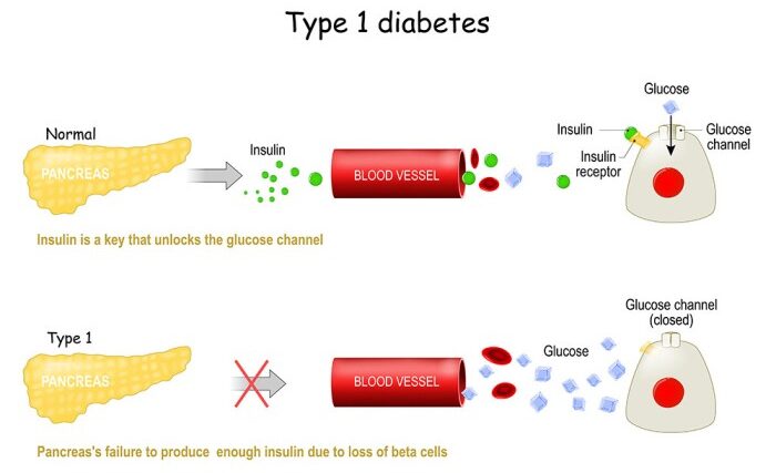
Formerly called insulin-dependent diabetes mellitus (IDDM) or "juvenile diabetes," T1DM is characterized by low or absent levels of endogenously produced insulin due to the targeted destruction of pancreatic beta cells.

Epidemiology
  • It is the most common endocrine disorder of childhood and adolescence.
  • Onset: Occurs predominantly in childhood, featuring a bimodal distribution with two peaks: one at 5-7 years of age (coinciding with increased exposure to infectious agents upon starting school), and another at puberty (coinciding with the physiological stress and counter-regulatory hormone surge of the pubertal growth spurt). However, it may present at any age.
  • Prevalence: Varies globally; for instance, in India, an average prevalence of Type I diabetes is estimated at 10 per 100,000 population.
Genetic Risk and Susceptibility

While most cases occur sporadically, there is a clear genetic component to the risk of developing Type 1 DM:

Relationship to Index Case Estimated Risk of Developing T1DM
If Mother has T1DM 2%
If Father has T1DM 7%
Sibling of index case 6%
Dizygotic (Fraternal) Twins 6% - 10%
Monozygotic (Identical) Twins 30% - 65%
Note: The fact that monozygotic twins only have a 30-65% concordance rate (rather than 100%) strongly indicates that while genetics provide the susceptibility, environmental triggers are absolutely necessary to initiate the autoimmune destruction.
Pathogenesis and Natural History

The natural history of Type 1 Diabetes involves an interplay between genetic susceptibility, environmental triggers, and immune dysregulation, progressing through distinct stages:

  1. Genetic Susceptibility: The baseline genetic predisposition.
  2. Exposure to Environmental "Triggers": Viral infections, dietary factors, or other unknown events that act as a catalyst.
  3. Initiation of Autoimmunity: The immune system begins to mistakenly target the beta cells in the Islets of Langerhans. Autoantibodies such as IAA (Insulin Autoantibodies), GADA (Glutamic Acid Decarboxylase Autoantibodies), and ICA (Islet Cell Autoantibodies) become positive.
  4. Preclinical Autoimmunity (Progressive Loss of β-cell function): As beta-cell mass declines over time, the body first loses its first-phase insulin response (the rapid burst of insulin immediately after eating). During this phase, glucose intolerance begins, but fasting blood sugars may remain normal.
  5. Onset of Clinical Disease (Overt Diabetes): Once approximately 80-90% of the beta-cell mass is destroyed, the remaining cells can no longer maintain normoglycemia, leading to clinical symptoms and the absence of C-peptide (a marker of endogenous insulin production).
  6. Transient Remission ("Honeymoon Period"): Shortly after initiating exogenous insulin therapy, the few remaining functional beta cells may temporarily recover and secrete insulin, drastically reducing exogenous insulin requirements. This period is transient and eventually followed by complete beta-cell failure.
  7. Established Disease: Absolute insulin deficiency requiring lifelong exogenous insulin.
  8. Development of Complications: Arising years later due to chronic microvascular and macrovascular damage.
Clinical Presentations & Diagnostic Criteria
Clinical Presentations
  • DKA (Diabetic Ketoacidosis): The most common initial presentation in pediatrics, presenting as a life-threatening medical emergency.
  • Classical Symptom Triad:
    • Polyuria: Excessive urination due to osmotic diuresis caused by glycosuria.
    • Polydipsia: Excessive thirst due to dehydration from polyuria.
    • Weight Loss: Despite normal or increased appetite (polyphagia), the body breaks down fat and muscle for energy due to the inability to utilize glucose.
  • Accidental Diagnosis: Detected incidentally during routine urinalysis (glycosuria) or blood tests for other illnesses.
Diagnostic Criteria

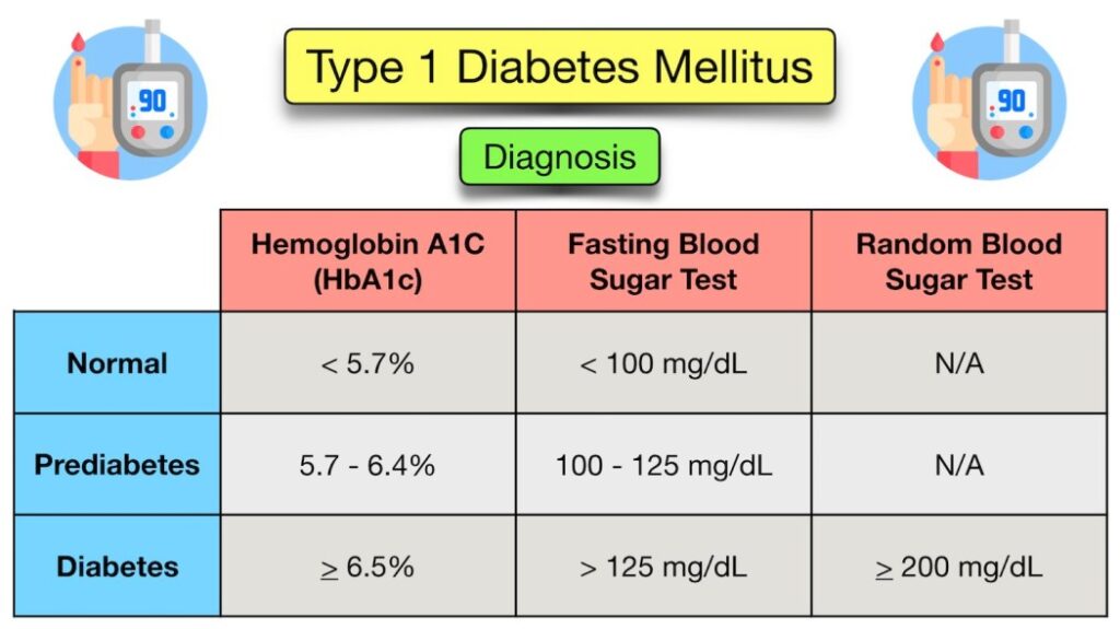
Diagnosis requires clear laboratory evidence of hyperglycemia.

  • Symptomatic Children: In children presenting with classic symptoms (polydipsia, polyuria, weight loss), a single Random Plasma Glucose > 11.1 mmol/L (200 mg/dL) is diagnostic.
  • Hemoglobin A1c: An HbA1c level of >= 6.5% is universally recognized as diagnostic criteria.
  • Asymptomatic or Equivocal Cases: A modified Oral Glucose Tolerance Test (OGTT using 1.75g/kg oral glucose, max 75g) may be needed. This is indicated for asymptomatic children with hyperglycemia (RBS >140) or symptomatic children with borderline hyperglycemia (RBS between 140 to 200).
Test Parameter IGT (Impaired Glucose Tolerance) / Pre-Diabetes Diabetic Threshold
Fasting Blood Glucose (FBG) 6.0 - 6.9 mmol/L (100 - 125 mg/dL) >= 7.0 mmol/L (126 mg/dL)
2 Hours post-OGTT 7.8 - 11.0 mmol/L (140 - 199 mg/dL) >= 11.1 mmol/L (200 mg/dL)
Clinical Pearl: Remember that acute infections in young, non-diabetic children can cause transient stress hyperglycemia without ketoacidosis. Always follow up to confirm a true diabetes diagnosis once the acute stress resolves.
Management and Treatment Elements

The successful management of T1DM is multifaceted and requires a dedicated team approach involving the physician, diabetes educator, dietitian, and the family.

Core Treatment Elements:
  1. Education
  2. Insulin therapy
  3. Glycemic control Monitoring
  4. Diet and meal planning
  5. Prevention and early detection of complications
1. Education

Educating the child and caregivers is the cornerstone of management. Key topics include:

  • Understanding the pathophysiology of Diabetes Type 1.
  • Acceptance of life-long insulin therapy and proper injection techniques.
  • Self-monitoring of blood glucose (SMBG) and maintaining accurate logs/records.
  • Recognition, prevention, and emergency management of Hypoglycemia & DKA.
  • Understanding the meal plan and carbohydrate counting.
  • Sick-day management protocols.
  • Awareness of possible long-term complications to encourage compliance.
2. Insulin Therapy

Insulin is a polypeptide made of two beta-chains, famously discovered by Banting & Best in 1921. Historically, animal types (porcine & bovine) were utilized before the introduction of highly purified human-like insulin via DNA-recombinant technology. Recently, more potent insulin analogs have been produced by changing specific amino acid sequences to alter absorption kinetics.

Insulin Type & Examples Onset Peak Time Duration
Rapid-acting Insulin:
Insulin lispro (Humalog), Insulin aspart (Novolog), Apidra
Begins to work at about 5 - 15 minutes. Peaks at about 1 - 2 hours. Continues to work for about 2 - 5 hours.
Regular or Short-acting Insulin:
Soluble Human Insulin (Actrapid, Humulin R)
Reaches the bloodstream within 30 minutes after injection. Peaks anywhere from 2 - 4 hours. Effective for approximately 3 - 8 hours.
Intermediate-acting Insulin:
Isophane Insulins: NPH (Insulatard), Lente
Reaches the blood stream about 2 to 4 hours after injection. Peaks 4 - 12 hours later. Effective for about 12 to 18 hours.
Long-acting Basal Analogues:
Insulin glargine (Lantus), Detemir (Levemir)
Reaches the bloodstream ~2 to 6 hours after injection. No pronounced peak ("peakless"). Usually effective for 18 - 24 hours. Provides a steady basal rate.
Pre-mixed Insulins:
Mixtard 30, Humulin M3, Novo Mix 30
Combines rapid/short acting with intermediate acting. E.g., Mixtard 30 means 30% Soluble (Regular) insulin and 70% Isophane (NPH). Kinetics depend on the mixture ratio.
Key Aspects of Insulin Therapy & Administration:
  • Aim: To perfectly mimic the natural, physiological pattern of insulin secretion (a steady basal rate to suppress hepatic glucose output, accompanied by meal-time boluses).
  • Concentrations: Insulin is available in 40, 80, & 100 Unit/ml concentrations. To prevent fatal dosage confusion, the WHO now strongly recommends U-100/ml as the only standard formulation. Special high-concentration preparations (U-500/ml) exist for specific profound insulin resistance scenarios.
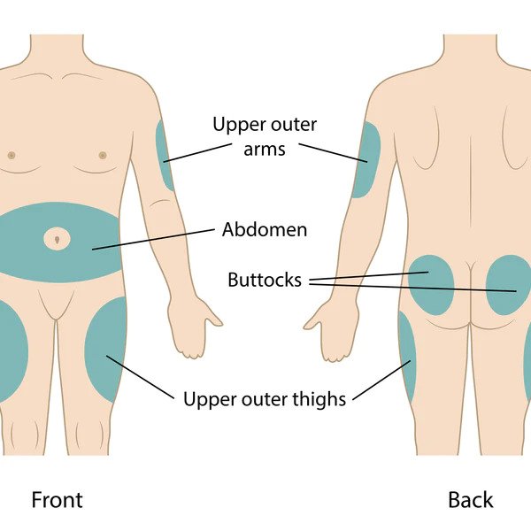
  • Administration Sites: Administered subcutaneously using syringes, pens, or pumps. Sites include:
    • Anterolateral thighs
    • Anterior and lateral abdominal wall
    • Posterior aspect of upper arms
    • Superolateral aspects of buttocks
Site Rotation: Following a regular pattern of using different sites and rotating within the same site is critically important to prevent lipohypertrophy (fatty lumps) or lipoatrophy (fatty depressions), which severely impair consistent insulin absorption.
Insulin Regimens and Dosing:

Initial Dose Estimation:

  • DKA / Overt symptoms: Total Daily Dose (TDD) = 0.8 to 1.0 unit/kg/day.
  • Incidentally diagnosed (lower starting doses required):
    • Toddlers & pre-school (2-5 yrs): 0.2 - 0.4 unit/kg/day
    • Pre-pubertal children (5-9 yrs): 0.5 - 0.8 unit/kg/day
    • Adolescents (9-14 yrs): 0.8 - 1.5 unit/kg/day (higher due to pubertal resistance).

Common Regimens:

  1. Split-Mix Regime (Twice Daily):
    • Typically utilizes Mixtard 30:70, OR a manual mix of NPH (2/3) + Regular insulin (1/3).
    • Given twice daily: 2/3 of the TDD given 45 mins Before Breakfast (BBF), and 1/3 of the TDD given 45 mins Before Dinner (BD).
    • Rule of thumb correction: 1 IU of Mixtard typically takes care of blood sugar ~50mg/dl above target.
  2. Basal-Bolus Regime (Multiple Daily Injections - MDI):
    • Provides superior physiological matching.
    • 30-50% of the TDD is given as one dose of long-acting basal insulin (Glargine, Detemir).
    • The remainder is divided into 3-4 doses of rapid-acting insulin given right before meals.
Advanced Dose Calculations (Bolus/Meal-Time):
  • Carbohydrate to Insulin Ratio (CIR): Indicates the amount of carbohydrate in grams covered by one unit of insulin.
    • Initial calculation rule: 500 / Total Daily Dose (TDD).
    • Accurate estimation: Based strictly on the amount of carbohydrate consumed, units administered, and pre/post-prandial glucose logging.
  • Insulin Sensitivity Factor (ISF): Represents the expected reduction in blood glucose by one single unit of rapid-acting insulin.
    • Initial calculation rule: 1800 / Total Daily Dose (TDD).
    • Correction Formula: For correcting pre-meal high sugars: (Actual BS - Target BS) / ISF = Correction Dose required.

Example Calculation

Patient: 10-year-old child, weighing 33 kg.
Total Insulin Requirement (TDD): 0.8 IU/kg * 33 kg = ~26 IU/day.
CIR (Carb Ratio): 500 / 26 = ~20 grams per unit.
ISF (Correction Factor): 1800 / 26 = ~70 mg/dL per unit.
Basal Regimen: Inj Glargine 40% of TDD = ~11 IU daily.

Scenario: If the child is taking 80g of carbohydrates for lunch, the base meal insulin needed is 80 / 20 (CIR) = 4 IU.
If their pre-lunch blood sugar is elevated at 200 mg/dL (Target is 130), the correction dose is (200 - 130) / 70 (ISF) = 1 IU.
Total Meal Dose: 4 IU (for carbs) + 1 IU (for correction) = 5 IU of Rapid (Lispro) insulin given before lunch.

Insulin Pump Therapy (CSII)

Continuous Subcutaneous Insulin Infusion (CSII) via battery-powered pumps provides a closer approximation of normal physiological plasma insulin profiles. It accurately delivers a small baseline continuous infusion of rapid-acting insulin (basal rate), coupled with customizable parameters for bolus therapy. The bolus insulin delivery is precisely determined by the amount of carbohydrate intake programmed by the user, cross-referenced with their real-time blood sugar level.

3. Monitoring of Glycemic Control

Consistent monitoring ensures therapy is effective and mitigates complications.

  • Self Monitoring of Blood Glucose (SMBG): Routine testing should occur fasting, before meals, 2 hours after meals, and during the night.
  • Real-time Continuous Glucose Monitoring (CGM): Subcutaneous sensors providing minute-by-minute trending.
  • Glycosylated Hemoglobin (HbA1C): Provides an average reflection of glycemic control over the preceding 2-3 months. Must be tested every 3-4 months.
  • Measuring Ketones in Urine: More sensitive and accurate for detecting early deterioration. Must be checked when Blood Glucose > 250 mg/dl, or during illness with fever, vomiting, abdominal pain, polyurea, drowsiness, or rapid breathing.
  • Urinary Glucose: A very crude indicator of hyperglycemia. It only reflects the glycemic level over the preceding several hours and is only positive if the renal threshold (usually ~180 mg/dL) is exceeded.
Suggested Target Blood Glucose Range
Time of Checking Target Plasma Glucose (mg/dL)
Fasting or preprandial 90 - 145
Postprandial 90 - 180
Bedtime 120 - 180
Nocturnal 80 - 162
Note for young children: For children < 5 years of age, slightly more relaxed targets are optimal to prevent severe neurological deficits from unrecognized hypoglycemia: 90-200 mg/dL during the day, and between 150-200 mg/dL at bedtime/night.
Adverse Effects & Practical Problems of Insulin Therapy
  • Adverse Effects: Hypoglycemia (most dangerous acute risk), Lipoatrophy (loss of subcutaneous fat at injection site), Lipohypertrophy (fat accumulation at injection site), Obesity/Weight gain (due to insulin's anabolic nature), Insulin allergy, and development of Insulin antibodies.
  • Practical Problems: Non-availability of insulin in poorer nations, poor injection techniques/site rotation, difficulties with insulin storage (cold chain) & transfer, errors in mixing insulin preparations, managing insulin scheduling during school hours, difficulties in adjusting the dose at home, complex sick-day management, and failure to recognize or treat hypoglycemia accurately.
Diet, Exercise, and Sick-Day Management
Diet Regulation
  • Regular meal plans utilizing calorie exchange options are highly encouraged.
  • Macronutrients: 50-60% of required energy should be obtained from complex carbohydrates. A diet low in salt, low in saturated fats, and high in fiber is heavily encouraged.
  • Distribution: Distribute the carbohydrate load evenly during the day, preferably structured as 3 main meals & 2 snacks, with absolute avoidance of simple sugars.
    • Split-Mix Regime: Requires strict meal timing. 6 meals daily: 3 major meals (70% of total calories) and 3 mid-meals/snacks (30% of total calories).
    • MDI Regime: Offers more flexibility. Mid-meals are not essential as the basal covers fasting periods. If taken, mid-meals should have less than 10-15 gm of carbohydrate.
  • Glycemic Index (GI): A ranking of carbohydrates on a scale from 0 to 100 according to how rapidly they raise blood sugar levels.
    • High GI: Rapidly digested, causing marked blood sugar spikes (e.g., corn flakes, potato, watermelon, biscuits, chocolates). Should be avoided.
    • Low GI: Slow digestion and absorption, producing gradual rises in blood sugar and insulin levels. Proven benefits for diabetics (e.g., most fruits, non-starchy vegetables, pasta, pulses, milk, curd).
Exercise

Exercise is a fundamental pillar of therapy. It profoundly decreases insulin requirements by increasing both the sensitivity of muscle cells to insulin and the direct, non-insulin-mediated utilization of glucose by skeletal muscle. However, because the body cannot shut off injected insulin, exercise can easily precipitate severe hypoglycemia in an unprepared diabetic patient if carbohydrates are not supplemented prior to activity.

Sick-Day Management

Managing diabetes during acute illnesses (colds, flu, gastroenteritis) is critically challenging. Insulin requirements may unexpectedly increase or decrease.

  • Stress Physiology: Fever, dehydration, and the physiological stress of illness cause hyperglycemia due to increased production of counter-regulatory hormones (cortisol, glucagon, epinephrine). Conversely, vomiting and loss of appetite can lead directly to hypoglycemia.
  • Ketosis Risk: The risk of ketosis is exponentially increased due to the combination of starvation (not eating) and dehydration.
  • Action Plan:
    • Take plenty of fluids to prevent dehydration.
    • Blood glucose and urine ketones must be monitored frequently (every 2-4 hours).
    • The presence of "moderate" or "large" ketones in the urine alongside hyperglycemia is a dangerous red flag, indicating severe insulin deficiency and high risk for impending DKA.
    • The child should be given additional correction doses of rapid-acting or regular insulin (e.g., 0.1 u/kg if RBS > 250), alongside oral fluids. Ketones must be rechecked in the next urine output.
    • Red Flag: If there is persistent vomiting with hyperglycemia and large ketones, or uncontrollable persistent hypoglycemia, the child must be taken to the emergency department immediately.
Complications and Pitfalls of Management
Pitfalls of Glycemic Management
  • Delayed diagnosis resulting in presentation at the DKA stage.
  • Misinterpreting the "Honeymoon period" as a cure.
  • Failure to properly diagnose and treat DKA and hypoglycemia.
  • Dawn Phenomenon vs. Somogyi Phenomenon: These conditions both result in elevated morning glucose but require opposite treatments.
    • Dawn Phenomenon: Blood glucose levels steadily increase in the early morning hours before breakfast. This is a normal physiologic process caused by the overnight surge of growth hormone secretion and increased clearance of insulin. While non-diabetics simply secrete more insulin to compensate, a T1DM child cannot, resulting in morning hyperglycemia. Treatment: Increase the evening basal insulin.
    • Somogyi Phenomenon: A theoretical rebound hyperglycemia. It occurs when a patient experiences undetected late-night or early morning hypoglycemia (e.g., from too much evening insulin). The body panics and mounts an exaggerated counter-regulatory hormone response (glucagon, epinephrine), heavily dumping glucose into the blood, leading to ambiguously elevated morning glucose levels. Treatment: Decrease the evening basal insulin or provide a bedtime snack. Continuous glucose monitoring or 3:00 AM fingersticks clarify which phenomenon is occurring.
Acute Complications
  1. Hypoglycemia: Defined as blood glucose < 70 mg/dL. The risk increases as the duration of diabetes increases.
    • Mild to moderate: Immediate oral intake of 0.3 g/kg of fast-acting glucose dissolved in a small amount of water (raises blood glucose by 45-65 mg/dL). Retest after 10-15 minutes and readminister if the response is inadequate. Note: Chocolate, milk, and fat-containing sweets are poor choices for emergency rescue as fat significantly delays the gastric emptying and absorption of glucose. Avoid treating with the child's favorite candies to prevent behavioral reinforcement.
    • Severe (Altered mental status, unconsciousness, or seizures): Glucagon is administered subcutaneously or intramuscularly. DOSE: 0.5 mg for < 12 years; 1.0 mg for > 12 years. If glucagon injections are unavailable, glucose gel or honey can be carefully rubbed into the buccal pouch.
  2. Diabetic Ketoacidosis (DKA): A life-threatening complication occurring more commonly in children with Type 1 DM than Type 2 DM.
    • Definition: Hyperglycemia (serum glucose > 200-300 mg/dL) combined with severe metabolic acidosis (blood pH < 7.3, serum bicarbonate < 15 mEq/L) and prominent ketonemia/ketonuria.
    • Signs & Symptoms: Nausea, vomiting, severe abdominal pain, fruity (acetone) odor on breath, tachycardia, low volume pulses, hypotension, impaired skin turgor, delayed capillary refill, profound dehydration, and rapid, deep sighing respirations known as Kussmaul respirations (a respiratory compensation for the metabolic acidosis).
    Classification of Diabetic Ketoacidosis
    Parameter Normal MILD DKA MODERATE DKA SEVERE DKA
    Venous CO2 (mEq/L) 20 - 28 16 - 20 10 - 15 < 10
    Venous pH 7.35 - 7.45 7.25 - 7.35 7.15 - 7.25 < 7.15
    Clinical State No change Oriented, alert but fatigued Kussmaul respirations; oriented but sleepy; arousable Kussmaul or depressed respirations; sleepy to depressed sensorium leading to coma
    DKA Treatment Protocol:
    • 1st Hour: Quick volume expansion. 10-20 ml/kg IV bolus of 0.9% NaCl (Normal Saline) or Lactated Ringer's. May be repeated. Keep patient NPO. Monitor I/O and neurological status closely. Have Mannitol at the bedside (1 g/kg IV push) ready for cerebral edema. Initiate continuous Insulin drip at 0.05 to 0.10 units/kg/hr.
    • 2nd Hour until DKA resolution: Change fluids to 0.45% NaCl. Continue Insulin drip. Crucially, when blood sugar drops below 250 mg/dL, add 5% glucose to the IV fluid to prevent hypoglycemia while insulin continues to clear ketones. Add 20 mEq/L Potassium Phosphate (If K < 3 mEq/L, give 0.5 to 1 mEq/kg as oral K or increase IV K to 80 mEq/L). Total IV rate should equal roughly 85 ml/kg + maintenance - bolus divided by 23hr.
    • Transition to Subcutaneous: Occurs after correction of dehydration and acidosis (No emesis, CO2 > 16, normal electrolytes). As oral feeds advance, IV fluids are reduced. The ideal time to transition is just before a meal. Rapid-acting insulin is given 15-30 mins prior, and regular insulin 1-2 hours prior to stopping the IV infusion to prevent rebound hyperglycemia.
  3. Cerebral Edema: The most dreaded, fatal complication of DKA treatment, often caused by rapid fluid shifts.
    • Management: Head end elevation. Give Mannitol 0.5-1 gm/kg immediately and repeat if no response in 30 mins to 2 hrs. 3% Hypertonic saline (5 ml/kg over 30 mins) can alternatively be given. Restrict IV fluids to 2/3 maintenance. Replace the remaining fluid deficit slowly over 72 hours rather than 24 hours. Intubation and hyperventilation may be required.
  4. Non-Ketotic Hyperosmolar Coma (NKHS): Uncommon in children, but exceptionally dangerous.
    • Features: Severe, massive hyperglycemia (blood glucose > 800 mg/dL), absence of (or only slight) ketosis, nonketotic acidosis, profound, severe dehydration, depressed sensorium or frank coma, and variable neurological signs (grand mal seizures, hyperthermia, hemiparesis, positive Babinski signs). Respirations are shallow. Serum osmolarity > 350 mOsm/kg.
    • Treatment: Requires rapid repletion of the vascular volume deficit, but very slow correction of the hyperosmolar state to avoid cerebral edema. 0.45% NaCl is administered (replacing 50% deficit in 1st 12hr, remainder over next 24hr). When glucose approaches 300 mg/dL, change to 5% dextrose in 0.2 NS. Insulin drip is lower (0.05 units/kg/hr) and delayed until the 2nd hour of fluid therapy. Aggressive potassium replacement (20 mEq/L) is required.
Late-Onset Complications & Monitoring

Chronic hyperglycemia damages blood vessels over decades, leading to microvascular and macrovascular disease.

  • Retinopathy: Damage to the retina leading to blindness. Screening: Fundal photography. Begin 5 years post-diagnosis in prepubertal children, and 2 years post-diagnosis in pubertal children. Frequency: 1-2 yearly.
  • Nephropathy: Kidney damage progressing to renal failure. Screening: Spot urine sample for albumin:creatinine ratio (detecting microalbuminuria). Timing: Same as retinopathy. Frequency: Annually.
  • Neuropathy: Nerve damage causing numbness, tingling, and pain. Screening: Physical examination (monofilament test). Screening timelines are less clear in children, but typically assessed 5 years after T1DM diagnosis.
  • Macrovascular Disease: Ischemic heart disease and stroke. Screening: Fasting lipid profile. Starts after age 2. Frequency: Prepubertal every 5 years; Pubertal within 6-12 months of diagnosis, then every 2 years.
  • Autoimmune Co-morbidities: T1DM patients frequently develop other autoimmune diseases.
    • Thyroid Disease: Screen via TSH at diagnosis, then every 2-3 years.
    • Celiac Disease: Screen via tissue transglutaminase (tTG) and endomysial antibodies at diagnosis, then every 2-3 years.
Recent Advances and Prevention Strategies
Recent Treatment Advances:
  • Pancreas & Islet Cell Transplantation: Whole pancreas transplants are typically reserved for diabetics suffering from end-stage renal disease who are already receiving kidney transplants (and thus already on immunosuppressants). Islet cell transplants (the ultimate theoretical treatment) are under trials globally with encouraging results, but graft rejection and the recurrence of the underlying autoimmunity are serious, persistent limitations.
  • Immune Modulation: Utilizing immunosuppressive therapy for newly diagnosed patients or high-risk children to prolong the "honeymoon phase" and preserve remaining beta cells. Drugs investigated include Nicotinamide and Mycophenolate.
  • Gene Therapy: Cutting-edge research attempting to block the immunologic attack against islet cells using DNA-plasmids encoding self-antigens, genes encoding cytokine inhibitors, or modifying gene-expressed islet-cell antigens like GAD.
Prediction and Prevention:

Prediction: Evaluating high-risk individuals utilizing sensitive and specific immunologic markers such as GAD Antibodies, GLIMA antibodies, and IA-2 antibodies. Having a single antibody presents a 2-6% risk of developing T1DM, two antibodies increase risk to 21-40%, and carrying more than two antibodies creates a 59-80% definitive risk.

Prevention Tiers:

  • Primary Prevention: Intervening before any disease process starts. Includes identification of the diabetes gene, theoretical tampering/re-education of the immune system, and elimination of triggering environmental factors.
  • Secondary Prevention: Intervening after antibodies appear but before overt clinical diabetes. Utilizes immunosuppressive therapies (e.g., cyclosporin) and GLP-1 agonists to delay onset.
  • Tertiary Prevention: Focuses on established disease. It demands tight metabolic control and excellent monitoring to prevent the devastating late-onset complications.
Nursing diagnosis for diabetes — 5 sample care plans
Care Plan #1 — Risk for Unstable Blood Glucose Level

This is one of the most frequently used nursing diagnoses for diabetes on the floor, especially for newly diagnosed patients or anyone whose regimen has just changed.

Scenario. A 52-year-old patient is newly diagnosed with type 2 diabetes during a hospital admission for cellulitis. A1C 9.4%. Fingerstick on admission 268 mg/dL. The patient says they haven’t checked sugars at home and don’t know how to use the meter the provider sent home with them.

Diagnosis (NANDA-I 2024–2026)

Risk for Unstable Blood Glucose Level related to new T2DM diagnosis, insufficient diabetes self-management knowledge, and acute illness.

(Risk-for diagnoses use “related to” only — no “as evidenced by.”)

Goals
  • By the end of the shift, the patient verbalizes the steps for checking a fingerstick glucose and demonstrates the technique with the bedside meter.
  • Pre-discharge, the patient identifies their personalized A1C and glucose targets and the three earliest signs of hypoglycemia.
  • Pre-discharge, the patient verbalizes when to call the provider (glucose < 70 or > 300 with symptoms, illness, ketones).
Interventions and rationales
No. Intervention Rationale
1 Monitor fingerstick glucose per facility schedule (typically AC and HS); document trends. Frequent monitoring catches both hyper- and hypoglycemia before clinical decompensation.
2 Administer insulin and oral agents as prescribed; verify dose, type, and timing using the Five Rights and double-check per policy. Insulin is a high-alert medication. Dosing errors are a leading source of inpatient adverse events.
3 Provide a structured 1:1 teach-back session covering meter use, target range, insulin pen technique, and the 15-15 rule. Active demonstration with teach-back is more durable than verbal instruction alone.
4 Coordinate meal trays with mealtime insulin within the facility’s recommended window (often 15 minutes pre / post tray delivery). Mismatched timing increases hypo- and hyperglycemia risk.
5 Refer to inpatient diabetes educator / DSMES program before discharge. Structured DSMES is associated with improved A1C, self-management behaviors, and care engagement.
6 Coordinate post-discharge primary care / endocrinology follow-up within 1–2 weeks. Early follow-up improves medication titration and reduces readmission.
Evaluation
  • Did the patient demonstrate accurate self-monitoring before discharge?
  • Did glucose stay within the individualized target range during hospitalization?
  • Did the patient verbalize hypoglycemia symptoms and rescue steps?
Care Plan #2 — Deficient Knowledge: Diabetes Self-Management

Scenario. A 67-year-old patient with T2DM is being discharged on basal-bolus insulin for the first time after years of metformin alone. They tell you, “I don’t really get when to take the long-acting versus the fast-acting. And I’m scared of needles.” Spouse expresses willingness to help but also has limited knowledge.

Diagnosis (NANDA-I 2024–2026)

Deficient Knowledge: Diabetes Self-Management related to new medication regimen, low prior exposure, and expressed fear of injection, as evidenced by patient stating, “I don’t get when to take which one” and reluctance to handle insulin pen during demonstration.

Goals
  • By the end of teaching session, the patient demonstrates correct insulin pen technique using a saline trainer.
  • Pre-discharge, the patient verbalizes the difference between basal and bolus insulin and when to use each.
  • Pre-discharge, the patient and the spouse together demonstrate fingerstick technique and verbalize what to do for a reading < 70 mg/dL.
Interventions and rationales
No. Intervention Rationale
1 Assess current knowledge, literacy level, cultural / language preferences, and learning style before teaching. Tailored education improves retention and reduces non-adherence.
2 Use teach-back: explain ➔ patient repeats ➔ correct gaps ➔ re-demonstrate. Teach-back is an AHRQ-recommended technique linked to better outcomes in low-health-literacy populations.
3 Provide written materials at a 5th–6th grade reading level with visuals. Avoid medical jargon. Roughly half of U.S. adults have low health literacy; visuals improve comprehension.
4 Demonstrate insulin pen priming, dial-up, injection sites (rotation), and disposal. Use saline pen first if needle anxiety is present. Building competence reduces fear and increases adherence.
5 Include the spouse / caregiver in every teaching encounter. Shared knowledge reduces medication errors at home.
6 Connect to outpatient DSMES program; provide written instructions for follow-up appointment. DSMES is associated with improved A1C and self-management behaviors.
7 Screen for diabetes distress with a validated tool (e.g., Diabetes Distress Scale). High distress predicts non-adherence and depression.
Evaluation
  • Did the patient and spouse demonstrate competence with insulin pen and meter?
  • Did the patient verbalize basal vs. bolus differences in their own words?
  • Was the DSMES referral placed and confirmed before discharge?
Care Plan #3 — Risk for Infection

Scenario. A 58-year-old patient with T2DM is post-op day 2 from a colectomy. Glucose has been running 220–280 mg/dL on sliding scale. WBC trending up from 11 to 14. Surgical site is dry and intact, but the IV site shows mild erythema.

Diagnosis (NANDA-I 2024–2026)

Risk for Infection related to hyperglycemia-impaired immune function, surgical wound, and indwelling vascular and urinary devices.

Goals
  • The patient maintains glucose within the inpatient target range. Per ADA 2026: for most noncritically ill hospitalized patients, 100–180 mg/dL when achievable without significant hypoglycemia; for most critically ill / ICU patients, 140–180 mg/dL. Initiate or intensify therapy for persistent hyperglycemia ≥ 180 mg/dL confirmed on two occasions in 24 hours.
  • The patient is free from signs of new infection (no fever, no purulent drainage, no rising WBC) by hospital day 5.
  • The patient verbalizes three signs of infection to report after discharge.
Interventions and rationales
No. Intervention Rationale
1 Monitor temperature, HR, BP, and WBC each shift; escalate per sepsis screening criteria. People with diabetes have a higher risk of severe sepsis and can present atypically (blunted fever response).
2 Target glucose 100–180 mg/dL for most noncritically ill inpatients when achievable safely; 140–180 mg/dL for critically ill / ICU patients. Notify the provider for persistent values ≥ 180 mg/dL confirmed on two occasions in 24 hours and anticipate regimen intensification — basal +/- prandial + correction insulin. Sliding-scale-only is discouraged for sustained hyperglycemia. Hyperglycemia impairs neutrophil function and wound healing; tighter targets without survival benefit increase hypoglycemia risk.
3 Assess surgical site, IV / central line, and Foley insertion sites every shift. Remove lines as soon as clinically appropriate. Each indwelling device is a portal of entry.
4 Use sterile technique for dressing changes; perform hand hygiene before and after every patient contact. Standard infection-prevention bundle.
5 Encourage incentive spirometry, early mobility, and adequate nutrition / protein intake. Reduces post-op pulmonary infection and supports wound healing.
6 Educate the patient on signs of incision infection — redness, warmth, swelling, drainage, fever, or new pain — and when to call. Empowers early outpatient detection.
Evaluation
  • Were glucose values within the inpatient target range during the shift?
  • No new signs of infection at any monitored site?
  • Did the patient teach back three signs of infection to report after discharge?
Care Plan #4 — Risk for Impaired Skin Integrity (Diabetic Foot)

Scenario. A 71-year-old patient with T2DM x 15 years presents to clinic with bilateral plantar calluses. Reports occasional “numbness like a sock is on.” Monofilament screening: cannot feel the 10-g filament at 6 of 10 plantar sites bilaterally. Pulses palpable but diminished. No active ulcer today.

Diagnosis (NANDA-I 2024–2026)

Risk for Impaired Skin Integrity related to peripheral sensory neuropathy, diminished protective sensation, and chronic hyperglycemia.

(Note: if an actual ulcer or breakdown is present, switch to Impaired Skin Integrity or Impaired Tissue Integrity depending on depth — Wagner grade ≥ 2 typically requires the tissue-integrity label.)

Goals
  • The patient verbalizes a daily foot-inspection routine and demonstrates use of a mirror to view plantar surfaces by the end of the visit.
  • The patient remains free of new foot ulcers, blisters, or calluses requiring intervention at the 3-month follow-up.
  • The patient identifies three specific situations requiring same-day call to the provider (any open lesion, redness, drainage, or sudden change in sensation).
Interventions and rationales
No. Intervention Rationale
1 At routine diabetes visits, remove shoes and socks for visual inspection (skin, nails, deformities, footwear). Perform comprehensive foot exam (visual + 10-g monofilament + 128-Hz tuning fork + pulses + ABI if indicated) at least annually, and more often for high-risk feet. ADA recommends at-least-annual comprehensive foot screening; high-risk patients (neuropathy, prior ulcer, deformity, PAD, loss of protective sensation) need more frequent surveillance.
2 Teach daily self-inspection: top, bottom, between toes; use a mirror or family member if the patient cannot see plantar surfaces. Catches early breakdown before progression.
3 Reinforce footwear teaching: well-fitting, closed-toe shoes; never barefoot; no hot water for soaks; check shoes for foreign objects before wearing. Protective sensation loss means the patient may not feel a pebble, blister, or burn.
4 Refer to podiatry for high-risk feet, deformities, callus debridement, or therapeutic footwear. Specialist care reduces amputation risk.
5 Coordinate glycemic management referral if A1C is uncontrolled. Improved glycemic control slows neuropathy progression.
6 Document the patient’s monofilament map and update at each visit. Allows trending of sensory loss over time.
Evaluation
  • Did the patient demonstrate daily inspection technique?
  • Are the feet free of new lesions, calluses, or open wounds at follow-up?
  • Was a podiatry / vascular referral placed when indicated?
Care Plan #5 — Overweight

Scenario. A 44-year-old patient with T2DM, BMI 34, sedentary office job. A1C 8.2%. Reports that meals are usually fast food on the road; “I know I should eat better but I don’t have time.” Spouse cooks dinner. No prior dietitian referral.

Diagnosis (NANDA-I 2024–2026)

Overweight related to energy intake exceeding energy expenditure, sedentary lifestyle, and reliance on calorie-dense convenience foods, as evidenced by BMI 34, A1C 8.2%, and patient-reported fast-food meal pattern.

(NANDA-I uses Overweight and Obesity as distinct diagnoses by BMI category. Verify the precise label, definition, and defining characteristics against your licensed NANDA-I 2024–2026 text. If your facility’s text uses an alternate label such as “Imbalanced Nutrition” or a health-management diagnosis, swap accordingly.)

Goals
  • The patient identifies two realistic dietary changes they’re willing to try this week (e.g., swap one fast-food meal for a packed lunch, switch sugary drinks to water or unsweetened).
  • The patient verbalizes one weekly activity goal aligned with the ADA recommendation of ≥ 150 minutes / week of moderate activity.
  • A1C decreases by ≥ 0.5% at the 3-month follow-up.
Interventions and rationales
No. Intervention Rationale
1 Refer to a registered dietitian nutritionist for individualized medical nutrition therapy. MNT can meaningfully improve glycemic outcomes and should be individualized by an RDN. Medicare covers MNT for diabetes.
2 Use motivational interviewing — explore what the patient values and is willing to change. Avoid prescriptive advice. Patient-led goal-setting outperforms top-down instruction in sustained behavior change.
3 Teach plate method or carb-counting basics, whichever fits the patient’s literacy and preference. Both approaches are ADA-endorsed and adaptable.
4 Discuss medication options for weight management when lifestyle alone is insufficient (GLP-1 RA, GLP-1 / GIP, SGLT2 inhibitor — provider-led). Newer agents offer cardio-renal protection plus weight reduction.
5 Encourage gradual activity — start with 10-minute walks if the patient is sedentary. Small consistent increases are more sustainable than large abrupt changes.
6 Screen for emotional eating, food insecurity, and barriers (work schedule, neighborhood food access). Social determinants drive nutrition behavior; an MNT plan that ignores them won’t stick.
Evaluation
  • Did the patient identify and attempt the dietary changes?
  • Is the patient meeting the activity goal at follow-up?
  • Has A1C decreased at the 3-month visit?
Hypoglycemia rescue (15-15 rule)
Conscious patient, able to swallow safely:
  1. Give 15 grams of fast-acting carbohydrate — examples: 4 oz juice, 4 oz regular soda, 3–4 glucose tablets, 1 tablespoon honey, 1 tube glucose gel.
  2. Wait 15 minutes and recheck blood glucose.
  3. If still < 70 mg/dL, repeat steps 1–2.
  4. Once back in range, give a complex carb + protein snack if the next meal is more than an hour away.
Severe hypoglycemia (unable to swallow safely or altered mental status):
  • Do not give oral glucose — aspiration risk.
  • Administer glucagon 1 mg IM / SC (Glucagon Emergency Kit) or 3 mg intranasal (Baqsimi) per facility protocol or family / school training.
  • In the hospital: D50W 25 g IV per provider order.
  • Place patient in recovery position, monitor airway, and call rapid response / 911 if outside hospital.

Special note for alpha-glucosidase inhibitors: Patients on acarbose or miglitol must rescue with glucose (dextrose) specifically — sucrose absorption is delayed.

Sick day rules

When a person with diabetes is sick (cold, flu, GI illness, infection), glucose can swing dramatically. Teach:

  • Do not stop insulin — even if not eating. Basal insulin almost always continues. Adjust per provider sick-day plan.
  • Check glucose every 3–4 hours (or per provider).
  • Check ketones (urine or blood) if T1DM and glucose > 250 mg/dL.
  • Hydrate — sip clear fluids; if unable to eat, take regular (non-diet) clear fluids in small amounts to provide some carbohydrate.
  • Know when to call: persistent vomiting, glucose > 300, ketones moderate or large, signs of DKA (Kussmaul respirations, fruity breath, abdominal pain), altered mental status.
  • If the patient takes an SGLT2 inhibitor (Jardiance, Farxiga, Invokana, etc.): follow the provider’s sick-day plan for temporary hold during acute illness, dehydration, or poor oral intake — and before scheduled procedures. Watch for euglycemic DKA: nausea, vomiting, abdominal pain, rapid breathing, or general malaise warrant evaluation even if the glucose isn’t very high. ADA convention is to stop most SGLT2 inhibitors ~3 days before scheduled surgery.

Every patient with diabetes should have a written sick-day plan personalized by their provider.

Diabetic foot care checklist
Daily — by the patient:
  • Wash with warm (not hot) water; pat dry, especially between toes
  • Inspect tops, bottoms, sides, and between toes (mirror or helper if needed)
  • Moisturize tops and bottoms (not between toes — fungal risk)
  • Wear clean, dry socks
  • Inspect shoes for foreign objects, rough seams, or wear before putting them on
  • Never go barefoot, not even at home
Periodically:
  • Trim toenails straight across; file rough edges
  • Have a podiatrist trim if vision, dexterity, or sensation are limited
  • Replace shoes when they show wear
At routine diabetes visits:
  • Remove shoes and socks
  • Visually inspect skin, nails, deformities, footwear, and pulses as clinically indicated
At least annually (and more often for high-risk feet — neuropathy, PAD, deformity, prior ulcer, loss of protective sensation):
  • Comprehensive foot exam with 10-g monofilament, 128-Hz tuning fork (vibration), and full vascular and neurologic assessment
  • Refer to podiatry for: prior ulcer, deformity (hammer toes, Charcot foot), peripheral artery disease, loss of protective sensation on monofilament screening, or any active foot lesion.

Quick Quiz

DM Type 1 Quiz

Medical Nursing - mobile-friendly and focused practice.

Privacy: Your details are used only for quiz tracking and certificates.

Diabetes Mellitus Type 1 Read More »

Hyperparathyroidism and Hypoparathyroidism

Hyperparathyroidism and Hypoparathyroidism

Hyperparathyroidism and Hypoparathyroidism

The parathyroid glands are critical regulators of calcium, phosphate, and bone metabolism. Pathologies affecting these glands typically manifest as disorders of calcium homeostasis, presenting either as hypercalcemia (hyperparathyroidism) or hypocalcemia (hypoparathyroidism).

1. ANATOMY AND PHYSIOLOGY OF PARATHYROID GLANDS
Embryology and Anatomy
  • Definition: Small endocrine glands located in the neck, usually situated behind the thyroid gland, responsible for producing parathyroid hormone (PTH).
  • Embryology: They develop early in gestation at approximately 6 weeks and migrate caudally by 8 weeks. The inferior glands derive from the 3rd pharyngeal pouch, while the superior glands derive from the 4th pharyngeal pouch.
  • Number: There are typically 4 parathyroid glands (two superior and two inferior).
  • Ectopic Locations: Because of their embryological migration, glands can sometimes be found in unusual locations.
    • Paraesophageal (28%)
    • Mediastinum (26%)
    • Intrathymic (24%)
    • Intrathyroidal (11%) - located entirely within the thyroid gland capsule.
    • Carotid sheath (9%)
    • High cervical (2%)
Historical Milestones
  • 1849: Sir Richard Owen provided the 1st accurate description of normal parathyroid glands after examining an Indian Rhinoceros.
  • 1879: Anton Wölfler described tetany in a patient after a total thyroidectomy, hinting at the glands' function.
  • 1880: Ivar Sandström, a Swedish medical student, grossly and microscopically described the parathyroid glands in humans.
  • 1909: Calcium measurement became possible, and its firm association with the parathyroids was established.
  • 1925: The 1st successful parathyroidectomy was performed on a 38-year-old man suffering from severe bone pain secondary to osteitis fibrosa cystica.
2. FUNCTIONS OF PARATHYROID HORMONE (PTH)
Synthesis and Metabolism of PTH

PTH is synthesized in the chief cells of the parathyroid gland as a larger precursor molecule. It undergoes sequential cleavage:

  1. Preproparathyroid hormone (the initial synthesized form).
  2. Cleaved into proparathyroid hormone.
  3. Cleaved finally into the active 84-amino-acid PTH.

Secreted PTH has an extremely short half-life of 2 to 4 minutes. In the liver, PTH is metabolized into an active N-terminal component and a relatively inactive C-terminal fraction.

Clinical Note: The short half-life of PTH is highly advantageous during parathyroidectomy surgery. Surgeons can measure intraoperative PTH levels; a drop of >50% within 10 minutes of removing a suspicious gland confirms the successful removal of the hypersecreting adenoma.
Primary Actions of Parathyroid Hormone

The overarching goal of PTH is to increase serum calcium levels and decrease serum phosphate levels. It achieves this via three main target organs:

  • Bone: Activates and increases the number of osteoclasts, which mobilizes (resorbs) calcium and phosphate from bone tissue into the blood.
  • Kidneys:
    • Increases renal tubular reabsorption of calcium (preventing its loss in urine).
    • Increases urinary phosphate excretion (phosphaturic effect), which prevents calcium-phosphate precipitation and effectively raises free ionized calcium.
    • Increases the conversion of Vitamin D to its most active form, 1,25-dihydroxyvitamin D3 (Calcitriol).
  • Gastrointestinal Tract: Indirectly increases GI calcium absorption via the actions of the newly activated Vitamin D.
PTH and Bone Physiology: The Osteoblast Paradox

The interaction between PTH and bone cells is complex and indirect:

  • PTH stimulates the osteocytic pump, increasing the permeability of the osteocytic membrane, allowing calcium to diffuse rapidly from bone fluid into the blood.
  • Receptor Specificity: Osteoblasts and osteocytes have PTH receptors. Osteoclasts do not have PTH receptors.
  • Signaling Mechanism: PTH stimulates osteoblasts/osteocytes, which then activate osteoclasts via a complex "signaling" system (physiologically known as the RANK/RANKL pathway). Therefore, PTH indirectly stimulates the formation of new osteoclasts.
  • Net Effect: While both osteoblastic (building) and osteoclastic (destroying) cell lines are activated by PTH, the clastic activity > blastic activity, resulting in net bone resorption.
Calcitonin: The Physiological Antagonist

Calcitonin is a 32-amino-acid-long peptide produced by the parafollicular cells (C cells) of the thyroid interstitium. It acts as the physiological antagonist to PTH.

  • Action: Temporarily lowers serum calcium levels.
  • Mechanism: Decreases osteoclastic bone resorption activity and stimulates a distal tubular-mediated calciuresis (calcium excretion in the kidney).
  • Stimulus: Secretion is directly stimulated by high blood calcium levels.
Etiology of Hypercalcemia (The Differential Diagnosis)

Before assuming hyperparathyroidism, it is critical to understand that approximately 80% of all hypercalcemia cases are caused by either Malignancy or Primary Hyperparathyroidism. The mnemonic VITAMINS TRAP helps recall the various causes:

Letter Etiology Letter Etiology
V Vitamins (A and D intoxication) T Thiazide diuretics, other drugs (Lithium)
I Immobilization (prolonged bed rest) R Rhabdomyolysis
T Thyrotoxicosis (Hyperthyroidism) A AIDS
A Addison's disease (Adrenal insufficiency) P Paget's disease, Parenteral nutrition, Pheochromocytoma, Parathyroid disease
M Milk-alkali syndrome
I Inflammatory disorders
N Neoplastic related disease (Malignancy)
S Sarcoidosis (Granulomatous diseases)
3. HYPERPARATHYROIDISM

Hyperparathyroidism is characterized by the excessive secretion of PTH, leading to disruptions in calcium and phosphate homeostasis.

Classification and Pathogenesis
  1. Primary Hyperparathyroidism:
    • Definition: The unregulated, autonomous overproduction of PTH resulting in abnormal calcium homeostasis (hypercalcemia).
    • Epidemiology: Incidence of 27 cases per 100,000 annually. Prevalence in the general population is 0.1%-0.3%, but it rises to >1% in women over 60 years old. There are approximately 50,000 new cases yearly.
    • Etiology:
      • Adenoma of a single parathyroid gland (most common, ~80-85%).
      • Multiple adenomas.
      • Parathyroid Hyperplasia (all 4 glands enlarged).
      • Parathyroid Carcinoma (rare, <1%).
  2. Secondary Hyperparathyroidism:
    • Definition: The overproduction of PTH secondary to a chronic abnormal stimulus for its production. It is a compensatory mechanism to maintain calcium levels in the face of a calcium-depleting condition.
    • Etiology:
      • Chronic Kidney Disease (CKD): Failing kidneys cannot excrete phosphate or activate Vitamin D. Hyperphosphatemia appears to be particularly important in the development of parathyroid hyperplasia.
      • Vitamin D Deficiency: Decreased calcium absorption from the intestine leads to chronic hypocalcemia, stimulating the parathyroid glands.
    • Pathophysiology in CKD: Decreased active Vitamin D and increased phosphate lower serum ionized calcium -> Parathyroid glands undergo hyperplasia to pump out more PTH -> Result is high PTH, normal/low Calcium, and severe bone disease (renal osteodystrophy).
  3. Tertiary Hyperparathyroidism:
    • Definition: Characterized by the development of autonomous hypersecretion of PTH causing hypercalcemia, occurring after long-standing secondary hyperparathyroidism.
    • Etiology: The exact etiology is unknown, but a change may occur in the set point of the calcium-sensing mechanism to hypercalcemic levels. The hyperplastic glands simply stop responding to negative feedback, even if the underlying cause (like kidney failure) is corrected (e.g., post-renal transplant).
Clinical Features of Hyperparathyroidism

The classical presentation of hyperparathyroidism is memorized by the rhyme: "Stones, Bones, Groans, and Moans."

General and Systemic Symptoms:
  • Muscular weakness and severe fatigue.
  • Poor appetite, anorexia, vomiting, constipation, and loss of weight.
  • Slow and/or shaken "duck" gait.
"Stones" (Renal Involvement):
  • Polyuria, polydipsia (nephrogenic diabetes insipidus due to calcium interfering with ADH).
  • Hyposthenuria (inability to concentrate urine) and alkaline reaction of urine.
  • Nephrolithiasis / Renal calcinosis: Calcium phosphate or calcium oxalate kidney stones (coral stones).
  • Renal infections and potential progression to renal failure.
"Bones" (Skeletal Involvement):

Continuous excessive PTH strips the skeleton of calcium, leading to profound radiological and clinical findings:

  • Severe osteoporosis and osteopenia; bone and joint pains.
  • Pathological bone fractures.
  • Osteitis Fibrosa Cystica: Advanced bone disease with cystic "brown tumors" replacing normal marrow.
  • Subperiosteal Resorption: Classical X-ray finding, especially visible on the radial aspect of the middle phalanges of the fingers.
  • Pepper-pot skull: Also called "salt and pepper appearance" on skull X-rays due to punctate decalcification.
  • Rugger jersey spine: Sclerotic bands on the superior and inferior endplates of vertebrae.
  • Dental Changes: Loss of teeth, Epulis (Giant cell tumor/granuloma of the jaw). Loss of lamina dura is a pathognomonic oral change seen on dental panoramas (radiopaque teeth standing out in contrast to radiolucent jaws).
"Abdominal Groans" (Gastrointestinal):
  • Peptic ulcer disease and bleeding (calcium stimulates gastrin secretion).
  • Calculous pancreatitis.
"Psychiatric Moans" & Neurological:
  • Headaches.
  • Mental status changes, confusion, depression, lethargy.
Cardiovascular & Other Soft Tissue Findings:
  • High blood pressure (Hypertension).
  • Heart palpitations and arrhythmias.
  • Left ventricular hypertrophy.
  • Accelerated vascular calcification.
  • Band Keratopathy: Deposition of calcium at the corneal/scleral junction. Common in long-standing hypercalcemia. Calcium deposition begins near the limbus at the 3 & 9 o'clock position (where there is less friction from the lids). The tear film is most alkaline in the most exposed area, precipitating calcium as a band running across the cornea.
Diagnostics and Hypercalcemic Crises
Calcium Status Values
Normal Total Serum Calcium 2.0 to 2.5 mmol/L
Normal Ionized Calcium 1.0 to 1.4 mmol/L
Hypercalcemia Total > 2.5 mmol/L OR Ionized > 1.4 mmol/L
Severe Hypercalcemia Total > 3.5 mmol/L
Hypercalcemic Crisis:

A medical emergency present when severe neurological symptoms (coma, stupor) or cardiac arrhythmias are present in a patient with a serum calcium > 3.5 mmol/L, or automatically when the serum calcium is > 4.0 mmol/L. ECG findings often include a shortened Q-T interval.

Algorithm of Diagnostics:
  • Clinical Presentation: General weakness, fatigue, bone pain, skeleton deformation, fractures.
  • Laboratory Data: Hypercalcemia, hypophosphatemia, hypercalcinuria. Increased levels of Alkaline Phosphatase (reflecting high bone turnover). High intact PTH levels.
  • Radiology (Bone Densitometry/DXA): Identifies osteoporosis/osteopenia.
    • T-score measures Standard Deviations (SD) from the young adult mean.
    • Normal: T above -1.
    • Osteopenia: T between -1 and -2.5.
    • Osteoporosis: T below -2.5.
    • Severe Osteoporosis: T below -2.5 AND fragility fractures.
  • Localization Studies (Imaging for Adenoma): Used primarily for surgical planning.
    • Ultrasound of the neck.
    • Colour Doppler (demonstrates typical vascularity of the adenoma).
    • CT scan / MRI: Especially useful for ectopic glands (e.g., identifying an adenoma in the upper mediastinum behind the oesophagus).
5. PRINCIPLES OF TREATMENT OF HYPERPARATHYROIDISM
Medical Treatment of Severe Hypercalcemia

In acute severe forms (Hypercalcemic Crises), the mainstay of therapy is immediate reduction of serum calcium:

Treatment Onset / Duration Mechanism & Advantages
Hydration with Saline Onset: Hours. Duration: During infusion. Adequate hydration dilutes serum calcium. Rehydration is invariably needed as patients are usually profoundly volume depleted.
Forced Diuresis Onset: Hours. Duration: During treatment. Saline plus Loop Diuretic (e.g., Furosemide) increases the rapid urinary excretion of calcium along with sodium, preventing its reabsorption by the renal tubules. (Note: Never use Thiazides as they retain calcium).
Bisphosphonates Onset: 1-2 days. Duration: Days to weeks. Bind to calcium in bone and completely inhibit osteoclast activity. High potency.
  • 1st Gen: Etidronate
  • 2nd Gen: Pamidronate
  • 3rd Gen: Zoledronate (High potency, rapid infusion, prolonged action >3 weeks)
Calcitonin Onset: Hours. Duration: 1-2 days. Rapid onset of action. Very useful as a fast adjunct in severe hypercalcemia while waiting for bisphosphonates to take effect.
Phosphate Onset: Hours (IV) to 24h (Oral). Oral is used for chronic management if hypophosphatemia is present. IV is highly potent but rarely used due to the massive risk of metastatic calcification (cardiac/renal decompensation).
Glucocorticoids Onset: Days. Oral therapy, particularly useful as an antitumor agent for hypercalcemia of malignancy (e.g., multiple myeloma, lymphomas).
Dialysis Onset: Hours. Useful in renal failure; can immediately reverse life-threatening hypercalcemia.
Surgical Treatment: Parathyroidectomy

The basic and definitive method of treatment for Primary Hyperparathyroidism is surgical.

Clinical Indications for Surgery (in asymptomatic or mild disease):
  • Significant symptoms of hypercalcemia.
  • Nephrolithiasis (kidney stones).
  • Decreased bone mass (T-score > 2 standard deviations below mean for age, i.e., osteoporosis).
  • Serum calcium > 2.5 mmol/L (or >1.0 mg/dL above the upper limit of normal).
  • Age < 50 years.
  • Infeasibility of long-term medical follow-up.
Surgical Strategy:
  • Surgeon must explore and find all four glands. Intraoperative frozen sections and rapid PTH measurements are highly useful.
  • If a single gland is enlarged (Adenoma): Removal is usually curative.
  • If multiple glands are enlarged: They are removed; normal-appearing glands are just biopsied.
  • If all 4 are enlarged (Generalized Parathyroid Hyperplasia): A subtotal parathyroidectomy is performed (removing 3 & 1/2 glands). The remaining half-gland is often re-implanted into a reachable muscle bed (like the forearm muscle) to maintain basic PTH levels and allow easy removal if hyperparathyroidism recurs.
Post-Operative Management:

Following successful surgery, the bones rapidly pull calcium from the blood to rebuild (a phenomenon known as "Hungry Bone Syndrome"). For the fastest restoration of bone structure, the following are recommended:

  • Diet fortified with calcium.
  • Calcium medications and Vitamin D3 supplementation.
  • Anabolic steroids (to promote tissue building).
  • Calcitonin.
  • Physiotherapy exercises and massage.
6. HYPOPARATHYROIDISM

Hypoparathyroidism is the state of decreased secretion or activity of parathyroid hormone (PTH).
Synonyms/Associated terms: Tetany, Hypocalcemia, DiGeorge Syndrome, Osteomalacia, Pseudohypoparathyroidism.

Categories and Pathogenesis

Hypoparathyroidism broadly falls into three pathophysiological categories:

  1. Deficient PTH secretion: The glands are absent, damaged, or suppressed.
  2. Inability to make an active form of PTH: Genetic defects in synthesis.
  3. Inability of the kidneys & bones to respond to PTH: Known as Pseudohypoparathyroidism.
    • Characterized by resistance to PTH.
    • Patients have normal or high PTH levels but exhibit tissue insensitivity to the hormone.
    • Clinically associated with mental retardation, skeletal deformities (e.g., short stature, short 4th and 5th metacarpals), collectively termed Albright Hereditary Osteodystrophy.
    • These rare individuals have plenty of PTH, but their organs simply do not behave appropriately to it due to receptor or G-protein defects.
Etiology of Hypoparathyroidism
1. Iatrogenic Causes (Most Common)
  • Post-Surgical: Operations designed to remove parathyroid glands for hyperparathyroidism. Accidental removal or devascularization during a total thyroidectomy or surgery for laryngeal/neck malignancy.
  • Extensive Irradiation: Radiation therapy to the face, neck, or mediastinum.
  • "Hungry Bone Syndrome": Develops acutely after a parathyroidectomy for severe hyperparathyroidism, as described above.
2. Autoimmune Causes
  • May exist alone (sporadic/familial) or as part of a syndrome. Average age of hypocalcemia onset is 7 years.
  • Type 1 Autoimmune Polyglandular Syndrome (APS-1): Also referred to as HAM syndrome (Hypoparathyroidism, Addison's, Mucocutaneous candidiasis). This involves the direct autoimmune destruction of the parathyroid glands.
3. Ion Deficiency / Metal Overload
  • Iron & Copper Overload: Hemochromatosis and Thalassemia (iron overload) or Wilson disease (copper overload) result in metal deposition in the glands, causing destruction and primary hypoparathyroidism.
  • Aluminum Deposition: Occurs within parathyroid glands of end-stage renal disease patients on hemodialysis.
  • Magnesium Imbalance: Hypermagnesemia decreases PTH release. Hypomagnesemia causes a reversible, functional primary hypoparathyroidism (magnesium is required for PTH exocytosis and peripheral receptor action).
4. Infiltration of the Parathyroid Glands
  • Granulomatous diseases (Sarcoidosis).
  • Amyloidosis.
  • Syphilis.
  • Progressive systemic sclerosis.
5. Neonatal Causes
  • Occurs in the unborn baby of a mother with hypercalcemia. The maternal hypercalcemia crosses the placenta and causes chronic suppression of the fetal parathyroid gland function. At birth, the maternal calcium supply is severed, and the newborn is at immediate risk of profound hypocalcemia.
6. Congenital Causes
  • Branchial Dysgenesis (DiGeorge Syndrome): A failure of the 3rd and 4th pharyngeal pouches to develop, leading to absent thymus and parathyroids, along with cardiac defects.
  • Isolated primary hypoparathyroidism (monogenic, autosomal dominant or recessive conditions).
  • Diabetic embryopathy.
7. CLINICAL FEATURES OF HYPOPARATHYROIDISM (TETANY)

Low serum calcium lowers the threshold for nerve action potentials, leading to severe neuromuscular hyperexcitability.

Neuromuscular & General Symptoms:
  • Muscle spasm or cramping (Tetany):
    • "Obstetrician Hand" (Carpal Spasm): The hand is fixed with wrist flexed, metacarpophalangeal joints flexed, and interphalangeal joints extended.
    • "Tip Foot" (Pedal Spasm): A sharp plantar bending of the foot with bent toes.
    • "Sardonic Smile" / "Fish Mouth": Spasm of the facial muscles.
    • Trismus: Severe spasm of the chewing (masseter) muscles.
  • Convulsions and seizures.
  • Paresthesias: Abnormal sensations such as numbness, tingling, or burning, especially noticeable around the mouth (perioral) and fingertips.
  • Anxiety.
Cardiovascular & Respiratory Symptoms:
  • Hoarseness & Stridor: Highly dangerous, caused by laryngospasm (spasm of the vocal cords) obstructing the airway.
  • Wheezing & Dyspnea: Due to bronchospasm.
  • Hypotension and resistance to digitalis drugs.
  • Refractory heart failure with cardiomegaly can occur in chronic cases.
Ectodermal & Chronic Findings:
  • Cataracts (calcium deposition in the lens).
  • Hair loss, dry skin, and malformed, brittle nails.
  • Poor tooth development in children (hypoplasia of enamel).
  • Candidiasis (yeast infection), especially noted in autoimmune polyglandular syndrome.
Specific Physical Signs of Latent Tetany
Sign Description
Chvostek's (Weiss) Sign An abnormal spasm of the facial muscles elicited by lightly tapping the patient's facial nerve just anterior to the earlobe (near the lower jaw).
Trousseau's Sign An indication of latent tetany in which a carpal spasm occurs when the upper arm is compressed by a blood pressure cuff inflated above systolic pressure for 3 minutes. (More sensitive and specific than Chvostek's).
Schlesinger's Symptom If the patient's lower limb is held at the knee joint and flexed strongly at the hip joint, there will soon be an extensor spasm at the knee joint, with extreme supination of the foot.
Hoffmann's Sign Increased excitability to electrical stimulation in sensory nerves; typically, the ulnar nerve is tested.
Laboratory Studies & Diagnosis
  • Calcium & Phosphorus: Hypocalcemia is characterized by abnormally low levels of calcium and high levels of phosphorous (hyperphosphatemia) in the blood.
  • PTH Levels:
    • Primary Hypoparathyroidism: Serum PTH is Low (↓), Calcium is Low (↓).
    • Pseudohypoparathyroidism: Serum PTH is High (↑), Calcium is Low (↓).
    • Secondary Hypoparathyroidism: (e.g. severe Vit D deficiency causing exhaustion of glands): Serum PTH Low/High, Calcium Low.
  • Vitamin D: Measurement of 25-hydroxy vitamin D is vital to exclude nutritional vitamin D deficiency as the root cause of the hypocalcemia.
  • Magnesium: Serum magnesium must be checked. Hypomagnesemia causes PTH deficiency.
  • ECG Findings: Typically shows a prolonged QT interval and various arrhythmias due to delayed ventricular repolarization.
  • Monitoring: A blood test every three months is recommended for patients whose serum calcium and symptoms are stable, with more frequent testing for those who are unstable.
8. PRINCIPLES OF TREATMENT OF HYPOPARATHYROIDISM
Dietary Steps:
  • Rich in Calcium: Dairy produce, almonds, legumes, dark leafy greens, blackstrap molasses, oats, sardines, prunes, apricots, and sea vegetables.
  • Low in Phosphorus: Must avoid phosphorus-rich items. This specifically means avoiding meat-heavy diets and carbonated soft drinks, which contain high levels of phosphorus in the form of phosphoric acid.
Medical Therapy (Chronic Management):
  • Oral Calcium Tablets (Calcium Salts):
    • Calcium Carbonate (Cal-Plus, Caltrate, Os-Cal 500): 1-2 g/day of elemental calcium PO (which equates to 2.5-5 g/day of actual calcium carbonate compound). It contains 400mg elemental Ca per 1g of salt.
    • Calcium Citrate (Citracal): 1-2 g/day elemental calcium PO. (Better absorbed in patients with low stomach acid). It contains 211mg elemental Ca per 1g of salt.
  • Vitamin D Preparations: Massive doses are required because PTH is not available to convert inactive Vitamin D to active Calcitriol in the kidney.
    • Ergocalciferol (D2): 50,000 - 100,000 U/day PO/IM. (100 times the physiological requirement). Treatment begins with a loading dose of 250,000 - 400,000 units.
    • Dihydrotachysterol (DHT): 125 - 250 mcg/d PO.
    • Calcifediol: 50 - 220 mcg/d PO.
    • Calcitriol (Active D3, Rocaltrol): 0.5 - 1 mcg/day PO. Preferred as it acts rapidly and bypasses the kidney activation step.
  • Synthetic form of PTH: Teriparatide.
9. FIRST AID FOR AN ACUTE ATTACK OF TETANY

An acute tetanic attack (especially with laryngospasm) is a life-threatening medical emergency requiring immediate intravenous calcium.

Phase Action Rationale / Details
Acute Resuscitation Administer IV Calcium Gluconate. 10–60 ml of 10% Calcium Gluconate diluted in 50 mL of 5% dextrose (D5W) or 0.9% Normal Saline, given slowly IV over 5 minutes.
Note: 1g of Calcium Gluconate provides 90 mg of elemental calcium.
Maintenance Infusion Set up a continuous IV infusion. Continuing hypocalcemia often requires a constant infusion: typically 10 ampules of calcium gluconate (or 900 mg of elemental calcium) mixed in 1 Liter of 5% dextrose or 0.9% sodium chloride, administered continuously over 24 hours.
Clinical Note: Intravenous calcium must be administered slowly and carefully, preferably with cardiac monitoring, as rapid infusion can cause cardiac arrhythmias or sudden cardiac arrest. Ensure the IV line is secure to prevent extravasation, as calcium is highly irritating to tissues and can cause necrosis.

Quick Quiz

Parathyrodism Quiz

Medical Nursing - mobile-friendly and focused practice.

Privacy: Your details are used only for quiz tracking and certificates.

Hyperparathyroidism and Hypoparathyroidism Read More »

Hyperthyroidism

Hyperthyroidism

Hyperthyroidism
I. Anatomy and Physiology of the Thyroid Gland
Introduction & Anatomy

The thyroid gland is a crucial and highly vascular component of the endocrine system. It is located at the anterior (front) of the neck, situated just below the larynx (Adam's apple) and wrapping around the anterior and lateral surfaces of the trachea (windpipe).

  • Shape & Structure: It is classically described as butterfly-shaped, consisting of a right and left lobe connected by a narrow band of tissue called the isthmus.
  • Physical Characteristics: The gland weighs between 20 and 60 grams on average in adults. It has a distinctive brownish-red color due to its incredibly rich blood supply (highly vascularized).
Principal Hormones

The thyroid gland produces three principle hormones that exert systemic effects:

  1. Thyroxine (T4): The primary prohormone secreted by the gland.
  2. Triiodothyronine (T3): The highly active, potent form of the hormone.
  3. Calcitonin: Produced by the parafollicular cells (C-cells), responsible for calcium homeostasis (it lowers blood calcium levels by inhibiting osteoclast activity).
Biosynthesis & Regulation of Thyroid Hormones

Iodine is an absolute elemental requirement for the synthesis of thyroid hormones. Without adequate dietary iodine, the gland cannot synthesize T3 or T4, often leading to compensatory hypertrophy (goiter).

Steps of Biosynthesis:
  • Uptake of Iodine: Active transport of iodide from the blood into the follicular cells.
  • Formation of Active Iodine: Oxidation of iodide to active iodine.
  • Thyroglobulin Synthesis: Binding of iodine to tyrosine residues on the thyroglobulin molecule, leading to the coupling and synthesis of T3 and T4.
Regulation (The Hypothalamic-Pituitary-Thyroid Axis):

Regulation operates strictly on a negative feedback loop. The Hypothalamus releases Thyrotropin-Releasing Hormone (TRH) → stimulates the Anterior Pituitary to release Thyroid-Stimulating Hormone (TSH) → stimulates the Thyroid Gland to synthesize and release T3 and T4. High circulating levels of T3/T4 will subsequently inhibit the release of both TRH and TSH.

Physiological Functions of the Thyroid Gland

Thyroid hormones affect virtually every cell and all the organs of the human body. Their primary role is the modulation of the Basal Metabolic Rate (BMR).

  • Metabolism & Weight: Regulates the exact rate at which calories are burned, profoundly affecting weight loss or weight gain.
  • Cardiovascular: Can speed up or slow down the heartbeat, altering cardiac output.
  • Thermoregulation: Can raise or lower core body temperature.
  • Gastrointestinal: Influences the rate at which food moves through the digestive tract (peristalsis).
  • Musculoskeletal: Controls the way muscles contract and the rate at which dying cells are replaced (bone and tissue turnover).
  • Growth & Development: Crucial in children for physical growth and proper maturation of the brain.
  • Neurological: Activation of the nervous system leads to improved concentration and faster reflexes.
II. Hyperthyroidism
DEFINITION AND INCIDENCE

Hyperthyroidism is the clinical condition that occurs due to the excessive production of thyroid hormone by the thyroid gland. Alternatively, it is defined as the hyperactivity of the thyroid gland leading to a sustained increase and excessive synthesis and release of thyroid hormones, resulting in an accelerated hypermetabolic state in the peripheral tissues.

Incidence: It is the second most common thyroid problem (behind hypothyroidism). It exhibits a strong gender predilection, affecting women eight times more frequently than men. The peak onset typically occurs between the ages of 20 and 40 years.

Types of Hyperthyroidism
Type Description & Pathophysiology
1. Primary Hyperthyroidism Arising directly from an intrinsic thyroid abnormality (the gland itself is at fault, autonomously overproducing hormones).
2. Secondary Hyperthyroidism Arising from a process outside of the thyroid gland, such as a TSH-secreting pituitary tumor driving an otherwise healthy thyroid to overproduce.
3. Apathetic Hyperthyroidism A highly deceptive, atypical presentation occurring primarily in the elderly (>70 years, though possible at any age).

Key Features: The typical hallmark features of thyroid hormone excess seen in younger patients (tachycardia, hyperactivity, prominent tremors) are completely blunted. The cardinal symptoms are instead depression and apathy.

Clinical Note: The absence of classical signs severely delays diagnosis. It is often misdiagnosed as a primary psychiatric disorder, dementia, or occult malignancy. Diagnosis is typically made incidentally during investigations for unexplained weight loss or worsening cardiovascular disease (e.g., new-onset atrial fibrillation).
Risk Factors
  • A family history of thyroid disease, particularly of Graves' disease.
  • Female sex.
  • A personal history of certain chronic autoimmune illnesses, such as Type 1 Diabetes Mellitus, Pernicious Anemia (Vitamin B12 deficiency), or Primary Adrenal Insufficiency (Addison's disease).
  • Advanced age (Over the age of 60).
  • Consuming an iodine-rich diet or taking medications containing high concentrations of iodine (e.g., Amiodarone, an antiarrhythmic drug).
  • Prior history of thyroid surgery or a pre-existing thyroid problem such as a goiter (swollen thyroid gland).
  • Exposure to radiation therapy, which may predispose a patient to develop hyperthyroidism or thyroid nodules.
  • Smoking: Women who smoke have nearly double the risk of developing Graves' disease compared to non-smokers.
III. Etiology & Pathophysiology
Etiology (Causes):
  1. Graves' Disease: The absolute most common cause, accounting for 50-80% of cases worldwide. It is an autoimmune disorder characterized by the presence of Thyroid-Stimulating Immunoglobulins (TSIs) that bind to TSH receptors, constantly stimulating the gland. Classic triad: Hyperthyroidism, Diffuse Goiter, and Exophthalmos (bulging eyes).
  2. Toxic Thyroid Adenoma: A benign, solitary tumor (nodule) in the thyroid that produces thyroid hormones independently of TSH control.
  3. Toxic Multinodular Goiter (Plummer's Disease): If there is more than one functioning, autonomous nodule, it is termed a multinodular goiter. It occurs more commonly in the elderly, especially those with a long-standing history of goiter.
  4. Thyroiditis: Inflammation of the thyroid gland (viral, autoimmune, or post-partum) which causes pre-formed T4 and T3 to "leak" out of the damaged gland and into the bloodstream. Often causes a transient hyperthyroidism followed by hypothyroidism.
  5. Excessive intake of thyroid hormone: Factitious hyperthyroidism or iatrogenic overdose of levothyroxine medication.
  6. Pituitary Tumors: An adenoma in the pituitary gland may produce an abnormally high, unregulated secretion of TSH, driving secondary hyperthyroidism.
  7. Excessive Iodine Intake: Can trigger hyperthyroidism in patients with pre-existing endemic goiters (Jod-Basedow phenomenon).
  8. Struma Ovarii: A very rare form of monodermal ovarian teratoma that contains mostly functioning thyroid tissue, leading to ectopic hyperthyroidism.
Pathophysiology:

The pathophysiology centers around a dramatic acceleration of metabolic processes. Graves' disease may be due to excessive stimulation of the adrenergic nervous system or direct effects of excessive levels of circulating Thyroid Hormone (TH).

  • Hyperthyroidism is characterized by a complete loss of the normal regulatory controls (negative feedback fails).
  • Because the action of TH on the body is stimulatory, hypermetabolism results alongside significantly increased sympathetic nervous system activity.
  • Excessive amounts of TH forcefully stimulate the cardiac system, increase the number and sensitivity of beta-adrenergic receptors (responsiveness), and increase peripheral blood flow.
  • Metabolism increases so greatly that it leads to a negative nitrogen balance, lipid depletion, a state of profound nutritional deficiency, and rapid weight loss despite increased appetite.
  • It also results in the altered secretion and metabolism of hypothalamic, pituitary, and gonadal hormones. If hyperthyroidism occurs before puberty, sexual development is notably delayed in both genders.
IV. Clinical Manifestations

The systemic hypermetabolic state produces a vast array of symptoms across nearly every bodily system.

Body System Signs & Symptoms
1. Cardiovascular
  • Systolic hypertension & widened pulse pressure.
  • Bounding, rapid pulse (90-160 bpm) with resting sinus tachycardia.
  • Greatly increased cardiac output and cardiac hypertrophy.
  • Systolic murmurs.
  • Dysrhythmias and Arrhythmias (Atrial Fibrillation is highly common).
  • Palpitations and Angina (due to increased myocardial oxygen demand).
2. Respiratory
  • Increased respiratory rate (tachypnea).
  • Dyspnea (shortness of breath) even on mild exertion due to skeletal muscle weakness and increased metabolic demand.
3. Gastrointestinal
  • Paradoxical presentation: Increased appetite and excessive thirst alongside profound weight loss.
  • Increased peristalsis leading to hyperactive bowel sounds.
  • Diarrhea and frequent urination.
  • Splenomegaly and Hepatomegaly (liver and spleen enlargement).
4. Integumentary
  • Skin: Warm, smooth, excessively moist, and flushed.
  • Hair: Fine, soft, and prone to significant hair loss (alopecia).
  • Palmar erythema (redness of the palms).
  • Diaphoresis (profuse sweating) and severe intolerance to heat.
  • Nails: Thin, soft nails that detach from the nail bed (a condition known as Onycholysis or "Plummer's nails").
5. Musculoskeletal
  • Profound fatigue and severe muscle weakness, especially proximal muscle weakness (difficulty climbing stairs or brushing hair).
  • Dependent edema.
  • Osteoporosis (due to increased bone turnover) and joint pain.
6. Nervous System
  • Nervousness, restlessness, and apprehensiveness.
  • A characteristic fine tremor of the fingers and toes; deterioration in handwriting.
  • Severe insomnia.
  • Emotional lability, delirium, irritability, and extreme agitation.
  • Lack of ability to concentrate; rapid exhaustion.
  • Hyperreflexia (hyperflexion of deep tendon reflexes).
  • In extreme untreated cases: stupor and coma.
7. Reproductive
  • Women: Menstrual irregularities, ranging from oligomenorrhea to complete amenorrhea; decreased fertility.
  • Men: Erectile dysfunction, decreased libido, and Gynecomastia (breast tissue enlargement).
8. Ophthalmic (Graves' Specific)
  • Exophthalmos: Bulging, protruding eyes caused by immune-mediated fluid accumulation and fat pad hypertrophy behind the globe. Seen ONLY in Graves' disease.
  • Associated with unblinking stare, eyelid retraction, and severe corneal dryness.
V. Diagnosis and Medical Management
Diagnosis
  1. History Taking: Inquire about family history, geographical area (iodine availability), dietary patterns, current medications (especially amiodarone or supplements), and classic signs (heat intolerance, increased sweating, weight loss).
  2. Physical Examination: Assess vital signs (tachycardia, elevated BP), weigh the patient, inspect for hand tremors, examine the eyes for exophthalmos, and palpate the neck for an enlarged thyroid gland (goiter) or nodules. Note the presence of warm, moist skin.
  3. Laboratory Tests:
    • Classic Profile: Increase in serum T4 and T3, and a profound decrease in TSH (in primary hyperthyroidism).
    • Measurements of Free Tri-iodothyronine (Free T3) and Free Thyroxine (Free T4).
    • Measurements of Total T3 and Total T4.
    • Cholesterol Level (usually abnormally low due to hypermetabolism).
    • Antibody Test: TSH-receptor antibodies (TRAb) or Thyroid-stimulating immunoglobulins (TSI) confirm Graves' disease.
    • Basal Metabolic Rate (BMR) - elevated.
  4. Radiologic Exams: Ultrasound (assesses gland size and nodules), Thyroid Scan (evaluates structure), Iodine uptake scan (differentiates Graves' vs. Thyroiditis based on high vs. low radioactive iodine uptake).
  5. Histologic Exam: Fine Needle Aspiration (FNA) Thyroid biopsy to rule out malignancy in nodules.
  6. Ophthalmological Examination: To formally determine the extent of exophthalmos and optic nerve involvement.
Medical Management

Management strictly depends on the exact cause, the age of the patient, the severity of the disease, and existing complications. The ultimate goal of therapy is to bring the metabolic rate to normal (a euthyroid state) as safely and as soon as possible. Three primary forms of treatment are available:

1. Pharmacotherapy
  • Antithyroid Medications (Thionamides): Drugs such as Carbimazole, Methimazole (Tapazole), and Propylthiouracil (PTU).
    Mechanism: They directly inhibit the synthesis of new thyroid hormones in the gland.
    Limitation: They do not destroy pre-formed circulating hormone, so they may take several weeks to months to become fully effective. Treatment must often be continued for 12-18 months.
  • Beta-Blockers: Drugs such as Propranolol or Metoprolol.
    Mechanism: These medications do not treat the underlying levels of thyroid hormone. Instead, they block the sympathetic nervous system overdrive, providing rapid symptomatic relief from anxiety, severe shaking (tremors), and dangerous tachycardias.
2. Radioactive Iodine (131 I) Therapy (Radioisotope Therapy)

This is often the treatment of choice for non-pregnant adults.

  • A specifically calculated small amount of radioactive iodine is taken orally.
  • The overactive thyroid cells preferentially absorb it, and the localized radiation destroys the hyperactive tissue.
  • This causes the thyroid gland to physically shrink and forces the levels of thyroid hormone to go down.
  • Clinical Consideration: It almost always eventually causes hypothyroidism. However, from a medical standpoint, hypothyroidism is vastly easier and safer to manage (requiring just a once-a-day levothyroxine supplement) than life-threatening hyperthyroidism.
3. Surgery (Thyroidectomy)

Surgical intervention involves Subtotal Thyroidectomy (leaving a small portion behind) or Total Thyroidectomy (entire gland removed). It is not extensively used as the first-line treatment because radioactive iodine is highly effective and non-invasive. Furthermore, surgery carries risks of bleeding, cutting the recurrent laryngeal nerve (making swallowing/speaking difficult), and inadvertently removing the parathyroid glands.

Indications for Surgery include:

  • A massive goiter causing tracheal compression or choking.
  • Patients who have been wholly unresponsive to or are allergic to antithyroid therapy.
  • Suspicion or confirmation of thyroid cancer.
  • Patients unresponsive to radiotherapy or pregnant women who cannot undergo radiation.

Endoscopic Thyroidectomy: A minimally invasive procedure where several small incisions are made and a scope is inserted to remove tissue. It is appropriate for patients with small nodules (less than 3 cm) and absolutely no evidence of malignancy. Advantages over open surgery include significantly less scarring, reduced pain, and a faster return to normal activity.

VI. Complications
Thyrotoxicosis / Thyroid Storm / Thyrotoxic Crisis

This is a supreme medical emergency. It is an acute, severe, and rare condition that occurs when massive, excessive amounts of thyroid hormones are dumped into the circulation. It is often triggered by stressors such as infection, trauma, or surgery in a patient with poorly controlled hyperthyroidism. If left untreated, it is usually fatal. With aggressive, proper treatment, the mortality rate is reduced substantially.

  • Symptoms Include:
    • Dangerously high fever (above 38.5°C / 101.3°F, sometimes reaching 105°F+).
    • Extreme tachycardia (heart rate can exceed 130-200 bpm).
    • Severely altered neurologic or mental state (agitation, delirium, psychosis, coma).
    • Massively exaggerated symptoms of baseline hyperthyroidism (severe vomiting, diarrhea, dehydration).
  • Management of Thyroid Storm:
    • Treatment is first and foremost directed toward relieving the immediate life-threatening symptoms.
    • Acetaminophen is given aggressively for the fever.
    • CRITICAL RULE: Aspirin is completely avoided. Aspirin chemically binds with the same serum transport proteins as T4, violently displacing the bound hormone and freeing additional, highly active T4 into the circulation, actively worsening the crisis.
    • Intravenous fluids (to combat dehydration from diarrhea/diaphoresis) and a cooling blanket may be ordered to forcefully cool the patient.
    • A Beta-adrenergic blocker, such as intravenous propranolol, is given immediately for tachycardia, arrhythmia, and sympathetic symptom control.
    • Oxygen is administered, and the head of the bed is elevated because the extreme metabolic rate massively increases the body's cellular oxygen requirements.
    • Once the life-threatening symptoms are stabilized and the patient is safe, the underlying thyroid dysfunction is definitively treated (PTU, iodine, steroids).
Other General & Surgical Complications:
  • General: Heart problems (fast heart rate, abnormal rhythm like A-fib, heart failure), Osteoporosis, progressive Airway Obstruction (from enlarging goiter), and severe Respiratory distress. Also, eventual Hypothyroidism (underactive thyroid) following treatment.
  • Surgery-Related Complications:
    • Immediate: Hemorrhage (life-threatening if it compresses the airway).
    • Short Term: Infection.
    • Long Term: Scarring of the neck, Hoarseness due to permanent damage to the recurrent laryngeal nerve (voice box), and Hypocalcemia/Hypoparathyroidism due to the inadvertent damage or removal of the parathyroid glands.
VII. Nursing Management & Care Plans
NURSING ASSESSMENT
  • Monitor the patient closely and continuously until normal thyroid activity is fully restored.
  • Monitor vital signs relentlessly. Immediately report any increases in pulse or blood pressure to the registered nurse or physician, as this signals impending crisis.
  • Monitor lung sounds carefully because crackles can indicate the onset of high-output heart failure.
  • Assess the level of anxiety and the patient's ability to cope with symptoms. Monitor weight trends, bowel function, and sleep patterns.
  • Assess the eyes for risk for injury caused by exophthalmos, and carefully note the degree of any muscle weakness.
  • CRUCIAL WARNING: Never palpate the thyroid gland of a patient with active hyperthyroidism. Palpation or rough handling can physically squeeze the gland, stimulating a massive release of thyroid hormone and precipitating a deadly thyrotoxic crisis.
NURSING DIAGNOSES, PLANNING, AND IMPLEMENTATION
No. Diagnosis & Interventions Rationale / Expected Outcome
1. Hyperthermia related to hypermetabolic state as evidenced by elevated temperature.
1 Monitor temperature closely. Administer acetaminophen as ordered. AVOID ASPIRIN. Aspirin increases free active circulating thyroid hormone. Acetaminophen safely reduces temperature.
2 Apply cooling blanket as ordered. Set it to 1-2 degrees below current temp. Wrap extremities with towels. External cooling combats the hypermetabolism. Wrapping extremities prevents shivering, which would counterproductively increase body heat.
3 Offer copious oral or IV fluids. Replaces massive fluid volumes lost through profound diaphoresis.
Expected Outcome: Patient's body temperature will remain within normal limits.
2. Diarrhea related to an increase in peristalsis as evidenced by frequent loose stools.
4 Provide a strict low-fiber diet and small, frequent meals of bland foods (e.g., bananas, rice, applesauce, toast - BRAT diet). High fiber heavily stimulates peristalsis. Bland foods are easily digested and less likely to worsen diarrhea.
5 Monitor electrolytes (especially sodium and potassium) and watch for severe dehydration. Chronic diarrhea forces the rapid loss of critical electrolytes and fluid volume.
6 Keep the perianal skin clean and dry; generously apply barrier cream. Protects delicate skin from caustic breakdown due to frequent stooling.
Expected Outcome: Maintain fluid and electrolyte balance.
3. Inadequate protein energy intake related to profoundly increased metabolism.
7 Consult dietician for a high-calorie diet (4000 to 5000 calories/day) with high protein, split into six meals per day. Massive caloric intake is mandatory to compensate for the hypermetabolic state and prevent severe negative nitrogen balance and muscle wasting.
8 Determine healthy weight for height, monitor weight strictly weekly. Monitor IV infusions, skin turgor, and vital signs. Tracks the effectiveness of nutritional interventions and overall hydration status.
Expected Outcome: Patient will have stable weight proportional to height.
4. Disrupted Sleep Pattern related to sympathetic stimulation as evidenced by difficulty sleeping.
9 Provide a quiet, restful, calm environment. Ask if music or earplugs are desired to mask environmental noise. Reduces external stimuli for a highly irritable, hyperactive nervous system.
10 Administer propranolol or sedatives strictly as ordered. Reduces the sympathetic nervous system overdrive, allowing the patient to finally rest.
Expected Outcome: Patient feels rested upon awakening.
5. Excessive Anxiety related to sympathetic stimulation as evidenced by patient statement.
11 Provide accurate information about the disorder; explain that proper medical treatment *will* correct these terrifying symptoms. Fear of the unknown produces anxiety. Understanding the physical cause of their emotional lability provides massive relief.
12 Offer massage, music, or other relaxation techniques; administer prescribed anti-anxiety agents or beta-blockers. Promotes active physical and mental relaxation.
Expected Outcome: Patient experiences and states reduced anxiety.
6. Risk for Injury related to hypermetabolic state and extreme eye involvement (Exophthalmos).
13 Administer lubricating saline eye drops aggressively. Advise the use of dark, tight-fitting glasses. Gently tape eyes shut with non-allergenic tape for sleeping if required. Protects protruding eyes from severe drying, light damage, and physical injury (since eyelids may fail to fully close over the bulging globe).
14 Elevate the head of the bed and provide a strict low-sodium diet. Gravity drainage and low sodium significantly decrease fluid accumulation (edema) behind the eyes.
15 Teach the patient to report eye pain or vision changes IMMEDIATELY. These are cardinal signs of pressure crushing the optic nerve, which causes permanent blindness if uncorrected.
Expected Outcome: Patient remains safe and free from injury.
Additional Standard Diagnoses:
  • 7. Fatigue related to hypermetabolic state with massively increased energy requirements.
  • 8. Knowledge Deficit related to disease process, prognosis, signs/symptoms, and long-term treatment.
VIII. Perioperative Care (Thyroidectomy)
Pre-Operative Care

All pre-operative nursing care should be provided identically to routine surgical procedures, with several highly specific additions:

  • Achieving Euthyroid State: The patient must be physically euthyroid (normal hormone levels) before the operation. Antithyroid drugs are used to suppress TH secretion, and specific iodine preparations (like Lugol's solution) are given prior to surgery.
    Rationale: Iodine profoundly reduces the size and the intense vascularity of the thyroid organ, massively diminishing the chance of catastrophic hemorrhage during the operation.
  • Nutrition & Cardiac Status: Provide a highly nutritious diet to counteract the ravages of hyperthyroidism. Heavily evaluate cardiac status (ECG) to screen for existing cardiac complications.
  • Emotional & Physical Prep: Explain the surgical procedures and approximate duration. Ensure the patient understands they will wake up with neck drain tubes and IV lines.
Post-Operative Care & Monitoring

Post-operative care involves routine surgical management coupled with hyper-vigilant monitoring of the neck and airway.

1. Respiratory & Hemorrhage Monitoring:
  • Monitor vital signs continuously, watching specifically for tachycardia and hypotension (classic signs indicating internal hemorrhage).
  • Monitor the neck dressing for drainage (a moderate amount of serosanguinous drainage is expected, but bright red or excessive blood is an emergency).
  • Watch for subtle signs of bleeding: Frequent swallowing or repeated clearing of the throat, rapid visible swelling of the neck, or any signs of respiratory distress (stridor).
  • CRITICAL INTERVENTION: Keep a complete tracheostomy set ready at the bedside for the first 48 hours. A rapid hematoma or severe edema can instantly crush the trachea, requiring emergency surgical airway intervention.
2. Parathyroid & Nerve Damage Monitoring:
  • Watch closely for changes in voice quality (extreme hoarseness or inability to speak), which indicate damage to the recurrent laryngeal nerve.
  • Monitor for Hypocalcemia (Tetany): Since the parathyroid glands are embedded on the back of the thyroid, they are easily damaged or accidentally removed. Monitor blood calcium levels (if it falls below 7 mg/dL, prepare for immediate IV calcium replacement).
  • Assess for clinical signs of hypocalcemic tetany: Irritability, severe twitching, intense spasms of hands and feet, and tingling/numbness of the toes and around the mouth.
    • Positive Chvostek's Sign (Weiss sign): When the facial nerve is gently tapped at the angle of the jaw (masseter muscle), the facial muscles on that same side will momentarily contract or twitch due to severe hyperexcitability of the nerves.
    • Positive Trousseau's Sign: Inflating a blood pressure cuff above systolic pressure for a few minutes elicits painful carpopedal spasms (claw-like curling of the hand and fingers).
3. Positioning & Comfort:
  • Keep the patient strictly in a Semi-Fowler's position. This facilitates optimal breathing mechanics and uses gravity to decrease surgical site edema.
  • Move the patient exceptionally carefully. Provide adequate, rigid support to the head and neck using sandbags or pillows on either side.
    Rationale: To prevent any sudden extension or flexing of the neck, which puts catastrophic tension on the fresh surgical sutures.
  • Administer pain medications as needed and prescribed.
Patient and Family Discharge Education
  • Teach the patient exactly how to take their new medications and the absolute importance of taking thyroid replacement medicine regularly (often for the rest of their lives).
  • Teach the patient the distinct clinical signs of both returning hyperthyroidism and newly developed hypothyroidism to report to the physician.
  • Emergency Warning: Instruct them to immediately report high fever, tachycardia, or extreme irritation/anxiety, as these are late-occurring signs of a post-operative thyroid storm.
  • Emphasize the strict necessity of routine, lifelong follow-up laboratory testing (TSH, Free T4) to ensure hormone replacement dosages are perfectly calibrated.

Quick Quiz

Hyperthyroidism Quiz

Medical Nursing - mobile-friendly and focused practice.

Privacy: Your details are used only for quiz tracking and certificates.

Hyperthyroidism Read More »

Splenomegaly and Hypersplenism

Splenomegaly and Hypersplenism

Splenomegaly and Hypersplenism
Splenomegaly

Splenomegaly is an abnormal enlargement of the spleen.

  • Etymology: The term comes from the Greek words "splen" (spleen) and "megas" (large).
  • Clinical Significance: A normal adult spleen is typically not palpable below the left costal margin (rib cage). Clinical splenomegaly is usually diagnosed when the spleen becomes palpable on physical examination. On imaging (e.g., ultrasound, CT scan), splenomegaly is generally defined by a spleen length greater than 12-13 cm in adults (though exact cut-offs can vary slightly by age, gender, and body habitus).
  • Significance: Splenomegaly is almost always a sign of an underlying disease rather than a disease in itself. It indicates that the spleen is actively involved in a pathological process.
Hypersplenism

Hypersplenism is a syndrome characterized by:

  1. Splenomegaly: An enlarged spleen (though in rare cases, hypersplenism can occur with a spleen of normal size or only mildly enlarged).
  2. Cytopenias: A reduction in one or more peripheral blood cell lines (red blood cells, white blood cells, and/or platelets). This can manifest as:
    • Anemia: Decreased red blood cell count.
    • Leukopenia: Decreased white blood cell count (particularly neutrophils).
    • Thrombocytopenia: Decreased platelet count.
    • Pancytopenia: A decrease in all three cell lines.
  3. Compensatory Bone Marrow Hyperplasia: The bone marrow attempts to compensate for the peripheral cytopenias by increasing production of the affected blood cell types.
  4. Correction of Cytopenias by Splenectomy: The cytopenias improve or resolve after removal of the spleen (splenectomy).
  • Mechanism: Hypersplenism occurs because the enlarged spleen becomes hyperactive in its normal functions. It traps and destroys blood cells and platelets at an accelerated rate, leading to their reduction in the circulation. The pooling of blood in the enlarged spleen also contributes to the cytopenias.
  • Relationship to Splenomegaly: Hypersplenism almost always occurs in the context of splenomegaly. While all hypersplenism involves splenomegaly, not all splenomegaly leads to hypersplenism. A person can have an enlarged spleen without evidence of increased destruction of blood cells (i.e., without cytopenias). Therefore, splenomegaly is a finding, and hypersplenism is a syndrome that often accompanies splenomegaly, involving both enlargement and increased splenic activity leading to blood cell destruction.
Normal Spleen Anatomy and Physiology
I. Anatomy of the Spleen
  1. Location:
    • The spleen is located in the left upper quadrant (LUQ) of the abdomen.
    • It sits just beneath the diaphragm, posterior to the stomach, and superior to the left kidney and splenic flexure of the colon.
    • It is generally protected by the 9th, 10th, and 11th ribs.
    • It is an intraperitoneal organ, suspended by various ligaments (gastrosplenic, splenorenal, phrenicocolic).
  2. Size and Weight:
    • In a healthy adult, the spleen is typically about 10-12 cm in length, 7 cm in width, and 3-4 cm in thickness.
    • It weighs approximately 150-200 grams.
    • It is usually ovoid or bean-shaped.
    • Crucially, a normal spleen is generally not palpable below the left costal margin in adults. Palpability usually indicates enlargement.
  3. Blood Supply:
    • The spleen is highly vascular. Its primary blood supply is from the splenic artery (a branch of the celiac trunk).
    • Venous drainage is via the splenic vein, which joins the superior mesenteric vein to form the hepatic portal vein. This rich blood flow is essential for its filtering functions.
  4. Internal Structure:
    • The spleen is encased in a fibrous capsule.
    • Its internal substance, the splenic pulp, is divided into two main components:
      • Red Pulp (approx. 75-80%): Rich in red blood cells, macrophages, and reticular cells. This is where old and damaged red blood cells are filtered and destroyed. It consists of splenic cords (cords of Billroth) and splenic sinusoids.
      • White Pulp (approx. 20-25%): Composed primarily of lymphatic tissue, similar to lymph nodes. It contains B lymphocytes, T lymphocytes, and macrophages, organized around central arterioles. This is the immune surveillance part of the spleen.
Physiology (Functions) of the Spleen

The spleen is a vital organ, often called the "lymph node of the blood" due to its immune functions, but it also has crucial roles in hematology.

  1. Hematological Functions:
    • Filtration and Culling (Quality Control): The red pulp removes old, damaged, rigid, or abnormal red blood cells (erythrocytes). As red blood cells pass through the narrow splenic sinusoids, healthy, flexible cells can squeeze through, while old, rigid cells are trapped and phagocytosed by macrophages. This process is called "culling."
    • Pitting: The spleen can also remove (pit) inclusions or parasites from red blood cells (e.g., Howell-Jolly bodies, malarial parasites) without destroying the entire cell.
    • Sequestration/Storage: The spleen acts as a reservoir for certain blood cells, particularly platelets (about one-third of the body's platelets are stored in the spleen) and, to a lesser extent, red blood cells. In conditions like splenomegaly, this storage function can become exaggerated, leading to lower counts in the peripheral circulation.
    • Erythropoiesis (Fetal Life): In fetal life, the spleen is a site of red blood cell production (extramedullary hematopoiesis). This capacity can be reactivated in adults under certain pathological conditions (e.g., severe bone marrow failure).
  2. Immunological Functions:
    • Immune Surveillance: The white pulp acts as a major secondary lymphoid organ. It filters blood-borne antigens, allowing lymphocytes and macrophages to initiate immune responses.
    • Antibody Production: B cells in the white pulp are activated to produce antibodies, especially against encapsulated bacteria (e.g., Streptococcus pneumoniae, Haemophilus influenzae type b, Neisseria meningitidis).
    • Phagocytosis: Splenic macrophages efficiently phagocytose bacteria, viruses, and other particulate matter from the blood.
    • Opsonization: The spleen plays a role in producing opsonins that enhance phagocytosis.
Etiologies of Splenomegaly

The causes of splenomegaly are diverse and can be broadly categorized based on the underlying pathological process affecting the spleen.

I. Infectious Causes

The spleen often enlarges as it works to filter pathogens and mount an immune response.

  1. Bacterial Infections:
    • Bacterial Endocarditis: Infection of the heart valves, leading to bacteremia and splenic involvement.
    • Salmonellosis (Typhoid Fever): Systemic bacterial infection.
    • Brucellosis: Zoonotic infection.
    • Tuberculosis: Can cause splenic involvement, especially disseminated TB.
    • Abscess: Localized collection of pus within the spleen.
  2. Viral Infections:
    • Infectious Mononucleosis (Epstein-Barr Virus - EBV): Very common cause, with lymphoid hyperplasia in the white pulp.
    • Cytomegalovirus (CMV): Another common viral cause.
    • HIV Infection: Especially in early stages or with opportunistic infections.
    • Hepatitis (A, B, C): Can cause mild splenomegaly.
  3. Parasitic Infections:
    • Malaria: Chronic infection causes massive splenomegaly (hyperreactive malarial splenomegaly).
    • Leishmaniasis (Kala-azar): Affects reticuloendothelial system.
    • Schistosomiasis: Liver fibrosis and portal hypertension lead to congestive splenomegaly.
    • Toxoplasmosis: Parasitic infection.
  4. Fungal Infections:
    • Histoplasmosis, Coccidioidomycosis: Systemic fungal infections.
II. Hematologic (Blood-Related) Causes

These conditions often involve increased destruction or production of blood cells, leading to splenic overactivity or infiltration.

  1. Hemolytic Anemias:
    • The spleen works harder to remove damaged or abnormal red blood cells.
    • Hereditary: Hereditary spherocytosis, hereditary elliptocytosis, thalassemia, sickle cell disease (though often leads to autosplenectomy in adults, can have acute sequestration crises in children).
    • Acquired: Autoimmune hemolytic anemia (AIHA).
  2. Myeloproliferative Neoplasms (MPNs):
    • Disorders of abnormal blood cell production in the bone marrow, often leading to extramedullary hematopoiesis (blood cell production outside the bone marrow, including the spleen).
    • Chronic Myeloid Leukemia (CML): Often causes massive splenomegaly.
    • Primary Myelofibrosis: Bone marrow scarring leads to extensive extramedullary hematopoiesis.
    • Polycythemia Vera: Overproduction of red blood cells.
    • Essential Thrombocythemia: Overproduction of platelets (less common cause of significant splenomegaly).
  3. Lymphoproliferative Disorders:
    • Cancers originating from lymphocytes.
    • Leukemias: Chronic Lymphocytic Leukemia (CLL), Hairy Cell Leukemia.
    • Lymphomas: Hodgkin lymphoma, Non-Hodgkin lymphoma (especially splenic marginal zone lymphoma, follicular lymphoma).
  4. Histiocytic Disorders:
    • Diseases involving abnormal proliferation of histiocytes (macrophages).
    • Gaucher Disease: Lysosomal storage disorder, leading to accumulation of glucocerebroside in macrophages.
III. Congestive Causes

Conditions that impede blood flow through the portal venous system, leading to blood backing up into the spleen.

  1. Portal Hypertension:
    • Liver Cirrhosis (most common): Increased resistance to blood flow in the liver.
    • Portal Vein Thrombosis: Clot in the portal vein.
    • Splenic Vein Thrombosis: Clot specifically in the splenic vein (can be localized, e.g., due to pancreatitis).
    • Budd-Chiari Syndrome: Obstruction of hepatic veins.
  2. Congestive Heart Failure: Severe, chronic right-sided heart failure can cause passive congestion.
IV. Infiltrative Diseases

Conditions where abnormal substances or cells accumulate in the spleen.

  1. Storage Diseases:
    • Gaucher Disease: (mentioned under hematologic, but fits here too): Accumulation of lipids.
    • Niemann-Pick Disease: Another lysosomal storage disorder.
    • Amyloidosis: Deposition of abnormal protein (amyloid) in tissues.
  2. Metabolic Disorders:
    • Sarcoidosis: Granulomatous inflammatory disease.
V. Inflammatory and Autoimmune Conditions

The spleen can enlarge as part of a systemic inflammatory or autoimmune response.

  1. Systemic Lupus Erythematosus (SLE): Autoimmune disease affecting multiple organs.
  2. Rheumatoid Arthritis: Especially Felty's syndrome (splenomegaly, rheumatoid arthritis, neutropenia).
  3. Sarcoidosis: Granulomatous disease.
VI. Other / Miscellaneous
  1. Cysts: Benign (e.g., congenital, post-traumatic, hydatid) or malignant (rare).
  2. Benign Tumors: Hemangiomas.
  3. Malignant Tumors: Primary splenic lymphoma (rare), metastatic cancer (very rare as the spleen usually does not get metastases).
Pathophysiology of Hypersplenism

Hypersplenism is fundamentally about an overactive spleen, leading to the premature destruction of healthy blood cells. This process involves a combination of splenic enlargement, exaggerated filtration, and sometimes increased immune activity.

I. Key Mechanisms Leading to Cytopenias in Hypersplenism

The primary pathophysiology revolves around three main processes occurring within the enlarged spleen:

  1. Exaggerated Sequestration (Pooling/Trapping):
    • Normal Spleen: A healthy spleen normally sequesters about one-third of the body's platelets and a small percentage of red blood cells. These cells are temporarily stored and can be released when needed.
    • Splenomegaly and Hypersplenism: When the spleen is enlarged, its volume increases significantly. This leads to an exaggerated pooling of blood within the splenic red pulp, sinusoids, and venous system.
    • Effect on Cytopenias: A much larger proportion of the body's circulating blood cells (RBCs, WBCs, and especially platelets) can become temporarily trapped or sequestered within the enlarged spleen. This reduces their numbers in the peripheral circulation, contributing to cytopenias (anemia, leukopenia, thrombocytopenia). The cells themselves might not be destroyed, but they are unavailable for function in the rest of the body.
  2. Increased Culling and Phagocytosis (Destruction):
    • Normal Spleen: The spleen's normal function is to filter and remove old, damaged, or abnormal blood cells (culling) and cellular debris, primarily by macrophages in the red pulp.
    • Splenomegaly and Hypersplenism: In an enlarged and hyperactive spleen, the blood cells spend a longer time navigating the tortuous splenic cords and sinusoids. This prolonged exposure, combined with an increased number and activity of macrophages, leads to an accelerated and premature destruction of even otherwise healthy or minimally abnormal blood cells.
    • Effect on Cytopenias: Macrophages in the spleen engulf and destroy red blood cells, white blood cells, and platelets at an increased rate, directly causing their reduction in the peripheral blood. This destruction is a major contributor to the cytopenias.
  3. Increased Immune-Mediated Destruction (less common, but can contribute):
    • In some conditions leading to splenomegaly (e.g., autoimmune diseases), the spleen's immune functions might be overactive.
    • This can lead to an increased production of antibodies against blood cells (e.g., autoantibodies in autoimmune hemolytic anemia or immune thrombocytopenic purpura), which then opsonize these cells, marking them for premature destruction by splenic macrophages.
    • While not the primary mechanism for all hypersplenism, it can exacerbate the process when underlying immune disorders are present.
II. The Cycle of Hypersplenism

This leads to a feedback loop:

  1. Underlying Disease: Causes splenomegaly (e.g., portal hypertension, myelofibrosis, chronic infection).
  2. Enlarged Spleen: Leads to increased sequestration and accelerated destruction of peripheral blood cells (RBCs, WBCs, platelets).
  3. Peripheral Cytopenias: Detected as anemia, leukopenia, and/or thrombocytopenia in the blood tests.
  4. Compensatory Bone Marrow Hyperplasia: The body attempts to counteract the peripheral cytopenias by stimulating the bone marrow to produce more blood cells. This is a key diagnostic feature of hypersplenism – a bone marrow that is working overtime, but the peripheral counts remain low due to splenic destruction.
  5. Perpetuation: The enlarged, overactive spleen continues to remove these newly produced cells, perpetuating the cycle of cytopenias.
III. Consequences of Cytopenias

The resulting low blood cell counts lead to the clinical manifestations of hypersplenism:

  • Anemia: Fatigue, weakness, pallor, shortness of breath.
  • Leukopenia (specifically neutropenia): Increased susceptibility to infections.
  • Thrombocytopenia: Increased risk of bleeding (petechiae, purpura, easy bruising, mucosal bleeding).
IV. Key Differentiating Point: Splenomegaly vs. Hypersplenism Pathophysiology
  • Splenomegaly: The pathophysiology here is primarily focused on why the spleen is enlarged. Is it due to:
    • Congestion (blood backing up)?
    • Increased work (filtering damaged cells in hemolytic anemia)?
    • Infiltration (cancer cells, storage material)?
    • Increased immune activity (infection, autoimmune disease)?
  • Hypersplenism: The pathophysiology is specifically focused on how that enlarged spleen then causes the premature destruction and/or sequestration of otherwise healthy or semi-healthy blood cells, leading to peripheral cytopenias despite an active bone marrow.
Clinical Manifestations of Splenomegaly and Hypersplenism

The clinical manifestations of splenomegaly and hypersplenism can range from asymptomatic to severe and life-threatening, depending on the degree of enlargement, the severity of cytopenias, and the nature of the underlying disease.

I. Clinical Manifestations of Splenomegaly (The Enlarged Spleen Itself)

These symptoms arise directly from the physical presence of an enlarged spleen.

  1. Abdominal Discomfort/Pain:
    • Left Upper Quadrant (LUQ) Discomfort/Heaviness: This is the most common complaint, often described as a dull ache or fullness. It's due to the stretching of the splenic capsule and pressure on surrounding organs.
    • Early Satiety: The enlarged spleen can press on the stomach, leading to a feeling of fullness after eating only a small amount. This can contribute to weight loss.
    • Referred Pain: Pain may be referred to the left shoulder (due to diaphragmatic irritation, particularly if the spleen is very large).
  2. Palpable Mass: On physical examination, the spleen can be felt below the left costal margin, sometimes extending significantly into the abdomen or even across the midline. This is the hallmark clinical sign.
  3. Hiccups: Less common, but can occur if the enlarged spleen irritates the diaphragm.
II. Clinical Manifestations of Hypersplenism (Due to Cytopenias)

These symptoms arise from the reduction in peripheral blood cell counts.

  1. Anemia (Due to Decreased Red Blood Cells):
    • Fatigue and Weakness: The most common symptom, due to reduced oxygen-carrying capacity.
    • Pallor: Pale skin, nail beds, and mucous membranes.
    • Dyspnea (Shortness of Breath): Especially on exertion.
    • Tachycardia (Rapid Heart Rate): The heart compensates by pumping faster to deliver oxygen.
    • Dizziness or Lightheadedness: Due to reduced oxygen supply to the brain.
  2. Leukopenia (Specifically Neutropenia, Due to Decreased White Blood Cells):
    • Increased Susceptibility to Infections: Patients may present with recurrent or unusually severe bacterial, fungal, or viral infections (e.g., pneumonia, cellulitis, oral thrush, urinary tract infections).
    • Fever: Often a sign of infection.
  3. Thrombocytopenia (Due to Decreased Platelets):
    • Bleeding Tendencies:
      • Petechiae: Pinpoint, non-blanching red or purple spots on the skin (often on lower extremities), indicating capillary bleeding.
      • Purpura: Larger patches of bleeding under the skin.
      • Ecchymoses (Bruising): Easy bruising with minimal trauma.
      • Mucosal Bleeding: Epistaxis (nosebleeds), gingival bleeding (gum bleeding), menorrhagia (heavy menstrual bleeding).
      • Gastrointestinal Bleeding: Blood in stool (melena or hematochezia) or vomit (hematemesis).
      • Hematuria: Blood in urine.
    • Prolonged Bleeding: After minor cuts or dental procedures.
Describe Diagnostic Methods
I. Clinical Assessment
  1. History Taking:
    • Symptoms of Splenomegaly: Ask about left upper quadrant discomfort, pain, early satiety, feelings of fullness, referred shoulder pain.
    • Symptoms of Cytopenias: Inquire about fatigue, weakness, pallor (anemia); recurrent infections, fever (leukopenia/neutropenia); easy bruising, petechiae, nosebleeds, heavy periods, GI bleeding (thrombocytopenia).
    • Symptoms of Underlying Disease: Explore fever, night sweats, weight loss (malignancy, chronic infection); jaundice, ascites, history of hepatitis (liver disease); joint pain, rashes (autoimmune disease); travel history, exposure (infectious diseases); family history (hereditary conditions).
    • Medication History: Some drugs can cause cytopenias or affect spleen size.
  2. Physical Examination:
    • Abdominal Palpation:
      • Palpation Technique: Patient should be supine, breathe deeply. Examiner starts palpating low in the left abdomen and moves upwards towards the costal margin.
      • Significance: A palpable spleen below the left costal margin in an adult generally indicates splenomegaly (a normal spleen is usually not palpable). The degree of enlargement can be estimated by how far below the costal margin it extends.
      • Characteristics: Assess for tenderness, consistency (firm vs. soft), and surface regularity.
    • Other Findings:
      • Lymphadenopathy: Enlarged lymph nodes can suggest infection, lymphoma, or leukemia.
      • Hepatomegaly: Enlarged liver, often accompanies splenomegaly (hepatosplenomegaly), particularly in liver disease or systemic conditions.
      • Signs of Anemia: Pallor of conjunctivae, nail beds.
      • Signs of Bleeding: Petechiae, purpura, ecchymoses.
      • Signs of Underlying Disease: Jaundice, ascites, spider angiomas (liver disease); rashes, joint swelling (autoimmune).
II. Laboratory Tests
  1. Complete Blood Count (CBC) with Differential:
    • Splenomegaly: May be normal or show varying degrees of cytopenias.
    • Hypersplenism: Characteristically shows:
      • Anemia: Decreased hemoglobin and hematocrit.
      • Leukopenia: Decreased total white blood cell count, often with neutropenia (decreased neutrophils).
      • Thrombocytopenia: Decreased platelet count.
    • Peripheral Blood Smear: Important for evaluating morphology of blood cells (e.g., spherocytes in hereditary spherocytosis, schistocytes in microangiopathic hemolytic anemia, teardrop cells in myelofibrosis) and for identifying abnormal cells (e.g., immature myeloid cells in CML, hairy cells in hairy cell leukemia).
  2. Reticulocyte Count:
    • Elevated in hemolytic anemias (bone marrow compensation for RBC destruction).
    • Can be high or normal in hypersplenism despite anemia (reflecting bone marrow's attempt to compensate).
  3. Liver Function Tests (LFTs):
    • To assess for underlying liver disease (e.g., cirrhosis causing portal hypertension). Elevated ALT, AST, bilirubin, alkaline phosphatase.
  4. Coagulation Studies (PT, aPTT, INR):
    • To assess clotting function, especially if there's thrombocytopenia or liver disease.
  5. Viral Serology:
    • Tests for EBV, CMV, HIV, hepatitis viruses (A, B, C) if infection is suspected.
  6. Autoimmune Markers:
    • ANA (antinuclear antibodies), RF (rheumatoid factor) if autoimmune disease is suspected.
  7. Bone Marrow Aspiration and Biopsy:
    • Purpose: To assess bone marrow cellularity and maturation.
    • Findings in Hypersplenism: Typically shows hypercellularity for the affected cell lines (e.g., erythroid hyperplasia in anemia, megakaryocytic hyperplasia in thrombocytopenia), indicating the bone marrow is actively trying to produce cells, but they are being destroyed in the spleen.
    • Also identifies primary bone marrow disorders (e.g., leukemia, lymphoma, myelofibrosis, storage disorders).
  8. Specific Tests for Underlying Conditions:
    • Gaucher cell stain if Gaucher disease suspected.
    • Hemoglobin electrophoresis for thalassemia, sickle cell disease.
    • Flow cytometry for lymphoid malignancies.
III. Imaging Studies
  1. Ultrasonography (Ultrasound):
    • First-line imaging: Non-invasive, widely available.
    • Confirms Splenomegaly: Measures splenic dimensions (length >12-13 cm usually indicates enlargement).
    • Evaluates Spleen Structure: Can detect cysts, infarcts, tumors, or abscesses.
    • Assesses Liver and Portal System: Crucial for identifying liver disease, portal hypertension (e.g., dilated portal vein, ascites), and portal/splenic vein thrombosis.
  2. Computed Tomography (CT) Scan (with contrast):
    • Provides more detailed anatomical information: More precise measurement of spleen size and morphology.
    • Better for characterizing lesions: Cysts, tumors, infarcts, abscesses.
    • Excellent for evaluating surrounding organs: Liver, lymph nodes, pancreas, and vasculature.
    • Detects Portosystemic Collaterals: In portal hypertension.
  3. Magnetic Resonance Imaging (MRI):
    • High soft-tissue resolution: Useful for specific characterization of splenic lesions and often for evaluating vascular anatomy, especially in complex cases.
  4. Echocardiography:
    • If endocarditis or heart failure is suspected.
Management and Treatment

The management of splenomegaly and hypersplenism is primarily directed at the underlying cause.

I. Treatment of the Underlying Cause (Primary Approach)

This is the most crucial aspect of management. If the underlying condition can be treated, the splenomegaly and hypersplenism will often resolve or improve.

  • Infections:
    • Bacterial: Antibiotics (e.g., for endocarditis, brucellosis).
    • Viral: Antivirals (e.g., for HIV, chronic hepatitis B/C), or supportive care (e.g., for mononucleosis).
    • Parasitic: Antiparasitic drugs (e.g., antimalarials, antileishmanials).
  • Hematologic Disorders:
    • Myeloproliferative Neoplasms (MPNs): Chemotherapy (e.g., hydroxyurea for CML, polycythemia vera), JAK inhibitors (e.g., ruxolitinib for myelofibrosis).
    • Leukemias/Lymphomas: Chemotherapy, radiation therapy, immunotherapy, stem cell transplantation.
    • Hemolytic Anemias: Corticosteroids (for autoimmune hemolytic anemia), immunoglobulins (IVIG), blood transfusions, disease-specific treatments (e.g., gene therapy for thalassemia, though not common for splenomegaly management).
  • Liver Disease/Portal Hypertension:
    • Treat the cause of liver disease: Antivirals for hepatitis, abstinence from alcohol, weight loss for NAFLD.
    • Manage portal hypertension: Beta-blockers to reduce portal pressure, diuretics for ascites, endoscopic variceal ligation for varices. Transjugular intrahepatic portosystemic shunt (TIPS) can decompress the portal system.
  • Autoimmune Diseases:
    • Immunosuppressants, corticosteroids (e.g., for SLE, rheumatoid arthritis).
  • Storage Diseases:
    • Enzyme replacement therapy (e.g., for Gaucher disease).
II. Supportive Care

While the underlying cause is being addressed, supportive measures are often necessary to manage the symptoms of hypersplenism.

  • Blood Transfusions:
    • Red Blood Cell Transfusions: For severe symptomatic anemia.
    • Platelet Transfusions: For severe thrombocytopenia, especially with active bleeding or prior to invasive procedures.
  • Growth Factors:
    • Granulocyte Colony-Stimulating Factor (G-CSF): Can be used to increase neutrophil counts in severe leukopenia/neutropenia, reducing infection risk.
  • Infection Prophylaxis:
    • Antibiotics may be used prophylactically in severely neutropenic patients.
III. Specific Therapies Directed at the Spleen

These interventions are considered when the hypersplenism is severe, unresponsive to primary therapy, or life-threatening.

  1. Splenectomy (Surgical Removal of the Spleen):
    • Indications:
      • Severe Symptomatic Cytopenias: When severe anemia, neutropenia, or thrombocytopenia significantly impact quality of life or pose a life-threatening risk (e.g., severe bleeding, recurrent severe infections) and are not responsive to other treatments.
      • Massive, Symptomatic Splenomegaly: When the enlarged spleen causes severe pain, early satiety leading to malnutrition, or risk of splenic rupture.
      • Diagnostic: Rarely, for definitive diagnosis of certain splenic pathologies (e.g., lymphoma, specific storage disorders) when less invasive methods are inconclusive.
      • Certain Hematologic Conditions: Often curative for hereditary spherocytosis, effective for immune thrombocytopenia (ITP) and autoimmune hemolytic anemia (AIHA) refractory to medical therapy, and sometimes beneficial in myelofibrosis.
    • Risks & Complications: (Will be detailed in Objective 8)
    • Pre-splenectomy Immunizations: Crucial due to increased risk of infection post-splenectomy (especially encapsulated bacteria). Vaccinations against Streptococcus pneumoniae, Haemophilus influenzae type b, and Neisseria meningitidis are mandatory.
  2. Partial Splenectomy (Splenic Embolization):
    • Indications: May be considered in selected cases of massive splenomegaly, especially when full splenectomy is contraindicated or carries very high risk. It aims to reduce spleen size and function while preserving some splenic tissue.
    • Procedure: Involves selectively occluding splenic arteries, causing infarction of part of the spleen.
    • Drawbacks: Risk of abscess formation, pain, recurrence of splenomegaly.
  3. Radiation Therapy:
    • Indications: Rarely used, but may be considered for palliation of severe pain from massive splenomegaly in patients who are not candidates for splenectomy (e.g., in advanced myelofibrosis or lymphoma). It aims to shrink the spleen and reduce pain.
    • Drawbacks: Can cause bone marrow suppression.
Nursing Diagnoses and Interventions for Splenomegaly and Hypersplenism

Nursing care for patients with splenomegaly and hypersplenism focuses on managing symptoms, preventing complications, educating the patient, and supporting them through their treatment journey.

I. Nursing Diagnoses Related to Splenomegaly
1. Impaired Comfort

Related to abdominal pressure from enlarged spleen, evidenced by patient report of left upper quadrant discomfort/pain, early satiety, and observed guarding.

  • Interventions:
    • Assessment: Routinely assess pain/discomfort level using a pain scale (0-10). Note location, quality, and aggravating/alleviating factors.
    • Positioning: Assist patient to positions of comfort; semi-Fowler's position may reduce diaphragmatic pressure.
    • Dietary Modifications: Offer small, frequent meals rather than large ones to reduce gastric distension and minimize early satiety. Suggest easily digestible foods.
    • Pharmacology: Administer prescribed analgesics as ordered. Evaluate effectiveness.
    • Non-pharmacological: Apply warm or cool compresses (if tolerated and not contraindicated), encourage relaxation techniques (deep breathing, guided imagery).
2. Inadequate protein energy intake

Related to early satiety and abdominal discomfort secondary to splenomegaly, evidenced by reported feeling of fullness after small meals, weight loss, and/or inadequate caloric intake.

  • Interventions:
    • Assessment: Monitor weight daily/weekly. Assess dietary intake and food preferences. Monitor lab values (albumin, prealbumin) for nutritional status.
    • Dietary Counseling: Collaborate with a dietitian to develop an individualized meal plan.
    • Meal Management: Provide small, frequent, nutrient-dense meals and snacks. Avoid gas-producing foods.
    • Timing: Offer food when patient is most comfortable and hungry.
    • Hydration: Encourage adequate fluid intake between meals rather than with meals to prevent early satiety.
3. Risk for Injury (Splenic Rupture)

Related to enlarged, fragile spleen.

  • Interventions:
    • Patient Education: Educate patient and family about avoiding contact sports, strenuous activities, heavy lifting, and any activities that could cause abdominal trauma.
    • Protection: Advise patient to wear loose clothing and avoid tight waistbands.
    • Monitoring: Instruct patient to report any sudden, severe left upper quadrant pain or signs of hypovolemic shock immediately.
    • Gentle Care: Perform abdominal assessments gently.
II. Nursing Diagnoses Related to Hypersplenism (Cytopenias)
1. Activity Intolerance

Related to anemia, evidenced by reported fatigue, weakness, dyspnea on exertion, and increased heart rate with activity.

  • Interventions:
    • Assessment: Monitor hemoglobin, hematocrit, vital signs before and after activity. Assess patient's perceived exertion level.
    • Energy Conservation: Assist patient in prioritizing activities. Encourage rest periods between activities.
    • Activity Planning: Plan activities when patient's energy level is highest. Provide assistance with activities of daily living (ADLs) as needed.
    • Oxygen Therapy: Administer supplemental oxygen as prescribed for dyspnea.
    • Patient Education: Teach energy conservation techniques and importance of balancing rest and activity.
2. Risk for Infection

Related to leukopenia/neutropenia, evidenced by decreased white blood cell count, and/or history of recurrent infections.

  • Interventions:
    • Assessment: Monitor CBC with differential, especially neutrophil count. Assess for signs of infection (fever, chills, redness, swelling, pain, discharge).
    • Hand Hygiene: Emphasize strict hand hygiene for patient, family, and healthcare providers.
    • Protective Environment: Implement neutropenic precautions if indicated (e.g., private room, limited visitors, no fresh flowers/fruit).
    • Skin Integrity: Maintain meticulous skin and mucous membrane care.
    • Patient Education: Educate patient and family on signs of infection to report, importance of avoiding crowds/sick individuals, and good personal hygiene. Administer vaccinations as indicated (e.g., influenza, pneumococcal).
3. Risk for Bleeding

Related to thrombocytopenia, evidenced by decreased platelet count, presence of petechiae/purpura, and/or history of easy bruising or bleeding.

  • Interventions:
    • Assessment: Monitor platelet count. Assess for signs of bleeding (petechiae, purpura, ecchymoses, epistaxis, hematuria, melena, gingival bleeding).
    • Injury Prevention: Institute bleeding precautions: use soft toothbrush, electric razor, avoid IM injections if possible, avoid aspirin/NSAIDs, prevent constipation, protect from falls/trauma.
    • Monitoring: Monitor for changes in neurological status (indicating potential intracranial bleed).
    • Pharmacology: Administer platelet transfusions as prescribed for active bleeding or prior to invasive procedures.
    • Patient Education: Educate patient and family about bleeding precautions and which signs of bleeding require immediate medical attention.
III. Nursing Diagnoses Related to Potential Splenectomy
1. Acute Pain

Related to surgical incision following splenectomy, evidenced by patient report of pain, grimacing, guarding, and increased vital signs.

  • Interventions:
    • Assessment: Routinely assess pain using a pain scale.
    • Pharmacology: Administer prescribed analgesics promptly and proactively.
    • Non-pharmacological: Repositioning, splinting incision with cough/deep breath, relaxation techniques.
    • Early Ambulation: Encourage early and progressive ambulation to promote comfort and prevent complications.
2. Risk for Ineffective Breathing Pattern

Related to incisional pain and diaphragmatic irritation post-splenectomy, evidenced by shallow respirations, decreased breath sounds, and patient reluctance to deep breathe/cough.

  • Interventions:
    • Assessment: Monitor respiratory rate, depth, effort, and breath sounds.
    • Pain Management: Ensure adequate pain control to allow for deep breathing.
    • Pulmonary Hygiene: Encourage deep breathing, coughing, and incentive spirometry every 1-2 hours while awake.
    • Positioning: Elevate head of bed to semi-Fowler's position.
    • Early Ambulation: Promotes lung expansion.
3. Risk for Infection (Overwhelming Post-Splenectomy Infection - OPSI)

Related to absence of splenic function, evidenced by history of splenectomy.

  • Interventions:
    • Patient Education (Crucial):
      • Educate about lifelong risk of OPSI.
      • Emphasize importance of mandatory immunizations (Pneumococcal, Hib, Meningococcal) as scheduled pre- and post-splenectomy.
      • Instruct to seek immediate medical attention for any fever (>100.4°F or 38°C) or signs of infection, emphasizing it's a medical emergency.
      • Advise wearing a medical alert bracelet/tag.
      • Discuss antibiotic prophylaxis if prescribed.
    • Monitoring: Closely monitor for signs of infection in the post-operative period and ongoing.
4. Inadequate health Knowledge

Regarding disease process, treatment, and self-care related to splenomegaly/hypersplenism and/or splenectomy.

  • Interventions:
    • Assessment: Evaluate patient's current knowledge and learning needs.
    • Teaching Plan: Develop an individualized teaching plan, using clear, understandable language and visual aids.
    • Topics: Include nature of the condition, purpose of diagnostic tests, medication regimen, symptoms to report, activity restrictions, dietary modifications, and specific post-splenectomy care (immunizations, OPSI warning signs).
    • Reinforcement: Provide written materials and allow ample time for questions. Involve family members in education.
    • Verify Understanding: Have patient demonstrate or verbalize understanding.
Potential Complications
I. Complications Related to Splenomegaly Itself
  1. Splenic Rupture:
    • Mechanism: An enlarged spleen is more fragile and susceptible to trauma (even minor trauma like a fall or sports injury). It can also rupture spontaneously in some conditions (e.g., infectious mononucleosis, malaria, certain lymphomas).
    • Clinical Presentation: Severe acute left upper quadrant pain, signs of hypovolemic shock (tachycardia, hypotension, pallor) due to internal bleeding.
    • Outcome: A medical emergency requiring immediate surgical intervention (splenectomy) or, in stable patients, sometimes non-operative management.
  2. Splenic Infarction:
    • Mechanism: Occurs when a portion of the spleen loses its blood supply, leading to tissue death. This can be due to thrombosis within the splenic vessels or rapid growth of the spleen outstripping its blood supply. It's more common in massive splenomegaly (e.g., CML, myelofibrosis).
    • Clinical Presentation: Acute, severe left upper quadrant pain, often radiating to the left shoulder, fever, and leukocytosis.
    • Diagnosis: Confirmed by CT scan.
    • Treatment: Pain management, supportive care; rarely, partial splenectomy or total splenectomy if severe.
  3. Pressure Effects on Adjacent Organs:
    • Early Satiety: As discussed, the enlarged spleen presses on the stomach, leading to reduced food intake and potentially malnutrition/weight loss.
    • Abdominal Pain/Discomfort: Chronic discomfort due to capsular stretching and pressure.
II. Complications Related to Hypersplenism (Due to Cytopenias)

These are the direct consequences of the reduced blood cell counts.

  1. Anemia-Related Complications:
    • Fatigue and Decreased Quality of Life: Chronic severe fatigue can significantly impair daily activities.
    • Cardiac Strain: Severe anemia forces the heart to work harder, potentially leading to or exacerbating heart failure, especially in individuals with pre-existing cardiac conditions.
  2. Leukopenia/Neutropenia-Related Complications:
    • Serious Infections: Increased risk of bacterial, fungal, and viral infections. These can be life-threatening and lead to sepsis if not promptly treated. The absence of a functional spleen (after splenectomy) further compounds this risk.
  3. Thrombocytopenia-Related Complications:
    • Bleeding: Increased risk of severe bleeding, ranging from mucosal bleeding (epistaxis, gingival) to gastrointestinal hemorrhage, intracranial hemorrhage (which can be fatal), or prolonged bleeding after trauma or surgery.
III. Complications Related to the Underlying Disease

These are highly variable and depend entirely on the primary etiology. Examples include:

  • Liver Failure (from chronic liver disease).
  • Variceal Hemorrhage (from portal hypertension).
  • Progression of Cancer (e.g., leukemia, lymphoma).
  • Systemic Manifestations of Autoimmune Disease (e.g., renal failure in SLE).
IV. Complications of Splenectomy (Post-Splenectomy Complications)

Splenectomy, while often life-saving for severe hypersplenism, carries its own set of significant risks.

  1. Surgical Complications (Early):
    • Bleeding: Intra-operative or post-operative hemorrhage.
    • Infection: Wound infection, intra-abdominal abscess.
    • Pancreatitis: Injury to the tail of the pancreas, which is in close proximity to the spleen.
    • Injury to Adjacent Organs: Stomach, colon, diaphragm.
    • Thrombosis: Increased risk of portal vein thrombosis or other venous thromboembolism immediately post-surgery.
    • Left Lower Lobe Atelectasis/Pneumonia: Due to pain inhibiting deep breathing.
  2. Overwhelming Post-Splenectomy Infection (OPSI) (Late & Life-Threatening):
    • Mechanism: The spleen is critical for filtering encapsulated bacteria (e.g., Streptococcus pneumoniae, Haemophilus influenzae type b, Neisseria meningitidis) and for producing opsonizing antibodies. Without a spleen, the body's ability to clear these pathogens is severely compromised.
    • Characteristics: Rapid onset, severe, overwhelming sepsis, often leading to death if not treated immediately.
    • Risk: Lifelong risk, though highest in the first few years post-splenectomy.
    • Prevention: Mandatory pre-splenectomy immunizations against encapsulated bacteria, lifelong prophylactic antibiotics in high-risk individuals (e.g., children, those with other immune deficiencies), and patient education (wear a medical alert bracelet, seek immediate medical attention for any fever).
  3. Thrombocytosis (Post-Splenectomy Thrombocytosis):
    • Mechanism: The spleen normally sequesters about one-third of the body's platelets. After splenectomy, these platelets are released into the circulation, leading to a transient or persistent elevation in platelet count.
    • Risk: Can increase the risk of thrombotic events (blood clots), especially in the portal vein system.
    • Management: May require antiplatelet agents or careful monitoring.
  4. Pulmonary Hypertension:
    • A rare but recognized long-term complication, though the exact mechanism is not fully understood.

Splenomegaly and Hypersplenism Read More »

lymph vessle

Lymphadenitis Lecture Notes

Lymphadenitis Lecture Notes

Lymphadenitis is a relatively common condition that refers specifically to the inflammation of one or more lymph nodes. It is characterized by enlargement, tenderness, and often hardening of the affected nodes.

While commonly associated with infection, it's important to remember that not all lymphadenopathy (enlarged lymph nodes) is lymphadenitis.

Key Characteristics of Lymphadenitis:
  • Inflammation: The hallmark of lymphadenitis is an inflammatory response within the lymph node(s). This is typically a reaction to a foreign substance (like bacteria, viruses, or toxins) or cellular debris that has been filtered from the lymph fluid.
  • Enlargement (Lymphadenopathy): The affected lymph nodes become noticeably swollen due to the influx of immune cells, fluid, and often pus within the node.
  • Tenderness: Inflamed lymph nodes are typically painful or tender to the touch, distinguishing them from many benign forms of lymphadenopathy.
  • Location: Lymphadenitis can occur in any lymph node group, but it is most commonly observed in superficial nodes such as the cervical (neck), axillary (armpit), and inguinal (groin) regions, as these are palpable and often drain areas prone to infection.
Distinguishing Lymphadenitis from Related Conditions

To fully understand lymphadenitis, it's helpful to differentiate it from other terms related to the lymphatic system:

Condition Definition Distinction
Lymphadenopathy This is a broader term that simply means enlarged lymph nodes. All lymphadenitis involves lymphadenopathy, but not all lymphadenopathy is lymphadenitis. Lymph nodes can be enlarged for various reasons (e.g., metastatic cancer, lymphoma, autoimmune diseases, benign reactive hyperplasia) without being acutely inflamed or tender. Lymphadenitis specifically implies inflammation.
Lymphangitis As we discussed, lymphangitis is the inflammation of the lymphatic vessels (the "pipelines" that carry lymph fluid). It is typically seen as red streaks extending from an infection site towards the regional lymph nodes. Lymphangitis affects the vessels, while lymphadenitis affects the nodes. They often occur concurrently because an infection traveling through the lymphatic vessels (lymphangitis) will typically lead to inflammation of the draining lymph nodes (lymphadenitis). However, one can occur without the other (e.g., isolated lymphadenitis from a local infection without visible streaking, or lymphangitis with only mild nodal involvement).
Lymphedema This is a chronic swelling (edema) caused by a malfunction or damage to the lymphatic system, resulting in the accumulation of protein-rich fluid in the interstitial space. It's a condition of impaired lymphatic drainage. Lymphadenitis is an acute inflammatory process of the nodes, while lymphedema is a chronic condition of fluid accumulation due to impaired lymphatic transport. Recurrent episodes of lymphadenitis (and lymphangitis) can contribute to the development or worsening of lymphedema due to damage to the lymphatic structures.
In essence:
  • Lymphadenitis = Inflamed lymph nodes (often enlarged and tender).
  • Lymphadenopathy = Enlarged lymph nodes (can be inflamed, or due to other causes).
  • Lymphangitis = Inflamed lymphatic vessels (often seen as red streaks).
  • Lymphedema = Chronic swelling from impaired lymphatic drainage.

Lymphadenitis is a key indicator that the body's immune system is responding to an antigen or insult, usually an infection, within the area drained by the affected lymph node(s).

Causes and Risk Factors of Lymphadenitis
I. Infectious Causes (Most Common)

The lymph nodes swell and become inflamed as they filter pathogens and immune cells from the lymph fluid draining from an infected area.

A. Bacterial Infections:

These are the most frequent cause of acute lymphadenitis, particularly in children.

  • Pyogenic Bacteria (Pus-forming):
    • Staphylococcus aureus and Streptococcus pyogenes (Group A Strep): These are the predominant causes. They typically originate from skin infections (e.g., cellulitis, impetigo, infected wounds, abscesses) or pharyngitis (strep throat).
    • Location: Often cause cervical lymphadenitis (from head/neck infections) or axillary/inguinal lymphadenitis (from limb/trunk infections).
  • Atypical Mycobacteria:
    • Mycobacterium avium complex (MAC) and Mycobacterium scrofulaceum: Can cause chronic, non-tender (initially), often unilateral cervical lymphadenitis, especially in immunocompetent children. Often referred to as scrofula when affecting the neck.
  • Cat Scratch Disease (Bartonella henselae):
    • Transmission: From a scratch, bite, or lick from an infected cat/kitten.
    • Presentation: Leads to tender, often significantly enlarged regional lymph nodes (usually axillary or cervical) weeks after exposure, sometimes with a primary skin lesion at the scratch site.
  • Tuberculosis (Mycobacterium tuberculosis):
    • Presentation: Can cause chronic lymphadenitis (tuberculous lymphadenitis or scrofula), particularly in the cervical region, often firm, matted, and sometimes draining. More common in immunocompromised individuals or those from endemic areas.
  • Tularemia (Francisella tularensis):
    • Transmission: From contact with infected animals (rabbits, rodents) or insect bites.
    • Presentation: Causes painful, often suppurative (pus-forming) regional lymphadenitis, typically axillary or inguinal, associated with an ulcer at the site of entry.
  • Plague (Yersinia pestis):
    • Transmission: Flea bites from infected rodents.
    • Presentation: Causes acutely painful, massively swollen and tender lymph nodes (buboes), often in the groin or armpit, in bubonic plague. Rare.
  • Sexually Transmitted Infections (STIs):
    • Chlamydia trachomatis (Lymphogranuloma Venereum - LGV): Causes inguinal lymphadenitis, often painful and suppurative.
    • Syphilis (Treponema pallidum): Can cause generalized lymphadenopathy, but primary syphilis may have regional lymphadenitis.
    • Chancroid (Haemophilus ducreyi): Causes painful genital ulcers with associated tender inguinal lymphadenitis.
B. Viral Infections:

Often cause generalized lymphadenopathy, but can present with prominent regional lymphadenitis.

  • Infectious Mononucleosis (Epstein-Barr Virus - EBV):
    • Presentation: Classic cause of generalized lymphadenopathy, but often with prominent, tender posterior cervical lymph nodes, along with fatigue, sore throat, and fever.
  • Cytomegalovirus (CMV): Similar to EBV, can cause mononucleosis-like syndrome with lymphadenopathy.
  • HIV (Human Immunodeficiency Virus):
    • Presentation: Acute HIV infection (seroconversion illness) often presents with generalized lymphadenopathy. Persistent generalized lymphadenopathy (PGL) is a common finding in later stages.
  • Adenovirus: Common cause of viral pharyngitis with cervical lymphadenitis, especially in children.
  • Herpes Simplex Virus (HSV): Primary genital herpes can cause tender inguinal lymphadenitis. Oral herpes can cause submandibular lymphadenitis.
  • Rubella (German Measles) and Measles: Cause characteristic rashes with associated lymphadenopathy.
  • Varicella-Zoster Virus (Chickenpox/Shingles): Can cause regional lymphadenitis draining the lesions.
C. Fungal Infections:

Less common, usually in immunocompromised individuals or specific geographic regions.

  • Histoplasmosis, Coccidioidomycosis, Blastomycosis: Systemic fungal infections can cause regional or generalized lymphadenopathy.
D. Parasitic Infections:
  • Toxoplasmosis (Toxoplasma gondii):
    • Transmission: From undercooked meat or cat feces.
    • Presentation: Often causes mild, asymptomatic cervical lymphadenopathy, but can be tender.
  • Filariasis: (Wuchereria bancrofti, Brugia malayi): Tropical infection transmitted by mosquitoes, leading to chronic lymphatic obstruction and lymphadenitis.
  • Leishmaniasis: Can cause regional lymphadenopathy depending on the form of the disease.
II. Non-Infectious Causes (Important to differentiate)

While infections are primary, other conditions can also cause lymphadenitis or lymphadenopathy that may be mistaken for it.

A. Autoimmune Diseases:
  • Systemic Lupus Erythematosus (SLE), Rheumatoid Arthritis: Can cause generalized lymphadenopathy, which may be tender, mimicking an inflammatory process.
  • Kawasaki Disease: Causes prominent unilateral cervical lymphadenopathy, often in children.
B. Malignancies:
  • Lymphoma (Hodgkin's and Non-Hodgkin's): Causes enlarged lymph nodes that are typically non-tender and firm/rubbery. However, rapid growth or necrosis can cause tenderness.
  • Leukemia: Can cause generalized lymphadenopathy.
  • Metastatic Cancer: Cancer cells spread from a primary tumor to regional lymph nodes, causing them to enlarge. These are typically firm, non-tender, and fixed.
C. Drug Reactions:
  • Certain medications (e.g., phenytoin, allopurinol) can cause drug-induced lymphadenopathy.
D. Sarcoidosis:
  • A systemic inflammatory disease that can cause generalized lymphadenopathy.
III. Predisposing Factors (Risk Factors)

These factors increase an individual's susceptibility to developing lymphadenitis.

  • Compromised Skin Barrier:
    • Skin lesions: Cuts, scrapes, insect bites, blisters, burns, rashes (e.g., eczema, psoriasis), fungal infections (e.g., tinea pedis). These provide entry points for pathogens.
    • Poor hygiene: Can increase bacterial colonization.
  • Immunocompromised States:
    • HIV/AIDS: Weakened immune system makes individuals more susceptible to opportunistic infections.
    • Diabetes Mellitus: Impaired immune function and circulation.
    • Corticosteroid use, chemotherapy, organ transplant recipients: Suppressed immune responses.
    • Elderly and very young: Often have less robust immune systems.
  • Proximity to Infection:
    • Any local infection (e.g., dental abscess, strep throat, otitis media, cellulitis, infected wound) will cause lymphadenitis in the draining lymph nodes.
  • Geographic Exposure: Travel to areas endemic for certain infections (e.g., tuberculosis, filariasis, fungal infections).
  • Animal Exposure: Pet cats (Cat Scratch Disease), wild animals (tularemia).
  • Intravenous Drug Use: Increased risk of skin and soft tissue infections.
Describe Pathophysiology of Lymphadenitis

Lymph nodes are critical components of the immune system, acting as filters for lymph fluid and as command centers for immune responses. When an infection or inflammatory process occurs in the body, the regional lymph nodes draining that area become activated, leading to lymphadenitis.

I. The Lymphatic Pathway and Initial Stimulus
  1. Lymph Production and Flow: Interstitial fluid from tissues is collected by lymphatic capillaries, forming lymph. This lymph, containing waste products, proteins, and sometimes pathogens or antigens, travels through increasingly larger lymphatic vessels.
  2. Afferent Lymphatic Vessels: Lymphatic vessels eventually converge and carry lymph into the lymph nodes via afferent lymphatic vessels.
  3. Antigen/Pathogen Entry: If an infection or inflammation is present in the tissue drained by a particular lymph node, pathogens (bacteria, viruses, fungi, parasites) or foreign antigens (e.g., from a wound, tumor cells) will be carried into the lymph node.
II. Immune Response Within the Lymph Node

Upon entry of pathogens or antigens, a cascade of immune events is triggered:

  1. Antigen Presentation:
    • As lymph flows through the lymph node's subcapsular sinus, the pathogens/antigens encounter resident immune cells, primarily macrophages and dendritic cells (APCs - Antigen-Presenting Cells).
    • These APCs engulf the pathogens, process their antigens, and then present these antigens on their cell surface to T lymphocytes in the paracortex of the lymph node.
  2. Lymphocyte Activation and Proliferation:
    • T-lymphocytes (T-cells): When naive T-cells recognize their specific antigen presented by an APC, they become activated. Activated T-cells proliferate rapidly (clonal expansion) and differentiate into effector T-cells (e.g., helper T-cells, cytotoxic T-cells) and memory T-cells.
    • B-lymphocytes (B-cells): B-cells in the cortical follicles of the lymph node also recognize specific antigens. With help from activated T-helper cells, B-cells proliferate, differentiate into plasma cells, and begin producing antibodies specific to the invading pathogen. This proliferation leads to the formation of germinal centers within the follicles.
  3. Influx of Other Immune Cells:
    • The inflammatory response within the lymph node triggers the release of cytokines and chemokines. These chemical messengers attract other inflammatory cells, such as neutrophils (especially in bacterial infections), monocytes, and additional lymphocytes, from the bloodstream into the lymph node.
III. Macroscopic Changes Leading to Swelling and Tenderness

The intense cellular activity and fluid shifts within the lymph node manifest as the clinical signs of lymphadenitis:

  1. Enlargement (Lymphadenopathy):
    • Cellular Proliferation: The rapid multiplication of T and B lymphocytes (clonal expansion) and the influx of other immune cells dramatically increase the number of cells within the lymph node, leading to its swelling.
    • Edema: Increased vascular permeability (a hallmark of inflammation) within the lymph node allows more fluid to leak from blood vessels into the tissue spaces of the node, contributing to swelling.
    • Inflammatory Exudate: In severe bacterial infections, there may be an accumulation of pus (a collection of dead neutrophils, bacteria, and tissue debris) within the lymph node, further contributing to its enlargement and potentially leading to abscess formation.
  2. Tenderness/Pain:
    • Capsular Stretching: The rapid increase in size stretches the fibrous capsule surrounding the lymph node. This stretching activates pain receptors within the capsule.
    • Inflammatory Mediators: The release of inflammatory mediators (e.g., bradykinin, prostaglandins, histamine) directly stimulates nerve endings within the lymph node, causing pain.
  3. Warmth and Redness:
    • Increased Blood Flow (Hyperemia): Inflammatory mediators cause local vasodilation and increased blood flow to the lymph node, leading to warmth and sometimes redness of the overlying skin.
IV. Resolution or Complication
  • Resolution: If the immune response is successful, the pathogens are cleared, the inflammatory process subsides, and the lymph node gradually returns to its normal size. Memory lymphocytes remain, ready for a faster response to future encounters with the same pathogen.
  • Complication (Suppuration/Abscess): If the bacterial infection is overwhelming or untreated, the intense inflammatory response, particularly with pyogenic bacteria, can lead to the formation of an abscess (a localized collection of pus) within the lymph node, requiring drainage.
  • Chronic Lymphadenitis: Persistent low-grade inflammation or an ongoing immune challenge can lead to chronic lymphadenitis, where the nodes remain enlarged and often firm due to fibrous tissue deposition. This can be seen in conditions like tuberculosis or some fungal infections.
Clinical Manifestations or signs and symptoms of lymphadenitis

The clinical manifestations of lymphadenitis are primarily characterized by local signs at the affected lymph node(s) and often accompanied by systemic symptoms, especially if the underlying cause is a widespread infection.

I. Local Clinical Manifestations (at the affected lymph node site)

These are the most direct signs of inflammation in the lymph node itself.

  1. Enlarged Lymph Nodes (Lymphadenopathy):
    • Size: Varies from slightly palpable to several centimeters in diameter.
    • Consistency:
      • Acute Bacterial: Often firm, but may become softer or fluctuant if an abscess forms.
      • Chronic (e.g., TB, Atypical Mycobacteria): May be firm to rubbery.
      • Malignancy: Typically firm, rubbery, or hard and non-tender.
    • Mobility:
      • Acute Infection: Usually mobile within the surrounding tissue.
      • Chronic/Malignancy: May become fixed or matted together.
    • Number: Can be solitary, multiple, or involve several adjacent nodes.
  2. Tenderness/Pain:
    • A cardinal sign of acute lymphadenitis. The nodes are painful to touch and often spontaneously painful.
    • Abscess formation: Pain often intensifies.
    • Chronic conditions (e.g., atypical mycobacteria, malignancy): May be non-tender or only mildly tender initially.
  3. Warmth and Redness (Erythema):
    • The skin overlying the inflamed lymph node may feel warm to the touch and appear red. This indicates significant superficial inflammation, often seen with acute bacterial infections.
  4. Edema/Swelling:
    • The surrounding tissue may also become swollen due to local inflammation and impaired lymphatic drainage.
  5. Skin Changes (overlying the node):
    • Acute: Skin may be taut, shiny, and erythematous.
    • Chronic/Suppurative: May develop thinning of the skin, discoloration (purplish), and eventually spontaneous drainage if an abscess ruptures.
    • Fistula formation: With chronic infections like TB or atypical mycobacteria, the node may drain spontaneously, forming a sinus tract (fistula) to the skin surface.
  6. Primary Infection Site:
    • Often, there will be a visible source of infection in the area drained by the affected lymph node. This could be a cut, scrape, insect bite, cellulitis, dental infection, pharyngitis, or otitis media.
    • Example: Cervical lymphadenitis may be associated with a sore throat, ear infection, or scalp lesion. Inguinal lymphadenitis may be linked to a foot infection or an STI.
II. Systemic Clinical Manifestations

These symptoms indicate a more widespread inflammatory response or a systemic infection.

  1. Fever and Chills:
    • Common with acute bacterial lymphadenitis or significant viral infections (e.g., infectious mononucleosis).
    • High fever can signal bacteremia or severe infection.
  2. Malaise and Fatigue:
    • Generalized feeling of unwellness, common with many infections.
  3. Anorexia:
    • Loss of appetite, particularly in children with severe infections.
  4. Headache and Myalgia:
    • General aches and pains, typical of systemic inflammatory responses.
  5. Night Sweats and Weight Loss:
    • These are more characteristic of chronic infections (e.g., tuberculosis, atypical mycobacteria, HIV) or malignancies (e.g., lymphoma).
  6. Associated Symptoms of Primary Infection:
    • Pharyngitis: Sore throat, difficulty swallowing (with cervical lymphadenitis).
    • Otitis Media: Earache (with cervical lymphadenitis).
    • Skin Infection: Redness, warmth, swelling at a distant site.
    • Mononucleosis: Extreme fatigue, sore throat, splenomegaly (enlarged spleen).
    • HIV: Rash, arthralgia, oral candidiasis.
III. Variation Based on Cause and Location
A. Cause-Specific Presentations:
  • Acute Bacterial (Staph/Strep): Rapid onset, very tender, warm, red, often with fever. Can quickly become fluctuant (abscess).
    • Example: Child with an infected cut on the finger develops painful, red, tender axillary lymphadenitis, often with fever.
  • Cat Scratch Disease: Subacute onset, often very large, tender, sometimes mildly warm nodes, weeks after cat exposure. May be purplish and spontaneously drain.
    • Example: Teenager develops a single, large (3-4 cm) tender node in the armpit 2 weeks after getting scratched by a kitten.
  • Atypical Mycobacterial: Chronic, slowly enlarging, usually non-tender initially, often in the neck. Can be firm, eventually become discolored (purplish) and form a draining fistula. Typically in children.
  • Tuberculosis: Chronic, firm, matted, often non-tender nodes, especially in the neck. May rupture and drain. Systemic symptoms like night sweats and weight loss are possible.
  • Viral (e.g., EBV/Mono): Generalized lymphadenopathy, but often very prominent, tender, posterior cervical nodes. Accompanied by significant fatigue, sore throat, and fever.
  • Malignancy (e.g., Lymphoma): Often firm, rubbery, non-tender, fixed nodes. Systemic "B symptoms" (fever, night sweats, weight loss) may be present.
B. Location-Specific Presentations:
  • Cervical Lymphadenitis (Neck): Most common. Associated with infections of the scalp, face, ears, mouth, teeth, pharynx, or upper respiratory tract. Can interfere with neck movement.
  • Axillary Lymphadenitis (Armpit): Associated with infections of the arm, hand, chest wall, or breast.
  • Inguinal Lymphadenitis (Groin): Associated with infections of the legs, feet, lower abdominal wall, buttocks, or sexually transmitted infections.
  • Generalized Lymphadenopathy: Enlargement of nodes in two or more non-contiguous regions. Suggests a systemic disease (e.g., mononucleosis, HIV, systemic lupus, leukemia, lymphoma).
Diagnostic Methods: Describe how lymphadenitis is diagnosed.
I. Clinical Assessment (History and Physical Examination)

This is the cornerstone of diagnosis and helps narrow down the differential diagnosis significantly.

A. History Taking:

The goal is to elicit information about the onset, characteristics, and associated symptoms, as well as potential exposures.

  • Onset and Duration:
    • Acute (days to weeks): Suggests acute infection (bacterial, viral).
    • Chronic (weeks to months): Suggests atypical mycobacteria, TB, fungal, toxoplasmosis, malignancy, or certain autoimmune diseases.
  • Characteristics of the Swelling:
    • Pain/Tenderness: Acute inflammation (e.g., bacterial) is usually painful. Non-tender nodes raise suspicion for malignancy or chronic causes.
    • Growth Pattern: Rapid growth vs. slow, insidious enlargement.
  • Associated Symptoms:
    • Systemic: Fever, chills, malaise, fatigue, night sweats, weight loss (suggestive of systemic infection, TB, malignancy, HIV).
    • Local: Sore throat, dental pain, skin lesion/wound, earache (to identify potential source of infection).
    • Rash, joint pain: Suggests viral infection or autoimmune disease.
  • Exposures:
    • Animal contact: Cat scratch (Cat Scratch Disease), tick/insect bites (Lyme disease, tularemia), rodent exposure (tularemia, plague).
    • Travel history: Exposure to endemic infections (e.g., fungal, parasitic).
    • Recent infections/illnesses: URI, skin infections, STIs.
    • Medication history: Certain drugs can cause lymphadenopathy.
    • Social history: IV drug use, sexual history (HIV, STIs).
    • Immunocompromise: HIV, diabetes, chronic illnesses, immunosuppressant medications.
B. Physical Examination:

A comprehensive examination is crucial, focusing on the affected lymph nodes and the areas they drain.

  • Palpation of Lymph Nodes:
    • Location: Identify involved node groups (cervical, axillary, inguinal, supraclavicular, epitrochlear).
    • Size: Measure in centimeters.
    • Consistency: Soft, firm, rubbery, hard.
      • Soft/Fluctuant: Suggests pus (abscess).
      • Rubbery: Often seen in lymphoma.
      • Hard/Stony: Often suggests metastatic cancer.
    • Tenderness: Acute inflammation causes tenderness.
    • Mobility: Mobile or fixed to surrounding tissues. Fixed nodes raise concern for malignancy or chronic inflammation.
    • Matting: Multiple nodes fused together. Suggests TB, sarcoidosis, or malignancy.
  • Inspection of Overlying Skin:
    • Redness, warmth, swelling, presence of discharge, sinus tracts/fistulas.
  • Search for Primary Source of Infection:
    • Head and Neck: Inspect scalp, ears, pharynx, tonsils, teeth, gums.
    • Upper Extremities: Inspect hands, arms, chest wall.
    • Lower Extremities: Inspect feet, legs, perineum, genitals.
    • Generalized: Look for rashes, other skin lesions.
  • Systemic Examination:
    • Vital Signs: Temperature (fever), heart rate.
    • General Appearance: Malaise, toxicity.
    • Other Lymph Node Chains: Palpate all major lymph node groups to determine if it's localized or generalized lymphadenopathy.
    • Liver and Spleen: Palpate for hepatosplenomegaly (suggests systemic infection, malignancy).
II. Laboratory Studies

These tests help identify the causative agent and assess the severity of the inflammatory response.

  • Complete Blood Count (CBC) with Differential:
    • Leukocytosis (high WBC count): Suggests bacterial infection.
    • Lymphocytosis/Atypical Lymphocytes: Suggests viral infections (e.g., EBV, CMV).
    • Eosinophilia: Suggests parasitic infections or allergic reactions.
    • Anemia, Thrombocytopenia: Can be seen in systemic infections or hematologic malignancies.
  • Inflammatory Markers:
    • Erythrocyte Sedimentation Rate (ESR) & C-Reactive Protein (CRP): Elevated in inflammatory conditions, can monitor response to treatment.
  • Specific Serology/Cultures:
    • Throat swab: For Streptococcus pyogenes (if pharyngitis is suspected).
    • Blood cultures: If patient is febrile or appears toxic (to rule out bacteremia).
    • Viral serology: For EBV, CMV, HIV (if suspected).
    • Toxoplasmosis serology: If exposure history or clinical suspicion.
    • Bartonella henselae serology: For Cat Scratch Disease.
    • PPD skin test (Tuberculin Skin Test) or IGRA (Interferon-Gamma Release Assay): For Tuberculosis.
    • STI screening: For Chlamydia, Syphilis, Chancroid (if inguinal lymphadenitis and risk factors).
  • Bacterial Culture from Node Aspiration/Biopsy:
    • If suppuration is suspected, aspiration of fluid for Gram stain and culture can identify bacterial pathogens and guide antibiotic therapy.
    • Atypical mycobacterial culture: Requires specific media.
III. Imaging Studies

Imaging is often used to assess the extent of nodal involvement, rule out abscess, or guide aspiration/biopsy.

  • Ultrasound (US):
    • First-line imaging for superficial nodes.
    • Can differentiate between solid lymphadenitis, abscess formation (fluctuant, anechoic/hypoechoic collection), and cystic lesions.
    • Can guide needle aspiration.
    • Assess vascularity (hypervascularity in inflammation).
  • Computed Tomography (CT) Scan:
    • Useful for assessing deeper lymph nodes (e.g., mediastinal, abdominal, retroperitoneal) or if ultrasound is inconclusive.
    • Can show extent of inflammation, involvement of surrounding structures, and signs of malignancy.
    • With contrast, can highlight abnormal vascularity.
  • Magnetic Resonance Imaging (MRI):
    • Provides excellent soft tissue detail, useful in complex cases or to evaluate neurovascular compromise. Less commonly used for initial diagnosis of uncomplicated lymphadenitis.
  • Chest X-ray:
    • May be indicated if systemic symptoms or suspicion of pulmonary TB, sarcoidosis, or malignancy.
IV. Lymph Node Biopsy

This is considered the definitive diagnostic tool when the diagnosis remains unclear despite thorough clinical and laboratory assessment, or when malignancy is strongly suspected.

  • Fine Needle Aspiration (FNA):
    • Less invasive. Collects cells for cytology (malignancy) and microbiology (Gram stain, culture, acid-fast bacilli stain).
    • Can be guided by ultrasound.
  • Excisional Biopsy:
    • Removal of the entire lymph node.
    • Provides the most comprehensive tissue for histopathology (to assess architecture, cellular changes, presence of granulomas, atypical cells, malignancy) and microbiology.
    • Often indicated for persistent, unexplained lymphadenopathy or strong suspicion of malignancy, TB, or atypical mycobacterial infection.
Outline Management and Treatment for lymphadenitis.

Management goals of lymphadenitis are primarily directed at treating the underlying cause, alleviating symptoms, and preventing complications.

I. General Supportive Care

These measures are beneficial regardless of the specific cause and aim to reduce discomfort and promote healing.

  1. Rest: Rest for the affected body part or general rest for the patient can help reduce inflammation and pain.
  2. Pain and Fever Management:
    • Analgesics/Antipyretics: Over-the-counter medications like acetaminophen (Tylenol) or non-steroidal anti-inflammatory drugs (NSAIDs) such as ibuprofen (Advil, Motrin) can reduce pain, inflammation, and fever.
  3. Local Heat/Cold Application:
    • Warm Compresses: Often recommended as they can improve blood flow, reduce swelling, and provide comfort, particularly for bacterial causes.
    • Cold Packs: May be used initially to reduce swelling and pain, especially if very acutely inflamed.
  4. Elevation: Elevating the affected limb (if applicable) can help reduce swelling by promoting lymphatic and venous drainage.
  5. Hydration: Ensuring adequate fluid intake, especially if fever is present.
II. Specific Medical Management

Treatment is tailored once the etiology is known or strongly suspected.

A. Antibiotic Therapy (for Bacterial Lymphadenitis):

This is the most common specific treatment.

  • Empiric Therapy:
    • Often initiated after cultures are taken but before results are back, based on the most likely pathogens.
    • Common choices: Penicillinase-resistant penicillins (e.g., dicloxacillin) or first-generation cephalosporins (e.g., cephalexin) are frequently used, as Staphylococcus aureus and Streptococcus pyogenes are the most common causes.
    • For suspected MRSA: Consider clindamycin, trimethoprim-sulfamethoxazole (Bactrim), or doxycycline, depending on local resistance patterns and severity.
    • Duration: Typically 7-14 days, but can be longer for more severe or chronic infections.
  • Culture-Directed Therapy:
    • Once culture and sensitivity results are available, antibiotics should be adjusted to target the specific organism.
  • Specific Bacterial Infections:
    • Cat Scratch Disease: Often self-limiting, but azithromycin may be used to shorten the course or for severe cases.
    • Atypical Mycobacteria: Requires long-term multi-drug therapy (e.g., clarithromycin, rifampin, ethambutol) for several months. Often managed by infectious disease specialists.
    • Tuberculosis: Requires multi-drug anti-tuberculous therapy for 6-9 months (e.g., isoniazid, rifampin, pyrazinamide, ethambutol).
    • STIs: Specific antibiotics depending on the pathogen (e.g., ceftriaxone for gonorrhea, doxycycline for chlamydia/syphilis).
B. Antiviral Therapy (for Viral Lymphadenitis):
  • Most viral lymphadenitis (e.g., EBV, CMV, adenovirus) is self-limiting and does not require specific antiviral medications.
  • HSV: Antivirals like acyclovir may be used for severe primary infections causing regional lymphadenitis.
  • HIV: Antiretroviral therapy (ART) is crucial for managing HIV infection and associated lymphadenopathy.
C. Antifungal/Antiparasitic Therapy:
  • Fungal: Specific antifungals (e.g., fluconazole, itraconazole, amphotericin B) are used for systemic fungal infections causing lymphadenitis, guided by culture.
  • Parasitic: Antiparasitic medications (e.g., pyrimethamine/sulfadiazine for toxoplasmosis) are used as appropriate.
D. Management of Non-Infectious Causes:
  • Autoimmune Diseases: Managed with immunomodulators or corticosteroids by rheumatologists.
  • Malignancy: Treatment depends on the type and stage of cancer (e.g., chemotherapy, radiation, surgery). This often involves oncologists.
III. Drainage Procedures (for Suppurative Lymphadenitis/Abscess)

If a lymph node becomes fluctuant (meaning it contains pus), drainage is necessary.

  1. Needle Aspiration:
    • Often performed under ultrasound guidance. A needle is inserted to withdraw pus.
    • Less invasive than incision and drainage.
    • Can provide material for Gram stain and culture.
    • May be repeated if pus reaccumulates.
  2. Incision and Drainage (I&D):
    • Surgical procedure to cut open and drain the abscess.
    • Often done for larger, well-formed abscesses or those that fail needle aspiration.
    • A drain may be placed to ensure complete evacuation of pus.
    • Culture of the drained material is crucial.
  3. Excisional Biopsy/Lymphadenectomy:
    • In some chronic or recurrent cases, especially for atypical mycobacteria, TB, or suspicion of malignancy, surgical removal of the entire affected lymph node(s) may be performed. This serves both diagnostic and therapeutic purposes.
IV. Nursing Interventions

Nurses play a vital role in the care of patients with lymphadenitis, focusing on assessment, administration of treatments, patient education, and comfort.

  1. Assessment:
    • Monitor vital signs (temperature, heart rate).
    • Assess the lymph node(s) regularly for changes in size, tenderness, warmth, redness, and the development of fluctuance.
    • Monitor the primary site of infection.
    • Assess for systemic symptoms (fever, malaise, signs of worsening infection).
    • Assess pain levels using a pain scale.
  2. Medication Administration:
    • Administer antibiotics/antivirals/antifungals as prescribed, ensuring correct dosage, route, and schedule.
    • Administer pain and fever medications.
  3. Wound Care (if drainage or I&D performed):
    • Perform dressing changes using aseptic technique.
    • Monitor for signs of infection at the drainage site (increased redness, swelling, purulent discharge).
    • Ensure drains (if present) are patent and properly functioning.
  4. Comfort Measures:
    • Apply warm compresses as ordered/needed.
    • Position for comfort, elevate affected limb.
    • Ensure adequate hydration.
  5. Patient Education:
    • Medication adherence: Emphasize the importance of completing the full course of antibiotics, even if symptoms improve, to prevent recurrence and resistance.
    • Wound care instructions: How to care for any drainage sites at home.
    • Symptom monitoring: What signs/symptoms indicate worsening condition or complications (e.g., increasing pain, fever, pus, red streaks, difficulty breathing/swallowing).
    • Prevention: Discuss ways to prevent future infections (e.g., good hygiene, wound care, avoiding scratching animals).
    • Follow-up: Stress the importance of follow-up appointments.
Potential Complications of lymphadenitis
I. Local Complications

These complications primarily affect the involved lymph node and surrounding tissues.

  1. Abscess Formation (Suppuration):
    • Description: This is the most common complication, especially with bacterial lymphadenitis (e.g., Staphylococcus aureus, Streptococcus pyogenes). It occurs when the inflammatory response leads to significant tissue necrosis and a localized collection of pus within the lymph node.
    • Clinical Presentation: The lymph node becomes increasingly tender, painful, fluctuant on palpation, and the overlying skin may become thinned, shiny, and discolored (purplish).
    • Management: Requires drainage (needle aspiration or incision and drainage) in addition to antibiotics.
  2. Cellulitis:
    • Description: The infection and inflammation can spread from the lymph node to the surrounding subcutaneous tissue, causing a spreading bacterial skin infection characterized by redness, warmth, swelling, and pain.
    • Management: Requires systemic antibiotics.
  3. Fistula/Sinus Tract Formation:
    • Description: If an abscess is left untreated or if it ruptures spontaneously, it can form a persistent tract (fistula or sinus) from the lymph node to the skin surface, continuously draining pus. This is particularly characteristic of chronic infections like atypical mycobacterial lymphadenitis or tuberculous lymphadenitis.
    • Management: Often requires surgical excision of the involved node and tract, in addition to specific antimicrobial therapy.
  4. Necrosis and Scarring:
    • Description: Severe inflammation and abscess formation can lead to tissue death within the lymph node. Even after resolution, significant scarring can occur, potentially altering the architecture of the node.
  5. Adhesions and Matting:
    • Description: Chronic inflammation can cause lymph nodes to adhere to surrounding tissues or to each other, forming "matted" nodes. This can make surgical removal difficult and may sometimes be a feature of malignancy, TB, or sarcoidosis.
II. Systemic Complications

These are more serious and involve the spread of infection beyond the lymph node, affecting the entire body.

  1. Bacteremia/Sepsis:
    • Description: This is a life-threatening complication where bacteria from the infected lymph node enter the bloodstream (bacteremia) and trigger a systemic inflammatory response (sepsis).
    • Clinical Presentation: High fever, chills, rapid heart rate (tachycardia), rapid breathing (tachypnea), confusion, low blood pressure (hypotension), and organ dysfunction.
    • Management: Requires urgent admission, IV antibiotics, fluid resuscitation, and supportive care in an intensive care setting.
  2. Persistent Generalized Lymphadenopathy (PGL):
    • Description: While not always a "complication" in the acute sense, persistent generalized lymph node enlargement can be a long-term consequence of certain chronic systemic infections (e.g., HIV, EBV, toxoplasmosis) or autoimmune diseases.
    • Clinical Significance: Requires ongoing monitoring and management of the underlying condition.
  3. Spread to Adjacent Structures:
    • Description: Depending on the location, a severe lymph node infection can spread to adjacent vital structures.
      • Cervical lymphadenitis: Can rarely cause airway compromise if nodes are very large, or spread to adjacent neck spaces, potentially leading to deep neck space infections (e.g., retropharyngeal abscess), jugular vein thrombophlebitis (Lemierre's syndrome), or erosion into blood vessels.
      • Mediastinal lymphadenitis: Can compress airways or blood vessels.
    • Management: Requires aggressive treatment of the infection and potentially surgical intervention to decompress affected structures.
III. Chronic Lymphatic Issues

These are long-term consequences that can affect lymphatic function.

  1. Lymphedema:
    • Description: Chronic or recurrent severe lymphadenitis, especially if associated with multiple surgical drainages or extensive scarring, can damage the lymphatic vessels. This damage can impair lymphatic drainage, leading to chronic swelling (lymphedema) in the region normally drained by the affected nodes. This is more common after extensive lymph node dissection (e.g., for cancer) but can occur secondary to severe infection.
    • Clinical Presentation: Persistent, often progressive swelling of a limb or body part, with skin changes (thickening, hardening).
    • Management: Physical therapy (manual lymphatic drainage, compression garments), skin care.
  2. Recurrent Lymphadenitis:
    • Description: In some individuals, particularly those with underlying immune deficiencies or recurrent exposure to infectious agents, lymphadenitis can recur.
    • Management: Requires identifying and addressing the underlying predisposing factors.
Integrating Nursing Diagnoses for Lymphadenitis
I. Acute Pain
  • Nursing Diagnosis: Acute Pain related to inflammation and swelling of lymph nodes, and potential abscess formation.
  • Related Factors: Inflammatory process, pressure on nerve endings, tissue swelling, capsular stretching, surgical incision (if I&D performed).
  • Defining Characteristics:
    • Subjective: Verbal reports of pain, grimacing, moaning, guarding behavior.
    • Objective: Increased heart rate, increased blood pressure, restlessness, irritability, facial mask of pain, tenderness on palpation, warmth, redness over affected area.
Nursing Interventions Rationale
Assess pain level using a standardized scale (e.g., 0-10) regularly. Provides baseline data and monitors effectiveness.
Administer prescribed analgesics (NSAIDs, acetaminophen) and evaluate effectiveness. Pharmacological relief is often necessary for inflammatory pain.
Apply warm or cool compresses as appropriate. Can improve comfort and reduce inflammation.
Encourage rest and comfortable positioning. Reduces strain and promotes comfort.
Educate on non-pharmacological pain relief methods (distraction, relaxation techniques). Augments pain control.
II. Hyperthermia
  • Nursing Diagnosis: Hyperthermia related to infectious process and increased metabolic rate.
  • Related Factors: Systemic infection, inflammatory response.
  • Defining Characteristics:
    • Elevated body temperature above normal range.
    • Warm, flushed skin.
    • Increased heart rate.
    • Chills (before fever spike).
    • Malaise, fatigue.
    • Tachypnea.
Nursing Interventions Rationale
Monitor vital signs, especially temperature, every 2-4 hours or as needed. Tracks fever trend.
Administer prescribed antipyretics. Lowers body temperature and increases comfort.
Provide tepid sponge baths. Facilitates heat loss through evaporation.
Encourage increased oral fluid intake to prevent dehydration. Replaces fluids lost through sweating and increased metabolism.
Remove excess clothing/blankets, provide light clothing. Promotes heat loss.
III. Risk for Infection (Secondary)
  • Nursing Diagnosis: Risk for Infection (Secondary) related to compromised skin integrity (from primary infection site or abscess rupture/drainage), presence of purulent drainage, or spread of causative organism.
  • Related Factors: Open wounds (cuts, scrapes), abscess formation, surgical incision/drainage, compromised immune response.
  • Defining Characteristics: (As a "risk for" diagnosis, defining characteristics are potential signs if the infection occurs)
    • Potential for increased redness, warmth, swelling, pain at site.
    • Potential for purulent drainage.
    • Potential for elevated WBC count, fever.
Nursing Interventions Rationale
Maintain strict aseptic technique during wound care (if applicable). Prevents introduction of new pathogens.
Administer prescribed antibiotics as scheduled and complete the full course. Eradicates the infection and prevents resistance.
Monitor primary infection site and lymph node for signs of worsening infection or new infection. Early detection allows for prompt intervention.
Educate patient on proper wound care and hygiene. Promotes healing and prevents spread.
Emphasize hand hygiene for patient and caregivers. Reduces transmission of microorganisms.
IV. Impaired Skin Integrity
  • Nursing Diagnosis: Impaired Skin Integrity related to inflammation, swelling, and potential rupture of lymph node abscess, or surgical incision.
  • Related Factors: Pressure from enlarged node, thinning of overlying skin, purulent drainage, surgical incision and drainage.
  • Defining Characteristics:
    • Redness, warmth, tenderness of overlying skin.
    • Presence of wound, sinus tract, or fistula.
    • Drainage (serous, purulent).
    • Skin discoloration (e.g., purplish hue).
Nursing Interventions Rationale
Assess skin surrounding the affected node regularly for changes. Monitors integrity and progression.
Keep the area clean and dry. Prevents maceration and secondary infection.
Perform wound care as prescribed, using appropriate dressings. Protects the wound and absorbs drainage.
Monitor for signs of secondary infection or delayed wound healing. Indicates need for further medical evaluation.
Protect fragile skin from further injury or irritation. Prevents worsening of skin breakdown.
V. Inadequate Health Knowledge
  • Nursing Diagnosis: Deficient Knowledge regarding disease process, treatment regimen, and self-care activities.
  • Related Factors: Lack of exposure/unfamiliarity with information, misinterpretation of information, cognitive limitation.
  • Defining Characteristics:
    • Verbalization of concerns or questions.
    • Inaccurate follow-through of instructions.
    • Inappropriate behaviors (e.g., stopping antibiotics prematurely).
Nursing Interventions Rationale
Assess patient's current knowledge and learning needs. Establishes a baseline for education.
Provide clear, concise information about lymphadenitis (cause, symptoms, expected course). Enhances understanding and reduces anxiety.
Educate on prescribed medications (purpose, dose, frequency, side effects, importance of completion). Promotes adherence and safety.
Teach proper wound care and signs of complications to report. Empowers self-care.
Explain the importance of follow-up appointments. Ensures ongoing monitoring.
Provide written materials to reinforce verbal teaching. Serves as a reference at home.
VI. Fatigue
  • Nursing Diagnosis: Fatigue related to inflammatory process, infection, increased energy demands, and interrupted sleep patterns.
  • Related Factors: Systemic infection, pain, fever, generalized malaise.
  • Defining Characteristics:
    • Verbal reports of overwhelming sustained exhaustion.
    • Lethargy, decreased energy.
    • Impaired ability to concentrate.
    • Decreased performance.
    • Sleep disturbances.
Nursing Interventions Rationale
Assess the severity and impact of fatigue. Determines the level of assistance needed.
Encourage rest periods and adequate sleep. Conserves energy for healing.
Assist with activities of daily living as needed. Reduces energy expenditure.
Encourage a balanced diet and adequate hydration. Supports metabolic needs.
Prioritize care activities to conserve patient energy. Prevents exhaustion.
VII. Excessive Anxiety
  • Nursing Diagnosis: Anxiety related to unknown diagnosis, potential for serious illness, pain, and uncertainty about prognosis.
  • Related Factors: Lack of knowledge, change in health status, fear of the unknown, discomfort.
  • Defining Characteristics:
    • Verbalization of worry, nervousness, apprehension.
    • Restlessness, irritability.
    • Increased heart rate, shortness of breath.
    • Difficulty concentrating.
Nursing Interventions Rationale
Provide clear and honest information about the diagnosis, treatment plan, and expected outcomes. Reduces fear of the unknown.
Encourage verbalization of feelings and concerns. Allows for emotional expression and validation.
Create a calm and supportive environment. Promotes relaxation.
Administer anti-anxiety medications if prescribed. Reduces severe anxiety levels.
Encourage relaxation techniques. Helps the patient manage stress.

Lymphadenitis Lecture Notes Read More »

lymph vessle

Lymphagitis Lecture Notes

Lymphangitis Lecture Notes

Lymphangitis is an acute inflammation of the lymphatic vessels, typically caused by a bacterial infection spreading into the lymphatic system from an infected site. It is characterized by the appearance of red streaks or lines, often tender and warm, extending proximally from the site of infection towards regional lymph nodes.

Key characteristics of lymphangitis include:
  • Acute Inflammation: It is a sudden onset inflammatory process.
  • Lymphatic Vessels: The primary site of inflammation is within the lymphatic channels themselves.
  • Infectious Etiology: Almost always caused by an infection, usually bacterial (most commonly Streptococcus pyogenes or Staphylococcus aureus).
  • Spread Pattern: The classic presentation is visible red streaks following the superficial lymphatic pathways, moving away from the infection source towards the trunk.
  • Systemic Symptoms: Often accompanied by systemic signs of infection such as fever, chills, malaise, and headache.
  • Lymphadenitis: Frequently associated with regional lymphadenitis (inflammation and enlargement of the lymph nodes draining the affected area).
II. Distinguishing Lymphangitis from Cellulitis

While lymphangitis and cellulitis often occur together, or one can precede the other, they are distinct conditions:

Cellulitis:
  • Definition: An acute, spreading bacterial infection of the dermis and subcutaneous tissue.
  • Appearance: Characterized by a localized area of redness (erythema), warmth, swelling, and tenderness that is typically diffuse, poorly demarcated, and spreads superficially. It does not usually present with distinct linear streaks.
  • Location: Affects the skin and the tissue directly beneath it.
  • Lymphatic Involvement: While cellulitis can lead to secondary lymphatic damage and can cause lymphangitis, the primary infection is in the tissue layers, not the lymphatic vessels themselves.
Lymphangitis:
  • Definition: Acute inflammation specifically of the lymphatic vessels.
  • Appearance: Distinctive red streaks or lines extending from the infection site towards the lymph nodes. The streaks may be palpable and tender. The skin between the streaks may appear normal, or there may be accompanying cellulitis.
  • Location: Within the lymphatic channels.
  • Initiating Event: Usually originates from a localized infection (e.g., cut, abrasion, insect bite, wound, ingrown toenail, or even an area of cellulitis) that breaches the skin barrier, allowing bacteria to enter the lymphatic system.
Analogy: Think of cellulitis as a broad, spreading infection across a field (the skin and subcutaneous tissue), whereas lymphangitis is like distinct, red "roads" (the lymphatic vessels) radiating from that infected field, indicating the infection is traveling along specific pathways.
III. Distinguishing Lymphangitis from Lymphedema

These two conditions represent different aspects of lymphatic system pathology:

Lymphedema:
  • Definition: A chronic condition characterized by the accumulation of protein-rich fluid in the interstitial space due to impaired lymphatic transport. It is a long-term swelling.
  • Appearance: Persistent, progressive swelling of a body part (e.g., limb). The skin changes develop gradually over time (thickening, hardening, hyperkeratosis). It does not typically present with acute red streaks unless an acute infection (like cellulitis or lymphangitis) is superimposed.
  • Etiology: Caused by primary (congenital) or secondary (e.g., surgery, radiation, filariasis) damage to the lymphatic system, leading to its inability to drain fluid effectively. It is a drainage problem.
  • Onset: Usually gradual, though it can become apparent after an acute trigger (e.g., surgery).
  • Symptoms: Heaviness, tightness, limb enlargement. Acute inflammatory signs are not characteristic unless infection is present.
Lymphangitis:
  • Definition: An acute infection and inflammation of the lymphatic vessels.
  • Appearance: Acute red streaks, often with systemic signs of infection. It is an active infection and inflammation of the vessels, not a chronic fluid accumulation.
  • Etiology: Caused by bacterial invasion of the lymphatic system. It is an infection problem.
  • Onset: Rapid, acute.
  • Symptoms: Red streaks, fever, chills, malaise.
Key takeaway: Lymphedema patients are at a significantly higher risk for developing cellulitis and lymphangitis because their compromised lymphatic system cannot effectively clear pathogens. An episode of lymphangitis can further damage the lymphatic system, potentially worsening existing lymphedema or even initiating it.
Causes, Infectious Agents, and Risk Factors
I. Common Causes and Initiating Events

Lymphangitis typically originates from a localized infection or injury that provides an entry point for bacteria into the lymphatic system. These initiating events can be quite varied:

  1. Skin Trauma/Breaks in the Skin Barrier:
    • Cuts, Scrapes, Abrasions: Even minor skin injuries can allow bacteria to enter.
    • Puncture Wounds: Including insect bites or stings, animal scratches or bites, splinters, or thorns.
    • Surgical Wounds: Post-operative incisions can become infected.
    • Burns: Especially if skin integrity is compromised.
    • Blisters and Ulcers: Both venous and arterial ulcers, or even friction blisters, can be entry points.
    • Tinea Pedis (Athlete's Foot): Fungal infections of the feet create cracks and fissures that bacteria can exploit.
    • Ingrown Toenails: Can lead to localized infection and subsequent lymphangitis.
    • Body Piercings/Tattoos: If not done or cared for aseptically.
  2. Existing Skin Infections:
    • Cellulitis: A pre-existing cellulitis can extend into the lymphatic vessels.
    • Abscesses or Boils: Localized collections of pus.
    • Infected Wounds: Any wound that has become colonized with bacteria.
II. Primary Infectious Agents

The vast majority of bacterial lymphangitis cases are caused by common skin bacteria.

  1. Streptococcus pyogenes (Group A Streptococcus - GAS):
    • Most Common Cause: This bacterium is a frequent cause of both cellulitis and lymphangitis. It produces enzymes (e.g., hyaluronidase) that facilitate its rapid spread through tissues, including lymphatic channels.
  2. Staphylococcus aureus (including MRSA):
    • Another Common Cause: While often associated with more localized infections like abscesses and boils, S. aureus can also cause diffuse cellulitis and lymphangitis. Methicillin-resistant S. aureus (MRSA) is an important consideration due to its antibiotic resistance.
  3. Other Bacteria:
    • Less commonly, other bacteria can be involved, especially in specific circumstances:
      • Pseudomonas aeruginosa: Often associated with water exposure or puncture wounds through footwear.
      • Pasteurella multocida: From animal bites (cats, dogs).
      • Erysipelothrix rhusiopathiae: Associated with handling fish, meat, or poultry (causes erysipeloid, a specific type of localized skin infection that can be followed by lymphangitis).
      • Anaerobes: In deep or necrotic wounds.
III. Predisposing and Risk Factors
  1. Compromised Lymphatic System (Most Significant Risk Factor):
    • Lymphedema (Primary or Secondary): Patients with pre-existing lymphedema have a severely impaired lymphatic drainage system. This leads to the accumulation of protein-rich fluid in the interstitial space, which acts as an excellent culture medium for bacteria. The damaged lymphatic vessels are also less able to clear pathogens. Recurrent infections are a hallmark complication of lymphedema.
    • Prior Lymph Node Dissection: E.g., axillary dissection for breast cancer, inguinal dissection for melanoma.
    • Radiation Therapy: To lymph node regions.
    • Surgery: Any surgery that potentially damages lymphatic vessels.
  2. Immunocompromised States:
    • Diabetes Mellitus: Impairs immune function, reduces circulation, and can lead to neuropathy, increasing risk of skin injury.
    • HIV/AIDS: Compromises the overall immune system.
    • Corticosteroid Use: Suppresses immune response.
    • Chemotherapy: Can lead to immunosuppression.
    • Chronic Kidney Disease/End-Stage Renal Disease: Often associated with immune dysfunction.
    • Malnutrition: Can impair immune function.
  3. Impaired Venous Circulation:
    • Chronic Venous Insufficiency (CVI): Can lead to venous stasis, skin breakdown (venous ulcers), and local edema, making the skin more vulnerable to infection and hindering immune response.
    • Peripheral Arterial Disease (PAD): Reduces blood flow, impairing wound healing and immune response.
  4. Breaks in Skin Integrity (as mentioned above): Any condition that makes the skin less intact increases risk.
  5. Obesity: Associated with impaired lymphatic function, chronic inflammation, and increased skin fold areas which can be prone to maceration and fungal infections (further compromising skin barrier).
  6. Fungal Infections: Tinea Pedis (Athlete's Foot): Creates skin fissures that serve as entry points for bacteria.
  7. Poor Hygiene: Can contribute to increased bacterial load on the skin.
  8. Trauma/Injury: Repetitive micro-trauma or significant injury to a limb can increase susceptibility.
Signs and Symptoms/Clinical Presentation
I. Local Manifestations (at the site of lymphatic inflammation)

The hallmark of lymphangitis lies in its distinctive local presentation:

  1. Red Streaks (Linear Erythema):
    • Description: This is the most characteristic and diagnostic sign. One or more fine, red lines or streaks appear on the skin.
    • Location/Direction: These streaks typically extend from the initial site of infection (e.g., a cut, wound, or patch of cellulitis) proximally (away from the injury, towards the body's core) along the course of the superficial lymphatic vessels. For example, from an infected finger up the arm towards the axilla, or from an infected toe up the leg towards the groin.
    • Appearance: The streaks are often slightly raised, tender to the touch, and warm. The skin between the streaks may appear normal, or there may be diffuse erythema if concurrent cellulitis is present.
  2. Tenderness and Pain: The affected lymphatic channels are usually quite tender and painful to palpation along the course of the red streaks.
  3. Warmth: Increased local skin temperature along the streaks due to the inflammatory process.
  4. Swelling (Edema): Localized swelling may be present around the initial infection site. The affected limb or area may also become diffusely swollen if concurrent cellulitis develops or if the lymphatic system is significantly compromised.
  5. Initial Site of Infection: Often, there is an identifiable primary lesion where the bacteria entered. This could be a small cut, abrasion, insect bite, wound, ingrown toenail, or an area of cellulitis. This primary site will typically show signs of inflammation (redness, swelling, warmth, pain) and sometimes pus or exudate.
  6. Lymphadenitis (Inflammation of Lymph Nodes):
    • Description: The lymph nodes that drain the affected area (regional lymph nodes) frequently become enlarged, tender, and firm. For example, in an arm infection, axillary lymph nodes (in the armpit) would be affected; for a leg infection, inguinal lymph nodes (in the groin) would be involved.
    • Significance: This indicates that the infection has reached the lymph nodes and they are actively trying to filter and contain the pathogens.
II. Systemic Manifestations (Generalized symptoms of infection)

Lymphangitis is not just a localized skin condition; the presence of infection within the lymphatic system often triggers a systemic inflammatory response.

  1. Fever: Often high-grade (e.g., 101°F/38.3°C or higher).
  2. Chills and Rigors: Sudden onset of shivering and sensations of cold, often preceding or accompanying a spike in fever.
  3. Malaise: A general feeling of discomfort, illness, or uneasiness; feeling "unwell."
  4. Fatigue: Profound tiredness and lack of energy.
  5. Headache: Common accompanying symptom of systemic infection.
  6. Anorexia: Loss of appetite.
  7. Myalgia: Generalized muscle aches and pains.
III. Progression
  • The local red streaks can appear quite rapidly after the initial infection, sometimes within hours.
  • Systemic symptoms (fever, chills) often develop concurrently with or shortly after the appearance of the red streaks.
  • If untreated, the infection can spread further, potentially leading to bacteremia (bacteria in the bloodstream) and sepsis (a life-threatening response to infection), or it can cause significant damage to the lymphatic system, exacerbating or initiating lymphedema.
  • In rare, severe cases, the affected lymphatic vessels can become necrotic or abscessed.
Pathophysiology of Lymphangitis
I. Bacterial Entry and Initial Colonization
  1. Breach of Skin Barrier: The process begins when the skin's protective barrier is compromised. This can be through a cut, scrape, insect bite, surgical incision, or even a pre-existing skin condition like athlete's foot or an ulcer.
  2. Bacterial Inoculation: Pathogenic bacteria, most commonly Streptococcus pyogenes or Staphylococcus aureus, gain entry into the superficial layers of the skin (dermis and subcutaneous tissue).
  3. Local Infection and Inflammation: The bacteria begin to multiply at the entry site, leading to a localized infection (e.g., a small cellulitis, abscess, or infected wound). The body's initial immune response triggers local inflammation, characterized by redness, warmth, swelling, and pain.
II. Invasion of Lymphatic Capillaries
  1. Proximity to Lymphatics: The superficial lymphatic capillaries form a dense network just beneath the skin's surface, intertwining with blood capillaries.
  2. Lack of Basement Membrane: Unlike blood capillaries, lymphatic capillaries typically lack a continuous basement membrane and have highly permeable, overlapping endothelial cells (often referred to as "flap valves"). This structural feature allows them to readily absorb interstitial fluid, proteins, cells, and, critically, pathogens from the tissue spaces.
  3. Bacterial Entry into Lymphatics: As bacteria multiply and inflammation increases, the bacteria, along with inflammatory exudate, can easily enter these highly permeable lymphatic capillaries. This is often facilitated by bacterial enzymes (e.g., hyaluronidase produced by Streptococcus) that break down connective tissue, making it easier for them to spread.
III. Spread Through Collecting Lymphatic Vessels
  1. Upstream Transport: Once inside the lymphatic capillaries, bacteria are transported by the normal flow of lymph fluid. This flow is unidirectional, moving from the periphery towards the central lymphatic system.
  2. Inflammation of Collecting Vessels: As the bacteria and toxins travel, they initiate an inflammatory reaction within the walls of the larger, collecting lymphatic vessels. This inflammation involves:
    • Vasodilation: Widening of the lymphatic vessels.
    • Increased Permeability: Leakage of fluid and inflammatory cells (neutrophils, macrophages) into the vessel wall and surrounding tissue.
    • Lymphatic Spasm/Obstruction: The acute inflammation can cause spasm and temporary obstruction of the lymphatic vessels, further impeding lymph flow and potentially contributing to local swelling.
  3. Visible Red Streaks: The inflammation of these superficial collecting lymphatic vessels makes them visible as the characteristic red streaks on the skin. The redness is due to the vasodilation and hyperemia (increased blood flow) in the vessels and the surrounding inflamed tissue. The streaks follow the anatomical course of the lymphatic drainage.
  4. Lymphangitis: This acute inflammatory process of the lymphatic vessels themselves is the definition of lymphangitis.
IV. Involvement of Regional Lymph Nodes
  1. Filtration and Immune Response: The lymphatic system includes lymph nodes strategically positioned along the lymphatic pathways. These nodes act as filters, trapping bacteria, cellular debris, and foreign particles.
  2. Lymphadenitis: When the bacteria reach the regional lymph nodes, they trigger a significant immune response. The nodes become inflamed, enlarged, tender, and sometimes painful – a condition known as lymphadenitis. This is a protective mechanism, attempting to localize and destroy the infection before it can spread further.
  3. Potential for Abscess Formation: In some cases, if the bacterial load is high or the immune response is overwhelmed, the lymph nodes can become severely infected and form abscesses.
V. Systemic Response and Complications
  1. Release of Inflammatory Mediators: As the infection progresses and the immune system responds, inflammatory mediators (e.g., cytokines, prostaglandins) are released into the bloodstream.
  2. Systemic Symptoms: These mediators are responsible for the systemic signs of infection, such as fever, chills, malaise, headache, and myalgia.
  3. Risk of Bacteremia and Sepsis: If the regional lymph nodes are unable to contain the infection, or if the bacterial load is overwhelming, bacteria can escape the lymph nodes and enter the general circulation (bloodstream).
    • Bacteremia: Presence of bacteria in the blood.
    • Sepsis: A life-threatening systemic inflammatory response to infection, potentially leading to organ dysfunction.
  4. Lymphatic Damage: Repeated or severe episodes of lymphangitis can cause permanent damage to the lymphatic vessels and valves. This chronic damage can lead to impaired lymphatic drainage and contribute to the development or worsening of secondary lymphedema.
Diagnostic Methods of Lymphangitis

Diagnosing lymphangitis primarily relies on a thorough clinical assessment, as its characteristic presentation is quite distinctive.

I. Clinical Assessment (History and Physical Examination)

This is the cornerstone of diagnosing lymphangitis.

  1. Patient History:
    • Recent Skin Trauma/Breach: Inquire about any recent cuts, scrapes, insect bites, puncture wounds, surgical incisions, or skin lesions (e.g., athlete's foot, blisters) that could have served as an entry point for bacteria.
    • Onset and Progression of Symptoms: Ask when the redness, pain, and systemic symptoms began and how they have evolved.
    • Systemic Symptoms: Document the presence and severity of fever, chills, malaise, headache, and fatigue.
    • Past Medical History: Specifically inquire about predisposing factors such as a history of lymphedema, diabetes, immunosuppression, or previous episodes of cellulitis/lymphangitis.
    • Travel History: (Less common, but relevant for unusual pathogens).
  2. Physical Examination:
    • Inspection:
      • Red Streaks: Look for the characteristic red, linear streaks extending proximally from a suspected primary infection site towards the regional lymph nodes. Note their number, length, and distribution.
      • Primary Infection Site: Identify and assess the initial source of infection (e.g., wound, abrasion, cellulitis). Note signs of inflammation, pus, or other discharge.
      • Skin Condition: Assess the overall skin condition of the affected limb, noting any signs of lymphedema (thickening, non-pitting edema), prior skin damage, or concurrent cellulitis.
    • Palpation:
      • Tenderness/Pain: Gently palpate along the red streaks to assess for tenderness and induration (hardening).
      • Warmth: Assess for increased warmth over the affected area.
      • Regional Lymph Nodes: Carefully palpate the lymph nodes draining the affected area (e.g., axillary nodes for arm involvement, inguinal nodes for leg involvement). Assess for enlargement, tenderness, and consistency (firmness).
    • Vital Signs: Monitor for fever, tachycardia, and other signs of systemic inflammatory response.
II. Laboratory Studies

These are primarily used to confirm the presence and severity of infection and guide antibiotic therapy.

  1. Complete Blood Count (CBC) with Differential:
    • White Blood Cell (WBC) Count: Typically elevated (leukocytosis), often with a "left shift" (increase in immature neutrophils), indicating a bacterial infection.
  2. Inflammatory Markers:
    • Erythrocyte Sedimentation Rate (ESR) and C-Reactive Protein (CRP): These will usually be elevated, indicating systemic inflammation. While non-specific, they can be useful for monitoring response to treatment.
  3. Blood Cultures:
    • Purpose: To identify the causative organism and determine its antibiotic susceptibility, especially if the patient is severely ill, septic, or immunocompromised, or if the infection is not responding to empiric antibiotics.
    • When to Obtain: Should be drawn before initiating antibiotic therapy.
    • Yield: Positive blood cultures are relatively uncommon in uncomplicated lymphangitis (estimated <10%), as the infection may be localized to the lymphatic system without true bacteremia.
  4. Wound/Swab Culture (from primary infection site):
    • Purpose: If there is an obvious primary lesion with purulent drainage, a culture of the exudate can help identify the pathogen and guide antibiotic selection.
    • Consideration: Surface cultures may not always reflect the deep tissue pathogen.
III. Imaging Studies (Generally Not Required for Uncomplicated Lymphangitis)

Imaging studies are usually reserved for atypical presentations, to rule out other conditions, or to assess for complications.

  1. Ultrasound:
    • Purpose: Can be used to rule out underlying abscess formation, deep vein thrombosis (DVT) in the leg (which can present with redness and swelling), or to evaluate for fluid collections. It can also visualize dilated lymphatic channels in severe cases.
    • Utility: Useful if the diagnosis is unclear or if complications are suspected.
  2. CT Scan or MRI:
    • Purpose: Rarely needed for uncomplicated lymphangitis. May be used in complex cases to delineate deeper infection, rule out osteomyelitis, or assess for extensive abscess formation, especially in the context of sepsis or failure to respond to treatment.
  3. Lymphoscintigraphy/Indocyanine Green (ICG) Lymphography:
    • Purpose: These are specialized tests used to assess lymphatic function and anatomy, primarily in the diagnosis and staging of lymphedema. They are not used for acute diagnosis of lymphangitis. However, they can be relevant retrospectively to assess lymphatic damage after recurrent episodes of lymphangitis, or to identify pre-existing lymphedema that predisposed the patient to lymphangitis.
IV. Differential Diagnosis

It's important to consider other conditions that might mimic lymphangitis:

  • Cellulitis: Often coexists, but diffuse redness without streaks suggests primary cellulitis.
  • Deep Vein Thrombosis (DVT): Can cause acute limb pain, swelling, and redness, but typically lacks the characteristic streaks and fever may be absent.
  • Erysipelas: A superficial form of cellulitis with sharply demarcated, raised borders, often on the face or lower extremities.
  • Contact Dermatitis: Allergic reaction causing redness and itching, usually without systemic symptoms or linear streaks of infection.
  • Tendonitis/Phlebitis: Local inflammation of tendons or veins can cause pain and some redness, but generally not the distinct streaking.
Management and Treatment for Lymphangitis

Goals of management of lymphangitis is to halt the spread of infection, alleviate symptoms, prevent complications, and preserve lymphatic function.

I. Medical Management
A. Antibiotic Therapy (Primary Treatment)

The prompt initiation of appropriate antibiotics is the cornerstone of lymphangitis treatment. The choice of antibiotic is initially empiric, targeting the most common causative organisms (Streptococcus pyogenes and Staphylococcus aureus), and may be adjusted based on culture results and susceptibility testing if available.

  1. Empiric Antibiotic Selection:
    • Coverage: Should cover both Group A Streptococci and Staphylococcus aureus.
    • Common Choices:
      • Oral: For mild to moderate cases in outpatient settings:
        • Penicillinase-resistant penicillins (e.g., dicloxacillin).
        • First-generation cephalosporins (e.g., cephalexin).
        • Clindamycin (if penicillin allergy or suspected MRSA).
        • Trimethoprim-sulfamethoxazole (TMP-SMX) or doxycycline (if MRSA is strongly suspected, but less reliable for strep).
      • Intravenous (IV): For severe cases, rapidly progressing infection, systemic toxicity, failure of oral therapy, or immunocompromised patients, requiring hospitalization:
        • Beta-lactam antibiotics (e.g., cefazolin, ceftriaxone, nafcillin, oxacillin).
        • Vancomycin (if MRSA is suspected or confirmed, or in penicillin-allergic patients).
        • Clindamycin.
    • Duration: Typically 7-14 days, depending on the severity of the infection and clinical response. Treatment should continue until all signs of infection have resolved and for at least a few days after.
  2. Adjusting Therapy:
    • If blood cultures or wound cultures yield a specific pathogen and susceptibility results are available, the antibiotic regimen can be narrowed (de-escalated) to a more targeted and potentially less broad-spectrum agent.
B. Pain Management
  • Analgesics: Over-the-counter pain relievers such as acetaminophen or NSAIDs (ibuprofen, naproxen) for mild to moderate pain. Stronger analgesics may be prescribed for severe pain.
II. Nursing Interventions and Supportive Care

Supportive care measures are vital for patient comfort, reducing inflammation, promoting healing, and preventing complications.

  1. Rest and Elevation:
    • Intervention: Encourage rest for the affected limb and elevate it above the level of the heart (e.g., using pillows).
    • Rationale: Reduces swelling, decreases pain, and promotes lymphatic and venous drainage.
  2. Immobilization (if severe):
    • Intervention: In severe cases, temporary immobilization of the affected limb may be beneficial.
    • Rationale: Reduces movement that could exacerbate pain and inflammation.
  3. Warm or Cool Compresses (Controversial, use with caution):
    • Intervention: Some sources suggest warm compresses for comfort and vasodilation; others suggest cool compresses for inflammation. Use carefully.
    • Rationale: Warmth can increase circulation and may aid in reabsorption of fluid, but excessive heat can also increase inflammation or macerate skin. Cool compresses can reduce local inflammation and pain. Crucially, avoid anything that can damage already compromised skin.
  4. Skin Care and Infection Control:
    • Intervention: Meticulous skin hygiene at the primary infection site and surrounding areas. Keep the area clean and dry. Avoid harsh soaps or rubbing.
    • Rationale: Prevents further bacterial invasion, promotes healing, and reduces the risk of secondary infections.
  5. Hydration:
    • Intervention: Encourage adequate oral fluid intake; IV fluids may be necessary for hospitalized patients, especially if febrile or vomiting.
    • Rationale: Prevents dehydration, supports immune function, and helps eliminate toxins.
  6. Monitoring for Complications:
    • Intervention: Closely monitor vital signs (temperature, pulse, blood pressure), assess for worsening redness, swelling, pain, spread of streaks, or signs of abscess formation. Monitor for signs of systemic toxicity (e.g., confusion, rapid breathing, hypotension).
    • Rationale: Early detection and intervention for complications like abscess, sepsis, or worsening infection.
  7. Patient Education:
    • Intervention: Educate the patient on:
      • The importance of completing the full course of antibiotics, even if symptoms improve.
      • Signs and symptoms of worsening infection (e.g., increased fever, spreading redness, pus, new pain) and when to seek immediate medical attention.
      • Strategies for preventing future episodes: meticulous skin care, prompt treatment of skin breaks, avoiding trauma, treating underlying conditions like tinea pedis, and managing lymphedema if present.
      • The chronic nature of lymphedema and its role as a risk factor for recurrent infections.
    • Rationale: Empowers the patient to manage their condition, adhere to treatment, and prevent recurrence.
  8. Management of Underlying Conditions:
    • Intervention: Address any predisposing factors, such as aggressive management of diabetes, treatment of tinea pedis, or ongoing lymphedema management.
    • Rationale: Reduces the risk of future episodes.
  9. Prophylactic Antibiotics (in selected cases):
    • Intervention: For individuals with recurrent episodes of lymphangitis/cellulitis, especially those with lymphedema, a physician may consider long-term low-dose prophylactic antibiotics.
    • Rationale: To prevent future infections, given the high risk of recurrence and potential for further lymphatic damage.
Potential Complications of Lymphangitis

While often treatable with antibiotics, lymphangitis can lead to severe and potentially life-threatening complications if left untreated, if the patient is immunocompromised, or if it becomes a recurrent issue. These complications can affect both general health and the long-term integrity of the lymphatic system.

I. Immediate and Acute Complications (often due to untreated or severe infection)
  1. Abscess Formation:
    • Mechanism: If the infection is not effectively controlled, bacteria can become localized, leading to the destruction of tissue and the formation of a collection of pus (abscess) within the lymphatic vessels or surrounding tissues, or even within the regional lymph nodes.
    • Consequences: Requires surgical drainage in addition to antibiotics. Can delay healing and cause more extensive tissue damage.
  2. Bacteremia and Sepsis:
    • Mechanism: If the infection overwhelms the local immune defenses and regional lymph nodes, bacteria can enter the bloodstream (bacteremia). This can trigger a widespread, dysregulated inflammatory response throughout the body (sepsis).
    • Consequences: Sepsis is a life-threatening condition that can lead to septic shock, multi-organ dysfunction (e.g., acute kidney injury, respiratory failure), and death. Prompt recognition and aggressive treatment are critical.
  3. Septic Thrombophlebitis:
    • Mechanism: Infection and inflammation of a vein wall that leads to thrombus (clot) formation within the vein, often localized to the area of infection.
    • Consequences: Can cause localized pain and swelling. Rarely, the clot can break off and travel to the lungs (pulmonary embolism), though this is more common with deep vein thrombosis.
  4. Osteomyelitis:
    • Mechanism: In rare cases, especially with deep puncture wounds or infections close to bone, the infection can spread directly or hematogenously to the bone, causing bone infection.
    • Consequences: Difficult to treat, often requiring prolonged antibiotic therapy and sometimes surgical debridement.
  5. Endocarditis:
    • Mechanism: If bacteria enter the bloodstream (bacteremia), they can travel to the heart and infect the heart valves, particularly in individuals with pre-existing heart valve abnormalities.
    • Consequences: Serious heart condition that can lead to valve damage, heart failure, and systemic emboli.
II. Long-Term and Chronic Complications (often due to recurrent episodes)
  1. Chronic Lymphedema:
    • Mechanism: This is arguably the most significant long-term complication of recurrent lymphangitis. Each episode of acute inflammation and infection within the lymphatic vessels can cause permanent damage to the delicate lymphatic capillaries and collecting vessels. This damage can include scarring, fibrosis, and destruction of the lymphatic valves, leading to impaired lymphatic transport capacity.
    • Consequences: Accumulation of protein-rich fluid in the interstitial space, resulting in chronic swelling, skin thickening, fibrosis, and increased susceptibility to further infections. This creates a vicious cycle where lymphedema predisposes to lymphangitis, which in turn worsens lymphedema.
  2. Recurrent Cellulitis/Lymphangitis:
    • Mechanism: Damaged lymphatic vessels and compromised lymphatic drainage (due to developing lymphedema) create a favorable environment for bacterial proliferation. The skin often becomes thicker, drier, and more prone to minor trauma, providing more entry points for bacteria.
    • Consequences: Patients can experience frequent, debilitating episodes of infection, requiring repeated antibiotic courses and hospitalizations, significantly impacting quality of life.
  3. Skin Changes (Chronic Venous-Lymphatic Insufficiency):
    • Mechanism: Chronic inflammation and fluid accumulation can lead to irreversible skin changes, often seen in the context of chronic lymphedema or venous insufficiency.
    • Consequences:
      • Hyperkeratosis: Thickening of the outer layer of the skin.
      • Papillomatosis: Development of small, wart-like growths.
      • Fissures and Cracks: Increased susceptibility to skin breakdown.
      • Pigmentation Changes: Discoloration of the skin.
      • Dermatoliposclerosis: Hardening and thickening of the skin and subcutaneous tissues.
  4. Impaired Quality of Life:
    • Mechanism: Chronic pain, recurrent infections, fear of infection, physical disfigurement, and functional limitations from lymphedema can significantly impact psychological well-being, social activities, and daily living.
Prevention Strategies of Lymphangitis

Prevention is paramount in managing lymphangitis, particularly in individuals prone to recurrent episodes.

I. Meticulous Skin Care and Hygiene (for all individuals, especially those at risk)
  1. Keep Skin Clean and Moisturize:
    • Intervention: Wash skin daily with mild soap, rinse thoroughly, and pat dry. Apply a pH-neutral, unscented moisturizer daily to prevent dryness and cracking.
    • Rationale: Clean skin reduces bacterial load. Moisturizing maintains skin barrier integrity, preventing fissures and dryness that can serve as entry points for bacteria.
  2. Prompt Treatment of Skin Breaks:
    • Intervention: Any cut, scrape, insect bite, blister, or skin lesion, no matter how small, should be thoroughly cleaned with soap and water and covered with a clean, sterile dressing. Apply antiseptic cream if advised by a healthcare professional.
    • Rationale: Minimizes the opportunity for bacteria to enter the lymphatic system.
  3. Foot Care (especially important for diabetics and lymphedema patients):
    • Intervention: Inspect feet daily for cuts, blisters, athlete's foot (tinea pedis), or other abnormalities. Wear clean, properly fitting shoes and socks. Treat tinea pedis aggressively with antifungal medications.
    • Rationale: Feet are common sites for initial infections, especially with conditions like athlete's foot which create entry points. Good foot care prevents these entry points.
  4. Nail Care:
    • Intervention: Trim fingernails and toenails carefully to avoid nicks or ingrown nails. Do not cut cuticles.
    • Rationale: Prevents small wounds that can become infected.
II. Avoiding Trauma and Injury
  1. Protect Skin from Injury:
    • Intervention: Wear gloves for gardening, housework, or other activities that might cause skin trauma. Use insect repellent to prevent bites. Be cautious with sharp objects.
    • Rationale: Directly prevents breaches in the skin barrier.
  2. Avoid Constriction:
    • Intervention: Avoid tight clothing, jewelry, or blood pressure cuffs on an affected limb (especially if at risk for lymphedema).
    • Rationale: Constriction can further impair lymphatic flow, potentially increasing local tissue pressure and susceptibility to infection.
III. Managing Predisposing Conditions (critical for at-risk populations)
  1. Lymphedema Management:
    • Intervention: For individuals with lymphedema, strict adherence to a comprehensive lymphedema management plan is crucial. This includes:
      • Manual Lymphatic Drainage (MLD): Performed by a trained therapist.
      • Compression Therapy: Wearing compression garments (sleeves, stockings, wraps) daily.
      • Exercise: Specific exercises to promote lymph flow.
      • Meticulous Skin Care: As described above, paramount for lymphedema patients.
    • Rationale: Effective lymphedema management reduces fluid accumulation, improves lymphatic function, and strengthens the skin barrier, thereby significantly reducing the risk of recurrent infections.
  2. Control of Chronic Diseases:
    • Intervention: For conditions like diabetes, strict blood glucose control is essential. Manage chronic venous insufficiency and other conditions that compromise skin integrity or immune function.
    • Rationale: Improves overall immune response, circulation, and tissue health, making the body more resilient to infection.
  3. Treatment of Fungal Infections:
    • Intervention: Promptly treat any fungal infections (e.g., tinea pedis, candidiasis) with appropriate antifungal agents.
    • Rationale: Fungal infections can create cracks and fissures in the skin, providing entry points for bacteria.
IV. Prophylactic Antibiotics (for select high-risk individuals)
  1. Consideration for Recurrent Episodes:
    • Intervention: In patients who experience frequent, severe, or rapidly recurrent episodes of lymphangitis (e.g., 2-3 episodes per year), especially those with underlying lymphedema, a healthcare provider may consider a course of long-term, low-dose prophylactic antibiotics.
    • Common Regimens: Oral penicillin V, erythromycin, or dicloxacillin.
    • Rationale: While not without risks (e.g., antibiotic resistance, side effects), prophylactic antibiotics can significantly reduce the frequency of infections in highly susceptible individuals, preventing further lymphatic damage and improving quality of life. This decision should be made in consultation with an infectious disease specialist or an experienced clinician.
V. Patient Education and Empowerment
  1. Awareness and Early Recognition:
    • Intervention: Educate patients about the signs and symptoms of lymphangitis and emphasize the importance of seeking medical attention at the first sign of infection.
    • Rationale: Early treatment can prevent the infection from escalating and causing more damage.
  2. Adherence to Treatment and Prevention Plans:
    • Intervention: Reinforce the importance of consistently following all prescribed treatments and preventive measures.
    • Rationale: Consistency is key to long-term prevention.
Nursing Diagnoses and Interventions for Lymphangitis
1. Acute Pain

Nursing Diagnosis: Acute Pain related to inflammatory process in lymphatic vessels and surrounding tissues, as evidenced by patient's verbal reports of pain, grimacing, guarding behavior, and tenderness on palpation.

Goals: Patient will report reduced pain level (e.g., from 8/10 to 3/10) within a specified timeframe, and demonstrate relaxed posture and facial expression.

Nursing Interventions Rationale
Assess pain characteristics: Ask patient to rate pain on a 0-10 scale, describe location, quality (e.g., throbbing, aching), and radiating patterns. Provides baseline data, helps monitor effectiveness of interventions, and guides appropriate pain management.
Administer prescribed analgesics: Provide pain medication (e.g., NSAIDs, acetaminophen, opioids if indicated) as ordered, and evaluate effectiveness after administration. Pharmacological pain relief is essential to manage acute inflammation and discomfort.
Implement non-pharmacological pain relief measures:
  • Elevate the affected limb: Place the limb above heart level using pillows.
  • Apply cool or warm compresses (with caution): Depending on patient preference and skin integrity, apply a cool pack (wrapped) to reduce inflammation or a warm pack for comfort and improved circulation.
  • Encourage rest and limited movement of the affected limb: Avoid unnecessary ambulation or strenuous activity.
  • Provide a comfortable, quiet environment: Reduce external stimuli.
  • Reduces swelling and inflammation by promoting venous and lymphatic drainage, thereby decreasing pressure on nerve endings.
  • Both can provide symptomatic relief by influencing local blood flow and nerve conduction. Caution is needed to prevent skin damage.
  • Rest reduces metabolic demands and prevents further irritation of inflamed tissues, minimizing pain.
  • Promotes relaxation and can reduce pain perception.
Educate patient on pain management techniques: Discuss the importance of reporting pain, medication schedules, and proper use of elevation/compresses. Empowers patient in their own pain management and promotes adherence.
2. Impaired Skin Integrity (or Risk for Impaired Skin Integrity)

Nursing Diagnosis: Impaired Skin Integrity related to inflammatory process, edema, and potential for skin breakdown at the primary infection site, as evidenced by redness, warmth, tenderness, and presence of an open wound/lesion.

Goals: Patient will demonstrate improved skin integrity, free from further breakdown, and the primary lesion will show signs of healing.

Nursing Interventions Rationale
Assess skin integrity regularly: Inspect the affected area and the primary infection site for changes in redness, warmth, swelling, presence of discharge, cracks, or signs of breakdown. Early detection of worsening conditions or new areas of damage allows for timely intervention.
Perform meticulous wound care (if applicable): Clean the primary lesion as prescribed (e.g., with mild soap and water or antiseptic), and apply appropriate dressings. Prevents further bacterial invasion, promotes healing, and protects the wound from external contaminants.
Maintain skin hygiene: Cleanse the entire affected limb gently with mild soap and water, and pat dry thoroughly. Reduces bacterial load on the skin surface, minimizing risk of secondary infection.
Apply moisturizer: Use a neutral pH, unscented moisturizer daily to intact skin, avoiding open lesions. Maintains skin hydration and elasticity, preventing dryness and cracking which can be entry points for bacteria.
Protect skin from trauma: Advise patient to avoid scratching, wearing tight clothing or jewelry, and to use caution with sharp objects. Prevents further damage to already compromised or vulnerable skin.
Monitor for signs of cellulitis or abscess formation: Observe for spreading redness, increased warmth, induration, or fluctuance. These are signs of worsening infection requiring prompt medical attention.
3. Risk for Infection (Spread/Recurrence)

Nursing Diagnosis: Risk for Infection (spread or recurrence) related to compromised lymphatic system, presence of pathogenic organisms, and potential for ineffective health management.

Goals: Patient will remain free from signs of worsening infection (e.g., no spread of red streaks, no new fever), and will verbalize understanding of prevention strategies for recurrence.

Nursing Interventions Rationale
Administer prescribed antibiotics: Ensure timely administration of antibiotics as ordered and monitor for side effects or allergic reactions. Directly targets the causative bacteria, halting the infection's progression.
Monitor vital signs and lab results: Regularly check temperature for fever spikes, and review WBC count, CRP, and ESR. Provides objective data on the body's inflammatory response and helps assess effectiveness of antibiotic therapy.
Observe for signs of infection spread: Closely monitor the extent of red streaks, new areas of redness, increased pain, or development of purulent drainage from the primary site or lymph nodes. Early detection of spread allows for timely modification of treatment.
Educate patient on completing antibiotic course: Emphasize the importance of taking all prescribed antibiotics, even if symptoms improve, and explain the risks of stopping early. Prevents antibiotic resistance and ensures complete eradication of the infection, reducing risk of recurrence.
Patient education on prevention strategies (as detailed in Objective 8):
  • Meticulous skin care.
  • Avoidance of trauma.
  • Foot care.
  • Lymphedema management.
  • Management of underlying conditions.
Empowers patient for early self-detection and prompt treatment, preventing severe episodes.
Discuss signs of recurrence: Teach patient what to look for and when to seek medical help (e.g., new redness, fever, pain). Empowers patient for early self-detection and prompt treatment, preventing severe episodes.
4. Inadequate Health Knowledge

Nursing Diagnosis: Inadequate health Knowledge regarding disease process, treatment regimen, and prevention strategies related to lack of exposure or unfamiliarity with lymphangitis.

Goals: Patient will verbalize understanding of lymphangitis, its treatment, and at least three prevention strategies for recurrence.

Nursing Interventions Rationale
Assess patient's current knowledge level: Ask open-ended questions about what they know regarding their condition. Identifies knowledge gaps and allows for individualized teaching.
Provide clear, concise information: Explain lymphangitis in simple terms, using visual aids if helpful. Cover causes, symptoms, diagnosis, treatment (antibiotics, supportive care), and potential complications. Improves patient's understanding and reduces anxiety.
Educate on prescribed medications: Explain purpose, dosage, schedule, potential side effects, and importance of completing the full course. Promotes medication adherence and safe use.
Teach preventive measures comprehensively: Review all points under "Risk for Infection" interventions, including skin care, trauma avoidance, lymphedema management, and recognizing early signs of recurrence. Equips patient with tools to prevent future episodes.
Encourage questions and provide opportunities for return demonstration: Allow patient to ask questions and, if appropriate (e.g., wound care), demonstrate techniques. Reinforces learning and ensures comprehension.
Provide written educational materials: Handouts or links to reliable online resources. Serves as a reference and reinforces verbal instructions.
5. Impaired Physical Mobility (if severe pain/swelling)

Nursing Diagnosis: Impaired Physical Mobility related to pain and swelling in the affected limb, as evidenced by reluctance to move, decreased range of motion, and verbal reports of discomfort with movement.

Goals: Patient will maintain optimal physical mobility, demonstrate ability to perform activities of daily living (ADLs) with minimal assistance, and verbalize methods to protect the affected limb during movement.

Nursing Interventions Rationale
Assess current mobility level and limitations: Determine how pain and swelling affect ambulation and ADLs. Establishes a baseline for intervention planning.
Assist with ADLs as needed: Provide support for bathing, dressing, and other self-care activities. Ensures patient's needs are met while minimizing strain on the affected limb.
Encourage gentle range-of-motion (ROM) exercises (if appropriate and not increasing pain): Once acute pain subsides, guide patient through gentle movements of unaffected joints and, if tolerated, very light movement of the affected limb. Helps prevent joint stiffness, muscle weakness, and promotes circulation, but avoid exacerbating inflammation.
Emphasize proper positioning and elevation: Reinforce the importance of elevating the limb during rest. Reduces edema, which can restrict movement.
Collaborate with physical therapy (if indicated): Refer for assessment and development of a tailored exercise program. Professional guidance can optimize recovery of mobility and function.
Educate on safety during ambulation: If ambulating, ensure patient has appropriate footwear and uses assistive devices if necessary. Prevents falls and injury to the affected limb.

Quick Quiz

Lymphangitis Quiz

Medical Nursing - mobile-friendly and focused practice.

Privacy: Your details are used only for quiz tracking and certificates.

Lymphagitis Lecture Notes Read More »

Polycystic Kidney Disease (PKD)

Polycystic Kidney Disease (PKD)

Polycystic Kidney Disease (PKD)

Polycystic Kidney Disease (PKD) is a genetic disorder characterized by the growth of numerous fluid-filled cysts within the kidneys. These cysts are non-cancerous but can grow very large and multiply, progressively replacing much of the normal kidney tissue.

  • Progressive Nature: PKD is a progressive disease. Over time, the expanding cysts impair the kidneys' ability to filter waste products from the blood, leading to kidney enlargement and a gradual decline in kidney function.
  • Systemic Involvement: While primarily affecting the kidneys, PKD is a systemic disease. It can cause cysts and other abnormalities in various other organs, including the liver, pancreas, spleen, ovaries, and brain, and is associated with cardiovascular complications.
  • Genetic Basis: PKD is one of the most common inherited kidney diseases. Its presence is due to specific gene mutations that affect protein production critical for kidney and other organ development and function.
Main Types of Polycystic Kidney Disease

There are two major forms of PKD, differentiated by their genetic inheritance patterns, typical age of onset, and clinical severity:

A. Autosomal Dominant Polycystic Kidney Disease (ADPKD)
  • Inheritance Pattern: ADPKD is the most common inherited kidney disease, accounting for about 90% of all PKD cases. It is inherited in an autosomal dominant manner. This means that if an individual inherits just one copy of the mutated gene from either parent, they will develop the disease. Each child of an affected parent has a 50% chance of inheriting the mutated gene and thus the disease.
  • Genetic Basis: The vast majority of ADPKD cases (approximately 85%) are caused by mutations in the PKD1 gene, located on chromosome 16. A smaller percentage (about 15%) are caused by mutations in the PKD2 gene, located on chromosome 4. Very rarely, mutations in other genes can cause ADPKD-like phenotypes.
  • Age of Onset: ADPKD typically manifests in adulthood, usually between the ages of 30 and 50, although cysts can be present from birth and symptoms can appear earlier or later.
  • Clinical Course: Characterized by bilateral renal cysts that gradually increase in size and number. This leads to progressive renal failure, with about 50% of patients developing end-stage renal disease (ESRD) by age 60. Extra-renal manifestations (e.g., liver cysts, intracranial aneurysms) are common.
  • Prevalence: Affects approximately 1 in 400 to 1 in 1,000 live births, making it the most common hereditary kidney disease.
B. Autosomal Recessive Polycystic Kidney Disease (ARPKD)
  • Inheritance Pattern: ARPKD is much rarer than ADPKD. It is inherited in an autosomal recessive manner. This means an individual must inherit two copies of the mutated gene (one from each parent) to develop the disease. Parents are typically unaffected carriers.
  • Genetic Basis: ARPKD is caused by mutations in the PKHD1 gene (Polycystic Kidney and Hepatic Disease 1), located on chromosome 6. This gene encodes fibrocystin, a protein important for kidney and bile duct development.
  • Age of Onset: ARPKD typically manifests in infancy or childhood, often presenting in utero or shortly after birth.
  • Clinical Course: Characterized by enlarged, cystic kidneys that can be detected prenatally. Renal cysts are typically much smaller and more numerous than in ADPKD, giving the kidneys a "sponge-like" appearance. ARPKD is also strongly associated with congenital hepatic fibrosis (scarring of the liver) and portal hypertension. Lung hypoplasia can occur in severe prenatal cases due to extreme kidney enlargement reducing fetal lung space. Progression to ESRD often occurs in childhood or adolescence.
  • Prevalence: Affects approximately 1 in 20,000 to 1 in 40,000 live births.
Key Differentiating Features:
Feature Autosomal Dominant PKD (ADPKD) Autosomal Recessive PKD (ARPKD)
Inheritance Autosomal Dominant (one mutated gene copy) Autosomal Recessive (two mutated gene copies)
Prevalence Common (1:400-1:1000) Rare (1:20,000-1:40,000)
Genetic Loci PKD1 (85%), PKD2 (15%) PKHD1
Age of Onset Typically adulthood (30-50 years), but can vary Infancy/childhood, often prenatal/neonatal
Kidney Cysts Fewer, larger, macroscopic cysts Many, smaller, microscopic cysts ("sponge-like" appearance)
Renal Prognosis ESRD by age 60 in ~50% of patients ESRD often in childhood/adolescence; variable severity
Liver Involvement Cysts are common, but functional impairment is rare Congenital Hepatic Fibrosis and portal hypertension are characteristic and can be severe
Other Organs Intracranial aneurysms, pancreatic cysts, diverticulosis Lung hypoplasia (due to severe renal enlargement in utero)
Etiology and Pathophysiology of Polycystic Kidney Disease

The etiology of PKD is purely genetic, driven by specific mutations that disrupt key cellular processes. The pathophysiology describes the cascade of events initiated by these genetic defects, leading to cystogenesis and ultimately organ dysfunction.

I. Etiology: The Genetic Basis of PKD

Both ADPKD and ARPKD are caused by mutations in specific genes that encode proteins crucial for normal kidney development and function. These proteins are often involved in cell-cell and cell-matrix interactions, mechanosensation, and cell signaling.

A. Etiology of Autosomal Dominant Polycystic Kidney Disease (ADPKD):
  1. PKD1 Gene Mutation:
    • Accounts for approximately 85% of ADPKD cases.
    • Located on chromosome 16p13.3.
    • Encodes for Polycystin-1 (PC1), a large integral membrane protein.
    • PC1 is thought to function as a receptor involved in cell-cell and cell-matrix adhesion, signal transduction, and mechanosensation (detecting fluid flow within renal tubules).
    • Mutations in PKD1 generally lead to a more severe disease phenotype and earlier onset of ESRD compared to PKD2 mutations.
  2. PKD2 Gene Mutation:
    • Accounts for approximately 15% of ADPKD cases.
    • Located on chromosome 4q21.
    • Encodes for Polycystin-2 (PC2), a smaller integral membrane protein that functions as a non-selective cation channel (particularly for calcium).
    • PC2 interacts with PC1, forming a complex that is believed to play a critical role in the primary cilia of renal tubular cells, acting as a mechanosensor.
    • Mutations in PKD2 typically result in a milder disease course and later onset of ESRD.
B. Etiology of Autosomal Recessive Polycystic Kidney Disease (ARPKD):
  1. PKHD1 Gene Mutation:
    • Accounts for nearly all cases of ARPKD.
    • Located on chromosome 6p12.2.
    • Encodes for Fibrocystin (also known as Polyductin), a large integral membrane protein with unknown precise function but localized to primary cilia and basal bodies of renal collecting duct cells and biliary epithelial cells.
    • Fibrocystin is believed to be important for cell-cell adhesion and proper tubular/ductal morphogenesis during development.
II. Pathophysiology: From Gene Mutation to Cyst Formation

Despite different genetic origins, the pathophysiology of cyst formation in both ADPKD and ARPKD shares common cellular pathways. The "two-hit hypothesis" is central to understanding cyst initiation in ADPKD.

A. The "Two-Hit Hypothesis" in ADPKD:
  • Individuals with ADPKD inherit one mutated copy of either PKD1 or PKD2.
  • The "first hit" is the inherited germline mutation.
  • The "second hit" is a somatic (acquired during life) mutation in the remaining normal copy of the gene in a specific renal tubular epithelial cell.
  • Once both copies of the gene are mutated (loss of heterozygosity) in that single cell, it loses normal control mechanisms and initiates uncontrolled proliferation and fluid secretion, leading to cyst formation. This explains why cysts develop focally and progressively over time.
B. Mechanisms of Cyst Formation (Shared Principles):
  1. Abnormal Cell Proliferation: Mutations in polycystins lead to dysregulation of cell cycle control. Affected renal tubular epithelial cells proliferate excessively, forming focal out-pouchings or dilatations of the renal tubules.
  2. Disrupted Fluid Secretion: Instead of maintaining the normal reabsorption/secretion balance, cystic epithelial cells actively secrete fluid into the cyst lumen. This secretion is driven by dysregulated chloride channels and subsequent osmotic water movement, causing the cyst to expand rapidly.
  3. Extracellular Matrix (ECM) Abnormalities: Structural integrity of renal tubules is compromised. Breakdown of basement membrane and alterations in ECM allow for outward budding and expansion of cysts.
  4. Inflammation and Fibrosis: Growing cysts compress adjacent normal kidney tissue, leading to local ischemia, inflammation, and fibrogenic pathways. This results in interstitial fibrosis (scarring) and tubular atrophy, driving progressive kidney function decline.
C. Pathophysiology Specifics for ADPKD:
  • Primary Cilia Dysfunction: Polycystin-1 and Polycystin-2 act as mechanosensors on primary cilia. When fluid flows through tubules, cilia bend, activating the PC1/PC2 complex and calcium influx. In ADPKD, mutations disrupt this mechanosensation and calcium signaling, leading to unchecked cell growth and altered fluid transport.
  • Renal Enlargement: Progressive growth of cysts causes kidneys to become enormously enlarged, displacing abdominal organs.
D. Pathophysiology Specifics for ARPKD:
  • Developmental Defects: Due to the severe nature of the PKHD1 mutation (two copies affected), defects are often apparent in utero.
  • Collecting Duct Involvement: Cysts primarily arise from collecting ducts, leading to diffuse involvement. Cysts are smaller and more numerous ("sponge-like").
  • Hepatic Fibrosis: Fibrocystin is expressed in bile ducts. Mutations lead to malformations and dilatations of intrahepatic bile ducts (Caroli's disease or congenital hepatic fibrosis), resulting in progressive liver fibrosis and portal hypertension.
E. Systemic Effects:
  • Hypertension: Caused by activation of the renin-angiotensin-aldosterone system (RAAS) due to localized ischemia and compression of renal vasculature.
  • Pain: Due to enlargement, rupture, hemorrhage, or infection.
  • Extra-renal Manifestations: Cysts in other organs (liver, pancreas, spleen) and structural abnormalities like intracranial aneurysms.
Clinical Manifestations of Polycystic Kidney Disease
I. Clinical Manifestations of ADPKD

ADPKD is characterized by a gradual onset of symptoms, typically in adulthood.

A. Renal Manifestations (Most Common and Impactful):
  1. Pain: Most frequent symptom.
    • Flank or Abdominal Pain: Chronic, dull, aching, due to sheer size of enlarged kidneys.
    • Acute Pain: Can result from Cyst Hemorrhage/Rupture (sudden, severe), Cyst Infection (fever, chills), or Nephrolithiasis (kidney stones).
    • Back Pain: Due to enlarged kidneys or musculoskeletal issues.
  2. Hypertension: One of the earliest manifestations (60-70% of patients), often preceding renal dysfunction. Accelerates kidney function decline and cardiovascular morbidity.
  3. Hematuria:
    • Gross Hematuria: Visible blood, often episodic from cyst rupture.
    • Microscopic Hematuria: Asymptomatic, detected on urinalysis.
  4. Recurrent UTIs or Cyst Infections: ADPKD patients are prone to UTIs which can ascend and infect cysts (difficult to treat).
  5. Palpable Abdominal Masses: Large, firm, nodular masses in the flanks.
  6. Progressive Renal Insufficiency/Failure: Gradual decline in GFR, leading to ESRD in ~50% of patients by age 60.
B. Extra-Renal Manifestations (Systemic Effects):
  1. Liver Cysts (Polycystic Liver Disease - PLD): Occurs in 80-90% of patients by age 60. More severe in women (estrogen influence). Usually asymptomatic but can cause mass effect symptoms.
  2. Intracranial Aneurysms (ICAs): Occur in 5-10% (up to 25% with family history). Risk of rupture leading to subarachnoid hemorrhage.
  3. Cardiac Abnormalities: Left Ventricular Hypertrophy (LVH), Valvular Heart Disease (Mitral valve prolapse), Aortic Root Dilatation.
  4. Hernias and Abdominal Wall Defects: Inguinal, umbilical, incisional hernias.
  5. Pancreatic Cysts: Often small and insignificant.
  6. Diverticulosis: Increased incidence in the colon.
II. Clinical Manifestations of ARPKD

Much more severe, often presenting in utero or shortly after birth.

A. Neonatal/Infantile Presentation (Severe Cases):
  1. Large, Bilateral Palpable Renal Masses: Kidneys massively enlarged, filling abdominal cavity.
  2. Pulmonary Hypoplasia: Major cause of mortality. Massively enlarged kidneys compress lungs in utero. Leads to respiratory distress at birth.
  3. Oligohydramnios/Anhydramnios: Reduced amniotic fluid due to lack of fetal urine production. Contributes to pulmonary hypoplasia and Potter sequence.
  4. Renal Insufficiency/Failure: Can be present at birth requiring dialysis.
  5. Hypertension: Common and often severe.
B. Childhood/Later Presentation (Less Severe Cases):
  1. Chronic Kidney Disease (CKD) Progression: Gradual decline leading to ESRD. Growth retardation, anemia, bone disease.
  2. Hypertension: Persistent and challenging.
  3. Hepatic Fibrosis and Portal Hypertension (Congenital Hepatic Fibrosis - CHF): A defining feature. Leads to Hepatomegaly/Splenomegaly, Esophageal Varices (risk of bleeding), Ascites, and Cholangitis.
  4. Growth Failure.
Diagnostic Procedures for Polycystic Kidney Disease
I. Imaging Studies (Primary Diagnostic Modality)
Modality Description & Findings
Renal Ultrasound
  • Role: First-line diagnostic tool. Non-invasive.
  • ADPKD Findings: Multiple bilateral cysts. Diagnostic criteria based on age/cyst number.
  • ARPKD Findings: Enlarged, hyperechogenic kidneys with poor corticomedullary differentiation. Oligohydramnios prenatally.
CT Scan
  • Role: More sensitive for smaller cysts and quantifying volume.
  • Use: Assessing complications (hemorrhage, infection) and calculating Total Kidney Volume (TKV) for prognosis.
MRI Scan
  • Role: Highly sensitive. Gold standard for monitoring disease progression (cyst growth/volume) in clinical trials.
  • Use: Visualizing complex cysts and detecting intracranial aneurysms.
II. Laboratory Tests
  • Blood Tests: Serum Creatinine & BUN (kidney function), Electrolytes, Hemoglobin/Hematocrit (anemia), Liver Function Tests (hepatic involvement).
  • Urinalysis: Hematuria, Proteinuria, Pyuria/Bacteriuria, Specific Gravity.
  • Urine Culture: If UTI or cyst infection suspected.
III. Genetic Testing (When Indicated)
  • Indications: Atypical presentation (no family history, early onset), ARPKD confirmation, Preimplantation Genetic Diagnosis (PGD), Living related kidney donors (to rule out preclinical disease), Prognostic information.
  • Methods: DNA Sequencing of PKD1, PKD2 (ADPKD) and PKHD1 (ARPKD).
IV. Other Diagnostic Considerations
  • Intracranial Aneurysm Screening: MRA of brain for high-risk ADPKD patients.
  • Cardiovascular Assessment: BP monitoring, echocardiography.
Medical Management of Polycystic Kidney Disease
I. General Supportive and Conservative Management
  1. Blood Pressure Control:
    • Goal: < 130/80 mmHg (or < 120/80).
    • Pharmacology: ACE inhibitors or ARBs are first-line (renoprotective, counteract RAAS).
  2. Pain Management:
    • Acute: Opioids (short-term), Acetaminophen. Caution with NSAIDs (worsen kidney function).
    • Chronic: Non-pharmacological (heat, massage), pain specialists, surgical cyst decompression (refractory cases).
  3. Dietary and Lifestyle:
    • Hydration (2-3 L/day) to suppress vasopressin.
    • Sodium Restriction, Protein Restriction (in advanced CKD).
    • Low-Oxalate diet (if stones), Caffeine avoidance (possible benefit).
    • Smoking cessation, Regular exercise.
  4. Infection Management: Prompt antibiotics. Lipophilic antibiotics (e.g., fluoroquinolones) preferred for cyst penetration.
  5. Kidney Stone Management: Fluids, alpha-blockers, lithotripsy.
II. Specific Pharmacological Management (ADPKD)
Vasopressin V2 Receptor Antagonists (Tolvaptan):
  • Mechanism: Blocks V2 receptors, reducing cAMP production and fluid secretion into cysts, slowing growth.
  • Indications: Rapidly progressive ADPKD.
  • Side Effects: Aquaretic effect (polyuria, thirst), risk of liver injury (requires LFT monitoring).
III. Management of Extra-Renal Manifestations
  • Polycystic Liver Disease: Somatostatin analogues, surgical decompression, or liver transplant for severe cases. Avoid estrogens.
  • Intracranial Aneurysms: Screening/monitoring. Surgical clipping/coiling if indicated.
IV. Management of ARPKD
  • Neonatal: Respiratory support (ventilation), Renal Replacement Therapy (RRT), aggressive BP control, nutritional support.
  • Congenital Hepatic Fibrosis: Monitor for portal hypertension, sclerotherapy for varices, shunt surgery, liver transplantation.
V. Management of ESRD
  • Dialysis: Hemodialysis or Peritoneal Dialysis.
  • Kidney Transplantation: Preferred treatment. May require native nephrectomy if kidneys are too large/infected.
Specific Nursing Diagnoses for Patients with PKD
I. Related to Renal Manifestations & Impaired Function
  • 1. Impaired Urinary Elimination
    • Related to: Kidney cyst formation, reduced concentrating ability.
    • Evidenced by: Polyuria, nocturia, hematuria.
    • Interventions: Monitor output, encourage fluids, pain management.
  • 2. Risk for Fluid Volume Excess
    • Related to: Decreased GFR.
    • Evidenced by: Edema, hypertension, weight gain.
    • Interventions: Fluid/sodium restriction, daily weights, diuretics.
  • 3. Risk for Electrolyte Imbalance
    • Specifics: Hyperkalemia, hyperphosphatemia.
    • Interventions: Monitor labs, dietary mods.
  • 4. Chronic Pain
    • Related to: Capsule distention, cyst rupture.
    • Interventions: Analgesics (avoid NSAIDs), heat/cold therapy.
  • 5. Risk for Infection
    • Related to: Cystic lesions, urinary stasis.
    • Interventions: Monitor vitals, antibiotics, hygiene.
  • 6. Fatigue
    • Related to: CKD, anemia, poor sleep.
    • Interventions: Manage anemia, rest periods.
  • II. Related to Extra-Renal/Systemic Effects
  • 7. Risk for Ineffective Cerebral Tissue Perfusion
    • Related to: Intracranial aneurysm rupture.
    • Interventions: Monitor BP, screen for headaches/neuro changes.
  • 8. Risk for Ineffective Health Maintenance
    • Related to: Complex management, lack of knowledge.
    • Interventions: Education on diet/meds/follow-up.
  • 9. Excessive Anxiety
    • Related to: Genetic nature, fear of kidney failure.
    • Interventions: Active listening, support groups.
  • 10. Compromised Family Coping
    • Related to: Hereditary nature, guilt, caregiver burden.
    • Interventions: Family meetings, counseling.
  • III. Related to Specific Treatments (e.g., Tolvaptan)
  • 11. Inadequate Health Knowledge (Tolvaptan)
    • Interventions: Educate on liver toxicity signs, need for hydration.
  • 12. Risk for Inadequate Fluid Volume
    • Related to: Aquaretic effect of Tolvaptan.
    • Interventions: Emphasize fluid intake.
  • IV. Nursing Diagnoses Specific to ARPKD
  • 13. Impaired Gas Exchange
    • Related to: Pulmonary hypoplasia.
    • Interventions: Ventilatory support, positioning.
  • 14. Inadequate Protein Energy Intake
    • Related to: Anorexia, compression.
    • Interventions: Nutritional support (NG tube), supplements.
  • 15. Risk for Bleeding
    • Related to: Esophageal varices (portal hypertension).
    • Interventions: Monitor for hematemesis/melena.
  • Nursing Interventions for Patients with PKD

    Comprehensive care addressing physiological, psychological, and educational needs.

    I. RENAL FUNCTION & COMPLICATIONS
    • Monitor Renal Function/Fluid Balance: I&O, daily weights, lab values (Creatinine, Electrolytes), signs of overload/deficit.
    • Manage Hypertension: Administer ACE/ARBs, educate on BP control and sodium restriction.
    • Pain Management: Assess pain, administer non-nephrotoxic analgesics, use heat/cold, positioning.
    • Prevent/Manage Infections: Monitor urine/fever, administer antibiotics, promote hygiene.
    • Address Fatigue: Manage anemia, plan activities.
    II. EXTRA-RENAL & SYSTEMIC MANIFESTATIONS
    • Intracranial Aneurysm (ICA) Education: Teach signs of rupture (sudden severe headache), strict BP control.
    • Liver Cysts (PLD): Monitor for abdominal distension/pain, avoid estrogens.
    III. PATIENT EDUCATION & PSYCHOSOCIAL SUPPORT
    • Disease Education: Genetics, progression, Tolvaptan specifics (liver monitoring, thirst).
    • Psychosocial Support: Listen to fears, refer for genetic counseling, connect with support groups.
    • Prepare for RRT: Early discussions on dialysis/transplant.
    IV. SPECIFIC INTERVENTIONS FOR ARPKD
    • Respiratory Support: Monitor status, ventilation.
    • Nutritional Management: Growth charts, specialized formulas, NG feeds.
    • Monitor for Bleeding: Signs of variceal bleeding.
    • Promote Development: Age-appropriate activities.
    Importance of Patient Education in PKD
    Rationale for Education:
    1. Promotes Adherence: To meds and lifestyle changes.
    2. Facilitates Self-Management: BP monitoring, symptom recognition.
    3. Reduces Anxiety: Demystifies disease, empowers patients.
    4. Enables Informed Decision-Making: Treatment choices, family planning.
    5. Improves Quality of Life.
    Key Areas for Education:
    • Disease Process: Genetics, prognosis.
    • Medication Management: Antihypertensives, Tolvaptan protocols, antibiotic adherence.
    • Lifestyle Modifications: Diet (sodium/fluid/protein), BP monitoring, exercise.
    • Symptom Management: Recognizing infection, aneurysm rupture, bleeding.
    • ESRD Management: Dialysis vs. Transplant.
    • Psychosocial: Coping strategies, genetic counseling.
    Role of Genetic Counseling in PKD

    Essential for addressing medical, psychological, and familial implications.

    I. Core Components
    • Information Provision: Diagnosis, Inheritance (Dominant 50% vs Recessive 25%), Prognosis.
    • Risk Assessment: For affected individuals and relatives.
    • Genetic Testing Guidance: Discussion of options, informed consent, predictive testing.
    • Psychosocial Support: Addressing guilt/fear, family communication.
    II. Scenarios Where Indicated
    • Newly diagnosed individuals.
    • Family history (at-risk adults, potential donors).
    • Atypical presentation.
    • Family Planning (Prenatal diagnosis, PGD).
    • Pediatric cases.
    III. Ethical Considerations
    • Confidentiality.
    • Non-directiveness.
    • Impact on family members ("right to know").
    • Genetic discrimination.
    • Testing of minors (generally deferred for adult-onset ADPKD).

    Polycystic Kidney Disease (PKD) Read More »

    URETHRITIS Lecture Notes

    Urethritis Lecture Notes
    Urethritis Lecture Notes

    Urethritis is an inflammatory condition of the urethra, the tube that carries urine from the bladder out of the body. In males, the urethra also carries semen. Inflammation of the urethra can be caused by various factors, but it is most commonly associated with infection.

    Key characteristics of urethritis include:
    • Inflammation: Swelling, redness, pain, and irritation of the urethral lining.
    • Location: Specifically affects the urethra, though it can sometimes coexist with or lead to inflammation in adjacent structures (e.g., cystitis, epididymitis).
    • Etiology: Primarily infectious, often sexually transmitted, but can also be due to non-infectious causes such as trauma or chemical irritation.
    Major Categories of Urethritis

    Urethritis is traditionally categorized based on the presence or absence of Neisseria gonorrhoeae, the bacterium that causes gonorrhea. This distinction is crucial because it guides diagnosis, treatment, and public health interventions.

    1. Gonococcal Urethritis (GU): Urethritis caused by infection with the bacterium Neisseria gonorrhoeae.
      • Characteristics:
        • Historically, it was the most common cause of bacterial urethritis.
        • Often associated with a more abrupt onset of severe symptoms.
        • Typically causes a purulent (pus-filled), copious discharge from the urethra, which is often described as yellow, greenish-yellow, or gray.
        • Diagnosis is confirmed by identifying N. gonorrhoeae in urethral specimens (e.g., Gram stain, nucleic acid amplification tests).
      • Clinical Significance: Requires specific antibiotic treatment regimens due to rising antimicrobial resistance and is a reportable sexually transmitted infection (STI).
    2. Non-Gonococcal Urethritis (NGU): Urethritis in which Neisseria gonorrhoeae is not identified as the causative agent.
      • Characteristics:
        • Now more common than gonococcal urethritis in many populations.
        • Symptoms tend to be less severe and may have a more gradual onset compared to GU.
        • Discharge, if present, is typically mucopurulent (mucus and pus) or clear/mucoid and often less copious than in GU. Some individuals may have no visible discharge.
        • A wide range of infectious and non-infectious agents can cause NGU.
      • Common Infectious Causes of NGU:
        • Chlamydia trachomatis (the most common cause of NGU).
        • Mycoplasma genitalium.
        • Ureaplasma urealyticum.
        • Trichomonas vaginalis (a parasitic protozoan).
        • Herpes Simplex Virus (HSV).
        • Adenovirus.
      • Non-Infectious Causes of NGU:
        • Trauma (e.g., catheterization, vigorous sexual activity).
        • Chemical irritation (e.g., spermicides, irritating soaps, lotions).
        • Foreign bodies in the urethra.
        • Reactive arthritis (Reiter's syndrome).
    Why the Distinction Matters: The categorization into GU and NGU is critical for several reasons:
    • Treatment: Different pathogens require different antibiotic regimens. Empirical treatment often covers both, but definitive treatment is pathogen-specific.
    • Partner Notification and Treatment: STIs necessitate contact tracing and treatment of sexual partners to prevent re-infection and further spread.
    • Public Health: Gonorrhea is a reportable disease, and surveillance is important for monitoring resistance patterns.
    • Prognosis and Complications: Untreated GU and specific causes of NGU (like Chlamydia) can lead to serious long-term complications (e.g., epididymitis, pelvic inflammatory disease, infertility).
    Etiological Agents and Risk Factors

    Urethritis can be caused by a variety of infectious microorganisms, primarily transmitted sexually, as well as by non-infectious factors.

    I. Etiological Agents (Causes):
    A. Infectious Causes (Most Common):
    1. Bacteria:
      • Neisseria gonorrhoeae: The causative agent of Gonococcal Urethritis (GU). It's a Gram-negative diplococcus.
      • Chlamydia trachomatis: The most common identifiable cause of Non-Gonococcal Urethritis (NGU). It's an obligate intracellular bacterium.
      • Mycoplasma genitalium: An increasingly recognized and significant cause of NGU, often associated with persistent or recurrent symptoms. Difficult to culture.
      • Ureaplasma urealyticum/parvum: These mycoplasma species are sometimes found in the urethra of asymptomatic individuals but can also cause NGU.
      • Other Bacteria (Less Common): Escherichia coli and other enteric bacteria (often associated with UTIs), Group B Streptococcus, Haemophilus influenzae, Neisseria meningitidis (rarely).
    2. Viruses:
      • Herpes Simplex Virus (HSV) Type 1 or 2: Can cause herpetic urethritis, often accompanied by vesicular lesions on the genitalia.
      • Adenovirus: Less common but reported.
    3. Protozoa:
      • Trichomonas vaginalis: A parasitic protozoan that commonly causes vaginitis in women but can also cause urethritis in both men and women.
    4. Fungi (Very Rare):
      • Candida albicans: Occasionally implicated, especially in immunocompromised individuals or those with diabetes.
    B. Non-Infectious Causes:

    These causes involve direct irritation or trauma to the urethral lining.

    • Trauma: Urethral Catheterization, Urethral Instrumentation (e.g., cystoscopy), Vigorous Sexual Activity, Foreign Bodies.
    • Chemical Irritation: Spermicides, Vaginal hygiene products/douches, Soaps/detergents/bubble baths, Topical medications or lubricants.
    • Allergic Reactions: To latex condoms, certain lubricants, or other substances.
    • Anatomical/Physiological Conditions: Urethral stricture, Reactive Arthritis (Reiter's Syndrome).
    II. Risk Factors:
    A. Sexual Risk Factors (Most Prominent):
    • Unprotected Sexual Intercourse: Especially with multiple partners. Lack of condom use significantly increases risk.
    • Multiple Sexual Partners: Increases exposure to various pathogens.
    • New Sexual Partner: Higher risk during the initial phase of a new sexual relationship.
    • History of STIs: Previous STIs indicate vulnerability and potential for recurrence or co-infection.
    • Sexual Contact with an Infected Partner: Direct exposure to an STI.
    • Anal Sex & Oral Sex: Can transmit pathogens like N. gonorrhoeae or HSV.
    B. Non-Sexual Risk Factors:
    • Urethral Instrumentation/Catheterization.
    • Use of Spermicides or Irritating Hygiene Products.
    • Personal Hygiene Practices.
    • Age: Sexually active young adults are often at higher risk.
    • Being a Male: Men typically have more overt symptoms due to a longer urethra.
    Pathophysiology of Urethritis

    The pathophysiology involves the entry of an offending agent or irritant into the urethra, leading to an inflammatory response within the urethral mucosa.

    1. Entry of Pathogen/Irritant: Introduction of microorganism or irritant into the urethral lumen (mostly during sexual contact).
    2. Adhesion and Colonization: Infectious agents adhere to epithelial cells.
      • N. gonorrhoeae uses pili and outer membrane proteins.
      • C. trachomatis invades and replicates within urethral epithelial cells.
    3. Local Tissue Damage and Immune Activation:
      • Direct damage: Cytopathic effects from pathogens or cellular injury from irritants.
      • Immune response: Recognition of foreign agent triggers local immune response.
      • Release of Inflammatory Mediators: Cytokines (TNF-α, IL-1, etc.), chemokines, prostaglandins.
      • Vasodilation and Increased Permeability: Increased blood flow and capillary permeability allow plasma proteins and immune cells to extravasate.
      • Immune Cell Recruitment: Neutrophils, macrophages, lymphocytes migrate to the site.
    4. Inflammation and Symptoms:
      • Dysuria: Due to irritation of nerve endings and swelling.
      • Urethral Discharge: Produced by increased fluid exudate, inflammatory cells (pus), and sloughed epithelial cells.
      • Urethral Pruritus/Itching: Nerve stimulation.
      • Erythema and Edema: Visible redness and swelling.

    Potential for Ascending Infection: If left untreated, inflammation can extend.
    In males: Epididymitis, prostatitis, orchitis, infertility.
    In females: Cervicitis, endometritis, pelvic inflammatory disease (PID), ectopic pregnancy, infertility.

    Clinical Manifestations of Urethritis
    I. Common Symptoms (Often More Prominent in Males):
    1. Dysuria (Painful or Difficult Urination): One of the most common first symptoms. Burning, stinging, or discomfort, usually at the beginning of urination.
    2. Urethral Discharge:
      • Gonococcal Urethritis (GU): Copious, purulent (pus-like) discharge, often yellow, green, or grayish. Abrupt onset (2-5 days).
      • Non-Gonococcal Urethritis (NGU): Scant, clear, or mucopurulent discharge. "Morning drop" at meatus. Gradual onset (1-3 weeks).
    3. Urethral Pruritus (Itching) or Irritation: Tingling or discomfort inside the urethra.
    4. Urinary Frequency and Urgency: Due to inflammation irritating nerve endings near the bladder neck.
    II. Symptoms Specific to Certain Etiologies:
    • Herpetic Urethritis (HSV): External vesicular lesions (blisters) or ulcers. Severe "external dysuria". Systemic symptoms (fever, malaise).
    • Trichomonal Urethritis: Discharge can be profuse, frothy, and malodorous. Pronounced pruritus.
    III. Presentation in Males vs. Females:
    Group Presentation & Characteristics
    Males
    • Symptoms generally more apparent and localized.
    • Dysuria, discharge, and pruritus are common.
    • ~25% of NGU can be asymptomatic.
    • Complications: Epididymitis, prostatitis, urethral strictures, infertility.
    Females
    • Often asymptomatic or subtle symptoms; diagnosis is challenging.
    • High likelihood of concurrent infections (cervicitis, vaginitis).
    • Symptoms: Vague dysuria, frequency, lower abdominal discomfort.
    • Often misdiagnosed as UTI.
    • Complications: Cervicitis, PID, chronic pelvic pain, ectopic pregnancy, infertility.
    IV. Asymptomatic Urethritis:

    A significant portion of individuals (especially with NGU) can be asymptomatic carriers. They can still transmit the infection and develop long-term complications, underscoring the importance of screening.

    Diagnostic Procedures for Urethritis
    I. Clinical Evaluation
    • Patient History: Sexual history (partners, condom use, practices), Symptom onset, Past medical history (STIs), Social history (irritants).
    • Physical Examination:
      • Males: Inspect meatus for erythema/discharge (may "milk" urethra), palpate for tenderness, examine testes/epididymis.
      • Females: Inspect meatus, speculum exam (cervicitis/vaginitis), bimanual exam (PID).
    II. Laboratory Procedures
    Test / Specimen Details & Findings
    Gram Stain of Urethral Discharge
    (Males)
    • Rapid, in-office test.
    • Positive for GU: Gram-negative intracellular diplococci (GNID) within PMNs. Highly specific.
    • Positive for NGU: Absence of GNID, but ≥5 PMNs per oil immersion field.
    Nucleic Acid Amplification Tests (NAATs)
    • Gold standard for Chlamydia trachomatis and Neisseria gonorrhoeae.
    • Highly sensitive and specific.
    • Can use urethral swabs, cervical/vaginal swabs, or First-Void Urine (FVU).
    • Can detect non-viable organisms.
    First-Void Urine (FVU) Tests
    • Leukocyte Esterase Test (LET): Detects enzymes from WBCs. Positive result or ≥10 PMNs per HPF indicates inflammation. Good screening tool.
    • NAATs on FVU: Widely used for screening due to non-invasiveness.
    Specific Tests for Other Etiologies
    • Mycoplasma genitalium / Ureaplasma: NAATs.
    • Trichomonas vaginalis: Wet mount (less sensitive), culture, or NAATs.
    • HSV: Viral culture or PCR (if lesions present).
    Medical Management of Urethritis
    I. GENERAL PRINCIPLES OF TREATMENT
    1. Empirical Treatment: Often initiated before lab results, covering N. gonorrhoeae and C. trachomatis simultaneously.
    2. Pathogen-Directed Treatment: Adjusted once specific pathogen is confirmed.
    3. Treatment of Sexual Partners: Partners from preceding 60 days should be evaluated/treated to prevent re-infection.
    4. Abstinence: No sex for 7 days after treatment or until partners are treated.
    5. Counseling: Safe sex practices and compliance.
    II. SPECIFIC TREATMENT REGIMENS (CDC Guidelines)
    A. Gonococcal Urethritis (GU) - Neisseria gonorrhoeae
    • Ceftriaxone 500 mg IM in a single dose (for < 150 kg).
    • (If ≥150 kg: Ceftriaxone 1 gram IM).
    • PLUS Doxycycline 100 mg orally twice a day for 7 days (to cover potential Chlamydia co-infection).
    • Alternative for Allergy: Gentamicin 240 mg IM + Azithromycin 2g orally.
    B. Non-Gonococcal Urethritis (NGU) - No N. gonorrhoeae
    • Doxycycline 100 mg orally twice a day for 7 days.
    • OR Azithromycin 1 gram orally in a single dose (less preferred due to resistance).
    • Rationale: Doxycycline is effective against Chlamydia, Mycoplasma, and Ureaplasma.
    C. Persistent or Recurrent NGU
    • If symptoms persist, retreat with a different regimen:
      • Moxifloxacin 400 mg orally daily for 7-14 days (covers M. genitalium).
      • OR Metronidazole 2g single dose (if Trichomonas suspected) PLUS Azithromycin 1g.
    D. Trichomonal Urethritis
    • Metronidazole 500 mg orally twice a day for 7 days.
    • OR Tinidazole 2 grams single dose.
    E. Herpetic Urethritis (HSV)
    • Antiviral medications (Acyclovir, Valacyclovir, Famciclovir) to suppress viral replication and manage symptoms.
    F. Supportive Care
    • Pain Relief: Acetaminophen, Ibuprofen.
    • Hydration: Adequate fluid intake.
    • Avoid Irritants: No perfumed soaps, douches, etc.
    Specific Nursing Diagnoses for Patients with Urethritis
    No. Diagnosis & Definition Related Factors & Characteristics
    1 Acute Pain
    Unpleasant sensory/emotional experience.
    • Related to: Inflammation, chemical irritation, biological injury.
    • Characteristics: Verbal reports ("burning when I pee"), guarding, dysuria, urethral tenderness.
    2 Impaired Urinary Elimination
    Dysfunction in urine elimination.
    • Related to: Urethral inflammation/edema, bladder irritation.
    • Characteristics: Dysuria, frequency, urgency, nocturia.
    3 Risk for Infection
    (Spread or Re-infection)
    • Related to: Insufficient knowledge, unprotected sex, non-adherence, lack of partner treatment.
    • Risk Factors: Multiple partners, infectious discharge.
    4 Inadequate Health Knowledge
    Deficiency of information.
    • Related to: Lack of exposure/familiarity.
    • Characteristics: Misunderstanding causes/treatment, non-adherence, high-risk behaviors.
    5 Disturbed Body Image
    Disruption in perception.
    • Related to: Shame/guilt of STI, social stigma, lesions/discharge.
    • Characteristics: "I feel dirty", avoidance of touching body parts.
    6 Social Isolation
    Aloneness perceived as negative.
    • Related to: Fear of transmission, shame.
    • Characteristics: Withdrawal from relationships/intimacy.
    Prevention of Urethritis
    I. Primary Prevention (Reducing Exposure):
    • Safe Sexual Practices: Consistent and correct condom use; limiting partners; monogamy; abstinence.
    • Regular STI Screening and Prompt Treatment.
    • Partner Notification and Treatment: Including Expedited Partner Therapy (EPT).
    • Avoidance of Urethral Irritants: Avoid perfumed soaps, spermicides; use proper catheterization technique; maintain hydration.
    • Vaccination: HPV vaccine (indirectly); research ongoing for Gonorrhea/Chlamydia vaccines.
    II. Secondary Prevention (Early Detection):
    • Awareness of Symptoms: Education to prompt medical attention.
    • Accessible Healthcare: Easy access to testing/treatment.
    III. Tertiary Prevention (Preventing Complications):
    • Adherence to Treatment: Completing full antibiotic course.
    • Follow-up: Appointments to ensure cure and rule out re-infection.

    URETHRITIS Lecture Notes Read More »

    Antibiotics and Antimicrobial Therapy

    Antibiotics and Antimicrobial Therapy

    Antibiotics and Antimicrobial Therapy
    1. INTRODUCTION & DEFINITIONS
    What Is Antimicrobial Therapy?

    Antimicrobial therapy is the use of medications to kill or stop the growth of microorganisms that cause infection. These microorganisms include:

    • Bacteria (treated with antibiotics)
    • Viruses (treated with antivirals)
    • Fungi (treated with antifungals)
    • Protozoa/Parasites (treated with antiprotozoals)
    • Worms (treated with antihelminthics)
    What Is an Antibiotic?

    The word "antibiotic" comes from Greek: Anti = "against", Bios = "life".

    • Original Definition: A substance produced by microorganisms (bacteria, fungi) that, in small amounts, inhibits the growth of or kills other microorganisms.
    • Modern Definition: In modern clinical practice, the term "antibiotic" has broadened to include:
      • Naturally derived compounds (from bacteria/fungi)
      • Synthetic agents (made in laboratories)
      • Semi-synthetic agents (modified natural compounds)

    Key Characteristic: Antibiotics specifically target bacteria. They are ineffective against viruses, fungi, or parasites.

    Antibiotics vs. Antimicrobial Agents

    The term "antimicrobial agent" is a broader category that encompasses any agent that kills or inhibits the growth of microorganisms. Antibiotics are a subset of antimicrobial agents — they specifically target bacteria.

    💡 Tutor Expansion: The Physiology of "Selective Toxicity"
    How does an antibiotic kill a bacterial cell without killing the human cells in your patient's body? This is based on a physiological concept called Selective Toxicity. Human cells are Eukaryotes (they have a nucleus and no cell wall). Bacteria are Prokaryotes (they have no nucleus and possess a rigid cell wall). Antibiotics exploit these anatomical differences—for example, targeting the cell wall (which humans don't have) or targeting bacterial ribosomes (which are structurally different from human ribosomes).
    Why Is This Important for Nurses in Uganda?

    In Ugandan communities, infections are very common due to contaminated water and food, poor sanitation, limited access to healthcare, overcrowding, and self-medication with antibiotics bought from shops without prescriptions. As nurses, you are the frontline defenders against infections. You must understand which antibiotic to give for which infection, how antibiotics work, how to prevent resistance, and how to teach communities about proper antibiotic use.

    2. TYPES OF ANTIMICROBIAL AGENTS
    Antimicrobial Agent Type What It Targets Examples How It Works
    1. Antibacterials (Antibiotics) Bacteria Penicillin, Ciprofloxacin, Vancomycin Interfere with specific bacterial cellular processes or structures
    2. Antivirals Viruses Acyclovir (herpes), Remdesivir (COVID-19), Oseltamivir (flu) Inhibit viral replication at various stages (entry, uncoating, reverse transcription, protease activity). Highly specific to viral processes — do NOT harm bacteria
    3. Antifungals Fungi (yeasts, molds) Fluconazole, Amphotericin B, Nystatin Target fungal cell membranes (ergosterol synthesis) or cell walls, distinct from bacterial or human cells
    4. Antiparasitics Parasites (protozoa, helminths) Mefloquine (malaria), Metronidazole (Giardia), Albendazole (worms) Diverse mechanisms depending on parasite — interfere with parasitic metabolism or structure
    5. Antiseptics Microorganisms on LIVING tissue (skin, mucous membranes) Alcohol, iodine, chlorhexidine Used before surgery, wound care, hand hygiene. NOT for internal use — too toxic
    6. Disinfectants Microorganisms on INANIMATE objects/surfaces Bleach, hydrogen peroxide, quaternary ammonium compounds For sterilizing equipment, cleaning surfaces. Too toxic for living tissue

    IMPORTANT NOTE: Antiseptics and disinfectants are NOT antibiotics. They are for external use only. Never give them by mouth or inject them!

    3. CLASSIFICATION OF ANTIBIOTICS

    Antibiotics can be classified in multiple ways, often with overlapping categories. We will focus on two primary classifications:

    A. Classification Based on Mode of Action
    Bactericidal Antibiotics
    • Definition: These drugs directly kill bacteria, leading to a rapid reduction in bacterial load. They can achieve bacterial eradication largely independent of the host's immune system.
    • Clinical Significance: Often preferred and sometimes critical in situations where the host immune system is compromised. Essential for: immunosuppressed patients, severe infections (endocarditis, meningitis), neutropenic patients. Ensure prompt clearance of the infection.
    • Examples:
      • Cell Wall Inhibitors: Penicillins (Benzylpenicillin, Amoxicillin, Ampicillin), Cephalosporins (Ceftriaxone), Carbapenems, Vancomycin
      • DNA Gyrase Inhibitors: Fluoroquinolones (Ciprofloxacin, Levofloxacin)
      • Cell Membrane Disrupters: Daptomycin, Polymyxins
      • Aminoglycosides: Gentamicin, Streptomycin (protein synthesis inhibitors but bactericidal)
    Bacteriostatic Antibiotics
    • Definition: These antibiotics inhibit bacterial growth and multiplication, preventing the infection from spreading and allowing the host's immune system to clear the remaining bacteria. They do NOT directly kill bacteria.
    • Clinical Significance: Rely on an intact and functioning immune system for successful infection eradication. In patients with healthy immune systems, they can be as effective as bactericidal drugs.
    • Examples:
      • Protein Synthesis Inhibitors: Tetracyclines (Tetracycline, Doxycycline), Macrolides (Erythromycin, Azithromycin), Clindamycin, Chloramphenicol
      • Folate Synthesis Inhibitors: Sulfonamides (Sulfamethoxazole), Trimethoprim

    IMPORTANT NOTE: The distinction between bactericidal and bacteriostatic is not always absolute. Some bacteriostatic agents can become bactericidal at higher concentrations, and some bactericidal agents may exhibit bacteriostatic effects at lower concentrations.

    B. Classification Based on Spectrum of Activity
    Narrow-Spectrum Antibiotics
    • Definition: Effective against a limited range of bacterial species. They target specific types of bacteria.
    • Clinical Significance: Preferred when the causative pathogen is known. Minimizes disruption to normal microbiota, reduces selective pressure for antibiotic resistance, and is often associated with fewer side effects.
    • Examples: Penicillin G (Benzylpenicillin), Penicillin V, Cloxacillin, Flucloxacillin, Isoniazid (specific for M. tuberculosis).
    Broad-Spectrum Antibiotics
    • Definition: Effective against a wide range of bacterial species, including both Gram-positive and Gram-negative bacteria.
    • Clinical Significance: Crucial for empirical therapy (treatment initiated before pathogen is identified, especially in sepsis) and mixed infections.
    • DISADVANTAGES: Significantly disrupt normal flora, higher risk of superinfections (C. difficile, candidiasis), and contribute heavily to antibiotic resistance.
    • Examples: Aminopenicillins, Tetracyclines, Third-generation Cephalosporins (Ceftriaxone), Fluoroquinolones, Carbapenems.
    4. BACTERICIDAL vs. BACTERIOSTATIC
    Bactericidal Antibiotics (KILL) Bacteriostatic Antibiotics (STOP)
    The bacteria actually die. The bacteria are still alive but cannot multiply. The patient's own immune system must kill the remaining bacteria.
    Best for:
    - Patients with weak immune systems (HIV, cancer, malnutrition)
    - Life-threatening infections (sepsis, endocarditis, meningitis)
    Best for:
    - Healthy patients with strong immune systems
    - Less severe infections
    🧠 Memory Tricks
    Bacteri-CIDAL = Cidal = KILL (like "homicide" or "suicide")
    Bacterio-STATIC = Static = STOP (like a "stop sign" or "stationary")
    Clinical Decision Guide:
    Patient Situation Best Choice Reason
    HIV patient with low CD4 Bactericidal Immune system too weak to clear bacteria
    Severe sepsis / Meningitis Bactericidal Must kill every bacterium quickly; cannot afford surviving bacteria
    Healthy adult with mild UTI Either Strong immune system can handle it
    Patient on chemotherapy Bactericidal Neutropenic — no white blood cells to help
    5. & 6. SPECTRUM OF ACTIVITY & GRAM STAIN
    • When to Use Broad vs Narrow: When culture results are known, use Narrow-spectrum. For severe infection with cultures pending, use Broad-spectrum.
    • Nursing Responsibility: Once culture results come back, remind the doctor to narrow the antibiotic to the most specific one. This is part of antimicrobial stewardship!

    Gram-Positive vs. Gram-Negative Bacteria: The Gram Stain Test divides bacteria based on their cell wall structure.

    • Gram-Positive (G+): Stain PURPLE/VIOLET — thick peptidoglycan layer. (e.g., Staphylococcus aureus, Streptococcus pneumoniae, Clostridium tetani)
    • Gram-Negative (G-): Stain RED/PINK — thin peptidoglycan layer + outer membrane. (e.g., E. coli, Salmonella typhi, Neisseria gonorrhoeae, Pseudomonas aeruginosa)
    💡 Tutor Expansion: The Outer Membrane & Endotoxin Shock
    Why do we care so much about Gram-Negative bacteria? Because their extra "outer membrane" contains Lipopolysaccharide (LPS), also known as Endotoxin. When bactericidal antibiotics destroy massive amounts of Gram-negative bacteria in the blood, the cells burst and release LPS. This triggers a massive immune response leading to severe vasodilation, hypotension, and potentially deadly Endotoxic Shock.
    7. MECHANISMS OF ACTION OF ANTIBIOTICS

    Antibiotics work by attacking different parts of the bacterial cell. Think of it like attacking a castle — you can attack the walls, the weapons factory, the command center, or the food supply!

    • Mechanism 1: Inhibition of Cell Wall Synthesis 🏰
      • What it does: Prevents bacteria from building their protective outer wall. The wall becomes weak, water rushes in, and bacteria burst and die.
      • Drugs: Penicillins, Cephalosporins, Carbapenems, Monobactams, Vancomycin.
      • Analogy: Like removing the bricks from a castle wall — the castle collapses!
    • Mechanism 2: Disruption of Cell Membrane 🛡️
      • What it does: Damages the cell membrane, causing contents to leak out.
      • Drugs: Polymyxin B, Colistin, Daptomycin.
      • Analogy: Like poking holes in a water balloon.
    • Mechanism 3: Inhibition of Protein Synthesis 🏭
      • What it does: Antibiotics attach to the bacterial ribosome (the "protein factory") and stop protein production.
      • 30S Inhibitors: Aminoglycosides (Bactericidal), Tetracyclines (Bacteriostatic).
      • 50S Inhibitors: Macrolides, Lincosamides, Chloramphenicol, Linezolid (All Bacteriostatic).
    💡 Tutor Expansion: Ribosomal Math (Why it doesn't harm humans)
    Bacterial ribosomes are smaller than human ribosomes. Bacterial ribosomes are called 70S (made of a 30S and 50S subunit). Human ribosomes are 80S (made of 40S and 60S subunits). Drugs like Macrolides specifically target the bacterial 50S subunit, completely ignoring the human 60S subunit. This is how protein synthesis inhibitors achieve selective toxicity!
    • Mechanism 4: Inhibition of Nucleic Acid Synthesis 🧬
      • What it does: Prevents bacteria from copying their DNA or making RNA.
      • DNA Replication Inhibitors: Fluoroquinolones (Ciprofloxacin) target DNA gyrase.
      • RNA Synthesis Inhibitors: Rifampin targets RNA polymerase.
    • Mechanism 5: Inhibition of Folate Synthesis 🍞
      • What it does: Bacteria must make their own folic acid to make DNA. Antibiotics block this process.
      • Drugs: Sulfonamides block Dihydropteroate synthase; Trimethoprim blocks Dihydrofolate reductase.
      • Key Point: Humans get folic acid from food, so these drugs are safe for us!
    8. CULTURE AND SENSITIVITY TESTING
    • Culture: Grows bacteria from a patient sample to identify the specific organism.
    • Sensitivity: Tests which antibiotics will kill that specific organism.
    Nursing Responsibilities:
    • Collect sample BEFORE giving antibiotics! If you give antibiotics first, the bacteria may be killed before they can be identified, making the test useless.
    • Use proper sterile technique, label correctly, transport quickly to the lab, and follow up on the results.
    Infection Site Sample Type Nursing Notes
    Blood Blood culture Clean skin with alcohol/chlorhexidine; collect 10-20 mL per bottle
    Urine Clean-catch midstream Teach patient to clean genital area first
    Sputum Deep cough specimen Early morning sample best; avoid saliva
    CSF Lumbar puncture Sterile technique; send immediately to lab
    9. EMPIRIC vs. DEFINITIVE THERAPY
    • Empiric Therapy: Giving broad-spectrum antibiotics based on the most likely bacteria before culture results are available (e.g., immediate ceftriaxone for meningitis).
    • Definitive Therapy: Giving the specific narrow-spectrum antibiotic that the culture and sensitivity test shows will work.
    🔄 The Clinical Pathway
    Collect CULTURE sample

    Start EMPIRIC therapy (broad-spectrum)

    Wait 24–72 hours for results

    Switch to DEFINITIVE therapy (narrow-spectrum)

    Complete full course
    PART 2: DETAILED DRUG CLASSES
    10.1 PENICILLINS

    Penicillins belong to the broader class of beta-lactam antibiotics. They were the first antibiotics discovered. They are Bactericidal and work by inhibiting Penicillin-Binding Proteins (PBPs), leading to a defective cell wall that ruptures due to osmotic pressure.

    Classification & Clinical Uses
    Subclass & Examples Spectrum & Features Clinical Uses
    Natural Penicillins:
    Penicillin G (IV/IM), Penicillin V (oral)
    Narrow (Gram+ cocci, syphilis, anthrax). Highly susceptible to beta-lactamase destruction. Strep infections, syphilis, anthrax, meningococcal prophylaxis.
    Aminopenicillins:
    Ampicillin, Amoxicillin
    Broad-spectrum. Gram+ and some Gram-. Often combined with inhibitors (Amoxicillin + Clavulanic acid = Co-amoxiclav). Respiratory infections, UTIs, GI/skin infections.
    Penicillinase-Resistant:
    Cloxacillin, Flucloxacillin
    Narrow. Stable against beta-lactamase producing S. aureus (MSSA). MSSA skin/soft tissue infections, osteomyelitis.
    Extended-Spectrum (Antipseudomonal):
    Piperacillin, Ticarcillin
    Very broad. Covers Pseudomonas. Almost always combined with inhibitors (Piperacillin + Tazobactam = Tazocin). Hospital-acquired infections, Pseudomonas, intra-abdominal infections.
    Repository Forms:
    Benzathine Penicillin, Procaine Penicillin
    Same as natural, but formulated for IM administration for slow, sustained release over weeks. Syphilis (single dose), rheumatic fever prophylaxis.
    Side Effects & Nursing Actions
    • Hypersensitivity Reactions: Ranges from mild rash to severe anaphylaxis (bronchospasm, hypotension). ALWAYS ask about penicillin allergy before giving!
    • GI Disturbances: Diarrhea, N/V. Risk of C. difficile pseudomembranous colitis.
    • Safe for Pregnancy/Breastfeeding: Category B. Excreted in breast milk in small amounts but generally safe.
    Pharmacology Table: Penicillins
    Common Drug Indications Typical Dosage (Adult) Contraindications Side Effects
    Amoxicillin Otitis media, sinusitis, respiratory infections, H. pylori eradication 500 mg PO every 8 hours (or 875 mg every 12 hours) History of severe allergic reaction to Penicillins or Cephalosporins Nausea, vomiting, diarrhea, skin rash, allergic reactions
    Benzylpenicillin (Pen G) Severe strep infections, neurosyphilis, meningitis, anthrax 1.2 to 24 million units/day IV/IM in divided doses Penicillin hypersensitivity Pain at injection site, seizures (high doses), anaphylaxis, hemolytic anemia
    Cloxacillin Staphylococcal skin/soft tissue infections (MSSA), osteomyelitis 500 mg PO every 6 hours (empty stomach) Penicillin allergy GI upset, rash, elevated liver enzymes, phlebitis (if IV)
    10.2 CEPHALOSPORINS

    Cephalosporins are also beta-lactam antibiotics, closely related to penicillins but more stable against enzymatic degradation.

    The "Generations" Concept
    Generation Examples Spectrum & Clinical Use
    First Gen Cephalexin, Cefadroxil, Cefazolin Excellent against Gram-positives (MSSA). Limited Gram-negative. Used for surgical prophylaxis (Cefazolin) and skin infections.
    Second Gen Cefuroxime, Cefaclor, Cefoxitin Good Gram-positive + Enhanced Gram-negative. Cefoxitin covers anaerobes. Used for pelvic/abdominal infections, gonorrhea.
    Third Gen Ceftriaxone, Cefotaxime, Ceftazidime Broadest Gram-negative coverage (Enterobacteriaceae). Ceftriaxone/Cefotaxime cross the blood-brain barrier (First-line for Meningitis!). Ceftazidime covers Pseudomonas.
    Fourth Gen Cefepime (IV) Combines 1st Gen Gram+ with 3rd Gen Gram- (including Pseudomonas). Reserved for severe hospital-acquired infections.
    Fifth Gen Ceftaroline, Ceftolozane/Tazobactam Ceftaroline uniquely covers MRSA! Reserved for highly resistant superbugs.
    ❓ Clinical Scenario: Cross-Reactivity
    Case: A patient is prescribed Cefuroxime for a severe respiratory infection. During admission, they state, "I am severely allergic to Penicillin. It makes my throat swell up." What is your nursing action?
    Answer: HOLD the Cefuroxime and call the doctor. Because Penicillins and Cephalosporins both have a beta-lactam ring, there is a 1-10% chance of cross-reactivity. In a patient with a history of severe anaphylaxis (throat swelling) to Penicillin, giving a cephalosporin could be fatal.

    Side Effects to Note: Disulfiram-like reaction with alcohol (severe flushing, vomiting) specifically with Cefotetan. Teach patients NO ALCOHOL during treatment! Bleeding risk (interferes with Vitamin K synthesis).

    Pharmacology Table: Cephalosporins
    Common Drug Indications Typical Dosage (Adult) Contraindications Side Effects
    Cephalexin (1st Gen) Uncomplicated skin/soft tissue infections, UTIs, strep pharyngitis 500 mg PO every 6 hours Severe beta-lactam allergy Diarrhea, nausea, rash, dyspepsia
    Cefuroxime (2nd Gen) Lower respiratory tract infections, Lyme disease, acute otitis media 250 - 500 mg PO every 12 hours Severe beta-lactam allergy Vaginal candidiasis, diarrhea, elevated transaminases
    Ceftriaxone (3rd Gen) Meningitis, severe pneumonia, gonorrhea, sepsis, typhoid 1 - 2 g IV/IM once daily Neonates (can displace bilirubin causing kernicterus), allergy Biliary sludging/gallstones, pain at IM site, hypersensitivity
    10.3 MACROLIDES

    Macrolides are broad-spectrum, generally bacteriostatic antibiotics that bind to the 50S ribosomal subunit. They are excellent alternatives for penicillin-allergic patients.

    Key Drugs & Uses:
    • Erythromycin: Older agent. Promotes gastric motility. Used for pertussis, neonatal conjunctivitis. Highly prone to drug interactions (CYP450 inhibitor).
    • Azithromycin: Longer half-life (once-daily). First-line for uncomplicated Chlamydia trachomatis (single dose).
    • Clarithromycin: Used in "Triple Therapy" to eradicate H. pylori in peptic ulcer disease.
    Side Effects (Remember "MACRO"):
    • Motility issues (Severe GI upset, take with food)
    • Arrhythmias (QT Interval Prolongation leading to torsades de pointes)
    • Cholestatic hepatitis
    • Rash
    • Ototoxicity (reversible hearing loss at high doses)
    Pharmacology Table: Macrolides
    Common Drug Indications Typical Dosage (Adult) Contraindications Side Effects
    Azithromycin Atypical pneumonia, Chlamydia, traveler's diarrhea, MAC prophylaxis 500 mg PO on day 1, then 250 mg daily for 4 days; OR 1g single dose (Chlamydia) History of cholestatic jaundice, concurrent use with pimozide, QT prolongation Nausea, diarrhea, abdominal pain, QT prolongation, ototoxicity
    Erythromycin Pertussis, diphtheria, prokinetic agent for gastroparesis 250 - 500 mg PO every 6 hours Hepatic impairment, use with statins (CYP3A4 inhibitor) Severe cramping/GI upset, hepatotoxicity, thrombophlebitis (IV)
    10.4 TETRACYCLINES

    Tetracyclines are broad-spectrum, bacteriostatic drugs that bind to the 30S ribosomal subunit.

    Key Drugs & Uses:
    • Doxycycline: First-line for Chlamydia, Lyme disease, Rocky Mountain Spotted Fever, Cholera, and Malaria prophylaxis.
    • Minocycline: Used for acne vulgaris and some MRSA skin infections.
    Crucial Side Effects & Nursing Rules:
    • Dental Staining & Bone Hypoplasia: Permanently turns developing teeth yellow/brown. NEVER give to pregnant women or children under 8 years old!
    • Chelation: Binds tightly to calcium, iron, magnesium. Teach patient: Separate from milk/dairy, antacids, and iron supplements by at least 2 hours!
    • Photosensitivity: Exaggerated sunburns. Teach patient to wear sunscreen and avoid direct sunlight.
    • Esophageal Irritation: Must be taken with a full glass of water and the patient must remain upright for 30 minutes.
    Pharmacology Table: Tetracyclines
    Common Drug Indications Typical Dosage (Adult) Contraindications Side Effects
    Doxycycline Chlamydia, Lyme disease, Malaria prophylaxis, Syphilis (if PCN allergic) 100 mg PO twice daily Pregnancy (Category D), children < 8 years old Photosensitivity, pill esophagitis, tooth discoloration, GI upset
    Tetracycline Acne vulgaris, H. pylori eradication, brucellosis 500 mg PO twice to four times daily Pregnancy, young children, severe renal impairment Vestibular toxicity (dizziness), photosensitivity, enamel hypoplasia
    10.5 AMINOGLYCOSIDES

    Aminoglycosides are potent, bactericidal antibiotics that bind to the 30S ribosomal subunit. Because they require an oxygen-dependent pump to enter bacteria, they ONLY work against aerobic Gram-negative bacteria (like Pseudomonas) and fail against anaerobes.

    Key Drugs:
    • Gentamicin / Tobramycin: Severe hospital-acquired Gram-negative infections, sepsis. (Given IV).
    • Streptomycin: Tuberculosis and Plague.
    • Neomycin: Used topically or orally for bowel sterilization (it is too toxic for IV use and is not absorbed in the gut).
    Crucial Side Effects (Remember "Ami-NO"):
    • Nephrotoxicity: Damage to renal tubules. Monitor BUN, Creatinine, and urine output. (Reversible).
    • Ototoxicity: Damage to cranial nerve VIII causing deafness, tinnitus, and vertigo. (Irreversible!)
    • Neuromuscular Blockade: Can cause respiratory paralysis if pushed too fast IV, or if given to patients with Myasthenia Gravis.
    ❓ Clinical Scenario: The Trough Level
    Case: Your patient is receiving IV Gentamicin every 12 hours. The doctor orders a "Trough level" to be drawn. When exactly should the nurse draw this blood sample, and why?
    Answer: The trough level should be drawn exactly 30 minutes before the next dose is due. Aminoglycosides have a very narrow therapeutic window. We draw the trough to ensure the drug is clearing the kidneys properly. If the trough is too high, the drug is accumulating and the patient is at extreme risk for Nephrotoxicity and Ototoxicity!
    Pharmacology Table: Aminoglycosides
    Common Drug Indications Typical Dosage (Adult) Contraindications Side Effects
    Gentamicin Severe Gram-negative sepsis, endocarditis, complicated UTIs 5 - 7 mg/kg IV once daily (weight-based dosing) Myasthenia Gravis, severe renal impairment (caution) Nephrotoxicity, ototoxicity, neuromuscular blockade
    Streptomycin Active Tuberculosis, plague, tularemia 15 mg/kg IM/IV once daily Pregnancy (auditory nerve damage in fetus) Vertigo, hearing loss, nephrotoxicity, neurotoxicity
    Other Important Antibiotics

    This section discusses several other commonly used and important antibiotics, each with unique properties, mechanisms, and clinical niches.

    1. Cotrimoxazole (Trimethoprim/Sulfamethoxazole - Septrin)

    Cotrimoxazole is a bactericidal combination antibiotic consisting of two synergistic components: sulfamethoxazole (a sulfonamide) and trimethoprim. It has a broad spectrum of activity and, despite increasing resistance, remains a vital agent for specific infections.

    • Mechanism of Action: Cotrimoxazole works by sequentially blocking the bacterial synthesis of folic acid, a crucial cofactor for the production of nucleotides (DNA and RNA) and proteins.
      • Sulfamethoxazole: Competitively inhibits dihydropteroate synthase, an enzyme involved in the incorporation of para-aminobenzoic acid (PABA) into dihydrofolic acid. Bacteria must synthesize their own folic acid, while humans obtain it from their diet, providing selective toxicity.
      • Trimethoprim: Inhibits dihydrofolate reductase, the enzyme responsible for converting dihydrofolic acid to tetrahydrofolic acid. The sequential blockade by these two drugs leads to a potentiation of their individual effects (synergy), making the combination more effective than either drug alone and often overcoming resistance to individual components.
    • Spectrum of Activity:
      • Good against many Gram-positive bacteria: Staphylococcus aureus (including MRSA in many communities), Streptococcus pneumoniae.
      • Good against many Gram-negative bacteria: E. coli, Klebsiella spp., Proteus spp., Enterobacter spp., Haemophilus influenzae, Moraxella catarrhalis, Salmonella spp., Shigella spp..
      • Excellent against opportunistic pathogens: Pneumocystis jirovecii (formerly carinii), Toxoplasma gondii, Nocardia spp..
      • No activity against: Pseudomonas aeruginosa, anaerobes, Mycoplasma, Chlamydia.
    • Clinical Uses:
      • Prophylaxis and treatment of Pneumocystis jirovecii Pneumonia (PCP): Especially in immunocompromised patients (e.g., HIV-positive patients).
      • Urinary Tract Infections (UTIs): For both acute and recurrent UTIs, particularly when local resistance patterns allow.
      • Acute Exacerbations of Chronic Bronchitis (AECB).
      • Pneumonia: Including community-acquired pneumonia when susceptible.
      • Bacterial Diarrhea: Caused by susceptible Salmonella, Shigella, or enterotoxigenic E. coli.
      • Prophylaxis of recurrent urinary tract infections in women.
      • Chronic Bacterial Prostatitis.
      • Nocardiosis.
      • Toxoplasmosis.
      • MRSA skin and soft tissue infections: In communities where MRSA remains susceptible.
    • Side Effects:
      • Gastrointestinal: Nausea, vomiting, diarrhea, loss of appetite, stomatitis.
      • Hypersensitivity Reactions: Skin rash (can be severe, e.g., Stevens-Johnson syndrome, toxic epidermal necrolysis), urticaria, pruritus.
      • Hematologic: Bone marrow suppression (folate deficiency), leading to anemia (megaloblastic), leukopenia, thrombocytopenia. This is more common with prolonged use, high doses, or in folate-deficient patients.
      • Renal: Crystalluria (especially with dehydration), interstitial nephritis, acute kidney injury (due to trimethoprim's effect on creatinine secretion).
      • Hepatic: Elevated liver enzymes, rarely hepatitis.
      • Hyperkalemia: Due to trimethoprim's anti-aldosterone effect, especially in elderly, renal-impaired, or those on ACE inhibitors/potassium-sparing diuretics.
      • Other: Headache, fever.
    • Contraindications:
      • Known hypersensitivity: To sulfonamides or trimethoprim.
      • Severe liver and renal impairment: Use with extreme caution or avoid.
      • Megaloblastic anemia due to folate deficiency.
      • Infants less than 2 months of age: Due to the risk of kernicterus.
    • Pregnancy and Breastfeeding:
      • Pregnancy: Use with caution, especially at term.
        • First Trimester: Sulfonamides are teratogenic in animal studies. While human data is mixed, some studies suggest a small increased risk of neural tube defects and cardiovascular malformations when used in the first trimester, likely due to folate antagonism. Folate supplementation may mitigate this risk.
        • Third Trimester/Near Term: Contraindicated at term (last few weeks) and during labor/delivery. Sulfonamides can displace bilirubin from albumin binding sites in the neonate, leading to elevated unconjugated bilirubin levels and a risk of kernicterus (bilirubin encephalopathy), especially in premature or jaundiced infants.
      • Breastfeeding: Generally discouraged. Sulfonamides enter breast milk and can pose a theoretical risk of kernicterus in young infants (especially those less than 1 month, jaundiced, or G6PD deficient) due to the same mechanism as in late pregnancy. Trimethoprim also enters breast milk but is considered safer. However, due to the sulfonamide component, an alternative is often preferred.
    Pharmacology Table: Cotrimoxazole
    Common Drug Indications Typical Dosage (Adult) Contraindications Side Effects
    Trimethoprim / Sulfamethoxazole (Cotrimoxazole / Septrin) PCP prophylaxis/treatment in HIV, UTIs, MRSA skin infections, Toxoplasmosis 800 mg SMX / 160 mg TMP (1 Double Strength tablet) PO every 12 hours Term pregnancy, infants < 2 months, severe folate deficiency, sulfa allergy Stevens-Johnson syndrome, hyperkalemia, megaloblastic anemia, crystalluria, GI upset
    2. Nitrofurantoin

    Nitrofurantoin is a synthetic bactericidal antimicrobial agent specifically used as a urinary tract antiseptic. It is highly effective against many common uropathogens and achieves very high concentrations in the urine, while systemic levels remain low.

    • Mechanism of Action: Nitrofurantoin is a prodrug that is rapidly reduced by bacterial flavoproteins within the bacterial cell to highly reactive intermediates. These reactive metabolites damage multiple bacterial macromolecules (DNA, RNA, proteins, cell wall components), leading to broad inhibition of bacterial metabolic processes and eventual cell death. Because it targets multiple sites, bacterial resistance develops slowly.
    • Spectrum of Activity:
      • Primarily effective against common Gram-negative uropathogens: E. coli (high susceptibility), Klebsiella spp., Enterobacter spp., Citrobacter spp..
      • Effective against some Gram-positive uropathogens: Staphylococcus saprophyticus, Enterococcus faecalis (including some VRE).
      • Not effective against: Proteus spp., Pseudomonas aeruginosa (intrinsic resistance).
      • Important: It does not achieve therapeutic concentrations in the blood or tissues, making it unsuitable for systemic infections (e.g., pyelonephritis, prostatitis). Its action is limited to the urine.
    • Indications:
      • Uncomplicated Urinary Tract Infections (UTIs): A first-line agent for acute cystitis in many guidelines, especially for E. coli infections.
      • Prophylaxis of Recurrent Urinary Tract Infections: For women with frequent UTIs.
    • Side Effects:
      • Gastrointestinal: Nausea, vomiting, diarrhea, loss of appetite. Taking with food can reduce these effects.
      • Pulmonary Reactions: Can range from acute (fever, chills, cough, dyspnea, chest pain, eosinophilia, usually reversible upon discontinuation) to chronic (pulmonary fibrosis, irreversible). More common with prolonged use in elderly patients.
      • Peripheral Neuropathy: Can be severe and irreversible, characterized by numbness, tingling, and weakness. Risk increases with renal impairment, prolonged use, and in elderly patients.
      • Hematologic: Hemolytic anemia (especially in G6PD deficient patients), leukopenia, megaloblastic anemia.
      • Hepatic: Elevated liver enzymes, rarely hepatitis or cholestatic jaundice.
      • Hypersensitivity Reactions: Rash, fever, chills.
      • Darkening of urine: A harmless side effect.
    • Contraindications:
      • Infants less than 3 months of age: Due to the risk of hemolytic anemia (unstable red blood cell membranes).
      • Known allergy to the drug.
      • Significant renal impairment (CrCl < 60 mL/min or < 30 mL/min depending on guidelines): Due to accumulation of the drug and increased risk of peripheral neuropathy, and reduced efficacy as therapeutic urinary concentrations may not be achieved.
      • Glucose-6-Phosphate Dehydrogenase (G6PD) Deficiency: Risk of hemolytic anemia.
    • Pregnancy and Breastfeeding:
      • Pregnancy: Not recommended at term (last few weeks) and during labor or delivery. Similar to sulfonamides, nitrofurantoin can cause hemolytic anemia in the neonate due to immature enzyme systems, particularly in premature infants or those with G6PD deficiency. It is generally considered safe during the second trimester for uncomplicated UTIs if other first-line agents are not suitable.
      • Breastfeeding: Not recommended during the first month of breastfeeding, or in infants with G6PD deficiency. Nitrofurantoin enters breast milk. While concentrations are usually low, the risk of hemolytic anemia in a young infant (especially neonates) or one with G6PD deficiency outweighs the benefits.
    Pharmacology Table: Nitrofurantoin
    Common Drug Indications Typical Dosage (Adult) Contraindications Side Effects
    Nitrofurantoin Acute uncomplicated cystitis, UTI prophylaxis 100 mg PO twice daily (Macrocrystal form) for 5-7 days CrCl < 30-60 mL/min, term pregnancy, G6PD deficiency Pulmonary fibrosis (chronic use), peripheral neuropathy, hemolytic anemia, GI upset, brown urine
    3. Chloramphenicol

    Chloramphenicol is a broad-spectrum bacteriostatic (and sometimes bactericidal at higher concentrations against very susceptible organisms) antibiotic. Its use has significantly declined due to severe, dose-related, and idiosyncratic side effects, leading to its reservation for serious, life-threatening infections where safer alternatives are ineffective or contraindicated.

    • Mechanism of Action: Chloramphenicol is a protein synthesis inhibitor. It binds reversibly to the 50S ribosomal subunit of susceptible bacteria, inhibiting the enzyme peptidyl transferase. This prevents the formation of peptide bonds between amino acids, thereby blocking protein chain elongation and bacterial growth. It can also inhibit mitochondrial protein synthesis in mammalian cells at high concentrations, which contributes to its toxicity.
    • Spectrum of Activity:
      • Broad spectrum: Effective against a wide range of Gram-positive, Gram-negative, and anaerobic bacteria.
      • Gram-positive: Staphylococcus aureus, Streptococcus pneumoniae, Streptococcus pyogenes.
      • Gram-negative: Haemophilus influenzae, Neisseria meningitidis, Salmonella typhi, E. coli, Klebsiella spp., Proteus spp..
      • Anaerobes: Bacteroides fragilis and other anaerobes.
      • Atypical: Rickettsia spp., Chlamydia spp., Mycoplasma spp..
    • Clinical Uses: Due to its toxicity profile, chloramphenicol is rarely a first-line agent. Its use is reserved for:
      • Life-threatening infections where no other effective and less toxic agents are available:
      • Bacterial Meningitis: Particularly in regions with high rates of resistance to other agents or in resource-limited settings.
      • Severe Typhoid Fever: Especially in cases of multi-drug resistant strains.
      • Rickettsial Infections: Such as Rocky Mountain spotted fever (when tetracyclines are contraindicated, e.g., in children).
      • Brain Abscesses: Due to its excellent CNS penetration and anaerobic activity.
      • Severe Anaerobic Infections.
      • Ophthalmic preparations: For bacterial conjunctivitis.
    • Side Effects: Chloramphenicol has several serious and potentially fatal side effects:
      • Bone Marrow Suppression (Dose-Related and Reversible): Manifests as anemia, leukopenia, and thrombocytopenia. This is predictable and related to dose and duration of therapy. Careful monitoring of blood counts is essential.
      • Aplastic Anemia (Idiosyncratic and Irreversible): A rare but often fatal complication that can occur days or weeks after therapy, even with short courses or low doses. It is not dose-related and involves complete failure of the bone marrow to produce blood cells.
      • "Grey Baby Syndrome": A severe and often fatal reaction in neonates and infants (especially premature) due to their inability to adequately metabolize and excrete chloramphenicol (deficient glucuronidation by the liver and immature renal function). Symptoms include abdominal distension, vomiting, hypothermia, irregular respiration, cyanosis, and ashen-grey skin color, followed by cardiovascular collapse and death.
      • Gastrointestinal: Nausea, vomiting, diarrhea, glossitis, stomatitis.
      • Hypersensitivity Reactions: Rash, fever.
      • Optic and Peripheral Neuritis: With prolonged use.
    • Contraindications:
      • Known allergy to the drug.
      • Pre-existing bone marrow suppression/dysfunction: Including aplastic anemia, myelosuppression from other drugs, or recent radiation/chemotherapy.
      • Minor infections: Should never be used for infections where safer agents are available.
      • Infants less than 2 weeks of age (or less than 1 month): Due to the high risk of Grey Baby Syndrome.
      • Porphyria.
    • Pregnancy and Breastfeeding:
      • Pregnancy: Generally contraindicated. Chloramphenicol crosses the placenta. Use in late pregnancy or near term carries a risk of Grey Baby Syndrome in the newborn. It should only be used in very severe, life-threatening maternal infections where no alternative is suitable, and the potential benefits clearly outweigh the catastrophic risks.
      • Breastfeeding: Contraindicated. Chloramphenicol is excreted into breast milk and can cause Grey Baby Syndrome or bone marrow suppression in the nursing infant. If chloramphenicol is essential for the mother, breastfeeding should be temporarily discontinued.
    Pharmacology Table: Chloramphenicol
    Common Drug Indications Typical Dosage (Adult) Contraindications Side Effects
    Chloramphenicol Life-threatening meningitis, severe typhoid fever, brain abscess, rickettsial infections 50 mg/kg/day PO/IV in divided doses every 6 hours Neonates/Premature infants, bone marrow suppression, mild infections Aplastic anemia (irreversible), Grey Baby Syndrome, reversible myelosuppression, optic neuritis
    REFERENCES
    • Burchum, J. R., & Rosenthal, L. D. (2022). Lehne's Pharmacology for Nursing Care (11th ed.). Elsevier.
    • Katzung, B. G., & Vanderah, T. W. (2021). Basic & Clinical Pharmacology (15th ed.). McGraw-Hill Education.
    • Vallerand, A. H., & Sanoski, C. A. (2023). Davis's Drug Guide for Nurses (18th ed.). F.A. Davis Company.
    • World Health Organization (WHO). (2021). WHO Model List of Essential Medicines (22nd List).

    Quick Quiz

    Antibiotics Quiz

    Pharmacology - mobile-friendly and focused practice.

    Privacy: Your details are used only for quiz tracking and certificates.

    Antibiotics and Antimicrobial Therapy Read More »

    Poliomyelitis

    Poliomyelitis Lecture nOTES

    Nursing Lecture Notes - Poliomyelitis

    Introduction to Poliomyelitis

    Poliomyelitis, commonly known as polio, is an infectious disease that has historically caused widespread fear due to its potential for causing permanent paralysis and death, particularly in children. While significant progress has been made towards its global eradication, understanding the disease remains crucial for healthcare professionals and public health initiatives. This section will introduce the disease, its causative agent, and its epidemiology.

    Poliomyelitis is derived from the Greek words "polios" (meaning gray), "myelon" (meaning marrow, referring to the spinal cord), and "-itis" (meaning inflammation). Therefore, literally, poliomyelitis refers to the "inflammation of the gray matter of the spinal cord."

    • Nature of the Disease: Polio is an acute, highly contagious viral infection.
    • Causative Agent: It is caused by the poliovirus.
    • Primary Target: While the virus initially replicates in the gastrointestinal tract, its most severe clinical manifestations arise from its invasion and damage to the central nervous system (CNS), specifically the motor neurons in the anterior horn of the spinal cord and the brainstem.
    • Clinical Spectrum: The infection can manifest in various ways, ranging from asymptomatic infection (which is the most common outcome) to severe paralytic disease, which is the most feared and recognized form.
    • Historical Context: Prior to the development of effective vaccines in the mid-20th century, polio epidemics were a regular and terrifying occurrence worldwide, earning it the moniker "infantile paralysis" due to its predilection for affecting young children.
    • Impact: The long-term consequences of paralytic polio include permanent muscle weakness, paralysis, skeletal deformities, and in severe cases involving respiratory muscles, death.

    The Causative Agent: Poliovirus

    The agent responsible for poliomyelitis is the poliovirus (PV), a highly adapted human pathogen.

    Classification:

    • Family: Picornaviridae (Pico = small, RNA = RNA virus).
    • Genus: Enterovirus (Enteron = intestine), indicating its primary site of replication and excretion.

    Viral Structure: Poliovirus is a small, non-enveloped RNA virus. The absence of an outer lipid envelope makes it particularly stable and resistant to environmental factors such as disinfectants, detergents, and acidic conditions (like stomach acid). This resilience contributes to its efficient fecal-oral transmission.

    Genomic Material: Its genetic material is a single-stranded positive-sense RNA genome.

    Serotypes (Immunological Types):

    There are three distinct immunological types (serotypes) of wild poliovirus (WPV), designated as Type 1, Type 2, and Type 3. These serotypes are antigenically distinct, meaning that immunity to one type does not confer significant protection against the other two. Therefore, effective vaccination requires protection against all three serotypes.

    Wild Poliovirus Type 1 (WPV1):

    • Significance: WPV1 is historically the most common cause of paralytic polio and the serotype that currently poses the greatest threat to global eradication efforts.
    • Status: It remains endemic in the last two polio-endemic countries (Afghanistan and Pakistan) and is responsible for all recent outbreaks of wild poliovirus.

    Wild Poliovirus Type 2 (WPV2):

    • Significance: WPV2 was successfully eradicated globally, with the last naturally occurring case confirmed in India in 1999.
    • Declaration: It was formally certified as eradicated in September 2015.
    • Vaccine Impact: Due to its eradication, and to minimize the risk of vaccine-associated paralytic polio (VAPP) and circulating vaccine-derived poliovirus (cVDPV) linked specifically to the Type 2 component of the Oral Polio Vaccine (OPV), the Type 2 component was removed from routine OPV use in a synchronized global switch in April 2016 (moving from trivalent OPV to bivalent OPV containing only Type 1 and Type 3).

    Wild Poliovirus Type 3 (WPV3):

    • Significance: WPV3 was also successfully eradicated globally, with the last naturally occurring case confirmed in Nigeria in 2012.
    • Declaration: It was formally certified as eradicated in October 2019.
    • Vaccine Impact: Following its eradication, the Type 3 component of OPV was also eventually phased out, leaving only Type 1 in the final stages of the eradication strategy where OPV is still used.

    The successful eradication of WPV2 and WPV3 represents monumental achievements in public health, demonstrating the feasibility of global disease eradication. The ongoing challenge is to achieve the same for WPV1.

    Epidemiology of Polio

    Understanding the epidemiology of poliovirus is fundamental to designing and implementing effective control and eradication strategies.

    A. Mode of Transmission:

    Poliovirus is highly contagious and primarily spreads through:

  • Fecal-Oral Route: This is the predominant mode of transmission. An infected person sheds poliovirus in their feces for several weeks, even if they show no symptoms. If these feces contaminate food, water, or hands, and then another person ingests these contaminated items, they can become infected. This route is facilitated by:
    • Poor sanitation.
    • Inadequate hand hygiene.
    • Contaminated water sources (e.g., sewage leakage into drinking water).
    • Contaminated food prepared by an infected individual.
  • Oral-Oral Route (less common): The virus can also be spread through droplets from sneezes or coughs from an infected individual, primarily affecting the pharynx. This mode is less significant than fecal-oral but can contribute to transmission, especially in crowded environments.
  • Incubation Period: The time from exposure to the onset of symptoms typically ranges from 7 to 14 days, but it can vary from 3 to 35 days.
  • Period of Infectivity: Infected individuals are most contagious from 7-10 days before and after the onset of symptoms. However, the virus can be shed in feces for several weeks (up to 6 weeks or longer) after infection, even in asymptomatic individuals.
  • B. Reservoirs:

    • Humans Only: A critical factor in the feasibility of polio eradication is that humans are the only known natural reservoir for poliovirus. Unlike many other diseases that can hide in animal populations, if the virus is eliminated from all humans, it has nowhere else to persist naturally. This makes global eradication a realistic, albeit challenging, goal.

    C. Historical Global Prevalence:

    • Widespread Before Vaccination: Prior to the widespread availability of polio vaccines in the mid-1950s (Salk's IPV) and early 1960s (Sabin's OPV), polio was endemic worldwide.
    • Epidemics: It caused devastating epidemics, particularly in developed countries where improved sanitation ironically led to a later age of exposure (children had less passive immunity from mothers) and thus a higher risk of paralytic disease.
    • Seasonal Pattern: In temperate climates, polio epidemics often occurred during the summer and fall months.
    • Public Fear: The disease instilled immense fear, leading to significant public health campaigns and a desperate search for a cure and prevention. It filled hospitals with paralyzed children and led to the widespread use of "iron lungs" for patients with respiratory paralysis.

    D. Current Restricted Geographical Distribution:

    • Dramatic Reduction: The Global Polio Eradication Initiative (GPEI), launched in 1988, has resulted in a dramatic reduction in polio cases (over 99.9% reduction) and a severe constriction of the geographical range of the wild poliovirus.
    • Endemic Countries (as of current status): As previously noted, Wild Poliovirus Type 1 (WPV1) is currently endemic in only two countries:
      • Afghanistan
      • Pakistan
      These countries represent the last strongholds where WPV1 transmission has never been interrupted.
    • Circulating Vaccine-Derived Poliovirus (cVDPV): While WPV has been largely confined, a new challenge has emerged: circulating vaccine-derived poliovirus (cVDPV). This occurs in areas with low population immunity where the weakened virus from the oral polio vaccine (OPV) can circulate for a prolonged period, mutate, and regain neurovirulence, behaving like wild poliovirus. cVDPV outbreaks are a growing concern in several countries across Africa and Asia, underscoring the importance of high vaccination coverage.
    • Imported Cases: Even countries declared polio-free can experience imported cases of WPV from the endemic countries, or cVDPV, necessitating robust surveillance systems.

    E. Silent Transmission by Asymptomatic Carriers:

    • The "Iceberg" Phenomenon: For every case of paralytic polio, there are hundreds, if not thousands, of individuals who are infected with the poliovirus but show no symptoms (asymptomatic carriers) or only mild, non-specific symptoms.
    • Public Health Challenge: These asymptomatic carriers are highly infectious and effectively shed the virus, silently spreading it within communities. This "silent transmission" is a major epidemiological challenge, as it means the virus is circulating far more widely than clinical cases would suggest. This necessitates population-wide vaccination campaigns and highly sensitive environmental surveillance (e.g., testing sewage samples) to detect virus circulation in the absence of reported paralysis.

    Pathophysiology of Poliovirus Infection

    The journey of the poliovirus through the human body is critical to understanding the wide spectrum of clinical outcomes, from unapparent infection to devastating paralysis.

    A. Viral Entry and Initial Replication:

    1. Entry: Poliovirus primarily enters the body through the mouth, usually via ingestion of contaminated food or water (fecal-oral route).
    2. Primary Replication Sites:
      • Oropharynx: The virus initially replicates in the lymphoid tissues of the oropharynx (tonsils, Peyer's patches).
      • Gastrointestinal Tract: It then moves down to the Peyer's patches and other lymphoid tissues of the small intestine. During this stage, the virus is shed in throat secretions for a short period and in feces for several weeks.
    3. Viremia (Minor and Major):
      • Minor Viremia: From the primary replication sites, the virus enters the bloodstream, leading to a transient, low-level viremia. In most cases (about 95-99%), the infection is contained at this stage, and the host's immune system clears the virus, resulting in asymptomatic infection or mild illness.
      • Major Viremia: In a small percentage of cases (1-5%), the virus continues to replicate in lymphoid tissue and spreads to other tissues, including deeper lymph nodes, brown fat, and muscle. This leads to a sustained, higher-level viremia. It is from this major viremia that the virus gains access to the central nervous system.

    B. Invasion of the Central Nervous System (CNS):

    1. Blood-Brain Barrier: Poliovirus gains access to the CNS by crossing the blood-brain barrier. The exact mechanism is not fully understood but is thought to involve transport across endothelial cells or via infected macrophages.
    2. Neural Pathways: Once in the bloodstream, the virus can also travel along peripheral nerves to reach the CNS. This "retrograde axonal transport" from infected peripheral sites to the spinal cord is another proposed pathway.
    3. Target Cells - Motor Neurons: Within the CNS, poliovirus has a distinct tropism (preference) for motor neurons. These are the nerve cells responsible for transmitting signals from the brain and spinal cord to muscles, initiating movement. The virus primarily attacks:
      • Anterior Horn Cells (AHC) of the Spinal Cord: These are the motor neurons that control skeletal muscle movement.
      • Motor Nuclei of the Brainstem: Affecting cranial nerves that control facial muscles, swallowing, and breathing.
    4. Destruction of Neurons: The poliovirus replicates within these motor neurons, leading to their destruction (lytic infection). This neuronal death is the direct cause of paralysis.
    5. Inflammation: The destruction of neurons triggers an inflammatory response in the surrounding tissues, contributing to the acute symptoms (pain, stiffness).

    C. Clinical Forms of Polio Infection:

    The outcome of poliovirus infection is highly variable, largely depending on whether the virus successfully invades the CNS and which parts it affects.

    1. Asymptomatic (Inapparent) Infection (90-95% of cases):
      • Description: The vast majority of individuals infected with poliovirus experience no symptoms whatsoever.
      • Pathophysiology: The virus replicates in the GI tract, and minor viremia occurs, but the immune system effectively clears the virus before it can reach or cause significant damage in the CNS.
      • Clinical Significance: These individuals are crucial for viral transmission as they shed the virus in their feces, contributing to the "silent spread" of polio within a population.
    2. Abortive Polio (Minor Illness) (4-8% of cases):
      • Description: A mild, non-specific illness lasting a few days, without evidence of CNS involvement.
      • Pathophysiology: The infection progresses to major viremia, causing systemic symptoms, but the immune response is robust enough to prevent CNS invasion.
      • Symptoms: Fever, malaise, headache, nausea, vomiting, abdominal pain, sore throat. These symptoms are indistinguishable from other common viral infections.
    3. Non-Paralytic Aseptic Meningitis (1-2% of cases):
      • Description: The virus invades the CNS, causing inflammation of the meninges (the membranes surrounding the brain and spinal cord), but without motor neuron destruction leading to paralysis.
      • Pathophysiology: Poliovirus enters the CNS, triggering an inflammatory response, but motor neurons are either not infected or not extensively damaged.
      • Symptoms: In addition to abortive polio symptoms, patients experience signs of meningeal irritation: stiff neck, back pain, muscle spasm, and sometimes a skin rash. Recovery is usually complete within 2-10 days. Diagnosis is confirmed by CSF analysis showing elevated white blood cells (predominantly lymphocytes) and normal glucose.
    4. Paralytic Polio (Less than 1% of cases):
      • Description: This is the most severe and feared form, characterized by muscle weakness and irreversible paralysis, resulting from the destruction of motor neurons in the CNS.
      • Pathophysiology: The virus replicates extensively in motor neurons of the spinal cord and/or brainstem, leading to their irreversible destruction. The extent and location of neuronal damage determine the pattern and severity of paralysis.
      • Phases:
        • Prodromal Phase: Often preceded by an abortive illness or aseptic meningitis.
        • Major Illness: Characterized by a new wave of fever, severe muscle pain, spasms, and the rapid onset of flaccid paralysis.
      • Clinical Significance: This is the form that leads to long-term disability and death.

    Clinical Manifestations of Paralytic Polio

    Paralytic polio is a devastating condition with a distinct clinical picture.

    A. General Signs and Symptoms of Acute Paralytic Polio:

    The onset of paralysis is typically preceded by a prodromal phase (fever, headache, nausea, vomiting) followed by a return of fever and other more severe symptoms.

    • Fever: Often biphasic (an initial fever followed by a period of relative normalcy, then a second, higher fever coinciding with paralysis onset).
    • Fatigue and Malaise: General feeling of unwellness.
    • Headache: Can be severe.
    • Nausea and Vomiting: Common, particularly in the prodromal phase.
    • Stiffness and Pain: Characteristically, patients develop severe muscle pain and spasms, particularly in the back, neck, and limbs. Stiffness of the neck and back (nuchal rigidity) is a common sign of meningeal irritation.
    • Muscle Tenderness: Muscles are often exquisitely tender to touch.
    • Rapid Onset of Paralysis: The hallmark of paralytic polio is the sudden, usually rapid (within hours to a few days) onset of muscle weakness progressing to paralysis.
    • Characteristic Paralysis:
      • Flaccid: The muscles are weak and limp, with reduced or absent reflexes (areflexia). This differentiates it from spastic paralysis (which involves increased muscle tone).
      • Asymmetric: The paralysis typically affects one side of the body more than the other, or one limb more than another. It is rarely symmetrical.
      • Proximal > Distal: Often affects proximal muscles (e.g., thigh, shoulder) more severely than distal muscles (e.g., foot, hand).
      • Lower Limbs > Upper Limbs: Paralysis is more common and often more severe in the legs than in the arms.

    B. Patterns of Paralysis:

    The pattern of paralysis depends on which motor neurons in the CNS are primarily affected.

  • Spinal Polio (Most Common):
    • Description: This form results from the destruction of motor neurons in the anterior horn of the spinal cord.
    • Clinical Features: Characterized by asymmetric flaccid paralysis affecting the muscles innervated by the damaged spinal cord segments. This most commonly affects the lower limbs, but can also affect the arms, trunk, and diaphragm.
    • Respiratory Involvement: Paralysis of the intercostal muscles and diaphragm can lead to respiratory failure, historically requiring mechanical ventilation ("iron lung").
  • Bulbar Polio (Less Common, More Severe):
    • Description: This form occurs when the poliovirus attacks the motor nuclei of the cranial nerves located in the brainstem (the "bulb" of the brain).
    • Clinical Features: Affects the muscles supplied by cranial nerves, leading to:
      • Dysphagia: Difficulty swallowing (due to paralysis of pharyngeal and laryngeal muscles), increasing the risk of aspiration.
      • Dysphonia/Aphonia: Difficulty speaking or loss of voice.
      • Facial Weakness: Asymmetric paralysis of facial muscles.
      • Respiratory Difficulties: Impairment of breathing and heart regulation centers in the brainstem, which can lead to rapid and severe respiratory failure and cardiac arrest. This is the most dangerous form, with a higher mortality rate.
  • Bulbospinal Polio:
    • Description: A combination of both spinal and bulbar paralysis, affecting both the limbs and the cranial nerve-innervated muscles.
    • Clinical Features: Patients present with symptoms of both spinal and bulbar polio, making this a particularly severe and life-threatening form. Respiratory compromise is very common.
  • C. Outcome of Paralysis:

    • Variable Recovery: The paralysis is typically maximal within a few days of onset. Some degree of motor function can return over weeks to months as uninjured neurons recover or collateral sprouting occurs. However, any motor neurons that are destroyed cannot be replaced, leading to permanent weakness or paralysis in the affected muscles.
    • Permanent Disability: Long-term consequences include muscle atrophy, limb deformities, joint contractures, and functional limitations requiring assistive devices (braces, wheelchairs) or surgery.
    • Mortality: Mortality rates for paralytic polio vary but are higher in bulbar polio (5-10%) and can be up to 25-75% if respiratory muscles are involved and ventilatory support is unavailable.

    Discussion of the Diagnosis of Polio

    Accurate and timely diagnosis of poliovirus infection, particularly paralytic polio, is crucial for patient management, public health surveillance, and confirming cases within the context of eradication efforts. Given the rarity of wild poliovirus today, differentiating polio from other causes of acute flaccid paralysis (AFP) is a primary diagnostic challenge.

    A. Clinical Suspicion:

    • Diagnosis often begins with clinical suspicion, especially in areas where polio is still endemic or where there are outbreaks of vaccine-derived poliovirus.
    • Any case of Acute Flaccid Paralysis (AFP), especially in a child under 15 years, must be investigated for polio. AFP is defined as the sudden onset of flaccid paralysis (loss of muscle tone) in one or more limbs, often accompanied by loss of deep tendon reflexes, in a child.
    • Key Clinical Features Suggestive of Polio: Rapid onset of asymmetric flaccid paralysis with absent deep tendon reflexes, absence of sensory loss, and fever at onset.

    B. Laboratory Confirmation (Gold Standard):

    Confirmation of poliovirus infection primarily relies on the detection and identification of the virus itself or specific antibodies.

  • Viral Isolation (Reverse Transcription Polymerase Chain Reaction - RT-PCR and Cell Culture):
    • Specimen Collection:
      • Stool Samples: This is the most important and reliable specimen for poliovirus isolation. Two stool samples (8-10g each) should be collected 24-48 hours apart, as early as possible after the onset of paralysis (within 14 days), and kept refrigerated. The virus is shed in feces for several weeks.
      • Throat Swabs: Can be collected early in the course of illness (within the first few days) as the virus replicates in the oropharynx, but stool samples are generally more productive.
      • Cerebrospinal Fluid (CSF): Poliovirus can be isolated from CSF in a small percentage of paralytic cases, but it is not the primary diagnostic sample due to lower viral load and difficulty in collection.
      • Environmental Samples (Sewage): Used for surveillance to detect the presence of poliovirus in communities, even in the absence of reported cases.
    • Procedure:
      • RT-PCR: Initially, nucleic acid amplification tests like RT-PCR are used to detect poliovirus RNA. This provides rapid results.
      • Cell Culture: Positive PCR samples are then typically cultured on susceptible cell lines (e.g., L20B cells) to isolate the live virus. This allows for further characterization.
    • Serotyping: Once isolated, the virus is identified as wild poliovirus (WPV1, WPV2, WPV3) or vaccine-derived poliovirus (VDPV) using specific serological tests and genetic sequencing. Genetic sequencing is critical to differentiate between wild types and VDPVs, and to trace the origin of outbreaks.
    • Interpretation: Isolation of poliovirus from stool samples in a case of AFP is definitive evidence of polio.
  • Serological Testing (Antibody Detection):
    • Method: Measures the presence and levels of antibodies (IgM, IgG) against poliovirus in the blood.
    • Significance:
      • IgM: Elevated IgM antibodies indicate recent infection.
      • Paired Sera (IgG): A four-fold or greater rise in neutralizing antibody titers between acute and convalescent serum samples (taken 3-4 weeks apart) is indicative of recent infection.
    • Limitations: Serology alone can be less specific than viral isolation for acute diagnosis as it cannot differentiate between infection due to wild virus, vaccine virus, or previous vaccination unless the patient is completely unvaccinated. It's more useful for assessing population immunity levels or confirming exposure in retrospect.
  • C. Cerebrospinal Fluid (CSF) Analysis (Importance in Suspected Cases):

    • Procedure: A lumbar puncture is performed to collect CSF.
    • Findings in Polio:
      • Early Stage (First few days): Elevated white blood cell count (pleocytosis), predominantly polymorphonuclear leukocytes (neutrophils), with mildly elevated protein.
      • Later Stage (After first week): White blood cells become predominantly lymphocytes, and protein levels may be more elevated. Glucose levels are usually normal.
    • Diagnostic Value: CSF analysis helps in differentiating polio from other neurological conditions (e.g., bacterial meningitis, which would show low glucose and predominantly neutrophils, or Guillain-Barré Syndrome, which typically shows high protein with few or no cells—albumino-cytological dissociation). While not diagnostic for poliovirus by itself, it provides supportive evidence of CNS inflammation and helps rule out other causes of AFP.

    D. Differential Diagnosis for Acute Flaccid Paralysis (AFP):

    It's important to remember that poliovirus is only one cause of AFP. Other conditions that can present with AFP include:

    • Guillain-Barré Syndrome (GBS)
    • Transverse Myelitis
    • Acute Myelitis caused by other viruses (e.g., Enterovirus D68, West Nile Virus)
    • Botulism
    • Tick Paralysis
    • Traumatic neuritis
    • Toxic neuropathies

    Excluding these conditions is a crucial part of the diagnostic process for suspected polio, especially in polio-free regions.

    Outline the Management of Acute Polio Infection

    Unfortunately, there is no specific antiviral drug or cure for poliovirus infection. Once paralysis sets in, the damage to motor neurons is largely irreversible. Therefore, management of acute polio infection is entirely supportive, aimed at alleviating symptoms, preventing complications, and maximizing functional recovery.

    A. No Specific Antiviral Treatment:

    1. Unlike some viral infections where antiviral medications can inhibit viral replication, there are no effective antiviral drugs against poliovirus currently available. Antibiotics are also ineffective as polio is a viral disease.
    2. The focus is entirely on supportive care.

    B. Supportive Care Strategies:

    1. Rest and Observation:
      • Patients require bed rest, especially during the acute phase.
      • Close monitoring for progression of paralysis, especially respiratory muscle involvement, is critical.
    2. Pain Management:
      • Acute polio often causes severe muscle pain, spasms, and tenderness.
      • Analgesics: Pain relievers (e.g., NSAIDs, opioids in severe cases) are used to manage pain.
      • Muscle Relaxants: May be used to alleviate muscle spasms.
      • Warm Compresses/Heat Therapy: Can provide comfort and reduce muscle stiffness.
    3. Respiratory Support:
      • This is the most critical aspect of care, particularly in bulbar and bulbospinal polio, or severe spinal polio affecting the diaphragm and intercostal muscles.
      • Monitoring: Continuous monitoring of respiratory function (e.g., respiratory rate, oxygen saturation, vital capacity) is essential.
      • Mechanical Ventilation: Patients with respiratory paralysis require immediate and continuous mechanical ventilation. Historically, this involved negative pressure ventilators like the "iron lung"; today, positive pressure ventilators are used.
      • Tracheostomy: May be necessary for prolonged ventilation or to manage airway secretions.
      • Airway Management: Careful attention to maintaining a clear airway, especially in bulbar polio where swallowing difficulties (dysphagia) increase the risk of aspiration. Suctioning of secretions is often needed.
    4. Nutritional Support and Hydration:
      • Maintaining adequate hydration and nutrition is important, especially in patients with fever, vomiting, or dysphagia.
      • Intravenous Fluids: May be necessary.
      • Nasogastric or Gastrostomy Tube Feeding: For patients with severe dysphagia to prevent aspiration and ensure adequate caloric intake.
    5. Bladder and Bowel Management:
      • Poliovirus can occasionally affect bladder and bowel function, leading to urinary retention or constipation.
      • Catheterization: May be required for urinary retention.
      • Laxatives/Stool Softeners: To manage constipation.
    6. Physical Therapy and Rehabilitation (Early and Ongoing):
      • Prevention of Deformities: This is paramount to minimize long-term disability.
        • Positioning: Proper positioning of limbs in functional alignment to prevent contractures and pressure sores.
        • Passive Range of Motion Exercises: Gentle exercises performed by a therapist or caregiver to maintain joint flexibility and prevent stiffness in paralyzed limbs. These should be started early, even during the acute painful phase, to the patient's tolerance.
        • Splinting/Bracing: To support weak limbs, prevent overstretching of muscles, and maintain proper joint alignment.
      • Muscle Strengthening (Post-Acute Phase): Once the acute phase resolves and pain subsides, active physical therapy is initiated to strengthen remaining muscle function, improve motor control, and teach compensatory strategies.
      • Occupational Therapy: To help patients adapt to daily living activities with their residual disabilities.
      • Assistive Devices: Prescription of braces, crutches, wheelchairs, or other aids to facilitate mobility and independence.
      • Psychological Support: Dealing with permanent paralysis and disability can be emotionally devastating. Psychological support for both the patient and their family is crucial.

    Discussion of Post-Polio Syndrome (PPS)

    Even individuals who recovered significantly from paralytic polio decades ago can experience a late-onset complication known as Post-Polio Syndrome (PPS). This condition highlights the long-term impact of poliovirus infection on the nervous system.

    A. Definition and Onset:

    • Late-Onset Complication: PPS is a condition that affects polio survivors, typically occurring 15 to 40 years or more after the initial paralytic poliovirus infection. It is not a recurrence of the original poliovirus infection (the virus is no longer present in the body).
    • Progressive Nature: PPS is characterized by a gradual and progressive weakening of muscles that were previously affected by polio and/or muscles that seemingly recovered fully or were unaffected by the initial infection.

    B. Characteristic Symptoms:

    The most common symptoms of PPS include:

    • New Muscle Weakness: This is the hallmark symptom. It can manifest as new weakness in muscles previously affected and/or in muscles that were thought to be spared or had recovered. This weakness is often asymmetric and slowly progressive.
    • Overwhelming Fatigue: Profound, often debilitating, fatigue that is not relieved by rest. This fatigue can be physical, mental, or both.
    • Muscle and Joint Pain: Chronic pain, often described as aching, burning, or cramping, in muscles and joints. This pain can be exacerbated by activity or changes in weather.
    • Muscle Atrophy: Wasting away of muscle tissue in affected areas.
    • New or Worsening Atrophy: Individuals may notice a reduction in muscle bulk in previously affected or seemingly unaffected limbs.
    • Functional Decline: Difficulty with activities of daily living that were previously manageable (e.g., walking, climbing stairs, lifting objects).
    • Cold Intolerance: Increased sensitivity to cold temperatures.
    • Sleep Disorders: Including sleep apnea.
    • Swallowing or Breathing Difficulties: In severe cases, especially if the original polio was bulbar, new or worsening dysphagia or respiratory insufficiency can occur.

    C. Hypothesized Pathophysiology:

    The exact mechanism of PPS is not fully understood, but the leading hypothesis centers on the degeneration of overused motor units in the aging nervous system.

    • Initial Polio Damage: The original poliovirus infection destroyed a significant number of motor neurons in the spinal cord and brainstem.
    • Compensatory Mechanism (Motor Unit Enlargement): To compensate for the lost neurons, surviving motor neurons "sprouted" new nerve endings. These new nerve endings re-innervated muscle fibers that had been orphaned by the death of their original motor neurons. This process created enlarged motor units—a single surviving motor neuron now controls a much larger number of muscle fibers than it normally would. This allows for significant functional recovery after acute polio.
    • Metabolic Overload and Degeneration: These enlarged motor units have to work much harder and are under increased metabolic stress. Over decades, this chronic overuse and metabolic demand eventually lead to:
      • Premature degeneration of the nerve sprouts from the enlarged motor units.
      • Eventual death of the compensating motor neurons themselves.
    • Progressive Weakness: As these enlarged motor units degenerate, muscle fibers once again become denervated, leading to new or worsening muscle weakness, fatigue, and atrophy.
    • Aging Factor: The normal aging process, which also involves a gradual loss of motor neurons, likely contributes to the onset and progression of PPS.

    D. Diagnosis and Management:

    • Diagnosis: PPS is a diagnosis of exclusion, based on the presence of the characteristic symptoms in an individual with a confirmed history of paralytic polio, after ruling out other medical conditions. There is no specific diagnostic test.
    • Management: Management is symptomatic and supportive:
      • Energy Conservation: Pacing activities, avoiding overuse, and adequate rest are crucial to manage fatigue and prevent further muscle damage.
      • Physical Therapy: Gentle, non-fatiguing exercises to maintain strength and flexibility, and the use of assistive devices (braces, walkers) to reduce strain on weakened muscles.
      • Pain Management: Medications and non-pharmacological approaches to address muscle and joint pain.
      • Lifestyle Modifications: Weight management, ergonomic adjustments, and assistive technology.

    Understanding PPS underscores the long-term public health burden of polio, even for those who survived the acute infection.

    Prevention of Polio: The Role of Vaccination

    Vaccination is the single most effective tool for preventing poliovirus infection and is the cornerstone of global polio eradication efforts. Without widespread vaccination, polio would undoubtedly resurge.

    A. Importance of Vaccination:

    1. Only Effective Prevention: As there is no cure for polio, prevention through vaccination is the only way to protect individuals and achieve global eradication.
    2. Herd Immunity: High vaccination coverage within a population creates "herd immunity," protecting even unvaccinated individuals by making it difficult for the virus to spread.
    3. Global Eradication: The GPEI relies entirely on achieving and maintaining high vaccination rates worldwide to interrupt poliovirus transmission permanently.

    B. Types of Polio Vaccines:

    1. Inactivated Poliovirus Vaccine (IPV) - Salk Vaccine:
      • Composition: Contains inactivated (killed) poliovirus of all three serotypes (Type 1, 2, and 3).
      • Administration: Given by injection (intramuscular or subcutaneous).
      • Advantages:
        • Safety: Cannot cause vaccine-associated paralytic polio (VAPP) because it contains only killed virus.
        • Systemic Immunity: Elicits a strong systemic antibody response, providing excellent individual protection against paralytic disease.
      • Disadvantages:
        • Limited Intestinal Immunity: Induces very little intestinal immunity. This means that while vaccinated individuals are protected from paralysis, they can still be infected with wild poliovirus and shed it in their feces, potentially transmitting it to unvaccinated individuals. This is a critical limitation for eradication.
        • Cost and Administration: More expensive per dose and requires trained health workers for administration (injection).
        • No Herd Immunity via shedding: Does not contribute to herd immunity by preventing intestinal infection and transmission as effectively as OPV.
      • Current Use: IPV is now used in almost all polio-free countries and is being increasingly incorporated into immunization schedules in countries transitioning away from OPV. The current global strategy emphasizes the use of at least one dose of IPV.
    2. Oral Poliovirus Vaccine (OPV) - Sabin Vaccine:
      • Composition: Contains live, attenuated (weakened) poliovirus of one, two, or all three serotypes.
        • Trivalent OPV (tOPV): Contained Type 1, 2, and 3 (no longer in use globally).
        • Bivalent OPV (bOPV): Contains Type 1 and 3 (currently in use globally after the Type 2 switch).
        • Monovalent OPV (mOPV): Contains only one serotype (used for outbreak response).
      • Administration: Given orally (drops into the mouth).
      • Advantages:
        • Easy Administration: Simple to administer, does not require trained personnel, making it ideal for mass vaccination campaigns, especially in remote areas.
        • Intestinal Immunity (Mucosal Immunity): Induces excellent intestinal (mucosal) immunity, which is crucial for blocking both infection and transmission of wild polioviovirus. This is its key advantage for eradication.
        • Herd Immunity via shedding: Vaccinated individuals can shed the attenuated vaccine virus in their feces, which can then circulate in communities (especially in areas with poor sanitation). This can indirectly immunize some unvaccinated contacts, contributing to herd immunity.
        • Cost: Generally less expensive per dose than IPV.
      • Disadvantages:
        • Risk of Vaccine-Associated Paralytic Polio (VAPP): In very rare cases (about 1 in 2.7 million first doses), the live attenuated virus in OPV can revert to a neurovirulent form and cause paralysis in the vaccinated individual or a close contact. This risk is primarily associated with the Type 2 component.
        • Circulating Vaccine-Derived Poliovirus (cVDPV): In areas with very low vaccination coverage and poor sanitation, the attenuated vaccine virus can circulate for a long time, undergoing genetic mutations that cause it to regain neurovirulence, leading to outbreaks of cVDPV. This is a significant challenge to eradication, especially for Type 2 (cVDPV2).
      • Current Use: OPV has been the primary tool for eradication campaigns due to its ability to block transmission. However, its use is being phased out or carefully managed to eliminate VAPP and cVDPV risks as wild poliovirus nears eradication.

    C. Global Polio Eradication Strategy (GPEI):

    The GPEI, led by WHO, UNICEF, Rotary International, CDC, and the Bill & Melinda Gates Foundation, employs a comprehensive strategy:

    1. High Vaccination Coverage: Achieving and maintaining extremely high coverage with both OPV and IPV.
    2. Switch from tOPV to bOPV: To eliminate the risk of Type 2 VAPP/cVDPV after WPV2 eradication.
    3. Outbreak Response: Rapid and targeted vaccination campaigns using monovalent OPV (mOPV) or bOPV in response to any detected poliovirus (WPV or cVDPV) to contain outbreaks.
    4. Surveillance: Robust surveillance systems, including AFP surveillance and environmental surveillance (wastewater testing), to detect all poliovirus cases and circulation.
    5. Containment: Rigorous biosafety measures in laboratories to contain all remaining poliovirus samples.
    6. Transition to IPV: Gradually transitioning all countries to an all-IPV schedule once wild poliovirus is fully eradicated, to completely eliminate the risks associated with OPV.

    Poliomyelitis Lecture nOTES Read More »

    Want notes in PDF? Join our classes!!

    Send us a message on WhatsApp
    0726113908

    Scroll to Top
    Enable Notifications OK No thanks