Surgical Nursing

TUMORS (NEOPLASMS)

TUMORS (NEOPLASMS)

Nursing Notes - Tumors

TUMORS (NEOPLASMS)

A Neoplasm is an abnormal mass of tissue whose growth exceeds and is uncoordinated with that of the normal tissues, and which persists in the same excessive manner after the cessation of the stimuli that evoked the change. These new abnormal growths are also commonly referred to as Tumors.

Etiology of Tumors

The exact cause of tumor development (oncogenesis) is often multifactorial and idiopathic. However, several factors are known to increase the risk of abnormal tissue growth:

  • Genetics: Predisposition due to inherited gene alterations. For example, mutations in the BRCA1 and BRCA2 genes are linked to an increased risk of breast and ovarian cancer.
  • Hereditary Factors: Certain families have a higher incidence of specific cancers, suggesting predisposing genetic factors.
  • Carcinogens: These are agents that can cause cancer by damaging cellular DNA.
    • Chemical Carcinogens: Tobacco smoke, asbestos, alcohol, aflatoxin (from fungus), and industrial chemicals like benzene.
    • Physical/Radiation Carcinogens: Exposure to ultraviolet (UV) rays from the sun, and ionizing radiation from X-rays and radioactive materials.
  • Viral and Microbial Infections: Certain viruses can integrate their genetic material into host cells, leading to malignant transformation. Examples include Human Papillomavirus (HPV) predisposing to cervical cancer, Hepatitis B and C viruses to liver cancer, and Epstein-Barr virus (EBV) to certain lymphomas.
  • Immunosuppression: A weakened immune system is less effective at detecting and destroying abnormal cells. This is seen in individuals with HIV/AIDS (predisposing to Kaposi's sarcoma) or those on immunosuppressive drugs after organ transplants.
  • Hormonal Disturbances: Imbalances in certain hormones, especially growth-related hormones or sex hormones (e.g., estrogen), can promote the growth of hormone-sensitive tumors.
  • Lifestyle Factors:
    • Substance Use: Excessive alcohol consumption and cigarette smoking are major risk factors for many cancers.
    • Diet: A diet high in processed meats and low in fruits and vegetables is linked to an increased risk of certain cancers, like colorectal cancer.
    • Obesity and Physical Inactivity: These contribute to a pro-inflammatory state and hormonal imbalances that can drive tumor growth.
  • Chronic Irritation and Inflammation: Prolonged inflammation can lead to increased cell turnover and a higher chance of DNA mutations (e.g., chronic acid reflux leading to esophageal cancer). Local trauma or injury is less commonly a direct cause but can be associated with inflammation.
  • Demographic Factors:
    • Age: The incidence of cancer increases significantly with age, as cellular repair mechanisms become less efficient over time.
    • Race and Geographical Distribution: Certain tumors are more common in specific racial groups or geographical locations, likely due to a combination of genetic, environmental, and lifestyle factors.
  • Classification of Tumors

    Tumors are broadly classified based on their clinical behavior, cellular origin, and potential to spread.
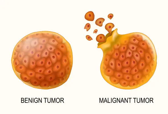
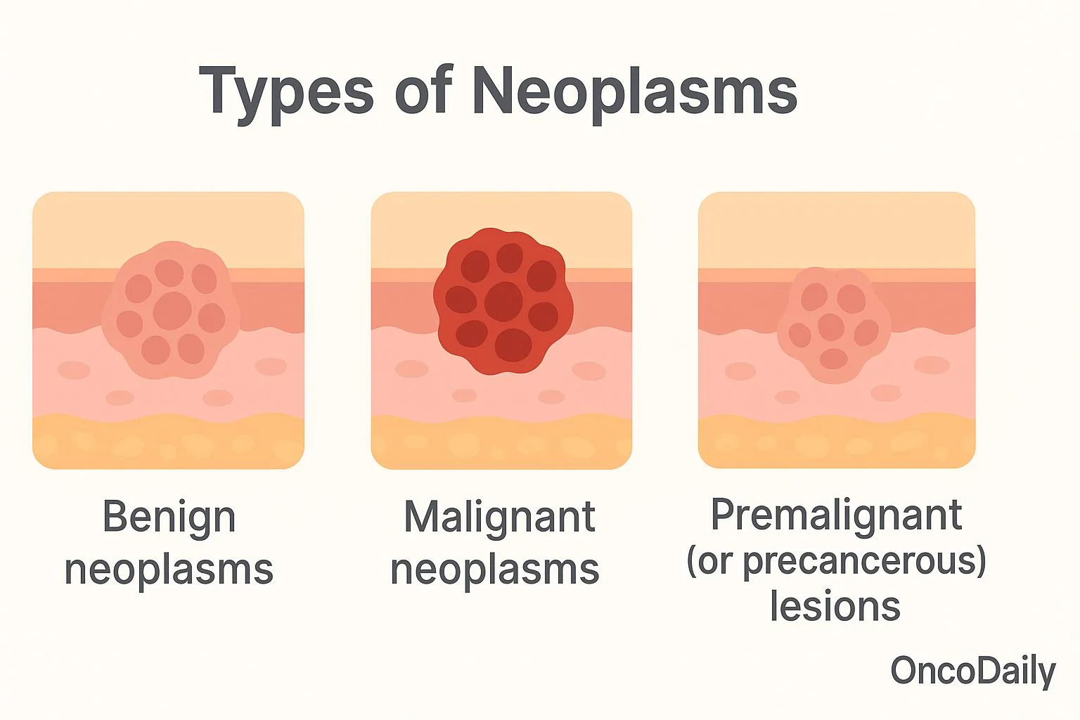
    Benign Tumors

    These are non-cancerous growths. They tend to grow slowly and are often enclosed in a fibrous capsule, which keeps them from spreading to surrounding tissues. While they do not metastasize (spread to distant sites), they can cause problems by exerting pressure on vital organs, nerves, or blood vessels. Histologically, the cells of a benign tumor are well-differentiated, meaning they closely resemble the normal cells of the tissue from which they originated. They generally do not return after surgical removal. However, some benign tumors have the potential to become malignant if left untreated.

    Malignant Tumors

    These are cancerous tumors. They are characterized by rapid, uncontrolled growth and the ability to invade and destroy surrounding tissues. Malignant cells are anaplastic, meaning they are poorly differentiated and do not resemble normal cells in shape, size, or function. A key feature is their ability to metastasize, where cells break away from the primary tumor and travel through the blood or lymphatic system to form secondary tumors (metastases) in other parts of the body. Malignant tumors have a high tendency to recur after removal.

    Premalignant (or Precancerous) Conditions

    This refers to conditions involving abnormal cells that are not yet cancerous but have an increased risk of developing into a malignant tumor over time. These conditions require close monitoring and sometimes treatment.

    Types of Benign Tumors

    Benign tumors are typically named by adding the suffix “-oma” to the name of the tissue of origin.

    Adenomas

    Benign tumors arising from the epithelial tissue of a gland or gland-like structure. Common examples include colon polyps and adenomas of the thyroid, pituitary, or liver. They can sometimes become cancerous and may require surgical removal.

    Fibromas (or Fibroids)

    Tumors of fibrous or connective tissue that can grow in any organ. They are very common in the uterus (leiomyomas, often called fibroids) and can cause symptoms like heavy bleeding or pelvic pain, necessitating removal.

    Hemangiomas

    A benign tumor formed by a collection of excess blood vessels. They often appear on the skin as "strawberry marks," especially in newborns, and most fade over time without treatment.

    Lipomas

    The most common benign tumor in adults, arising from fat cells (adipose tissue). They are slow-growing, soft, movable lumps often found on the neck, shoulders, or back. Treatment is usually only necessary if they become painful or grow rapidly.

    Meningiomas

    Tumors that develop from the meninges, the membranes surrounding the brain and spinal cord. Most are benign and slow-growing. Symptoms (headaches, seizures, weakness) depend on their location and may require treatment.

    Myomas

    Tumors that grow from muscle tissue. Leiomyomas grow from smooth muscle (e.g., in the uterus, stomach). Rhabdomyomas are rare benign tumors of skeletal muscle.

    Neuromas

    Benign tumors that grow from nerves. Neurofibromas and schwannomas are other examples, which can occur anywhere in the body.

    Osteomas

    Benign tumors of bone. The most common type is an osteochondroma, which usually appears as a painless bump near a joint, like the knee or shoulder.

    Papillomas

    Growths that project in finger-like fronds from epithelial tissue. They can appear on the skin, cervix, or in breast ducts. Some are caused by HPV and may require removal to rule out cancer.

    Nevi (Moles)

    Very common noncancerous growths on the skin.

    Types of Premalignant Conditions

    Actinic Keratosis

    Also known as solar keratosis, these are crusty, scaly patches of skin caused by sun exposure, especially in fair-skinned individuals. A percentage of these can progress to squamous cell carcinoma.

    Cervical Dysplasia

    Abnormal cell changes on the surface of the cervix, usually detected by a Pap smear. It is considered premalignant and is at risk of developing into cervical cancer if not treated.

    Metaplasia of the Lung

    A change in the cells lining the bronchi (airways), often caused by smoking, where glandular cells are replaced by squamous cells. This can be a precursor to cancer.

    Leukoplakia

    Thick, white patches that form on the gums, inside the cheeks, or on the tongue, which cannot be scraped off. It is strongly linked to tobacco use and a small percentage can become cancerous.

    Types of Malignant Tumors

    Malignant tumors are named based on their tissue of origin.

    Carcinoma

    The most common type of cancer, forming from epithelial cells that line the surfaces of the body. Examples include adenocarcinoma (from glandular cells), squamous cell carcinoma, and basal cell carcinoma. They can occur in the lung, breast, prostate, colon, and skin.

    Sarcoma

    Cancers that arise from connective and supportive tissues such as bone, cartilage, fat, muscle, and nerves. Examples include osteosarcoma (bone) and liposarcoma (fat).

    Blastoma

    Tumors derived from embryonic (precursor) cells. They are more common in children. Examples include neuroblastoma (nerve cells), retinoblastoma (eye), and medulloblastoma (brain).

    Germ Cell Tumors

    These arise from reproductive cells (sperm or eggs) and most often occur in the testicles or ovaries. Teratomas are a common type.

    Leukemia and Lymphoma

    These are cancers of the blood-forming cells and immune system. Leukemia is a cancer of the bone marrow that leads to an overproduction of abnormal white blood cells. Lymphomas (e.g., Hodgkin's and non-Hodgkin's) are cancers of the lymphatic system. Note: All lymphomas are malignant.

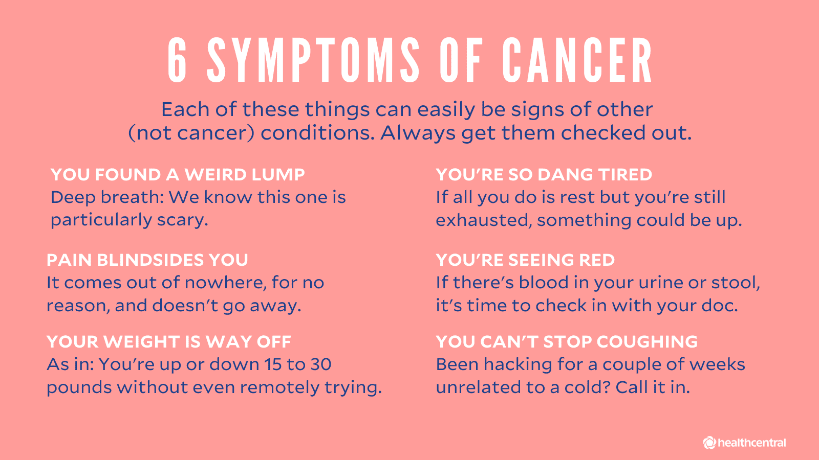
    Clinical Features, Diagnosis, and Staging

    Modes of Spread (Metastasis)

    Malignant tumors spread via several pathways:

    • Local Extension: Direct invasion into adjacent tissues.
    • Lymphatic Spread: Tumor cells enter lymphatic vessels and travel to regional lymph nodes.
    • Hematogenous (Blood) Spread: Tumor cells penetrate blood vessels and are carried to distant organs like the liver, lungs, and brain.
    • Transcoelomic Spread: Malignant cells spread across body cavities like the peritoneum or pleura.
    • Tumor Seedlings: Accidental transplantation of tumor cells to new sites, for instance, during a surgical procedure.

    Diagnosis and Investigations

    Diagnosis involves a combination of history, physical examination, and investigations.

    • Biopsy: The definitive diagnosis of cancer is made by examining a tissue sample under a microscope.
      • Excisional Biopsy: Removal of the entire tumor or suspicious area.
      • Incisional or Core Biopsy: Removal of a sample directly from the tumor.
      • Needle Aspiration Biopsy: Removal of fluid or a small tissue sample with a needle.
    • Imaging Studies: To determine the tumor's location, size, and extent of spread. These include CT scans, MRI scans, X-rays, Ultrasounds, and Mammograms.
    • Endoscopy: To visualize internal organs and obtain biopsies (e.g., colonoscopy for colon polyps, endoscopy for stomach tumors).
    • Blood Tests: Can include complete blood counts and checks for tumor markers (substances produced by cancer cells).
    • Cytological Examinations: Such as a Pap smear to examine cells from the cervix for abnormalities.

    Tumor Staging and Grading

    Once cancer is diagnosed, it is staged and graded to plan treatment and predict prognosis.

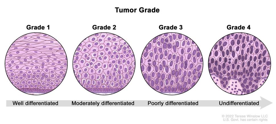
    • Grading: Describes how abnormal the cancer cells look under a microscope (degree of differentiation). A lower grade (e.g., Grade 1) means the cells are well-differentiated and look more like normal cells, suggesting a slower-growing cancer. A higher grade (e.g., Grade 3 or 4) means the cells are poorly differentiated or anaplastic, suggesting a more aggressive cancer.
    • Staging: Describes the size of the tumor and how far it has spread from its original location. The most common system is the TNM system:
      • T (Tumor): Describes the size and extent of the primary tumor.
      • N (Nodes): Indicates whether the cancer has spread to nearby lymph nodes.
      • M (Metastasis): Indicates whether the cancer has metastasized to distant parts of the body.

    Management of Malignant Tumors

    Treatment can be curative (aiming to cure the disease) or palliative (aiming to relieve symptoms and improve quality of life when a cure is not possible).

    Curative Treatment

    Often involves a combination of modalities:

    • Surgery: The physical removal of the tumor, often with a margin of surrounding healthy tissue. It is a primary treatment for many solid tumors.
    • Radiotherapy: Uses high-energy radiation to kill cancer cells and shrink tumors. It can be used alone or in combination with other treatments.
    • Cytotoxic Chemotherapy: The use of drugs to kill rapidly dividing cancer cells. It is particularly effective for metastatic disease or blood cancers.

    Palliative Management

    Focuses on symptom control and maintaining quality of life.

    • Surgery: Can be used to relieve symptoms, such as debulking a tumor that is causing an obstruction or pain, even if it cannot be completely removed.
    • Radiotherapy: Effective for shrinking tumors to relieve pain, especially from bone metastases.
    • Hormone Therapy: Blocks or lowers the amount of hormones that some cancers (like breast and prostate) need to grow.
    • Targeted Therapy and Immunotherapy: Newer treatments that target specific molecular characteristics of cancer cells or boost the body's own immune system to fight cancer.
    • Cytotoxic Chemotherapy: Can be used in lower doses to control tumor growth and manage symptoms. Drugs used include:
      • Alkylating agents (e.g., cyclophosphamide)
      • Antimetabolites (e.g., methotrexate, fluorouracil)
      • Plant alkaloids (e.g., vincristine)
      • Cytotoxic antibiotics (e.g., doxorubicin)
      • Platinum compounds (e.g., cisplatin)
      • Monoclonal antibodies (e.g., trastuzumab)
    • Symptom Control:
      • Pain Relief: Use of analgesics (from non-opioids to strong opiates), as well as nerve blocks with phenol or alcohol.
      • Supportive Drugs: Anti-emetics for nausea, tranquilizers, and hypnotics.
      • Maintenance of Morale: Providing psychological, emotional, and spiritual support is a crucial part of holistic care.

    Prevention of Tumors and Cancers

    Many cancers can be prevented through healthy lifestyle choices and regular screening.

    • Early Screening and Detection: Regular check-ups, self-examinations (e.g., self-breast exam), and recommended screenings (e.g., Pap smears, colonoscopies, mammograms).
    • Reduce Carcinogen Exposure: Minimize exposure to radiation, toxic chemicals, and excessive sun (use sunscreen).
    • Healthy Lifestyle:
      • Maintain a healthy weight and exercise regularly.
      • Eat a balanced diet rich in fruits, vegetables, and fiber.
      • Limit alcohol consumption.
      • Avoid smoking and chewing tobacco.

    Table 1: Difference between Benign and Malignant Tumors

    Characteristic Benign Tumors Malignant Tumors
    Cancerous No, they are non-cancerous. Yes, they are cancerous.
    Rate of Growth Slow Rapid
    Capsule Typically encapsulated (enclosed in a capsule). Not encapsulated; invasive.
    Invasion Does not invade surrounding tissues; grows by expansion. Invades and destroys surrounding tissues.
    Metastasis Does not metastasize. Frequently metastasizes to distant sites.
    Cell Differentiation Well-differentiated; cells resemble normal tissue. Poorly differentiated (anaplastic); abnormal cell structure.
    Recurrence Rarely recurs after surgical removal. Frequently recurs after removal.
    Systemic Effects Usually localized effects (e.g., pressure on organs). Can cause systemic effects like weight loss, fatigue (cachexia).

    TUMORS (NEOPLASMS) Read More »

    FLUID AND ELECTROLYTE IMBALANCE

    FLUID AND ELECTROLYTE IMBALANCE

    Nursing Notes - Burns

    FLUID AND ELECTROLYTE IMBALANCE

    Fluid and electrolyte balance is a dynamic process that is crucial for life and homeostasis.

    Electrolytes

    Electrolytes in body fluids are active chemicals (cations that carry positive charges and anions that carry negative charges). The major cations in the body fluid are sodium, potassium, calcium, and hydrogen ions. The major anions are chloride, bicarbonate, phosphate, sulphate, and proteinate ions. The chemicals unite in varying combinations. Therefore, electrolyte concentration in the body is expressed in terms of milliequivalents (mEq) per litre. A milliequivalent is defined as being equivalent to the electrochemical activity of 1mg of hydrogen.

    Approximately 60% of a typical adult’s weight consists of fluids (water and electrolytes). Factors that influence the amount of body fluid are age, gender, and body fat. In general, younger people have a higher percentage of body fluid than older people, and men have proportionately more body fluid than women. People who are obese have less fluid than those who are thin because fat cells contain little water.

    FLUID VOLUME DISTURBANCES OR ELECTROLYTE IMBALANCE OR DISORDERS

    An electrolyte disorder occurs when the levels of electrolytes in the body are either too high or too low. Electrolytes are naturally occurring elements and compounds in the body. They control important physiologic functions.

    SODIUM IMBALANCES

    Sodium is the most abundant electrolyte in the ECF. Its concentration ranges from 135-145 mEq per litre. Sodium has a major role in controlling water distribution throughout the body because it does not easily cross the cell membrane and because of its abundance and high concentration in the body. Sodium also functions in establishing the electrochemical state necessary for muscle contraction and transmission of nerve impulses.

    SODIUM DEFICIT (HYPONATREMIA)

    Hyponatremia refers to a serum sodium level that is less than 135 mEq/L (135mmol/L). Sodium imbalance often occurs with a fluid imbalance because the same hormones regulate both sodium and water balance.

    CLINICAL MANIFESTATIONS
    • Poor skin turgor
    • Dry mucosa
    • Headache
    • Decreased saliva production
    • Orthostatic fall in blood pressure
    • Nausea and vomiting
    • Abdominal cramping
    • Neurological changes which include: Altered mental status, Status epilepticus, and coma
    • Anorexia
    • Feeling of exhaustion
    SIGNS OF INTRACRANIAL PRESSURE
    • Lethargy
    • Confusion
    • Muscle twitching
    • Hemiparesis
    • Focal weakness
    • Papilledema
    • Seizures and death may occur.
    CAUSES
    • Excessive diaphoresis
    • Diuretics (high ceiling diuretics)
    • Wound drainage (especially gastrointestinal)
    • Decreased secretion of aldosterone
    • Hyperlipidemia
    • Kidney diseases (scarred distal convoluted tubule)
    • Nothing by mouth
    • Low salt diet
    • Cerebral salt wasting syndrome
    • Hyperglycemia
    • RELATIVE SODIUM DEFICITS (DILUTIONAL): Excessive ingestion of hypotonic fluids, fresh water submersion, Kidney failure (nephrotic syndrome), Irrigation with hypotonic fluids, Heart failure.
    MANAGEMENT
    • When possible, the underlying cause is treated.
    • Intravenous infusion of normal saline is used for slow and gradual correction.
    • Monitoring therapy can help restore sodium balance in mild Hyponatremia. This includes increasing oral sodium intake and restricting oral fluid intake.
    • The nurse’s responsibility for this patient includes skin protection, safety, monitoring, patient and family teaching, and administering prescribed drugs.
    HYPERNATREMIA

    Hypernatremia is excess sodium in the blood, in which the serum level is over 145 mEq/L.

    CAUSES

    ACTUAL SODIUM EXCESSES:

    • Hyperaldosteronism
    • Kidney failure, Heart failure, Liver failure
    • Corticosteroids
    • Cushing’s syndrome or disease
    • Excessive oral sodium ingestion (salt intake)
    • Excessive administration of sodium-containing IV fluids.

    RELATIVE SODIUM EXCESSES:

    • Nothing by mouth, severe burns
    • Increased rate of metabolism
    • High fever
    • Hyperventilation
    • Infection
    • Excessive diaphoresis
    • Watery diarrhea
    • Dehydration.
    CLINICAL FEATURES
    • Pitting edema
    • Puffiness of the face
    • Increased urination
    • Often dilated jugular veins
    • Features of pulmonary oedema
    • Body temperature may increase mildly
    • A primary characteristic of Hypernatremia is thirst.
    • Dry, sticky mucous membranes
    • A rough, dry tongue and lethargy which can progress to coma.
    MANAGEMENT

    Treatment depends on the cause.

    • Infusion of a hypotonic electrolyte solution (e.g., 0.3% sodium chloride) or an isotonic non-saline solution (e.g., dextrose 5% in water).
    • Diuretics also may be prescribed to treat the sodium gain.
    • Nutrition therapy to prevent or correct mild Hypernatremia involves ensuring adequate water intake, especially among older adults.
    • Dietary sodium is restricted when kidney problems are present.
    • Collaboration with a dietitian to teach the patient how to determine the sodium content of food, beverages, and drugs is important.
    • Nursing actions for patient safety include skin protection, monitoring, and patient and family teaching about sodium excess.

    POTASSIUM IMBALANCES

    Potassium is the major cation of the intracellular fluid. It is particularly important for regulating heart function and helps in maintaining healthy nerves and muscles. Almost all foods contain potassium; it is high in meat and fish but less so in eggs, bread, and cereal grains. A deficit of potassium in the blood is called hypokalemia.

    HYPOKALEMIA

    Hypokalemia is an electrolyte imbalance in which the serum potassium level is below 3.5 mEq/L. It can be life-threatening because every body system is affected.

    CAUSES
    • Actual potassium deficits: Inappropriate or excessive use of drugs (e.g., Diuretics, Digitalis, Corticosteroids); Increased secretion of aldosterone; Cushing's syndrome; Diarrhea; Vomiting; Wound drainage (especially gastrointestinal); Prolonged nasogastric suction; Heat-induced excessive diaphoresis; Kidney failure.
    • Relative potassium deficits: Alkalosis; Hyperalimentation; Hyperinsulinism; Total parenteral nutrition; Water intoxication; IV therapy with potassium-poor solutions.
    CLINICAL FEATURES
    • Fatigue, Anorexia, Nausea, and vomiting
    • Muscle weakness
    • Polyuria, Decreased bowel motility
    • Ventricular asystole or fibrillations
    • Paresthesias, Leg cramps
    • Decreased blood pressure
    • Abdominal distention, Hypoactive reflexes
    MANAGEMENT
    • Conventional measures such as increased intake in the daily diet or oral potassium supplements are good for mild to moderate hypokalemia.
    • IV replacement therapy for potassium loss is typically 40-80 mEq/day. Examples include potassium chloride, potassium gluconate, and potassium citrate.
    • IV K+ is given for severe loss, and the amount depends on the degree of loss.
    • Oral potassium preparations can be taken as liquids or solids.
    • Diuretics that increase the kidney's excretion of potassium (e.g., furosemide/Lasix) can cause hypokalemia and should be monitored.
    • Nutrition therapy involves collaboration with a dietitian to teach the patient how to increase dietary potassium intake.
    • Respiratory monitoring is performed at least hourly for severe hypokalemia; monitor pulse, cough reflex, among others.
    HYPERKALEMIA

    Hyperkalemia is an electrolyte imbalance in which the serum potassium level is higher than 5.0 mEq/L. A level above 5.5 mEq/L is considered more severe.

    COMMON CAUSES
    • Over-ingestion of potassium-containing foods or medications (e.g., Salt substitutes, Potassium chloride)
    • Crush injury, Burns
    • Rapid infusion of potassium-containing IV solution, Bolus IV potassium injections
    • Transfusions of whole blood or packed cells
    • Adrenal insufficiency, Kidney failure, Addison’s disease
    • Potassium-sparing diuretics, Angiotensin-converting enzyme inhibitors (ACEIs)
    RELATED POTASSIUM EXCESSES
    • Tissue damage, Acidosis, Hyperuricemia, Uncontrolled diabetes mellitus
    CLINICAL MANIFESTATIONS
    • Muscle weakness, twitching, palpitations
    • Bradycardia, Hypotension
    • Tingling and burning sensations followed by numbness in the hands and feet
    • Increased motility with diarrhea and hyperactive bowel sounds; bowel movements are frequent and watery
    • Flaccid paralysis, Paresthesias, Intestinal colic, Cramps, Abdominal distension
    • Irritability, Anxiety
    MANAGEMENT
    • In non-acute situations, restricting dietary potassium and potassium-containing medications may correct the imbalance.
    • Administration of cation-exchange resins (e.g., sodium polystyrene sulfonate) orally or as retention enemas.
    • If serum potassium levels are dangerously elevated, it may be necessary to administer IV calcium gluconate with caution.
    • Monitor blood pressure to detect hypotension.
    • IV administration of regular insulin and a hypertonic dextrose solution causes a temporary shift of potassium into cells.
    • Loop diuretics such as furosemide (Lasix) increase the excretion of potassium.
    • Beta-2 agonists such as Albuterol (Ventolin) can be effective in decreasing potassium.
    • The nurse must caution the patient about using salt substitutes sparingly if they are taking other supplementary forms of potassium.
    • Observe the patient's general condition, vital signs, and GI symptoms.
    • Prevention includes avoiding potassium-rich foods if prescribed and checking labels of beverages for high potassium content.

    CALCIUM IMBALANCES

    More than 99% of the body’s calcium (Ca++) is located in the skeletal system, where it is a major component of bones and teeth. It is a divalent cation that exists in both a bound form (attached to serum proteins like albumin) and an ionized (free) form. The body functions best when calcium levels are maintained between 9.0 and 10.5 mg/dL. Calcium enters the body via dietary intake, and its absorption requires active vitamin D. It is a vital mineral used to stabilize blood pressure, control skeletal muscle contraction, and build strong bones and teeth.

    HYPOCALCEMIA

    Hypocalcemia is an electrolyte imbalance in which the total serum calcium (Ca2+) level is below 9.0 mg/dL or 2.25 mmol/L.

    COMMON CAUSES OF HYPOCALCEMIA
    • Actual calcium deficits: Inadequate oral intake of calcium, Lactose intolerance, Malabsorption (e.g., Celiac, Crohn's), Inadequate intake of vitamin D, End-stage kidney disease, Steatorrhea, Wound drainage, Hypoparathyroidism, Pancreatitis, Massive subcutaneous infections, Massive transfusions of citrated blood, Chronic diarrhea, Burns, Alcoholism.
    • Relative calcium deficits: Hypoproteinemia, Alkalosis, Immobility, Removal of the parathyroid gland.
    CLINICAL MANIFESTATIONS
    • Numbness and tingling of fingers
    • Positive Trousseau's sign and Chvostek's sign
    • Seizures, Bronchospasms
    • Anxiety, Impaired clotting time
    • Diarrhea, Anorexia, Nausea, and vomiting
    • Abdominal distention and pain are common
    MANAGEMENT
    • Acute symptomatic hypocalcemia is life-threatening and requires prompt treatment with IV administration of calcium salts (e.g., calcium gluconate, calcium chloride).
    • Vitamin D therapy may be instituted to increase calcium absorption from the GI tract.
    • Calcium-containing foods include milk products, green leafy vegetables.
    • Aluminum hydroxide or calcium acetate may be prescribed to decrease elevated phosphorus levels before treating hypocalcemia.
    • Educate the patient about foods rich in calcium and the potential need for supplements.
    • Advise the patient to reduce alcohol and caffeine intake and to stop smoking, as these can inhibit calcium absorption or increase its excretion.
    HYPERCALCEMIA (CALCIUM EXCESS)

    Hypercalcemia is an electrolyte imbalance in which the total serum calcium level is above 10.5 mg/dL or 2.62 mmol/L. The excitable tissues most affected are the heart, skeletal muscles, nerves, and intestinal smooth muscles.

    CAUSES OF HYPERCALCEMIA
    • Actual calcium excesses: Excessive oral intake of calcium, Excessive oral intake of vitamin D, Kidney failure, Use of Thiazide diuretics, Malignancies (e.g., leukemia), Hyperparathyroidism, Paget’s disease, Prolonged immobilization.
    • Relative calcium excess: Use of glucocorticoids, Dehydration, Digoxin toxicity.
    CLINICAL MANIFESTATIONS
    • Increased heart rate and blood pressure
    • Cyanosis and pallor
    • Muscular weakness, Hypoactive deep tendon reflexes
    • Constipation, Anorexia, Nausea, and vomiting
    • Polyuria and polydipsia, Dehydration
    • Lethargy, Deep bone pain, Pathologic fractures
    • Flank pain, Calcium stones, Hypertension
    MANAGEMENT
    • Treating the underlying cause is essential (e.g., chemotherapy, parathyroidectomy).
    • Discontinue IV solutions or oral drugs containing calcium or vitamin D.
    • IV normal saline (0.9% sodium chloride) is given to increase kidney excretion of calcium.
    • Thiazide diuretics are replaced with diuretics that enhance calcium excretion, such as furosemide (Lasix).
    • Administer drugs that inhibit calcium reabsorption from bone, such as phosphorus, calcitonin, and prostaglandin synthesis inhibitors (aspirin, NSAIDs).
    • Implement cardiac monitoring for patients with hypercalcemia.

    PHOSPHORUS IMBALANCES

    Normal serum level of phosphorus ranges from 3.0 to 4.5 mg/dL. It is essential to the function of muscles and red blood cells, the formation of ATP, and maintaining acid-base balance. It also provides structural support to bones and teeth.

    PHOSPHORUS DEFICITS (HYPOPHOSPHATEMIA)

    Hypophosphatemia is an electrolyte imbalance in which the serum phosphorus level is below 3.0 mg/dL. Because phosphorus and calcium are interrelated, a decrease in serum phosphorus can cause an increase in serum calcium.

    CAUSES OF HYPOPHOSPHATEMIA
    • Malnutrition, Starvation
    • Use of aluminum hydroxide-based or magnesium-based antacids
    • Hyperparathyroidism, Hypercalcemia, Kidney failure, Malignancy
    • Hyperglycemia, Hyperalimentation, Respiratory alkalosis, Uncontrolled diabetes mellitus
    • Alcohol abuse or withdrawal, Vitamin D deficiency, Diarrhea, Burns, and severe wounds
    CLINICAL MANIFESTATIONS
    • Paresthesia, Muscle weakness
    • Bone pain and tenderness, Chest pain
    • Confusion, Cardiomyopathy, Respiratory failure
    • Seizures, Tissue hypoxia, Increased susceptibility to infections, Nystagmus
    • On laboratory investigation, the serum phosphorus level is less than 2.5mg/dl.
    MANAGEMENT
    • Discontinue drugs that promote phosphorus loss (e.g., antacids, osmotic diuretics, calcium supplements).
    • Oral replacement with phosphorus along with vitamin D may correct moderate deficits.
    • IV phosphorus is given cautiously and slowly for severe cases (less than 1 mg/dL).
    • Nutrition therapy involves increasing the intake of phosphorus-rich foods while decreasing calcium-rich foods.
    PHOSPHORUS EXCESS (HYPERPHOSPHATEMIA)

    Hyperphosphatemia is an electrolyte imbalance in which the serum phosphorus level is above 4.5 mg/dL. High levels are generally well-tolerated by most body systems.

    CAUSES
    • Certain cancer treatments, Tumor lysis syndrome
    • Acute and chronic renal failure
    • Excessive intake of phosphorus, Vitamin D excess
    • Respiratory and metabolic acidosis
    • Hypoparathyroidism, Volume depletion
    • Leukemia/lymphoma treatment with cytotoxic drugs
    • Increased tissue breakdown, Rhabdomyolysis
    CLINICAL MANIFESTATIONS
    • Tetany, Tachycardia
    • Anorexia, Nausea, and vomiting
    • Signs and symptoms of associated hypocalcemia
    • Hyperactive reflexes
    • Soft tissue calcifications in lungs, kidneys, heart, and cornea
    • Lab analysis shows serum phosphorus level exceeds 4.5mg/dl.
    MANAGEMENT
    • Management often entails managing the associated hypocalcemia.
    • Give Vitamin D orally or parenterally.
    • Restrict dietary phosphorus; promote excretion with loop diuretics and volume replacement with saline.
    • Dialysis may also lower phosphorus levels.
    • Advise the client to avoid phosphate-containing laxatives and enemas.

    CHLORIDE IMBALANCES

    Chloride (Cl-) is the major anion of the ECF. The normal plasma concentration ranges from 98 to 106 mEq/L. It enters the body through dietary intake and is important in the formation of hydrochloric acid in the stomach and in maintaining acid-base balance.

    CHLORIDE EXCESS (HYPERCHLOREMIA)

    Hyperchloremia exists when the serum level of chloride exceeds 107 mEq/L. Hypernatremia, bicarbonate loss, and metabolic acidosis can occur with high chloride levels.

    CLINICAL MANIFESTATIONS
    • Tachypnea, Weakness, and lethargy
    • Deep and rapid respirations
    • Diminished cognitive ability
    • Hypertension; pitting oedema
    • Dysrhythmias
    MANAGEMENT
    • Correcting the underlying cause and restoring electrolyte, fluid, and acid-base balance are essential.
    • Ringer's lactate solution may be administered.
    • IV sodium bicarbonate may be given to increase bicarbonate levels, which promotes renal excretion of chloride ions.
    • Diuretics may be administered to eliminate chloride.
    • Monitor vital signs, arterial blood gas values, and patient status.
    • Educate the patient about diet and maintaining adequate hydration.
    HYPOCHLOREMIA

    Hypochloremia is a serum chloride level below 97 mEq/L.

    CAUSES
    • Addison’s disease
    • Reduced chloride intake or absorption
    • Untreated diabetic ketoacidosis
    • Excessive sweating, Vomiting, and nausea
    • Gastric suctioning, Diarrhea, Draining fistulas and ileostomies
    • Rapid removal of ascitic fluid with high sodium content
    • IV fluids that lack chloride (e.g., dextrose and water)
    SIGNS AND SYMPTOMS
    • Agitation, Irritability
    • Tremors, Muscle cramps, Hyperactive deep tendon reflexes, Hypertonicity, Tetany
    • Slow, shallow respirations
    • Seizures, Dysrhythmias, Coma
    MANAGEMENT
    • Administer IV normal saline (0.9% sodium chloride) or half-strength saline (0.45% sodium chloride).
    • If the patient is using a diuretic, it may be discontinued or another one prescribed.
    • Nursing care is similar to that for other electrolyte imbalances.

    MAGNESIUM IMBALANCES

    Magnesium (Mg++) is an abundant intracellular cation. The normal serum Mg+ level is 1.3 to 2.3 mg/dL. It is the most abundant intracellular cation after potassium and plays a role in both carbohydrate and protein metabolism. Magnesium balance is important for neuromuscular function, as it acts directly on the myoneural junction. It also affects cardiovascular activity, producing vasodilation. 60% of magnesium is deposited in bone and soft tissues; it is absorbed in the small intestine and excreted by the kidneys.

    MAGNESIUM DEFICITS (HYPOMAGNESEMIA)

    Hypomagnesemia refers to a below-normal serum magnesium concentration (<1.3 mg/dL) and is frequently associated with hypokalemia and hypocalcemia.

    CAUSES
    • Chronic alcoholism, Malabsorptive disorders
    • Hyperthyroidism, Hyperaldosteronism
    • Diuretic phase of renal failure
    • Diabetic ketoacidosis
    • Refeeding after starvation, Parenteral nutrition
    • Chronic laxative use, Diarrhea
    • Acute myocardial infarction, Heart failure
    • Certain pharmacological agents (e.g., gentamicin)
    CLINICAL MANIFESTATIONS
    • Neuromuscular irritability
    • Positive Trousseau's sign and positive Chvostek's sign
    • Insomnia, Mood changes, Anorexia
    MANAGEMENT
    • Mild deficits can be corrected by diet alone (e.g., green leafy vegetables, nuts, seeds, seafood, peanut butter, cocoa).
    • Oral magnesium salts (oxide or gluconate form) can be administered but may produce diarrhea.
    • IV parenteral magnesium can be administered for severe hypomagnesemia.
    • Monitor vital signs frequently during magnesium administration.
    • Monitor urine output.
    • Calcium gluconate must be readily available to treat hypocalcemic tetany or hypermagnesemia.
    MAGNESIUM EXCESS (HYPERMAGNESEMIA)

    Hypermagnesemia occurs when the serum magnesium level is over 2.3 mg/dL. It is a rare electrolyte abnormality because the kidneys efficiently excrete magnesium.

    CONTRIBUTING FACTORS
    • Renal failure
    • Diabetic ketoacidosis, Adrenocortical insufficiency
    • Increased absorption due to intestinal hypomotility
    • Lithium intoxication
    • Extensive soft tissue injury (e.g., trauma, shock, sepsis, cardiac arrest)
    SIGNS AND SYMPTOMS
    • At mildly increased levels: low blood pressure (vasodilation), nausea, vomiting, weakness, facial flushing.
    • At higher concentrations: lethargy, difficulty speaking (dysarthria), drowsiness.
    • Severe untreated cases: Coma, cardiac arrest.
    • Platelet clumping and delayed thrombin formation.
    MANAGEMENT
    • Avoid administering magnesium to patients with renal failure.
    • Discontinue parenteral and oral magnesium salts.
    • IV calcium gluconate antagonizes the cardiovascular and neuromuscular effects of magnesium.
    • The nurse monitors the level of consciousness and vital signs, noting hypotension and shallow respirations.
    • Identify and assess patients at risk for hypermagnesemia.

    FLUID AND ELECTROLYTE IMBALANCE Read More »

    Hemorrhage

    HAEMORRHAGE: Nursing Lecture Notes

    Nursing Notes - Burns

    HAEMORRHAGE: Nursing Lecture Notes

    Haemorrhage, commonly known as bleeding, is the loss of blood from the circulatory system, specifically from blood vessels. It is a critical medical condition that, if uncontrolled, can lead to severe physiological compromise and death. The body possesses intrinsic defence mechanisms, primarily through the process of clotting (hemostasis), to prevent excessive blood leakage. However, these mechanisms can be deficient due to underlying diseases, absence of essential clotting factors, or the use of anticoagulant medications.

    Types of Haemorrhage

    Haemorrhage is classified based on several key characteristics to aid in diagnosis, prognosis, and management. These classifications include:

    1. The type of blood vessel involved.
    2. The location or situation of the haemorrhage.
    3. The time of occurrence or duration of the haemorrhage.
    Classification by Blood Vessels Involved

    The characteristics of bleeding often provide clues as to which type of blood vessel has been compromised:

    1. Arterial Haemorrhage:
  • Appearance: Characterized by bright red blood due to its high oxygen content.
  • Flow Pattern: Blood spurts rhythmically with each heartbeat, reflecting the pulsatile nature of arterial blood flow under high pressure.
  • Rate of Loss: Blood loss is typically rapid and significant, often more profound than from a vein of a corresponding size, due to the higher pressure within arteries.
  • Bleeding from Vessel Ends: Blood loss can occur from both ends of the severed vessel.
  • 2. Venous Haemorrhage:
  • Appearance: The bleeding is characterized by dark purplish-red blood due to its lower oxygen content.
  • Flow Pattern: Blood flows at a more even, gentle, and continuous rate, lacking the pulsatile nature of arterial bleeding.
  • Rate of Loss: While it can still be substantial, the rate of blood loss is generally less rapid than that of arterial haemorrhage for comparable vessel sizes.
  • 3. Capillary Haemorrhage:
  • Appearance: Blood typically oozes slowly over the surface of the wound. It is darkish red in colour.
  • Rate of Loss: Although the immediate rate of loss is slow, over several hours, continuous oozing can result in considerable and clinically significant blood loss, especially over large surface areas (e.g., abrasions, extensive burns).
  • Classification by Time or Duration of Haemorrhage

    The timing of haemorrhage relative to an injury or surgical procedure provides important diagnostic and prognostic information:

    1. Primary Haemorrhage:
  • Definition: This refers to bleeding that occurs immediately at the time of injury or surgical incision.
  • Mechanism: It continues until it spontaneously ceases through the body's natural hemostatic mechanisms (e.g., vasoconstriction, platelet plug formation, coagulation) or is controlled by artificial means (e.g., direct pressure, ligation, cauterization).
  • Examples: A simple cut finger, bleeding during an operative incision.
  • 2. Reactionary or Intermediate Haemorrhage:
  • Definition: Bleeding that occurs within the first 24 hours following an injury or surgical operation.
  • Mechanism: It often results from the resolution of vasoconstriction that was initially induced by shock, hypothermia, or drugs administered at the time of injury or operation. Small blood vessels that were initially cut but could not bleed due to these factors begin to bleed as blood pressure rises and normal physiological responses return.
  • Common Sites: This type of haemorrhage is common following operations on highly vascular organs such as the kidney, thyroid gland, breast, and uterus (e.g., total hysterectomy).
  • Additional Contributing Factors:
    • Increased intravascular pressure due to actions such as coughing or vomiting.
    • Increased venous pressure.
    • Physical excitement or administration of stimulant drugs.
  • Clinical Tip: To minimize the risk, restrict the number of visitors to a minimum in the immediate postoperative period to reduce patient excitement and physical exertion.
  • 3. Secondary Haemorrhage:
  • Definition: This type of bleeding occurs later, typically between 36 hours to 48 hours, or even up to 10-14 days after the initial injury or operation.
  • Mechanism: It is usually indicative of an underlying complication, often related to infection or mechanical irritation, leading to the erosion or sloughing of blood vessel walls.
  • Common Causes:
    • Sepsis: Bacterial infection leading to inflammation and enzymatic destruction of vessel walls.
    • Enzymatic Action: For example, the action of pepsin on a bleeding peptic ulcer, eroding the vessel.
    • Mechanical Pressure: Persistent pressure from a drainage tube or foreign body (e.g., bone fragment) eroding a vessel.
    • Presence of Carcinoma: Malignant tumors can erode blood vessels, leading to chronic or acute bleeding.
  • Classification by Situation or Location of Haemorrhage

    This classification distinguishes whether the blood loss is visible externally or contained within body cavities:

    1. External or Revealed Haemorrhage:
    • Definition: This is bleeding that is directly visible, either from an open wound on the body surface or from a natural body orifice (e.g., epistaxis from the nose, hematemesis from vomiting blood, melena/hematochezia from the rectum).
    • Visibility: Blood is immediately apparent and can be quantified relatively easily.
    2. Internal or Concealed Haemorrhage:
    • Definition: This refers to bleeding that occurs into an internal body cavity or tissue space, where the blood loss is not immediately visible externally.
    • Locations: Common sites include the peritoneal cavity (e.g., ruptured spleen), pleural cavity (e.g., hemothorax), retroperitoneal space, lumen of hollow organs (e.g., intestines, stomach, bladder), or within the tissues of a limb (e.g., large hematoma).
    • Diagnosis: Since the bleeding is concealed, diagnosis relies heavily on the patient's symptoms and signs of hypovolemia and shock. It may be "revealed" later if the blood exits the body (e.g., vomited blood, blood passed per rectum) or by the formation of bruising and swelling on the surface of the body.

    Clinical Picture: Signs and Symptoms of Haemorrhage

    The clinical presentation of haemorrhage varies depending on the amount, rate, and duration of blood loss. Symptoms and signs reflect the body's compensatory mechanisms attempting to maintain vital organ perfusion, followed by the failure of these mechanisms as blood loss becomes severe. The progression is often categorized into stages of shock.

    Early Symptoms and Signs (Compensatory Stage / Class I & II Haemorrhage)

    These signs indicate the body's initial attempts to compensate for blood loss (up to 15-30% of blood volume). The sympathetic nervous system is activated.

    Neurological/Mental Status:
    • Restlessness and Anxiety: Often one of the earliest signs, resulting from cerebral hypoperfusion and increased catecholamine release.
    • Increased Thirst: Due to fluid shifts and activation of the renin-angiotensin-aldosterone system.
    Cardiovascular:
    • Slightly Increased Pulse Rate (Mild Tachycardia): The heart beats faster to maintain cardiac output despite reduced blood volume.
    • Blood Pressure (BP) Maintained or Slightly Lowered: Due to peripheral vasoconstriction attempting to shunt blood to vital organs. Orthostatic hypotension may be present.
    Integumentary (Skin):
    • Pallor (Paleness): Due to vasoconstriction and reduced blood flow to the skin.
    • Coldness: Skin feels cool to the touch (subnormal temperature, e.g., 36.9°C), also due to peripheral vasoconstriction.
    • Slightly Clammy Skin: Due to increased sweating from sympathetic activation.
    Renal:
    • Oliguria (Reduced Urine Output): The kidneys conserve fluid and blood flow is shunted away from them.
    Symptoms and Signs of Severe Haemorrhage (Decompensatory & Irreversible Stages / Class III & IV Haemorrhage)

    These signs manifest when compensatory mechanisms are overwhelmed, and blood loss exceeds 30-40% of total blood volume. This leads to profound organ hypoperfusion and cellular dysfunction.

    Neurological/Mental Status:
    • Lethargy, Drowsiness, Confusion: Progressive worsening of cerebral hypoperfusion.
    • Decreased Responsiveness: Leading to stupor and eventually coma.
    • Blindness, Tinnitus (Buzzing in the Ears): Severe cerebral ischemia.
    Cardiovascular:
    • Extreme Pallor: Face becomes ashen white, indicating severe cutaneous vasoconstriction and lack of circulating blood.
    • Profound Coldness: Core body temperature may drop significantly (e.g., 36°C or lower), indicating severe hypothermia and circulatory collapse.
    • Pulse: Very rapid in rate (severe tachycardia, >120 bpm), thready in volume (barely palpable), and often irregular in rhythm, indicating a severely compromised cardiac output.
    • Blood Pressure: Extremely low (severe hypotension), indicating failed compensation and impending circulatory collapse.
    • Low Venous Pressure: Due to severely depleted intravascular volume.
    Respiratory:
    • Air Hunger: The patient gasps for breath, with respirations becoming rapid and sighing (Kussmaul-like breathing), as the body attempts to compensate for metabolic acidosis resulting from anaerobic metabolism.
    • Dyspnea: Difficult or labored breathing.
    Renal:
    • Diminished Urine Volume: Progressing to anuria (no urine production), which may result in acute renal failure due to prolonged renal ischemia.
    Other Systemic Effects:
    • Extreme Thirst: Persists and worsens.
    • Metabolic Acidosis: Due to widespread anaerobic metabolism and lactic acid accumulation.
    • Eventual Multi-Organ Dysfunction Syndrome (MODS): Leading to irreversible organ damage and death.

    Management of Haemorrhage: Principles of Care

    Effective management of haemorrhage is time-sensitive and aims to stop the bleeding, restore circulating blood volume, optimize tissue perfusion, and treat any underlying coagulopathy.

    Immediate Priorities (The "ABCDE" Approach):
    1. Airway: Ensure a patent airway. If the patient's consciousness is compromised, intubation may be necessary to protect the airway and facilitate ventilation.
    2. Breathing: Assess respiratory effort and oxygenation. Administer high-flow oxygen (e.g., via non-rebreather mask) to maximize oxygen delivery to tissues. Provide ventilatory support if needed.
    3. Circulation: This is paramount in haemorrhage.
      • Direct Pressure: Apply direct pressure to any visible external bleeding site.
      • Large-Bore IV Access: Establish at least two large-bore intravenous (IV) lines for rapid fluid and blood product administration.
      • Fluid Resuscitation: Begin rapid infusion of crystalloid solutions (e.g., 0.9% Normal Saline, Lactated Ringer's) while awaiting blood products.
      • Blood Transfusion: Initiate blood product transfusion (e.g., packed red blood cells, fresh frozen plasma, platelets) as soon as possible, especially for significant haemorrhage. Consider massive transfusion protocols if appropriate.
      • Identify and Stop Bleeding: Promptly identify the source of bleeding and take definitive steps to control it (e.g., surgical intervention, endoscopic intervention, interventional radiology embolization, tourniquet for severe limb trauma).
    4. Disability (Neurological Status): Assess the patient's level of consciousness (e.g., AVPU scale, GCS) to monitor cerebral perfusion.
    5. Exposure and Environment: Fully expose the patient to identify all injuries and bleeding sites. Prevent hypothermia by covering the patient with warm blankets, as hypothermia exacerbates coagulopathy.
    Ongoing Management and Monitoring:
    • Continuous Monitoring: Continuously monitor vital signs (heart rate, blood pressure, respiratory rate, oxygen saturation), ECG, and urine output. An arterial line may be used for continuous blood pressure monitoring.
    • Laboratory Monitoring: Serial blood tests, including complete blood count (CBC), electrolytes, coagulation profile (PT, PTT, fibrinogen), blood type and cross-match, and lactate levels (to assess tissue perfusion and acidosis).
    • Temperature Control: Maintain normothermia; hypothermia can worsen coagulopathy and acidosis.
    • Correct Coagulopathy: Administer specific clotting factors, cryoprecipitate, or prothrombin complex concentrates (PCCs) as indicated, especially if the patient is on anticoagulants or has a pre-existing coagulopathy. Consider tranexamic acid (TXA) as an antifibrinolytic.
    • Pain Management: Administer analgesia cautiously, considering its potential effects on blood pressure and mental status.
    • Prevent Complications: Implement strategies to prevent acute kidney injury, acute respiratory distress syndrome (ARDS), disseminated intravascular coagulation (DIC), and multi-organ dysfunction syndrome (MODS).
    • Definitive Treatment: Address the underlying cause of the haemorrhage once the patient is stabilized.

    Management and Interventions

    Effective management of haemorrhage is time-sensitive and requires a multi-faceted approach. The primary goals are to:

    1. Arrest the haemorrhage: Control and stop the bleeding at its source.
    2. Restore blood volume: Replenish lost blood and fluids to maintain adequate circulation.
    3. Manage the extravasated blood: Address the consequences of blood accumulating outside the vessels, and support the body's physiological responses.
    I. Arrest of Haemorrhage: Controlling the Bleeding Source

    The methods to control bleeding depend on whether the haemorrhage is revealed (external) or concealed (internal).

    A. Arrest of Revealed (External) Haemorrhage

    Most forms of external haemorrhage can be controlled by applying pressure directly or indirectly to the bleeding site. The choice of method depends on the severity and nature of the bleeding:

    Direct Pressure (Pad & Bandage):
    • Method: This is the simplest, most effective, and often the first line of treatment. Apply a clean, sterile pad directly to the bleeding wound and secure it firmly with a bandage.
    • Mechanism: Direct pressure compresses the bleeding vessels, allowing clots to form.
    • Advantages: Highly effective, causes minimal damage, and can be performed quickly.
    Digital Pressure (Indirect Pressure):
    • Method: Fingers are used to apply firm pressure over the pressure point of an artery that supplies the wounded area, proximal to the injury.
    • Mechanism: Temporarily occludes the main arterial blood supply to the limb or area.
    • Application: Commonly used in areas where direct pressure might be difficult or less effective, such as on the neck (e.g., carotid artery pressure point in severe facial bleeding). It provides temporary control until definitive measures can be taken.
    Elevation of the Limb:
    • Method: Raising the injured limb above the level of the heart.
    • Mechanism: Reduces hydrostatic pressure in the veins, which can help control venous bleeding.
    • Application: A classical method for controlling bleeding from ruptured varicose veins of the leg or other venous injuries.
    Application of Tourniquet:
    • Method: A constricting band applied proximally to an injury on a limb. Tourniquets include devices like the Samway anchor, Esmarch’s Elastic bandage, or inflatable cuffs.
    • Application: **Use ONLY for the control of heavy, life-threatening bleeding from a limb when other methods have failed or are not feasible.**
    • Dangers: If left on for more than 30 minutes, it carries significant risks such as gangrene, nerve damage, and reperfusion injury upon removal. Requires careful application and monitoring.
    Surgical Ligation:
    • Method: Surgically tying off the bleeding vessel with sutures.
    • Application: Necessary if bleeding continues despite less invasive measures or for larger vessels.
    Coagulation (Electrocautery/Diathermy):
    • Method: Application of heat (via electrical current) to the bleeding point to seal small vessels.
    • Application: Commonly used in surgical settings for precise haemostasis.
    Therapeutic Embolisation:
    • Method: Deliberate occlusion of bleeding blood vessels by introducing embolic materials (e.g., coils, particles, glues) through an angiographic catheter under imaging guidance.
    • Application: Common in controlling bleeding from internal sources like oesophageal varices, gastric ulcers, or arterial bleeds in inaccessible locations. Examples of emboli include lyophilized human dura mater.
    Packing:
    • Method: Insertion of sterile gauze or specialized hemostatic dressings into a wound or cavity to apply internal pressure.
    • Application: A temporary measure for very severe bleeding, often used in theatre to control sudden haemorrhage or for diffuse bleeding that is difficult to ligate.
    Styptics/Topical Haemostatics:
    • Method: Substances capable of causing bleeding to stop when applied locally.
    • Examples: Include topical thrombin, collagen, gelatin sponges, oxidized regenerated cellulose (Oxycel). Some natural substances like snake venom or adrenaline can also act as styptics.
    • Application: Used locally in certain cases for low-pressure bleeding from capillaries and venules.
    B. Arrest of Concealed (Internal) Haemorrhage

    Controlling internal haemorrhage is more challenging as direct pressure is often not possible. Management focuses on internal pressure, addressing the underlying cause, and enhancing coagulation.

    Surgical Ligation/Repair:
    • Method: Direct surgical intervention to identify and ligate or repair the bleeding vessel.
    • Application: Often the definitive treatment for ruptured organs (e.g., ruptured spleen, liver laceration) or major vessel injuries.
    Empty Organ of Blood Clot if Possible:
    • Method: Removing blood clots from a hollow organ can allow it to contract and seal bleeding vessels.
    • Application: For severe bleeding from the bladder, passing a catheter and emptying it of clots can help the bladder contract and tamponade bleeding.
    Encouraging Vessels to Contract (Pharmacological):
    • Method: Administration of medications that promote vasoconstriction.
    • Examples:
      • Adrenaline (Epinephrine): Can be added to saline or sodium bicarbonate for washing out an organ to encourage vessel constriction (e.g., in some urological procedures, often done two-hourly).
      • Ergometrine: Used post-partum to stimulate uterine contractions and reduce bleeding after the birth of the placenta.
      • Vasopressin (Pitressin): Can be used effectively in the control of bleeding from oesophageal varices by causing splanchnic vasoconstriction.
    Increasing Blood Coagulability:
    • Method: Administering agents that correct clotting factor deficiencies.
    • Application: Very valuable when the mechanism of clotting is deficient.
      • Vitamin K (IM): Important in jaundiced patients or those with liver dysfunction who are bleeding due to impaired synthesis of Vitamin K-dependent clotting factors.
      • Factor VIII Concentrate: Indicated in patients with Haemophilia A.
      • Fresh Frozen Plasma (FFP), Platelets, Cryoprecipitate: Administered to provide clotting factors or platelets as needed.
    Internal Packing/Haemostatic Agents:
    • Method: Using specialized materials to provide internal pressure or promote clotting.
    • Examples:
      • Gauze soaked in adrenaline can be effective in certain sites (e.g., nasal packing for epistaxis).
      • Oxycel (oxidized regenerated cellulose), Fibrin glue, or a piece of the patient’s own crushed muscle can be used to promote local haemostasis in surgical beds.
    Antibiotics:
    • Method: Systemic antibiotic administration.
    • Application: Essential in secondary haemorrhage, especially when caused by infection, to control sepsis which contributes to vessel wall breakdown.
    Internal Pressure (Balloon Tamponade):
    • Method: Applying pressure from within a lumen using an inflatable balloon.
    • Application: Applied by the balloon of a triluminal tube (e.g., Sengstaken-Blakemore tube) in bleeding oesophageal varices or by the balloon of a Foley catheter in a post-prostatectomy cavity.
    Antifibrinolytic Therapy:
    • Method: Use of medications that inhibit the breakdown of blood clots.
    • Example: Achieved by the use of Tranexamic Acid (TXA), which stabilizes clots and reduces bleeding in various conditions.
    II. Restoration of Blood Volume and Oxygen Carrying Capacity

    Replacing lost fluid and blood is crucial to maintain adequate circulation and tissue perfusion.

  • Immediate Priorities (The "ABCDE" Approach in an Emergency):
    • Airway: Ensure a patent airway. Intubation may be necessary if consciousness is compromised to protect the airway and facilitate ventilation.
    • Breathing: Assess respiratory effort and oxygenation. Administer high-flow oxygen (e.g., via non-rebreather mask) to maximize oxygen delivery to tissues. Provide ventilatory support if needed.
    • Circulation: This is paramount.
      • Large-Bore IV Access: Establish at least two large-bore intravenous (IV) lines (e.g., 14-16 gauge) for rapid fluid and blood product administration. Central venous access may be needed in severe cases.
      • Fluid Resuscitation: Begin rapid infusion of crystalloid solutions (e.g., 0.9% Normal Saline, Lactated Ringer's) as initial volume expanders while awaiting blood products. Monitor response.
      • Blood Transfusion: Initiate blood product transfusion (e.g., packed red blood cells to increase oxygen-carrying capacity; fresh frozen plasma for clotting factors; platelets for thrombocytopenia) as soon as possible, especially for significant haemorrhage. Consider massive transfusion protocols (MTP) for severe, ongoing bleeding.
    • Disability (Neurological Status): Assess the patient's level of consciousness (e.g., AVPU scale, GCS) to monitor cerebral perfusion and detect neurological changes.
    • Exposure and Environment: Fully expose the patient to identify all injuries and bleeding sites. Prevent hypothermia by covering the patient with warm blankets, as hypothermia significantly exacerbates coagulopathy and metabolic acidosis.
  • Ongoing Monitoring and Support:
    • Continuous Monitoring: Continuously monitor vital signs (heart rate, blood pressure, respiratory rate, oxygen saturation), ECG for cardiac rhythm, and hourly urine output via an indwelling urinary catheter (a sensitive indicator of renal perfusion). An arterial line provides continuous and accurate blood pressure monitoring.
    • Laboratory Monitoring: Frequent serial blood tests are essential:
      • Complete Blood Count (CBC): To monitor hemoglobin and hematocrit.
      • Electrolytes and Renal Function Tests: To assess fluid and electrolyte balance and kidney function.
      • Coagulation Profile: PT, PTT, fibrinogen to assess clotting status.
      • Blood Type and Cross-match: For blood product compatibility.
      • Lactate Levels: To assess tissue perfusion and severity of acidosis.
      • Arterial Blood Gases (ABGs): For oxygenation, ventilation, and acid-base balance.
    • Temperature Control: Actively maintain normothermia using warming blankets and warmed fluids.
    • Correct Coagulopathy: Actively manage any identified clotting factor deficiencies by administering specific factor concentrates, cryoprecipitate, or prothrombin complex concentrates (PCCs), especially if the patient is on anticoagulants or has a pre-existing coagulopathy.
  • A. Nursing Diagnoses for Patients with Haemorrhage (Examples)

    Nursing diagnoses are clinical judgments about individual, family, or community responses to actual or potential health problems/life processes. For haemorrhage, they often focus on perfusion, fluid balance, and anxiety.

    • Deficient Fluid Volume related to active blood loss, as evidenced by hypotension, tachycardia, decreased urine output, cool/clammy skin, and altered mental status.
    • Ineffective Tissue Perfusion (specify: Cerebral, Cardiopulmonary, Renal, Gastrointestinal, Peripheral) related to hypovolemia and decreased oxygen-carrying capacity, as evidenced by altered mental status, oliguria, delayed capillary refill, weak pulses, or abnormal ABGs.
    • Decreased Cardiac Output related to reduced preload (due to blood loss), as evidenced by hypotension, tachycardia, and signs of hypoperfusion.
    • Risk for Shock related to uncompensated blood loss.
    • Anxiety/Fear related to threat to health status, perceived loss of control, and critical illness.
    • Risk for Imbalanced Body Temperature (Hypothermia) related to hypovolemia, decreased metabolic rate, and rapid fluid resuscitation.
    • Acute Pain related to injury or invasive procedures, as evidenced by patient report, guarding behavior, or vital sign changes.
    B. Nursing Interventions for Haemorrhage

    Nursing interventions are actions designed to achieve patient outcomes related to the nursing diagnoses. These are broad categories and require specific adaptation based on the individual patient's condition and the type of haemorrhage.

    1. Prioritize ABCs and Rapid Response:
      • Immediately assess and maintain airway patency, breathing effectiveness, and circulation.
      • Activate rapid response team/code team according to facility protocol for acute haemorrhage.
      • Stay with the patient; do not leave an acutely bleeding patient unattended.
    2. Control Bleeding (Nursing Actions):
      • Apply direct, firm pressure to any external bleeding site using sterile dressings. Elevate the affected limb if appropriate.
      • Prepare and assist with tourniquet application if indicated for life-threatening limb haemorrhage (monitor time).
      • Prepare for and assist with surgical or interventional radiology procedures for definitive bleeding control.
      • Ensure all lines, drains, and tubes are securely in place to prevent accidental dislodgement.
    3. Fluid and Blood Volume Resuscitation:
      • Establish and maintain multiple large-bore IV access sites.
      • Administer prescribed IV fluids (crystalloids) and blood products (PRBCs, FFP, platelets) rapidly, using rapid infusers if available, and monitor patient response.
      • Monitor for signs of fluid overload or transfusion reactions.
      • Ensure warmed fluids and blood products are used to prevent hypothermia.
    4. Continuous Assessment and Monitoring:
      • Monitor vital signs (BP, HR, RR, SpO2, Temp) continuously (e.g., every 5-15 minutes or more frequently in acute phase).
      • Assess level of consciousness (LOC) and neurological status frequently for signs of cerebral hypoperfusion.
      • Monitor hourly urine output via indwelling catheter; report output less than 0.5 mL/kg/hour.
      • Assess skin color, temperature, and capillary refill for signs of peripheral perfusion.
      • Monitor dressing for increasing saturation and measure blood loss (e.g., weigh pads, assess drainage in collection devices).
      • Review and trend laboratory results (Hgb, Hct, lactate, coagulation studies, electrolytes).
      • Assess for signs of internal bleeding if concealed haemorrhage is suspected (e.g., increasing abdominal girth, distension, pain, bruising, changes in bowel sounds, persistent hypotension despite fluid resuscitation).
    5. Oxygenation and Respiratory Support:
      • Administer oxygen as prescribed to maintain SpO2 >94%.
      • Monitor respiratory effort and patterns; prepare for ventilatory support if respiratory distress or failure occurs.
    6. Maintain Normothermia:
      • Use warming blankets, warmed IV fluids, and control room temperature to prevent and treat hypothermia.
    7. Pain and Anxiety Management:
      • Administer analgesics as prescribed, carefully monitoring for effects on vital signs.
      • Provide emotional support, calm reassurance, and clear, concise explanations to the patient and family. Address their fears and anxiety.
      • Create a calm environment as much as possible.
    8. Prevent Complications:
      • Maintain strict asepsis for all invasive procedures (IV insertion, catheter care) to prevent infection.
      • Implement measures to prevent pressure injuries due to immobility and hypoperfusion.
      • Initiate DVT prophylaxis as soon as appropriate and ordered.
      • Monitor for signs of acute kidney injury or multi-organ dysfunction.
    9. Documentation and Communication:
      • Accurately and timely document all assessments, interventions, and patient responses.
      • Communicate effectively and frequently with the interdisciplinary team (physicians, respiratory therapists, lab, blood bank) regarding patient status and changes.
      • Handover critical information thoroughly.
    Nursing Notes - Burns

    Special Types and Terms of Haemorrhage

    Haemorrhage can manifest in various specific ways depending on its anatomical location, and certain terms are used to describe these particular presentations.

    Specific Types of Haemorrhage

    These are haemorrhages that are identified by their site of external manifestation or unique characteristics:

    Epistaxis (Nosebleed):
    • Description: Bleeding from the nose.
    • Common Causes:
      • Injury to the nose (trauma).
      • Fracture base of the skull (indicating severe trauma).
      • Ulceration of the mucus membrane of the nose (e.g., from dryness, digital manipulation).
      • Bleeding disorders (e.g., leukemia, haemophilia).
      • Local infections like rhinitis.
      • Venous congestion associated with heart diseases (e.g., heart failure).
      • Hypertension (high blood pressure).
    • Management:
      • Initial First Aid: The patient should sit upright, leaning slightly forward (not backward, to prevent blood from flowing down the throat), and firm pressure should be applied to the soft cartilaginous part of the nostrils for 10-15 minutes.
      • Sponge the face with cold water or apply a cold compress to the bridge of the nose.
      • If bleeding persists, medical attention is required.
      • Medical Interventions:
        • The nose may be packed with sterile gauze, sometimes impregnated with vasoconstrictors like adrenaline, or specialized nasal packing devices.
        • The nasal plug/pack is typically left in situ for 24-48 hours, with careful monitoring due to the risk of infection (sepsis) and potential airway obstruction.
        • Recurrent or persistent bleeding may be treated by chemical (e.g., silver nitrate) or electrical (electrocautery) cauterization of the bleeding vessel.
        • In severe cases, surgical ligation of feeding arteries or interventional radiology embolization may be necessary.
  • Haemoptysis:
    • Description: This is the coughing up of blood from the respiratory tract (lungs or bronchial tubes). The blood is typically bright red, frothy (mixed with air), and alkaline. It is often mixed with sputum.
    • Common Causes:
      • Pulmonary diseases (e.g., Tuberculosis (TB), Bronchiectasis, Pneumonia, Lung abscess).
      • Lung cancer (bronchogenic carcinoma).
      • Benign tumours of the respiratory tract.
      • Injury to the lungs or chest (trauma).
      • Pulmonary embolism (especially with infarction).
      • Venous congestion into the lungs (e.g., severe heart failure, mitral stenosis).
      • Blood disorders (e.g., leukemia, coagulopathies).
      • Rupture of an aortic aneurysm into a bronchus (rare but life-threatening).
      • Foreign body aspiration.
    • Management:
      • Immediate Action: Severe cases require urgent medical assessment and treatment to secure the airway and control bleeding.
      • Patient Care:
        • Maintain a calm environment and reassure the patient (care of the mind).
        • Position the patient sitting up to aid breathing and prevent aspiration; usually, the bleeding side down if known, to protect the contralateral lung.
        • Ensure total rest.
        • Frequent mouth washes to remove the taste of blood.
        • Provide non-stimulating fluids.
        • Keep the patient warm.
      • Medical Interventions:
        • Collect blood for Hemoglobin (HB) estimation, blood grouping, and cross-matching for potential transfusion.
        • Blood transfusion if bleeding is severe and causing hemodynamic instability.
        • Administer antitussives (e.g., codeine, morphine) to suppress cough, which can exacerbate bleeding, and to provide sedation.
        • Treat the underlying cause (e.g., antibiotics for infection, chemotherapy/radiation for cancer, bronchoscopic intervention).
        • Bronchoscopy for localization and intervention (e.g., laser coagulation, balloon tamponade).
        • In severe cases, surgical resection may be considered.
  • Haematemesis:
    • Description: This is vomiting blood from the upper gastrointestinal (GI) tract (esophagus, stomach, or duodenum). The blood may be bright red (indicating active, fresh bleeding) but is more often brown, resembling "coffee grounds" due to the action of gastric acid on hemoglobin. It is acidic.
    • Common Causes:
      • Peptic ulcers (gastric or duodenal ulcers).
      • Acute gastritis (inflammation of the stomach lining, often due to corrosive drugs like NSAIDs/Aspirin, or alcohol taken on an empty stomach).
      • Gastric cancer.
      • Oesophageal varices (dilated veins in the esophagus, often due to portal hypertension, e.g., in liver cirrhosis).
      • Mallory-Weiss tear (tear in the esophageal lining due to forceful vomiting/retching).
      • Swallowed blood (e.g., after severe epistaxis or haemoptysis).
      • Fracture base of the skull (blood from nasopharynx tracking down).
      • Post-operative bleeding after nose and throat surgeries.
      • Blood disorders (e.g., leukemia, coagulopathies).
    • Management:
      • Initial Assessment: Immediate assessment of hemodynamic stability.
      • Investigations:
        • Collect blood for HB, grouping, and cross-matching.
        • Stool for occult blood test.
      • Patient Care:
        • Ensure absolute rest and quietness.
        • Frequent monitoring of vital signs.
        • Provide emotional support.
      • Medical Interventions:
        • Fluid resuscitation and blood transfusion if indicated.
        • Administer morphine for pain and sedation as needed, while carefully monitoring respiratory status and vital signs.
        • Specific Treatment According to Cause:
          • Proton pump inhibitors (PPIs) for ulcers/gastritis.
          • Endoscopic intervention (e.g., banding, sclerotherapy for varices; clipping, coagulation for ulcers).
          • Surgical intervention for refractory cases or severe bleeds not amenable to endoscopy.
        • General nursing care including NPO (nothing by mouth) and monitoring for further bleeding.
  • Melaena:
    • Description: This is the passage of dark, tarry, sticky stools (faeces) with a characteristic foul odor. It results from bleeding in the upper GI tract, where blood has been digested and altered by intestinal bacteria. Usually indicates bleeding from a site high in the GIT (esophagus, stomach, duodenum, or small bowel).
    • Common Causes:
      • Duodenal ulcers (most common cause).
      • Gastric ulcers.
      • Gastritis.
      • Bleeding from the small bowel.
      • Swallowing of a large amount of blood (e.g., from severe epistaxis or haemoptysis).
      • Certain medications like iron supplements (can cause dark stools, but not true melaena, which is positive for occult blood) or bismuth subsalicylate.
    • Investigation: Stool for occult blood (guaiac test) confirms the presence of blood. Endoscopy is usually required to identify the source.
    • Management: As for internal haemorrhage, focusing on hemodynamic stabilization, identifying the source, and definitive treatment (often endoscopic or medical).
  • Haematuria:
    • Description: Is the passage of blood in urine, making it appear pink, red, or dark brown/cola-colored. It can be macroscopic (visible to the naked eye) or microscopic (detectable only with urinalysis).
    • Common Causes:
      • Trauma to the urinary tract (e.g., ruptured kidney, bladder injury).
      • Urinary tract infections (UTIs).
      • Renal calculi (kidney stones) – often associated with pain.
      • Chronic kidney infection (pyelonephritis).
      • Tuberculosis (TB) of the kidney.
      • Post-operative causes (e.g., prostatectomy, bladder surgery).
      • Growths/tumours in the bladder, kidney, or prostate (can be painless haematuria, requiring urgent investigation).
      • Leukemia or other blood disorders affecting clotting.
      • Inflammation of the urinary tract (e.g., cystitis, glomerulonephritis, bilharzia/schistosomiasis).
      • Certain medications (e.g., anticoagulants).
    • Management:
      • Less Severe Cases: Rest in bed and reassurance, along with treatment of the underlying cause.
      • More Severe Cases: If there's significant damage to the bladder or kidneys, or a mass, surgical intervention (e.g., to remove stones, excise tumors, repair trauma) may be indicated.
      • Specific treatment varies significantly according to the underlying cause. This may include antibiotics for infection, medical management for kidney disease, or interventional procedures for stones/tumors.
  • Special Terms for Haemorrhage from Specific Sites/Contexts

    These terms describe the location of blood accumulation or specific bleeding patterns:

    • Haemothorax: Bleeding into the pleural cavity (space between the lungs and the chest wall). Often due to chest trauma or lung pathology.
    • Haemoperitoneum: Bleeding into the peritoneal cavity (abdominal cavity). Often associated with ruptured organs (e.g., spleen, liver) or major vessel injury.
    • Haemarthrosis: Bleeding into a joint space. Common in individuals with bleeding disorders like haemophilia or following trauma.
    • Menorrhagia: Excessive or prolonged menstrual bleeding at regular intervals.
    • Metrorrhagia: Irregular, acyclic uterine bleeding occurring between expected menstrual periods.
    • Menometrorrhagia: Prolonged or excessive bleeding occurring at irregular and frequent intervals.
    • Haemopericardium: Bleeding into the pericardial sac (the sac surrounding the heart). Can lead to cardiac tamponade, a life-threatening condition.
    • Haematomyelia: Bleeding into the spinal cord parenchyma.
    • Haematoma: A localized collection of extravasated blood, usually clotted, in an organ, space, or tissue (e.g., a bruise).
    • Ecchymosis: A discoloration of the skin resulting from bleeding underneath, typically caused by bruising. Larger than petechiae.
    • Petechiae: Small (1-2 mm), pinpoint, non-blanching red or purple spots on the skin caused by minor hemorrhage.
    • Purpura: Red or purple discolored spots on the skin that do not blanch on pressure, caused by bleeding underneath the skin. Larger than petechiae but smaller than ecchymoses.

    HAEMORRHAGE: Nursing Lecture Notes Read More »

    BURNS NURSES REVISION UHPAB (1)

    BURNS LECTURE NOTES

    Nursing Notes - Burns

    BURNS

    Burns are injuries to the skin due to extremes of temperature i.e cold or hot, chemicals or radiations. Burns occur when there is injury to the tissues of the body caused by heat, chemicals, electric current or radiations.

    Anatomical review of the skin.
    • Skin is the largest organ of the body that protects against injury, loss of fluid and from infection.
    • It also maintains a constant body temperature with sebum, hair follicles. The skin has got two layers;
    • -Epidermis (outer layer)
    • and -Dermis (inner layer).
    • Under the skin is sebaceous tissue mainly fat.
    • The top part of the skin (epidermis) is made up of fat cells which are constantly shed and are replaced by new cells which come from underneath the layer.
    • The epidermis has got an oily layer called sebum produced by sebaceous gland. It prevents heat loss (it thickens when it’s cold).
    • Sebum makes the skin water proof, makes skin supplies plethoric.
    • The dermis contains blood vessels, nerve, muscles, sweat glands, hair follicles, sebaceous glands; the ends of the sensory nerves in the dermis register sensation from the body surface.

    TYPES OF BURNS

    Thermal burns

    These can be caused by flame, flash, scald, or contact with hot object.

    Chemical burns

    These are the result of tissue injury and destruction from necrotizing substance. Chemical burns are most commonly caused by acids; however alkalis can also cause a burn e.g. cleaning agents, drain cleaners and lye’s.

    Electrical burns

    These result from coagulation necrosis that is caused by intense heat generated from an electrical current. It can also result from direct damage to nerves and vessels causing tissue anoxia and death. The severity of the electrical injury depends on the amount of voltage, tissue resistance, current pathways, and surface area in contact with the current and on the length of time the current flow was sustained.

    Smoke and inhalation injury

    It results from inhalation of hot air or noxious chemicals that can cause damage to the tissues of the respiratory tract. Smoke inhalation injuries are an important determinant of motility in the fire victims.

    • Carbon monoxide poisoning.
    • Inhalation injury above the glottis, it is thermally produced and above is chemically produced.
    • Inhalation injury below the glottis is related to the length of exposure to smoke or toxic fumes.
    Cold thermal injury

    These are due to extreme cold temperatures e.g. frost bite, freezing metals.

    Irradiations

    I.e. sun burn, radiation therapy, medical therapy e.g. treatment of cancer of the cervix.

    SCALDS

    Are injuries caused by moist heat, and hot liquids?

    CLASSIFICATION OF BURN INJURY

    Burns are classified according to;

    • Depth of the burn.
    • Extent of the burn.
    • Location of the burn.
    DEPTH OF THE BURN

    In the past, burns were defined by degrees; first degree, second degree and third degree burns. They now advocate more explicit definition categorizing the burn according to the depth of skin destruction.

    • SUPERFICIAL BURNS: Involves only the outer most skin layer. They have redness, swelling, and tenderness. It usually heals well, if first aid is given promptly and if blisters don’t form. Burns from sun, charcoal stove. Are also known as first degree burns.
    • PARTIAL THICKNESS BURNS: The damage to epidermis is severe, we almost always have blister formation and very painful. Completely destroys the epidermis. Blisters form because of fluid released from the damaged tissue, usually heal well but may be fatal if more than 30% of skin is involved. Also known as second degree burns.
    • FULL THICKNESS/DEEP BURNS: The dermis is involved including other structures like muscles, bones. All layers involved blood vessels, fat and nerves. There is either no pain or minimal. This may mislead that the burns are not severe. You need immediate help; the skin is pale and charred (like toasted meat).
    LOCATION OF BURN
    • The location of the burn wound is related to the severity of the burn injury. Burns to the face and neck and circumferential burns of the chest may inhibit respiratory function by virtue of mechanical obstruction secondary to edema or scar formation.
    • These injuries may also indicate the possibility of inhalation injury and respiratory mucosal damage.
    • Burns of the hands, feet, joints, and eyes are of concern because they make self-care very difficult and may jeopardize future function.
    • The ears and nose, composed mainly of cartilage, are susceptible to infection because of poor blood supply to the cartilage.
    • Burns of buttocks and genitalia are highly susceptible to infection.
    • Circumferential burns of the extremities can cause circulatory compromise distal to the burn with subsequent neurologic impairment of the affected extremity.
    • Patient may develop compartment syndrome from direct heat damage to the muscles, multiple intravenous access attempts or pre burn vascular problems.

    EXTENT OF A BURNT AREA.

    Two commonly used guides for determining the total body surface area (TBSA) affected or the extent of a burn wound are the Lund-Browder chart and rule of nines. Only partial thickness burns and full thickness burns are included when calculating the burnt area because it is more accurate. The patient’s age, in proportions to relative body area size, is taken into account. For irregular or odd-shaped burns, the palmar surface of the patient’s hand is considered to be approximately 1% of the TBSA.

    USES OF WALLACE’S RULE OF 9 (for Adults)
    • Head and neck is 9% (NB. The head alone is 8% and the neck is 1%).
    • Each arm is 9% (both arms carry 18%).
    • Anterior trunk-18% (chest and abdomen).
    • Posterior trunk-18% (from neck to symphysis, coccyx).
    • Each lower limb-18% (both limbs 36%).
    • Perineal/genital area-1%.
    WALLACE’S RULE IN CHILDREN (slight difference)
    • Head -18%
    • Arms -9%
    • Chest and trunk -18%
    • Back of trunk -18%
    • Legs -14%
    • Perineal and genital area -1%
    Use of Lund Browder’s chart
    Head7%
    Neck2%
    Anterior trunk13%
    Posterior trunk13%
    Rt buttock2.5%
    Lt buttock2.5%
    Genitalia1%
    Rt upper arm4%
    Lt upper arm4%
    Rt lower arm3%
    Lt lower arm3%
    Rt hand2.5%
    Lt hand2.5%
    Rt thigh9.5%
    Lt thigh9.5%
    Rt leg7%
    Lt leg7%
    Rt foot3.5%
    Lt foot3.5%
    Total100%

    PREDISPOSING FACTORS & ASSESSMENT

    PREDISPOSING FACTORS
    • Age, children and old (weak)
    • Disease-commonly epilepsy, leprosy
    • alcoholism, and cigarette smoking
    • Occupation-e.g. electricians, industrial workers, alcohol brewers
    • Poverty e.g crowded kitchen.
    • Fights (wrangles and conflicts)
    • Race e.g. frost bite common in whites
    • Skin bleaching.
    SIGNS AND SYMPTOMS OF BURNS
    • History of involvement with any of the cause of burns.
    • Blistering due to vasodilation hence collection of serum between the dermis and epidermis.
    • Necrosis due to coagulation of proteins.
    • Functional impairment of the temperature regulation process of the burnt area.
    • Shock due to fluid loss and blood loss (hypovoleamic shock).
    • Shock can also occur due to severe pain (neurogenic shock).
    • Toxaemia depending on the type and cause of burns. Histamines and adenocytes produced are released from the burnt surface and they find their way into the blood stream.
    ASSESSMENT OF BURNS
    • Circumstances and cause of burns i.e. where and when did it occur.
    • Was the airway affected? Assess whether it was in closed spaces (inhaled hot gases).
    • Assess the extent, location and depth. The bigger the burn, the higher the extent (%) the greater the surface area.

    CRITERIA FOR ADMISSION OF BURNT PATIENT.

    • Burns involving the airways
    • Full thickness.
    • Admit all children for observation
    • The bigger the surface area above 5% superficial burns.
    • Special areas involved e.g. face, hands, joints, neck, and genitalia.
    • Circumferential burns give a tourniquet effect may cause gangrene.
    • Electric burns because all electric burns are said to be deep until proved otherwise.
    • Chemical burns, can continue burning for several days.
    • If you are not sure; below 15% burns, GIT absorption is intact, oral route work in fluid replacement.

    FIRST AID FOR BURNS.

    AIMS
    • Maintain an open airway.
    • Minimize the risk of infection
    • Treat any other associated injuries
    • Make sure you watch for signs of shock.
    • Make sure you check for signs of respiratory distress.
    ACHIEVING THE FIRST AID MANAGEMENT
    • Decrease temperature /stop fire if possible.
    • Call for help.
    • Evacuate the patient; pour water on the affected area.
    • Undress the patient.
    • Assume the airway has been affected until proved otherwise continue pouring water on the burnt area for minimally 20min to reduce injury i.e neutralized heat.
    • Lie patient down but avoid the burnt area touching the ground.
    • Pour water on burns for 20mins.
    • Continue pouring water until pain stops.
    • Put on gloves.
    • Remove rings, shoes, watches, necklace, belts, stockings and clothes before tissue damage.
    • Cover the injured area with sterile cloth or sterile dressing.
    • Record details of injuries.
    • Regularly monitor and record the vital signs and the level of consciousness, urine output.
    • Treat shock if present.
    • Re-assure and give words of hope.
    • Avoid over cooling the patients especially children and elderly because they may get hypothermia.
    • Do not remove anything stuck on the burnt wound to prevent spread of infections and more injuries.
    • Do not touch the burnt area with your fingers.
    • Do not apply lotions on the burn apart from anti-septic.
    • Do not burst any blisters.
    • If burns are on the face do not cover them for easy assessment of respiratory distress.

    FOR AIRWAY BURNS

    Burns of the face, mouth, throat, nose, airway passages, are serious because the airway passage rapidly becomes swollen because of inflammation.

    How to assess for airway burns.
    • History taking.
    • Respiratory rate increased.
    • Examine the nostrils i.e there is no soot.
    • Examine the nasal hair i.e if they are burnt, short with a Taft.
    • There would be damaged to the skin around the nose and mouth.
    • Has difficulty in breathing.
    • Has hoarse voice due to inflammation of vocal cords.
    AIMS OF MANAGEMENT
    • To recognize the airway burns.
    • To maintain the airway and after take the patient to hospital management.
    First Aid Management for Airway Burns
    • Open the mouth (airway) and check whether he is breathing.
    • Sweep the tongue.
    • If not breathing, give rescue breaths, mouth to mouth. Put patient in a recovery position and call for help.
    • Take the steps to improve the airway e.g remove clothes or unbutton, clear the place.
    • Re assure the patient.
    • Monitor and record vital observations until help arrives.
    Interventions in the hospital

    Put patient on oxygen therapy. Intubate the patient with endotracheal tube, connected to oxygen cylinder.

    FOR CHEMICAL BURNS

    The commonest cause of chemical burns in Uganda are domestic fights and it’s commonly women to women.

    FIRST AID
    • Ensure your safety.
    • Disperse the powerful chemical by wiping away the chemical, pouring water (plenty) for about 30min. This dilutes the chemical.
    • Arrange to transfer patient to hospital but label the chemical if you have identified it.
    • Do not attempt to neutralize the chemical with another chemical.
    • Ensure that you remove contaminated clothing.
    • If the face has been burnt, expect the burns of the airway. Make sure that the airway is open and functioning.
    How do you recognize chemical burns
    • There may be chemicals in the vicinity.
    • The pain is intense and stinging (itching).
    • Later discoloration, blistering and peeling of the skin forming wound.
    • Supportive treatment with anti-inflammatory drugs, anxiolytics, painkillers.
    • Re-assure the patient.

    FOR ELECTRIC BURNS

    These occur when electricity passes through the body, person a conductor through which electricity passes. Most of the visible damage occurs at points of entry and exit of the current. You may have an internal tract where wounds are mainly concentrated. The current follows mainly muscle, nerves and blood vessels. If it follows the nerves, it can cause cardiac arrest which is the commonest cause of death in electric burns.

    NOTE:

    The current will cause muscle spasms which may prevent patient from breaking contact with electric source hence continues electric shock. Switch off the main switch. Do not touch a patient with live hands or metallic materials to break the contact. Assess the ABC immediately. Shout for help. Be safe, do something and waste no time.

    FIRST AID {AIMS}
    • Ensure your safety first.
    • Ensure that electric source is disconnected or blocked i.e you may use your shoes or clothes to disconnect the source from patient.
    • Flood the exit and entry points with water to cool the burn and prevent further burning process.
    • Protect the burn from infection.
    • Re-assure
    • Give treatment for shock.

    ASSESSMENT FOR BURNS TO THE EYE.

    Patient is usually unconscious or semi-conscious. If eyes are burnt with chemicals, it will cause scarring and blindness so gets water and wash the eyes to dilute and disperse the acid. Let them not rub the eyes (don’t touch the eyes), continue pouring water in the eyes.

    SIGNS AND SYMPTOMS OF EYE BURNS.
    • Continue watering the eyes
    • Swollen
    • redness
    Treatment
    • Have gloves on.
    • Lie the patient with the affected eye low and most so that water does not affect the rest of the face.
    • Open that eye and run cold water for more than 30 minutes.
    • Make sure that the water is penetrating into all parts of the eye. Open eye with your hands if they cannot open.
    • Get a clean bandage and close the eye until the opthalmist comes.
    • Try to identify the chemical and record or label.

    GENERAL MANAGEMENT OF BURNS

    Aims of management
    • To arrest bleeding.
    • To prevent the condition from worsening.
    • To preserve life.
    • To correct electrolyte imbalances. Etc

    N.B Burns with a TBSA greater than 15% the following is done. It is a surgical emergency so quit assessment and immediate care is needed plus quick admission. (Immediate nursing care).

    Airway maintenance

    Through opening and clearing the airway, In case of a suspected cervical spine, keep movements of the neck to a minimum and never hyperflexion or hyperextend to head or neck. If smoke inhalation is suspected intubate before oedema makes it difficult. The head of the bed is elevated and nasal pharynx suction is done incase of excessive secretions.

    Breathing and ventilation.

    Expose the chest and make sure that chest expansion is adequate. Always provide oxygen in severe burns or when inhalation injury is suspected give 4-8 hr/min. Assess breathing sounds and respiratory rate. Monitor for hypoxia. Encourage aggressive pulmonary care e.g. turning, coughing and deep breathing.

    Circulation and hemorrhage control

    Stop bleeding with direct pressure. Check capillary if greater than 2secs it means hypovolaemia. Monitor pulse and check pallor which occurs with 30%. Insert 2 large bore peripheral IV lines in superficial burns.

    Assessment of the neurological status.

    This is done through using a glasgowcoma scale. This helps to check the levels of consciousness that is checking;

    • Alertness (A)
    • Response to vocal stimuli (V)
    • Response to painful stimuli (U)
    • Unresponsive.

    Examine the pupils for light reaction. Hypoxia can cause reduced levels of response. Keep the patient flat and covered with a sterile sheet to relieve the pain induced by circulatory air currents. Keep the patient warm and check for any adherent clothing, cut around it, when removing the cloth i.e cut around the edges of the clothes disturbing the wound as little as possible.

    GENERAL NURSING CARE OF A BURN PATIENT

    Maintenance of an aseptic environment.

    All attendants must wear capes, gowns, masks and cover shoes. Hands should be washed thoroughly. Cleaning should consist of sloughing skin and use of aseptic solution like hibitane or savlon, the surface is then dried with warm air or sterile dressing (gauze). Afterwards the burnt area is treated by either the exposure method or closed of dressing.

    Management of wounds.
    • Nurse the patient in a special room to prevent infections (burns are normally sterile). Make sure that you maintain asepsis as much as possible.
    • Avoid touching the wound with bear hands i.e. use sterile gloves and use a disinfectant after attending to the patient.
    • You must have a mask while examining the patient.
    • Use the mosquito net to protect the patient from flies.
    • Limit visitors as these increase the risk of infection we give definitive treatment (dress) after resuscitation for burns involving the eyes attend to airway then the burnt eyes and resuscitation later.

    FLUID REPLACEMENT

    Always replace the lost fluids, can be IV or orally since fluid absorption in the GIT is now very poor. IV fluids are recommended. If an adult loses 15% of the body fluid or as little as 10% in a small child, this will lead to shock. Replacement needs to be continued for at least 48hours.

    In deep burns, plasma is given as this is what the patient is losing in 48hours. Towards the end of 48hours, whole blood is given to replace RBCs destroyed, later N/S to replace electrolytes. Glucose to replace energy loss.

    Fluid Replacement Calculation (Parkland Formula basis)

    The volume of fluid replacement (Y) = (weight in kg X surface area of burns) mls / 2. This volume is given over 8 hours.

    Example: Y = (70kg X 20%) / 2 = 700mls. Y=700mls in 4 hours so multiply it by 2 = 1400mls in first 8hours.

    • Y=1400mls in the next 16hours
    • Y=1400mls in the next 24hours

    But adults require 3 liters in 24hours with or without burns (normal physiological fluid requirement). How much is needed Z = (3000x1)/24 = 125mls per hour.

    The rate of fluid loss in children below 6yrs is twice that of adults hence double the fluids to be replaced.

    Management of Wounds

    • Nurse the patient in a special room to prevent infections (burns are normally sterile).
    • Avoid touching the wound with bear hands i.e. use sterile gloves and use a disinfectant after attending to the patient.
    • You must have a mask while examining the patient.
    • Use the mosquito net to protect the patient from flies.
    • Limit visitors as these increase the risk of infection.
    EXPOSED METHOD

    Nothing touches the burn except air and anti-bacterial agent e.g. hibitane, ghee and honey. This indicates for burns of the face especially scalds. It is good for areas that are difficult to dress e.g. perineum, buttocks, face, Axilla.

    OCCLUSIVE / CLOSED METHOD.

    This method keeps the wound sterile, also aims at applying anti-bacterial agents. E.g. ghee, honey, neomycin cream, tetracycline, hibitane etc.

    PROCEDURE FOR BURN DRESSING APPLICATION

    This procedure emphasizes a "no-touch" sterile technique to prevent infection.

    1. Ensure Sterility: Utilize a number touch technique, meaning no human hand shall directly touch the burn or the dressing materials, except when sterile. All instruments used must be sterile.
    2. Consider Sedation: Administer appropriate sedation if required, to ensure patient comfort and cooperation during the procedure.
    3. Clean the Area: Gently clean the burn wound and the surrounding healthy skin with Chlorhexidine solution.
    4. Manage Blisters: Leave any blisters intact; do not puncture them, as they provide a natural protective barrier against infection.
    5. Apply Impregnated Gauze: Using a sterile spatula, carefully apply the impregnated gauze directly onto the burn wound.
    6. Apply Dry Gauze: Cover the impregnated gauze with at least 2cm of dry gauze.
    7. Add Cotton Wool: Place approximately 3cm of cotton wool over the dry gauze layer.
    8. Secure with Crepe Bandage: Apply a crepe bandage to secure all layers of the dressing.
    9. Extend Dressing Margins: Ensure the entire dressing extends beyond the wound margin by about 10cm to provide adequate coverage and protection.

    Prevention of Burns

    • Treat the epileptics, teach them, and mobilize the community about epileptics with burns.
    • Raised fire places.
    • Keep flues out of the houses e.g. petrol.
    • Keep chemical in raised places and out of reach of children.
    • Avoid bleaching.
    • Keep children out of hot or fire places.

    COMPLICATIONS OF BURNS

    • Shock.
    • Excessive oedema, quite dangerous if burns are of the face, neck as it causes obstruction of the airway and oesophagus.
    • Renal failure; due to failure to give adequately fluids.
    • Toxaemia and infections; infection of the burnt area causing sepsis resulting in septicaemia, gas gangrene and tetanus.
    • Depression of the bone marrow.
    • Contractures.
    • Keloid formation
    • Electrolyte imbalance
    • Anaemia due to haemolysis.
    • Thrombosis due to plasma loss.
    • GIT bleeding, ulcers develop due to increased production of gastric acid.
    • Paralytic ileus.
    • Sepsis.
    • Neuromas
    • Cosmetic disfigurement
    • Mal-function of the body part

    BURNS LECTURE NOTES Read More »

    Surgical Shock

    Surgical Shock

    Nursing Notes - Surgical Shock

    COMMON SURGICAL CONDITIONS

    SHOCK

    Definition
    • Shock is a state of poor perfusion with impaired cellular metabolism manifesting with severe pathophysiological abnormalities. It is due to circulatory collapse and tissue hypoxia. Shock is meant by ‘inadequate perfusion` to maintain normal organ function.
    • Shock is a life-threatening medical condition characterized by inadequate tissue perfusion and oxygenation, leading to cellular dysfunction, widespread organ damage, and if uncorrected, irreversible organ failure and death. It's not simply low blood pressure, but rather a critical imbalance between the demand for oxygen and nutrients by the cells and the body's ability to deliver them.

    • The condition associated with circulatory collapse when the arterial blood pressure is too low to maintain an adequate supply of blood to the tissues.
    • The failure of the circulatory system to adequately supply oxygen to the tissues.

    ETIOLOGY AND PATHOPHYSIOLOGY

    Shock has a multitude of causes. The most common cause of shock is severe blood loss i.e. if it exceeds 1.2 liters.

    The circulation may fail because of the following:
    Sudden malfunction of the heart. This may occur as a result of:
    • Coronary arterial occlusion with acute myocardial ischaemia.
    • Trauma with structure damage to the heart
    • Toxaemia – bacterial or viral
    • Effects of drugs
    Deficient oxygenation of the blood in the lungs as a result of:
    • Postoperative atelectasis and pneumonia
    • Thoracic injuries, particularly tension pneumothorax, bruising and laceration of the lungs
    • Obstruction of the pulmonary artery by an embolus.
    • Disturbances of lung function following surgery and anesthesia.
    Reduction in the blood volume (oligaemia or hypovolaemia). This may occur from the loss of:
    • Whole blood – haemorrhage
    • Plasma – significant in burns
    • Water and electrolyte which occurs in: Peritonitis, Intestinal obstruction and paralytic ileus, Severe diarrhoea and vomiting.
    Miscellaneous: there are a number of other conditions that may lead to shocked state with low blood pressure:
    • Adrenal deficiency
    • The common faint. The arterioles in the muscle relax
    • Over dosage of drugs eg analgesic like pethedine
    • Following therapy with beta blocking agents for angina, hypertension etc
    • Noxious stimuli, such as pain, if severe with cause vasodilation
    • Systolic dysfunction: it is the inability of the heart to pump forward like in myocardial infarction and cardial myopathy
    • Diastolic dysfunction: it is the inability of the heart to fill e.g. cardiac tamponade, ventricular hypertrophy and cardial myopathy
    • Dysrhythmias eg in bradyrhythmias and tarchyrhythmias
    • Structural factors like valvular stenosis or regurgitation, ventricular septal rapture
    • Internal bleeding like fracture of long bones, ruptured spleen heamopneumothorax and severe pancreatitis
    • Fluid shift like in burns and cysts
    • Spinal anesthesia
    • Vasomotor center depression

    Types, and Clinical Manifestations

    Types of Shock: Categorization by Underlying Pathophysiology

    Shock is broadly classified into several types based on the primary physiological mechanism causing the inadequate tissue perfusion. While these types have distinct primary causes, they often share common clinical features and can coexist or lead to one another.

    1. Hypovolemic Shock (Inadequate Circulating Volume)
  • Definition: Results from a significant reduction in circulating intravascular fluid volume, leading to decreased venous return to the heart, reduced cardiac preload, and consequently, decreased cardiac output.
  • Pathophysiology: The heart has insufficient blood to pump effectively, leading to a drop in blood pressure and inadequate tissue perfusion. The body attempts to compensate by increasing heart rate (tachycardia) and constricting peripheral blood vessels (vasoconstriction) to shunt blood to vital organs.
  • Causes:
  • Hemorrhage (Absolute Hypovolemia):
    • Trauma (external or internal bleeding)
    • Gastrointestinal bleeding (e.g., peptic ulcer, variceal bleeding)
    • Post-surgical bleeding
    • Obstetric hemorrhage (e.g., postpartum hemorrhage)
    • Aortic rupture
  • Fluid Loss (Relative Hypovolemia/Third Spacing):
    • Severe Dehydration: Vomiting, diarrhea, inadequate fluid intake.
    • Severe Burns: Massive fluid shifts from intravascular space into interstitial space.
    • Peritonitis/Bowel Obstruction: Fluid sequestration within the abdominal cavity or bowel lumen.
    • Diabetic Ketoacidosis (DKA) / Hyperosmolar Hyperglycemic State (HHS): Profound osmotic diuresis.
    • Excessive Diuretic Use.
  • 2. Cardiogenic Shock (Pump Failure)
  • Definition: Occurs when the heart's pumping ability is severely impaired, leading to a significant reduction in cardiac output despite adequate intravascular volume. The heart simply cannot pump enough blood to meet the body's demands.
  • Pathophysiology: Decreased myocardial contractility and/or structural issues prevent effective forward flow of blood, leading to decreased cardiac output, increased pulmonary and systemic venous pressures, and subsequent tissue hypoperfusion.
  • Causes:
    • Myocardial Infarction (MI): Especially extensive anterior or left ventricular MI, which damages a significant portion of the heart muscle.
    • Severe Arrhythmias: Tachyarrhythmias (e.g., ventricular tachycardia, atrial fibrillation with rapid ventricular response) or bradyarrhythmias that significantly reduce ventricular filling time or heart rate.
    • Valvular Heart Disease: Acute severe mitral regurgitation, aortic stenosis.
    • Cardiomyopathies: Acute exacerbation of chronic heart failure.
    • Myocarditis: Inflammation of the heart muscle.
    • Acute Papillary Muscle Rupture.
  • 3. Distributive Shock (Vasogenic Shock / Abnormal Vasodilation)
  • Definition: Characterized by severe peripheral vasodilation, leading to a maldistribution of blood volume within the vascular system. Despite normal or increased total blood volume, there is a relative hypovolemia as the vascular "container" expands, causing insufficient blood return to the heart and decreased tissue perfusion.
  • Pathophysiology: Loss of sympathetic vasomotor tone, or release of excessive vasodilatory substances, causes widespread arterial and/or venous dilation. This leads to a profound drop in systemic vascular resistance (SVR) and a pooling of blood in the peripheral circulation, effectively decreasing central venous pressure and cardiac preload.
  • Subtypes and Causes:
    a. Septic Shock:
    • Definition: A life-threatening organ dysfunction caused by a dysregulated host response to infection, leading to persistent hypotension requiring vasopressors to maintain mean arterial pressure (MAP) ≥ 65 mmHg and having a serum lactate level > 2 mmol/L despite adequate fluid resuscitation.
    • Pathophysiology: Triggered by severe infection (bacterial, viral, fungal). Pathogen-associated molecular patterns (PAMPs) and danger-associated molecular patterns (DAMPs) released from pathogens and damaged host cells activate a complex inflammatory cascade. This leads to widespread endothelial dysfunction, microcirculatory alterations, profound vasodilation, increased capillary permeability (fluid leakage into interstitial spaces leading to relative hypovolemia and edema), and myocardial depression.
    • Causes: Severe infections, particularly with Gram-negative bacteria (e.g., *E. coli, Klebsiella, Pseudomonas*) or Gram-positive bacteria (e.g., *Staphylococcus aureus, Streptococcus pneumoniae*). Common sources include pneumonia, urinary tract infections, abdominal infections (e.g., appendicitis, diverticulitis), and skin/soft tissue infections.
    • Clinical Features: Often presents as "warm shock" in early stages (warm, flushed skin, bounding pulses) due to vasodilation, progressing to "cold shock" as compensatory mechanisms fail and cardiac output falls.
    b. Anaphylactic Shock:
    • Definition: A severe, life-threatening systemic allergic reaction characterized by rapid onset of profound vasodilation, increased vascular permeability, and bronchoconstriction.
    • Pathophysiology: Exposure to an allergen triggers a massive release of inflammatory mediators (e.g., histamine, leukotrienes, prostaglandins) from mast cells and basophils. These mediators cause widespread vasodilation and leakage of fluid from capillaries into the interstitial space, leading to circulatory collapse and airway obstruction.
    • Causes: Exposure to allergens such as insect stings, certain foods (e.g., peanuts, shellfish), medications (e.g., antibiotics, NSAIDs), or latex.
    c. Neurogenic Shock:
    • Definition: Occurs due to loss of sympathetic nervous system tone, leading to widespread vasodilation and pooling of blood in the periphery. Unlike other forms of shock, the heart rate may be paradoxically normal or even bradycardic.
    • Pathophysiology: Damage to the sympathetic nervous system (typically above T6) interrupts the normal vasoconstrictive impulses to peripheral blood vessels. This results in unopposed parasympathetic activity, leading to profound vasodilation and often bradycardia.
    • Causes:
      • Spinal cord injury (most common cause).
      • Spinal anesthesia.
      • Guillain-Barré Syndrome.
      • Severe head trauma (less common as a primary cause).
      • Certain drugs (e.g., ganglionic blockers, adrenergic antagonists).
    d. Endocrine Shock (e.g., Adrenal Crisis, Myxedema Coma):
    • Definition: Shock resulting from acute hormonal deficiencies that disrupt normal cardiovascular function and metabolic processes.
    • Causes: Adrenal crisis (acute adrenal insufficiency leading to severe hypotension refractory to fluids and vasopressors due to lack of cortisol) or myxedema coma (severe hypothyroidism leading to decreased cardiac output, bradycardia, and hypothermia).
    4. Obstructive Shock (Extracardiac Obstruction to Blood Flow)
  • Definition: Occurs when there is a physical obstruction to blood flow, either into or out of the heart, leading to reduced cardiac output. The "pump" (heart) is functioning, but its ability to fill or eject blood is physically blocked.
  • Pathophysiology: Blockage of major blood vessels or mechanical compression of the heart or great vessels impedes venous return, ventricular filling, or cardiac ejection, resulting in decreased cardiac output and tissue hypoperfusion.
  • Causes:
    • Pulmonary Embolism (PE): Massive PE obstructs blood flow from the right ventricle into the pulmonary circulation.
    • Cardiac Tamponade: Accumulation of fluid or blood in the pericardial sac, compressing the heart and preventing adequate ventricular filling.
    • Tension Pneumothorax: Air accumulation in the pleural space collapses the lung and shifts the mediastinum, compressing the great vessels and heart.
    • Constrictive Pericarditis (severe acute exacerbation).
    • Critical Valvular Stenosis (less common as primary obstructive shock).
  • 5. Vasovagal Shock (Neurocardiogenic Syncope)
    • Definition: While often presenting as syncope (fainting), severe forms can lead to a transient state of shock. It's characterized by a sudden, exaggerated reflex response that results in both widespread peripheral vasodilation and bradycardia.
    • Pathophysiology: Triggered by certain stimuli (e.g., pain, fear, emotional stress, prolonged standing, specific odors). The vagus nerve is overstimulated, leading to parasympathetic activation (bradycardia) and sympathetic inhibition (vasodilation), causing a temporary drop in blood pressure and cerebral perfusion.
    • Clinical Significance: Usually self-limiting and resolves upon lying down. Rarely life-threatening unless associated with significant trauma from a fall. Not considered a true "shock state" in the critical care sense as it's typically transient and reversible with simple measures.

    Recognition Features of Shock / Signs and Symptoms of Shock

    The signs and symptoms of shock are a reflection of the body's compensatory mechanisms attempting to maintain vital organ perfusion, followed by the failure of these mechanisms as shock progresses. The specific presentation can vary slightly depending on the type and stage of shock.

    I. Early / Compensatory Stage (Body's attempt to maintain vital organ perfusion)

    In this initial stage, the body activates its sympathetic nervous system and hormonal responses to maintain blood pressure and vital organ blood flow. This often leads to increased heart rate and vasoconstriction.

    Cardiovascular:
    • Rapid Pulse (Tachycardia): The earliest and most consistent sign. The heart beats faster to compensate for reduced cardiac output.
    • Normal to Slightly Decreased Blood Pressure: The body is still able to maintain BP through vasoconstriction.
    Integumentary (Skin):
    • Pale, Cool, Clammy Skin: Due to peripheral vasoconstriction shunting blood away from the skin to vital organs. The clamminess is due to diaphoresis (sweating) caused by sympathetic stimulation.
    • Delayed Capillary Refill: >2 seconds (indicates poor peripheral perfusion).
    Neurological:
    • Restlessness, Anxiety, Agitation: Early signs of cerebral hypoperfusion and catecholamine release.
    • Increased Thirst: Due to fluid shifts and activation of the renin-angiotensin-aldosterone system.
    Renal:
    • Oliguria: Decreased urine output (< 0.5 mL/kg/hr) as kidneys conserve fluid and blood flow is shunted away.
    Respiratory:
    • Slightly Increased Respiratory Rate: Due to metabolic acidosis (from anaerobic metabolism) and increased oxygen demand.
    II. Progressive / Decompensatory Stage (Compensatory mechanisms begin to fail)

    As shock progresses, the compensatory mechanisms become overwhelmed, leading to widespread cellular hypoxia, anaerobic metabolism, and accumulation of lactic acid. Organ function begins to deteriorate.

    Cardiovascular:
    • Hypotension: Significant drop in systolic blood pressure (<90 mmHg or MAP <65 mmHg) or a drop of >40 mmHg from baseline. This is a critical sign that compensation has failed.
    • Weak, Thready Pulse: Rapid but difficult to palpate, indicating profound vasoconstriction and low stroke volume.
    Integumentary:
    • Progressively Colder, Mottled Skin: Especially in extremities (e.g., "grey-blue skin," cyanosis of lips and nail beds) due to severe peripheral vasoconstriction and pooling of deoxygenated blood.
    Neurological:
    • Lethargy, Drowsiness, Confusion: Worsening cerebral hypoperfusion.
    • Decreased Responsiveness to Stimuli.
    Gastrointestinal:
    • Nausea, Vomiting: Due to reduced blood flow to the GI tract.
    • Abdominal Pain.
    Respiratory:
    • Rapid, Shallow Breathing (Tachypnea): The body's attempt to compensate for metabolic acidosis.
    Metabolic:
    • Increasing Lactic Acidosis: Due to anaerobic metabolism.
    III. Irreversible / Refractory Stage (Widespread cellular and organ damage)

    In this final stage, cellular and organ damage becomes so severe that it is irreversible, even with aggressive interventions. Multi-organ dysfunction syndrome (MODS) develops, leading inevitably to death.

    Cardiovascular:
    • Profound Hypotension: Unresponsive to fluids and vasopressors.
    • Severe Tachycardia or Bradycardia: With eventual cardiac arrest.
    • Absent Peripheral Pulses.
    Neurological:
    • Unconsciousness, Coma.
    • Fixed, Dilated Pupils.
    • Loss of Reflexes.
    Respiratory:
    • Gasping for Air (Agonal Respirations): Severe respiratory distress.
    • Respiratory Failure.
    Renal:
    • Anuria: Complete cessation of urine production.
    • Acute Kidney Injury.
    Metabolic:
    • Severe Lactic Acidosis: Uncorrectable.
    • Electrolyte Imbalances.
    Other:
    • Disseminated Intravascular Coagulation (DIC): Widespread clotting and bleeding.
    • Multi-Organ System Failure (MOSF).
    Special Considerations: Warm Shock (Septic Shock in Early Stages)

    While most forms of shock present with cool, clammy skin due to vasoconstriction, early septic shock (hyperdynamic or "warm shock" phase) can present differently due to the profound systemic vasodilation:

    • Warm, Dry, Flushed Skin: Due to peripheral vasodilation.
    • Rapid, Strong (Bounding) Pulse: Indicating a hyperdynamic state and decreased systemic vascular resistance.
    • Fever: Evidence of underlying infection.
    • Hyperventilation: To compensate for metabolic acidosis.
    • Despite these initial "warm" signs, tissue perfusion is still inadequate at the microcirculatory level, and this phase rapidly progresses to decompensated ("cold") shock if not aggressively treated.

    MANAGEMENT OF SHOCK

    AIMS
    • To treat the cause
    • To improve cardiac function
    • To improve tissue perfusion
    Emergency treatment for shock
    • Help patient to lie down and place patient in supine position
    • Cover patient and keep him or her warm
    • Raise and support her legs as high as possible
    • Administer oxygen if possible
    • Determine underlying cause and treat if possible e.g. applying pressure for bleeding.
    • Lessen any tight clothing, undo anything that constrict the neck, chest and wrist
    • Monitor breathing, pulse and response
    • Monitor and record vital observation like pulse breathing, monitor level of response, if the casualty become unconscious, open the airway and check breathing.
    General management
    • Treat the cause e.g. arrest haemorrhage, drain pus etc.
    • Fluid replacement e.g. plasma normal saline dextrose ringers lactate, plasma expanders maximum 1 liter can be given in 24hours.
    • Blood transfusion is done whenever necessary, hypotonic solutions like dextrose are poor volume expanders and so should not be used in shock.
    • Inotropic agents e.g. dopamine, dobutamine, adrenaline infusions.
    • Correction of acid base balance. Acidosis is corrected by using 8.4 sodium bicarbonate intravenously.
    • Steroid is often life saving. 500- 1000mg of hydrocortisone can be given. It improves perfusion, reduces the capillary leakage and systemic inflammatory effects.
    • Antibiotics in patients with patients with sepsis; proper control of blood sugar and ketosis in diabetic patients.
    • Catheterization to measure urine output (30 – 50mls/hour or > 0.5 ml/kg/ hour should be maintained).
    • Nasal oxygen to improve oxygenation or ventilator support with intensive care unit monitoring has to be done.
    • Haemodialysis (a process of removing a waste part e.g. kidney) may be necessary if kidneys are not functioning.
    • Control pain using morphine (4mg iv).
    • Injection ranitidine iv or omeprazole iv or pantoprazole iv.
    • Activated c protein, it is beneficial as it prevents the release of inflammatory response.
    • Diuretics, mannitol is an osmotic that neither absorbed in the renal tubules nor metabolized. It may be given when acidosis and Oliguria have been corrected but if oliguria persist frusemide may also be given.
    • Anticoagulants may occasionally be indicated if micro- circulatory thrombosis is suspected.

    Prevention of shock

    Pre operative measures
    • Take thorough history which include biographic data, medical history, obstetric history, gynaecological.
    • Assess the level of consciousness.
    • Take the baseline vital observation which include temperature, pulse, respiration and blood.
    • General body assessment from head to toe to rule out abnormalities like oedema, hemorrhage, cyanosis and pallor.
    • If there is external heamorrhage arrest the bleeding by positioning the patient.
    • Empty the bladder by passing a catheter.
    • Antibiotic prophylaxis is given to prevent sepsis.
    • Take investigation such as hemoglobin estimation, cross matching, blood grouping and cross matching, clotting factor, malaria slide etc.
    • Give anxiolytics to allay anxiety and give pain killer to reduce pain.
    • Resuscitate patient with iv fluids.
    • Reassure the patient.
    • The patient should be educated about physical exercises which are done post operatively.
    • Circulatory collapse should be avoided by strenuous measures if all possible.
    • Preoperatively patient should be fit as possible from the point of view of the circulatory system: His blood should be a adequate in quality and volume, His tissues should be hydrated adequately, He should be mobile so that there is no stagnation in the circulatory system.
    Intra operatively
    • Patient is kept warm on his journey from the ward to the theater and back.
    • Fear is allied and tranquiller are commonly used pre- operatively.
    • The blood pressure is monitored continuously and recorded more so for the serious cases.
    • Blood and fluid replacement is commenced in good time and the patient is monitored using fluid balance chart.
    • Major operations are commenced only after satisfactory infusions have been established.
    • The head of the bed is lowered if the blood pressure falls (Trendelenburg position).
    • The anesthetist induces and maintains an adequate level of anesthesia ensuring good oxygenation and tissue perfusion.
    Post operatively measures
    • Fluid and electrolyte replacement (saline, 5% dextrose, Hartman solution, plasma and blood as indicated).
    • Position the patient in a recovery position.
    • Maintain air way patent.
    • Give antibiotics to prevent infections.
    • Give inflammatory drugs.
    • Check the conscious level of the patient.
    • Initiate exercise like coughing, deep breathing and ambulation to aid normal circulation.
    • Rest and relieve of pain are continued to prevent shock.

    General Nursing Considerations and Principles for Patients in Shock

    Nursing care for a patient in shock is complex, dynamic, and requires rapid assessment, intervention, and continuous monitoring. The primary goals are to optimize tissue perfusion, restore hemodynamic stability, identify and treat the underlying cause, and prevent complications. Nurses work collaboratively with the medical team to implement a comprehensive plan of care.

    Nursing Diagnoses)

    Nursing diagnoses guide the individualized care plan for patients. Examples for a patient in shock might include:

    • Decreased Cardiac Output related to altered preload, afterload, contractility, or heart rate, as evidenced by hypotension, tachycardia, altered mental status, decreased urine output, and cool, clammy skin.
    • Ineffective Tissue Perfusion (Cardiac, Cerebral, Renal, Gastrointestinal, Peripheral) related to hypovolemia, impaired cardiac pump function, maldistribution of blood flow, or obstruction, as evidenced by pallor, cyanosis, delayed capillary refill, weak peripheral pulses, altered mental status, oliguria, and increased serum lactate.
    • Impaired Gas Exchange related to ventilation/perfusion mismatch, increased metabolic demand, or pulmonary edema, as evidenced by tachypnea, dyspnea, abnormal blood gas values, and cyanosis.
    • Deficient Fluid Volume related to active fluid loss, failure of regulatory mechanisms, or third-space fluid shift, as evidenced by hypotension, tachycardia, decreased urine output, and dry mucous membranes.
    • Risk for Infection related to invasive procedures, compromised immune status, or presence of underlying infection.
    • Acute Confusion related to decreased cerebral perfusion, metabolic imbalances, or hypoxia, as evidenced by disorientation, agitation, or altered level of consciousness.
    • Risk for Imbalanced Body Temperature related to altered metabolic rate, infection, or environmental factors.
    • Anxiety/Fear related to critical illness, threat of death, or unpredictable prognosis.

    Nursing Interventions (General Principles - specific actions depend on the type of shock)

    Interventions are aimed at supporting vital organ function, addressing the underlying cause, and minimizing further deterioration. These often fall into categories of hemodynamic support, respiratory support, infection control, and monitoring.

    1. Optimize Hemodynamic Status and Perfusion:
    • Fluid Resuscitation: Administer intravenous fluids (crystalloids or colloids) as prescribed and monitor response (e.g., blood pressure, heart rate, urine output, central venous pressure).
    • Vasopressors/Inotropes: Administer vasoactive medications (e.g., norepinephrine, dopamine, dobutamine) as prescribed to improve blood pressure and cardiac output, titrating carefully to desired effect and continuously monitoring for adverse effects (e.g., arrhythmias, tissue ischemia).
    • Blood Product Administration: Administer blood transfusions (e.g., packed red blood cells, plasma, platelets) for hemorrhagic shock as indicated.
    • Positioning: Position the patient to optimize cardiac output and venous return (e.g., modified Trendelenburg for hypovolemic shock if tolerated and not contraindicated).
    • Maintain Body Temperature: Prevent hypothermia, which can worsen acidosis and coagulopathy. Use warming blankets if necessary.
    2. Support Respiratory Function and Oxygenation:
    • Oxygen Therapy: Administer high-flow oxygen via appropriate device (e.g., non-rebreather mask).
    • Airway Management: Assess and maintain a patent airway. Prepare for and assist with intubation and mechanical ventilation if respiratory failure is imminent or present.
    • Ventilator Management: Monitor ventilator settings, ensure proper oxygenation and ventilation, and prevent ventilator-associated complications.
    • Arterial Blood Gas (ABG) Monitoring: Frequently assess ABG results to monitor oxygenation, ventilation, and acid-base balance.
    3. Monitor and Assess Continuously:
    • Vital Signs: Monitor heart rate, blood pressure (preferably arterial line), respiratory rate, and oxygen saturation continuously and frequently.
    • Cardiac Monitoring: Continuous ECG monitoring for arrhythmias and signs of ischemia.
    • Neurological Status: Assess level of consciousness, pupillary response, and motor function frequently to detect changes in cerebral perfusion.
    • Urine Output: Insert an indwelling urinary catheter and monitor hourly urine output as a sensitive indicator of renal perfusion and overall hemodynamic status.
    • Skin Assessment: Monitor skin color, temperature, turgor, and capillary refill for changes in perfusion.
    • Laboratory Values: Monitor serial laboratory tests (e.g., CBC, electrolytes, lactate, renal and liver function tests, coagulation studies) to track response to treatment and detect complications.
    • Fluid Balance: Accurately track all intake and output.
    • Pain Assessment: Administer analgesia as needed, considering its effects on hemodynamics.
    4. Identify and Treat the Underlying Cause:
  • For Septic Shock:
    • Administer broad-spectrum antibiotics promptly after obtaining cultures.
    • Identify and control the source of infection (e.g., drainage of abscess, removal of infected line).
  • For Cardiogenic Shock:
    • Administer medications to improve cardiac contractility or reduce afterload as prescribed.
    • Prepare for and assist with interventions like angioplasty, thrombolysis, or intra-aortic balloon pump (IABP) insertion.
  • For Hypovolemic Shock:
    • Identify and stop the source of bleeding or fluid loss.
    • Administer fluids/blood products.
  • For Obstructive Shock:
    • Prepare for and assist with interventions to relieve obstruction (e.g., pericardiocentesis for tamponade, needle decompression/chest tube for tension pneumothorax).
    5. Prevent Complications:
    • Infection Control: Maintain strict aseptic technique for all invasive procedures (e.g., IV line insertion, Foley catheter care, wound care).
    • Skin Integrity: Implement pressure injury prevention strategies (e.g., frequent repositioning, pressure-relieving devices) due to poor perfusion and immobility.
    • Nutrition: Initiate enteral or parenteral nutrition as soon as feasible to support metabolic needs and gut integrity.
    • Psychological Support: Provide emotional support to the patient and family, explain procedures, and answer questions honestly.
    • Deep Vein Thrombosis (DVT) Prophylaxis: Administer prophylactic anticoagulants or use pneumatic compression devices as ordered.
    6. Documentation and Communication:
    • Document all assessments, interventions, and patient responses accurately and in a timely manner.
    • Communicate effectively with the interdisciplinary team (physicians, respiratory therapists, pharmacists, etc.) regarding patient status, changes, and concerns.
  • Surgical Shock Read More »

    GANGRENE

    GANGRENE

    Nursing Notes - Asepsis & Investigations

    GANGRENE

    Definition
  • Gangrene is necrosis and subsequent decay of body tissues caused by infection or thrombosis or lack of blood flow.
  • Gangrene refers to the localized death and decomposition of body tissue resulting from obstructed circulation or bacterial infection.
  • Gangrene is a condition that involves death and decay of tissue usually in the extremities due to loss of blood supply.
  • The best of all possible treatments is revascularization of the affected organ, which can reverse some of the effects of necrosis and allow healing.
  • Gangrene is a complication of necrosis “cell death” characterized by the decay of body tissues, which become black and appearing “rotten”.
  • Causes: Ischemia and Infection as Primary Drivers
  • Ischemia (Insufficient Blood Supply): The most common underlying cause. When tissues do not receive adequate oxygen and nutrients via blood flow, their cells begin to die. This can be due to:
    • Thrombosis: Formation of a blood clot within a blood vessel, obstructing flow.
    • Embolism: A piece of clot, fat, or other material travels and lodges in a blood vessel, blocking it.
    • Atherosclerosis: Hardening and narrowing of arteries, leading to chronic reduction in blood flow, especially to the extremities.
    • Vasoconstriction/Vasospasm: Severe narrowing of blood vessels (e.g., in Raynaud's phenomenon).
    • External Compression: Pressure on blood vessels (e.g., from tight casts, prolonged immobility leading to pressure ulcers).
  • Infection: Certain aggressive bacterial infections can directly cause tissue destruction and necrosis, even with initially intact blood supply. The bacteria produce toxins that kill cells and tissues. This is particularly true for:
    • Gas Gangrene: Caused predominantly by *Clostridium perfringens* and other anaerobic bacteria, which produce potent toxins and gas within tissues.
    • Streptococcal and Staphylococcal Infections: While less common as a primary cause of widespread gangrene compared to clostridial species, severe invasive infections (e.g., necrotizing fasciitis) can rapidly lead to tissue death.
  • Trauma/Injury: Severe crush injuries, frostbite, burns, or other physical or chemical violence can directly damage tissues and blood vessels, creating an environment ripe for ischemia and/or infection.
  • Combination of Factors: Often, gangrene arises from a combination of compromised circulation and secondary bacterial infection, where ischemic tissue becomes highly susceptible to colonization by pathogens.
  • Types of Gangrene

    Dry Gangrene
    • Dry gangrene begins at the distal part of the limb due to ischemia and often occurs in the toes and feet of elderly patients due to arteriosclerosis (abnormal thickening and hardening of the arterial walls).
    • Dry gangrene spreads slowly until it reaches the point where the blood supply is inadequate to keep tissue viable.
    • The affected part is dry, shrunken and dark black, resembling mummified flesh.
    • If the blood flow is interrupted for a reason other than severe bacterial infection, the result is a case of dry gangrene.
    • People with impaired peripheral blood flow, such as diabetics, are at greater risk of contracting dry gangrene.
    • The early signs are a dull ache and sensation of coldness in the affected area.
    • If caught early, the process can sometimes be reversed by vascular surgery.
    • If necrosis sets in, the affected tissue must be removed and treated like a case of wet gangrene.
    Wet Gangrene
    • Wet gangrene occurs in naturally moist tissue and organs such as the mouth, bowel, lungs, cervix, and vulva.
    • Bedsores occurring on body parts such as the sacrum, buttocks and heels (not in “moist” areas) are also categorized as wet gangrene infections.
    • In wet gangrene, the tissue is infected by microorganisms, which cause tissue to swell and emit a foul odour.
    • Wet gangrene usually develops rapidly due to blockage of venous and/or arterial blood flow.
    • The affected part is saturated with stagnant blood which promotes the rapid growth of bacteria.
    • The toxic products formed by bacteria are absorbed causing systemic manifestation of bacteria and finally death.
    • The affected part is soft, putrid, rotten and dark.
    • The darkness in wet gangrene occurs due to the same mechanism as in dry gangrene.
    Gas Gangrene
    • Gas gangrene is a bacterial infection that produces gas within tissues.
    • It is a deadly form of gangrene usually caused by bacteria.
    • Infection spreads rapidly as the gases produced by bacteria expand and effect healthy tissue.
    • Gas gangrene is caused by environmental bacteria; Clostridium perfringens.
    • It can also be from; Group A Streptococcus, Staphylococcus aureus & Vibrio vulnificus.
    • These Bacteria are mostly found in soil.
    • These environmental bacteria enter the muscle through a wound and cause necrotic tissue and powerful toxins.
    • These toxins destroy nearby tissue, generating gas at the same time.
    • Gas gangrene can cause necrosis, gas production, and sepsis.
    • Progression to toxemia and shock is often very rapid.
    • Because of its ability to quickly spread to surrounding tissues, gas gangrene should be treated as a medical emergency.
    Internal Gangrene
  • Description: Gangrene affecting one or more internal organs, such as the intestines, gallbladder, appendix, or other abdominal organs.
  • Causes: Occurs when blood flow to the organ is blocked, leading to ischemia and subsequent necrosis. Common causes include:
    • Strangulated Hernia: A loop of intestine becomes trapped and its blood supply is cut off.
    • Volvulus: Twisting of the intestine.
    • Intussusception: A portion of the intestine telescopes into another.
    • Ischemic Colitis: Reduced blood flow to the colon.
    • Acute Mesenteric Ischemia: Blockage of major arteries supplying the intestines.
  • Clinical Significance: A surgical emergency. Necrotic bowel or organs can perforate, leading to peritonitis and severe sepsis.
  • Fournier's Gangrene
  • Description: A rare, but rapidly progressive and life-threatening form of necrotizing fasciitis (a severe soft tissue infection) affecting the perineum, external genitalia, and perianal region.
  • Etiology: Typically polymicrobial, involving a combination of aerobic and anaerobic bacteria, originating from infections in the genitourinary tract, perianal area, or skin breaks.
  • Affected Population: More common in men, but can occur in women and children. Risk factors include diabetes, alcoholism, immunosuppression, and local trauma.
  • Clinical Course: Characterized by sudden onset of severe pain, swelling, erythema, and crepitus in the affected areas, rapidly progressing to necrosis, skin sloughing, and systemic toxicity.
  • Urgency: A surgical emergency requiring aggressive debridement, broad-spectrum antibiotics, and supportive care.
  • Meleney's Gangrene (Progressive Bacterial Synergistic Gangrene)
    • Description: A rare, chronic, and progressively spreading necrotizing soft tissue infection, often occurring as a complication of surgery (especially abdominal surgery) or trauma.
    • Etiology: Caused by a synergistic infection, typically involving a microaerophilic non-hemolytic Streptococcus and a Staphylococcus aureus.
    • Clinical Presentation: Patients develop exquisitely painful, rapidly enlarging skin lesions, often one to two weeks after an operation. The lesion has a characteristic appearance: a central area of necrosis and ulceration surrounded by a purplish zone, which is then surrounded by an outer ring of erythema.
    • Management: Requires aggressive debridement and targeted antibiotic therapy.

    Risk Factors & Clinical Picture: Who is at Risk and What to Look For

    General Risk Factors for Gangrene Development

    Any condition that impairs blood flow, compromises the immune system, or increases susceptibility to severe infections can elevate the risk of gangrene.

  • Vascular Diseases:
    • Atherosclerosis: Hardening and narrowing of arteries, leading to Peripheral Arterial Disease (PAD).
    • Peripheral Arterial Disease (PAD): Critical reduction of blood flow to the limbs.
    • Raynaud's Phenomenon: Severe vasoconstriction in fingers and toes, though typically not severe enough to cause gangrene unless prolonged and severe.
    • Severe Vasculitis: Inflammation of blood vessels.
  • Metabolic Conditions:
    • Diabetes Mellitus: A leading cause. Imbalanced blood sugar levels damage blood vessels (micro- and macroangiopathy) and nerves (neuropathy), reducing sensation and blood flow, making feet especially vulnerable to injury and infection.
  • Lifestyle Factors:
    • Smoking: Significantly damages blood vessels, accelerates atherosclerosis, and reduces oxygen delivery to tissues.
    • Obesity: Contributes to diabetes and vascular disease.
    • Alcoholism: Can lead to malnutrition and a weakened immune system.
    • Intravenous Drug Use (IVDU): Can cause local infections, abscesses, and damage to blood vessels at injection sites.
  • Compromised Immunity:
    • Weak Immune System: Conditions like HIV/AIDS, cancer, chemotherapy, or long-term corticosteroid use impair the body's ability to fight infection.
  • Trauma & Local Injury:
    • Serious Injury or Trauma: Crush injuries, deep penetrating wounds, frostbite, severe burns, and scalds can directly damage tissues and blood vessels, leading to ischemia and providing entry points for bacteria.
    • Surgery: While rare, can introduce bacteria or compromise blood supply if not managed carefully.
  • Infections:
    • Direct infection by highly virulent bacteria (e.g., *Clostridium perfringens*, Group A Streptococcus) can cause gangrene even in initially healthy tissue, particularly in necrotizing fasciitis.
  • Signs and Symptoms: Recognizing the Clinical Presentation

    Symptoms typically begin suddenly and can worsen rapidly, especially in wet or gas gangrene. Clinical presentation varies by type but generally includes:

  • Localized Signs (Specific to the affected area):
    • Pain: Moderate to severe pain, which can be disproportionate to the visible injury (especially in gas gangrene or necrotizing fasciitis). Pain may initially be dull or aching, progressing to intense, throbbing, or burning.
    • Skin Discoloration:
      • Dry Gangrene: Initial pallor, progressing to dull red, purple, then ultimately black, resembling mummified tissue.
      • Wet/Gas Gangrene: Initial pallor or bronze discoloration, rapidly progressing to dark red, purplish, or black.
    • Swelling (Edema): Progressive and often rapid swelling around the affected area. The tissue may feel tense and firm.
    • Blisters/Bullae: Formation of vesicles or large bullae (blisters) filled with brown, foul-smelling, or serosanguineous (blood-tinged) fluid.
    • Foul Odor: A putrid, sweetish, mousy, or decaying smell emanating from the affected tissue, particularly in wet and gas gangrene due to bacterial activity.
    • Skin Breakdown: Ulceration, sloughing of skin, and visible decay.
    • Crepitus: A palpable crackling or crunching sensation when the affected area is pressed, indicating the presence of gas in the subcutaneous tissues (a hallmark of gas gangrene).
  • Systemic Signs (Indicating widespread infection/sepsis):
    • Fever: Moderate to high-grade fever, often accompanied by chills and rigors.
    • Tachycardia: Rapid heart rate.
    • Tachypnea: Rapid breathing.
    • Hypotension: Low blood pressure, especially as sepsis progresses to septic shock.
    • Diaphoresis: Profuse sweating.
    • Altered Mental Status: Confusion, disorientation, stupor, or delirium, progressing to coma in severe cases.
    • Oliguria/Anuria: Decreased or absent urine output due to kidney injury.
    • Nausea, Vomiting, Abdominal Pain: If internal organs are affected.
    • General Malaise: Feeling unwell, weakness, fatigue.
  • Diagnostics: Confirming the Diagnosis and Guiding Treatment

    Diagnosis of gangrene is primarily clinical, but diagnostic tests are crucial for confirming the type, identifying the causative organism, assessing the extent of tissue damage, and guiding treatment.

  • Clinical Assessment: History taking (risk factors, onset of symptoms, pain characteristics) and physical examination (visual inspection, palpation for crepitus, assessment of pulses, temperature, sensation).
  • Laboratory Tests:
    • Complete Blood Count (CBC): Marked leukocytosis (elevated white blood cell count) with a left shift is typical, indicating bacterial infection. Anemia may also be present.
    • Inflammatory Markers: Elevated C-reactive protein (CRP) and erythrocyte sedimentation rate (ESR). Procalcitonin levels can also be useful in assessing the severity of bacterial infection.
    • Blood Cultures: To identify bacteremia and systemic infection. Crucial for guiding systemic antibiotic therapy.
    • Electrolytes, Renal Function Tests (BUN, Creatinine): To assess for fluid and electrolyte imbalances and kidney injury, especially with sepsis.
    • Liver Function Tests (LFTs): To assess for liver involvement.
    • Blood Glucose: Especially important for diabetic patients to assess control.
  • Microbiological Studies:
    • Gram Stain of Fluid/Tissue Aspirate: Rapid identification of bacterial morphology (e.g., Gram-positive rods suggestive of Clostridium).
    • Aerobic and Anaerobic Tissue/Fluid Culture and Sensitivity: Definitive identification of causative organisms and their susceptibility to antibiotics. This is critical for targeted therapy.
  • Imaging Studies:
    • Plain X-rays: Can reveal gas in soft tissues (subcutaneous emphysema), especially useful for suspected gas gangrene. May also show foreign bodies or underlying bone involvement (osteomyelitis).
    • Ultrasound: Can show fluid collections, tissue edema, and sometimes gas. Also useful for assessing blood flow (Doppler ultrasound).
    • Computed Tomography (CT) Scan: Provides detailed cross-sectional images, clearly delineating the extent of soft tissue involvement, fascial plane involvement, and the presence and distribution of gas. Essential for pre-surgical planning.
    • Magnetic Resonance Imaging (MRI): Offers superior soft tissue contrast, invaluable for assessing muscle involvement, edema, and differentiating between viable and non-viable tissue. Can be particularly useful for identifying necrotizing fasciitis early.
    • Angiography (CT Angiography, MR Angiography, Conventional Angiography): To visualize arterial blood flow and identify blockages in cases of suspected dry gangrene or critical limb ischemia, guiding revascularization procedures.
  • Tissue Biopsy: In ambiguous cases, a biopsy of affected tissue for histological examination can confirm necrosis and rule out other conditions.
  • Management of Gangrene

    Managing necrotizing infections like gangrene requires a multi-faceted approach, integrating medical, surgical, and comprehensive nursing interventions. Historically, methods like maggot therapy (biodebridement) were used for necrotic tissue. While largely superseded by antibiotics, maggot therapy has seen a resurgence in specific chronic wound care cases due to its efficacy in consuming only devitalized tissue.

    I. Medical & Surgical Management (Collaborative Care)

    A. On Admission & Initial Assessment:
    • Patient Placement: Admit to a surgical ward; consider isolation precautions (e.g., contact precautions) if the infection is highly virulent or there's significant exudate/drainage. Barrier nursing principles are paramount to prevent cross-contamination.
    • Positioning: Position the patient for comfort and to optimize circulation to unaffected areas. Elevate affected limbs if swelling is present, unless contraindicated by arterial insufficiency. Frequent repositioning is essential to prevent pressure injuries.
    • Vital Signs & General Observation: Obtain and meticulously record baseline vital signs (temperature, pulse, respiration, blood pressure, oxygen saturation). Observe for signs of systemic infection (JACCOLD: Jaundice, Anemia, Cyanosis, Clubbing, Oedema, Lymphadenopathy, Dehydration – though for acute infection, focus more on fever, tachycardia, tachypnea, hypotension, altered mental status). Assess for signs of sepsis and septic shock.
    • Intravenous Access: Establish immediate IV access for fluid resuscitation, antibiotic administration, and other necessary medications, according to physician's orders.
    • Pain Assessment: Perform a comprehensive pain assessment using an appropriate pain scale.
    B. Investigations:

    Prompt diagnostic testing is crucial for identifying the causative organism and assessing the extent of systemic involvement.

    • Wound Culture & Sensitivity: Aspirate fluid or tissue from the wound for Gram stain, aerobic and anaerobic culture, and sensitivity testing to guide targeted antibiotic therapy.
    • Blood Cultures: Obtain blood cultures (typically two sets from different sites) to identify bacteremia and potential sepsis.
    • Imaging Studies:
      • X-ray: To determine the presence of gas (crepitus/bubbles) in soft tissues (suggestive of gas gangrene) or bone involvement (osteomyelitis).
      • CT/MRI: Provide more detailed visualization of soft tissue involvement, extent of necrosis, and gas patterns.
    • Hematological Studies:
      • Complete Blood Count (CBC): To assess for leukocytosis (elevated WBC count, indicating infection), anemia (due to chronic disease or blood loss), and platelet count.
      • Coagulation Profile (PT/INR, PTT): To assess clotting status, especially in severe sepsis or disseminated intravascular coagulation (DIC).
      • C-Reactive Protein (CRP) & Erythrocyte Sedimentation Rate (ESR): Inflammatory markers indicating systemic inflammation.
    • Biochemical Studies:
      • Electrolytes, Renal Function Tests (BUN, Creatinine): To monitor for fluid and electrolyte imbalances and assess kidney function, especially with antibiotic use or sepsis.
      • Liver Function Tests (LFTs): To assess for liver involvement/damage.
      • Blood Glucose: Especially important for diabetic patients, as hyperglycemia can worsen infections.
    • Type & Crossmatch: Prepare for potential blood transfusions to correct anemia or support hemodynamic stability, especially in severe cases or impending surgery.
    C. Treatment (Pharmacological & Surgical):
    • Antibiotic Therapy:
      • Administer broad-spectrum antibiotics intravenously immediately after cultures are drawn, without waiting for results. Examples include high-dose penicillin (e.g., Penicillin G 2.4 million units IV q4-6h) for clostridial infections, along with synergistic agents like clindamycin to inhibit toxin production.
      • Cephalosporins (e.g., ceftriaxone), fluoroquinolones (e.g., ciprofloxacin), and metronidazole (for anaerobic coverage, typically 500mg IV q6-8h) are often part of combination therapy, tailored to suspected pathogens.
      • Antibiotics alone are often insufficient because they may not adequately penetrate ischemic or necrotic muscles.
    • Antitoxins: In specific cases (e.g., gas gangrene caused by *Clostridium perfringens*), antitoxin administration may be considered, though its efficacy is debated and it's less commonly used than antibiotics.
    • Analgesia: Aggressive pain management is crucial. Administer analgesics as prescribed, such as diclofenac 75mg IM, or opioids like tramadol IV or IM, titrated to effect. Consider patient-controlled analgesia (PCA) for severe pain.
    • Blood Transfusion: Administer packed red blood cells as indicated to correct anemia and improve oxygen-carrying capacity, especially in hemodynamically unstable patients.
    • Surgical Intervention:
      • Emergent Debridement: The most critical intervention. Surgical debridement to remove all necrotic, infected tissue is often life-saving. This involves extensive incision and drainage to establish a larger wound opening for aeration (as many causative bacteria are anaerobic) and promote drainage. Repeat debridements may be necessary.
      • Amputation: If the limb is extensively gangrenous, non-viable, or threatening the patient's life due to uncontrolled infection, amputation (surgical removal of the limb) may be necessary to prevent further spread and save the patient's life.
      • Revascularization: For arterial gangrene, restoring blood flow to the affected area (e.g., bypass surgery, angioplasty) is the best treatment option, as it addresses the underlying ischemia. This may precede or follow debridement depending on the clinical situation.
    • Hyperbaric Oxygen Therapy (HBOT): Administration of 100% oxygen at increased atmospheric pressure. This can inhibit the growth of anaerobic bacteria, enhance the killing power of phagocytes, and promote angiogenesis and wound healing. It's often used as an adjunctive therapy, particularly in gas gangrene.

    II. Nursing Diagnoses & Interventions

    A. Impaired Tissue Integrity (Related to necrotic tissue, infection, impaired circulation)
  • Interventions:
    • Wound Care & Debridement Assistance:
      • Assist physician/surgeon with debridement procedures (surgical, mechanical, enzymatic, autolytic).
      • Perform meticulous wound care with strict aseptic technique (medical and surgical asepsis). Ensure all equipment and linens are autoclaved and sterile.
      • Irrigate wounds as prescribed (e.g., with normal saline, hydrogen peroxide for specific anaerobic infections).
      • Apply prescribed dressings (e.g., moist-to-dry, specialized antimicrobial dressings, negative pressure wound therapy [NPWT]).
      • Observe the wound closely for bleeding, oozing, increased exudate, foul odor, changes in color, and signs of spreading infection (e.g., cellulitis, crepitus). Document findings thoroughly.
      • Monitor drainage and secretions. Implement appropriate isolation precautions (e.g., contact precautions) based on institutional policy and pathogen.
      • Educate patient and family on proper hand hygiene and avoiding contamination of the wound.
    • Circulatory Management:
      • Elevate affected limb if edema is present (unless contraindicated by arterial disease) to promote venous return.
      • Assess peripheral pulses, capillary refill, skin color, and temperature regularly in the affected and unaffected limbs.
      • Avoid restrictive clothing or bedding that could impede circulation.
    • Nutritional Support: Ensure adequate protein, calorie, vitamin (especially C and A), and mineral (zinc) intake to promote wound healing. Consult with a dietitian.
  • B. Acute Pain (Related to tissue damage, surgical incisions, infection)
  • Interventions:
    • Pain Assessment & Management:
      • Regularly assess pain intensity, characteristics, and location using an appropriate pain scale (e.g., 0-10 numeric scale).
      • Administer prescribed analgesics proactively and on a schedule, rather than waiting for severe pain. Utilize a multimodal approach (e.g., opioids, NSAIDs, adjuncts).
      • Evaluate the effectiveness of pain medication and adjust as needed in collaboration with the physician.
      • Teach and encourage non-pharmacological pain relief methods (e.g., relaxation techniques, guided imagery, distraction, repositioning, application of heat/cold if appropriate and safe).
    • Comfort Measures:
      • Ensure patient is in a comfortable position, using pillows for support.
      • Maintain a quiet and calming environment. Minimize unnecessary disturbances.
  • C. Risk for Infection / Sepsis (Related to invasive procedures, compromised immune status, necrotic tissue)
  • Interventions:
    • Infection Control:
      • Adhere to strict hand hygiene before and after all patient contact and procedures.
      • Maintain sterile technique for all invasive procedures (e.g., dressing changes, IV insertion, catheterization).
      • Monitor for signs of systemic infection (fever, chills, tachycardia, hypotension, altered mental status, increased WBC count).
      • Administer prescribed antibiotics on time and monitor for adverse effects.
      • Maintain meticulous Foley catheter care (if applicable) to prevent urinary tract infections.
      • Ensure proper care of all IV lines to prevent phlebitis or line infections.
    • Fluid & Electrolyte Balance: Monitor intake and output, urine specific gravity, and electrolyte levels. Administer IV fluids as prescribed to maintain hydration and perfusion.
    • Early Ambulation: Encourage early mobilization as tolerated to prevent complications like pneumonia and deep vein thrombosis (DVT), which can worsen systemic compromise.
  • D. Disturbed Sleep Pattern (Related to pain, hospital environment, anxiety)
  • Interventions:
    • Environmental Modifications:
      • Optimize sleep environment: dim lights, control noise on the ward (e.g., wear soft-soled shoes, lower voices, prompt treatment of alarms).
      • Bundle nursing care to minimize nighttime interruptions.
    • Comfort & Relaxation:
      • Administer pain medication before sleep if pain is a factor.
      • Offer warm beverages (if allowed), back rubs, or a quiet conversation.
      • Suggest relaxation techniques or play soft, calming music for those who find it helpful.
    • Daytime Activities: Encourage appropriate daytime activity and exposure to natural light to help regulate circadian rhythm.
  • E. Self-Care Deficit (Related to pain, weakness, impaired mobility)
  • Interventions:
    • Hygiene & Personal Care:
      • Assist with daily bed baths or showers as tolerated. Maintain personal hygiene.
      • Perform oral care at least twice daily, or more frequently if patient is NPO or has a dry mouth.
      • Provide meticulous skin care, especially for pressure areas, by repositioning every 2 hours and using pressure-relieving devices.
    • Elimination: Ensure regular bowel and bladder elimination. Offer bedpan/urinal frequently or assist to commode/bathroom as mobility allows. Monitor for constipation or urinary retention.
    • Encourage Independence: Promote patient independence in self-care activities as much as possible to foster a sense of control and recovery.
  • III. Long-Term Management & Discharge Planning

    A. Patient Education:

    Comprehensive patient and family education is vital for successful recovery and prevention of recurrence.

    • Medication Compliance: Emphasize the importance of completing the full course of antibiotics, even if symptoms improve, to prevent antibiotic resistance and recurrence.
    • Wound Care at Home: Provide clear, step-by-step instructions (and demonstration) on wound care, dressing changes, and signs of infection to report immediately.
    • Activity Restrictions: Explain any activity restrictions or limitations on the affected limb/area.
    • Nutrition: Reinforce the importance of a balanced diet to support healing.
    • Recognition of Complications: Educate on signs and symptoms of potential complications (e.g., worsening infection, fever, increasing pain, changes in wound, signs of sepsis) and when to seek immediate medical attention.
    • Lifestyle Modifications: Discuss management of underlying conditions (e.g., strict glycemic control for diabetics, smoking cessation for PVD).
    B. Follow-up Care:
    • Schedule follow-up appointments with the surgeon, wound care specialist, and primary care provider.
    • Arrange for home health nursing services if indicated for complex wound care or continued monitoring.
    • Provide contact information for emergencies and questions.

    IV. Potential Complications

    Close monitoring for and prompt intervention against complications are essential for positive patient outcomes.

  • Local Complications:
    • Disfiguring or disabling, permanent tissue damage (e.g., loss of limb function, scarring).
    • Osteomyelitis (bone infection).
    • Recurrence of infection.
  • Systemic Complications:
    • Sepsis: Life-threatening organ dysfunction caused by a dysregulated host response to infection.
    • Septic Shock: Sepsis with persistent hypotension requiring vasopressors and elevated lactate.
    • Acute Kidney Injury/Failure (due to sepsis or nephrotoxic medications).
    • Liver damage/Jaundice (due to sepsis, severe infection, or certain medications).
    • Disseminated Intravascular Coagulation (DIC).
    • Acute Respiratory Distress Syndrome (ARDS).
  • Neurological Complications (often due to severe sepsis/shock):
    • Stupor
    • Delirium
    • Coma
  • Functional Complications:
    • Chronic pain.
    • Impaired mobility and functional limitations requiring rehabilitation.
    • Psychological distress (anxiety, depression, body image issues).
  • GANGRENE Read More »

    Proptosis

    Proptosis / Exophthalmos

    Proptosis

    Proptosis of the eye, also known as exophthalmos, is a condition where one or both eyes bulge or protrude from their normal position in the eye sockets. 

    It can be caused by various factors affecting the structures around the eyes. 

    Causes and Risk Factors:

    1. Thyroid Eye Disease: One of the common causes of proptosis is thyroid eye disease, also known as Graves’ ophthalmopathy. It occurs when the immune system mistakenly attacks the tissues around the eyes, causing inflammation and pushing the eyes forward.

    2. Orbital Cellulitis and Infections: Infections in the eye socket, known as orbital cellulitis, can lead to swelling and proptosis.

    3. Orbital Tumors: Benign or malignant tumors in the eye socket can cause the eyes to bulge out. These growths need to be evaluated and treated promptly.

    4. Trauma or Injury: Severe injuries to the eye or orbit can displace the eye from its normal position, resulting in proptosis.

    5. Allergic Reactions: Severe allergic reactions in and around the eyes can cause swelling and push the eyes forward.

    Risk Factors:

    • Thyroid disorders, such as hyperthyroidism (overactive thyroid)
    • Previous history of eye injuries or surgeries
    • Family history of thyroid eye disease or other eye conditions
    • Certain infections that can affect the eye socket and surrounding tissues

    Classifications of Proptosis:

    Proptosis, also known as exophthalmos, can be classified based on different criteria

    Based on Onset:
    a. Acute Proptosis: Sudden onset of bulging eyes, often associated with infections, trauma, or inflammatory conditions.
    b. Chronic Proptosis: Gradual and persistent eye protrusion, frequently linked to conditions like thyroid eye disease or slow-growing tumors.

    Based on Cause:
    a. Thyroid-Related Proptosis: Caused by thyroid eye disease, usually associated with hyperthyroidism (Graves’ ophthalmopathy).
    b. Inflammatory Proptosis: Resulting from infections or autoimmune disorders that lead to eye inflammation and swelling.
    c. Neoplastic Proptosis: Caused by benign or malignant tumors within the orbit.
    d. Traumatic Proptosis: Arising from injuries or fractures involving the eye and surrounding structures.
    e. Allergic Proptosis: Due to severe allergic reactions affecting the eye and eye socket.

    Based on Uni or Bilaterality:
    a. Unilateral Proptosis: Affecting only one eye, often seen in localized conditions or trauma to one eye.
    b. Bilateral Proptosis: Involving both eyes, commonly observed in systemic or thyroid-related causes.

    Based on Severity:
    a. Mild Proptosis: Minimal eye protrusion with no significant impact on vision or eye function.
    b. Moderate Proptosis: Noticeable eye bulging with mild-to-moderate impact on eye movement and visual acuity.
    c. Severe Proptosis: Pronounced eye protrusion with significant visual impairment, restricted eye movement, and potential complications.

    Eye Structure: Anatomy of the eye

    It consists of several important parts:

    1. Cornea: The clear front part that allows light to enter the eye.
    2. Iris: The colored part of the eye that controls the size of the pupil.
    3. Pupil: The black center that regulates the amount of light entering the eye.
    4. Lens: Located behind the iris, it focuses light onto the retina.
    5. Retina: The back of the eye where images are formed and sent to the brain through the optic nerve.
    6. Optic Nerve: Carries visual information from the retina to the brain for processing.

    Orbit and Eye Socket:

    The orbit, also called the eye socket, is a bony cavity in the skull that houses the eye and its surrounding structures. The orbit is made up of several bones, including the frontal bone, maxilla, zygomatic bone, and others. It not only protects the eye but also provides support and attachment points for the eye muscles.

    Within the orbit, there are important soft tissues that include:

    1. Extraocular Muscles: These muscles control the movement of the eye in different directions.
    2. Fat Tissue: Provides cushioning and support for the eye within the orbit.
    3. Blood Vessels and Nerves: Supply nutrients and transmit sensory information to and from the eye.

    Pathophysiology

    Proptosis occurs when there is an abnormal increase in the volume of tissue within the orbit, causing the eye to bulge forward. This can happen due to swelling, growths, or displacement of structures within the eye socket. As a result of proptosis, the eye is pushed out of its normal position, which can lead to several effects:

    1. Visible Bulging: The affected eye(s) may appear more prominent than the other eye due to the forward displacement.
    2. Limited Eye Movement: Proptosis can hinder the normal movement of the eye because of the increased pressure within the confined space of the orbit.
    3. Exposure of the Eye Surface: The bulging eye may have difficulty closing fully, leading to problems with lubrication and dryness.
    4. Vision Problems: Proptosis can impact the alignment of the eyes, leading to double vision (diplopia) or blurred vision.
    signs of Proptosis

    Signs and Symptoms of Proptosis

    A. Bulging or Protruding Eye(s): One of the most noticeable signs of proptosis is when one or both eyes appear to bulge or protrude from their normal position within the eye sockets. The affected eye(s) may look larger and more prominent than usual, which can be concerning for the person experiencing this symptom.

    B. Redness and Swelling: Proptosis often leads to redness and swelling around the affected eye(s) and the surrounding tissues. The increased pressure within the eye socket can cause inflammation, making the eye area appear puffy and irritated.

    C. Vision Changes and Diplopia (Double Vision): Changes in vision are common with proptosis. The displaced position of the eye can disrupt the normal alignment, leading to double vision (diplopia). This occurs when the images seen by each eye do not merge properly, resulting in two overlapping images instead of a single clear image.

    D. Pain or Discomfort: Patients with proptosis may experience varying degrees of pain or discomfort around the affected eye(s) and the surrounding area. The pressure and stretching of tissues within the eye socket can cause pain, which may worsen with eye movement or touch.

    E. Eyelid Abnormalities: Proptosis can affect the position and function of the eyelids. Some patients may experience difficulty fully closing the affected eye, leading to incomplete blinking and potential corneal exposure, which can cause dryness and irritation.

    F. Photophobia (Light Sensitivity): Increased protrusion of the eye can make it more sensitive to light, leading to discomfort or pain when exposed to bright lights.

    G. Watery Eyes: Proptosis can disrupt the normal tear flow and drainage, resulting in excessive tearing (epiphora).

    H. Displacement of the Eye Muscles: The abnormal position of the eye may cause the extraocular muscles (responsible for eye movement) to become misaligned, leading to limited or abnormal eye movements.

    I. Changes in Eye Appearance: Aside from bulging, proptosis may cause changes in the appearance of the eye(s), such as a widened palpebral fissure (the opening between the upper and lower eyelids) or changes in the position of the iris.

    J. Pressure Sensation: Some individuals with proptosis may describe a feeling of pressure or heaviness around the eyes due to the increased tissue volume within the eye socket.

    Diagnosing Proptosis

    Diagnosis of Proptosis

    Clinical Examination by Healthcare Professionals: The first step in diagnosing proptosis involves a thorough clinical examination by healthcare professionals, such as ophthalmologists or eye specialists. During the examination, the following assessments may be performed:

    1. Visual Acuity Test: To assess how well the patient can see at various distances using an eye chart.
    2. Eye Movement Examination: To check for any limitations or abnormalities in the movement of the affected eye(s).
    3. Pupil Examination: To evaluate the size and reaction of the pupils to light.
    4. Eye Pressure Measurement: To check for increased intraocular pressure, which may be associated with certain eye conditions.
    5. Slit-Lamp Examination: A specialized microscope used to examine the front structures of the eye, including the cornea, iris, and lens.
    6. Fundoscopy: To visualize the back of the eye (retina and optic nerve) using an ophthalmoscope.

    Imaging Studies (MRI, CT Scan) for Accurate Assessment: Imaging studies are essential to get a detailed view of the eye and the structures within the orbit. The two most common imaging modalities used for proptosis diagnosis are:

    1. Magnetic Resonance Imaging (MRI): This non-invasive technique uses powerful magnets and radio waves to create detailed images of the eye, orbit, and surrounding soft tissues. MRI helps identify any abnormal growths, inflammation, or changes in the eye and orbital structures.

    2. Computed Tomography (CT Scan): CT scans provide cross-sectional images of the eye and orbit, offering precise information about the bony structures and any abnormalities present. It helps in identifying fractures, tumors, or other conditions affecting the eye socket.

    Differential Diagnosis

    1. Thyroid Eye Disease (Graves’ Ophthalmopathy): This autoimmune condition is one of the common causes of proptosis and may be associated with other signs of hyperthyroidism.

    2. Orbital Cellulitis: An infection of the tissues around the eye, causing redness, swelling, and pain.

    3. Orbital Tumors: Benign or malignant growths that can push the eye forward.

    4. Allergic Reactions: Severe allergies can cause eye swelling and redness.

    5. Traumatic Eye Injury: Severe eye injuries may lead to eye displacement and proptosis.

    Management of Proptosis

    Medical Management:

    1. Treating Underlying Conditions (e.g., Thyroid Disorders): If proptosis is caused by an underlying condition like thyroid eye disease, the primary focus of treatment is managing the underlying disorder. For instance, in Graves’ ophthalmopathy, controlling the overactive thyroid with medications, radioactive iodine, or surgery may help stabilize or improve eye symptoms.

    2. Corticosteroids and Immunosuppressive Therapy: In certain cases of proptosis associated with inflammation or autoimmune conditions, corticosteroids may be prescribed. These anti-inflammatory medications help reduce swelling and inflammation around the eyes. In more severe cases, immunosuppressive therapy may be used to modulate the immune response and manage the underlying cause.

    Surgical Interventions: (Pre and Post operative care)

    1. Orbital Decompression Surgery: Orbital decompression is a surgical procedure performed to alleviate pressure in the eye socket by creating additional space. It involves removing or reshaping parts of the bony orbit to allow the displaced eye to move back to a more normal position. This surgery is commonly used for patients with proptosis due to thyroid eye disease or other conditions causing compression of the optic nerve.

    2. Orbital Tumor Removal: If proptosis is caused by benign or malignant tumors within the orbit, surgical removal may be necessary. The goal is to excise the tumor while preserving the surrounding eye structures and restoring a more natural eye position.

    3. Eye Realignment Surgery: In cases of proptosis resulting from muscle imbalances or nerve problems, eye realignment surgery may be recommended. This procedure aims to reposition the affected eye(s) to improve alignment and reduce double vision.

    Nursing Care for Patients with Proptosis

    Patient Education:

    1. Understanding the Diagnosis and Treatment Plan: Nurses play a vital role in educating patients about their proptosis diagnosis, explaining the underlying cause, and discussing the treatment options available. They should ensure that patients comprehend the information, addressing any questions or concerns they may have.

    2. Eye Care and Hygiene: Nurses should provide guidance on proper eye care and hygiene practices to prevent complications like dry eyes and corneal exposure. This includes instructing patients on how to use lubricating eye drops, avoiding eye rubbing, and maintaining a clean eye area to reduce the risk of infections.

    Monitoring and Assessment:

    1. Visual Acuity Checks: Regular visual acuity assessments should be performed to monitor changes in the patient’s vision. Record and report any abnomarities in visual acuity to the healthcare team promptly.

    2. Assessing for Complications: Monitoring for potential complications related to proptosis, such as signs of optic nerve compression, corneal exposure, and eye infections. Regular assessments can help detect these issues early, allowing for timely intervention.

    Emotional Support for Patients and Families:

    1. Addressing Psychological Impact: Having proptosis can significantly impact a patient’s emotional well-being and self-esteem. Nurses should provide empathetic support, actively listen to patients’ concerns, and offer reassurance to help alleviate anxiety or distress related to the condition.

    2. Encouraging Coping Mechanisms: Nurses can recommend stress-reducing techniques and coping mechanisms to help patients manage their emotions and cope with the challenges of living with proptosis. Encouraging patients to engage in hobbies, relaxation techniques, or support groups can be beneficial.

    3. Positioning the Patient After Surgery: Following orbital surgeries, nurses will help position the patient to minimize swelling and promote comfort. Elevating the head of the bed and keeping the patient’s head elevated can help reduce post-operative swelling and pressure around the eyes.

    Other measures, 
    • Lubricating Eye Drops: For patients experiencing dry eye symptoms due to incomplete eye closure, artificial tears or lubricating eye drops can help keep the eyes moist and reduce discomfort.

    • Eye Protection: Patients with proptosis should be advised to wear appropriate eye protection, such as safety glasses or goggles, to safeguard the eyes from potential injury.

    • Eye Patching: In cases where there is significant corneal exposure, eye patches may be used to protect the cornea and promote healing.

    • Vision Therapy: For patients with residual double vision, vision therapy exercises may be prescribed to help improve eye muscle coordination and reduce the impact of diplopia.

    • Psychological Support: Dealing with proptosis and its effects on appearance and vision can be emotionally challenging for patients. Providing psychological support and counseling can help patients cope with the condition and boost self-esteem.

    Proptosis / Exophthalmos Read More »

    Self study questions for nurses and midwives

    Questions and Answers

    Questions and Answers

    Medicine

    Hyperglycemia
    1. Mrs. Loyce a thirty three year old female patient has been admitted with signs and symptoms of hyperglycemia.

                  (a). Manage Loyce from the time of her admission up to discharge.

                  (b) Differentiate between hyperglycemia and hypoglycemia.

                  (c) Explain how you can prevent a diabetic foot.

    SOLUTIONS

    a). Hyperglycemia– refers to chronically high blood glucose level .it is usually over 240mg/dl.

    Hypoglycemia– refers to dangerously low blood glucose levels that drop below 70mg/dl

    However the sign and symptoms of hyperglycemia includes:-

    • Blood glucose over 240mg/dl
    • More urine output than normal
    • Increased thirst (polydipsia)
    • Dry skin and mouth (dehydration)
    • Nausea and vomiting
    • Decreased appetite
    • Easy fatiquability ,drowsiness or no energy 

    Management of Loyce from the time of admission up to discharge

    Aims of management

    1. To reduce blood glucose level to normal 
    2. To prevent further complication 
    3. To provide basic nursing care  
    4. To alley anxiety

    ACTUAL MANAGEMENT

    • Mrs. Loyce is received in female medical ward given a seat and rapport created to alley anxiety
    • Brief history taking of the patient’s condition including the demographic data

    Admission

    •  The patient is admitted in female medical ward in a clean admission bed with clean linens in a well lit room free from dust and well ventilated

    Position

    • The patient adopts any comfortable position under nurse’s supervision

    Observations 

    1. Vital observations .temperature, pulse respiration and blood pressure of the patient are taken and recorded in the observation chart. So that incase of any deviation from normal, it can be managed appropriately.
    2. Specific observations .this includes observing the patient for jaundice, anemia ,cyanosis, clubbing, oedema, lymphadenitis, dehydration, urine colour and smell. Findings recorded and reported to the doctor.

    General observation 

    • This is done from head to toe to rule out any abnormalities.

    Inform the doctor: As soon as the observations are done the doctor is informed who will come and carry out his assessments (confirm the nurses findings) and may order for the following investigations

    Investigations

    1. Specific investigations
    • Haematology 

        -Blood for random blood sugar

        – Renal function test

       – Complete blood count

        – Blood electrolytes

    • Urinalysis to rule out presence of acetone & ketones, urine protein, blood in urine etc 
    1. b) General investigations

        – HIV serology

         – When results are out, the doctor makes a diagnosis and may prescribe the following supportive treatment

    • Intravenous fluids normal saline 3 liters while monitoring blood pressure until blood glucose level is lowered to normal
    • Insulin administered intradermal .it can be pre-breakfast or pre- supper depending on doctor’s prescription 
    • Antibiotics e.g. ceftriaxone 2g in case of any sign of infections 

    Specific nursing care

    • Diet. -The patient is given low sugar diet ,low fats diet and  diet rich in vitamins 
    • Elimination. – Bladder is monitored for urine output using the fluid balance chart (FBC) and recorded on the chart.
    • Bowel. Patient is encouraged to empty the bowel whenever necessary
    • Exercise:- passive exercise in acute state eg massaging the patients toes and fingers to aid circulation 
    • Active exercise e.g. deep breathing exercise to prevent hypostatic pneumonia, lower limbs to prevent DVT, the patient is encouraged to move around the ward 

    General nursing care

    • Rest and sleep by restricting number of visitors and noise in the room should be minimized
    •  Personal hygiene e.g. skin care, oral care and bed linens changed whenever it is soiled.
    • Environmental hygiene of the ward .the ward should be maintained clean and free  from horrible dour that may discomfort the patient 
    • Psychotherapy e.g. the nurse allows relatives to stay with the patient and also may invite religious leaders who may update the  patients spiritually 

    Investigations before discharge.

     When the patient’s condition has improved, the doctor may order for investigations like:-

    • Urinalysis
    • Blood  for blood sugar & CBC
    • Renal function test

    When the results are satisfactory, the doctor writes a discharge form and the patient is discharged

      Advice on discharge

    • Take medications as prescribed 
    • Come back for review on the schedule date
    • Avoid injuries that can cause damage to the skin
    • The patient is advised on diet as follows
    •  Food with reduced sugar 
    • Fatty food should be limited
    • Diet should contain vitamins
    • Improve on life style for example cessation of smoking ,alcoholism etc. 
    • Differences between hyperglycemia and hypoglycemia

    Hyperglycemia

    Hypoglycemia

    • High blood glucose level  more than 240mg/dl 
    • Low blood glucose level 
    • The onset is gradual over few days 
    • Onset is sudden over minutes 
    • Urine contains large amount of sugar and acetone 
    • Urine has no sugar and acetone 
    • Insulin is administered in most cases 
    • Glucose is given 
    • The skin is worm and dry 
    • The skin is pale, cold and sweaty 
    • Patients become gradually drowsy and lethargic 
    • Patient is confused ,restless and anxious 
    • Breath is deep and fast in most cases 
    • Shallow breath 
    • Fruity smell of the breath due to acetone
    • No fruity smell, acetone absent 
    • Rapid pulse rate 
    • Normal pulse rate 


    • Prevention of diabetic foot

    Diabetic foot is a neurological condition that occurs during diabetes. However, it can be prevented from occurring through the following ways:-

    • Maintain and keep the blood glucose level low in a target range to prevent  complications by administering insulin and advising on diet for example reduce on intake of sugar and fatty foods 
    • Examine and screen the patient’s feet daily for senses, colour, cuts, swelling, pain and temperature for early interventions incase of any.
    • Wash and dry feet paying much attention between the toes.
    • Turn the patient 2 hourly to prevent excessive pressure on the pressure areas of the foot to prevent pressure sores 
    • Wash and dry feet paying much attention between the toes.
    • Turn the patient 2 hourly to prevent excessive pressure on the pressure areas of the foot to prevent pressure sores 
    • Wear for the patient shoes and stockings to prevent injury to the feet .the shoes should be of appropriate size. Always check the shoes before wearing.
    • Trim the patients nail. This is done using the nail file to prevent under growing nails that can cause infections. 
    • Keep the skin soft and smooth by rubbing the skin with lotion over  the top and bottom to prevent cracks
    • Massage the feet to maintain blood circulation and the patient is encouraged not to cross the legs for long time because this can cut off circulation for the feet
    • Protect the feet from cold and hot water since this can impair the senses 
    • Exercise the foot by moving it for about  5 minutes and teach the patient how to do it (physiotherapy)
    •  

    Health educate the patient (Loyce) on the following;

    • Importance of wearing  a well fitting pair of shoes
    • Not to move bare foot 
    • Check her foot before putting on shoes
    • Seek medical assistance in case of any injury
    • Put on gumboots incase of farming activities
    Pulmonary Tuberculosis

    2. Joseph an adult patient has been diagnosed with pulmonary tuberculosis.

     (a) Outline ten signs and symptoms of PTB.

    (b) Describe his management using nursing process from the time of admission up to discharge.

     (c) List five complications of TB.

    SOLUTIONS

    Tuberculosis (TB)

    This is a chronic lung disease caused by a bacillus called mycobacterium of the genus mycobacterium tuberculosis.

    It can also occasionally be caused by other strains of mycobacteria including mycobacterium bovis which is found in animals.

    TB is of two types;

    1. Pulmonary TB
    2. Extra pulmonary TB

    PULMONARY TB:

    Type of TB that affects mainly the lungs and is the most common type of TB.

    SIGNS AND SYMPTOMS.

    • Fever and chills
    • Night sweats
    • Productive or non productive cough
    • Weight loss
    • Fatigue
    • Cough for more than 3 weeks.
    • Coughing up blood (Haemoptysis)
    • Chest pain
    • Significant figure clubbing may occur
    • Lymphdenopathy which is a sign of bacterial infection.
    • Aneroxia
    • Insomnia

    ASSESSMENT

    NURSING DIAGNOSIS

    PLAN/GOAL∕EXPECTED OUTCOME

    INTERVENTION OR IMPLEMENTATION

    RATIONALE

    EVALUATION

    Chest Pain

    A cute chest pain related to inflammatory response secondary to disease process as evidenced by patient coughing out blood and reporting pain.

    Relieve pain within 24 hours.


    Patient will be free from pain until discharge.

    Admit the patient on the medical ward specifically the TB unit.


    Take vital observations i.e. TPRIBP and weight.


    Position the patient in sit up position.


    Re-assure the patient.

    Inform doctor to prescribe drugs and order for investigations.


    Administer prescribed 

    analgesics like 1m diclofenac  

    75mg stat then later tabs paracetamol 1g tds x 3/7. 

    For proper management


    As baseline and for future reference.


    To relieve pressure of the abdominal organs onto the diaphragm.


    To allay the patient’s anxiety


    For proper assessment and management of the patient.


    To relieve pain.

    Goal met, patient was relieved from pain after 24 hours and patient was free from pain at discharge.

    Cough 

    Altered respiratory patterns related to disease process as evidenced by patient having cough for more than 3 weeks.

    Patient will have normal respiratory patterns until discharge.

    Maintain the patient in the sit up position.


    Do investigations as ordered.


    Do sputum analysis and chest x – ray, erythrocyte sedimentation rate (ESR).


    Complete blood count  (CBC)


    Administer prescribed anti TB drugs and give the right regimen (6EHRZ + 2EH)


    Administer prescribed supportive drugs like multi-vitamins i.e. Folic acid

    For comfortibility.


    To confirm the causative agent and to rule out the involvement of other organs like the heart and complications.


    To help in the re-epithelialisation and boost the patient’s appetite.


    To destroy the causative organism

    Goal met, patient reports normal respiratory patterns until discharge.

    Fever 

    Altered thermoregulation / body temperature related to disease process as evidenced by the patient hot on touch also the patient reporting fevers for the last 3 weeks.

    To normalize the body temperatures wit in 24 hours and maintain within normal ranges until discharge.

    Expose the patient.


    Tepid sponge the patient.


    Maintain the already prescribed antipyretics.


    Take temperature 4 hourly until discharge.

    To allow cool air to reach the patient’s skin.


    To cool the external body.


    To act on the temperature regulating centres in the brain.


    As baseline and for future comparison.

    Goal met, patient’s temperatures normalized after 24 hours and the patient’s temperatures were maintained with in the normal ranges till discharge.

    Weight loss

    Altered nutrition less than body requirements related to loss of appetite as evidenced by the patient reporting having lost weight for the last 3 weeks or months.

    To nourish the patient throughout his stay on the ward.

    Encourage nourishing diet.


    Encourage oral care and continue with prescribed multi-vitamins.

    To nourish the patient.


    To boost the patient’s appetite.

    Goal met, patient was well nourished at discharge. 

    Fatigue 

    Activity intolerance related to disease process as evidenced by patient unable to perform activities of daily life.

    Patient will perform activities of daily living throughout his stay on the ward.

    Encourage patient to carryout activities of daily living such as bathing, eating, toileting, oral care and going to the urinals by himself  

    To improve on patient’s general hygiene and improve on the appetite 


    To avoid complications that may arise as a result of over staying in bed. 

    Goal met, patient is able to perform activities of daily living at discharge.

    Insomnia 

    Altered sleeping patterns related to night sweats and irritating cough secondary to disease process as evidenced by the patient reporting not sleeping well.

    Patient will have normal sleeping patterns during his stay on the ward.

    Minimize noise and visitors on the ward.


    Switch off light (bright lights)


    Administer prescribed sedatives like tabs diazepam 5mg OD or PRN 


    Continue re-assuring the patient.

    To enable the patient have enough rest.

    To induce sleep.

    To alley patient anxiety


    COMPLICATIONS ARE;

    • Plueral effusion
    • Pericardial effusion
    • Empyema (pus in the pleural cavity)
    • Pneumothorax
    • Lung fibrosis
    • Lung collapse (Atelectasis)
    • Extra TB due to spread of the infection to other organs.
    Nephrotic Syndrome

    3. An adult male patient has been brought to medical ward with features of nephrotic syndrome

               (a) List five cardinal signs and symptoms of nephrotic syndrome

                (b) Describe his management from admission up to discharge.

                (c) Mention five likely complications of this condition.

    SOLUTIONS

     (a) NEPHROTIC SYNDROME.

    Is a syndrome caused by many diseases that affect the kidney characterized by severe and prolonged loss of protein in urine especially albumen, retention of excessive salts and water, increased levels of fats.

    FIVE CARDINAL SIGNS AND SYMPTOMS.

    • Massive protenuria.
    • Generalized edema.
    • Hyperlipidemia.
    • Hypoalbuminemia.
    • Hypertension.

    (b) MANAGEMENT.

    Aims of management

    • To prevent protein loss in urine.
    • To prevent and control edema.
    • To prevent complications.

     ACTUAL MANAGEMENT.

    • Admit the patient in medical ward male side in a warm clean bed in a well ventilated room and take the patients particulars such as name, age, sex, religion, status.
    • General physical examination is done to rule out the degree of oedema and other medical conditions that may need immediate attention.
    • Vital observations are taken such as pulse, temperature, blood pressure recorded and any abnormality detected and reported for action to be taken.
    • Inform the ward doctor about the patient’s conditions and mean while the following should be done.
    • Position the patient in half sitting to ease and maintain breathing as the patient may present with dyspnoea due to presence of fluids in the pleural cavity.
    • Weigh the patient to obtain the baseline weight and daily weighing of the patient should be done to ascertain whether edema is increasing or reducing which is evidenced by weight gain or loss.
    • Monitor the fluid intake and output using a fluid balance chart to ascertain the state of the kidney.
    • Encourage the patient to do deep breathing exercises to prevent lung complications such as atelectasis.
    • Provide skin care particularly over edematous area to prevent skin breakdown.
    • On doctor’s arrival, he may order for the following investigations.
    • Urine for culture and sensitivity to identify the causative agent.
    • Urine analysis for proteinuria and specific gravity.     
    • blood for; 
    • Renal function test, it will show us the state of the kidney function.
    • Cholesterol levels; this will show us the level of cholesterol in blood.
    • Serum albumen; this will show us the level of protein or albumen in blood.
    • The doctor may prescribe the following drugs to be administered;
    • Diuretics, such as spirinolactone 100-200mg o.d to reduce edema by increasing the fluid output by the kidney.
    • Antihypertensives such as captoril to control the blood pressure.
    • Infusion albumen 1g/kg in case of massive edema ascites and this will help to shift fluid from interstitial spaces back to the vascular system.
    • Plasma blood transfusion to treat hypoalbuminemia.
    • Cholesterol reducing medication to have the cholesterol levels in blood such as lovastatin.
    • Anticoagulants to reduce the blood ability to clot and reduce the risk of blood clot formation e.g. Hepanine. 
    • Immune suppressing medications are given to control the immune system such as prednisolone if the cause is autoimmune. 
    • Antibiotics such as ceftriaxone to treat secondary bacterial infections.
    • The doctor may order for renal transplant if the chemotherapy fails.


      Routine nursing care.


    • Continuous urine testing is done to see whether proteinuria is reducing or increasing.
    • Encourage the patient to take a deity rich in carbohydrates and vitamins but low in protein and salts.
    • Ensure enough rest for the patient as this will reduce on body demand for oxygen and hence prevent fatigue.
    • Promote physical comfort by ensuring daily bed bath, change of position, oral care and change of bed linen. 
    • Re-assure the patient to alley anxiety and hence promote healing.
    • Ensure bladder and bowel care for the patient.

    ADVICE ON DISCHARGE 

    The patient is advised on the following:

    • To take a deity low in salt and protein.
    • Drug compliance.
    • Personal hygiene. 
    • Stop using drugs like heroin, NSAID’s.
    • Screening and treating of diseases predisposing or causing the disease.
    • To come back for review on the appointment given.

     COMPLICATIONS.

    • Acute kidney failure.
    • Kidney necrosis.
    • Ascites.
    • Pyelonephrosis.
    • Cardiac failure
    • Pulmonary embolism.
    • Atherosclerosis.
    • Deep venous thrombosis.

    Surgical Nursing

    Fractures

    Josephine a thirty year old female patient has been involved in a road traffic accident and sustained a compound fracture.

    (a) Outline ten signs and symptoms of fracture.

    (b) Discuss the negative factors that can influence healing of a bone.

    (c) Describe the healing of a bone.

    (d) Mention ten complications of fractures.

    SOLUTIONS

    1. a) History from the patient or the on lookers.
    • Pain aggravated by movement
    • Tenderness over the fractured limb
    • Loss of function of the affected part or the whole limb
    • Deformity
    • Shortening of the limb
    • Abnormal mobility at the affected area
    • Creepers or grating of the bone ends as they move each other
    • Swelling of the affected part
    • Shock may occur
    • The bone may be seen out if it’s a compound fracture

    b)

    • Tissue fragments between bone ends; Splinters of dead bone (sequestrate) and soft tissue fragments not removed by phagocytosis delay healing.
    • Deficient blood supply; this delays growth of granulation tissue and new blood vessels. Hypoxia also reduces the number of osteoblasts and increases the number of chondrocytes that develop from there common parent cells. This may lead to cartilaginous union of fracture which results in a weaker repair.
    • Poor alignment of bone ends: This may result in the formation of a callus that heals slowly and often results in permanent disability
    • Continued mobility of bone ends; Continuous movement results in fibrosis of the granulation tissue followed fibrous union of the fracture.
    • Miscellaneous; this include
    • Infection; pathogens enter through broken skin, although they occasionally be blood borne, healing will not occur until infection resolves
    • System illness 
    • Malnutrition
    • Drugs e.g. Corticosteroids
    • Aging

    c)

    • Following a fracture the broken ends of a bone a joined by the deposition of a new bone. This occurs in several stages
    • Hematoma forms between the ends of the bone and in the surrounding soft tissues.
    • There follows development of acute inflammation and accumulation of inflammatory exudates, continuing microphages that phagocytosis the hematoma and small fragments of a bone without blood supply(this takes place about five days). Fibroblasts migrate to the site, granulation tissue and the new capillaries develop.
    • New bone forms as large numbers of osteoblasts secretes spongy bone, which unit the broken ends, and is protected by the outer layer of the bone and cartilage, this new deposits of bone and cartilage are called callus.
    • Over the next few weeks, the callus matures and the cartilage is gradually replaced by new bone
    • Reshaping of the bone continues and gradually the medullary canal is re –opened through the callus (in weeks or month). In time the bone heals completely with callus tissue replaced with mature compact bone. Often the bone is thicker and stronger at the repair site that originally, and the second is more likely to occur at a different site.
    1. d) Complications of fractures are divided in to two.

    General complications.

    • Local complications
    • General complications are;
    • Hemorrhage which may lead in to shock.
    • Fat embolism
    • Infections
    • Hypostatic Pneumonia
    • Damage to the nearby structures

    Local complications

    • Keloids
    • Loss of function
    • Damage to the nerves
    • Necrosis
    • Delayed union of bones; this may be as a result of incomplete reduction, inadequate immobilization, lack of blood supply to areas, infection which disrupt formation
    • Malunion of the bones; this when there’s failure of bone fragments to unit. This as a result of a big gap between the fragment
    Hyperthyroidism

    1.Define:

    (a) Hyperthyroidism:

    (b)Hypothyroidism

    (c) Thyrotoxicosis

    2. Outline the differences between hyperthyroidism and hypothyroidism

    3. Describe the management of a patient with hyperthyroidism.

    4. Mention seven complications which are likely to occur following a thyroidectomy.

    SOLUTIONS

    The hub of excellence 

    a)  Hyperthyroidismthis is a condition in which there is high circulating thyroid hormone in blood.
    b) Hypothyroidism-this a condition in which there are low circulating thyroxin hormone in blood.
    c) Thyrotoxicosis– it is a state of hyper secretion of  thyroxin by the thyroid gland.

    2. Differences between hyperthyroidism and hypothyroidism

    Hyperthyroidism 

    Hypothyroidism 

    It is characterized  by  excessive thyroxin production. 

    Characterized by  insufficient thyroxin production. 

    Characterized by weight loss with increased appetite and diarrhea 

    Characterized by weight gain 

    More commonly caused by  an auto immune response to specific anti bodies 

    Can be of congenital cause 

    T4( thyronine ) levels are elevated 

    The serum Thyroid stimulating hormone is elevated  in an attempt to produce more thyroxin 

    Commonly occurs in women than men, usually at age of 20 to 40 years 

    Common in women of ages 30 to 60 years 

    Surgery is always indicated incase medication  and radio therapy has failed 

    Primarily managed by hormonal replacement therapy 


    3. Management

    • Patient is admitted on a medical ward for complete bed rest. 
    • Reassure patient and relatives.
    • Vitals are taken and doctor informed
    • Thorough physical assessment is done 

      Pre operative tests are ordered by the doctor and blood taken for the following tests;

    •  Serum thyroxin estimation- which levels are elevated in hyperthyroidism
    • Serum tri-thyroxin(T3) 
    • TSH estimation –to rule out hypothyroidism
    • Thyroid antibody measurement in cases of autoimmune thyroidism 
    • Radio active iodine uptake and scan for both diagnosis and treament.
    • FNAC- for cytology to rule out any malignancy
    • Fibre optic laryngoscopy-to view the vocal cords

    Pre operative preparation(immediate)

    • Patent’s HB is checked
    • Cross match  and book 2 units of blood
    • x-ray chest thoracic inlet
    • Shaving of the neck skin, upper part of the chest, the axilla and the upper arms

    Pre operative drugs are also given as ordered by the doctor  to  bring patient to euthyroid state including;

    • Carbimazole 10-20mg start 8hourly,several weeks then stopped 10days to surgery
    • Propranolol 120-160 mg daily in divided doses. this is continued up to operation day 
    • Lugol’s iodine 0.3-0.9mls T.D.S. for 10 days-to reduce vascularity 
    • Diazepam 5mg 12 hourly to sedate the patient 
    • Digitalis incase of atrial fibrilation

     Meanwhile, specific  pre operative nursing care  includes;

    • Daily measuring of the neck circumference to monitor progression of thyroid enlargement
    • Monitor serum electrolyte levels and check for hyperglycaemia
    • Monitor for signs of heart failure e.g date dyspnoea
    • Ensure nutritious diet with adequate calorie, proteins
    • Minimize physical and emotional stress
    • Re assure patient and family that mood swings will disappear with Rx.
    • Monitor frequency and characteristic of stool and give anti diarrhaels as ordered

    Post operative management

    • Post operative bed is prepared and patient put in lateral position till recovery, then propped up supported by back rest.
    • Monitor vitals including BP, Respirations. Give oxygen incase respirations are fast, shallow
    • Report any respiratory difficulty for prompt management
    • Ensure little fluid intake to clean the mouth.  

    Specific nursing care

    • Ensure constant drainage in a drainage bottle or dressing
    • Intubation if there is respiratory edema.
    • Closely observe for hemorrhage.
    • Ensure a calm environment, and possibly give drugs to encourage sleep.
    • Care of drain and sutures; change drainage 24 hourly and sutures removed on third day or fourth day.
    • Minimize patient’s neck movement to minimize neck pain.
    • Give analgesics 1g start to reduce pain.
    • 2 hourly vitals’ taking including temperature, respiration and blood pressure to monitor for any complications like thyroid storm or infections.
    • Give antibiotics; ceftriaxone 2g 24 hourly

    4. Complications of thyroidectomy

    • Hemorrhage due  to hyper ventilation of the thyroid gland
    • Thyroid crisis (thyroid storm); characterized by rapid pulse, raised temperature, profuse,      sweating, and confusion.
    • Tetany; due to removal or trauma to parathyroid glands- it’s characterized by tingling and numbness of the face, lips and hands.
    • Soreness of the throat. 
    • Hoarseness –due to damage to the recurrent laryngeal nerve
    • Hypothyroidism due to thyroid removal
    • Recurrent thyrotoxicosis
    • Respiratory obstruction –due to laryngeal edema.
    • Wound  infection
    Shock

    1.An adult male patient has been brought to S.O.P.D with featured of shock.

    (a)Define shock

    (b) Explain seven types of shock that you know.

    (c) Describe how you would manage a patient with hypovolemic shock.

    SOLUTIONS

    a). shock.

    Is the failure of the circulatory system to maintain adequate tissue perfusion of the vital organs like the heart and kidney, brain?

    PATHOPHYSIOLOGY

    • Heart:-due to the reduced fluid volume in the body(blood) caused by vaso constriction leads to inadequate blood supply to the heart which decreases cardiac output hence less amount of blood reach the brain leading to hypoxia eventually shock occurs.
    1. B) . seven types of shock.
    • Hemorrhagic shock.

    It occurs due to severe blood loss causes are as follows:- Obstetric emergencies e.g. post partum heamorrhage, abortion etc, Trauma i.e. RTA, gun shot

    • Septic shock

    It occurs as result of bacteria multiplying in the blood and releasing toxins in the circulation leading to pooling of blood in the capillaries and blood vessels. It occurs in diabetic wounds, crutch wounds, burns.

    • Carcinogenic shock ; this is when the heart fails to maintain tissue perfusion leading to shock. It results from the following; Heart attack, Myocardial infarction
    • Neurogenic shock. 

    This is generalized vasodilatation due to stimulation of Vegas nerve e.g. due to strong pain.

    • Anaphylactic shock.

    This is due to hypersensitivity reaction which results from exposer to allergens leading to sudden cardiac arrest or respiratory distress. It can be due to reaction to drugs, foods.

    • Hypovolemic shock.

    This is due to loss of body fluids through diarrhea, vomiting, burns etc.

    • Ologenic shock.

    This is due to either receiving of good or bad news: – emotional upset

    C). management

    • It’s a surgical emergency that requires immediate intervention 

    Aims of management 

    • To maintain functions of the vital organs like the brain and heart
    • To improve circulation 
    • To prevent complications 
    • To promote patients comfort 

    Admission 

    • The patient is received and quickly admitted in surgical ward in warm well ventilated room  
    • The relatives of the patient are reassured 
    • Patient is put in semi porn Position with the head turned to one side for easy drainage of secretions and to prevent the tongue from falling back 
    • The foot of the bed is elevated to aid return of blood to the circulatory center
    • Quick assessment done

    Assess the consciousness of the patient using Glasgow coma scale. This is performed as follows .

    PARAMETERS

    SCORE

    Eye opening 

    • Spontaneously
    • To speech
    • To pain 
    • None

    4

    3

    2

    1

    Best verbal response

    • Oriented
    • Disoriented
    • Inappropriate
    • Incompressible
    • No response

    5

    4

    3

    2

    1

    Best motor response

    • Obeys command
    • Localized pain
    • With draw or flexion
    • Extension with rigidity
    • None response

    6

    5

    4

    3

    2

    1

    Total response for 3 is 15


    Observations i.e.

    • Vital observations like temperature, pulse, respiration and BP (blood pressure).
    • General observations such as level of dehydration, skin color for cyanosis.
    • Doctor is informed 

    Specific management

    Air way:-

    • Artificial air way is put in position and sanction is done whenever necessary to avoid blockage of the air way with secretion and falling back tongue
    • Air way piece is insitu to prevent back flow of the tongue.

    Breathing:

    • Patient is administered oxygen 5-8 liters per minute in order to ventilate the lungs and increase tissue perfusion.

    Circulation:-

    • Plan c of management of dehydration applies
    • An intravenous line is established to re hydrate patient with intravenous fluids like normal saline  0.9% and ringers lactate(se) allows it to run faster at a drop /rate of 40drops per minute
    • Continue monitoring the patient’s condition for over flow 

    Investigations 

    Doctor orders for the following investigations below.

    Blood 

    • Hb, grouping and cross matching
    • CBC (complete blood count, Ph of the blood showing decreased Ph (acidic Ph)

    ECG (Electrocardiogram)

    • To check for the activities of the heart.

    EEG (.electro encephalogram):- 

    • To check for the activities of the brain.

    Urinalysis:

    • To determine kidney function

    Specific nursing care

    • Patient is provided with warmth by adding additional blanket but not to overheated transfuse the patient incase patient is anemic with whole blood and to improve blood volume in the circulation
    • Foot of the bed is elevated to aid return of blood into the circulatory center.
    • Continue monitoring vitals that is TPR/BP, to detect deviation from the normal.

    General nursing care

    Hygiene:-

    • Ensure patient s hygiene by daily oral care, care of the skin , finger nails, patients hair, daily bathing of patient if able and if patient unable to bath by self carry out bed bath for patient as well much attention is on the pressure areas.

    Diet:-

    • The patient is feed on well balanced diet and light diet which can digest easily. Feeding is done using naso gastric tube, when patients condition improves give patient food orally.

    Rest and sleep:-

    • Adequate rest is ensured by limiting visitors; minimize noise in the room or ward etc.

    Psychotherapy:-

    • This includes care of mind by counseling, give adequate information about the illness to the patient and the relatives or family members

    Physiotherapy: – 

    • This is done by helping and encouraging the patient to carry out some light exercise that is passive and active exercise such as deep breathing exercise. 

    Elimination 

    • Care of the bladder and bowel check if patient is passing urine out normally and avoid constipation by encouraging plenty of fluids and light diet is suitable 
    • Pass catheter incase of incontinence to prevent wetting of the bed or soiling of the bed linen
    • Give bed pan to the patient if passing out stool normally.
    • Establish fluid balance chart in order to monitor fluid input and output.
    Tracheostomy

    A male patient aged 40 yrs has been brought to S.O.P.D with features of an airway obstruction, upon assessment the surgeon recommended for a tracheotomy.

    1. Define tracheotomy.
    2. Mention 10 indications of a tracheotomy.
    3. Describe the pre and post operative management of the patient up to discharge.
    4. Mention 5 likely complications which may occur following a tracheotomy.

    SOLUTIONS

    Tracheotomy is the artificial opening through the neck into the trachea to relieve sudden airway obstruction

    Indications of a tracheotomy

    These are divided into two i.e. obstructive conditions of the larynx and paralysis or spasm of the respiratory muscles or respiratory failure.

    • Obstructive conditions of the larynx
    • Acute laryngitis e.g. in diphtheria
    • Carcinoma of the larynx
    • A cute oedema of the glottis
    • Foreign body in the airway
    • Trauma to the trachea
    • Severe burns of the mouth or involving the larynx
    • Severe neck or mouth injuries
    • Paralysis or spasms of the respiratory muscles failure
    • Paralysis of the respiratory muscles
    • Respiratory failure
    • Tetanus
    • Following thyroidectomy
    • Surgery around the box (larynx) that prevents normal breathing and swallowing

    Pre-operative management of the patient for tracheostomy

    Aims

    • To relieve sudden airway obstruction
    • To alley patient’s anxiety
    • To prevent likely complications to occur .

    Admission:  The patient is admitted to the surgical ward in a well ventilated room and all procedures are done within this time.

    Nurse patient relationship / rapport: A positive nurse patient relationship is created to alley patients, anxiety; explain the nature of condition is having to the patient and what is going to be done.

    Observation: Both general and vital observations are done to know the state of condition in which the patient is in starting with general observations then vital observations that is temperature, tube, respiration and pressure monitor patients conditions 

    Investigation: The doctor will order for investigation i.e. Hb, biopsy 

    Consent form: It’s obtained from the patient after through explanation towards what is going to be done in theatre to relieve airway obstruction

    Shaving: This is done immediately before the patient being taken to theatre for operation

    Theatre gown: The patient is offered with a theatre gown before going in for operation, all other items like bangle and dentures are removed there and then 

    Premedication: Will be administered to the patient if any was prescribed by the doctor

    Informing of theatre staffs: They are informed before the patient is taken for theater for the operation

    Patient taken of theatre: The patient will then be taken to theatre for operation by two nurses who will handle the patient to theatre staff

    Post – operative bed: After the nurses have handled over the patient in theatre they will come and make a postoperative bed and all its accompaniments

    In theatre : A patient well be positioned in a supine position with the neck hyper extended over the shoulder which brings the tracheal orifice closer to the surface. An incision is made on the trachea and the tracheal tube inserted into the opening and secured in position with tapes tied around the neck

    Post operative management 

    Prepare an emergency tray at the bed side with tapes tied around the neck

    • Sterile tracheal dilators, 
    • Sterile suction catheters
    • Sterile gloves
    • Suction machine with half an inch of savlon in the suction bottle
    • Bowl of savlon
    • Gallipot with saline to act as a lubricant

    After completion of the operation, the theatre team will inform ward nurse s to come all collect the [patent taken to the ward

    On the ward

    Position: The patient should lie flat in bed had turned on one side hourly for easy drainage, when patient a wakens he should be probed up and kept in this position for 48 hours

    Observations: Vital observations are done ½  hourly for ½ hourly for 2 hours , 1 hourly , 2 hourly for 6b hours then when the patient stabilizers they are done twice a day , much emphasis is put on the respiration rates , observe the tube to see if its not blocked.

    Medication: Drugs like antibiotics to treat or prevent any infections like – IV ceftriaxone1-2 gms o.d for 5-7 days 

    Analgesics e.g. in diclofenac 75 mg IM 8hry for 24 hours then  paracetamol tablets 1gm tds for 3 days 

    Oxygen may be administered via the tracheotomy mask or tube 

    Care of the tubes: Frequent suction is very important at least 2 hourly then later PRN , it’s done by anesthetist and a nurse , the inner tube is washed with sodium bicarbonate , sterilized and replaced as required a supply of sterile tubes be readily available

    Keep the tube covered loosely with gauze to prevent entry of cockroaches and other insects especially at night

    Care of the incision wound: It should be cleaned daily using a suitable antiseptic and new dressings replaced, key hole dressing is used

    Exercises: Deep breathing exercises are carried out under the direction by a physiotherapist, suction must be readily available for the nurse to suck the secretions

    Provide a bell, book and pen for easy communication in acute phase

    Diet: Swallowing may be very difficult especially in the  acute phase but small amounts of fluids can usually be taken and if the patient fails to tolerate intravenous fluids are given , when the condition improves they are stopped

    Hygiene : Is should be observed throughout to prevent respiratory infections, hands scrubbed and worn to prevent cross infection. Tubes must be stylized before re use

    Psychological care: This is maintained throughout the patient’s stays on the ward to alley anxiety

    Bowel and bladder care: By taking of plenty of oral sips and roughages to prevent constipation

    Rest and sleep: Ensure that the patient facts enough reset a sleep by minimizing noise, dimming lights during the night and covering the tube to prevent entry of insects into the tube 

    Discharge : When the patient’s condition is satisfaction will be discharged home and follow up date given

    N.B 

    In case the tracheostomy is permanent like in cases of career ,  the patient will be returning for reviews and increase where its temporarily when the obstruction has resolved the tube is removed and the wound left to close

    Advice on discharge

    • Take drugs as prescribed
    • Maintain proper hygiene
    • Maintain / keep follow up dates

    Complications

    • Haemorrhage 
    • Shock 
    • Infections
    • Respiratory failure
    • Emphysema
    • Nerve injury including paralysis
    • Scarring
    • Damage to the thyroid gland

    Pharmacology

    Malaria

    Opio aged 10 years with a body weight of 18 kg was diagnosed with severe malaria. The doctor prescribed IV artesunate and requested the nurse to calculate the right dose.

    1. State any four cardinal symptoms of severe malaria.
    2. Calculate the dose of IV artesunate you would give to Opio.
    3. Outline the steps you would take as you administer IV artesunate.
    4. Mention any 3 drugs used in the prevention of malaria.

    SOLUTIONS

    Malaria: Is an acute illness characterized by fever and other clinical features which is caused by infection with the malaria parasites of the genus anopheles mosquito.

    TRANSMISSION: Malaria is transmitted from one person to another through the bite of an infected anopheles mosquito.

    Plasmodium species

    • P. Falciparum
    • P. Malarie
    • P. Vivax
    • P. Ovale 

    No.1

    • Altered mental state / confusion.
    • Convulsions.
    • Severe anemia.
    • Prostration.
    • Difficulty in breathing.

    No.2

    Artesunate dosage   =3.0mg × body weight

      =3mg ×18

      = 54mg

    Therefore: The dosage of Artesunate to be given to Opio is 54mg.

    No. 3

    • Identify the patient.
    • Create a rapport and weigh the patient.
    • Identify the drug and check for the expiry date.
    • Reconstitute the drug by mixing Sodium bicarbonate with Artesunate powder.
    • Shake approximately for 2 minutes until the dissolved solution will be cloudy.
    • The reconstituted solution will be clear in about 1 minute. Discard if not clear.
    • Insert the needle to remove air.
    • Inject the required volume of saline in to the reconstituted solution.
    • Artesunate solution is now ready for use.
    • With draw the required dose in m/s according to the route of administration.
    • Give slow IV injection 3-4 minutes per minute or injection in the appropriate site by deep IM.

    No.4

    • Chloroquine
    • Sulphurdoxine.
    • Doxycycline.
    • Mefloquine.
    Hypertension

    MN, a 44 year old truck driver was diagnosed with hypertension by his doctor after registering a BP of 160/ 95 mmhg on 3 separate clinic visits. MN weighs 107 kgs and his height is 1.7 M tall. He smokes an average of 16 cigarettes per day and drinks 4 bottles of beer every evening. The doctor prescribed Nifedipine 20mg 12 hourly for 30 days.

    1. To which class of anti hypertensive does Nifedipine belong?
    2. Name any other 3 drugs that belong to the same class as Nifedipine.
    3. Give 4 common side effects associated with the use of Nifedipine.
    4. Besides drug treatment, give any 5 advices you would give to Opio in order to effectively control his blood pressure.
    5. Mention any 4 complications associated with poorly managed hypertension.

    SOLUTIONS

    1.  Nifedipine belongs to a group of antihypertensive called calcium channel blockers

    Calcium channel blockers act by decreasing calcium uptake into cardiac and smooth muscles by blocking slow calcium channels which reduces on the vascular tone that results into reduction in peripheral resistance thus controlling blood pressure

           2. Examples of other calcium channel blockers

    • Amlodipine   Tablets 10mg, 5mg
    • Nicardipine   Capsules 20mg, 30mg
    • Felodipine     Tablets 2.5mg, 5mg, 10mg
    • Nimodipine   Tablets 30mg

         3. Side effects associated with the use of Nifedipine

    • Peripheral edema
    • Flushing
    • Hypotension
    • Visual disturbances
    • Headache
    • Dizziness
    • Fatigue 
    • Fast heart rate 

         4.  Advises which can be given to Mr. Opio to effectively control his blood pressure

    • Health educating Opio about dangers of smoking
    • Health educating him about the dangers of excessive alcohol consumption
    • Eating much less than usual to reduce the weight 
    • Eating fat free foods
    • Doing enough exercises
    • Visiting the clinic regularly for blood pressure checking
    • Teaching him about the warning signs of elevated blood pressure 

         5. Complications of poorly managed hypertension

    • Renal failure
    • Glomeronephritis
    • Heart failure
    • Retinopathy 
    • Un explained abortions
    • Intra uterine growth retardation  i.e. in pregnant mothers
    • Cerebral vascular accident
    • Hypertensive encephalopathy
    • Impotence
    • Brain damage

    Mental Health

    Causes of Mental Illnesses
    1. Mental illness is very common in Uganda, Write down the general causes of mental illness.

    SOLUTIONS

    CAUSES OF MENTAL ILLNESS.

    The chief cause of mental illness is unknown i.e. it is idiopathic.

    However research states a number of factors responsible for causing mental illness.

    These factors are either;

    • Predisposing factors
    • Precipitating factors
    • Perpetuating factors.

    PREDISPOSING FACTORS 

    • These factors determine an individual’s susceptibility to mental illness. They interact with triggering factors resulting into mental illness. Examples include; Genetic risk factor, physical damage to the central nervous system (the brain and spinal cord).

    PRECIPITATING FACTORS.

    • These are events that occur shortly before the onset of the disorder. I.e. they trigger the onset of the disease. Examples include; physical stress and psychosocial stress.

    PERPETUATING FACTORS.

    • These factors are responsible for aggravating or prolonging the disease already existing in an individual. Examples include; psychosocial stress.

    Thus, etiological factors of mental illness can be;

    • Biological factors
    • Physiological factors
    • Psychological factors
    • Social factors

    BIOLOGICAL FACTORS

      • Genetic risk factor; According to research individuals born in families with parents and relatives who have suffered from mental illness, are susceptible to developing mental illness once exposed stressful conditions. This is because the predisposition gene is passed on from the parents to the offspring.
      •   Biochemical; This regards the neurotransmitters (provide medium for transmission of impulses). Any imbalances in the levels of the neurotransmitters in the brain may result into mental illness as shown below.

    Neurotransmitter related state

    Mental disorder

    Increased in dopamine level

    Schizophrenia

    Decrease in nor epinephrine level

    Depression

    Decrease in serotonin level

    Alzheimer’s disease

    Decrease in gamma amino butyric acid

    Anxiety

    Decrease in glutamate level

    Psychotic thinking

    Brain damage; This may be as a result of;

    • Infections e.g. HIV infection, neurosphilis, encephalitis etc.
    • Injury that involves loss of the brain tissue.
    • Intoxication; toxins that can damage the brain tissue e.g. alcohol.
    • Vascular damage; damage to blood vessels leading to poor blood supply to the brain, subdural hemorrhage, intracranial hemorrhage, etc.
    • Tumors; brain tumors
    • Degenerative diseases; dementia.

    Physiological factors; The functioning of the body changes at certain critical periods in life i.e., puberty, pregnancy, menstruation, peurperium and delivery. Coupling these physiological changes with maladaptive psychological capacity makes an individual susceptible to mental ill health.
    Psychological factors; 

    • Personality; It has been observed that specific personality types are more prone to certain psychological disorders, e.g. Schizoid personality (unsocial and reserved) are vulnerable to schizophrenia under stressful situations.
    • Strained interpersonal relationships at home, school and work.
    • Childhood insecurity due to parent’s over strictness, rejection and unhealthy comparisons.
    • Social and recreational deprivation; which may result into boredom, isolation and alienation.
    • Marriage problems e.g. forced bachelorhood, childlessness and many children.
    • Sexual difficulties.
    • Stress and frustrations.

    Social factors;

    • Poverty.
    • Unemployment.
    • Injustice.
    • Insecurity.
    • Migration.
    • Urbanization.
    • Gambling.
    • Alcoholism.
    • Prostitution.
    • Divorce.
    • Religions.
    • Traditions.
    Psychiatric Emergencies

    Psychiatric emergencies are very common in the community.

    1. Mention all the psychiatric emergencies.
    2. How can we prevent psychiatric emergencies?
    3. Nakimbugwe, a psychiatric patient has completely refused to eat food and she wants to starve herself to death, How can you manage such a patient?

    SOLUTIONS

    • The psychiatric emergencies.
      1. Aggression and violence; Aggression is an intended behavior that can cause pain, harm directly to one self or others either physically or verbally whereas violence is an intention to use physical force/power to threatened action against one’s self, other person or group resulting into injury.
      2. Suicidal attempts; This is a type of deliberate self-harm and is defined as an intentional human act of killing oneself.
      3. Delirium tremens; A type of delirium caused by abrupt withdraw from excessive taking of alcohol or substance of abuse
      4. Status Epilepticus; This is said to occur when a seizure lasts too long or when seizures occur close together and the person doesn’t recover between seizures.
      5. Catatonic stupor; This refers to decreased motor activity or being emotionless or being unresponsive to the environment stimuli although he or she is conscious
      6. Hysterical attacks; This personality disorder due to the upbringing. Individual of this category present with exaggeration, attention seeking, want over protection, very sensitive to pain and also want to be cared about
      7. Furor Epilepticus; The sudden unprovoked attacks of intense anger and violence to which individuals with psychomotor epilepsy are occasionally subject.
      8. Panic attacks; This is a psychiatric emergency characterized by periods of intensive fear, which occurs suddenly without accompanying danger but person thinks or perceives that there is danger
      9. Total insomnia; Sleeping disorder characterized by loss of sleep of an individual
      10. Food refusal; Psychiatric eating disorder characterized by abandoning of oneself to eat food
      11. Severe depression; Excessive type of depression characterized by persisted low mood or sadness

       2. How we can prevent psychiatric emergencies. 

    Psychiatric emergencies are life threatening and therefore they should be attended to urgently to prevent complications and save life. I.e. the ways include;

    • Proper counseling and guidance of patients with stress disorders 
    • Proper management of psychiatric conditions
    • Early diagnosis and treatment of psychiatric conditions
    • Health education of the people about the predisposing factors to severe mental illness
    • Equipping heath skilled workers on how to manage the psychiatric conditions by regular CME’s.
    • Ensuring drug compliance to prevent relapses and progression to severity

    3. Management of food refusal

    On admission

    Patient is hospitalized in a psychiatric unit and a rapport is created in order to gain confidence of the patient in the health unit and the healthy worker

    Assessment

    • Subjective data; Here history is obtained of any chronic illness, and any history about mental illness in the family
    • Objective data; physical examination from head to toe and general appearance of the patient to rule out any underlining conditions
    • Mental data; This involves the emotional response, concentration, orientation , memory and perception.

    Investigation

    • Do an FBC to rule out any infection
    • VDL test to rule out syphilis
    • Do an RBS  to check the amount of sugar levels of the patient
    • Do a urinalysis for ketones

    Nursing care

    • Daily weighing of the patient is paramount
    • Monitor status of skin and mucous membranes 
    • Encourage the patient to verbalize feelings of not wanting food.
    • Maintenance of a strict output and input chart
    •  Avoid discussions that focus on food and weight gain
    • Allow patient to take packed foods and fluids
    • Encourage family to participate in education regarding connection between family process and the patient’s disorders
    • Control vomiting by making the bathroom inaccessible for at least 2hours
    • Eating must be supervised by the nurse and a balanced diet of atleast 3000 calories should be provided in 24hours

    Drugs

    • Give appetite stimulants like multivitamins
    • Give antidepressants like Amitriptyline 25mg-75mgs

    Family therapy; Educate and counsel the family to accept the patient

    Psychotherapy; If the patient’s condition improves, assist the patient to sit and move around and encourage her by respecting her suggestions

    Individual therapy; Talk politely to the patient and make him aware that she is important by respecting her decisions

    Bi-Polar

    Bipolar Affective Disorder is one of the common conditions patients present with.

    1. What is bipolar affective disorder?
    2. Mention the signs and symptoms of Bipolar Affective Disorder?
    3. How would you manage a patient with bipolar affective disorder?

    SOLUTIONS

      1. Bipolar affective disorder– is an affective/ mood disorder characterized by alternating attacks of Mania and Depression separated by episodes of normal mood
    1. Signs and symptoms of Bipolar affective disorder

    Manic episode

    • Persistently elevated mood
    • Increased psychomotor activity
    • Flight of ideas
    • Poor judgement
    • Pressure of speech
    • Lack of insight
    • Delusions of grandeur and persecution
    • Decreased food intake due to over activity
    • Dressed in flamboyant clothes. In severe cases, there is poor self care
    • Decreased need for sleep (less than 3hrs)
    • Increased libido
    • Decreased attention and concentration
    • High risk activity
    • Irritability
    • Increased sociabilities
    • Impulsive behavior
    • High risk activities e.g. reckless driving, foolish business investment, distributing money or articles to unknown people

    Depressive episode

    • Decreased psychomotor activity
    • Persistent low mood/ sadness
    • Social withdrawal
    • Loss of energy
    • Hopelessness, unworthlessness and powerlessness
    • Fatigue
    • Delusion of persecution, sin, control, unworthiness, hypochondriasis
    • Decreased food intake due to lack of appetite
    • Auditory hallucinations
    • Avolition i.e. lack of will to act
    • Ambivalence i.e. two opposing ideas
    • Anhedonia i.e. inability to experience pleasure
    • Insomnia
    • Physiological symptoms e.g. headache, backache, chest pain, amenorrhea, decreased libido, abdominal pain
    • Tearfulness
    • Pessimistic
    • Recurrent thoughts of death
    • Slow speech/ poverty of ideas
    • Negativism

        3. Management of Bipolar affective disorder

    Manic phase

    Aims of management

    1. To alleviate delusions and hallucination
    2. To alleviate hyperactivity
    3. To prevent possible injury and aggression
    4. To calm down the patient
    5. To restore normal food intake
    6. To restore normal sleep pattern

    Management

    • Assessment to obtain baseline data and the basis for evaluation. It focuses on the severity of the disorder, causes, patients’ resources, mood and affect, thinking, perceptual ability, sleep disturbance, changes in energy level. 

    Obtain both objective and subjective data from the patient

    Objective data

    • Disturbed speech
    • Rapid speech
    • Loud pressured speech
    • Over activity
    • Mood lability
    • Weight changes

        Subjective data

    • Feelings of joy
    • Rapid mood swing
    • Sleep disturbance 
    • Delusions and hallucinations
    1. Admit the patient on an acute non-storeyed psychiatric ward with minimum furniture, free from harmful objects with reduced environmental stimuli to prevent possible harm to self or others.
    2. Form a positive nurse-patient relationship to win the patients’ trust and confidence
    3. Encourage patient to verbally express his feelings to relieve tension and hostility
    4. Have sufficient staff to show strength to the patient and convey contrl over the situation
    5. Reassure patients and relatives to allay anxiety
    6. Encourage performance of planned activities to channel excess energy into socially acceptable behaviours
    7. Formulate a contract and set limits on manipulative behavior, explain the consequences if limits are violated
    8. Stay with the patient as hyperactivity increases to offer support and provide a feeling of security
    9. Keep the patient occupied most of the time during day, discourage day sleep eliminate uncomfortable stimuli at bed time, avoid caffeine containing drinks  at bed time, administer prescribed hypnotics to promote sleep and rest of the patient
    10. Teach the patient relaxation techniques e.g. deep breathing exercise, diversion techniques e.g. listening to music to cope with anxiety
    11. To restore normal food intake:
    • Serve the patient meals on time
    • Involve patient in food preparation
    • Serve meals in clean and attractive dishes
    • Fruits should be provided unpeeled
    • Provide patients with foods that the patient can eat while moving
    • Encourage patient to sit down and eat
    • Provide a balanced diet
    • Ensure adequate fluid intake
    • Monitor fluid intake and output
    • Weigh the patient regularly

          12. Encourage the patient to interact with others to promote communication

          13. Positive reinforcement for desired behaviours

          14. Involve family members in the management of this patient

          15. Administer prescribed drugs i.e.

    • Major tranquilizers such as Chlorpromazine 100-600mg daily in divided doses, Haloperidol 5-60mg daily
    • Mood stabilizers such as carbomazepine200-1000mg daily, Lithium carbonate300-1500mg daily in divided doses, Sodium valporate600-2600mg daily
    • Anxiolytics and sedatives such as Diazepam 5-20mg daily in divided doses

           16. Monitor side effects of drugs 

            17. ECT

            18. Health educate patient and family members about side effects and how to manage them, increased fluid intake, drug compliance 

            19. Advice on discharge


    Depressive episode

    Aims of management

    • To promote possible harm self and others
    • To restore normal nutritional status
    • To restore normal sleep pattern
    • To restore normal communication

           Interventions

    • Assessment to obtain baseline information and determine the basis for evaluation. It focuses on severity, risk for suicide, causes, resources available, Mood, affect, thinking, somatic complaints. Obtain both objective and subjective data

    Objective data

    • Alteration of activity
    • Poor personal hygiene
    • Apathy
    • Altered social interaction
    • Impaired cognition
    • Delusions

    Subjective data

    • Anhedonia
    • Worthlessness, hopelessness, helplessness
    • Suicidal idea
    1. Admit the patient on a non-stored  open psychiatric ward with limited furniture, free from dangerous objects to prevent possible harm to self
    2. Form a therapeutic nurse- patient relationship to win patients’ trust and confidence
    3. Closely supervise the patient during meals and medication time
    4. Form a contract with the patient not to harm self. This gives a degree of responsibility of his safety
    5. Explore feelings of anger and help the client direct them towards intended object
    6. Accept the clients’ feelings, spend time with the patient, focus on the strengths and accomplishments and minimize failures to build patients’ self esteem
    7. Teach patient assertive and communication skills to promote self esteem
    8. Allow the patient to participate in goal setting and decision making regarding own core to increase his or her feelings of control
    9. Positive reinforcement for desired behavior
    10. Close supervision is always required when recovering from the disease
    11. Involve patient in groups as he improves to promote communication
    12. Ensure quiet and peaceful environment, give warm bath to the patient, do not allow patient to sleep during day, sedatives, plan day activities basing on patients’ interest to improve night sleep
    13. Closely monitor food and fluid intake, maintain input and output chart, record patients’ weight regularly, serve patient with the food he likes, feed the patient on small but frequent meals, encourage more fluid intake to restore normal nutrition. Feed patient on roughage diet and green vegetables to prevent constipation
    14. Administer prescribed drugs i.e.
    • Antidepressants such as
    • SSRI’s  e.g. Fluoxetine 20-60mg daily, Paroxetine, Sertraline, Citalopram
    • Tricyclic antidepressants e.g. Amitriptyline 25-75mg Nocte, Imipramine 25-150mg
    • MAOIs’ e.g. Phenelzine
    • Others e.g. Maprotiline
    Mania

    Nakibirye, a mentally ill is presenting with a provisional diagnosis of mania.

    1. Define the term mania.
    2. What are the causes of mania?
    3. Mention the signs and symptoms of mania.
    4. What medical treatment will be given to this patient with mania?

    SOLUTIONS

    1. Mania is a mood disorder characterized by self important ideas, mood changes consisting of elation, irritability and over activity sustained over a long period of time

          2. Causes of mania

    The actual cause is idiopathic but however there are factors that are believed to contribute to its occurrence.

    They include:

    1. Predisposing factors
    2. Precipitating factors
    3. Perpetuating factors/ maintaining factors

    (I) PREDISPORSING FACTORS

    These are factors that may operate from early life or people are born with them. 

    • Hereditary: Mania is believed to have been passed on from the parents/ relatives who suffered from it to children
    • Uterine environment: This includes factors like maternal drug abuse while pregnant which can be transplacental and causes effect to the fetus
    • Personality: People with difficult personalities like the paranoid are predisposed to mania due to their irritative mood
    • Biochemical factor: This includes the abnormal secretion of neuro transmitters and hormones like over secretion of serotonin, dopamine, acetylcholine, adrenaline hormone stimulates the hyperactivity of the body.

    (II) PRECIPITATING FACTOR

    These are factors which occur shortly before the onset of the illness and appear to have induced the disorder for example: 

    • Physical and social factors like upbringing of children: Which can be due to too much freedom/ permissiveness given to children by parents when growing up?
    • Maternal deprivation: This creates a depressive mood at early childhood due to inadequate maternal love provided to the child but later mania may be developed as denial to the depression.
    • Anxious parents: For example parents who expect much from the child and hence drive child’s mind to go for bigger positions (like in leadership if at school) in order to sustain the parents. 
    • Physical stressors: These include changes that which occur for example during adolescents
    • Psychological situations: Financial achievements like acquiring a job, winning money/prizes. Fulfilled goals in life like education at higher levels like masters degree, PHD.
    • Marriage and partnership: Being wedded/ introduced especially among women by their husbands. Becoming pregnant for example among women once pronounced infertile by community.
    • Drug abuse like alcohol abuse marijuana, khaki etc: Trauma to the brain for example through accidents involving the head. Brain tumor like brain cancer can precipitate mania. Infections like syphilis, meningitis that affect the brain tissues may precipitate mania .

    (iii) PERPECUATING FACTOR

    1. Continuous drug abuse during the illness.
    2. Poor drug compliance during the illness.
    3. Loss of a job due to the disorder.
    4. Difficulty personality maintaince for example psyclothemic who have mood swing

      4. What medical treatment will be given to this patient with mania? 

    • Mania can be managed with/ without treatment depending on the cause.
    • The patient is admitted on psychiatric ward in a side room with no furniture’s, open sealing or an y other metals to avoid injuries to the patient.
    • She is given the following medical treatment as prescribed by the psychiatric doctor.
    • Anti-psychotic drugs-  to control psychotic features like hallucination for example chlorpromazine initially 100-200mg 8hourly,then daily doses of up to 300mg are given as a single dose at night.

    OR.

    • Iv sterazine 5-10mg every 12hours;then adjust according to the response up to 40mg or more daily may be required in severe or persistent cases..

    OR.

    • IV haloperidol 5-10mg for every 12 hours; then assessment is made according to response.
    • An additional dose of diazepam 5-20mg 12hourly for 3/7 its given with chlorpromazine (above)
    • If patients condition improves is given tablets diazepam 10mg once at night to allow patient rest.
    • She can also be given a mood stabilizer for example. 
    • Carbamezapine 200mg once a day until a condition stabilizers

    OR

    • Tabs: sodium valporate 200-500mg 12 hourly

    OR

    • Tabs: lithium carbonate 300mg once a day till condition stabilizers.
    • In case of extra pyramidal side effects, tablets artane is given 2mg-5mg once daily.

    Supportive treatment

    • Family planning is initiated for example IM depoprouera 150mg for every 3 months since she has a high libido.
    • I.V fluids like normal saline/ dextrose 10% for rehydrating the patient.
    • A nutritious diet is provided to the patient to boast the immunity.
    • Psychotherapy like counseling incase patient gains insight.
    • And investigations are carried out to find out the underlying cause for example rapid plasma reagent (RPR) to R/O for syphilis , serology to R/O HIV/AIDS , and a CT-scan to R/O brain tumors.

    Pediatrics

    Immunisation, Cold chain
    1. Define the term immunization.
    2. Outline the current immunization schedule.
    3. Describe the cold chain system.

    SOLUTIONS

    Definition.

    Immunization is the process of introducing a weakened or killed vaccine into the body in an attempt to increase the body’s ability to fight against immunizable diseases.

    UGANDA NATIONAL EXPANDED PROGRAMME ON IMMUNIZATION (UNEPI)

    Vaccine

    Doses

    No of dosage

    Interval between dosage

    Minimum age to start.

    Route of administration.

    Site of administration.

    Storage temperature.

    Remark

    BCG

    -0.05mls up to 11months

    -0.1mls after 11months.

    1

    None

    -At birth 

    -At 1st contact.

    I M

    -Right upper arm.

    +2-+8

    -Use diluents provided for BCG ONLY.

    Not to be given to children with symptomatic HIV/AIDS.

    -Discard reconstituted vaccine after 6 hrs.

    Use sponge method.

    DPT+ Hep B – Hip


    0.5mls

    3

    1 month

    At 6 weeks.

    I M

    -Outer aspect of the left thigh.

    +2-+8

    -Don’t freeze

    -Don’t place directly on ice.

    Use sponge method.

    PCV

    0.5mls

    3

    1 month

    At 6 weeks.

    I M

    -Outer aspect of the thigh.

    +2-+8

    -Don’t freeze

    Use sponge method.

    Polio O.P.V


    3 drops

    0+3

    1 month

    -At birth OPV

    -First contact.

    Orally

    Mouth

    +2-+8

    -Use diluents provided.

    -Discard used vial.

    Use sponge method.

    Measles

    0.5mls

    1

    None

    At 9 months

    1st contact.

    S/C

    -Left upper aspect of the arm.

    -Outer aspect of the thigh.

    +2-+8

    -Use diluents provided.

    Use sponge method.

    Tetanus toxoid

    0.5mls

    5

    TT1-TT2=1mnth

    TT2-TT3=6mnth

    TT3-TT4=1yr

    TT4-TT5=1yr. 

    -At child bearing age 1st contact

    Pregnant mother

    I M

    -Upper arm or 

    -Outer aspect of the thigh.

    +2-+8

    -Don’t freeze

    -Don’t place vial directly on the ice pack.

    Use sponge method.

    HPV

    0.5mls

    2

    HPV 1:At first contact with a girl in primary 4 or aged 10 years for those in the community

    HPV2; 6 months after HPV1.

    Girls in primary 4 or 10 years old girls who are out of school.

    IM

    Left upper arm.

    +2 to 8

    Don’t freeze 

    Use conditioned ice packs

    Use sponge method


    IPV

    0.5mls

    1

    None

    At 14 weeks (or first contact after that age)

    IM

    Outer upper aspect of right thigh2.5cm from PCV injection site.

    +20c to +80c

    -Do not freeze

    -Use conditioned ice packs.

    -Use sponge method.


    b).       Cold chain – Refers to the set of equipments or containers in which vaccines are stored at specified temperatures and transported from the moment of manufacture to the time of administration. It is essential to ensure an unbroken cold chain for vaccines right from the manufacturer (producer) to the person being vaccinated. The specified temperature range is 35° F (2°C) to 45°F (8°C),the system involves personnels, equipments, vaccines, supplies and procedures.

    If the vaccines get warm, their potency (effectiveness) is lost, especially those containing live organisms such as polio and measles. On the other hand, vaccines made from toxoids such as Tetanus and diphtheria, and suspended dead organisms such as whooping cough (pertussis) must not be frozen as this will make them loose potency. Vaccines must be stored at their own correct temperatures all the time. The cold chain must not be broken. If the cold chain is broken, Vaccines may loose potency and become useless.

    DIAGRAMATIC REPRESENTATION OF THE COLD CHAIN.


    Manufacturer of vaccines


    Airport


    Central vaccine store


    Regional or District store


    Mobile or Outreach post


    Health centre


    Immunization post


    Recipient (Mothers, children)

    The chain travels in this way;

    1. From the manufacturer to the airport, vaccines are carried in deep freezers in the aeroplane.
    2. From the airport, to the general medical vaccines stores and they are carried in freezers or cold boxes.
    3. From the general medical vaccines stores to the regional (Districts). They are carried in a refrigerated van, in a refrigerator, cold boxes or vaccine carriers.
    4. From the district to the health units, they are carried in the vaccine carriers or cold boxes.
    5. From the health unit to the outreach site, the vaccine should be wrapped in black polythene bags and carried in a well packed vaccine carrier with ice packs.

       In the chain vaccines should be separated into those that can be frozen and those that must not be frozen.

    The temperature monitoring devices used in the cold chain are; Thermometers and vaccine vial monitors (VVMS).

    The equipments used in the cold chain are;

    1. Cold rooms
    2. Freezers and Refrigerators 
    3. Vaccine carriers
    4. Ice packs.
    5. Thermometers.

    COLD ROOMS

    Cold rooms are large, specially constructed rooms or self- contained buildings located at national and in some cases regional levels for storage of large quantities of vaccines that last for 12 months or more.

    They have a 24-hour temperature monitoring system with an alarm, a recorder, and a back up generation that will turn on automatically when the regular power is interrupted.

    FREEZERS AND REFRIGERATORS

    Freezers and refrigerators are used at the district, regional and central stores.

    Freezers are used for freezing icepacks and storing some vaccines, particularly OPV that need to be kept at temperatures below 0°c. Other vaccines are stored in refrigerators, which are also used for chilling diluents before mixing with freeze- drained Ice lined refrigerations, which are used at the central and regional levels, are capable of maintaining temperatures below =8°c even when electricity fails as many as 16 of every 24 hours, day after day.

    HOW TO CARE FOR REFRIGERATORS.

    COLD BOXES

    Cold boxes are insulated containers that are lined with icepacks to keep vaccines and diluents cold. They are normally used to transport vaccines from the central level to the regions, regions to districts, and sometimes from districts to the service delivery levels (immunization posts). In some developing countries, Refrigerated vehicles are used instead of cold boxes.

    However, these vehicles are expensive to buy, and are subject to frequent mechanical breakdowns, a good cold box works as well, or even better.

    Cold boxes are used for temporary storage of vaccines when a refrigerator is out of order, or being defrosted.

    VACCINE CARRIERS

    These are insulated containers that are lined with icepacks to keep vaccines and diluents cold. They are more portable, are commonly used to transport vaccines from distinct stores to smaller health facilities and to outreach sessions (immunization posts).

    ICE PACKS

    An icepack is a flat rectangular plastic container designed to be filled with clean water, frozen and then used to keep vaccine. Icepacks must be placed in a cold box or vaccine carrier in a precise way, So their size is important. One extra set of ice packs should be available so that while one set is being frozen at a temperature of (-) 25°C, the other is being used. Freezing icepacks is a process that usually takes at least 24 hours. The icepacks are different from vaccine carriers and should be as per the guide of the manufacturer.

    THERMOMETERS

    Health unit staffs use alcohol thermometers to monitor the temperature of vaccines in refrigerators, cold boxes and vaccine carriers.

    VACCINES

    • It is stored at a temperature of +2°C to+8°C.
    • Restricted BCG and Measles vaccine should not be used beyond 6 hours.

            Only use the diluents supplied and packaged by the manufacturer with the vaccine since the diluents is specifically designed for the needs of that vaccine, with respect to volume, HP level and chemical properties.

    The diluents may be stored outside the cold chain as it may occupy the space of the fridge but keep diluents for at least 24 hours before use in the fridge to ensure that the vaccine and diluents are at +2°C to 8°C when being reconstituted. Otherwise, it can lead to thermal shock that is, the death of some or all the essential live organisms in the vaccine. Store all the diluents and droppers with the vaccine in the vaccine carrier during transportation. Diluents should not come in contact with the ice packs.

          Any vials that are expired or frozen or with VVMS beyond the discard point, should not be kept in the cold chain.

    Questions and Answers Read More »

    nursing exam Nursing Management nursing exam

    Nursing Exam Question Approach

    Nursing Exam Question Approach.

    In this comprehensive guide, we will explore and provide examples of the Question Approach for nurses preparing for their nursing exams. We will cover question types such as EXPLAIN, OUTLINE, DESCRIBE, MENTION, IDENTIFY, STATE, LIST, WHAT, and GIVE. 

    Also, we will dive into question approaches specifically related to Nursing Management questions, including specific nursing interventions, considerations, concerns, issues, and interventions.

    The EXPLAIN Question Approach

    In Simple Terms: "Explain" means to give details and reasons. You need to show *how* or *why* something happens, not just what it is.

    The EXPLAIN question requires nurses to provide a detailed explanation of a particular concept, process, or condition. When approaching an EXPLAIN question, follow these steps:

    Step 1: Understand the question

    Carefully read the question and identify the main topic or concept that needs to be explained. Pay attention to any specific instructions or requirements.

    Step 2: Organize your response

    Create an outline or mental map of the key points you want to include in your explanation. Start with a concise introduction that provides context and a clear thesis statement. Then, present your main points in a logical order, supporting them with relevant evidence or examples.

    Step 3: Provide a thorough explanation

    Elaborate on each key point using clear and concise language. Use appropriate nursing terminology and provide examples or case studies to enhance your explanation. Aim to cover all relevant aspects of the topic while maintaining a coherent and structured response.

    Example EXPLAIN question:

    “Explain the pathophysiology of diabetes mellitus and its effects on the body.”

    Sample response:

    Diabetes mellitus is a chronic metabolic disorder characterized by high blood glucose levels due to impaired insulin secretion, insulin action, or both. The pathophysiology of diabetes involves multiple factors that contribute to the development and progression of the disease.

    Firstly, in type 1 diabetes, an autoimmune process leads to the destruction of insulin-producing beta cells in the pancreas. This results in a deficiency of insulin and requires external insulin administration. On the other hand, type 2 diabetes is primarily characterized by insulin resistance, where the body’s cells become less responsive to insulin.

    Insulin is a hormone produced by the beta cells of the pancreas, and its main function is to regulate glucose metabolism. In diabetes, the lack of insulin or the body’s inability to use it effectively leads to hyperglycemia. Persistently high blood glucose levels can have detrimental effects on various organs and systems in the body.

    The effects of diabetes on the body are many. It can lead to macrovascular complications, such as cardiovascular disease, stroke, and peripheral vascular disease. Also, microvascular complications may arise, affecting small blood vessels in the eyes, kidneys, and nerves. Diabetes can also increase the risk of infections, slow wound healing, and cause diabetic neuropathy and nephropathy.

    The OUTLINE Question Approach

    In Simple Terms: "Outline" means to create a structured summary. Use main headings and sub-points to show the main parts of a topic in a clear, organized way.

    The OUTLINE question requires nurses to present a structured overview of a particular topic, process, or care plan. When approaching an OUTLINE question, follow these steps:

    Step 1: Analyze the question

    Carefully read the question and identify the main components or aspects that need to be outlined. Pay attention to any specific instructions or requirements.

    Step 2: Organize your response

    Create a clear and logical outline for your response. Identify the main headings or sections that you will include and arrange them in a coherent order. Each section should address a specific aspect of the topic or process.

    Step 3: Provide detailed information

    Under each heading or section, provide detailed information, explanations, or examples related to that particular aspect. Use concise and informative language, ensuring that your outline is well-structured and easy to follow.

    Example OUTLINE question

    “Outline the steps involved in the nursing process.”

    Sample response:
    The nursing process is a systematic framework that guides nurses in delivering patient-centered care. It consists of five essential steps: assessment, diagnosis, planning, implementation, and evaluation.

  • Assessment: Gather relevant patient data, including physical, psychological, social, and cultural aspects. Perform a comprehensive health history and physical examination. Utilize assessment tools and techniques to collect objective and subjective data. Document and organize the collected data systematically.
  • Diagnosis: Analyze the assessment data to identify health problems, risks, or potential complications. Formulate nursing diagnoses based on the identified issues. Ensure that the diagnoses are accurate, concise, and specific.
  • Collaborate with other healthcare professionals when necessary.
  • Planning: Establish patient-centered goals and outcomes in collaboration with the patient.
  • Develop a nursing care plan that includes evidence-based interventions and strategies. Prioritize nursing actions based on the urgency and importance of each goal. Ensure that the care plan is feasible, realistic, and adaptable.
  • Implementation: Execute the planned nursing interventions effectively and efficiently.
  • Provide safe and compassionate care while considering the patient’s preferences. Document the implementation process and any modifications made. Collaborate with the interdisciplinary healthcare team to deliver comprehensive care.
  • Evaluation: Assess the patient’s response to the nursing interventions and the achievement of goals. Compare the actual outcomes with the expected outcomes. Modify the care plan if needed based on the evaluation findings. Document the evaluation results and communicate them to the healthcare team.
  • The DESCRIBE Question Approach

    In Simple Terms: "Describe" means to paint a picture with words. You need to give a detailed account of the characteristics or features of something.

    The DESCRIBE question requires nurses to provide a detailed account or characterization of a particular topic, condition, or intervention. When approaching a DESCRIBE question, follow these steps:

    Step 1: Understand the question

    Carefully read the question and identify the main topic or subject that needs to be described. Pay attention to any specific instructions or requirements.

    Step 2: Provide a comprehensive description

    Offer a thorough and comprehensive description of the topic or subject. Include relevant details, characteristics, features, or components. Use clear and concise language to ensure clarity and understanding.

    Step 3: Use appropriate terminology

    Utilize appropriate nursing terminology to accurately describe the topic or subject. This will demonstrate your knowledge and understanding of the nursing concepts related to the question.

    Step 4: Provide examples (if applicable)

    Enhance your description by providing relevant examples. These real-life scenarios will help illustrate the topic or subject and provide a practical way for understanding.

    Example DESCRIBE question:

    “Describe the stages of wound healing.”

    Sample response:
    Wound healing is a complex process that involves several distinct stages. Understanding these stages is essential for nurses to provide appropriate wound care and promote optimal healing.

  • Hemostasis: This initial stage begins immediately after the injury occurs.
  • Blood vessels constrict to reduce blood flow and prevent excessive bleeding. Platelets aggregate to form a temporary clot. The clotting process releases various growth factors and cytokines, initiating the subsequent stages of healing.
  • Inflammatory phase: This phase typically lasts for 2-3 days.
  • Inflammation occurs as a response to tissue injury. Vasodilation and increased vascular permeability allow immune cells to migrate to the wound site. Neutrophils arrive first to eliminate debris and prevent infection. Macrophages then remove dead tissue and release additional growth factors to stimulate healing.
  • Proliferative phase: This phase generally occurs between days 3 and 20.
  • New blood vessels form to supply oxygen and nutrients to the wound. Fibroblasts produce collagen, which provides structural support for wound healing. Epithelial cells migrate from the wound edges to resurface the wound. Granulation tissue forms, consisting of new blood vessels, fibroblasts, and extracellular matrix.
  • Maturation phase: This final phase can last for several months to years.
  • Collagen fibers reorganize and remodel, increasing the wound’s tensile strength. Scar tissue forms, but it may not possess the same strength and flexibility as the original tissue. The scar gradually becomes more refined and fades over time.

    The MENTION / IDENTIFY / STATE Question Approach

    In Simple Terms: These words mean "give a short, direct answer." Just name the specific facts or points asked for, without extra explanation.

    The MENTION/IDENTIFY/STATE question requires nurses to highlight or state specific information or facts related to a particular topic or condition. When approaching such a question, follow these steps:

    Step 1: Understand the question

    Carefully read the question and identify the specific information or facts that need to be mentioned, identified, or stated. Pay attention to any specific instructions.

    Step 2: Provide a direct response

    Offer a direct and concise response to the question. Avoid unnecessary elaboration or providing excessive details beyond what is asked.

    Step 3: Use precise language

    Use precise and accurate language to give the required information. Ensure that your response aligns with the question and provides the requested details.

    Step 4: Provide examples if applicable

    If the question allows for it or if it helps clarify the information, you can provide relevant examples or scenarios to support your response.

    Example MENTION / IDENTIFY / STATE question:

    “Mention/ Identify/ State the types of delusions.”

    Sample response:

  • Grandiose delusions; the patient believes s/he is somebody great /important ,knowledgeable or powerful contrary to the social cultural ,religious background and experiences.
  • Delusion of guilty and worthlessness; the patient believes s/he is not worth to live even though there’s nothing to justify this belief.
  • Delusions of jealousy; the patient believes that spouse/partner is being unfaithful even when there is no evidence to suggest so.
  • Delusion of persecution: the patient believes they’re being deliberately wronged, conspired or harmed by another person or agency even when there’s no evidence to suggest so.
  • Religious delusions; the individual believes he or she has a special link with God that is out keeping with people of the same religious belief.
  • Delusions of control, influence or phenomenon; these are three types; belief that the person performs activities as a result of an extreme force.
  • The LIST Question Approach

    In Simple Terms: "List" means to present points one after another, usually with a short description for each one.

    The LIST question requires nurses to present a series of items, factors, or elements related to a specific topic or condition. When approaching a LIST question, follow these steps:

    Step 1: Understand the question

    Carefully read the question and identify the specific items or factors that need to be listed. Pay attention to any specific instructions or requirements.

    Step 2: Organize your response

    Create a well-structured list that presents the items or factors in a logical and coherent order. Consider using bullet points or numbered lists for clarity.

    Step 3: Provide concise descriptions

    Under each item or factor, provide a concise description or explanation. Keep your descriptions clear, informative, and relevant to the question.

    Example LIST question:

    “List the risk factors for cardiovascular disease.”

    Sample response:

  • Hypertension: High blood pressure increases the strain on the heart and blood vessels, leading to an increased risk of cardiovascular problems.
  • High cholesterol levels: Elevated levels of LDL (low-density lipoprotein) cholesterol, also known as “bad” cholesterol, can lead to the formation of plaque in the arteries, restricting blood flow and increasing the risk of cardiovascular events.
  • Smoking: Tobacco smoke contains harmful chemicals that can damage blood vessels and promote the development of atherosclerosis.
  • Obesity: Excess body weight, especially when concentrated around the abdomen, increases the risk of hypertension, high cholesterol, and diabetes, all of which contribute to cardiovascular disease.
  • Sedentary lifestyle: Lack of regular physical activity can contribute to obesity, hypertension, and other risk factors for cardiovascular disease.
  • Diabetes mellitus: Individuals with diabetes have a higher risk of developing cardiovascular disease due to the impact of chronically elevated blood glucose levels on blood vessels and the heart.
  • Family history: Having a close relative, such as a parent or sibling, with a history of cardiovascular disease increases an individual’s risk.
  • Age and gender: Advancing age and being male are additional risk factors for cardiovascular disease.
  • The WHAT Question Approach

    In Simple Terms: "What" asks for a definition. Give a clear, simple explanation of the term or concept.

    The WHAT question requires nurses to provide an explanation or definition of a specific term, concept, or procedure. When approaching a WHAT question, follow these steps:

    Step 1: Understand the question

    Carefully read the question and identify the specific term, concept, or procedure that needs to be explained or defined. Pay attention to any specific instructions.

    Step 2: Provide a clear explanation or definition

    Offer a clear and concise explanation or definition of the term, concept, or procedure. Use simple language and avoid unnecessary words.

    Step 3: Provide examples if applicable

    If the term, concept, or procedure can be further elucidated with examples or scenarios, provide relevant and practical illustrations to enhance understanding.

    Example WHAT question:

    “What is sepsis?”

    Sample response:

    Sepsis is a potentially life-threatening condition that occurs when the body’s response to an infection becomes unregulated, leading to widespread inflammation and organ dysfunction. It is often triggered by bacterial, fungal, or viral infections, but can also result from other sources of inflammation.

    Nursing Exam Question Approach Read More »

    Cataract

    Cataract

    Cataract

    Cataract refers to the clouding or opacity of the eye’s lens, leading to impaired vision. This condition occurs when proteins in the lens clump together, causing light to scatter as it passes through the lens. This prevents a sharply defined image from forming on the retina, resulting in blurred or diminished vision. 

    Cataracts can develop in one or both eyes but do not spread from one eye to the other.
    The loss of transparency, or opacity formation is called Cataract.

    Cataract vision

    WHEN EYES WORK PROPERLY

    In a healthy eye, light passes through the cornea and pupil, and the lens focuses this light to produce clear, sharp images on the retina. When a cataract forms, the lens becomes cloudy, which disrupts this process. The light becomes scattered, and the image that reaches the retina is blurred. As a cataract progresses, it can severely impact vision, making daily tasks like reading, driving, and recognizing faces difficult.

    • Light passes through the cornea and the pupil to the lens.
    • The lens focuses light and produces clear, sharp images on the retina.
    • As a cataract develops, the lens becomes clouded, which scatters the light and prevents a sharply defined image from reaching the retina. As a result, vision becomes blurred.
    • Cataract can occur to one eye or both

    Risk Factors for Cataracts in Adults

    Cataracts are primarily associated with aging, but several other factors can increase the risk:

    • Age: The most significant risk factor, with cataracts being prevalent in older adults.
    • Sunlight (UV light) Exposure: Prolonged exposure to ultraviolet radiation from the sun can increase the risk.
    • Smoking: Tobacco smoke contains harmful chemicals that can damage the lens.
    • Diabetes: High blood sugar levels can cause changes in the lens, leading to cataracts.
    • Trauma: Both blunt and penetrating injuries to the eye can cause cataracts.
    • Family History: A genetic predisposition can increase the likelihood of developing cataracts.
    • Corticosteroid Therapy: Long-term use of corticosteroids can contribute to cataract formation.
    • Radiation Exposure: Exposure to radiation, including X-rays and other forms of ionizing radiation, can increase the risk.
    • Electrical Injury: Electric shocks can cause cataracts due to the energy damaging the lens.
    • Myotonic Dystrophy: This genetic disorder can lead to early-onset cataracts.
    • Ocular Inflammation (Uveitis): Chronic inflammation of the uvea can damage the lens and lead to cataract formation.

    Causes of Cataracts

    • Aging: The most common cause, leading to changes in the lens over time.
    • Ocular Diseases: Conditions like diabetes mellitus and uveitis can cause cataracts.
    • Previous Ocular Surgery: Surgery for conditions like glaucoma can increase the risk of cataracts.
    • Systemic Medications: Prolonged use of steroids and phenothiazines can contribute to cataract formation.
    • Trauma: Injuries to the eye, including those involving intraocular foreign bodies, can lead to cataracts.
    • Ionizing Radiation: Exposure to X-rays and UV rays can damage the lens.
    • Congenital Factors: Some infants are born with cataracts due to maternal illnesses like rubella or genetic conditions.
    • Inherited Abnormalities: Conditions like myotonic dystrophy, Marfan syndrome, and high myopia can predispose individuals to cataracts.
    • Dehydration: Severe dehydration, such as that seen in cholera victims, can increase the risk, as noted in some cases in India.

    Types of Cataracts

    1. Acquired Cataracts

    • Age-Related Cataract: The most common type, typically developing after age 40.
    • Presenile Cataract: Occurs in individuals younger than the typical age range for cataracts.
    • Traumatic Cataract: Results from an injury to the eye.
    • Drug-Induced Cataract: Caused by prolonged use of certain medications, such as corticosteroids.
    • Secondary Cataract: Develops as a result of other medical conditions like diabetes or ocular inflammation.

    2. Congenital Cataracts

    • Inborn Cataract: Present at birth and often associated with genetic conditions or maternal infections.

    Classifications of Cataracts

    Age-Related Cataract Classification


    A. Morphological Classification

    NUCLEAR CATARACT

    Nuclear Cataracts: Symptoms, Causes, and Treatment

    Nuclear Cataract: Occurs in the central nucleus of the lens, often leading to a yellowing or browning of the lens. This type can progress slowly over years. Most common.

    CORTICAL CATARACT

    Cortical cataracts symptoms, causes and treatment - Neoretina Blog

    Cortical Cataract:  Occur on the outer edge/layer of the lens (cortex). Begins on the outer edge of the lens, characterized by white, wedge-shaped opacities that spread towards the center. This type often causes issues with glare.

    SUBCAPSULAR CATARACT


    •  Occur just under the capsule of the lens.
    •  Starts as a small, opaque area.
    •  It usually forms near the back of the lens, right in the path of light on its way to the retina.
    •  It’s interferes with reading vision
    •  Reduces vision in bright light
    •  Causes glare or halos around lights at night.

    POSTERIOR SUBCAPSULAR CATARACTS

    Symptoms, causes and treatment for posterior subcapsular cataracts


    • Posterior Subcapsular Cataracts: Begins at the back of the lens (posterior pole) and spreads to the periphery or edges of the lens. It can be developed when: Part of the eye is chronically inflamed or Heavy use of some medications (steroids). 
    • Affects vision more than other types of cataracts because the light converges at the back of the lens. Dilating drops are useful in this type by keeping the pupils large and thus allow more light into the eye.
    B. Maturity Classification

    IMMATURE CATARACT

    Immature cuneiform senile cortical cataract. EyeRounds.org: Online Ophthalmic Atlas

    • Immature Cataract: The lens is partially opaque, with some areas remaining clear. Vision is still possible but may be significantly impaired.

    MATURE CATARACT

    Hunting the Great White

    • Mature Cataract: The lens is completely opaque, leading to a significant reduction in vision. The lens may appear pearly white. 
    •  Lens appears pearly white
    •  Mature cataract, with obvious white opacity at the Centre of pupil.

    HYPERMATURE CATARACT ( Morgagnian)

    Morgagnian Cataract. EyeRounds.org - Ophthalmology - The University of Iowa

    Hypermature Cataract (Morgagnian): The lens cortex becomes liquefied, and the lens nucleus may sink within the capsule. This can lead to a wrinkled anterior capsule and potentially severe complications.

    • Intumescent: The proteins in the lens break down and the lens absorbs water and becomes swollen, appearing milky white.
    • Liquefactive/Morgagnian Type: Cortex undergoes auto-lytic liquefaction and turns uniformly milky white. The nucleus loses support and settles to the bottom.

    CONGENITAL CATARACT

    Samprathi Eye Hospital and Squint Centre

    Congenital Cataract Classification

    • Occur in about 3:10000 live births.
    • 2/3 of case are bilateral (half of the cause can be identified)
    • The most common cause is genetic mutation usually.
    •  It can cause amblyopia(lazy eye) in infants.

    It is divided to:

    1. Systemic Association

    • Metabolic Disorders: Conditions like galactosemia and galactokinase deficiency can cause cataracts in infants.
    • Prenatal Infections: Infections like congenital rubella can lead to cataract formation in newborns.
    • Chromosomal Abnormalities: Genetic syndromes such as Down syndrome, Patau syndrome, and Edward syndrome are associated with a higher risk of congenital cataracts.

    2. Non-Systemic Association

    • Idiopathic Cases: In some cases, the cause of congenital cataracts is unknown.

    Clinical Presentation of Cataracts

    • Blurred Vision: Gradual loss of clarity, leading to difficulty in seeing fine details.
    • Reduced Visual Acuity: Difficulty in seeing both near and distant objects.
    • Night Vision Problems: Increased difficulty seeing in low light or at night.
    • Glare Sensitivity: Bright lights, such as sunlight or car headlights, may cause discomfort or halos.
    • Halos Around Lights: Rings of light may appear around bright sources.
    • Double Vision: Seeing two images of a single object, typically in one eye.
    • Color Distortion: Colors may appear faded or yellowed.

    Differential Diagnosis

    • Glaucoma: Increased intraocular pressure leading to optic nerve damage.
    • Diabetic Retinopathy: Damage to the retinal blood vessels due to diabetes.
    • Hypertensive Retinopathy: Retinal damage caused by high blood pressure.
    • Age-Related Macular Degeneration: Deterioration of the central part of the retina.
    • Retinitis Pigmentosa: A group of genetic disorders causing retinal degeneration.
    • Trachoma: A bacterial infection leading to roughening of the inner eyelid.
    • Onchocerciasis (River Blindness): A parasitic infection that can cause blindness.
    • Vitamin A Deficiency: Can lead to night blindness and, in severe cases, total blindness.

    Clinical Findings / Investigations 

    • The most common objective finding associated with cataracts is decreased visual acuity.
    • This is measured with an office wall chart or near-vision card.

    1. VISUAL ACUITY

    Visual acuity - Wikipedia

     Acuity refers to the sharpness of vision or how clearly you see an object.
    • In this test, the doctor checks to see how well you read letters from across the room
    • Eyes are tested one at a time, while the other eye is covered.
    • Using the chart with progressively smaller letters from top to bottom, to determine the level of vision.

            2. SLIT LAMP EXAM (SLE)

    Frontiers | Comprehensive Compositional Analysis of the Slit Lamp Bacteriota
    • SLE allows the ophthalmologist to see the structures of the eye under
    magnification.
    • The microscope is called a slit lamp because it uses an intense slit of light to illuminate your cornea, iris, and lens.
    • These structures are viewed in small sections to detect any small
    abnormalities.

            3. DILATED EXAM

    Why is a Pupil Dilation Test Important for You? - Looking Glass Optical
    • Dilating drops are placed in the eyes to dilate the pupils wide and provide a better view to the back of the eyes.
    • It allows the ophthalmologist to examine the lens for signs of a cataract and, if needed, determine how dense the clouding is.
    • It also allows for examination of the retina and the optic nerve.
    • Dilating drops usually keep your pupils open for a few hours before
    their effect gradually wears off.

             4. REFRACTION

    “Which is clearer?” “One or two?” Learning Manifest Refraction

    • This is performed by your doctor to see if the decrease in vision is simply
    due for need for new glasses, or if there is another process at work that accounts for the decrease in visual acuity.

    Treatment/Management of Cataracts

    1. Non-Surgical Management.

    • Glasses: Cataracts alter the refractive power of the natural lens, so glasses can help maintain good vision.
    • Make sure that eyeglasses or contact lenses are the most accurate prescription possible.
    • Patient Advice:
    1. Lighting: Improve home lighting with more or brighter lamps.
    2. Sunglasses: Wear sunglasses outdoors to reduce glare.
    3. Night Driving: Limit night driving.

    2. Surgical Management.

    Indications:

    • Changes in eyeglasses no longer improve vision.
    • Quality of life is significantly impacted.
    • Cataract removal is likely to improve vision (when visual acuity cannot be improved with glasses).

    Surgical Techniques:

    Phacoemulsification:

    • Phacoemulsification
    • Procedure: A tiny, hollowed tip uses high-frequency (ultrasonic) vibrations to break up the cloudy lens (cataract). The same tip is used to suction out the lens.
    • Advantages: Minimally invasive, precise, and generally results in faster recovery.

    Extracapsular Cataract Extraction (ECCE):

    • Procedure: The nucleus and cortex are removed from the capsule, leaving behind the intact posterior capsule, peripheral anterior capsule, and zonules.
    • Advantages: Preserves the capsular bag, reducing the risk of complications like vitreous prolapse.

    Intracapsular Cataract Extraction:

    • Procedure: The entire lens (nucleus, cortex, and capsule) is removed as a single piece after breaking the zonules.
    • Advantages: Eliminates the risk of posterior capsular opacification (after-cataract).
    • Disadvantages: Increased risk of complications like vitreous prolapse and retinal detachment.

    3. Pre-Operative Assessment:

    • General Health Evaluation:
      • Blood pressure check.

      • Assessment of patient’s ability to cooperate with the procedure and lie flat during surgery.

    • Eye Drop Instillation Instruction: Teach patients how to instill eye drops correctly.

    • Reassurance and Consenting: Provide reassurance and obtain informed consent.

    • Intraocular Pressure: Ensure normal intraocular pressure or adequate control of pre-existing glaucoma.

    4. Post-Operative Care:

    • Discharge: Patients are usually discharged home the same day.
    • Follow-Up: Patients are seen in the office the next day, the following week, and then again after a month to monitor healing progress.

    • Patient Advice:

      • Discomfort: Mild discomfort is normal for a couple of days after surgery.

      • Eye Patch/Shield: Wear an eye patch or protective shield the day of surgery.

      • Exertion: Avoid strenuous exertion to prevent increased pressure in the eyeball.

      • Trauma: Avoid ocular trauma.

    • Medications: The doctor may prescribe medications to prevent infection and control eye pressure:

      • Steroid drops: To reduce inflammation.

      • Antibiotic drops: To prevent infection.


    Complications of Cataract Surgery

    • Infective Endophthalmitis: A rare but severe infection that can lead to vision loss.
    • Suprachoroidal Hemorrhage: Severe intraoperative bleeding that can cause permanent vision loss.
    • Uveitis: Inflammation of the uvea, more common in patients with diabetes or a history of ocular inflammation.
    • Ocular Perforation: A rare but serious complication.
    • Refractive Error: Incorrect intraocular lens power can lead to residual vision problems.
    • Posterior Capsular Rupture and Vitreous Loss: Can increase the risk of retinal detachment.

    Nursing Care Plan for Cataracts

    Assessment

    Nursing Diagnosis

    Goals/Expected Outcomes

    Interventions

    Rationale

    Evaluation

    Patient reports blurred vision, difficulty seeing at night, and sensitivity to glare

    Disturbed Sensory Perception related to cataract formation as evidenced by blurred vision, difficulty seeing at night, and sensitivity to glare

    To improve visual acuity and reduce sensory disturbances within 2 weeks

    – Assess visual acuity using a Snellen chart or other appropriate tools

    – Educate the patient about cataract symptoms and the impact on vision

    – Advise on environmental modifications, such as using brighter lights and reducing glare

    – Encourage the patient to use magnifying aids or reading glasses as needed

    – Regular assessment of visual acuity helps in monitoring the progression of cataracts

    – Patient education empowers the patient with knowledge about their condition

    – Environmental modifications can help manage symptoms and improve quality of life

    – Magnifying aids can assist in daily activities and reading

    – Patient reports improved ability to see clearly and manage symptoms with environmental modifications

    Patient expresses concern about vision loss and the need for surgery

    Anxiety related to vision loss and surgical intervention as evidenced by patient expressing concern and fear about the procedure

    To reduce anxiety and improve the patient’s understanding of the treatment plan within 1 week

    – Provide information about cataract surgery, including the procedure, risks, and benefits

    – Reassure the patient that cataract surgery is a common and effective treatment

    – Discuss postoperative care and recovery expectations

    – Offer emotional support and address any specific concerns or fears

    – Providing information helps alleviate fear and confusion about the surgery

    – Reassurance and education can reduce anxiety and increase patient comfort

    – Understanding postoperative care and recovery helps prepare the patient for the process

    – Emotional support fosters a positive therapeutic relationship

    – Patient reports feeling less anxious and demonstrates understanding of the surgical procedure and recovery process

    Assessment of preoperative and postoperative visual acuity and any changes

    Ineffective Health Maintenance related to inadequate knowledge of postoperative care as evidenced by patient’s lack of understanding of care instructions

    To ensure proper adherence to postoperative care and monitor visual changes within 1 week

    – Provide detailed instructions on postoperative care, including eye drop administration, avoiding eye strain, and recognizing signs of complications

    – Schedule follow-up appointments to monitor recovery and visual acuity

    – Educate the patient on signs of infection or complications, such as increased redness, pain, or vision changes

    – Review the importance of attending follow-up appointments and adhering to care instructions

    – Detailed instructions help prevent complications and promote proper healing

    – Follow-up appointments are crucial for monitoring progress and addressing any issues

    – Early recognition of complications can prevent further problems and improve outcomes

    – Adherence to care instructions ensures optimal recovery and visual improvement

    – Patient demonstrates proper postoperative care practices and reports no signs of complications

    – Visual acuity improves as expected and follow-up appointments are attended

    Patient reports difficulty performing daily activities and decreased quality of life due to vision changes

    Impaired Functional Ability related to decreased visual acuity as evidenced by difficulty performing daily activities and decreased quality of life

    To enhance functional ability and quality of life through improved visual acuity within 4 weeks

    – Assess the impact of visual changes on daily activities and quality of life

    – Collaborate with an occupational therapist to address functional limitations and recommend adaptive strategies

    – Provide resources for low vision aids and support services

    – Encourage the patient to engage in activities they enjoy to improve overall well-being

    – Assessment of impact helps tailor interventions to the patient’s specific needs

    – Occupational therapy can provide strategies and tools to improve daily functioning

    – Resources for low vision aids and support services can enhance independence and quality of life

    – Encouraging engagement in enjoyable activities supports emotional and psychological well-being

    – Patient reports improved ability to perform daily activities and an enhanced quality of life

    Patient has difficulty understanding and following medication regimens and postoperative care

    Knowledge Deficit related to unfamiliarity with postoperative medication and care instructions as evidenced by patient’s questions and confusion

    To improve patient understanding and adherence to the medication and care regimen within 1 week

    – Provide clear, written instructions on medication administration and postoperative care

    – Demonstrate the proper technique for administering eye drops and caring for the eye

    – Use teach-back methods to confirm understanding and clarify any questions

    – Schedule a follow-up call or visit to review instructions and address any issues

    – Written instructions reinforce verbal teaching and provide a reference for the patient

    – Demonstration ensures proper technique and reinforces learning

    – Teach-back methods confirm understanding and allow for clarification of doubts

    – Follow-up calls or visits provide additional support and address any remaining concerns

    – Patient demonstrates correct medication administration and adherence to postoperative care instructions


    Cataract Read More »

    Scroll to Top
    Enable Notifications OK No thanks