Nurses Revision

Uncategorized

Disease Transmission and Control

Transmission Cycle and Modes of Transmission

Transmission Cycle and Modes of Transmission

Why this matters: Infection prevention is not simply memorising a list of diseases. A nurse or EMT must be able to follow how an organism moves from its source to a person who can become infected, then select the practical action that breaks that movement. This lesson explains the chain of infection, the major modes of transmission and the precautions used in clinical and community practice.

Learning objectives

By the end of this lesson, the learner should be able to:

  • Define the transmission cycle, chain of infection, reservoir, portal of exit, portal of entry and susceptible host.
  • Describe the six links in the chain of infection and explain how each link can be interrupted.
  • Differentiate direct contact, indirect contact, droplet, airborne, vehicle-borne and vector-borne transmission.
  • Apply Standard Precautions and appropriate additional precautions in common care situations.
  • Use a transmission-cycle approach to reason through clinical scenarios, including respiratory illness, diarrhoea, wound care and sharps injuries.
1. Core concepts

Transmission cycle: the sequence by which an infectious agent leaves its usual source, reaches another person and enters a body site where it may establish infection.

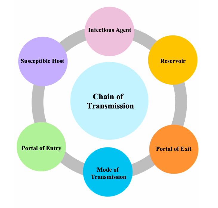
Chain of infection: a teaching model that divides this sequence into six linked stages: infectious agent, reservoir, portal of exit, mode of transmission, portal of entry and susceptible host.

Mode of transmission: the mechanism by which the agent is transferred, for example hands, a contaminated object, droplets, air, food, water, blood or an insect vector.

Infection requires more than the mere presence of a micro-organism. The organism must be capable of causing disease, leave or be carried from a source, survive long enough to reach a new host, enter through a suitable portal and overcome that person's defences. The chain model is useful because breaking one link can stop transmission.

Colonisation, contamination and infection are not the same
  • Contamination means an organism is present on a surface, item, food, water, skin or specimen. It may not be multiplying or causing harm.
  • Colonisation means organisms live and multiply on or in the body without tissue invasion or symptoms. Normal flora commonly colonise skin, mouth, bowel and vagina.
  • Infection occurs when organisms invade or multiply in tissues and trigger local or systemic disease.
  • Infectious disease is infection that can be transmitted directly or indirectly to another person; not every infection is readily transmissible between people.
2. The six links in the chain of infection
LinkWhat it meansExamplesHow to break the link
1. Infectious agentA microorganism capable of causing disease.Bacteria, viruses, fungi, protozoa, helminths; sometimes toxins produced by microbes.Accurate diagnosis, targeted antimicrobial treatment when indicated, sterilisation/disinfection and antimicrobial stewardship.
2. ReservoirThe usual habitat in which the agent lives, grows or persists.Humans, animals, water, soil, food, equipment or a health-care environment.Case management, cleaning, safe waste disposal, vector control, food and water safety.
3. Portal of exitThe route by which the agent leaves the reservoir.Cough droplets, sputum, blood, faeces, urine, wound drainage, genital secretions, placental/umbilical blood.Respiratory hygiene, wound dressings, safe injection practice, covering containers and proper linen/waste handling.
4. Mode of transmissionHow the agent travels to another person.Hands, equipment, droplets, air, food, water, needles, transfusion, mosquitoes or flies.Hand hygiene, PPE, cleaning, safe food/water, sharps safety and transmission-based precautions.
5. Portal of entryThe route by which the agent enters the new host.Respiratory tract, mouth, gastrointestinal tract, broken skin, eyes, nose, genital tract, bloodstream.Aseptic technique, wound care, PPE, safe devices, condoms where appropriate, vaccination and post-exposure procedures.
6. Susceptible hostA person whose defences are insufficient to prevent infection.Newborns, older adults, malnourished people, pregnant women, patients with chronic disease, burns or immunosuppression.Immunisation, nutrition, chronic-disease care, prophylaxis when prescribed and early recognition/treatment.
Link 1: Infectious agent

The infectious agent may be a bacterium, virus, fungus, protozoan or helminth. Its ability to cause disease depends on properties such as infectious dose, virulence, ability to attach to cells, toxin production, resistance to drying or disinfectants, and ability to evade immune responses. For example, a small number of some organisms may be enough to initiate infection, while others require a large inoculum.

Clinical point: Antimicrobials are not a substitute for infection prevention. They may reduce an agent in a reservoir, but unnecessary or incorrect use selects resistant organisms and can make future infection harder to treat.
Link 2: Reservoir

A reservoir is the natural place where an agent normally survives or multiplies. A human reservoir can be a symptomatic patient, a person in the incubation period, a person with a mild infection, or an asymptomatic carrier. Animals may be reservoirs for zoonotic infections. Environmental reservoirs include contaminated water, damp equipment, soil and improperly stored food.

  • Human reservoir: respiratory secretions, skin lesions, blood, bowel, urinary and genital tract.
  • Environmental reservoir: water systems, sinks, poorly disinfected equipment, contaminated linen, food-preparation surfaces and waste.
  • Animal/vector reservoir: animals or arthropods that maintain an organism in nature; this differs from a simple mechanical carrier.
Link 3: Portal of exit

Organisms leave a reservoir through a body fluid or route. The important question is not only “what disease is this?” but also “what material can carry the organism out?” Respiratory organisms may leave in secretions generated by coughing, sneezing, talking or procedures. Enteric organisms leave in faeces or vomitus. Blood-borne organisms leave in blood and can be transferred by needles, unsafe injections or transfusion. A wound with drainage can be a portal of exit when the drainage contaminates hands, linen or equipment.

3. Modes of transmission

The mode of transmission is the link nurses most often interrupt directly. A single infection can have more than one possible route; the dominant route determines the most useful precaution.

A. Contact transmission

Contact transmission is transfer through physical touch. It is the commonest mechanism for many health-care-associated infections.

TypeHow it occursExamples and prevention
Direct contactImmediate person-to-person transfer through touching skin, mucosa or body fluids.Touching an infected wound without gloves; intimate/sexual contact; exposure to drainage. Use hand hygiene, gloves when exposure is anticipated, dressings and safe care of lesions.
Indirect contactTransfer through an intermediate contaminated object, often called a fomite.Hands move organisms from one patient to another; a stethoscope, bed rail, thermometer, dressing trolley, linen or phone becomes contaminated. Clean equipment between patients and perform hand hygiene at the correct moments.

A frequent error is to think gloves replace hand hygiene. Gloves can become contaminated; hands may become contaminated during removal, and gloves may spread organisms from a “dirty” task to a clean task. Clean hands before putting on gloves and immediately after taking them off.

B. Droplet transmission

Droplet transmission occurs when relatively large respiratory particles are propelled a short distance and land on another person's mouth, nose or eyes, or on nearby surfaces. Coughing, sneezing, shouting and some clinical procedures increase respiratory secretion exposure. The key prevention approach is source control, spatial separation, hand hygiene, eye/face protection according to risk and appropriate masking as indicated by the clinical situation and local policy.

Remember: droplets are not the same as airborne particles. Droplet spread is generally associated with close range; airborne spread involves small particles that can remain suspended and travel with air currents. Follow current local IPC guidance for disease-specific precautions.
C. Airborne transmission

Airborne transmission involves very small particles or droplet nuclei that remain suspended in air for longer periods and may be inhaled by susceptible persons. Controls rely on reducing exposure to contaminated air: early recognition, prompt placement according to facility capacity, good ventilation, limiting unnecessary movement, respiratory protection where required and following the facility’s tuberculosis/COVID-19/other respiratory infection policy. Opening windows alone is not a complete plan; patient placement, airflow, duration of exposure and PPE must be considered together.

D. Vehicle-borne transmission

A vehicle is a non-living medium that carries an agent to many people. Common vehicles include food, water, medications, blood, intravenous fluids, improperly processed milk and contaminated medical products. This route can produce clusters because several people share the same vehicle.

  • Food/water: prevent through safe sourcing, cooking, storage, handwashing, latrine use and protecting water from faecal contamination.
  • Blood and blood products: screen donors, use sterile single-use equipment, maintain cold chain/quality systems and follow transfusion safety procedures.
  • Medicines/fluids: prepare aseptically, label correctly, avoid using contaminated multi-dose containers unsafely and maintain equipment.
E. Vector-borne transmission

A vector is a living carrier, usually an arthropod. Biological transmission occurs when the organism develops or multiplies inside the vector before transfer. Mechanical transmission occurs when an insect simply carries organisms on its body from contaminated material to food or a surface. Malaria is a familiar example of biological vector-borne transmission via infected female Anopheles mosquitoes. Prevention includes environmental control, eliminating breeding sites where feasible, insecticide-treated nets, protective clothing, early diagnosis and community measures directed by public-health programmes.

F. Blood-borne, inoculation and iatrogenic transmission

Blood-borne transmission occurs when infectious blood or selected body fluids enter the bloodstream or contact non-intact skin or mucous membranes. It may occur through a needlestick injury, cuts from contaminated sharps, unsafe injections, unsafe transfusion, re-use of equipment or occupational exposure. Iatrogenic transmission means transmission related to medical care or procedures.

Sharps-safety routine: use a new sterile needle and syringe for every patient and every injection; do not recap used needles unless an approved specific procedure requires it; activate safety devices if supplied; dispose of sharps immediately at point of use; report and manage exposures urgently using the facility post-exposure protocol.
4. Portals of entry and the susceptible host

The same route may be both a portal of exit for one person and an entry portal for another. Common portals of entry include:

  • Respiratory tract: inhalation of droplets or airborne particles.
  • Gastrointestinal tract: ingestion through contaminated fingers, food, water or utensils.
  • Genitourinary tract: sexual contact or instrumentation.
  • Broken skin: wounds, burns, ulcers, dermatitis, invasive lines and injection sites.
  • Mucous membranes: eyes, nose and mouth exposed to splashes.
  • Placenta/vertical pathway: transmission from mother to baby before, during or shortly after birth for some infections.

Host susceptibility changes with age, nutritional status, pregnancy, vaccination history, chronic illness, use of immunosuppressive therapy, skin integrity, invasive devices and dose of exposure. In clinical practice, prevention is particularly important for neonates, people with burns, surgical patients, patients living with HIV or receiving cancer therapy, and frail older adults.

5. Breaking the chain: practical infection-prevention actions
Where action is aimedPractical measuresWhy it works
Agent/reservoirEarly assessment, testing as ordered, appropriate treatment, cleaning/disinfection, safe disposal of excreta and waste.Reduces the organism at its source or prevents its persistence in the environment.
Portal of exitRespiratory hygiene, tissues/masks where indicated, covering draining wounds, closed drainage systems, containing spills.Prevents infectious material reaching hands, surfaces or air.
Mode of transmissionHand hygiene; cleaning shared equipment; safe food/water; PPE; ventilation; safe injections; environmental and vector control.Stops transfer between the source and another person.
Portal of entryAseptic technique, skin preparation, device care, wound protection, eye/face protection when splash risk exists.Protects vulnerable body sites from inoculation.
Susceptible hostVaccination, nutrition, prevention/treatment of chronic disease, prescribed prophylaxis and patient education.Strengthens defences or reduces the likelihood of severe disease.
Standard Precautions

Standard Precautions are the minimum infection-prevention practices used for the care of every patient, regardless of known or suspected infection status. They include hand hygiene; risk-based use of gloves, gown, mask and eye/face protection; respiratory hygiene; safe injection and sharps practice; cleaning and disinfection of equipment and environment; safe handling of linen, waste and specimens; and appropriate placement of patients. They are based on the principle that blood, body fluids, secretions, excretions, non-intact skin and mucous membranes may carry infectious organisms.

Transmission-Based Precautions

When a disease is known or suspected to spread by a particular route, additional contact, droplet or airborne precautions may be required on top of Standard Precautions. The exact arrangement depends on the patient condition, facility capacity and national/facility guidance. Nurses should identify the suspected route early, communicate it during handover, post appropriate signage where used locally and avoid moving the patient unnecessarily.

6. Applying the cycle: clinical scenarios

Scenario 1: Coughing patient in triage. A patient arrives coughing and febrile in a crowded waiting area. Respiratory secretions are the portal of exit; droplets or, for selected diseases, airborne particles may be the mode; eyes, nose and mouth are portals of entry for staff and other patients. Priorities are rapid screening, respiratory hygiene/source control, spacing or placement as available, hand hygiene, appropriate PPE and communication with the receiving team.

Scenario 2: Dressing a heavily draining wound. Drainage is the portal of exit. Unclean hands, a reused dressing trolley or improperly cleaned equipment can become the transmission mode. Protect the wound, prepare clean supplies, use gloves and other PPE according to splash risk, avoid touching clean items with contaminated gloves, clean the environment/equipment and perform hand hygiene after glove removal.

Scenario 3: Diarrhoea outbreak on a ward. Faeces are the portal of exit and the mouth is the portal of entry. Hands, food, water, shared toilets and surfaces may act as vehicles/fomites. Strengthen handwashing with soap and water where appropriate, safe disposal of excreta, cleaning of high-touch surfaces, safe food/water, patient placement and rapid reporting according to facility procedures.

Scenario 4: Used needle after injection. Blood is the reservoir material and portal of exit; the hollow needle can inoculate blood through broken skin, creating a portal of entry. Do not recap. Place it immediately in a sharps container. If an exposure occurs, wash the area without squeezing aggressively, report at once and follow the occupational exposure/post-exposure protocol without delay.

7. Nursing and EMT responsibilities
  • Perform hand hygiene at the correct moments and before moving from a contaminated task to a clean task on the same patient.
  • Assess the likely transmission route from symptoms, diagnosis, procedures and body-fluid exposure—not from appearance alone.
  • Select PPE by risk assessment: what splash, contact or respiratory exposure is likely?
  • Use aseptic non-touch technique for invasive procedures, dressing changes and preparation of sterile items.
  • Clean shared equipment between patients and ensure reusable items are reprocessed correctly.
  • Use safe sharps practice and report occupational exposures immediately.
  • Educate patients and caregivers about hand hygiene, cough etiquette, safe food/water, wound care and when to seek help.
  • Document and communicate isolation/precaution needs at transfer and handover while maintaining confidentiality and dignity.
8. Common examination mistakes
  • Writing that airborne and droplet transmission are identical. State the difference in particle behaviour and control measures.
  • Calling every object a reservoir. An object may be a contaminated fomite; a reservoir is the habitat in which the agent normally survives or multiplies.
  • Listing PPE without explaining which link it interrupts. Link each item to portal of exit, mode of transmission or portal of entry.
  • Forgetting that an asymptomatic carrier may be a human reservoir.
  • Writing “use gloves” as the only answer. Include hand hygiene, correct removal, cleaning and safe workflow.
  • Assuming antibiotics break every chain. They do not treat viral infections and misuse contributes to resistance.
Quick revision questions
  1. Name the six links in the chain of infection in the correct order.
  2. Differentiate a reservoir, a portal of exit and a vehicle using an example of diarrhoeal disease.
  3. Give three actions that break contact transmission in a busy outpatient department.
  4. Why are hand hygiene and equipment cleaning both necessary when gloves have been used?
  5. Explain how a needlestick injury can complete the transmission cycle.
  6. A patient is coughing in triage. Identify the likely portal of exit, possible modes of transmission and three immediate nursing actions.
Further reading
Revision tip: For any infection-control question, draw six small boxes for the chain. Put the case details into the boxes, then state one action that breaks the most important link. This gives a clear, clinically reasoned answer instead of a memorised list.

Transmission Cycle and Modes of Transmission Read More »

Microorganisms and Their Characteristics

Microorganisms and Their Characteristics

Microorganisms and Their Characteristics

Micro-organisms differ in size, cellular organisation, shape, habitat, nutrition, metabolism, environmental requirements, growth, reproduction and capacity to cause disease. These features help the nurse and laboratory worker predict where a microbe survives, how it spreads, what specimen to collect and which infection-prevention measures are important.

Core idea: a characteristic is a feature used to describe, classify or identify a micro-organism. A laboratory combines morphology, staining, culture, biochemical tests, antigen tests and molecular methods rather than relying on one characteristic alone.
Learning Objectives
  • Describe the major characteristics used to classify and identify micro-organisms.
  • Explain how nutrients, oxygen, temperature, pH and water affect microbial growth.
  • Differentiate major forms of microbial reproduction and growth.
  • Relate microbial characteristics to virulence, resistance and infection prevention.
1. Characteristics Used to Describe Micro-organisms
CharacteristicQuestion answeredClinical or laboratory importance
Cellular organisationProkaryotic, eukaryotic or acellular?Bacteria are prokaryotic; fungi/protozoa are eukaryotic; viruses are acellular.
MorphologyWhat is the size, shape and arrangement?Cocci, bacilli, vibrios, yeasts, mould hyphae and viral shape give early clues.
Staining reactionHow does it behave with stains?Gram-positive, Gram-negative and acid-fast findings guide early assessment.
MetabolismHow does it obtain energy and nutrients?Fermentation and enzyme tests give biochemical identification clues.
Environmental needsWhat oxygen, temperature, pH, moisture or salt level supports growth?Determines specimen transport, culture method, food safety and survival.
Reproduction / life cycleHow does it multiply, persist and spread?Binary fission, budding, cysts, spores and host-cell viral replication.
Genetic and antigenic featuresWhat genes or surface antigens are present?Useful in PCR, strain typing, outbreak investigation and resistance testing.
VirulenceHow does it cause or worsen disease?Capsules, toxins, enzymes, adhesins and biofilm formation can increase pathogenicity.
2. Morphological Characteristics
Size, shape and arrangement

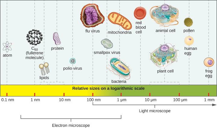
Size is measured in micrometres (µm) or nanometres (nm). A typical bacterium is around 1 µm, while many viruses are around 100 nm. Fungi and protozoa are often larger than bacteria, but detailed identification still needs microscopy.

  • Bacteria: cocci, bacilli, coccobacilli, vibrios, spirilla/spirochaetes and branching filamentous forms.
  • Arrangement: pairs, chains, clusters, tetrads and packets result from the plane of cell division and whether cells stay attached.
  • Fungi: unicellular yeasts, multicellular moulds with hyphae, and dimorphic forms.
  • Protozoa: may move by pseudopodia, flagella or cilia; many have trophozoite and cyst stages.
  • Viruses: helical, icosahedral, complex, enveloped or non-enveloped.
Remember: morphology is an early clue, not final identification. Gram-positive cocci in clusters suggest staphylococci, but culture or another validated test is needed to confirm the organism.
3. Cellular Organisation and Staining
GroupKey characteristicWhy it matters
BacteriaProkaryotic; no true nucleus; 70S ribosomes; usually peptidoglycan cell wall.Can be stained, cultured and tested for antimicrobial susceptibility.
FungiEukaryotic; chitin/glucan wall and ergosterol-containing membrane.Structural differences provide antifungal drug targets.
ProtozoaSingle-celled eukaryotes with a nucleus and specialised feeding/movement structures.Correct timing and preservation of blood, stool or other specimens is important.
VirusesAcellular DNA or RNA genome in capsid, with or without envelope.Need host cells or antigen/molecular testing; ordinary bacterial culture media will not grow them.

Gram staining separates bacteria by cell-envelope structure. Gram-positive bacteria retain crystal violet and appear purple because of a thick peptidoglycan wall. Gram-negative bacteria have thin peptidoglycan plus an outer membrane and appear pink/red after counterstaining.

4. Habitat and Ecological Characteristics

A habitat is where an organism normally lives, grows or persists. Microbes occur in soil, water, food, sewage, plants, animals, skin, mucous membranes and healthcare environments. Their habitat depends on nutrients, moisture, temperature, pH, oxygen and suitable surfaces.

  • Free-living organisms survive independently in the environment.
  • Commensals live on or in the body without causing disease in their normal site.
  • Mutualists benefit both microbe and host.
  • Parasites benefit at the host's expense.
  • Opportunists cause disease when immunity is low, barriers are broken, normal flora is disturbed or devices are present.
Reservoir versus source

A reservoir is the usual habitat in which an agent lives and multiplies. A source is the person, animal, object or substance from which a host acquires infection. Correctly identifying both is important during infection-prevention investigation.

5. Nutritional and Metabolic Characteristics

Cellular microbes need energy, carbon, water and essential chemical elements. Their metabolic pattern helps classify and identify them. Catabolism breaks substances down to release energy; anabolism uses energy to build cellular material.

TermMeaningImportance
PhototrophUses light as energy source.Common among environmental microbes and algae.
ChemotrophUses chemical compounds as energy source.Most medically important bacteria and fungi.
AutotrophUses carbon dioxide as major carbon source.Important in environmental microbiology.
HeterotrophUses organic compounds for carbon.Most human pathogens.
FermentationEnergy production without oxygen as terminal electron acceptor.Supports growth in low-oxygen environments and aids biochemical identification.
6. Environmental Requirements for Growth
Temperature

Each microbe has minimum, optimum and maximum temperatures. Many human pathogens are mesophiles and grow best near body temperature. Cold slows growth of many microbes but does not sterilise food or equipment.

pH, moisture and salt

Most pathogens prefer near-neutral pH, while acidophiles prefer acidic conditions and alkaliphiles prefer alkaline conditions. Water is essential for metabolism. High salt or sugar lowers available water and inhibits many organisms, although some tolerate it well.

Oxygen requirements
TypeOxygen relationshipPractical implication
Obligate aerobeRequires oxygen.Grows where oxygen is available.
Obligate anaerobeOxygen is harmful or lethal.Needs oxygen-free specimen collection and transport.
Facultative anaerobeGrows with or without oxygen, usually better with oxygen.Common among human-associated bacteria.
MicroaerophileRequires low oxygen concentration.Needs special culture conditions.
Aerotolerant anaerobeDoes not use oxygen but tolerates it.Can survive in oxygenated environments while fermenting.
7. Growth, Reproduction and Persistence

Most bacteria multiply by binary fission: DNA replicates, the cell elongates, a septum forms and one cell divides into two daughter cells. The generation time is the time for a population to double under stated conditions.

  1. Lag phase: cells adapt, repair and synthesise enzymes; cell number changes little.
  2. Log/exponential phase: rapid division; many antimicrobials work best on actively dividing cells.
  3. Stationary phase: nutrient depletion and waste accumulation; growth rate equals death rate.
  4. Death phase: viable cells decline as conditions become unfavourable.
  • Budding: common in yeasts.
  • Fragmentation: occurs in filamentous forms.
  • Cysts/endospores: enhance survival in different groups; bacterial endospores are survival structures, not reproductive structures.
  • Viral replication: occurs only after a virus enters a compatible host cell.
8. Biofilms, Virulence and Resistance

A biofilm is a structured community of microbes attached to a surface and embedded in a self-produced matrix. Biofilms form on teeth, wounds, urinary catheters, vascular lines and other devices, and can make organisms more difficult to remove or treat.

  • Adhesins and pili support attachment to tissue or devices.
  • Capsules may reduce phagocytosis and promote persistence.
  • Toxins and enzymes may damage tissue or help spread.
  • Gene transfer can spread antimicrobial-resistance genes between bacteria.
  • Antigenic variation helps some organisms avoid host immunity.
Nursing relevance: prevent device-associated infection through hand hygiene, aseptic insertion, closed-system care, daily review of device need, correct cleaning and prompt removal of unnecessary devices.
9. Applying Characteristics in Nursing Practice
Deep wound with offensive discharge

A contaminated deep wound with swelling and foul discharge may involve anaerobic organisms. Collect ordered specimens correctly, use appropriate transport, monitor for sepsis, practise standard precautions and escalate deterioration quickly.

Recurrent catheter-associated symptoms

Think about microbial adherence and biofilm. Do not break the closed system unnecessarily; collect urine from the sampling port using aseptic technique according to policy, review catheter need and report fever, flank pain, confusion or sepsis indicators promptly.

Food kept at room temperature

Food held in an unsafe temperature range may allow rapid growth of some bacteria. Safe food handling needs hygiene, thorough cooking, prompt refrigeration and prevention of cross-contamination.

Common Examination Mistakes
  • Equating every microbe with a pathogen.
  • Confusing growth with increase in cell size only; it usually means increase in cell number.
  • Calling all anaerobes unable to tolerate oxygen.
  • Confusing bacterial endospores with reproduction.
  • Assuming biofilms are only dental plaque.
  • Forgetting that metabolic traits are used for laboratory identification.
Quick Revision Questions
  1. State eight characteristics used to classify or identify micro-organisms.
  2. Explain how temperature, pH, moisture and oxygen influence growth.
  3. Differentiate the five oxygen-requirement groups.
  4. Describe the four phases of the bacterial growth curve.
  5. Explain why biofilms matter in catheter and wound care.
  6. How can biochemical characteristics help laboratory identification?
Further Reading

Microorganisms and Their Characteristics Read More »

Structure of Micro-organisms

Structure of Micro-organisms

Structure of Micro-organisms

Micro-organisms, commonly called microbes, are organisms or infectious agents that are too small to be seen clearly with the unaided eye. They are found in soil, water, air, food, animals, plants, the human body and healthcare environments. Some are useful or harmless; some cause disease when they enter the wrong site, multiply excessively, produce toxins or overcome host defences.

This topic explains what microbes are made of, how they differ structurally and why the structure of a microbe matters in microscopy, staining, treatment, infection prevention and nursing care.

Clinical connection: structure predicts behaviour. For example, bacterial cell-wall structure influences Gram staining and antibiotic susceptibility; the presence of a viral envelope influences susceptibility to some disinfectants; and bacterial endospores require much stronger decontamination methods than ordinary vegetative cells.
Learning Objectives
  • Define micro-organism, pathogen, commensal and opportunistic pathogen.
  • Classify microbes as cellular prokaryotes, cellular eukaryotes or acellular infectious agents.
  • Describe the major structures of bacteria, fungi, protozoa and viruses.
  • Relate bacterial morphology and cell-wall structure to laboratory identification and patient care.
  • Apply microbiology structure knowledge to infection prevention and safe nursing practice.
1. What Is a Micro-organism?

A micro-organism is a microscopic living organism. In health sciences, the term is commonly used for bacteria, fungi, protozoa and microscopic parasites. Viruses are also studied in microbiology although they are acellular: they are not made of cells and can reproduce only inside a living host cell.

TermMeaningExample / clinical significance
MicrobeA microscopic organism or infectious agent.Bacterium, fungus, protozoan or virus.
PathogenA micro-organism capable of causing disease.Mycobacterium tuberculosis causes tuberculosis.
Commensal / normal floraA microbe that normally lives on or in the body without causing disease in its usual site.Some gut bacteria assist normal body processes but may cause infection if introduced into wounds or blood.
Opportunistic pathogenA microbe that causes disease when host defences are reduced or when it reaches an abnormal site.Candida may overgrow after antibiotics, in diabetes or immunosuppression.
ContaminantA microbe unintentionally introduced into a specimen, item or area.Skin flora contaminating a poorly collected blood sample may give a misleading result.
Important distinction: infection does not always mean disease

Colonisation means microbes are present and multiplying without tissue invasion or symptoms. Infection means invasion and multiplication in the host; disease occurs when infection produces tissue damage or symptoms. A nurse must collect specimens correctly and interpret results with the patient's clinical condition, not merely the presence of an organism.

2. Broad Classification of Micro-organisms
A. Cellular Micro-organisms
GroupCell typeNucleus / organellesTypical examplesKey point
BacteriaProkaryoticNo true nucleus; no membrane-bound organelles.Staphylococcus, Streptococcus, Escherichia coli, Vibrio choleraeUsually reproduce by binary fission; have 70S ribosomes; most have peptidoglycan cell walls.
ArchaeaProkaryoticNo true nucleus; structurally and genetically distinct from bacteria.Environmental archaeaImportant in classification; not a routine major cause of human infection.
FungiEukaryoticTrue nucleus and membrane-bound organelles.Candida, Aspergillus, CryptococcusCell wall contains chitin and glucans; can occur as yeasts or moulds.
ProtozoaEukaryoticTrue nucleus and organelles.Plasmodium, Giardia, EntamoebaUsually single-celled; many have trophozoite and cyst stages.
HelminthsEukaryotic multicellular parasitesTrue nuclei and organ systems.Roundworms, tapeworms, flukesAdult worms are visible, but eggs/larvae are microscopic and are studied in microbiology/parasitology.
Microscopic algaeEukaryoticTrue nucleus and chloroplasts in many species.Mostly environmental speciesUsually not central to routine human infection but important in general classification.
B. Acellular Infectious Agents
  • Viruses: consist of nucleic acid enclosed in a protein coat, sometimes with a lipid envelope. They depend on host cells to replicate.
  • Prions: infectious misfolded proteins that contain no nucleic acid. They are rare but important because they are unusually resistant to ordinary decontamination.
  • Viroids: small infectious RNA molecules mainly associated with plant disease; they are not a routine cause of human disease.
High-yield classification: Bacteria and archaea are prokaryotes. Fungi, protozoa, helminths and algae are eukaryotes. Viruses are acellular.
3. Size: Why a Microscope Is Needed

Microbes differ greatly in size. A typical bacterium is around 1 micrometre (µm), while many viruses are measured in nanometres (nm). Most protozoa and fungal cells are larger than bacteria, but they still require microscopy for detailed examination.

Approximate scaleWhat may be seenPractical meaning
10–100 nmMany virusesToo small for ordinary light microscopy; special methods are needed.
0.5–5 µmMany bacteriaUsually visualised after staining by light microscopy.
5–100 µmYeasts, protozoa, some fungal elementsMay be seen with light microscopy; morphology helps identification.
Micrometres to centimetresHelminth eggs/larvae to adult wormsEggs/larvae may be found microscopically; adult worms may be visible.
Units to know
  • 1 millimetre (mm) = 1,000 micrometres (µm).
  • 1 micrometre (µm) = 1,000 nanometres (nm).
  • Therefore, a 100 nm virus is far smaller than a 1 µm bacterium.
4. Prokaryotic and Eukaryotic Cell Structure
FeatureProkaryotic cell: bacteriaEukaryotic cell: fungi / protozoa / helminthsWhy it matters
NucleusNo membrane-bound nucleus; DNA lies in a nucleoid.True nucleus enclosed by a nuclear membrane.Major classification feature.
DNAUsually one circular chromosome; may have plasmids.Multiple linear chromosomes inside nucleus.Plasmids can carry antimicrobial-resistance genes.
OrganellesNo membrane-bound organelles.Mitochondria, endoplasmic reticulum, Golgi apparatus and other organelles present.Provides different possible drug targets.
Ribosomes70S ribosomes.80S cytoplasmic ribosomes.Some antibacterial medicines selectively target bacterial ribosomes.
Cell wallUsually peptidoglycan; absent in Mycoplasma.Fungal wall contains chitin/glucans; protozoa lack a bacterial-type peptidoglycan wall.Cell-wall differences influence staining and therapy.
DivisionUsually binary fission.Mitosis; some have sexual stages.Explains growth and life cycles.
5. General Structure of a Bacterial Cell

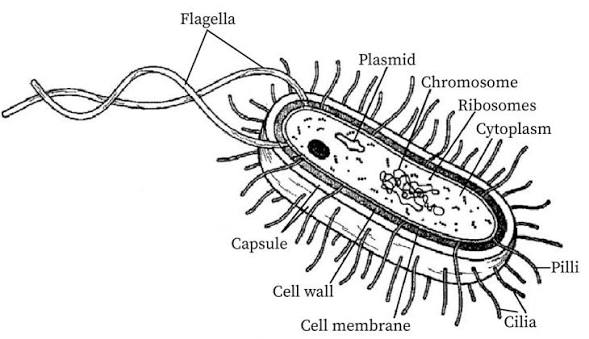
A bacterium is a complete living prokaryotic cell. Although small, it can take in nutrients, make proteins, generate energy, grow, respond to its environment and reproduce independently by binary fission.

Essential Bacterial Structures
StructureDescriptionMain functionClinical relevance
Capsule / glycocalyxOuter sticky layer in some bacteria, often polysaccharide.Adherence, protection from drying and sometimes resistance to phagocytosis.May increase virulence; can help bacteria form biofilms on catheters and devices.
Cell wallRigid layer outside the cell membrane; in most bacteria contains peptidoglycan.Maintains shape and prevents osmotic lysis.Basis of Gram staining; target of several antibiotics.
Cell membraneSelective phospholipid-protein barrier beneath wall.Controls entry/exit; involved in energy generation and transport.Damage to the membrane can kill the cell; essential in every bacterium.
CytoplasmWater-based internal matrix containing enzymes, nutrients, ribosomes and inclusions.Site of many metabolic reactions.Metabolism determines growth requirements and laboratory tests.
NucleoidRegion containing the main circular bacterial chromosome.Stores essential genetic information.Genetic changes can alter virulence or drug susceptibility.
PlasmidsSmall extra-chromosomal circular DNA molecules.Carry additional traits that can be shared between bacteria.May carry genes for antimicrobial resistance or toxin production.
70S ribosomesSmall protein-synthesising particles.Make bacterial proteins.Target of several antibacterial drug groups.
FlagellaLong, whip-like protein filaments.Motility and chemotaxis.Motility can assist spread through tissues or fluids.
Pili / fimbriaeShort, hair-like surface projections.Attachment; some sex pili transfer DNA between bacteria.Adherence supports colonisation; DNA transfer can spread resistance.
EndosporeDormant, highly resistant survival form produced by some bacteria.Survives heat, drying and chemicals; not a reproductive method.Bacillus and Clostridium spores require strict decontamination procedures.
Bacterial endospores: do not confuse with fungal spores

A bacterial endospore is a survival structure formed inside one bacterial cell under unfavourable conditions. One vegetative cell forms one endospore and, when conditions improve, one vegetative cell emerges. It does not increase bacterial numbers. Fungal spores are commonly reproductive structures.

6. Bacterial Shape and Arrangement

Morphology means the form, shape and structural arrangement of an organism. In the laboratory, shape and arrangement give early clues but do not identify a species by themselves.

ShapeDescriptionTypical arrangement / example
CocciSpherical or oval bacteria.Pairs (diplococci), chains (streptococci), clusters (staphylococci), tetrads or packets.
BacilliRod-shaped bacteria.Single rods, pairs, chains or palisades; Escherichia coli is a common rod.
CoccobacilliVery short rods that may appear oval.Intermediate appearance between cocci and bacilli.
VibriosComma-shaped curved rods.Vibrio cholerae is the major clinical example.
SpirillaRigid spiral-shaped cells with external flagella.Usually environmental organisms.
SpirochaetesFlexible, slender corkscrew-shaped bacteria.Motile by axial filaments; includes Treponema species.
Filamentous / branching bacteriaLong branching filaments that can resemble fungi.Includes actinomycetes such as Actinomyces.
Memory aid: Staph = clusters like a bunch of grapes; Strep = chains like a string of beads; Diplo = pairs; Bacilli = rods; Vibrio = comma.
7. The Bacterial Cell Envelope and Gram Staining

The cell envelope includes the cell membrane and structures outside it. Gram staining divides bacteria into Gram-positive and Gram-negative groups according to differences in their cell-wall envelope. This is one of the first and most useful laboratory tests in bacteriology.

FeatureGram-positive bacteriaGram-negative bacteria
PeptidoglycanThick layer.Thin layer.
Outer membraneAbsent.Present outside the thin peptidoglycan.
Stain appearanceRetains crystal violet: purple/blue.Decolourised then takes counterstain: pink/red.
Outer membrane componentsNo lipopolysaccharide outer membrane.Contains lipopolysaccharide; lipid A is associated with endotoxin activity.
Practical implicationCell-wall characteristics influence antibiotic selection.Outer membrane can limit entry of some antimicrobial agents.
Limitations of the Gram stain
  • It is a rapid classification test, not a final species identification.
  • Old, damaged or poorly stained cultures may give unreliable results.
  • Mycobacterium has a waxy cell wall and is better assessed with acid-fast staining.
  • Mycoplasma lacks a cell wall, so it does not Gram stain in the usual way.
8. Structure of Fungi

Fungi are eukaryotic organisms. Their cell wall contains chitin and glucans, and their cell membrane contains ergosterol rather than cholesterol. These structural differences are important targets for antifungal therapy.

  • Yeasts: unicellular fungi, often round or oval; many reproduce by budding. Example: Candida.
  • Moulds: multicellular fungi formed by branching filaments called hyphae; a mass of hyphae is called a mycelium. Example: Aspergillus.
  • Dimorphic fungi: can change form, often existing as moulds in the environment and yeasts/tissue forms in the host depending on conditions.
  • Fungal spores: may be reproductive and dispersal structures; they are different from bacterial endospores.
Nursing relevance: prolonged broad-spectrum antibiotics, diabetes, indwelling devices, malnutrition and immunosuppression can increase susceptibility to fungal overgrowth or infection. Observe for oral thrush, vulvovaginal symptoms, skin-fold lesions and signs of invasive infection in high-risk patients.
9. Structure of Protozoa and Helminths
Protozoa

Protozoa are single-celled eukaryotes. They have a nucleus, cytoplasm, cell membrane and specialised structures for movement, feeding or attachment. Depending on the group, they may move by pseudopodia, flagella or cilia. Many parasitic protozoa have a feeding/multiplying trophozoite stage and a resistant/transmission cyst stage.

  • Plasmodium: malaria parasite with stages in mosquitoes and human liver/red cells.
  • Entamoeba histolytica: may form cysts and trophozoites; can cause amoebic dysentery.
  • Giardia duodenalis: has flagella and an attachment disc; causes diarrhoeal disease.
Helminths

Helminths are multicellular parasitic worms. Their eggs and larval forms are often examined microscopically in stool, urine, blood or tissue specimens. They include nematodes (roundworms), cestodes (tapeworms) and trematodes (flukes).

10. Structure of Viruses

A virus is not a cell. The complete infectious particle is called a virion. It contains a genome of either DNA or RNA, never both as its genomic material, enclosed by a protein coat called a capsid. Some viruses also have a lipid envelope derived from host-cell membranes.

Viral componentDescriptionImportance
GenomeDNA or RNA carrying viral genetic instructions.Directs production of new viral components inside host cells.
CapsidProtein coat surrounding the genome, built from capsomeres.Protects genome and helps determine viral shape.
EnvelopeLipid membrane present in enveloped viruses.Contains viral glycoprotein spikes; relatively fragile to drying, heat, detergents and some disinfectants.
Spikes / attachment proteinsSurface proteins on capsid or envelope.Bind specific receptors on host cells and influence tissue tropism.
Viral enzymesPresent in some viruses.Assist replication when host-cell enzymes are insufficient.
Virus morphology
  • Helical: capsid proteins arranged in a spiral around nucleic acid.
  • Icosahedral: roughly symmetrical capsid with 20 triangular faces.
  • Complex: more complicated structure, for example bacteriophages that infect bacteria.
  • Enveloped or non-enveloped: enveloped viruses have a lipid envelope; non-enveloped viruses have only nucleic acid and capsid.
Infection-prevention link: enveloped viruses are generally more easily disrupted by soap and many routine disinfectants than non-enveloped viruses. However, all cleaning and disinfection must follow the product instructions, recommended contact time and local infection-prevention policy.
11. From Structure to Safe Nursing Practice
  • Specimen collection: a correct specimen from the correct site, collected with aseptic technique and labelled accurately helps the laboratory distinguish true infection from contamination.
  • Hand hygiene: breaks transfer of bacteria, fungi, viruses and parasites between patients, staff, equipment and the environment.
  • Device care: capsules and biofilms help some bacteria adhere to catheters, cannulae and other devices; follow aseptic insertion and maintenance bundles.
  • Decontamination: bacterial spores are highly resistant; use approved sterilisation/disinfection methods rather than assuming all organisms are equally easy to kill.
  • Antimicrobial stewardship: bacterial structures provide drug targets, but unnecessary antibiotics select resistant bacteria. Antibiotics do not treat viral infections.
  • Patient education: explain that not every “germ” causes disease, but hygiene, safe water/food, immunisation, correct medicine use and early care-seeking reduce infection risk.
Clinical scenario

A patient with a urinary catheter develops fever and cloudy urine. The nurse should not simply report “infection.” Consider the device as a potential surface for bacterial adherence and biofilm formation; assess the patient, obtain specimens correctly when ordered, maintain aseptic technique, review catheter need, monitor vital signs and escalate signs of sepsis promptly.

Common Examination Mistakes
  • Writing that viruses are prokaryotes. Viruses are acellular, not prokaryotic or eukaryotic cells.
  • Confusing bacterial endospores with fungal reproductive spores.
  • Stating that all microbes cause disease. Many are harmless, beneficial or opportunistic only under certain conditions.
  • Forgetting that Gram-negative bacteria have an outer membrane and thin peptidoglycan.
  • Calling every round bacterium “staphylococcus.” Shape and arrangement are clues, not a full identification.
  • Using antibiotics for viral illness without a bacterial indication.
Quick Revision Questions
  1. Classify bacteria, fungi, protozoa, helminths and viruses as prokaryotic, eukaryotic or acellular.
  2. Draw and label a typical bacterial cell, stating one function of each structure.
  3. Differentiate a bacterial endospore from a fungal spore.
  4. Compare Gram-positive and Gram-negative bacterial cell envelopes.
  5. Describe the structures of an enveloped virus and explain why the envelope matters for infection prevention.
  6. Explain how microbial structure influences specimen collection, antibiotic use and decontamination.
Further Reading

Structure of Micro-organisms Read More »

Emergency Care Governance and Health Facility Teams

Emergency Care Governance and Health Facility Teams

Governance is the way a health system is directed, regulated, financed, monitored and held accountable. In emergency care, governance is what connects a national policy to the person who answers a call, starts first aid, drives an ambulance, triages at the gate, receives a patient in the emergency unit and arranges onward referral. Strong governance makes emergency care safe, coordinated, equitable and continuously improving.

Learning Objectives
  • Explain governance, regulation, management and leadership in emergency care.
  • Describe international, national and local bodies that shape emergency care in Uganda.
  • Identify the roles of a functional health-facility emergency team.
  • Demonstrate safe communication, escalation and structured handover.
1. Meaning of Governance in Emergency Care
ConceptMeaningExample in an emergency unit
GovernanceSetting direction, rules, accountability and oversight for the health system.National standards for ambulances, referral, data reporting and emergency-care quality.
LeadershipInfluencing people to work toward a shared goal.A team leader coordinates a resuscitation while maintaining calm, role clarity and patient dignity.
ManagementPlanning, organising, staffing, budgeting and controlling resources.The facility ensures oxygen, staff rosters, triage space, drugs, equipment maintenance and incident review.
RegulationRules and enforcement that protect the public and professional standards.Registration, scope of practice, ethical requirements and action on unsafe or unqualified practice.
AccountabilityDuty to explain actions, meet standards and correct failures.A delayed referral is reviewed through records and system learning, not hidden or blamed informally.

Key principle: governance is not only “what happens in Kampala.” Every provider contributes through honest documentation, respect for scope, reporting hazards, accepting supervision and treating patients fairly.

2. International Bodies and Their Roles
World Health Organization (WHO)

WHO provides technical guidance, norms and tools that countries adapt to their own resources. Its Emergency Care System Framework describes essential functions at the scene, during transport, in the emergency unit and into early inpatient care. WHO guidance supports governments to assess gaps and strengthen systems for everyday emergencies and major incidents.

  • Develops global standards, evidence-based guidance and training resources.
  • Supports emergency, critical and operative care systems and universal health coverage.
  • Promotes quality care, data use, disaster preparedness and patient safety.
Other international partners
  • International Committee of the Red Cross (ICRC): supports humanitarian action, conflict/injury care and Basic Emergency Care resources with WHO.
  • UN agencies and development partners: may support maternal/newborn health, immunisation, disease control, supply chains, data and emergency preparedness.
  • Professional associations and academic institutions: contribute training, ethics, research and clinical standards.

Important: International guidance informs practice but does not replace Uganda’s laws, Ministry of Health policies, professional regulation, local protocols or your authorised scope.

3. National and Local Governance in Uganda
Level / bodyCore roleEmergency-care relevance
Ministry of HealthNational policy, stewardship, standards, strategic planning, coordination and monitoring.Leads implementation of the National EMS Policy and development of the national emergency-care system.
National EMS structures / referral networkCoordinate emergency-care administration and service delivery across levels.Link pre-hospital response, communication, ambulance transport, emergency units and referral hospitals.
Professional councilsRegistration, discipline, ethics, education standards and professional scope.Protect patients by ensuring providers work competently and accountably.
Local governments and district health teamsLocal planning, supervision, coordination and community engagement.Identify local emergency risks, strengthen referral routes and organise preparedness with facilities.
Health-facility managementLocal readiness, staffing, supplies, records, safety and service improvement.Ensures the emergency unit can function 24 hours and link safely to the next level.
Communities and civil societyDemand accountability, support prevention, first response and appropriate care-seeking.Community awareness can reduce delay in recognising danger signs and seeking help.
National policy context

Uganda’s National EMS Policy was approved in 2021 to guide establishment of a functional ambulance and referral system. The National EMS Strategic Plan covers emergency-care administration and delivery for obstetric, medical, surgical, paediatric and other emergencies. The policy direction is to build one connected system—not isolated ambulances or one emergency room working alone.

4. The Health Facility Emergency Team

A safe emergency response is a team function. The exact staff available differs by level of care, but roles must be clear before a crisis occurs.

Team memberTypical roleWhat good teamwork looks like
Team leader / senior clinicianSets priorities, assigns roles, makes escalation decisions and communicates the plan.Uses short, clear commands; checks understanding; reassesses and calls for help early.
EMT / first responderScene assessment, primary assessment, immediate life support within scope, monitoring, transport and handover.Brings concise facts, anticipates deterioration and maintains equipment readiness.
Nurse / midwifeTriage, observations, emergency nursing/midwifery interventions, medicines within authority, documentation and patient support.Recognises danger signs, coordinates flow and protects privacy/dignity.
Medical officer / clinicianDiagnosis, definitive treatment decisions, supervision, referral and admission/discharge decisions.Provides timely review, delegates safely and confirms the plan.
Laboratory, pharmacy and imaging staffTime-critical tests, blood/medicines and diagnostic support.Receive clear requests, communicate critical results and maintain safe supply systems.
Driver / dispatcher / porter / securityTransport, communication, safe access and operational support.Are included in drills, know call pathways and never work outside their role.
Minimum team behaviours during resuscitation
  1. Declare the leader and roles: “I am leading. You manage airway; you monitor; you document; you call the referral hospital.”
  2. Use closed-loop communication: sender gives a clear instruction; receiver repeats it; sender confirms completion.
  3. Speak up early: any team member may raise a safety concern, deteriorating vital sign, missing equipment or unclear order.
  4. Keep a shared mental model: state the working problem, current priorities, next step and escalation trigger.
  5. Debrief: after the event, discuss what went well, what failed and what must change in the system.
5. Referral and Handover: Governance at the Bedside

Handover is an accountability moment. The receiving provider must receive enough information to continue care safely. A useful format is IMIST or SBAR.

IMIST elementWhat to state
I – IdentificationPatient name/approximate age, sex and identifiers if known.
M – Mechanism / medical complaintWhat happened, onset and relevant mechanism or illness.
I – Injuries / informationKey findings, suspected injuries and major risks.
S – SignsVital signs, conscious level, glucose or other available observations, and trends.
T – Treatment and trendsWhat has been done, response, times, medicines/fluids/oxygen authorised, and pending needs.
Applied Scenarios
Scenario 1: Sudden collapse in the outpatient department

Several people give instructions at once. No one is documenting; the oxygen cylinder is empty.

  • Governance failure: unclear team leadership, poor equipment readiness and no defined role allocation.
  • Immediate response: call for emergency support, assign a leader/roles, begin approved assessment and life support, obtain alternative oxygen/transfer plan.
  • System action after care: report the stock/readiness failure, review the equipment-check SOP and strengthen duty handover.
Scenario 2: Referral with no receiving-facility notification

An unstable patient arrives after a long journey, but the receiving team has not been told and no written referral note is available.

  • This is not a complete referral. The absence of communication and documentation creates duplication, delay and patient-safety risk.
  • The EMT/nurse should give an immediate structured verbal handover, document available facts and report the referral-chain gap through the correct channel.
6. Common Examination Points
  • Governance sets direction and accountability; management organises resources; leadership mobilises people; regulation protects the public.
  • Emergency care requires coordinated functions from scene through transport, emergency unit and early inpatient care.
  • Good teams use role allocation, closed-loop communication, early escalation and debriefing.
  • Professional accountability includes recognising and reporting a system failure, not hiding it.
Revision Questions
  1. Differentiate governance, leadership, management and regulation.
  2. List five actors involved in Uganda’s emergency-care system and state one role for each.
  3. Why must a driver, dispatcher and security worker be included in emergency preparedness?
  4. Explain closed-loop communication with an emergency example.
  5. What information should be included in an IMIST handover?
Further Reading

Emergency Care Governance and Health Facility Teams Read More »

Policies and Guidelines for Emergency Care

Policies and Guidelines for Emergency Care

Emergency care is time-sensitive care for acute illness and injury. A patient with severe bleeding, airway obstruction, sepsis, stroke, eclampsia or major trauma cannot wait for care to be improvised. Policies, standards, guidelines, protocols and standard operating procedures (SOPs) turn the aim of the right care, at the right time, in the right place into safe daily practice for the EMT and the whole emergency-care team.

Why this matters: clear rules reduce dangerous variation, guide early escalation, protect patient rights, support fair triage and make it possible to review whether care was safe and timely.
Learning Objectives
  • Differentiate policy, standard, guideline, protocol, procedure and standing order.
  • Explain the policy principles that guide safe emergency care from first contact to handover.
  • Apply scope of practice, triage, consent, confidentiality, documentation and referral rules to EMT cases.
  • Relate everyday EMT practice to Uganda's emergency-care and referral-system direction.
1. Key Documents That Guide Emergency Care

These documents are related, but they are not interchangeable. In examinations, define the term first, then give a practical emergency-care example.

DocumentMeaningWhat it answers for the EMTExample in emergency care
PolicyA high-level statement that sets direction, authority, priorities and accountability.What must the service achieve and who is responsible?A national policy establishing an organised emergency medical services and referral system.
StandardA minimum measurable level of acceptable quality or performance.What level of care, staffing, equipment or safety is acceptable?An ambulance or emergency unit must have trained staff, essential equipment, medicines, records and infection-prevention measures.
GuidelineEvidence-informed recommendations that help a clinician make safe decisions.What is the recommended approach?ABCDE assessment, triage guidance and early recognition of a deteriorating patient.
ProtocolA defined sequence for a particular clinical situation, often with criteria, limits and escalation points.What do I do now, in this situation?Management of severe hypoglycaemia, uncontrolled external bleeding or suspected sepsis within authorised scope.
Procedure / SOPStep-by-step instructions for carrying out one task consistently.How exactly should the task be done?Cleaning the ambulance, checking oxygen, completing a patient-care record, moving a patient or giving a structured handover.
Standing orderPre-authorised instruction allowing specified trained staff to provide stated care under stated conditions.What may I initiate without waiting for a patient-specific order?Immediate authorised first aid or treatment under the service's approved clinical governance arrangements.
A simple hierarchy to remember
  1. Law and regulation set the legal boundaries for professional practice.
  2. National policy sets the health-system direction and accountability.
  3. National or service standards and clinical guidelines describe expected quality and recommended care.
  4. Local protocols and SOPs translate these requirements into workable steps for a facility, ambulance or team.
  5. Clinical judgement is used within that framework; it does not justify working outside scope or ignoring safety rules.
2. Why Emergency Care Needs Clear Policies and Guidelines

Emergency decisions are often made with incomplete history, distressed relatives, limited resources and a changing patient condition. A good system therefore makes the safe action the expected and repeatable action, rather than relying on memory or personal habit.

  • Patient safety: supports correct identification, infection prevention, safe medicine use, monitoring, safe transfer and early treatment of life threats.
  • Equity: ensures that triage is based on clinical urgency, not money, status, gender, age, disability, nationality or personal connections.
  • Continuity of care: makes sure the patient story, findings, treatment and response travel with the patient from scene to receiving team.
  • Accountability: records show what was found, what was done, when, by whom, the reason for referral and the outcome of handover.
  • Team coordination: clarifies roles, who must be called, when to pre-alert a receiving unit and how to manage overcrowding or multiple casualties.
  • Quality improvement: delays, adverse events, near misses and outcomes can be reviewed so the service improves rather than repeats mistakes.
Exam tip: when asked for the importance of a policy, do not write only “it guides workers.” Link it to patient safety, fairness, continuity, accountability and quality improvement.
3. Core Policy Areas for EMT Practice
3.1 Scope of practice, competence and escalation

Work within your training, certification, equipment, supervision and authorised protocols. A task may be appropriate for one practitioner but unsafe for another. Recognising limits early is professional practice, not weakness.

  • Know what you are authorised and competent to assess, initiate, administer, monitor and document.
  • Call for a higher-level clinician or additional resources early when the patient's need exceeds your scope or available equipment.
  • Never allow pressure from relatives, colleagues or a crowd to make you perform an unauthorised or unsafe procedure.
  • When care is delegated, confirm that the task is appropriate, the person is competent and supervision is available where required.
  • Document deterioration, advice received, escalation made and the patient's response.
3.2 Scene safety, infection prevention and personal protection

Emergency care begins before touching the patient. The EMT must assess hazards to self, colleagues, patient and bystanders.

StepActionReason
1Pause, observe and identify hazards: traffic, fire, electricity, violence, chemicals, unstable structures, weapons, crowds and body fluids.Entering an unsafe scene can create more casualties and stop care for the original patient.
2Use standard precautions: hand hygiene, appropriate PPE, safe sharps practice, cleaning and waste disposal.Every patient may carry an infection risk; precautions protect both patient and provider.
3Request police, fire, rescue, additional ambulance support or crowd control when needed.Early support prevents unsafe delay and supports coordinated rescue.
4Reassess hazards throughout care, transport and handover.Scenes and patient risk can change quickly.
3.3 Triage and prioritisation

Triage is the rapid sorting of patients according to urgency so that immediately life-threatening conditions are identified first. It is both a clinical and an ethical process: available resources are directed according to need while every patient is treated with dignity.

  • Use a structured assessment such as ABCDE: Airway, Breathing, Circulation, Disability and Exposure/Environment.
  • Reassess whenever a patient deteriorates, after treatment, before transport and at handover.
  • In a multiple-casualty incident, follow the incident command and triage system used by the service; do not treat patients merely in arrival order.
  • Communicate respectfully with waiting patients and relatives, explaining delays without disclosing another patient's private information.
3.4 Assessment, treatment and clinical governance

Use the approved clinical pathway: scene safety, primary survey, immediate life-saving interventions, focused history/examination, repeated observations and destination decision. Record baseline and repeat vital signs. Do not delay transport for non-essential procedures when the patient needs definitive care.

Clinical governance means: the organisation is responsible for maintaining safe, effective care through training, supervision, protocols, audit, incident reporting, equipment checks and continuous improvement.
3.5 Referral, transport and handover

Referral is not simply “sending a patient away.” It is a coordinated transfer of responsibility to the level of care that can meet the patient's needs. Select the destination according to the clinical condition, available service, travel time and local referral arrangements.

  • Stabilise immediately reversible life threats within your authorised scope.
  • Contact or pre-alert the receiving facility when a time-critical patient is coming.
  • Monitor during transport; repeat observations and record changes or interventions.
  • Use a structured handover, for example SBAR: Situation, Background, Assessment and Recommendation.
  • State the patient's identity, chief problem, mechanism/onset, key findings, vital-sign trends, treatment, response, allergies where known, risks and outstanding needs.
  • Confirm that a named receiving provider has accepted handover before leaving the patient.
3.6 Consent, confidentiality, dignity and safeguarding

Explain care in clear language the patient can understand, seek consent where the patient has capacity and involve the patient in decisions whenever possible. In a life-threatening emergency where a patient lacks capacity and delay would cause serious harm, emergency care is generally provided in the person's best interests according to law and approved protocol.

  • Expose only the body area necessary for assessment or treatment; preserve warmth and privacy.
  • Do not discuss patient details where bystanders can hear, or share images and records without proper authority.
  • Record refusal of care, what information was given, capacity assessment where relevant, risks explained and the advice/escalation provided.
  • Follow local safeguarding and reporting pathways when a child, older person or vulnerable adult may be experiencing abuse, neglect or exploitation.
3.7 Documentation, incident reporting and quality improvement

“Not documented” may be interpreted as “not done.” Documentation must be factual, timely, legible and free from blame or personal opinion.

  • Date, time, location, patient identity and presenting problem/mechanism of injury.
  • Initial and repeated vital signs, mental status, pain score and relevant assessment findings.
  • Care given, medicine dose/route/time where applicable, response and complications.
  • Referral decision, destination, pre-alert, transport details and the person receiving handover.
  • Consent/refusal issues, equipment failures, safety incidents, safeguarding concerns and required signatures.
4. Uganda's Emergency-Care Policy Direction

Uganda's National Emergency Medical Services Policy (2021) provides a policy foundation for strengthening emergency care, including an organised emergency medical services, ambulance and referral system. The national EMS strategic plan describes an emergency-care continuum that links community response, pre-hospital care, transport, health facilities and definitive care.

The EMT's SAFER sequence
  1. S — Scene safety: assess hazards, wear appropriate PPE and call for support.
  2. A — Assess: conduct an ABCDE primary survey and identify immediate threats.
  3. F — Follow authorised guidance: work within scope, protocol and available resources.
  4. E — Escalate and refer: call for help, choose an appropriate destination and pre-alert where necessary.
  5. R — Record and hand over: document clearly and transfer responsibility safely.
Key point: a good emergency response is not finished when the patient enters a vehicle or reaches a doorway. It ends after safe handover, accurate documentation and linkage to the care the patient still needs.
5. Applying Policy to Clinical Scenarios
Scenario 1: Road traffic crash at night

A motorcyclist is lying on the roadside with severe leg bleeding. Traffic is still moving and bystanders are pulling the patient up.

  • Policy-guided priorities: protect the scene, use PPE, request support, control catastrophic bleeding, assess ABCDE, prevent heat loss, avoid unnecessary movement, reassess and arrange urgent transport.
  • Referral rule: pre-alert the receiving facility with mechanism of injury, bleeding control, consciousness, vital-sign trend and estimated arrival time.
  • Common mistake: focusing on the visible limb injury while failing to check airway, breathing, circulation or hidden trauma.
Scenario 2: Unconscious patient with no relatives present

A patient is found unconscious in a market. No relative is available and the patient cannot communicate.

  • Assess safety, ABCDE and immediately life-threatening causes; seek available history from reliable witnesses.
  • Provide urgent care within authorised protocol when delay would risk serious harm; maintain privacy and dignity.
  • Document the patient condition, absence of an available decision-maker, interventions, reasons for emergency action and handover details.
Scenario 3: Crowded emergency department

Several patients arrive together, including a calm patient with chest pain, a crying child with fever and a patient with obvious severe respiratory distress.

  • Do not use “first come, first served” for critical illness.
  • Apply triage, call the team and start authorised immediate care for the most unstable patient.
  • Reassess everyone because the apparently stable patient may deteriorate while waiting.
  • Communicate respectfully with families and protect privacy.
6. Common Examination Mistakes
  • Confusing a policy with a protocol: policy gives direction; protocol gives a specific action sequence.
  • Writing that triage means “treating the first patient to arrive.” It means prioritising according to clinical urgency.
  • Assuming consent is never needed in emergency care. Seek it whenever the patient has capacity; know the emergency exception and document the reason for action.
  • Forgetting that EMT safety comes before patient contact: unsafe rescuers become additional casualties.
  • Writing “handover given” without stating what a safe handover contains.
  • Using vague records such as “patient was okay.” Record observations, findings, action, response and time.
Quick Revision Questions
  1. Differentiate a policy, standard, guideline, protocol and SOP, using one emergency-care example for each.
  2. Explain five reasons why emergency services require clear policies and guidelines.
  3. Outline the EMT's responsibilities in referral, transport and SBAR handover.
  4. Describe how you would apply SAFER at a road traffic incident.
  5. Discuss consent, confidentiality and dignity when caring for an unconscious patient.
  6. List the information that must be documented in an emergency patient-care record.
Further Reading

Policies and Guidelines for Emergency Care Read More »

Health Service Financing, Resource Mobilisation, Efficiency and Sustainability

Health Service Financing, Resource Mobilisation, Efficiency and Sustainability

Health services need resources every day—not only during a crisis. Financing determines how money is raised, pooled and used. Resource mobilisation brings together money, people, equipment, information, transport, supplies and partnerships. Efficiency and sustainability ensure that today’s service does not exhaust the ability to care tomorrow.

Learning Objectives
  • Explain revenue collection, pooling and purchasing in health financing.
  • Identify finance sources and non-financial resources needed for health services.
  • Differentiate fair allocation, technical efficiency and allocative efficiency.
  • Apply sustainability principles to ambulance, emergency-unit and referral services.
1. Core Functions of Health Financing
FunctionMeaningWhy it matters
Revenue collectionRaising funds through taxes, government budgets, insurance contributions, grants, donations or direct payments.Determines resources available for staff, medicines, oxygen, buildings and emergency transport.
PoolingCombining funds so the cost of illness is shared across people.Protects households from carrying the full financial risk when illness or injury strikes.
PurchasingDeciding what services, providers and inputs are paid for, and how payment is organised.Can encourage availability, quality, prevention and appropriate referral rather than volume alone.

When health care relies heavily on payment at the time of illness, patients may delay treatment, avoid referral or sell assets. Financial protection is therefore a clinical and equity issue—not only an accounting issue.

2. Sources of Funds and Resources
SourceStrengthRisk to manage
Government revenueCan support public health, essential services and vulnerable populations.Needs transparent planning, fair allocation and predictable release.
Insurance / prepaymentShares risk before illness occurs.Benefit packages and payment arrangements must protect access and quality.
Partners and external aidCan expand technical support, commodities and service capacity.Must align with national priorities and avoid parallel, short-lived systems.
Household paymentMay fill immediate gaps.Can cause hardship and delay care; never use ability to pay as a triage criterion.
Community/private/in-kind supportMay provide transport, facilities, volunteers, blood drives or supplies.Needs accountability, quality assurance, maintenance and clear role boundaries.
3. Resource Mobilisation Beyond Money

Resources are not only cash. A functioning emergency service needs a competent workforce, leadership, time, beds, oxygen, medicines, blood, vehicles, communications, data, buildings, maintenance, community trust and reliable referral agreements.

Ambulance-service example

Buying an ambulance is a capital investment. Sustainable operation also needs trained staff, dispatch and communication, fuel, tyres, servicing, oxygen, PPE, monitoring equipment, authorised medicines, documentation, receiving-facility links and a replacement plan. Without these recurrent inputs, the vehicle may exist but the service fails.

Exam point: resource mobilisation includes identifying needs, mapping assets, prioritising gaps, obtaining support, tracking use and maintaining what has been obtained.
4. Fair Distribution and Equity

Allocation is how scarce resources are shared across programmes, areas and facilities. Fair allocation considers population size, disease burden, poverty, remoteness, emergency risk, existing capacity, workforce gaps and cost of reaching underserved groups. It is not simply giving every facility the same amount.

Equity question

“Who has the greatest unmet need, and what additional support is required for them to achieve a comparable outcome?”

  • A remote facility may need extra transport/communication support because distance raises the cost of safe referral.
  • A high-trauma road corridor may need more emergency readiness because injury burden is higher.
  • A facility with a young catchment population may need stronger maternal, newborn and paediatric readiness.
5. Efficiency and Sustainability
Technical and allocative efficiency
  • Technical efficiency: producing a service with the least waste while maintaining quality, e.g., stock rotation and preventative maintenance.
  • Allocative efficiency: directing resources toward interventions with the greatest health benefit for population need, e.g., funding life-saving emergency readiness rather than low-value spending.

Cutting essential staff, safety steps or oxygen may reduce a budget line today but increase complications, transfers, disability and cost later. Economy never means informal rationing, discrimination or unsafe shortcuts.

Practical efficiency actions
  • Use stock cards and first-expiry-first-out practice to reduce losses and expiry.
  • Maintain oxygen systems, vehicles and equipment before failure occurs.
  • Use approved checklists, protocols and referral criteria to reduce error and duplication.
  • Match tasks to competence; provide supervision, training and rest to reduce burnout and mistakes.
  • Use service data to identify bottlenecks such as delayed triage, repeat transfer or stock-outs.
6. What Makes a Service Sustainable?

A sustainable service continues to provide safe, quality care over time without exhausting people, funds or the environment. It needs recurring funding, local ownership, supply-chain reliability, workforce development, maintenance, resilience to shocks, accurate data and honest performance review.

AreaSustainable practiceUnsafe / unsustainable practice
EquipmentService, calibration, training, spare parts and replacement plan.Buying equipment with no user training, maintenance or consumables.
SuppliesForecasting, stock monitoring and safe storage.Emergency purchasing after stock-out or allowing expiry.
WorkforceTraining, supervision, fair rosters and support after stressful events.Chronic overload, unclear scope and no skills refreshment.
EnvironmentSafe waste segregation, rational energy/water use and infection prevention.Unsafe reuse of single-use items or disposal that harms staff/community.
Applied Scenario
Scenario: New oxygen concentrators are donated

A facility receives concentrators, but there is no training, voltage protection, maintenance contact, spare parts plan or record of who checks them each shift.

  1. Problem: resource acquisition occurred without full mobilisation and sustainability planning.
  2. Risk: equipment may fail during an emergency, resulting in unsafe care and wasted investment.
  3. Correct action: assign accountability, train users, establish daily checks and maintenance, document faults, plan power backup and include replacement/repair in the budget.
7. EMT Responsibilities
  • Use supplies carefully; report loss, breakage, expiry and stock-outs promptly.
  • Check vehicle/equipment at start and end of duty; report defects before they become emergencies.
  • Complete patient-care and referral records accurately; data justify resources and reveal service gaps.
  • Never decide who deserves care based on money, status or personal connection.
  • Participate in drills, audit, quality improvement and feedback within scope.
Revision Questions
  1. Explain revenue collection, pooling and purchasing.
  2. Why can out-of-pocket payment delay emergency care?
  3. List eight resources needed for a sustainable ambulance service.
  4. Differentiate technical and allocative efficiency.
  5. Why is equal allocation not always equitable allocation?
Further Reading

Health Service Financing, Resource Mobilisation, Efficiency and Sustainability Read More »

Quality and Cost of Medical Care

Quality and Cost of Medical Care

Quality and cost are not enemies. High-quality care prevents avoidable harm, repeat visits, prolonged admission, disability and waste. Low-cost care that is unsafe, ineffective, unfair or delayed is not good value. Hospital economy therefore asks how limited resources can produce the best possible health outcomes without denying essential care or compromising dignity.

Learning Objectives
  • Define quality of medical care and its six major dimensions.
  • Differentiate direct, indirect, capital and recurrent costs.
  • Explain efficiency, value and cost-effectiveness without confusing them with “cheap care.”
  • Apply quality improvement to realistic emergency-service problems.
1. Dimensions of Quality Care
DimensionMeaningEmergency-care example
SafeAvoids preventable harm caused by care itself.Correct patient identification, hand hygiene, safe oxygen/medicine practice and fall prevention.
EffectiveUses evidence-based care likely to benefit the patient.Early recognition and management of shock within current protocol and scope.
TimelyReduces harmful waits and delays.Immediate triage of airway, severe breathing, major bleeding, altered-consciousness and obstetric emergencies.
EfficientAvoids waste while maintaining needed quality.Prepared emergency packs, correct stock rotation and avoiding duplicate tests/referrals.
EquitableQuality does not vary unfairly by income, location, sex, age, disability, language or status.Triage priority is based on clinical urgency—not payment or connections.
People-centredRespects dignity, values, preferences, privacy and information needs.Explain procedures, preserve modesty and include the patient in decisions where possible.
Mnemonic: Safe, Effective, Timely, Efficient, Equitable, People-centred = SETEEP.
2. Understanding the Cost of Care

Cost is the value of resources used to deliver care. It is more than the price printed on a receipt. A facility spends on staff time, medicines, oxygen, diagnostic tests, equipment, buildings, fuel, power, maintenance, transport, cleaning and information systems. The patient and household also carry major costs.

Cost typeMeaning / exampleWhy it matters
Direct medical costMedicines, tests, supplies, procedures, blood, bed use or oxygen.Requires rational ordering, storage and use.
Direct non-medical costTransport, meals, accommodation for a caregiver.Can delay referral, follow-up and recovery.
Indirect costLost income, caregiving time and reduced productivity.Shows why early effective treatment benefits households and society.
Capital costBuilding, ambulance, monitor, generator, oxygen plant or major equipment.Must include training, maintenance and replacement planning—not purchase only.
Recurrent costSalaries, fuel, consumables, medicines, service contracts and maintenance.Must be funded continuously or a service soon stops functioning.
3. Efficiency, Value and Cost-Effectiveness
Efficiency

Technical efficiency means producing a service with the least waste while maintaining standards. Allocative efficiency means directing resources toward interventions with the greatest health benefit for the population’s needs. Both are important.

Cost-effectiveness

Cost-effectiveness compares resources used with health outcomes. It does not mean “choose the cheapest action.” An inexpensive but ineffective intervention wastes resources. A slightly more costly action that prevents death, disability, repeat transfer or long admission may be excellent value.

High-value emergency practices
  • Accurate triage and early treatment of life threats.
  • Correct referral communication and complete handover.
  • Daily equipment/oxygen checks and preventative maintenance.
  • Hand hygiene and infection prevention to reduce hospital-acquired harm.
  • Rational use of medicines, fluids, blood and diagnostics under approved protocols.
  • Clear documentation to prevent duplicate treatment and delays.
4. Measuring Quality: Structure, Process and Outcome
Measure typeWhat it assessesExamples
StructureResources and conditions in place before care.Staff numbers/skills, beds, oxygen, equipment, medicines, transport and protocols.
ProcessWhat providers actually do during care.Triage time, ABCDE assessment, documentation completeness, hand hygiene, referral notification.
OutcomeResults for patients and populations.Mortality, complications, recovery, readmission, patient experience and functional outcome.

Reducing expenditure while mortality, complications or patient dissatisfaction rise is not a successful improvement. Quality and cost must be reviewed together.

Quality Improvement Scenario
Scenario: Repeated emergency referral delays

A facility finds that patients are often transferred late because oxygen cylinders are empty and the ambulance is not ready. Staff blame each other after every event.

  1. Define the problem: measure how often equipment/transport readiness fails and when.
  2. Look for root causes: unclear duty checklist, delayed refill, missing maintenance schedule, poor handover or no assigned responsibility.
  3. Test a change: introduce a signed start-of-shift readiness checklist, escalation contact and weekly review.
  4. Re-measure: check whether oxygen/ambulance readiness and referral time improve.
  5. Learn without blame: focus on the system that allowed the failure, while maintaining individual accountability for honest practice.
5. The EMT Contribution to Quality and Economy
  • Assess accurately and prioritise life-threatening conditions.
  • Work within approved protocol; check identity, allergies, doses and equipment.
  • Use PPE and safe waste/sharps practices to prevent avoidable infection and injury.
  • Record interventions, time and response honestly; records support audit and continuity.
  • Report stock-outs, damaged equipment and near misses before they become patient harm.
  • Never use “saving money” as a reason to skip essential safety steps or discriminate against a patient.
Common Examination Points and Mistakes
  • Quality care is safe, effective, timely, efficient, equitable and people-centred.
  • Capital costs buy/build major assets; recurrent costs keep services running.
  • Cost-effectiveness is not the same as cheapest care.
  • Structure, process and outcome measures should be interpreted together.
  • Avoid: informal rationing, unnecessary tests, poor stock rotation, ignored equipment faults and blaming patients for system barriers.
Revision Questions
  1. List and explain six dimensions of quality.
  2. Differentiate capital, recurrent, direct and indirect costs.
  3. Why can the cheapest clinical option be poor value?
  4. Give two examples each of structure, process and outcome indicators.
  5. How can accurate documentation improve both quality and efficiency?
Further Reading

Quality and Cost of Medical Care Read More »

Factors Affecting Health Care Delivery

Factors Affecting Health Care Delivery

Health care delivery means getting the right care, to the right person, at the right time, safely and respectfully. A poor outcome is rarely caused by one problem alone. Behaviour, population characteristics, environment, household resources, staff, supplies, leadership and referral systems can combine to delay care or reduce its quality.

Learning Objectives
  • Classify behavioural, demographic, environmental, social and health-system factors affecting delivery.
  • Apply the three-delays model to emergency situations.
  • Explain equity and person-centred care in the presence of barriers.
  • Identify practical EMT actions and system issues that require escalation.
1. Behavioural and Social Factors
FactorHow it affects deliveryProvider response
Health literacyPeople may not recognise danger signs, understand instructions or know where to seek help.Use plain language, demonstrations and teach-back: “Please show me how you will do this at home.”
Beliefs, stigma and fearMay delay care for mental health, HIV, sexual health, substance-related problems or violence.Listen without judgement, protect privacy and explain urgency respectfully.
Risk behavioursUnsafe driving, alcohol/drug use, violence, poor diet and poor treatment adherence can increase illness or injury.Treat urgent problems first; offer brief prevention advice only when safe and within role.
Communication barriersLanguage, hearing/vision impairment, low literacy or cognitive impairment may lead to errors.Adapt communication, allow time, use accessible methods and authorised support where appropriate.
Gender and power relationsA person may be unable to decide, travel or pay independently.Respect autonomy, assess safety and link to appropriate support without exposing the patient to harm.
2. Demographic Factors

Age, sex, pregnancy, disability, household composition, occupation, migration, population density and location shape both health need and service use.

  • Children: can deteriorate quickly; require size-appropriate equipment, dosing within scope, warmth and caregiver communication.
  • Pregnancy and postpartum period: requires rapid recognition of bleeding, severe headache, convulsions, sepsis and obstructed labour danger signs.
  • Older persons: may have multiple conditions, atypical presentation, polypharmacy, falls risk and social isolation.
  • People with disability: may face physical access barriers, communication barriers or discriminatory assumptions.
  • Mobile/displaced populations: may lack records, social support, a usual facility or secure transport.
Never assume: demographic group tells you about risk patterns; it does not replace a respectful, individual assessment.
3. Environmental Factors
FactorEffect on careEMT / facility preparation
Distance, terrain and road conditionDelays response/referral; makes transport unsafe.Know routes and alternates; communicate early; stabilise before transfer.
Floods, rain and heatInterrupt access, increase injury and alter disease patterns.Maintain PPE, water safety, emergency contacts and surge plans.
Water, sanitation and housingIncrease diarrhoeal disease, infection risk, burns, injury and outbreaks.Use infection prevention and report suspected clusters through proper channels.
Workplace/community safetyInjury, violence, chemical exposure and unsafe scenes.Scene safety first; request relevant support; never enter avoidable danger.
Electricity/telecommunicationsDisrupt oxygen, diagnostics, cold chain, lighting, communication and records.Check contingency arrangements and keep clear, portable documentation.
4. Health-System Factors

Care depends on trained staff, essential medicines, functional equipment, diagnostics, beds, blood, transport, financing, leadership, data and a reliable referral system. Failure in one area may make all other work less effective.

The three delays model
  1. Delay in deciding to seek care: poor recognition, fear, stigma, cost concerns, beliefs or lack of household decision-making power.
  2. Delay in reaching care: distance, transport, weather, road condition, security, communication or referral barriers.
  3. Delay in receiving quality care: crowding, staff shortage, poor triage, missing oxygen/medicines, delayed assessment, weak communication or unavailable definitive care.

One patient may experience all three delays. The model is especially useful for maternal and newborn emergencies but also explains trauma, sepsis, asthma, stroke and other time-critical conditions.

Applied Scenarios
Scenario 1: Motorcycle-crash survivor in a trading centre

He delays coming because he fears police involvement and has no cash. Heavy rain later makes transport difficult. On arrival, the facility has no working suction and the referral hospital was not called.

  • Delay 1: fear and financial concern.
  • Delay 2: rain and transport.
  • Delay 3: equipment and communication failure.
  • EMT action: scene/primary assessment, authorised emergency support, early escalation and fact-based documentation of equipment and referral gaps.
Scenario 2: Deaf patient with severe abdominal pain

The waiting area is crowded. Staff repeatedly call the patient’s name but she does not respond and is left waiting.

  • This is an access and equity problem, not patient “non-cooperation.”
  • Adapt communication, ensure triage is based on clinical need, preserve dignity and avoid assumptions.
5. Equity, Quality and Patient-Centred Care

Equity means reducing unfair and avoidable differences in health and access. Equal treatment is not always equitable treatment. A patient with disability, language barrier, remote residence, poverty or unsafe home situation may need additional support to obtain the same chance of a good outcome.

  • Ask: “What stopped you from coming earlier?” rather than blaming the patient.
  • Ask: “Can you reach the referral facility? Do you understand the plan? Who can support you?”
  • Keep clinical priority separate from payment status, social standing or personal connection.
  • Protect privacy and consent while seeking family/support involvement appropriately.
6. Actions Within the EMT Role
  1. Perform structured patient and scene assessment; recognise time-critical conditions.
  2. Remove immediate barriers you can safely address: clear explanation, privacy, warmth, positioning, pain relief within scope and early referral communication.
  3. Escalate stock-outs, failed equipment, unsafe routes, violence risk and delayed transfer.
  4. Document facts—not blame—and safeguard confidential information.
  5. Use service data, drills and incident reports to support prevention and improvement.
Common Examination Points
  • Factors affecting delivery may be behavioural, demographic, environmental, socioeconomic or health-system related.
  • The three delays are decision to seek care, reaching care and receiving quality care.
  • Equity requires attention to barriers; it is not simply identical treatment for everyone.
  • Scene safety and communication are determinants of both patient and staff safety.
Revision Questions
  1. Give three behavioural factors that delay care.
  2. Explain the three delays using an obstetric or trauma example.
  3. How can environmental conditions affect both access and disease pattern?
  4. Why is a patient with disability at risk of inequitable care?
  5. List five health-system factors required for safe referral.
Further Reading

Factors Affecting Health Care Delivery Read More »

Uganda Health Care System: Actors, Sectors, Building Blocks and Public Health Services

Uganda Health Care System: Actors, Sectors, Building Blocks and Public Health Services

Emergency care cannot work as an isolated room or ambulance. A patient’s outcome depends on a connected health system: community first aid and care-seeking, a safe road/transport route, a prepared facility, trained staff, oxygen and medicines, accurate information, financing, leadership and follow-up. This lesson explains the actors, sectors and building blocks that make this possible in Uganda.

Learning Objectives
  • Identify the main actors and sectors influencing health in Uganda.
  • Explain the WHO health-system building blocks with EMT examples.
  • Describe public-health services and their link to emergency care.
  • Apply reforms, accountability and systems thinking to a patient journey.
1. Health-System Actors

An actor is an individual, community, organisation or institution that influences health. Good systems coordinate actors around the patient and population instead of leaving each group to work alone.

ActorMain roleEmergency-care connection
Ministry of HealthNational policy, standards, stewardship, programmes, monitoring and coordination.Sets direction for EMS, referral systems, quality, workforce, supplies and health information.
Local governments / district health teamsLocal planning, supervision, coordination and community engagement.Map local risks, review referral delays, support preparedness and coordinate facilities.
Public health facilitiesDeliver promotive, preventive, curative, rehabilitative and palliative services.Provide the referral ladder from community/PHC to hospitals and specialised care.
Private-not-for-profit and private-for-profit providersDeliver services, employ staff, supply goods and may partner with public programmes.Need clear standards, timely referral communication and safe continuity of care.
Professional councils and training institutionsRegistration, ethical standards, scope, education and discipline.Protect patients by ensuring competent, accountable providers.
Communities, patients and civil societyPrevention, first response, care-seeking, participation and accountability.Recognise danger signs, support referral and give feedback on access/quality.
Partners, insurers, suppliers and researchersTechnical support, finance, commodities, logistics, data and innovation.Can strengthen emergency readiness when aligned with national systems and sustainable maintenance.
2. Health Is Influenced by Many Sectors

The health sector cannot prevent all emergencies alone. Road design influences crash injury; water and sanitation influence outbreaks; education supports health literacy; agriculture affects nutrition; housing affects respiratory and injury risk; labour and industry shape occupational risk; environment and urban planning affect floods, heat and pollution.

SectorExample of effect on healthEmergency-care implication
Transport and roadsSpeed control, helmets, road conditions and ambulance access.Prevention of road trauma and reduced referral delay.
Water, sanitation and housingSafe water, waste disposal, drainage and ventilation.Reduced outbreaks, poisoning, injury and environmental-health emergencies.
EducationLiteracy, girls’ education, first-aid knowledge and health communication.Earlier recognition of danger signs and better treatment adherence.
Agriculture and food systemsFood security, nutrition, pesticide exposure and livelihood.Reduced malnutrition and poisoning; stronger resilience during shocks.
Environment and climateFloods, heat, vector breeding, air pollution and disaster risk.Preparedness for seasonal access disruption and disease/injury surges.
Security and justiceViolence prevention, safe public spaces and protection of staff/patients.Safer scene access and reduced violence-related injury.
Systems thinking: when a patient arrives late after a crash, do not see only the injury. Consider prevention, traffic safety, first aid, transport, communication, triage, facility readiness and rehabilitation as one chain.
3. WHO Health-System Building Blocks
Building blockWhat it meansPractical EMT / facility example
Service deliverySafe, effective, timely, equitable, integrated and people-centred services.Rapid triage, resuscitation, referral and discharge/follow-up that link across levels.
Health workforceSufficient, competent, supported and fairly distributed staff.Staff trained in assessment, teamwork, communication, safety and scope of practice.
Health informationReliable data for patient care, surveillance, planning and improvement.Complete vital signs, incident record, referral note, handover and service data.
Medical products and technologiesQuality-assured medicines, oxygen, diagnostics and equipment.Daily check of oxygen, suction, PPE, airway equipment, stock expiry and vehicle readiness.
Health financingFunds raised, pooled and used to support services fairly.Care and transfer should not be dangerously delayed by financial barriers.
Leadership and governancePolicy, regulation, accountability, participation and strategic direction.Clear emergency protocols, incident reporting, supervision, audit and corrective action.
The building blocks are interdependent

An ambulance without fuel is a financing problem; without oxygen it is a product/supply problem; without trained staff it is a workforce problem; without a dispatch/referral record it is an information problem. The patient experiences all of these as one failure in service delivery.

4. Public-Health Services and EMT Responsibilities

Public health protects the health of whole populations. Its services include health promotion, disease prevention, surveillance, immunisation, maternal/newborn/child health, nutrition, environmental health, injury prevention, outbreak preparedness, response and risk communication.

  • Health promotion: supports healthy choices and safer communities.
  • Prevention: reduces disease or injury before it occurs, e.g., immunisation, road safety, sanitation and antenatal care.
  • Surveillance: detects unusual patterns, priority diseases and outbreaks through timely reporting.
  • Preparedness: plans staff, supplies, communication and referral for predictable hazards or major incidents.
  • Response and recovery: protects communities during events and restores essential services afterward.
The EMT’s public-health lens
  • At a scene, ask whether there is a hazard, cluster of illness, violence risk, unsafe road, poison exposure or infection-control problem affecting others.
  • Use PPE, safe waste practice and standard precautions to prevent harm beyond the individual patient.
  • Report suspected outbreaks or unusual events through the authorised facility/district pathway.
  • Give brief, accurate prevention advice when it does not delay urgent care.
Applying the System
Scenario: A facility receives three children with severe diarrhoea from one village

One child is critically dehydrated; two have moderate illness. The ward has limited IV supplies and the water source in the village is reported to be contaminated.

  1. Clinical service: triage and manage life threats immediately using approved protocol.
  2. Information: record residence, onset, numbers affected and key findings; alert the appropriate supervisor/public-health channel.
  3. Products: review ORS/fluids, infection-prevention supplies and safe water arrangements.
  4. Governance: coordinate facility, district and community response; do not treat each child as unrelated if a cluster is suspected.
  5. Community action: risk communication, safe water and early care-seeking messages can prevent further cases.
5. Reform, Accountability and Patient-Centred Care

Health-system reform is intentional change to improve access, quality, equity, efficiency or financial protection. Reforms may change service organisation, referral systems, financing, supply chains, workforce deployment, information systems or community participation. In 2025–2030, Uganda’s National Health Compact and Ministry strategic direction emphasise stronger health systems and access to quality care.

Accountability works in several directions: upward to supervisors and regulators, downward to patients and communities, and horizontally among colleagues. An EMT is accountable for accurate assessment, respectful care, working within scope, confidentiality, prompt escalation and reporting unsafe conditions.

Common Examination Points
  • Health is influenced by multiple sectors; health services alone cannot remove all determinants.
  • The six WHO building blocks are service delivery, workforce, information, products/technologies, financing and governance/leadership.
  • Public health focuses on populations and prevention as well as response.
  • Health-system improvement requires data, community participation, accountability and sustainable resources.
Revision Questions
  1. List six health-system building blocks and give one emergency-care example for each.
  2. Name four non-health sectors that affect emergency demand.
  3. Differentiate public health from individual clinical care.
  4. How can an EMT contribute to outbreak surveillance?
  5. Why is an ambulance without fuel an example of a systems failure rather than only a driver’s problem?
Further Reading

Uganda Health Care System: Actors, Sectors, Building Blocks and Public Health Services Read More »

Key Health System Terms and Demographic Statistics of Uganda

Key Health System Terms and Demographic Statistics of Uganda

A health system is more than a hospital building. It includes people, institutions, money, information, medicines, transport, laws and community action that work together to promote health, prevent illness, treat disease, rehabilitate patients and provide palliation. For an EMT, these terms explain who provides care, where patients move, why delays occur and how emergency demand is planned.

Learning Objectives
  • Define essential health-system, service-delivery and population terms.
  • Differentiate rates, ratios, proportions and indicators.
  • Interpret Uganda’s 2024 population and household figures correctly.
  • Relate demographic patterns to emergency-care demand, referral and preparedness.
1. Essential Health-System Terms
TermMeaningEmergency-care relevance
Health systemAll organisations, people and actions whose primary purpose is to promote, restore or maintain health.Includes community first response, transport, referral, facility care, data and leadership—not only the emergency room.
Health serviceAn activity that promotes health, prevents disease, diagnoses, treats, rehabilitates or provides palliation.Triage, resuscitation, immunisation, referral and rehabilitation are all health services.
Primary health care (PHC)Essential, people-centred care close to where people live, linked to prevention, community participation and referral.Early recognition of danger signs at PHC can prevent avoidable emergency deterioration.
Public healthOrganised action to protect and improve the health of populations.Surveillance, outbreak control, road-safety work and health education change the emergency case load.
ReferralTransfer of responsibility to a more appropriate provider or level of care, with communication and feedback.Requires stabilisation, records, transport, receiving-facility notification and structured handover.
AccessPractical ability to obtain needed care in time.A facility may exist but be inaccessible because of cost, distance, disability, insecurity, stigma or poor transport.
CoverageProportion of people receiving a needed intervention.Coverage must be timely and good quality; a service recorded as “received” may still be ineffective.
Universal health coverage (UHC)People receive quality services they need without financial hardship.Emergency treatment and referral should not be dangerously delayed by inability to pay.
Access is not the same as availability

A service is available when it exists. It is accessible when a person can actually obtain it. For example, an ambulance stationed in a town may be available but inaccessible to a rural patient during flooding, at night without communication, or when the household cannot afford transport to the pickup point.

Exam point: availability, accessibility, acceptability, affordability and quality are all needed before a service can be considered genuinely usable.
2. Demography and Population Measures

Demography is the study of human populations: their size, distribution, composition and change through births, deaths and migration. Population data do not diagnose an individual patient; they help planners estimate needs for staff, ambulances, beds, medicines, maternity care, injury prevention and emergency readiness.

MeasureHow to read itUse in health planning
Population sizeTotal number of people in a defined place at a specified time.Estimates demand for staff, commodities and services.
Population densityNumber of people per unit of land area, often per km².Dense urban areas may require rapid response and mass-casualty plans; remote areas need stronger referral transport.
Age structureDistribution of people by age groups.Guides maternal/newborn, paediatric, adolescent, injury and older-person services.
Sex distributionNumbers or proportion of males and females in a population.Supports planning for reproductive, maternal and gender-responsive services.
Dependency ratioDependants, commonly children and older adults, relative to working-age people.Signals household caregiving and financing pressure.
MigrationMovement into or out of an area.Can rapidly alter demand in towns, borders, settlements and work sites.
UrbanisationGrowth in people living in urban areas.Changes traffic injury, violence, pollution, crowding and emergency-department demand.
Rate, ratio, proportion and indicator
  1. Rate: compares events with a population and time, e.g., deaths per 1,000 population per year.
  2. Ratio: compares two quantities, e.g., number of nurses to population.
  3. Proportion: is a part of a whole, commonly shown as a percentage.
  4. Indicator: a defined measure used to monitor performance, health status or service coverage.

Never compare figures without checking the numerator, denominator, place, period and case definition. A rise in “reported cases” may mean more illness, better reporting, increased access—or a combination.

3. Uganda Census Snapshot: What the Numbers Mean

Uganda’s National Population and Housing Census 2024 recorded 45,905,417 people and 10,698,913 households. The final report also describes population change since the 2014 Census. These national figures guide planning, but an EMT or facility manager must still use current district, facility and catchment data when planning local services.

How the 2024 data matter for emergency care
  • Large, growing population: increases need for a trained workforce, medicines, infrastructure, referral capacity and data systems.
  • Young population structure: makes maternal, newborn, child and adolescent health, injury prevention and paediatric emergency readiness especially important.
  • Urban growth: may concentrate road traffic crashes, workplace injuries, overcrowding, violence and longer waiting times.
  • Rural and hard-to-reach areas: make early recognition, stabilisation, reliable communication and transport essential.
  • Household factors: affect nutrition, care-seeking, treatment adherence, transport and ability to complete referral.
EMT Planning Application
The four questions before planning a service
  1. Who lives here? Consider age groups, pregnancy, disability, occupation, migration and vulnerability.
  2. What emergencies occur here? Review trauma, obstetric emergencies, infections, chronic disease crises, poisoning, violence and seasonal patterns.
  3. How do patients reach care? Map roads, telephones, transport, floods, security issues and referral delays.
  4. Where can patients be safely referred? Know the service capacity, communication contacts and feedback pathway.
Scenario: Flooded rural road and child with severe breathing difficulty

A child is brought to a lower-level facility late at night after several hours of travel. The road to the referral hospital is partly flooded and the oxygen cylinder is nearly empty.

  • Demographic/system interpretation: physical geography, transport, supplies and household access combine to produce delay.
  • Immediate EMT action: structured assessment, authorised emergency support, early escalation, communication with receiving facility and safe transfer planning.
  • Data/management action: document the delay, route problem and equipment gap. These are system-planning facts, not reasons to blame the family.
4. Social Determinants of Health

Social determinants of health are the conditions in which people are born, grow, live, work and age. Income, education, food security, housing, water, sanitation, gender, disability, social support, employment, transport and safety influence who becomes ill, who is injured and who reaches care in time.

DeterminantPossible emergency impactRespectful provider response
Poverty / financial hardshipDelayed care, missed follow-up, inability to buy transport or medicines.Do not shame; explain urgency, use appropriate referral/social support pathways and document barriers.
Low health literacyDanger signs may not be recognised early.Use plain language, visual demonstration and teach-back.
Distance / poor transportLate arrival and deteriorating condition.Stabilise, communicate early, plan safe transfer and report system gaps.
Stigma or discriminationDelayed presentation for mental health, HIV, sexual health or substance-related problems.Protect confidentiality; use non-judgemental language and person-centred care.
Unsafe housing / environmentBurns, injury, infection, heat/flood-related emergencies.Treat immediate needs and give practical prevention advice when appropriate.
5. Health Information, Ethics and Data Quality

Health information supports individual care, surveillance, planning and accountability. An EMT record should be complete, accurate, timely, legible and confidential. It must never be invented or altered to hide a delay or mistake.

  • Document patient identifiers, time of onset/injury, observations, assessment, intervention, response, referral and handover.
  • Use aggregate data for service improvement while protecting individual privacy.
  • Report suspected outbreaks, unusual clusters, violence patterns or hazards through authorised channels.
  • Remember: data are only as useful as their completeness and definition.
High-Yield Examination Points
  • A health system includes all actors and resources influencing health—not only health facilities.
  • Primary health care emphasises essential, people-centred care close to communities and links to referral.
  • Rate contains time; ratio compares quantities; proportion is part of a whole.
  • Uganda’s NPHC 2024 reported 45,905,417 people and 10,698,913 households.
  • Demographic data guide planning; individual patient assessment still guides immediate clinical care.
Revision Questions
  1. Differentiate availability, access and coverage.
  2. Explain the difference between a rate, ratio and proportion.
  3. State the Uganda 2024 population and household figures.
  4. Give four ways demographic patterns affect emergency-care planning.
  5. List five social determinants that can delay emergency care.
Further Reading

Key Health System Terms and Demographic Statistics of Uganda Read More »

Want notes in PDF? Join our classes!!

Send us a message on WhatsApp
0726113908

Scroll to Top
Enable Notifications OK No thanks