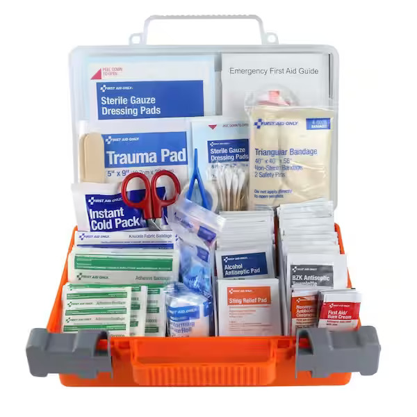
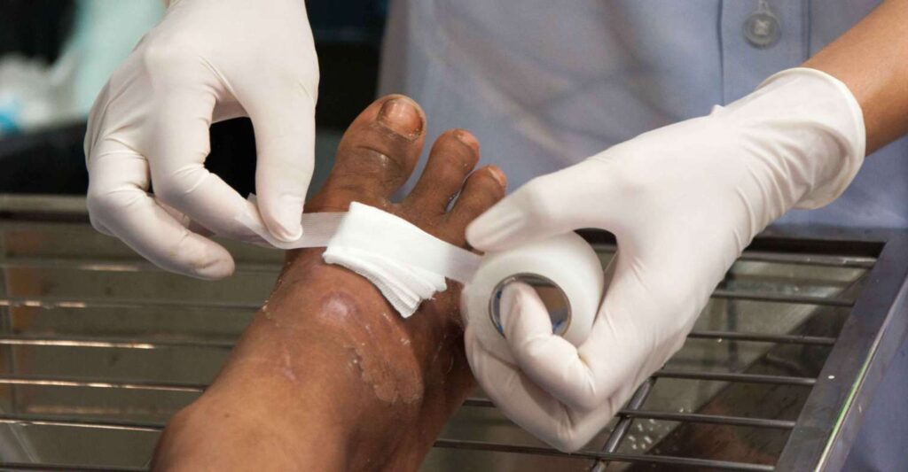
A first aid kit is a portable container/box stocked with equipment, materials and basic medicines used by a trained first aider to give immediate care to an injured or suddenly ill person before professional medical help is available. First aid kit or medical kit is a collection of supplies and equipment used to give immediate medical treatment, primarily to treat injuries and other mild or moderate medical conditions.
📝 Exam Tip: When defining a first aid kit, ensure you mention that it is portable, used by a trained person, and serves to bridge care until professional help arrives.
It is mandatory to have a first aid kit in every workplace like school, college, house, and vehicles. It should be kept at such a place that is easily accessible. Also, everyone should be aware of it. It should be labeled as “First Aid” and should have a red cross on a white background. From time to time, its items should be checked and replaced. All the required items should be available and ready for use at all times.
1.2 Principles/Qualities of a Good First Aid Kit
A good first aid kit must meet the following criteria:
Portable and easy to carry to the scene.
Water-resistant and dust-proof (protects the sterile contents).
Durable and firmly closable (the lid must fit tight).
Clearly LABELLED "FIRST AID KIT" in bold, visible letters (often with a white cross on a green background).
Contents must be organised (have compartments; frequently used items placed on top).
Contents appropriate to the setting (a home kit is very different from a military combat kit).
Stocked with non-expired items; checked regularly.
Kept in an accessible but child-safe location.
Simple to open in an emergency (but not so simple that toddlers can open it).
Accompanied by a list of contents and emergency numbers taped inside the lid.
Components of a First Aid Kit (Minimum Contents)
The minimum contents of the first aid box are as follows:
Torch (01) & Thermometer (01)
Tongue Depressor (Disposable ice cream spatula)
Writing pad and Pen/pencil
Bandages of various types, Gauze pieces, Cotton
Eye pads, Plaster, Safety pins
Scissors & Tourniquet
ORS packets & Glucose packets
Methylated spirit, Tincture of iodine, Tincture of benzoin
1.3 Components of a First Aid Kit (Detailed)
As a nurse, you must know exactly what is inside the kit and how to improvise if an item runs out.
GROUP A: WOUND CARE ITEMS
1. Sterile gauze swabs/pads: Used for cleaning wounds, as a primary wound dressing, and to apply direct pressure to bleeding. Improvisation: Clean boiled-and-cooled cloth, or clean cotton from a brand new pillow (last resort).
2. Cotton wool: Used for cleaning around (not inside) wounds, applying antiseptic, and padding. Improvisation: Soft clean cloth.
3. Adhesive dressings (Plasters/Band-Aids): Various sizes for minor cuts, blisters, and small grazes.
4. Roller bandages (5cm, 7.5cm, 10cm): Used to hold dressings in place, apply even pressure, support sprains, and immobilise. Improvisation: Clean torn sheets, pagne (kitenge) cloth.
5. Triangular bandages: Extremely versatile! Used for arm slings, broad-fold/cravat bandages, immobilising limbs, or improvising a splint tie. Improvisation: A square piece of clean cloth folded diagonally.
6. Adhesive tape (Zinc oxide/plaster): Securing dressings and splints.
7. Elastic (Crepe) bandage: Provides compression for sprains/strains and joint support.
8. Antiseptic solutions:
Hydrogen Peroxide (H2O2): Used for DIRTY/contaminated wounds. It effervesces (bubbles) to lift dirt and debris out of the wound.
Povidone-iodine (Betadine): Used for CLEAN wounds.
Surgical Spirit: Used to clean intact skin around the wound. NEVER put spirit inside an open wound — it damages exposed tissue and delays healing.
Plain soap and clean water: The first-line and best/cheapest method for wound cleaning.
9. Antibiotic ointment/cream: E.g., Gentian violet or tetracycline eye ointment (as per local protocol) to prevent infection in minor wounds.
10. Sterile saline/Eye wash: For irrigating eyes and flushing wounds.
11. Sterile eye pads: Specifically shaped for eye injuries.
🚨 EXAM TRAP: Antiseptic Selection
Examiners love asking: "Which antiseptic for a dirty wound and why?" Answer: Hydrogen Peroxide (H2O2) because its effervescence (bubbling action) physically lifts debris and grit out of the wound bed, and it has virucidal/bactericidal properties.
GROUP B: BLEEDING CONTROL ITEMS
12. Tourniquet: (Commercial windlass type preferred). Used as a last-resort to control life-threatening limb bleeding. Improvisation: A wide cloth + a strong stick. NEVER use wire or thin cord (it cuts tissue). ALWAYS write the time of application on the casualty.
13. Hemostatic dressing/gauze: Special gauze coated with clotting agents used for packing deep wounds.
14. Safety pins: Securing bandages, slings, and fastening splints.
15. Scissors: (Bandage scissors with a blunt tip to avoid cutting the patient) for cutting bandages, gauze, and exposing wounds by cutting clothing.
16. Tweezers/Forceps: Removing splinters, stings, ticks, and debris.
GROUP C: MEDICINES (As per scope/protocol)
17. Oral analgesics: Paracetamol (pain/fever), Ibuprofen (pain/inflammation — avoid in bleeding/ulcer/asthma), Aspirin 300 mg (have the casualty chew it if suspecting a heart attack).
18. Oral Rehydration Salts (ORS): For dehydration from diarrhoea, vomiting, or heat illness.
19. Antihistamine tablets/cream: For allergic reactions, insect bites/stings.
20. Glucose powder/tablets or sugar sachets: For hypoglycaemia (low blood sugar).
21. Antacid tablets: For heartburn and severe indigestion.
22. Anti-diarrhoeal: (Only per local medical guidance).
23. Anti-emetic: (Per protocol/scope).
GROUP D: PROTECTIVE/PPE ITEMS
24. Disposable gloves: (Latex/Nitrile — use Nitrile if there is a latex allergy). To avoid contact with blood and body fluids.
25. Face masks: To protect the casualty and first aider from respiratory droplet spread.
26. Face shield / Pocket mask: With a one-way valve for safer rescue breaths during CPR.
27. Apron/Gown: To protect clothing during heavy bleeding care or childbirth.
GROUP E: INSTRUMENTS & DIAGNOSTIC ITEMS
28. Digital thermometer: To check temperature.
29. Torch/Flashlight + Spare batteries: Essential for night scenes and examining pupil reactivity.
Contents: Elastic bandages, triangular bandages, splints, cold packs/instant ice, antiseptics, gloves, scissors, ORS. Kept by the team first aider at every game.
MILITARY / Combat Kit:
Purpose: War/combat wounds — severe bleeding, chest wounds, blast injuries.
Contents: Tourniquets, hemostatic dressings, field dressings, bandages, chest seals, morphine (for trained personnel), airway adjuncts. Rugged, camouflaged design carried on body armour.
FIRST RESPONDER Kit (Ambulance/Community Responder):
Purpose: Pre-hospital care while awaiting evacuation.
Choose a large, flexible, water-resistant plastic container with a tight-fitting lid.
Make a list of items needed (based on common home accidents in your specific family: are there children? elderly? risk of burns?).
Stock the container with all the listed items.
Inform ALL adult family members where the kit is kept.
Train adults how to use each item (e.g., practice bandaging together!).
Label the outside boldly "FIRST AID KIT".
Store safely — easily accessible to adults, but OUT of children's reach.
Inspect regularly: check expiry dates; restock immediately after every use; tick against the checklist.
1.6 Care and Maintenance of the Kit
Keep it closed, clean, dry, and away from direct sunlight (Ugandan heat rapidly degrades drugs and destroys glove elasticity).
Check monthly: expiry dates, completeness, and the sterility of sealed items.
Replace items immediately after use so the kit is always 100% ready.
Never store strong, unlabelled medicines loose in the box (major child safety hazard).
Wash reusable instruments; sterilise them per protocol.
Keep the emergency-number card updated.
1.7 Why Each Expired Item is Dangerous (Essay Depth)
As a nurse, do not just say "it is bad." Explain the clinical danger:
Expired antiseptics lose potency ➔ failure to kill bacteria ➔ severe wound infection.
Expired gloves perish, become brittle and tear during use ➔ massive blood exposure (HIV/HBV risk for the first aider).
Expired drugs may be completely ineffective (casualty gets no relief) or toxic (causes poisoning).
Damp/Expired gauze is no longer sterile ➔ introduces deadly hospital-acquired infections directly into the bloodstream.
Topic 2: Personal Protection (Standard Precautions)
2.1 Definitions
Personal Protection: Measures taken by the first aider to protect themselves and others from injury or infection while giving care.
Standard Precautions: (Formerly known as "universal precautions"). This means treating ALL blood and body fluids (except sweat) from EVERY casualty as potentially infectious, regardless of the casualty's known status — because in an emergency, their status is often UNKNOWN.
Body substances requiring precautions: Blood, vomit, saliva, sputum, urine, faeces, pus, wound drainage, semen/vaginal fluids, amniotic fluid, Cerebrospinal fluid (CSF). (Note: Sweat is generally considered low risk).
2.2 Why it Matters — Transmissible Infections
Nurses and first aiders are exposed to high-risk fluids. You must protect yourself from:
HIV: Needle-stick risk is ~0.3%; mucous membrane exposure is ~0.09%. Post-Exposure Prophylaxis (PEP) is highly effective if started within 72 hours.
Hepatitis B (HBV): Much more infectious than HIV via blood (needle-stick risk is ~30% if unvaccinated!). A highly effective vaccine is available — all nurses should be fully vaccinated!
Hepatitis C (HCV): Needle-stick risk is ~1.8%; there is no vaccine available.
Others: Ebola/Marburg (epidemics in Uganda!), Cholera, Typhoid, TB, COVID-19, and other respiratory viruses.
Uganda-specific Context: Due to Ebola outbreaks (2014 West Africa, 2022 Uganda Sudan ebolavirus outbreak, 2025 Kampala/Wakiso outbreak) ➔ ALWAYS take precautions; never assume a casualty is "safe".
2.3 PPE — When to Use Each Item
PPE Item
Protects
When to Use
Gloves (Nitrile/Latex)
Hands from blood/fluids
Touching blood, wounds, mucous membranes, body fluids; handling contaminated items.
Face mask (Surgical)
Mouth/Nose
Casualty with coughing/respiratory symptoms (TB, COVID); or when splashes are likely.
Eye goggles / Face shield
Eyes
Splashing/spraying blood or fluids is likely (e.g., major arterial bleeding, explosive vomiting, suctioning).
Apron / Gown
Body/Clothing
Heavy bleeding, vomiting, cholera care, assisting in childbirth.
Heavy contamination, epidemic/Operating Theatre settings.
Resuscitation face shield / pocket mask / Ambu bag
Mouth
Giving rescue breaths — NEVER do bare mouth-to-mouth where a barrier exists.
2.4 Hand Hygiene
WHEN: Before contact with the casualty, immediately after contact with blood/fluids, after removing gloves, and after the whole event is concluded.
HOW: Use soap + running water, rub all surfaces vigorously for 40–60 seconds; dry with a clean towel or air dry. If there is absolutely no water available: use an alcohol-based hand rub (≥60% alcohol) and rub until completely dry.
Remember: Gloves are NOT a substitute for hand washing! Gloves can have microscopic holes. Always wash hands after doffing gloves.
2.5 Steps of Donning and Doffing PPE (Order Matters!)
DONNING (Putting on):
Remove all jewellery and watches; ensure nails are short; cover your own cuts with a waterproof plaster.
Perform Hand Hygiene.
Put on the Gown/Apron.
Put on the Face mask (cover nose + mouth; mould the metal nose clip).
Put on Eye protection (goggles/face shield).
Put on Gloves (pull them over the gown cuffs if wearing one).
DOFFING (Taking off — The highest risk step!):
Reverse order, avoiding touching the contaminated outside surfaces:
Sharps Safety: Never recap, bend, or break needles by hand. Use yellow puncture-proof safety boxes and dispose immediately at the point of use. Never pass a sharp hand-to-hand — place it in a kidney dish/tray.
Waste Disposal Colour-Coding (Memorize this!):
BLACK bins/bags: General (non-infectious) waste: food remains, packaging, ordinary paper.
RED bins/bags: Highly infectious waste and Infectious waste: HIV/HBV waste, laboratory cultures (must burn/incinerate), blood-soaked dressings, used gloves, specimen containers.
BROWN bins/bags: Pharmaceutical and laboratory waste.
If you suffer a needle-stick injury or blood splash:
First aid: Wash the wound thoroughly with soap and running water. If splashed in mucous membranes (eyes/mouth), flush with plain water/saline for 10+ minutes.
Do NOT: Do not apply strong bleach or caustics. Do not squeeze or suck the wound!
Report: Immediately report to the occupational health officer or Sister-in-charge.
Risk assessment & Treatment: Start HIV PEP within 72 hours (sooner is better; ideally ≤2 hours). Administer Hepatitis B immunoglobulin/vaccine if unvaccinated.
Follow-up: Take baseline labs (HIV, HBsAg, HCV), receive counselling, and return for follow-up testing at 6 weeks, 12 weeks, and 6 months.
Document: Write a formal incident report.
💡 EXAM TIP: Cleaning, Disinfection & Sterilisation (Levels)
Examiners love to ask the difference between these three levels:
Cleaning: The physical removal of dirt/organic matter with soap + water. (This is always the first step, as dirt blocks disinfectants).
Disinfection: Kills most pathogens (but not all spores). Methods include boiling for 20 mins, using chlorine solutions (0.05% for hands/skin; 0.1% for surfaces/instruments per MoH), and 70% alcohol for intact skin/instruments.
Sterilisation: Kills ALL microbes, including highly resistant bacterial spores. Achieved by Autoclaving (121°C, 15 psi, 15–20 mins). Note: Boiling is NOT sterilisation; it is only high-level disinfection!
2.10 Scene Safety Behaviours (Beyond PPE)
Wear highly reflective clothing at road traffic accidents (night risk is huge!).
Switch off vehicle engines, apply the parking brake; put on hazard lights; assign a bystander far back to warn oncoming traffic.
Do not enter unstable buildings, fast-flowing flood water, or fires without professional rescue services.
Beware of crowd violence — ask the police to secure the scene when handling mob justice or riot casualties.
Quick Quiz
First Aid Kit and Life Threatening Conditions Quiz
First Aid
Privacy: Your details are used only for quiz tracking and certificates.
First Aid: Principles, Practice, and Legal Considerations
1. Introduction & Definition of First Aid
First aid is the immediate, temporary care given to a person who is suddenly injured or taken ill, using available materials, before professional medical treatment can be obtained.
As a nursing student, when asked to define First Aid in an exam, ensure you include all these elements for full marks:
Initial/temporary: It is not a substitute for definitive medical care; it bridges the critical gap until professional help arrives.
Given by a trained person (first aider): While anyone can help, a qualified first aider is formally trained by a recognized institution (e.g., Uganda Red Cross Society, St. John Ambulance).
Uses available materials: Improvisation is highly allowed and expected (e.g., using a clean shirt as a pressure dressing or a strong stick as a splint).
Given at the scene: Wherever the casualty is found (on the roadside, at home, or in the workplace).
Aimed at the 3 Ps: Preserve life, Prevent worsening (further harm/illness or injury), and Promote recovery.
A person trained by a recognised institution and qualified to give first aid.
Don't just say "anyone who helps".
Casualty / Victim
A person who is suddenly injured or ill and requires immediate treatment.
Focus on "sudden".
Bystander
A person at the scene who is called upon by the first aider to help (e.g., calling an ambulance, holding a dressing, controlling crowds).
Bystanders HELP.
Passer-by
Someone who passes the scene but does NOT stop to help.
Passes = passes on.
Onlooker
Someone who simply watches the scene but gives NO help.
Looks only.
Scene
The exact place where the accident/emergency occurred.
Location of incident.
Accident
A sudden, unexpected event causing injury or damage.
Unplanned.
Emergency
A serious, unexpected situation requiring immediate action to prevent loss of life or limb.
"Urgent + immediate intervention."
Complication
A secondary negative effect arising from the condition itself OR from poor/incorrect management by the rescuer.
Has two causes – memorise both!
Triage
Sorting casualties according to severity/priority, treating the most life-threatening first when resources are limited.
From French "trier" = to sort.
First aid kit
A box containing specialized equipment and materials used to give first aid.
The essential toolkit.
2. Aims, Objectives & Importance of First Aid
The aims of First Aid are built strictly on the 3 Ps. Let us break them down in detail.
P1: PRESERVE LIFE
Your primary goal is to keep the casualty alive until medical help arrives. This is achieved through ensuring ABC:
A – Airway: Make sure the airway is OPEN. If the casualty is unconscious, the tongue often falls back and blocks the airway. Use the head-tilt chin-lift maneuver to open the trachea. Clear any visible obstruction (like food, vomit, or broken teeth).
B – Breathing: Check for breathing using your senses:
LOOK: For chest/abdominal rising and falling movements.
LISTEN: For breath sounds at the casualty's mouth.
FEEL: For breath on your cheek.
Action: If not breathing, commence artificial respiration (mouth-to-mouth or using an Ambu bag).
C – Circulation: Check blood flow and perfusion. Feel for a pulse (check the carotid artery in adults, and the brachial artery in infants). Also, check the skin colour for critical signs:
Cyanosis: Bluish discoloration, especially on the lips/tongue, indicating a severe lack of oxygen.
Jaundice: Yellow discoloration of the skin and sclera (eyes), indicating liver issues.
Pallor: Extreme paleness/whitish skin indicating poor blood flow, shock, or severe anaemia.
Action: If there is no pulse/no circulation, start CPR immediately. Control any life-threatening severe bleeding. Treat for shock by elevating the legs if appropriate.
P2: PROMOTE RECOVERY
Help the casualty recover and prevent their condition from deteriorating while waiting for the ambulance:
Apply splints for suspected fractures. (Preventing bone movement stops the sharp bone edges from causing further internal tissue/nerve damage).
Apply sterile dressings to wounds to stop bleeding and prevent infection.
Place the casualty in the recovery position (lateral recumbent). This is crucial because it protects the airway in an unconscious, breathing casualty by allowing vomit/fluids to drain out, preventing fatal choking.
P3: PREVENT FURTHER COMPLICATIONS (Worsening)
Stop the injury or illness from getting worse before reaching the hospital:
Maintain correct body alignment, especially with a suspected spinal injury – do not move them unnecessarily! Bending a broken spine can paralyze or kill the casualty.
Prevent exposure. Keep them warm using blankets to prevent hypothermia, which heavily worsens shock.
Remove danger from the casualty, or carefully remove the casualty from danger (only if it is safe for you to do so).
Arrange swift transportation/referral to a hospital for definitive care.
📝 Essay Points: The Importance of First Aid
If asked to write an essay on the importance of First Aid, expand on these 8 key points:
Saves lives: Directly achieves the preservation of life through ABC management.
Prevents the condition from worsening: Stopping bleeding keeps the patient stable.
Reduces pain and suffering: Proper splinting stops the agony of broken bones grinding together.
Prevents permanent disability: Correct handling of spinal injuries ensures the patient can walk again.
Provides psychological comfort: Reassuring the casualty and panicking relatives calms the situation.
Bridges the gap: Covers the crucial time between the injury happening and reaching professional medical care.
Mass casualty management: Allows for Triage, ensuring the maximum number of lives are saved with limited resources.
3. Principles, Pillars & Golden Rules of First Aid
A. The Three Pillars of First Aid Assessment
Accurate assessment of any casualty rests solidly on 3 pillars. This is how you figure out what is wrong:
HISTORY TAKING:
Gathering information about the injury/illness: what exactly happened, when it happened, where, and what interventions have been done so far. You get this directly from the casualty (if conscious) OR from bystanders (if the casualty is unconscious).
In your secondary survey, use the AMPLE history mnemonic:
A – Allergy: Are they allergic to any drugs (like Penicillin) or foods?
M – Medication: Are they currently taking any medicines? (e.g., insulin, blood thinners).
P – Past medical history: Do they have asthma, epilepsy, or a heart condition?
L – Last meal: When did they last eat or drink? (Crucial if they need emergency surgery/anaesthesia).
E – Events: What events led up to the incident? (e.g., "He grabbed his chest, then fell").
SIGNS (Objective):
Signs are what YOU observe/find as the trained first aider. They are objective facts you can physically verify.
Mnemonic: Signs = SEEN (Objective, observable).
Examples: Active bleeding, a swollen ankle, visible bone deformity, cyanosis (blue lips), dilated pupils, smelling alcohol on their breath, or watching them vomit.
SYMPTOMS (Subjective):
Symptoms are what the CASUALTY complains of. You cannot see a symptom; you must rely on what they tell you.
Mnemonic: Symptoms = SAID (Subjective, complained).
Examples: "My chest hurts" (Pain), "I feel like vomiting" (Nausea), thirst, dizziness, a headache, or feeling short of breath.
🚨 EXAM TRAP: Signs vs. Symptoms
Examiners love to test if you know the difference. Be ready to classify them correctly in a multiple-choice setting. Rule: "Bleeding is a SIGN (you see it), pain is a SYMPTOM (they say it)."
B. Scope of First Aid (The 4 Main Steps)
Emergency assessment: Find out the problem via history, observation, and a head-to-toe physical examination. This phase also ensures the safety of the first aider.
Diagnosis: Identify the casualty's specific problem from your assessment findings (e.g., you see heavy bleeding ➔ Diagnosis: Severe Haemorrhage).
Immediate treatment: Give the appropriate first aid for the identified problem (e.g., applying a tight pressure dressing to stop the bleeding).
Referral/Transport: Arrange transfer to the nearest hospital. The method of transport depends entirely on the nature and severity of the injury (e.g., suspected spinal injury requires a rigid stretcher and ambulance, not a boda-boda).
C. Methods of Assessing a Casualty
History taking: Asking AMPLE questions.
Observation: Looking around the scene for clues (e.g., empty poison bottles, live wires) and looking at the casualty's general state.
Physical examination: A careful, systematic head-to-toe check for hidden injuries.
D. Importance of Assessing a Casualty First
Why not just start treating immediately? Because assessing first:
Protects you and bystanders: Identifies hidden scene dangers (live electrical wires, fire, speeding traffic, toxic gases).
Prioritizes life-threats: Identifies conditions in order of priority (ABC).
Leads to correct diagnosis: Helps collect accurate signs and symptoms.
Forms the basis of Triage: In mass casualties, assessment tells you who needs the doctor first.
Prevents harm: Stops you from giving inappropriate treatment (e.g., giving water to a patient needing surgery).
E. The 13 Golden Rules of First Aid
Memorize this list. As a certificate student, knowing these rules guides your entire approach to emergency care:
Be calm and act quickly but carefully: Panic spreads fast and causes deadly mistakes.
Assess the scene first: Use history, observation, and physical exam. Do not rush in blindly.
Ensure safety (Danger first!): For yourself, the casualty, and bystanders. You absolutely cannot help if you become a second casualty yourself.
Call for help early: Call bystanders to assist. Call 999/112 (Uganda national emergency line) or the Uganda Red Cross toll-free at 0800 211 088 for an ambulance.
Follow the ABC / DRABC approach: Treat life threats strictly in order of priority.
Reassure the casualty and bystanders constantly: Talk calmly; severe anxiety heavily worsens clinical shock.
Preserve life ➔ promote recovery ➔ prevent complications: Always follow the 3 Ps.
Do no further harm: Avoid unnecessary movement (especially with suspected fractures or spinal injury). Never give food or drink to a casualty who may need emergency surgery or anaesthesia.
Keep the casualty warm: Cover them with a blanket or sheet. This prevents hypothermia and combats shock.
Improvise when equipment is lacking: Being resourceful saves lives (e.g., use clean torn clothing as bandages, or clean plastic bags over your hands if you have no gloves).
Arrange transport/referral: To the nearest appropriate hospital after giving first aid.
Hand over properly to medical personnel: Give a clear verbal report: what happened, what you found, what you did, and how the casualty responded.
Document (crucial for nurses): Write down the time of the incident, findings, interventions, and the casualty's response.
4. Legal and Ethical Considerations in First Aid
Providing first aid involves interacting with people in vulnerable states. You must understand your legal and ethical boundaries to protect both the patient and yourself from lawsuits.
A. Consent
You cannot touch a person without their permission. There are three types of situations:
Expressed consent: The casualty is conscious, alert, and agrees to treatment. Always ask first: "My name is Mary, I am a nurse/trained first aider. Can I help you?"
Implied consent: The casualty is unconscious or unresponsive. In this case, the law assumes consent (a reasonable person would want their life saved). It is also implied when any delay would risk their life.
Refusal of care: A conscious adult of sound mind has the legal right to refuse your help. If they say no, respect it! Do not force them. Instead, explain the severe risks of refusing, stay near them, and call for professional help.
Children: Get consent from a parent or guardian if they are present. If the situation is life-threatening and no guardian is around, treat the child immediately under implied consent.
B. Duty of Care and Abandonment
Once you voluntarily start giving first aid, you establish a "Duty of Care." You must not leave the casualty until:
A rescuer of equal or higher ability (like a doctor or paramedic) takes over, OR
The scene becomes too dangerous/unsafe for you to continue, OR
The casualty is formally handed over to EMS/medical personnel.
Leaving halfway without a valid reason is called Abandonment, and you can be sued for it.
C. Negligence
Negligence means failing to provide a standard of care, resulting in harm. Understand the three levels:
Ordinary negligence: Honest mistakes a reasonable person might make under extreme pressure (e.g., accidentally cracking a brittle rib while performing life-saving CPR). The law generally protects you here.
Gross negligence: Reckless, conscious disregard for safety. (e.g., giving powerful drugs beyond your training level, dragging a casualty with a broken spine by the legs, or performing unnecessary surgical procedures). The law will NOT protect you here.
Willful misconduct: Intentionally causing harm to the casualty. Never protected.
D. The Good Samaritan Principle (Important Context)
This is a legal principle protecting people who voluntarily give reasonable emergency care in good faith, without expecting payment, and within their level of training.
Ugandan Context: In Uganda (and Africa generally), there is NO formal, written Good Samaritan statute passed by parliament. However, protection comes from common-sense common law principles: act in good faith, within your training, and do not charge money.
As a nurse in Uganda: Volunteer care done carefully and within your scope of practice is highly defensible in court. Reckless care, or demanding payment at the scene, destroys your protection.
Never accept payment for first aid at the roadside scene.
Do not perform procedures beyond your certificate training (e.g., an untrained student trying to cut open a throat for a surgical airway = gross negligence).
E. Confidentiality
Keep all information about the casualty totally private. Do not discuss their medical condition, HIV status, or cause of injury with curious onlookers, the media, or post photos on social media! Share medical information ONLY with the ambulance or hospital team taking over.
F. Other Ethical Issues
Right to privacy/dignity: Shield the casualty from curious onlookers. Expose only the specific body area being treated. Respect cultural norms and modesty (especially for female casualties).
Honesty: Do not lie or promise outcomes you cannot guarantee (e.g., never say, "You will be completely fine"). Instead, reassure them honestly: "Help is coming, and you are being cared for."
Non-maleficence & Beneficence: The core medical ethics: Do good, and avoid doing harm.
Reporting requirements: In Uganda, certain cases MUST be reported to the police (e.g., Road traffic accidents, assault, gunshot wounds, suspected child abuse). Document accurately and avoid altering the crime scene more than is necessary to save the life.
G. Records / Documentation (Nurse-Specific)
As a nurse, your memory is not enough; you must write it down. Record the exact time of the incident, time of arrival, condition found (vital signs), the AMPLE history, specific first aid given, how the casualty responded, the time and mode of transport, destination, and who you handed the patient over to. Accurate records protect both you and the casualty legally.
📝 EXAM STRATEGY: Legal & Ethical Considerations
If asked: "Discuss the legal and ethical considerations when giving first aid."
Structure your answer logically: Consent ➔ Duty of care/abandonment ➔ Negligence / Good Samaritan ➔ Confidentiality ➔ Dignity ➔ Documentation. Writing one strong, detailed paragraph for each of these 6 points will guarantee full marks.
5. Chain of Survival
Definition: The Chain of Survival is a rapid sequence of critical actions that must occur immediately after a sudden cardiac arrest to maximize the patient's chance of survival. The concept is that "the chain is only as strong as its weakest link."
The 6 Links (Classic Teaching - Still tested heavily)
Early recognition of cardiac arrest & activation of EMS: Recognize that the patient is unresponsive with no normal breathing. Immediately call for help (999/112, or Uganda Red Cross ambulance 0800 211 088).
Early CPR: High-quality chest compressions to keep blood flowing to the brain. (Rate of 100–120 compressions per minute, depth of 5–6 cm in adults, allowing full chest recoil, with minimal interruptions).
Early defibrillation: Using an Automated External Defibrillator (AED) to shock the heart as soon as it is available. Survival drops by about 10% for every minute the shock is delayed!
Advanced resuscitation: Interventions provided by EMS paramedics or the hospital team (advanced airway management, IV drugs, ECG monitoring).
Post-cardiac arrest care: Specialized ICU care, targeted temperature management (cooling the brain), and treating the underlying cause of the arrest.
Recovery: Long-term rehabilitation, physical/cognitive/psychological support, and follow-up care.
2025 AHA Updates (Crucial for up-to-date marks)
The American Heart Association (AHA) continuously updates these guidelines. For 2025, you must know these changes:
The AHA replaced the old four separate chains (adult/paediatric, in/out-of-hospital) with ONE unified 6-link chain of survival applicable to all ages and settings.
There is a massive new emphasis on prevention and preparedness as a surrounding concept before the arrest even happens.
Choking updates: Formal guidance requires 5 back blows alternating with 5 abdominal thrusts for adults. For infants: 5 back blows alternating with 5 chest thrusts.
Opioid overdose updates: If an overdose is suspected, administer Naloxone + commence CPR immediately.
Compression rules: The rate is strictly 100–120/minute for ALL ages. For adults, depth must be at least 5 cm, but avoid going deeper than 6 cm.
"Rescue breaths" are now simply called "breaths". Telecommunicator CPR (dispatcher-assisted CPR over the phone) is now given Class 1 priority.
🧠 SIMPLE PUBLIC MNEMONIC: CALL — PUSH — SHOCK
Call emergency services ➔ Push hard and fast (hands-only CPR) ➔ Shock (use the AED).
Why the Chain Matters (Essay Tip): Explain the "weakest link" concept. Each link is time-critical. Without early recognition, CPR never starts. Without early defibrillation, CPR alone rarely restores a normal heart rhythm. Without advanced hospital care, the brain and heart suffer re-injury. Without recovery support, survivors face severe lifelong disability.
6. Role of the Nurse in Emergencies
Different people at the scene have different responsibilities. You must distinguish between what a basic first aider does, what a trained nurse does, and what a bystander does.
Roles of the First Aider (Any trained person)
Assess the scene and ensure safety (Danger first).
Carry out the primary survey (DRABC) and secondary survey.
Give immediate life-saving care (ABC, CPR, bleeding control, placing in recovery position).
Call and arrange for professional help and transport.
Reassure and comfort the casualty and their relatives.
Prevent further injury (immobilization, warmth, stopping unnecessary movement).
Hand over the casualty with a clear verbal report to the EMS/medical team.
Specific Roles of the NURSE (Expanded for Nursing Exams)
As a nursing student, your scope of practice is much wider than a basic first aider. If an exam asks for YOUR role, include these 12 clinical points:
Triage: Sort casualties accurately in mass emergencies to prioritize life threats.
Advanced Assessment: Conduct rapid primary surveys (DRABC), take accurate vital signs, and perform a thorough secondary head-to-toe survey using SAMPLE history.
Life Support: Perform high-quality CPR, artificial ventilation (using an Ambu bag if available), advanced bleeding control, and clinical shock management.
Wound Care: Professional cleaning, sterile dressing, bandaging, and splinting.
Drug Administration: Administer emergency drugs within your legal scope and protocols (e.g., Adrenaline for anaphylaxis, Aspirin for suspected cardiac chest pain, oral Glucose for severe hypoglycaemia).
Airway Management: Positioning, using mechanical suction, and inserting airway adjuncts (e.g., Guedel airway) within your scope.
Coordination: Mobilize bystanders, communicate professionally with EMS/doctors, and arrange formal referral and transport.
Clinical Documentation: Write an accurate, legal record of findings, interventions, and patient response.
Communication & Psychosocial Support: Counsel the casualty and family, communicate with the community, and provide psychological debriefing after traumatic events.
Health Education & Prevention: Teach first aid in the community and schools, advocating for emergency preparedness (like having first aid kits).
Infection Prevention: Strictly use standard precautions and safe handling of sharps/blood to prevent HIV/Hepatitis transmission at the scene.
Follow-up Care: Continued monitoring of vital signs during transport, post-emergency care, and completing referral paperwork.
🚑 OSCE FOCUS: Roles of Bystanders (Common Exam Question)
Examiners often ask how you utilize bystanders. Bystanders are crucial. Instruct them clearly to:
Call for help / call the ambulance (999/112 or 0800 211 088).
Help control bleeding (instruct them to apply direct pressure or hold a dressing tightly).
Help move the casualty only when instructed and safe to do so.
Fetch the first aid kit, blankets, or water as directed.
Give information/history about what they saw happen.
Guide and direct the ambulance to the exact scene location.
Control the crowd and keep noisy onlookers away.
Provide comfort and reassurance to crying relatives.
7. The First Aider – Qualities of a Good First Aider
Definition: A first aider is a person trained by a recognised institution and formally qualified to give first aid.
11 Essential Qualities
To be effective in an emergency, a first aider must possess these specific traits (expand on each point in an essay):
Knowledgeable & Skilled: Must be properly trained, competent in first aid techniques, and keep their skills up to date.
Observant / Critical: Must notice subtle, critical signs that others miss (e.g., pale skin color, abnormal breathing patterns, unequal pupils, or the smell of poison/alcohol).
Confidential: Must strictly keep the casualty's medical and personal information private, sharing it only with doctors.
Trustworthy & Honest: Must safeguard the casualty's property (wallets, phones) and return it safely. Must be completely honest in their medical reports.
Resourceful & Improvisative: Must be able to think outside the box and use available materials when medical supplies run out (e.g., using clean polythene bags as gloves, or strong sticks and umbrellas as splints).
Courageous & Brave: Must act decisively despite unpleasant sights, massive blood, or danger (after ensuring reasonable safety protocols).
Empathetic & Compassionate: Must put themselves in the casualty's shoes, offering genuine hope, and calming their deep fears.
Calm & Composed: Must think clearly and systematically under massive pressure. They do not panic, because panic spreads to the casualty.
Patient: Must persist with long, exhausting resuscitation efforts (like continuous CPR) without giving up too soon.
Physically Fit: Performing CPR, lifting heavy patients, and carrying them to safety requires significant physical strength and stamina.
Good Communicator: Must give clear, authoritative instructions to bystanders, hand over effectively to paramedics, and provide soothing reassurance to the casualty.
📝 EXAM STRATEGY: Memorizing Qualities
Group them logically in your mind to remember all 11 for an essay:
Professional Traits: Knowledgeable, Observant, Confidential, Trustworthy.
Reach accident spot quickly. This will help to save life of the casualty.
Shout for help. Organize labour or onlookers or bystanders to help in any possible way. Make sure there are enough people to help you.
Be calm, methodical and quick. By doing so, you can minimize the pain and the effect of the injuries, which may save life. Handling casualty clumsily will make the final recovery difficult.
Remove the casualty from danger or danger from casualty.
Look for the following:
Is there failure of breathing?
Is there severe bleeding?
Is the shock high or severe? Is there any signs/ symptoms of shock?
Attend to these and then treat easily observable injuries.
Start artificial respiration, if the casualty is not breathing, it must begin at once, as every second gained is helpful.
Stop bleeding by pressing on the pressure point, press firmly on the bleeding area for at least a few minutes (minimum 3 minutes) by watch – take help if available.
Treat shock.
Avoid handling casualty unnecessarily.
Note:Never give anything by mouth to the patient who is unconscious.
Use the first aid articles if available (All trains, railway stations, Lorries and buses keep first aid box). Make use of material so obtained. In case, first aid box is not available, improvise and make use of available resources.
Assess the situation sensibly in regard to medical aid treatment which may be needed.
Make a written note on the general condition and your findings about the casualty.
Inspect the area: Take the casualty away from live wire, fallen walls, beams, fire, broken gas chamber, moving machinery, etc. to safer place.
Clear the crowd with polite words. Do not allow people to crowd around the casualty as the casualty needs fresh air. If a doctor is present, he will guide you. Any other first aider should be asked to help, otherwise take the assistance of by standers by giving them correct instructions.
Note the weather: If it is not raining, too hot or cold, treat in open, otherwise move the casualty into an airy room. If no suitable house or shelter is available nearby, it’s best to protect the casualty with an umbrella or a sheet of cloth or even a newspaper.
Reassure the casualty by soft words and encourage talking. This will help the casualty to take things lightly and lie quietly. This will help in recovery.
Arrange for dispatch of the casualty to the care of a doctor or to a nearby hospital. At the same time inform relatives as to where the casualty is being taken to.
Do not attempt too much. You are only a first aider, give minimum assistance so that condition does not become worse and life can be saved.
Do's and Don'ts
Do not forget that you are not a doctor hence, do not attempt to overdo things.
Do not handle the victim unnecessarily as that condition may worsen.
Do not expose the casualty unnecessarily.
Do not open any wounds / dressing, if bandaged previously by anybody.
Do not move any fracture case without putting proper splints.
Do not tie tourniquet at bleeding site and forget about it.
Attend to casualty as per priority.
Attend to children and women first.
Ensure self – safety and security before jumping into heroic attempts to save casualty.
Follow precautions in handling communicable / infectious cases.
Never declare any casualty dead, it is that doctor’s job.
The Management of the Case
The first aider must always:
Respond quickly to calls for assistance, the saving of a life may depend on promptness of action.
Adopt a calm and methodical approach to the casualty, quick and confident examination and treatment will relieve pain and distress, lessen the effect of injury and may save life. Time spent on long and elaborate examination of a casualty may be time lost in his ultimate recovery.
Treat obvious injuries and conditions endangering life such as failure of breathing, severe shock, before making a complete diagnosis.
Take first aid material. If this is immediately available. If standard equipment is not available the first aider must depend on material to hand which will have to be provided as required.
Study the surroundings carefully. These may influence the action to be taken and therefore require careful consideration for example:
Danger: From falling building, moving machinery, electric current, fire, poisonous gases and similar hazards.
Weather: If the accident occurs out of doors, the casualty may be treated in the open if the weather is fine, if the weather is bad, he must be removed to shelter as soon as is reasonably possible.
Shelter: Note houses and buildings near at hand, whether occupied or unoccupied and whether likely to be particularly useful, such as a chemist’s shop, otherwise, temporary shelter may be provided by means of umbrellas, rugs and the like.
Assistance: Crowds must be tactfully controlled. If a doctor is present, work under his direction. If not, ask if anyone with knowledge of first aid is present. If neither is available make use of bystanders to the best advantage.
Reassure the casualty by speaking encouragingly to him. Warm him to be still and tell him that he is in trained hands.
Step by Step Action to be Taken by the First Aider
Examination and Diagnosis: This is taking account of the casualty‘s history and that of incident, symptoms, signs and level of responsiveness.
History: This is the full story of how the incident occurred or the illness began, and should be taken directly from the casualty and a responsible bystander wherever possible. Never hurry the casualty and remember to pass on all information you have obtained when skilled help arrives.
Symptoms: These are sensations that the casualty feels and describes to you the most useful of these is pain. If the casualty is unconscious or unreliable because dazed (confused) or in shock, their diagnosis cannot be based on symptoms but has to be based on information obtained from bystanders and signs.
Sign: These are details ascertained by you using your senses – sight, touch, hearing and smell. These may be signs of injury such as: bleeding, swelling, deformity, or signs of illness such as raised temperature and rapid or regular pulse.
Key Interventions / Actions:
Cardiopulmonary resuscitation (every second).
Control bleeding.
Treat shock and special care of unconscious cases.
Fracture immobilization.
Burn cover, with clean washed or dressing and treat shock.
Eye, nose and ear injuries.
Multiple superficial injuries.
Transportation.
Responsibilities of a First Aider in the Management of Casualties
Gain access to the patient in easiest and safest way.
Observe the accidents scene and assess the situation.
If necessary, direct others to direct traffic keep bystanders at a safe distance and make essential telephone calls. Turn off all engines that may be still running.
To find out whether is unconscious, conscious alive dead.
Identify the disease or condition from which the casualty is suffering.
Give immediate, appropriate and treatment considering priority of the first aid measures. Such as first priority will be of restoration of breathing and circulation, while second will be stopping the bleeding.
Should bear in mind that a casualty may have more than one injury and that some casualties will require more urgent attention than others.
Arranging without delay for shifting of the casualty to a doctor, hospital or home according to the condition in such a manner that injury is not complicated or the victim is not subjected to unnecessary discomfort.
Keeping the record of the patient and of incidence, addresses and witness.
Once a first aider has voluntarily started care, he should not leave the scene, or stop the care until a qualified and responsible person relieves him.
To report your observations to those taking over care of the casualty and to give further assistance if required.
To prevent cross infection between yourself and casualty as much as possible.
Limitations of the First Aider
The first aider should be observant with the rules or objectives of first aid and act quickly and vigilantly.
He should inspire confidence in the patient and others closely related to the patient.
To save lives, there are three conditions that call for first aid: stoppage of breathing, severe bleeding and shock.
If breathing movements are not proper, the lips, tongue and finger nails become blue, in such a situation, artificial respiration should be started immediately.
If there is heavy bleeding: It may be from wounds through one or more large vessels. In this condition, pressure should be applied directly over the wound. For this, a clean handkerchief or a pad may be kept on the wound and pressed firmly with one or both hands, then apply affirm bandage.
The important factor to be attended immediately is shock. Shock accompanies severe injury or emotional disturbance. Cold and clammy skin, beads of perspiration on the fore head and palms. Pale face, nausea and vomiting are the common symptoms of shock.
Skills Required for the First Aider
Control the scene of accident.
Gain access to the patient.
Evaluate the scene in terms of safety and possible cause of accident.
Determine diagnostic signs and relate those to possible injuries or sudden illnesses that require emergency care.
Perform the necessary ABC’S of emergency care:
Open air way.
Breathing (breathlessness - provide artificial ventilation).
Circulation (pulseless - provide one and two rescuer cardiopulmonary resuscitation).
Bleeding control (haemorrhage controlled by direct pressure and elevation, pressure points and tourniquets).
Diagnosis and care for shock.
Diagnosis and care for open and closed fractures, sprains (tearing of ligaments), strains (muscle injured by overstretching) and dislocations, including cold treatment and basic splinting techniques.
Diagnosis and care for soft tissue and internal injuries including basic dressing and bandaging techniques.
Detect and care for poisoning including alcohol and drug abuse.
Diagnosis and care for heart attack, stroke, diabetes, coma, insulin shock, and epileptic or other seizures.
Diagnosis and care for facial injuries, head injuries, neck and spinal injuries and chest injuries including fracture ribs and penetrating chest wounds.
Diagnosis and care for burns and smoke inhalation.
Diagnosis and care for exposure to heat and cold, which includes heat exhaustion, heat cramps, heat stroke, hypothermia and frostbite.
Assist in child birth and care of the new born.
Psychological and proper emergency care to victims of crisis and disasters.
Perform proper transformation techniques.
Quick Quiz
First Aid Intro Quiz
First Aid
Privacy: Your details are used only for quiz tracking and certificates.
Define Gout and differentiate it from other forms of arthritis.
Explain the Pathophysiology of Gout, specifically focusing on uric acid metabolism and crystal formation.
Identify the Risk Factors and triggers associated with developing gout and gout flares.
Describe the Clinical Presentation of acute gouty arthritis, chronic tophaceous gout, and intercritical gout.
Discuss the Diagnostic Criteria and key laboratory/imaging findings used to confirm a diagnosis of gout.
Explain the Pharmacological Management Strategies for both acute gout flares and long-term uric acid-lowering therapy (ULT).
Identify Non-Pharmacological Management Strategies and lifestyle modifications crucial for preventing gout flares.
Describe Potential Complications associated with chronic gout.
Definition and Characteristics
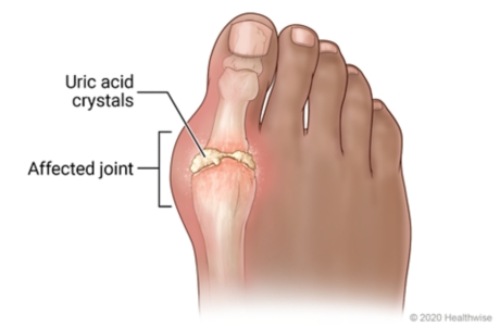
Gout is a metabolic disorder characterized by elevated serum uric acid levels and deposits of urate crystals in synovial fluids and surrounding tissues.
It is derived from the Latin word “Gutta” meaning a “drop” (of liquid).
Gout also is a kind of arthritis that occurs when uric acid builds up in blood and causes joint inflammation, it can be acute or chronic.
Acute: The affected joints often appear reddened and swollen and are sensitive to touch. The pain is described as a burning sensation. The development of acute gout is typically triggered by trauma, alcohol use, surgery, and systemic infection.
Chronic: This is characterized by visible deposits of urate crystals (tophi) that form nodules and may be painful during gout attacks.
Unlike Osteoarthritis (OA), which is primarily a "wear and tear" condition affecting cartilage, gout is characterized by sudden, severe attacks of pain, swelling, redness, and tenderness in the joints. It is fundamentally a metabolic disorder related to the body's handling of uric acid.
Gout is a type of inflammatory arthritis caused by the deposition of monosodium urate (MSU) crystals in the joints, tendons, and surrounding tissues. These crystals form when there are persistently high levels of uric acid (a waste product from the breakdown of purines) in the blood, a condition known as hyperuricemia.
When MSU crystals precipitate and accumulate in a joint, they trigger a potent inflammatory response, leading to the characteristic symptoms of a "gout flare" or "gouty attack." Over time, if left untreated, chronic hyperuricemia can lead to recurrent flares, joint damage, and the formation of visible chalky deposits called tophi.
Differentiation from other forms of Arthritis
Condition
Underlying Cause
Key Features & Diagnostics
Osteoarthritis (OA)
Primarily mechanical wear-and-tear and age-related degeneration of joint cartilage.
Pathology: Cartilage breakdown, osteophyte formation, subchondral sclerosis. No crystal deposition.
Onset: Gradual, progressive over years.
Symptoms: Pain worse with activity, relieved by rest; morning stiffness typically brief (<30 mins); bony enlargement; crepitus.
Monosodium Urate (MSU) Crystal Deposition: These are the specific crystals that cause the inflammation.
Acute Inflammatory Arthritis: Characterized by sudden, severe, often monoarticular (affecting one joint) attacks.
Classic "Podagra": Most commonly affects the metatarsophalangeal (MTP) joint of the big toe.
Cause
Gout is associated with the presence of hyperuricemia (high blood levels of urate, or serum urate levels greater than ~6.8 mg/dl).
Hyperuricemia: Gout occurs when urate crystals accumulate in your joint, causing the inflammation and intense pain of a gout attack. Urate crystals can form when you have high levels of uric acid in your blood.
NOTE: Not everyone with hyperuricemia develops gout as this condition requires two essential processes to develop – crystallization and inflammation. When uric acid levels become elevated, crystals will form in the joints, which will then trigger the inflammatory process.
Pathophysiology of Gout
Gout is fundamentally a disease of uric acid dysregulation. Its pathophysiology revolves around the production, breakdown, and excretion of uric acid, leading to hyperuricemia and subsequent crystal formation and inflammation.
I. Uric Acid Metabolism:
Origin of Uric Acid:
Uric acid is the final end-product of purine metabolism in humans.
Purines are naturally occurring compounds found in all body cells and in virtually all foods. They are building blocks of DNA and RNA.
Sources of purines:
Endogenous (internal): About two-thirds of the body's uric acid comes from the normal breakdown of cells and tissues.
Exogenous (dietary): About one-third comes from purine-rich foods and beverages (e.g., red meat, seafood, alcohol).
Breakdown Process: Purines are metabolized through a series of enzymatic reactions, with xanthine oxidase being a key enzyme in the final steps, converting hypoxanthine to xanthine, and then xanthine to uric acid.
Excretion of Uric Acid:
Uric acid is primarily excreted by the kidneys (about two-thirds) and to a lesser extent by the gastrointestinal tract (about one-third).
Renal excretion involves complex processes of filtration, reabsorption, and secretion in the renal tubules.
II. Hyperuricemia (Elevated Uric Acid Levels):
Hyperuricemia is the prerequisite for gout, defined as a serum uric acid level generally above 6.8 mg/dL (400 µmol/L). This is the saturation point at physiological temperature and pH at which monosodium urate (MSU) crystals can begin to form in tissues.
Hyperuricemia typically results from one of two main mechanisms, or a combination of both:
Uric Acid Underexcretion (Most Common - ~90% of cases):
The kidneys do not efficiently excrete uric acid. This can be due to:
Conditions with high cell turnover (e.g., myeloproliferative disorders, chemotherapy-induced tumor lysis syndrome, psoriasis).
High fructose consumption (fructose metabolism increases purine breakdown).
III. Monosodium Urate (MSU) Crystal Formation and Deposition:
When serum uric acid levels consistently exceed the saturation point (6.8 mg/dL), MSU crystals can precipitate out of solution.
These crystals prefer to deposit in:
Cooler body temperatures: This explains why gout often affects peripheral joints like the big toe (MTP joint), ankles, knees, wrists, and fingers.
Avascular or relatively avascular tissues: Cartilage, tendons, ligaments.
Damaged joints: Pre-existing joint damage (e.g., from OA or trauma) can provide nucleation sites for crystal formation.
Over time, these crystals accumulate in the joint synovium, cartilage, subchondral bone, and other soft tissues (leading to tophi).
IV. The Acute Gout Flare (Inflammatory Response):
The presence of MSU crystals alone does not always cause symptoms. An acute gout flare is triggered when these crystals are suddenly released from the synovial lining or when new crystals form, provoking a powerful inflammatory cascade:
Crystal Recognition: Inflammatory cells, particularly macrophages and neutrophils, recognize the MSU crystals as foreign bodies.
Phagocytosis: These cells attempt to engulf (phagocytose) the crystals.
Inflammasome Activation: The engulfed MSU crystals activate the NLRP3 inflammasome within the macrophages.
Cytokine Release: Activation of the inflammasome leads to the production and release of potent pro-inflammatory cytokines, especially interleukin-1 beta (IL-1β).
Inflammatory Cascade: IL-1β then amplifies the inflammatory response, recruiting more neutrophils and other inflammatory cells to the joint. This leads to the classic signs of inflammation:
Pain: Due to nerve stimulation and pressure from swelling.
Redness (Erythema): Due to vasodilation.
Swelling (Edema): Due to increased vascular permeability and fluid accumulation.
Heat: Due to increased blood flow.
Loss of Function: Due to pain and swelling.
Resolution: Eventually, the inflammatory process subsides, often through mechanisms involving anti-inflammatory cytokines, clearance of crystals, and neutrophil apoptosis. This natural resolution can take days to weeks if untreated.
V. Chronic Gout and Tophus Formation:
If hyperuricemia persists and gout flares are left untreated, chronic accumulation of MSU crystals can lead to:
Tophi: These are visible or palpable chalky deposits of MSU crystals, typically surrounded by chronic inflammatory cells. They commonly form in soft tissues (e.g., ear helix, elbows, fingers, Achilles tendon, around joints). Tophi can cause chronic pain, joint damage, and functional impairment.
Chronic Gouty Arthritis: Persistent inflammation and joint destruction.
Renal Complications: Urate nephropathy (kidney damage from crystal deposition in the renal interstitium) and uric acid kidney stones.
Risk Factors and Triggers associated with developing gout and gout flares.
This helps us identify individuals predisposed to gout, while recognizing triggers allows patients to manage their lifestyle to prevent acute flares.
I. Risk Factors for Developing Gout (Chronic Hyperuricemia):
These factors primarily contribute to sustained elevated uric acid levels, which is the prerequisite for gout.
Genetics/Family History: A strong family history of gout significantly increases an individual's risk. This is often due to inherited predispositions that affect uric acid production or, more commonly, its renal excretion.
Gender and Age:
Men are significantly more likely to develop gout than women, especially before menopause. This is partly due to men typically having higher uric acid levels and women having estrogen, which promotes renal uric acid excretion.
Risk increases with age for both sexes. After menopause, women's risk approaches that of men due to declining estrogen levels.
Obesity/Overweight: Obesity is strongly linked to hyperuricemia and gout. Adipose tissue is metabolically active and can contribute to increased uric acid production, and obesity is also associated with reduced renal uric acid excretion.
Hypertension (High Blood Pressure): Often co-occurs with hyperuricemia.
Dyslipidemia: Part of the metabolic syndrome cluster.
Kidney Disease (CKD): Impaired renal function leads to reduced uric acid excretion.
Dietary Factors (Chronic High Intake):
High Purine Foods: Regular consumption of large quantities of red meat (especially organ meats like liver, kidney), certain seafood (shellfish, sardines, anchovies, herring, mackerel).
High Fructose Corn Syrup/Sugar-Sweetened Beverages: Fructose metabolism directly increases purine turnover and uric acid production.
Alcohol Consumption: Particularly beer and spirits. Alcohol increases uric acid production and impairs its renal excretion. Wine appears to have a lesser effect.
Medical Conditions/Other Causes of High Cell Turnover: Psoriasis, Myeloproliferative disorders, Hemolytic Anemia, Tumor Lysis Syndrome.
II. Triggers for Acute Gout Flares:
These factors can cause a sudden change in uric acid levels or dislodge pre-existing crystals, provoking an acute inflammatory attack.
Sudden Changes in Serum Uric Acid Levels:
Rapid increase: Heavy consumption of purine-rich foods/beverages, Dehydration.
Initiation of Uric Acid Lowering Therapy (ULT): Ironically, when starting allopurinol or febuxostat, uric acid levels drop rapidly, which can cause existing crystals to destabilize and shed, triggering a flare. This is why ULT is usually started with flare prophylaxis.
Rapid decrease: Aggressive dieting/fasting.
Alcohol Consumption: Even moderate alcohol intake can trigger a flare.
Dehydration: Increases the concentration of uric acid.
Trauma/Injury to a Joint: A minor injury, surgery, or prolonged pressure.
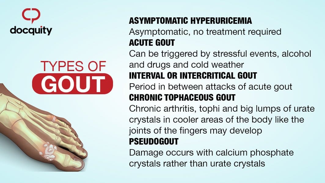
Gout progresses through several stages if left untreated, each with clinical characteristics.
I. Asymptomatic Hyperuricemia:
Description: This is the initial stage where a person has elevated serum uric acid levels (hyperuricemia) but experiences no symptoms of gout, no crystal deposition-related pain, and no history of gout flares.
Clinical Significance: While not considered "gout" at this stage, it is a precursor. Not everyone with asymptomatic hyperuricemia will develop gout (estimates vary, but it's often around 10-20% over a lifetime). Treatment is generally not recommended unless specific co-morbidities exist or uric acid levels are extremely high (>13 mg/dL).
II. Acute Gouty Arthritis (The Gout Flare):
This is the most common and recognizable presentation of gout. It's characterized by a sudden, exquisitely painful inflammatory attack.
Onset: Typically very sudden, often waking the patient from sleep.
Location:
Monoarticular: Usually affects a single joint in about 80-90% of initial attacks.
Podagra: The classic presentation involves the first metatarsophalangeal (MTP) joint of the big toe. This occurs in about 50% of first attacks and up to 90% of affected individuals at some point.
Other Joints: Ankle, knee, midfoot, wrists, fingers, elbows. Rarely affects axial joints in initial attacks.
Symptoms (Classic Signs of Inflammation): Severe Pain (throbbing, crushing, burning), Swelling, Erythema (shiny, bright red/purplish), Warmth, Tenderness (extreme sensitivity).
Duration: If untreated, typically resolves spontaneously within 3-10 days. Desquamation (peeling) of skin may occur.
III. Intercritical Gout (Intermittent Gout):
Description: This refers to the symptom-free periods between acute gout flares. During this phase, the patient has no symptoms, and the affected joints may appear normal. However, MSU crystals are still present.
Clinical Significance: Hyperuricemia usually persists, and ongoing crystal deposition can occur. Without ULT, subsequent attacks become more frequent, severe, and polyarticular.
IV. Chronic Tophaceous Gout:
This stage develops in individuals with long-standing, uncontrolled hyperuricemia and recurrent acute attacks. It typically takes 10-20 years to develop if gout is left untreated.
Description: Characterized by the formation of tophi – visible or palpable deposits of monosodium urate crystals. These appear as firm, chalky, painless (unless inflamed or infected) nodules.
Location of Tophi: Soft tissues around joints, Helix of the ear, Olecranon bursa, Prepatellar bursa, Achilles tendons. Can also develop in organs like kidneys.
The gold standard for diagnosis remains the identification of MSU crystals.
I. Gold Standard for Diagnosis: Synovial Fluid Analysis
The most definitive way to diagnose gout is by identifying monosodium urate (MSU) crystals in the synovial fluid (joint fluid) aspirated from an affected joint.
Negatively birefringent: When viewed under polarized light with a red compensator, they appear yellow when parallel to the compensator axis and blue when perpendicular to it.
Presence of Leukocytes: High white blood cell count (neutrophils). Also rule out septic arthritis.
Serum Uric Acid: While hyperuricemia (> 6.8 mg/dL) is a prerequisite, a normal uric acid level does NOT rule out gout during an acute flare. Levels can transiently drop during an attack.
Inflammatory Markers: Elevated ESR and CRP (non-specific).
Imaging Findings:
X-rays: Early gout may be normal. Chronic gout shows "Punched-out" erosions with sclerotic borders ("overhanging edge" sign).
Ultrasound: Can visualize MSU crystals as a "double contour sign".
Dual-Energy CT (DECT): Can definitively identify MSU crystals.
III. Differential Diagnosis:
Septic Arthritis (Crucial to rule out).
Pseudogout (CPPD).
Rheumatoid Arthritis.
Psoriatic Arthritis.
Cellulitis.
Osteoarthritis.
Pharmacological Management Strategies
The pharmacological management of gout has two distinct goals:
Rapidly alleviate the pain and inflammation of an acute gout flare.
Prevent future flares, joint damage, and tophus formation by lowering and maintaining serum uric acid levels below the saturation point.
I. Management of Acute Gout Flares:
The primary aim during an acute flare is to reduce pain and inflammation quickly. Treatment should be initiated as early as possible after symptom onset.
Dosing: Typically prescribed at high doses initially, then tapered over several days.
Considerations: Effective and generally well-tolerated. Contraindications include peptic ulcer disease, significant renal impairment, cardiovascular disease, and anticoagulant use.
Colchicine:
Mechanism: Disrupts neutrophil function and reduces the inflammatory response to MSU crystals by inhibiting microtubule assembly. Most effective when started within 24-36 hours of symptom onset.
Dosing: Low-dose colchicine (e.g., 0.6 mg once or twice daily) is often preferred for acute flares due to better tolerability compared to older high-dose regimens. Initial dose followed by a lower dose an hour later, then maintenance until flare resolves or for several days.
Considerations: Side effects include diarrhea, nausea, vomiting, abdominal pain. Dosing must be adjusted in patients with renal or hepatic impairment. Drug interactions are common (e.g., with CYP3A4 inhibitors like clarithromycin, diltiazem, verapamil, and P-glycoprotein inhibitors).
Corticosteroids:
Mechanism: Potent anti-inflammatory and immunosuppressive effects.
Administration: Can be given orally (e.g., prednisone), intramuscularly, or via intra-articular injection (directly into the affected joint).
Considerations: Useful when NSAIDs or colchicine are contraindicated or ineffective, or for polyarticular attacks. Intra-articular injections are particularly useful for monoarticular flares. Side effects include hyperglycemia, increased blood pressure, fluid retention, and mood changes.
Second-line/Alternative Agents (for refractory cases or specific contraindications):
IL-1 Inhibitors (e.g., Anakinra, Canakinumab):
Mechanism: Block the action of interleukin-1 (IL-1), a key cytokine in the inflammatory cascade of gout.
Considerations: Used in severe, refractory cases or when other agents are contraindicated. Administered via injection. Very expensive.
II. Long-Term Uric Acid-Lowering Therapy (ULT):
The goal of ULT is to reduce the body's uric acid burden, dissolve existing MSU crystals, prevent new crystal formation, and ultimately eliminate gout flares and tophi. The target serum uric acid level is generally < 6 mg/dL (360 µmol/L), and often < 5 mg/dL (300 µmol/L) in patients with severe disease, frequent flares, or tophi.
When to Initiate ULT: ULT is typically recommended for patients with:
Recurrent gout flares (two or more per year).
Presence of tophi (clinical or radiographic).
Gouty arthritis with evidence of joint damage on imaging.
Gout with chronic kidney disease (CKD stage 2 or higher).
History of uric acid kidney stones.
First gout flare if very severe or with extremely high serum uric acid (>9 mg/dL).
Important Considerations for Initiating ULT:
Prophylaxis: An acute flare can be triggered when starting ULT due to the rapid change in serum uric acid levels causing crystal shedding. Therefore, flare prophylaxis with low-dose colchicine or low-dose NSAIDs is usually recommended for the first 3-6 months (or longer if indicated) after initiating ULT.
Do NOT start ULT during an acute flare. Wait until the acute flare has subsided. If a patient is already on ULT, they should continue it during a flare.
Main Classes of ULT Agents:
Xanthine Oxidase Inhibitors (XOIs): These are the first-line agents for most patients.
Mechanism: Inhibit the enzyme xanthine oxidase, thereby blocking the final steps in uric acid production.
Examples:
Allopurinol:
Dosing: Start low (e.g., 50-100 mg daily) and titrate up gradually (e.g., by 50-100 mg every 2-4 weeks) to achieve the target uric acid level. Max dose often 800 mg/day, but depends on renal function.
Considerations: Generally well-tolerated. Side effects include rash, gastrointestinal upset. Allopurinol Hypersensitivity Syndrome (severe, potentially fatal reaction with rash, fever, eosinophilia, liver/kidney dysfunction) is rare but serious, especially in patients with HLA-B*5801 allele (more common in certain Asian populations) and those with renal impairment or starting on high doses. Renal dosing is crucial.
Febuxostat:
Dosing: Start at 40 mg daily, can increase to 80 mg daily if target not met.
Considerations: Can be used in patients with mild-to-moderate renal impairment without dose adjustment. Was previously associated with a higher risk of cardiovascular death compared to allopurinol in some studies, leading to a black box warning, but recent data suggests this risk may be less pronounced or restricted to specific populations.
Uricosuric Agents:
Mechanism: Increase the excretion of uric acid by the kidneys by inhibiting its reabsorption in the renal tubules.
Examples:
Probenecid:
Dosing: Start low and gradually titrate.
Considerations: Requires good renal function (creatinine clearance > 50 mL/min). Not effective in overproducers of uric acid. Side effects include gastrointestinal upset, rash. Patients must maintain good hydration to prevent kidney stone formation. Contraindicated in patients with a history of uric acid kidney stones.
Lesinurad: (often used in combination with an XOI, usually allopurinol, in refractory cases)
Considerations: Used to boost the efficacy of XOIs when target UA not achieved. Renal safety concerns.
Uricase (Pegloticase):
Mechanism: An enzyme that converts uric acid into allantoin, a more soluble and easily excreted substance.
Example: Pegloticase (IV infusion).
Considerations: Reserved for severe, refractory chronic gout, especially with large tophi, where other ULTs have failed or are contraindicated. High risk of infusion reactions and anti-drug antibodies, requiring careful monitoring.
Non-Pharmacological Management
Non-pharmacological management aims to reduce serum uric acid levels, minimize triggers for acute flares, and promote general well-being. These strategies should be discussed with every patient with gout.
I. Dietary Modifications:
The goal is not to eliminate purines entirely, as many healthy foods contain them, but to reduce intake of high-purine foods and those that increase uric acid production or impair its excretion.
Limit or Avoid High-Purine Foods:
Organ Meats: Liver, kidney, sweetbreads.
Certain Seafood: Anchovies, sardines, herring, mussels, scallops, trout, tuna, haddock. (Note: other fish and seafood in moderation are generally acceptable and beneficial for health).
Red Meats: Limit consumption (e.g., beef, lamb, pork) to moderate portions.
Reduce Fructose Intake:
Sugar-Sweetened Beverages: Avoid sodas, fruit juices (especially high-fructose corn syrup), and other sugary drinks. Fructose metabolism significantly increases uric acid production.
Processed Foods: Be mindful of hidden sugars (fructose) in many processed snacks and foods.
Fruits: While fruit contains natural fructose, whole fruits also provide fiber and other nutrients and are generally considered acceptable in moderation. The concern is with concentrated fructose from drinks.
Moderate Alcohol Consumption (or Avoid):
Beer and Spirits: Strongest association with gout flares due to increased purine load and impaired uric acid excretion. Best to avoid or severely limit.
Wine: Generally considered to have a weaker association with flares, but moderation is still advised.
Overall: Total alcohol intake should be limited, especially during periods of high risk or frequent flares.
Embrace Healthy Dietary Patterns:
Low-Fat Dairy Products: Studies suggest that dairy products (especially skim milk, yogurt) may actually help lower uric acid levels and reduce gout risk.
Complex Carbohydrates: Whole grains, vegetables, and fruits are encouraged.
Vegetables: Almost all vegetables (including purine-rich ones like spinach, mushrooms, asparagus, cauliflower) have not been shown to increase gout risk and are part of a healthy diet.
Hydration: Drink plenty of water throughout the day (at least 8-10 glasses) to help the kidneys flush out uric acid.
II. Weight Management:
Achieve and Maintain a Healthy Weight: Obesity is a significant risk factor for hyperuricemia and gout. Gradual weight loss can lower uric acid levels and reduce the frequency and severity of flares.
Avoid Crash Diets or Rapid Weight Loss: Fasting or very rapid weight loss can paradoxically increase uric acid levels and trigger flares. Gradual and sustained weight loss is preferred.
III. Regular Exercise:
Moderate Physical Activity: Regular exercise, combined with a healthy diet, helps with weight management and overall metabolic health, which can indirectly benefit gout.
Avoid Overexertion or Joint Trauma: While exercise is good, activities that cause excessive joint stress or trauma could potentially trigger a flare in a susceptible joint.
IV. Hydration:
Adequate Fluid Intake: Drinking plenty of water helps to dilute uric acid in the urine and promotes its excretion, reducing the risk of crystal formation and kidney stones.
V. Review Medications with a Physician:
Diuretics and Low-Dose Aspirin: If a patient is taking medications known to raise uric acid levels (e.g., thiazide diuretics, low-dose aspirin), their physician should evaluate if alternative medications are suitable or if the benefits outweigh the risks.
Start ULT with Prophylaxis: As discussed in Objective 6, patients initiating uric acid-lowering therapy should always be on concurrent anti-inflammatory prophylaxis to prevent initial flares.
VI. Identify and Avoid Personal Triggers:
Patients should be encouraged to keep a diary to identify their individual triggers, which can vary from person to person (e.g., specific foods, stress, minor trauma, dehydration).
Avoiding these identified personal triggers can significantly reduce flare frequency.
VII. Lifestyle Modifications during an Acute Flare:
Rest: Rest and elevate the affected joint.
Ice: Apply ice packs to the inflamed joint for short periods (e.g., 20 minutes at a time) to help reduce swelling and pain.
Avoid Trauma: Protect the joint from any pressure or impact.
Prognosis of gout and potential complications.
For emphasizing the importance of consistent management and patient adherence to treatment plans.
I. Prognosis with Effective Treatment:
With modern pharmacological and non-pharmacological management, the prognosis for gout is generally very good.
Symptom Control: Consistent adherence to uric acid-lowering therapy (ULT) can effectively lower serum uric acid levels below the target threshold (<6 mg/dL, or <5 mg/dL for severe cases).
Flare Prevention: Maintaining target uric acid levels will prevent the formation of new MSU crystals and facilitate the dissolution of pre-existing crystals, thereby dramatically reducing the frequency and severity of acute gout flares. Many patients can achieve a flare-free state.
Tophus Resolution: Existing tophi can shrink and even completely disappear over time with sustained low uric acid levels. This can reverse joint damage and restore function in some cases.
Prevention of Joint Damage: By preventing crystal deposition and inflammation, ULT can halt or reverse progressive joint destruction and deformity.
Improved Quality of Life: Patients experience less pain, better joint function, and a significant improvement in their overall quality of life.
Reduced Comorbidities: While gout itself doesn't cause some comorbidities, effective management can indirectly improve outcomes for associated conditions like kidney disease and cardiovascular health, especially by addressing systemic inflammation and metabolic issues.
II. Potential Complications:
Without proper management, gout progresses through its natural history, leading to significant and often irreversible complications.
Recurrent and More Severe Acute Flares:
Flares become more frequent, often polyarticular (affecting multiple joints), more severe, and of longer duration.
The intercritical periods (between flares) may shorten, or patients may experience continuous low-grade inflammation.
Chronic Tophaceous Gout:
This is a hallmark of untreated, long-standing gout. Tophi are crystal deposits that can form in:
Joints and surrounding soft tissues: Leading to chronic pain, stiffness, persistent swelling, and ultimately, irreversible joint damage, deformity, and significant functional disability.
Bursae: (e.g., olecranon, prepatellar) causing inflammation and swelling.
Ear helix: Characteristic deposits that can disfigure.
Tendons: (e.g., Achilles tendon) leading to pain, dysfunction, and potential rupture.
Internal organs: Although less common and often only detected on advanced imaging, tophi can deposit in kidneys or heart valves, contributing to organ dysfunction.
Joint Destruction and Deformity:
The persistent presence of MSU crystals and chronic inflammation leads to erosion of cartilage and bone, resulting in a severe form of arthritis that can mimic other inflammatory arthropathies. This can lead to permanent loss of joint function.
Kidney Complications:
Uric Acid Nephrolithiasis (Kidney Stones): Elevated uric acid levels increase the risk of forming uric acid kidney stones, which can cause severe pain, urinary tract obstruction, infection, and kidney damage.
Urate Nephropathy (Gouty Nephropathy): Chronic deposition of MSU crystals in the renal interstitium can lead to chronic inflammation, fibrosis, and progressive decline in kidney function. This can contribute to end-stage renal disease.
Psychosocial Impact:
Chronic pain, disability, and the unpredictable nature of flares can lead to depression, anxiety, social isolation, and impaired quality of life.
Difficulty performing daily activities, working, and engaging in hobbies.
Association with Cardiovascular and Metabolic Diseases:
While hyperuricemia and gout are often associated with cardiovascular disease, hypertension, diabetes, and metabolic syndrome, the exact causal relationship is complex and actively researched. However, it is clear that untreated gout exists within a cluster of metabolic disturbances that collectively increase morbidity and mortality risks. Effective gout management, particularly by addressing underlying metabolic issues, may contribute to better overall health outcomes.
Nursing Diagnoses and Interventions for a Patient with Gout.
Acute Pain related to inflammation in the affected joint(s) secondary to uric acid crystal deposition, as evidenced by patient's report of severe pain, guarding behavior, grimacing, and joint redness/swelling.
Impaired Physical Mobility related to pain and inflammation in the affected joint(s), as evidenced by reluctance to move the affected limb, limited range of motion, and difficulty with ambulation.
Inadequate health Knowledge related to disease process, dietary restrictions, medication regimen, and prevention strategies, as evidenced by patient's questions about gout, stated misconceptions, or observed non-adherence.
Risk for Ineffective Health Maintenance related to potential for non-adherence to long-term uric acid-lowering therapy, dietary modifications, and lifestyle changes.
Risk for Impaired Skin Integrity related to presence of tophi and chronic inflammation (for chronic tophaceous gout).
Excessive Anxiety related to unpredictable nature of gout flares, chronic pain, and impact on daily life, as evidenced by patient's verbalization of worry, restlessness, or irritability.
Nursing Interventions for Each Diagnosis:
1. Nursing Diagnosis: Acute Pain
Interventions:
Action
Rationale
Assess Pain Characteristics: Regularly assess pain level using a standardized scale (e.g., 0-10), location, quality (throbbing, crushing), and aggravating/alleviating factors.
Provides baseline data, monitors effectiveness of interventions, and helps identify triggers.
Administer Prescribed Medications: Administer NSAIDs, colchicine, or corticosteroids as ordered by the physician, ensuring correct dosage and timing. Educate on potential side effects.
These are the primary pharmacological interventions to reduce inflammation and pain during an acute flare.
Apply Non-Pharmacological Pain Relief Measures: Apply cold compresses/ice packs to the affected joint for 15-20 minutes at a time, several times a day.
Cold therapy helps reduce inflammation, swelling, and pain by vasoconstriction.
Position for Comfort and Joint Protection: Elevate the affected limb. Encourage resting the joint; avoid placing weight or pressure on the affected area (e.g., use a bed cradle to keep sheets off the big toe).
Elevation reduces swelling. Rest minimizes mechanical stress and irritation to the inflamed joint, reducing pain.
Provide a Quiet and Calm Environment: Ensure the patient's room is conducive to rest and sleep.
Reduces sensory overload, promoting relaxation and pain tolerance.
Educate on Pain Management at Home: Teach patient how to recognize early signs of a flare and initiate prescribed abortive therapies (e.g., colchicine) promptly.
Early intervention is key to minimizing the duration and severity of a flare.
2. Nursing Diagnosis: Impaired Physical Mobility
Interventions:
Action
Rationale
Assess Mobility Status: Evaluate the patient's current functional abilities, range of motion, gait, and need for assistive devices.
Establishes baseline and guides appropriate interventions.
Encourage Rest During Acute Flares: Advise the patient to avoid weight-bearing on the affected joint during the acute inflammatory phase.
Prevents further irritation and potential damage to the inflamed joint, allowing it to heal.
Assist with ADLs as Needed: Provide assistance with activities of daily living (ADLs) such as hygiene, dressing, and toileting to conserve energy and minimize pain.
Supports patient independence within pain limits and prevents undue strain on affected joints.
Provide Assistive Devices: Provide crutches, a cane, or a walker as appropriate and teach correct usage.
Enhances safe ambulation and reduces stress on affected joints.
Gradual Mobilization: Once the acute pain subsides, encourage gentle, progressive range-of-motion exercises within pain limits. Refer to physical therapy as indicated.
Prevents joint stiffness, strengthens surrounding muscles, and promotes return to normal function.
Educate on Joint Protection Techniques: Teach principles of joint protection, such as using the strongest joints for tasks and avoiding prolonged static positions.
Minimizes stress on joints and helps prevent long-term damage.
3. Nursing Diagnosis: Inadequate health Knowledge
Interventions:
Action
Rationale
Assess Current Knowledge Level: Ask open-ended questions about the patient's understanding of gout, its causes, triggers, and treatment.
Identifies gaps, misconceptions, and learning needs.
Educate on the Disease Process: Explain gout in simple terms, including the role of uric acid, crystal formation, and the inflammatory response. Use visual aids if available.
A clear understanding of the disease promotes acceptance and adherence to the treatment plan.
Review Medication Regimen: Explain the purpose, dosage, schedule, potential side effects, and importance of adherence for all prescribed medications (acute flare meds, ULT, and flare prophylaxis). Emphasize that ULT must be taken long-term, even when feeling well.
Prevents medication errors, enhances adherence, and ensures patient safety. Highlight the importance of prophylactic therapy when starting ULT.
Provide Detailed Dietary Education: Review specific dietary recommendations (limit high-purine foods, fructose, alcohol; encourage low-fat dairy, plenty of water, healthy whole foods). Provide written materials.
Dietary modifications are crucial for managing uric acid levels and preventing flares.
Discuss Lifestyle Modifications: Educate on the importance of weight management, adequate hydration, and moderate exercise.
These factors significantly impact uric acid levels and overall health.
Emphasize Flare Prevention Strategies: Teach patient to identify and avoid personal triggers. Explain the importance of early intervention for flares.
Empowering the patient to take an active role in preventing attacks.
Provide Resources: Offer contact information for support groups, reputable websites (e.g., Arthritis Foundation), or dietitians.
Provides ongoing support and reliable information.
Verify Understanding: Ask the patient to "teach back" the information in their own words.
Confirms comprehension and retention of learned material.
4. Nursing Diagnosis: Risk for Ineffective Health Maintenance
Interventions:
Action
Rationale
Individualize the Care Plan: Involve the patient in setting realistic goals and choosing interventions that fit their lifestyle and preferences.
Increases patient ownership and likelihood of adherence.
Reinforce Long-Term Nature of Gout: Educate that gout is a chronic condition requiring ongoing management, even during symptom-free periods. Emphasize that stopping ULT often leads to recurrence.
Addresses common misconception that treatment can stop once symptoms resolve.
Address Barriers to Adherence: Explore potential barriers such as cost of medications, side effects, forgetfulness, cultural beliefs, or lack of social support. Collaborate with the healthcare team (e.g., social work, pharmacy) to address these.
Proactive identification and mitigation of barriers improve adherence.
Provide Tools for Adherence: Suggest medication reminders (alarms, pill boxes), food diaries, or tracking apps.
Practical tools can help patients maintain complex regimens.
Encourage Regular Follow-up: Stress the importance of regular appointments with the healthcare provider for monitoring uric acid levels, assessing joint health, and adjusting treatment as needed.
Ongoing medical supervision is essential for effective long-term management and early detection of complications.
Promote Self-Efficacy: Acknowledge and praise patient efforts in managing their condition. Focus on successes and empower them to problem-solve challenges.
Builds confidence and motivates continued adherence.
Assess Skin Regularly: Inspect skin over tophi for redness, warmth, swelling, breaks in integrity, or signs of infection.
Early detection of skin compromise or infection allows for prompt intervention.
Maintain Skin Hygiene: Gently clean affected areas with mild soap and water, ensuring thorough drying.
Reduces bacterial load and prevents skin breakdown.
Protect Affected Areas: Advise patient to wear loose-fitting clothing and footwear to avoid friction or pressure on tophi. Use padding as needed.
Prevents mechanical injury and ulceration.
Monitor for Signs of Infection: Educate patient and family about signs of infection (increased pain, purulent drainage, fever, spreading redness) and when to seek medical attention.
Early recognition and treatment of infection are crucial.
Reinforce ULT Adherence: Emphasize that effective ULT can shrink tophi, thereby reducing pressure and the risk of skin breakdown.
ULT is the primary long-term strategy for managing tophi.
6. Nursing Diagnosis: Excessive Anxiety
Interventions:
Action
Rationale
Assess Level of Anxiety: Observe for signs of anxiety (restlessness, irritability, worry, rapid speech) and ask the patient to describe their feelings.
Allows for appropriate tailoring of interventions.
Provide Clear and Consistent Information: Reiterate information about gout management, emphasizing that it is treatable and flares can be prevented with adherence.
Knowledge reduces fear of the unknown and provides a sense of control.
Encourage Expression of Feelings: Create a supportive environment where the patient feels comfortable discussing their fears, concerns, and frustrations.
Allows for emotional release and helps identify specific sources of anxiety.
Teach Relaxation Techniques: Instruct the patient in deep breathing exercises, guided imagery, or progressive muscle relaxation.
Helps manage physical symptoms of anxiety and promotes a sense of calm.
Promote Effective Coping Strategies: Discuss past successful coping mechanisms and help the patient adapt them to their current situation.
Builds on existing strengths and promotes self-management.
Encourage Support Systems: Involve family or significant others in education and care, or suggest support groups.
A strong support system can buffer stress and provide emotional comfort.
Collaborate with Healthcare Team: Refer to social work, psychology, or spiritual care as needed for severe or persistent anxiety.
Provides specialized support for complex emotional needs.
Quick Quiz
Gout Quiz
Medical Nursing
Privacy: Your details are used only for quiz tracking and certificates.
Wound dressing is a method of carrying out surgical dressing and operative treatment with an aim to prevent the entry of Microorganisms into the wound.
Indications for wound dressing
To protect the wound from further injury or infection
To absorb exudates such as pus or serum.
To immobilize and support the injured part.
To apply pressure on the wound to control bleeding or approximate the wound
To provide psychological and physical comfort for the patient.
Wound: A cut or break in the normal continuity of the skin or body structure internally or externally.
Classification of Wounds
Wounds can be classified based on manner of production, bacterial content, extent, and time. Below is a detailed breakdown of each classification:
1. Classification by Manner of Production
Abraded Wound (Abrasion)
Caused by friction that removes the superficial layer of the skin.
Commonly occurs due to falls on rough surfaces, such as sand, concrete, or gravel.
Incised Wound
Resulting from a sharp cutting instrument that produces a clean and well-defined separation of tissue.
Example: Surgical incisions or cuts made by a sharp knife.
Contused Wound
Caused by a blunt object, leading to significant injury to the soft tissue.
Characterized by bruising (hemorrhage) and swelling due to damaged blood vessels.
Example: Injuries from a blow, impact from a falling object, or trauma from a blunt force.
Lacerated Wound
Involves tearing of tissue, resulting in irregular and ragged wound edges.
Commonly caused by injuries from glass, metal, machinery accidents, or animal bites.
Penetrating Wound
A wound that pierces through deep tissues and may enter a body cavity or organ.
Example: Stab wounds caused by knives, long nails, or gunshot injuries.
Punctured Wound
Made by a sharp, narrow, and pointed object.
Usually deep with a small entry point, increasing the risk of infection.
Example: Injuries caused by nails, splinters, or glass fragments.
2. Classification by Bacterial Content
Clean Wound
Contains no pathogenic organisms and is made under sterile conditions.
Example: Surgical wounds created with aseptic techniques.
While surgical wounds are clean, the skin cannot be completely sterilized, making some microbial presence inevitable. However, the body’s immune system prevents infection.
Contaminated Wound
A wound that contains a significant number of microorganisms.
All accidental wounds fall into this category since they occur in an uncontrolled environment where aseptic precautions are absent.
Septic (Infected) Wound
A wound infected by pathogenic microorganisms that lead to tissue destruction and pus formation.
Even a previously clean or contaminated wound can become septic if unsterile techniques are used during dressing or if the body’s immune response fails.
3.Classification by Extent
Open Wound
There is a break in the skin or mucous membrane, exposing the underlying tissue to external contaminants.
Open wounds pose a higher risk of infection due to potential entry of microorganisms and foreign objects.
Example: Incisions, abrasions, lacerations, and puncture wounds.
Closed Wound
The skin remains intact, but underlying tissue is damaged.
Internal bleeding, swelling, or bruising (hematoma) may occur.
Example: Contusions (bruises) caused by blunt trauma.
4. Classification by Time
Acute Wound
A wound that heals within four weeks.
Includes surgical wounds, minor cuts, and abrasions that heal without complications.
Chronic Wound
A wound that fails to heal within four weeks and remains in the inflammatory phase of healing.
Chronic wounds may be associated with conditions such as diabetes, poor circulation, or infection.
Example: Pressure ulcers, diabetic foot ulcers, and venous leg ulcers.
WOUND HEALING
Wound healing refers to the body’s natural process of replacing destroyed tissue with new, living tissue.
This complex biological process involves multiple phases and can be influenced by various internal and external factors.
Factors Affecting Wound Healing
Several factors determine the rate and effectiveness of wound healing:
1. Age
Younger individuals tend to heal faster due to higher cellular activity and collagen production.
Elderly individuals may experience delayed healing due to reduced skin elasticity, lower immune response, and slower cell regeneration.
2. Nutritional Status
Proper nutrition is essential for wound healing. Deficiencies in proteins, carbohydrates, lipids, vitamins (especially A, C, and E), and minerals (such as zinc and iron) can delay the process.
Proteins are crucial for cell growth and tissue repair.
Vitamin C is essential for collagen formation, while Vitamin A aids in immune function and epithelial cell formation.
3. Type of Wound
Clean surgical wounds heal faster than contaminated or infected wounds.
Deep wounds with tissue loss take longer to heal than superficial wounds.
4. Blood Supply to the Affected Area
Adequate blood circulation ensures oxygen and nutrient delivery to the wound, promoting faster healing.
Conditions like diabetes, peripheral artery disease, and smoking can impair circulation and slow healing.
5. Presence of Foreign Bodies
Dirt, debris, sutures, or other foreign materials in the wound can delay healing and increase infection risk.
6. Infection and Foreign Bodies in the Wound
Infections introduce bacteria into the wound, causing inflammation, pus formation, and delayed healing.
The presence of bacteria prevents new tissue from forming properly.
7. Lack of Rest of the Affected Part
Continuous movement or strain on a wound can prevent proper tissue formation and delay healing.
Immobilization and rest allow new cells to regenerate effectively.
8. Hemorrhage (Excessive Bleeding)
Uncontrolled bleeding can prevent clot formation, delaying the healing process.
Blood loss reduces oxygen supply to the wound, which is crucial for tissue repair.
9. Presence of Dead Space in the Wound
Dead space refers to empty spaces between tissues where fluid can accumulate, increasing infection risk.
Proper wound closure techniques (suturing or packing) help eliminate dead spaces.
10. Malnutrition
An inadequate supply of proteins, carbohydrates, lipids, vitamins, and trace elements can slow down all phases of wound healing.
11. Medications
Certain medications can impair the healing process, such as:
NSAIDs (Non-Steroidal Anti-Inflammatory Drugs): May interfere with inflammation, which is crucial for wound healing.
Chemotherapy and Immunosuppressive Drugs: Reduce cell proliferation, slowing tissue repair.
Corticosteroids: Suppress the immune response and delay new tissue formation.
12. Stress, Anxiety, and Depression
Emotional and psychological stress can negatively affect immune function and hormone balance, leading to slower wound healing.
13. Underlying Diseases
Conditions such as diabetes, autoimmune disorders, anemia, and cancer can impair wound healing by reducing immune function, circulation, and tissue regeneration.
14. Infection
A wound that becomes infected requires additional time to heal due to the presence of bacteria that compete with new tissue growth.
Infections can lead to chronic wounds if left untreated.
Types of Wound Healing (Wound Closure)
1. Healing by Primary Intention (First Intention)
The wound edges are brought together (approximated) using sutures, staples, or adhesive strips.
Occurs in clean, minimal tissue loss wounds such as surgical incisions.
Healing is quick with minimal scarring.
2. Healing by Secondary Intention (Granulation Healing)
Happens when there is significant tissue damage or infection, preventing the wound edges from being approximated.
The wound heals from the bottom up, filling with granulation tissue (new connective tissue and blood vessels).
Requires daily wound dressing as the open wound is at risk of infection.
Healing time is longer, and scarring is more prominent.
Phases of Wound Healing
The wound healing process consists of four overlapping phases, commonly referred to as the “cascade of healing.”
1. Hemostasis Phase (Bleeding Control Phase): The immediate response to physical injury, ensuring that bleeding is controlled.
Includes:
Vasoconstriction (narrowing of blood vessels to reduce bleeding).
Platelet response (platelets form a clot at the injury site).
Biochemical response (release of clotting factors to stabilize the wound).
2. Inflammatory Phase: Damaged cells release cytokines that attract white blood cells to fight infection.
Key events:
Histamine, serotonin, and kinins cause temporary blood vessel constriction, followed by dilation to allow immune cells to reach the wound.
Neutrophils arrive within 24 hours to remove bacteria and dead tissue.
3. Proliferative Phase: Begins once neutrophils have cleared cellular debris.
Key processes:
Fibroblasts migrate to the wound and produce collagen (Type III initially) to provide structural support.
Angiogenesis (formation of new blood vessels) starts within 48 hours.
Wound strength increases significantly during this phase.
This phase lasts up to 3 weeks.
4. Maturation (Remodeling) Phase: Begins around week 3 and continues for 9 to 12 months.
Collagen Type III is replaced with Collagen Type I, increasing tensile strength up to 80% of normal skin.
The wound contracts, and scar tissue forms.
Care of Wounds
Dressing Methods
Dressing Method – Covers the wound to promote healing.
Non-Dressing Method – Leaves the wound open to air for healing.
Advantages of Dressing
Absorbs wound drainage.
Protects from contamination (feces, urine, vomit, etc.).
Provides immobilization and prevents mechanical injuries.
Helps with hemostasis (prevents bleeding).
Provides psychological and physical comfort for the patient.
Advantages of Non-Dressing Method
Prevents bacterial growth by eliminating warmth and moisture.
Allows better observation of the wound.
Facilitates bathing without disrupting healing.
Avoids allergic reactions from adhesive tapes.
More economical and comfortable for the patient.
Disadvantages of Non-Dressing Method
Exposure of large wounds may cause anxiety for some patients.
Increased risk of contamination in an unclean environment.
Qualities of a Good Dressing
Sterile– Free from microorganisms.
Lightweight– Comfortable and non-bulky.
Porous– Allows air circulation to prevent moisture buildup.
Types of Dressings
Dry Dressing
Used for clean wounds.
Typically made of 4 to 8 layers of gauze, applied after antiseptic treatment.
Wet Dressing
Used for infected wounds with pus, softening discharge and promoting drainage.
Made of moistened antiseptic gauze with multiple layers.
Pressure Dressing
Applied with firm bandages to control bleeding and reduce oozing.
Commonly used for trauma or post-surgical wounds.
General Rules for Wound Dressing
Wound infections occur when microorganisms contaminate the wound, often originating from the ward environment. The primary sources of contamination include:
Sources of Wound Infection in the Ward
Airborne Contaminants – Dust particles or infected droplets from the nose and mouth of patients, visitors, and medical staff.
Hands of Healthcare Providers – Bacteria and pathogens from nurses, doctors, and other staff may transfer to wounds if proper hand hygiene is not followed.
Improper Dressing Techniques – Inadequate sterilization and incorrect handling of wounds can introduce infections.
Use of Unsterile Instruments – Dressing materials and instruments that are not properly sterilized can be a source of infection.
To prevent these risks and minimize wound infections, the following essential rules must be followed:
General Rules for Wound Dressing
No.
Rules
Rationale
1.
All bed making, mopping of the floor and dusting must be finished at least one hour before the dressing round is started.
To prevent spread of infections.
2.
Before the dressing round, wash the trolley with soap and water and dry it.
3.
Before each dressing, wipe the trolley shelves with a disinfectant using a mopper.
4.
Sterile articles are placed on the top shelf, un-sterile articles on the bottom shelf.
5.
Clean wounds are always dressed first
6.
Limit movements in the ward and windows near to the patient being dressed must be closed.
To prevent cross infection.
7.
Do not carry out dressing when having a focal wound or droplet infection.
8.
If possible 2 nurses should be available to carry out dressing.
To prevent contamination and save time.
9.
Apply universal infection prevention and control before and after each procedure.
To prevent spread of infections.
10.
Nails must be short, watches and rings should be removed.
11.
Masks are worn if required and once in position they must not be handled.
• When removing the mask, handle only the tapes and dispose off immediately.
• Never put a used mask in the uniform pocket.
To prevent spread of infections
12.
Lotions: The dressing assistant should pour only enough lotion for one dressing. Unused lotion must be disposed off when clearing the trolley.
To avoid wastage and cross infection.
13.
The trolley is reset for each dressing.
14.
All used equipment must be decontaminated, washed with soap water, brushed, dried and sterilisation.
To be ready for next dressing
15.
The trolley is cleaned with disinfectant.
Wound Dressing Procedures
Dressing a Clean Wound
A clean wound is a superficial wound caused by uncontaminated sharp objects.
It may occur electively (e.g., surgical incision) or accidentally (e.g., cuts from broken glass or sharp metal).
Purpose of Dressing a Clean Wound
To keep the wound clean and free from infection.
To prevent the wound from further injury and contamination.
To hold medications applied locally in place.
To immobilize the wound edges, promoting faster healing.
To apply pressure, minimizing bleeding and swelling.
Requirements for Clean Wounds
Top Shelf
Bottom Shelf
Bed Side
Sterile dressing pack containing:
– 2 dressing towels
– 2 non-toothed dissecting forceps
– 2 dressing forceps
– 3 gallipots
– 1 for swabs
– 1 for the lotion
– 1 for gauze dressing
– A pair of stitch scissor or a clip remover if required
– A dressing mackintosh and towel
– Receiver for soiled dressing
– Receiver for used instruments
– A bottle of antiseptic lotions
– A drum for dressing
– A drum for swabs
– A tray with bandages, scissors, safety pins, strapping
– A container of Cheatle forceps
– A pair of gloves and a pair of clean glove
– A bowl
– Hand washing equipment
Extra Requirements For Dirty Wound
– Probe
– Sinus forceps
– Hydrogen peroxide
– Pus swab
– Laboratory form
– Hypotonic saline
– Pedal bin
Bed-Side Requirements
Hand washing equipment
Screen for patient privacy
Safety box for disposal of sharps
A good source of ligh
Procedure
Steps
Action
Rationale
1.
Refer to general rules.
–
2.
Dressing assistant positions the patient.
To maintain sterility.
3.
Place a mackintosh and towel under the part to be dressed.
Provides comfort and prevents soiling of bed linen.
4.
Dressing assistant puts on clean gloves, removes the bandage, and loosens the strapping.
For easy removal of the old dressing.
5.
Dressing assistant removes gloves, washes hands, opens the dressing pack, and adds any additional sterile equipment using Cheatle forceps.
To arrange materials for easy use and maintain sterility.
6.
Adds sterile cleaning solution required.
To prevent the spread of infections.
7.
Dressing assistant puts on clean gloves, removes the dressing, and discards it in the receiver.
To prevent the spread of infections.
8.
Dressing nurse washes hands thoroughly with soap and water and dries with a sterile towel.
To reduce the spread of infections.
9.
Puts on sterile gloves.
To maintain surgical asepsis.
10.
Drapes the wound with a dressing towel.
To provide a sterile environment.
11.
Using forceps, swabs the wound, discarding each swab after use (first the center, then each side of the wound, working from the middle outwards).
To minimize the spread of infection.
12.
For a dirty wound, perform necessary toileting as prescribed, which may involve the removal of stitches or clips, probing the wound, or packing the wound.
To promote healing.
13.
Applies dressing to cover the wound and puts additional dressing if oozing or discharge is anticipated.
To protect the wound and prevent soiling of the linen.
14.
Places used instruments in a receiver.
To avoid cross infections.
15.
Removes gloves, applies strapping or a bandage on the wound as required.
–
16.
Washes hands, clears away, and leaves the patient comfortable.
To maintain hygiene and sterility.
17.
Documents the procedure and reports accordingly.
For continuity of care and follow-up.
Dressing of Septic Wound
Septic wound is characterized by the presence of pus, dead skin and offensive odour in the wound.
Purpose of Dressing a Septic Wound
To absorb discharge from the wound.
To apply pressure and prevent excessive fluid buildup.
To apply local medications for infection control.
To reduce pain, swelling, and further tissue injury.
Need irrigation: As for clean wounds and dirty wounds which may not need irrigation, however with addition of the following, on the top shelf.
Receiver containing large syringe and fine catheter
Receiver for used lotion
Procedure
Step
Action
Rationale
1
Explain procedure to the patient
To gain patient cooperation and reduce anxiety.
2
Clean trolley or tray and assemble sterile equipment on one side and surgically clean items on the other side. Make sure the tray or trolley is covered.
To maintain asepsis and prevent contamination of sterile supplies.
3
Drape patient and position comfortably.
To provide privacy and comfort for the patient during the procedure.
4
Place the rubber sheet and its cover under the affected part.
To protect the bed linen from becoming soiled.
5
First remove the outer layer of the dressing.
To expose the inner dressing and wound site.
6
Wear gloves if necessary. Use forceps to remove the inner layer of the dressing smoothly and discard therefore caps.
To prevent contamination of the wound and protect healthcare worker from exposure to infectious materials.
7
Observe the wound and check if there is drainage rubber or tube.
To assess the wound’s condition and identify any complications.
8
Take specimens for culture or slide if ordered (Do not cleanse wounds with antiseptic before you obtain the specimen.)
To accurately identify any infectious organisms present in the wound.
9
Start cleaning the wound from the cleanest part of the wound to the most contaminated part using antiseptic solution. (Hydrogen peroxide 3%) is commonly used for septic wounds). Discard the cotton ball used for cleaning after each stroke over the wound.
To prevent the spread of contamination from the dirtier areas to the cleaner areas.
10
Cleanse the skin around the wound to remove the plaster gum with benzene or ether.
To ensure proper adhesion of the new dressing.
11
Use cotton balls for drying the skin around the wound properly.
To create a clean, dry surface for the new dressing.
12
Dress the wound and make sure that the wound is covered completely.
To protect the wound from infection and promote healing.
13
Fix dressing in place with adhesive tape or bandages.
To secure the dressing and prevent it from dislodging.
14
Leave the patient comfortable and tidy.
To promote patient well-being and satisfaction.
15
Cleanse and return equipment to its proper places.
To maintain a clean and organized work environment.
16
Discard soiled dressings properly to prevent cross infection in the ward.
To prevent the spread of infection to other patients and healthcare workers.
NB:
If sterile forceps are not available, use sterile gloves.
Immerse used forceps, scissors and other instruments in strong antiseptic solution before cleansing and discard soiled dressing properly.
In a big ward it is best to give priorities to clean wounds and then to septic wounds, when changing dressings, as this might lessen the risk of cross infection.
Consideration should be given to provide privacy for the patient while dressing the wound.
Wounds should not be too tightly packed in effort to absorb discharge as this may delay healing.
Wound Irrigation
Wound irrigation is the process of removing foreign materials, reducing bacterial contamination, and clearing cellular debris or exudate from the wound surface.
It is a critical step in wound management, helping to maintain a clean environment that promotes optimal healing.
The procedure must be vigorous enough to achieve effective cleansing but gentle enough to prevent additional tissue trauma or the unintentional spread of bacteria and foreign particles deeper into the wound.
Since wound irrigation involves bodily fluids, splashing and spraying can occur due to the use of pressure. To ensure the safety of healthcare providers, proper personal protective equipment (PPE) such as gloves, masks, eye protection, and gowns must be worn.
Essential Steps of Wound Irrigation
Assessing the Wound – Evaluate the wound’s size, depth, level of contamination, and presence of infection.
Wound Anesthesia– If necessary, provide local anesthesia to minimize patient discomfort during irrigation.
Wound Periphery Cleansing – Clean the skin around the wound using antiseptic solutions to prevent external contamination.
Irrigation with Solution Under Pressure – Flush the wound using an appropriate solution with controlled pressure to remove debris and bacteria effectively.
Indications for Wound Irrigation
Wound irrigation is recommended for both acute and chronic wounds, especially when:
The wound is contaminated with debris or foreign materials.
The wound will undergo suturing, surgical repair, or debridement.
The wound has exudate buildup, which may delay healing.
Contraindications for Wound Irrigation
Wound irrigation may not be necessary or should be carefully performed in the following situations:
Contraindication
Reason
Highly vascular areas (e.g., scalp wounds)
Excessive irrigation may not be required due to the scalp’s rich blood supply, which naturally aids in cleansing.
Wounds with fistulas or sinuses of unknown depth
Irrigation could push bacteria and debris deeper into the wound or surrounding body spaces, leading to complications.
Extensive tissue damage or fragile wounds
Excessive irrigation pressure can worsen tissue injury.
Wound Cleansing Agents
Various wound cleansing agents are available, each with different bactericidal properties:
Cleansing Agent
Bactericidal Action
Effect on Healthy Tissue
Povidone-Iodine Solution
Strong against both gram-positive and gram-negative bacteria
Mildly toxic to healthy cells and granulation tissues
Chlorhexidine
Strongly bactericidal against gram-positive bacteria, less effective against gram-negative bacteria
Generally safe but may cause irritation
Hydrogen Peroxide
Strong against gram-positive bacteria, less effective against gram-negative bacteria
Can damage healthy tissue and delay healing
Irrigation Solutions for Wound Cleansing
Different irrigation solutions can be used based on wound type and availability:
Irrigation Solution
Properties
Usage Considerations
Normal Saline (0.9%)
Non-toxic, similar in tonicity to body fluids
Most commonly used due to safety and effectiveness
Sterile Water
Non-toxic but hypotonic, may cause cell lysis
Suitable when saline is unavailable but should be used cautiously
Potable Water
Readily available, no significant difference from sterile water in infection rates
Used when sterile water or saline is unavailable
Requirements
2 Receivers
Rubber sheet and its cover
Solutions (Hydrogen Peroxide or Normal Saline)
Adhesive tape or bandage
Bandage scissors
Sterile Syringe (with desired amount of solution) and Catheter
Sterile Forceps (2)
Procedure
Step
Action
Rationale
1
Explain the procedure to the patient and organize the needed items.
To gain patient cooperation and ensure efficiency.
2
Drape and position patient.
To provide privacy and comfort.
3
Put a rubber sheet and its cover under the part to be irrigated.
To protect the bed linen from becoming soiled.
4
Remove the outer layer of the dressing.
To expose the inner dressing.
5
Remove the inner layer of the dressing using the first sterile forceps.
To maintain sterility during dressing removal.
6
Put the receiver under the patient to receive the outflow.
To collect the irrigation fluid and prevent mess.
7
Use a syringe with the desired amount of solution fitted with the catheter.
To deliver a controlled amount of irrigation fluid.
8
Use forceps to direct the catheter into the wound.
To ensure the catheter reaches the desired area of the wound.
9
First inject the solution such as hydrogen peroxide at body temperature gently and wait for the flow. This must be followed by normal saline for rinsing.
Hydrogen peroxide helps to loosen debris, while normal saline rinses away the debris and remaining peroxide.
10
Make sure the wound is cleaned and dried properly.
To prepare the wound for dressing and prevent maceration.
11
Dress the wound and check if it is covered completely.
To protect the wound from infection.
12
Secure dressing in place with adhesive tape or bandage.
To keep the dressing in place.
13
Leave the patient comfortable and tidy.
To promote patient well-being.
14
Record the state of the wound.
To monitor healing progress.
15
Clean and return equipment to its proper place.
To maintain a clean and organized environment.
NB:
Keep patient in a convenient position. According to the need so that solution will flow from wound down to the receiver.
Complications
Wound irrigation should be avoided if the wound is actively bleeding, as it can disrupt clot formation and exacerbate hemorrhage. Incomplete or inadequate wound irrigation can lead to several complications:
Persistent Debris: Failure to thoroughly remove debris, foreign bodies, or necrotic tissue increases the risk of infection and delayed healing.
Sinus Formation: In abscesses, inadequate irrigation can result in the persistence of purulent discharge, potentially leading to chronic sinus tract formation.
Infection: Retained bacteria and contaminants can promote local or systemic infection.
Cytotoxicity: While povidone-iodine is a common antiseptic, excessive use or direct instillation into deep wounds can be cytotoxic, impairing wound healing. It should be used carefully, primarily on wound edges, and avoided in large quantities within the wound.
Wound Assessment
Wound assessment is a critical process in wound management that allows healthcare professionals to determine the appropriate treatment plan and monitor healing progression.
It involves evaluating the type, severity, and condition of the wound, along with assessing for signs of infection, complications, or delayed healing.
Both initial and ongoing wound assessments should be conducted systematically in collaboration with the treating team to ensure optimal patient care.
Key Factors in Wound Assessment
The following considerations are essential for a comprehensive wound assessment:
Type of Wound – Categorized as acute or chronic based on duration and healing progression.
Aetiology (Cause of Wound) – Includes surgical wounds, lacerations, ulcers, burns, abrasions, traumatic injuries, pressure injuries, and neoplastic wounds.
Wound Location & Surrounding Skin – Important for understanding healing potential and the impact on mobility or function.
Tissue Loss – Determines whether the wound is superficial, partial-thickness, or full-thickness.
Clinical Appearance of Wound Bed – Indicates the stage of healing and tissue viability.
Measurement & Dimensions – Includes both two-dimensional and three-dimensional wound assessments.
Wound Edges – Assessed for color, contraction, elevation, and rolling, all of which impact healing.
Exudate (Wound Drainage) – Evaluated for quantity, color, consistency, and odor to detect infection or complications.
Presence of Infection – Identified by local or systemic indicators of bacterial overgrowth.
Pain – Helps assess wound progression and potential underlying complications.
Previous Wound Management – Important for evaluating treatment effectiveness and necessary modifications.
1. Type of Wound
Wounds can be classified based on terminology related to their cause and general healing characteristics.
Wound Type
Description
Surgical Wound
Incision made during a medical procedure under sterile conditions.
Burn
Caused by heat, chemicals, electricity, or radiation.
Laceration
A deep cut or tear in the skin due to trauma.
Ulcer
A wound caused by prolonged pressure, infection, or vascular insufficiency.
Abrasion
Superficial wound caused by friction removing the skin’s surface.
Traumatic Wound
Resulting from external force, such as accidents, falls, or injuries.
Pressure Injury (Bedsore)
Skin and tissue damage due to prolonged pressure, especially in bedridden patients.
Neoplastic Wound
Caused by malignant tumors breaking down skin tissue.
2. Tissue Loss
The depth of a wound determines the level of tissue loss:
Tissue Loss Classification
Description
Superficial Wound
Involves only the epidermis (outer layer of the skin).
Partial-Thickness Wound
Affects both the epidermis and dermis.
Full-Thickness Wound
Extends beyond the dermis into subcutaneous tissue, possibly reaching muscles, bones, or tendons.
3. Clinical Appearance of the Wound Bed
The wound bed provides insight into the healing process. Different tissue types indicate the stage of healing and whether intervention is required.
Wound Bed Appearance
Description
Granulating
Healthy red/pink moist tissue, indicating active healing. Contains newly formed collagen, elastin, and capillary networks. Bleeds easily.
Epithelializing
Thin, pink or whitish layer forming over the wound. Signifies new skin formation over granulation tissue.
Sloughy
Yellow or whitish tissue, made up of dead cells and fibrin. Must not be confused with pus.
Necrotic
Black, dry, or grey dead tissue. Prevents healing and may require debridement.
Hypergranulating
Excess granulation tissue, extending beyond the wound margins. Often caused by infection, irritants, or bacterial imbalance.
4. Wound Measurement
A proper wound assessment requires accurate measurement of its size and depth.
Measurement Method
Description
Two-Dimensional Assessment
Uses a paper tape measure to record the length and width (in mm). Commonly used for chronic wounds.
Three-Dimensional Assessment
Depth is measured using a dampened cotton tip applicator. Helps assess cavity wounds or tracking (tunneling wounds).
5. Wound Edges
The edges of the wound give valuable insight into healing progress.
Wound Edge Feature
Indication
Pink edges
Indicate new tissue growth and healing.
Dusky edges
Suggest hypoxia (lack of oxygen) in the wound.
Erythema (redness)
May indicate inflammation or cellulitis.
Contracting wound edges
Show wound contraction, a normal part of healing.
Raised wound edges
Suggest hypergranulation, which may need intervention.
Rolled edges
Edges rolling inward may delay healing and require corrective action.
Changes in sensation
Increased pain or numbness should be investigated.
6. Exudate (Wound Drainage)
Exudate plays a critical role in healing but requires careful monitoring.
Functions of Exudate in Healing
Provides nutrients and growth factors for cell metabolism.
Contains white blood cells to fight infection.
Cleanses the wound by flushing out bacteria and debris.
A Panic Attack is an abrupt surge of intense fear or intense discomfort that reaches a peak within minutes, and during which time, four or more of the following symptoms occur:
Palpitations, pounding heart, or accelerated heart rate.
Sweating.
Trembling or shaking.
Sensations of shortness of breath or smothering.
Feelings of choking.
Chest pain or discomfort.
Nausea or abdominal distress.
Feeling dizzy, unsteady, light-headed, or faint.
Chills or heat sensations.
Paresthesias (numbness or tingling sensations).
Derealization (feelings of unreality) or depersonalization (being detached from oneself).
Fear of losing control or "going crazy."
Fear of dying.
Key Characteristics of a Panic Attack:
Abrupt onset: Symptoms appear suddenly, not gradually.
Peak intensity: Reach their peak within 10 minutes (though they can be shorter or longer).
Intense fear/discomfort: The emotional experience is overwhelming.
Multiple physical and cognitive symptoms: Not just one or two symptoms, but a cluster.
Can be expected or unexpected:
Expected Panic Attack: Occurs in anticipation of a feared situation (e.g., someone with social anxiety having a panic attack before a public speaking event).
Unexpected Panic Attack: Occurs "out of the blue" without an obvious trigger. These are particularly central to Panic Disorder.
Panic Disorder
Panic Disorder is a type of anxiety disorder characterized by recurrent, unexpected panic attacks. The diagnosis is made when an individual experiences:
Recurrent, unexpected panic attacks.
At least one of the attacks has been followed by 1 month (or more) of one or both of the following:
Persistent concern or worry about additional panic attacks or their consequences (e.g., losing control, having a heart attack, "going crazy"). This is often referred to as anticipatory anxiety.
A significant maladaptive change in behavior related to the attacks (e.g., behaviors designed to avoid having panic attacks, such as avoidance of exercise or unfamiliar situations, or avoidance of places where previous panic attacks occurred). This often leads to the development of agoraphobia.
Key Characteristics of Panic Disorder:
Core Feature: The unexpected nature of the panic attacks. It's not just about having panic attacks, but having them without an obvious trigger, leading to a fear of having more panic attacks.
Anticipatory Anxiety: A constant state of worry about when and where the next attack will strike, leading to hypervigilance for bodily sensations.
Behavioral Change/Avoidance: People start to avoid situations, places, or even physical sensations (like increased heart rate from exercise) that they associate with previous panic attacks or fear might trigger one. This avoidance can become very pervasive.
Differentiation from Other Anxiety Disorders
It's crucial to distinguish Panic Disorder from other anxiety disorders, as treatment approaches can vary.
Generalized Anxiety Disorder (GAD):
Panic Disorder: Characterized by acute, intense, episodic panic attacks, often unexpected, followed by worry about future attacks. The anxiety is typically episodic and focused on the panic attacks themselves.
GAD: Characterized by chronic, excessive, pervasive, and difficult-to-control worry about a variety of everyday life events.
Social Anxiety Disorder (Social Phobia):
Panic Disorder: Attacks are often unexpected, and the primary fear is of the panic attack itself.
Social Anxiety Disorder: Panic attacks are expected and triggered by specific social or performance situations.
Specific Phobia:
Panic Disorder: Attacks are often unexpected.
Specific Phobia: Panic attacks are expected and consistently triggered by exposure to a specific object or situation.
Post-Traumatic Stress Disorder (PTSD):
Panic Disorder: Focus on unexpected panic attacks and anticipatory anxiety.
PTSD: Panic attacks can occur, but they are typically expected and triggered by trauma-related reminders or flashbacks.
Obsessive-Compulsive Disorder (OCD):
Panic Disorder: Anxiety is related to the recurrence of panic attacks.
OCD: Anxiety is triggered by obsessions and relieved by compulsions.
Signs and Symptoms of a Panic Attack
A panic attack is an abrupt surge of intense fear or discomfort accompanied by a cluster of specific symptoms. These can be categorized as follows:
1. Physical/Somatic Symptoms:
These are often the most prominent and distressing, leading many individuals to believe they are having a medical emergency (e.g., heart attack, stroke).
Respiratory: Sensations of shortness of breath, smothering, feelings of choking.
Gastrointestinal: Nausea or abdominal distress.
Neurological/Vestibular: Dizziness, unsteadiness, light-headedness, faintness, paresthesias, trembling or shaking.
Thermoregulation: Chills or heat sensations, sweating.
2. Cognitive Symptoms:
These involve distorted thoughts and misinterpretations that fuel the fear.
Fear of losing control or "going crazy."
Fear of dying.
Derealization: Feelings of unreality.
Depersonalization: Being detached from oneself.
3. Emotional Symptoms:
The core emotional experience is intense fear.
Intense fear: Overwhelming and often unprovoked terror.
Apprehension: A sense of impending doom or danger.
Diagnostic Criteria for Panic Disorder (based on DSM-5-TR)
For a diagnosis of Panic Disorder, the following criteria must be met:
A. Recurrent Unexpected Panic Attacks:
The individual must experience recurrent, unexpected panic attacks.
"Unexpected" means the attack occurs without an obvious trigger or cue. This is a critical distinction from panic attacks that are always tied to a specific situation (e.g., a phobic situation).
A Panic Attack itself is defined by the abrupt surge of intense fear or intense discomfort that reaches a peak within minutes, during which four or more of the following 13 physical and cognitive symptoms occur: 1. Palpitations. 2. Sweating. 3. Trembling. 4. Shortness of breath. 5. Feelings of choking. 6. Chest pain. 7. Nausea. 8. Feeling dizzy. 9. Derealization. 10. Fear of losing control. 11. Fear of dying. 12. Paresthesias. 13. Chills or heat sensations.
B. Subsequent Persistent Concern or Behavioral Change:
At least one of the panic attacks has been followed by 1 month or more of one or both of the following:
Persistent concern or worry about additional panic attacks or their consequences. (Anticipatory anxiety).
A significant maladaptive change in behavior related to the attacks. (Avoidance of exercise, unfamiliar situations, social withdrawal).
C. Exclusion of Substance/Medical Condition:
The disturbance is not attributable to the physiological effects of a substance (e.g., a drug of abuse, a medication) or another medical condition. This emphasizes the importance of a thorough medical workup.
D. Exclusion of Other Mental Disorder:
The disturbance is not better explained by another mental disorder. For example, the panic attacks are not exclusively due to Social Anxiety Disorder, Specific Phobia, OCD, PTSD, or Separation Anxiety Disorder.
Development of Agoraphobia (often co-occurs with Panic Disorder):
While Agoraphobia can be diagnosed independently, it frequently develops as a direct consequence of Panic Disorder.
The fear of having a panic attack in situations where escape is difficult leads to avoidance of these situations (e.g., public transportation, open spaces).
In severe cases, individuals with agoraphobia may become housebound.
Cause of panic attacks
The cause of panic attack is unknown (idiopathic) but the following are thought to trigger panic attacks;
I. Biological Factors
Genetic Predisposition:
Panic Disorder often runs in families. First-degree biological relatives of individuals with Panic Disorder are at a higher risk (up to 4-8 times higher) of developing the disorder themselves.
Neurochemical Imbalances:
Several neurotransmitter systems are implicated in anxiety and panic:
Norepinephrine: Overactivity in the locus coeruleus is thought to contribute to the physiological arousal.
Serotonin: Dysregulation in serotonergic systems is well-established in many anxiety disorders.
GABA (Gamma-aminobutyric acid): Reduced GABAergic activity can lead to increased neuronal excitability.
Interoceptive Sensitivity and False Suffocation Alarm Theory:
Interoception: Individuals with Panic Disorder often have heightened sensitivity to normal bodily sensations.
False Suffocation Alarm Theory: Suggests that a subset of individuals have a hypersensitive "suffocation alarm" system in the brainstem.
II. Psychological Factors
Cognitive Misinterpretation of Bodily Sensations:
Individuals with Panic Disorder tend to catastrophically misinterpret normal bodily sensations as signs of impending catastrophe.
Anxiety Sensitivity:
Defined as the fear of anxiety-related sensations due to beliefs that these sensations have harmful consequences.
Conditioning and Learning Theories:
Classical Conditioning: A neutral stimulus can become associated with the intense fear of a panic attack.
Operant Conditioning (Negative Reinforcement): Avoiding situations that might trigger panic provides immediate relief, making avoidance more likely in the future.
III. Environmental/Social Factors
Stressful Life Events:
Panic attacks often first occur during periods of significant stress, such as job loss or major life transitions.
Childhood trauma: A history of abuse or trauma is a significant risk factor.
Substance Use and Withdrawal:
Stimulants: Caffeine, nicotine, and illicit stimulants can induce anxiety.
Alcohol/Sedative-Hypnotic Withdrawal: Withdrawal from substances can lead to severe anxiety and panic attacks.
Parenting Styles/Attachment:
Certain parenting styles or insecure attachment styles may contribute to a child's vulnerability to anxiety disorders.
Nursing Concerns/Impact and Complications
I. Impairment in Daily Functioning
The constant threat of unexpected panic attacks and the associated anticipatory anxiety and avoidance behaviors can severely disrupt nearly every aspect of an individual's life:
Occupational/Academic: Difficulty concentrating, avoidance of work/school, reduced productivity.
Social Life: Withdrawal from social activities, fear of embarrassment, significant reduction in social support.
Relationships: Strain on family and romantic relationships, dependence on others.
Leisure and Hobbies: Inability to participate in previously enjoyed activities.
Independence: In severe cases, individuals may become housebound.
II. Comorbidity with Other Mental Health Disorders
Panic Disorder rarely occurs in isolation. High rates of comorbidity are a significant challenge.
Major Depressive Disorder: 50-65% of individuals with Panic Disorder will experience a major depressive episode.
Other Anxiety Disorders: GAD, Social Anxiety Disorder, Specific Phobias, and PTSD can co-occur.
Substance Use Disorders: Increased risk of developing alcohol or substance use disorders as a form of "self-medication."
Personality Disorders: Certain personality disorders can co-occur, making treatment complex.
III. Physical Health Consequences
Cardiovascular Risk: Chronic activation of the sympathetic nervous system may contribute to an increased risk of cardiovascular disease.
Gastrointestinal Issues: Chronic anxiety can exacerbate conditions like IBS.
Sleep Disturbances: Difficulty falling or staying asleep.
Increased Healthcare Utilization: Frequent visits to emergency rooms leading to numerous diagnostic tests.
Headaches and Chronic Pain: Increased muscle tension can lead to headaches.
IV. Impact on Quality of Life
Reduced overall life satisfaction.
Feelings of helplessness, hopelessness, and demoralization.