Otitis media is an acute or chronic inflammation of the middle ear . It commonly occurs in children.
Acute Otitis media: implies rapid onset of disease associated with 1 or more of the following symptoms:
- Otalgia
- Fever
- Otorrhea
- Recent onset of anorexia
- Irritability
- Vomiting
- Diarrhea
These symptoms are accompanied by abnormal otoscopic findings of the tympanic membrane, which may include: opacity, bulging, erythema, middle ear effusion.
Chronic otitis media:
Chronic otitis media is a chronic inflammation of the middle ear that persists at least 6 weeks and is associated with otorrhea through a perforated tympanic membrane, an indwelling tympanostomy tube.
Table of Contents
ToggleCause
- Streptococcus pneumoniae
- Hemophilus influenzae
- Moraxella Catarrhalis
- Group A beta-hemolytic streptococcus
- Respiratory viruses
- Enlarged tonsils or adenoids (small lumps of tissue at the back of the throat, above the tonsils) may block the Eustachian tube.
The earache usually subsides within 8 hours of initiation of appropriate antibiotic therapy.
Predisposing factors
- In children, developmental alterations
of the Eustachian tube (short, wide, & straight) - An immature immune system
- Frequent infections of the upper respiratory mucosa all play major roles in acute otitis media development.
- Furthermore, the usual lying-down position of infants favors the pooling of fluids, such as formula.
Signs
- Earache
- Fever
- Red and bulging tympanic membrane, ± presence of fluid in the middle ear, ± ear discharge, ear itch.
- In younger children, irritability, restlessness, crying and sometimes pulling at the ear may be the only
symptoms. - Tenderness behind the ear
- Pus discharge for less than 14 days
- Tinnitus
- Bulging of the eardrum
- Hearing loss
- Lying down, chewing, and sucking can also cause painful pressure changes in the middle ear, so a child may eat less than normal or have trouble sleeping.
- On examination, tympanic membrane is red and mastoid process is tender
- There is redness of the eardrum
- A high temperature (fever) of 38°C (100.4°F) or higher
- Patient feels very ill/being sick – lack of energy
- Slight deafness
- Babies with ear infections will be hot and irritable. As babies are unable to communicate the source of their discomfort, it can be difficult to tell what is wrong with them.
Pathophysiology
The ear is responsible for hearing and balance and is made up of three parts — the outer ear, middle ear, and inner ear. Hearing begins when sound waves that travel through the air reach the outer ear, or pinna, which is the part of the ear that’s visible. The sound waves then travel from the pinna through the ear canal to the middle ear, which includes the eardrum (a thin layer of tissue) and three tiny bones called ossicles. When the eardrum vibrates, the ossicles amplify these vibrations and carry them to the inner ear.

- The inner ear translates the vibrations into electric signals and sends them to the auditory nerve, which connects to the brain. When these nerve impulses reach the brain, they’re interpreted as sound.
The Eustachian Tube
- To function properly, the middle ear must be at the same pressure as the outside world. This is taken care of by the eustachian tube, a small passage that connects the middle ear to the back of the throat behind the nose.
- By letting air reach the middle ear, the eustachian tube equalizes the air pressure in the middle ear to the outside air pressure. (When your ears “pop” while yawning or swallowing, the eustachian tubes are adjusting the air pressure in your middle ears.) The eustachian tube also allows for drainage of mucus and other debris from the middle ear into the throat.
- Sometimes, the eustachian tube may malfunction. For example, when someone has a cold or an allergy affecting the nasal passages, the eustachian tube may become blocked by congestion in its lining or by mucus within the tube. This blockage will allow fluid to build up within the normally air-filled middle ear.
- Otitis media is the result of dysfunction of the Eustachian tube.
The Eustachian tube, which connects the middle ear to the naso-pharynx, is normally closed, narrow and, directed downward,
preventing organisms from the pharyngeal cavity from entering the middle ear.
It opens to allow drainage of secretions produced by middle ear mucosa and to equalize air pressure between the middle ear
and outside environment.
Impaired drainage causes the pathological condition due to retention of secretion in the middle ear.
Diagnosis/Investigations
- History taking
- Pus swab for microscopy, culture and sensitivity.
Physical examination of the ear using Otoscope
To examine the ear, an otoscope is used, a small instrument similar to a flashlight. This device has a magnifying glass and a light source at the end. It is used to study the inside of the ear.
Other test or investigations which can be done include:
- Tympanometry: Tympanometry measures how the ear drum reacts to changes in air pressure. A healthy ear drum should move easily if there is a change in air pressure. During a tympanometry test, a probe placed into ear changes the air pressure at regular intervals while transmitting a sound into the ear. The probe measures how sound reflects back from the ear, and how changes in air pressure affect these measurements. If less sound is reflected back when the air pressure is high, it usually indicates an infection.
- A computer tomography (CT) scan may be used if it is thought the infection may have spread out of the middle ear . A CT scan takes a series of X-rays and uses a computer to assemble the scans into a more detailed image of the skull.
- Tympanocentesis involves draining fluid out of the middle ear using a small needle. The fluid can then be tested for bacteria or viruses that could be responsible for the infection.
Management
- Amoxycillin is 1st line antibiotic.
- In patients who are penicillin-allergic, trimethoprim-sulphasoxazole is the drug of choice.
- Second line antibiotics include
amoxycillin/clavulanate, ampicillin/salbactam or a cephalosporin - Children under the age of 2 yrs are at higher risk of developing recurrent episodes, chronic otitis media and serious septic complications.
- Give Antibiotics like caps Amoxicillin 500mgs QID for 5 days (in adults), children 15mgs per kg body weight
- Or tabs Erythromycin 500mg QID for 5 days (Adults) if the patient is allergic to penicillin’s
- Give analgesics like Paracetamol 1g tds for 3 days to control pain OR Tabs ibuprofen 400mgs tds for 3 days.
- Topics antibiotics like Gentamycin ear drops can be applied 2 drops tds for 5 days after ear wicking
- Review after 5 days, If eardrum still red repeat the above treatment
Surgical Management
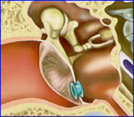
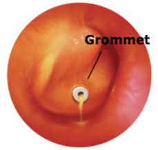
- Grommets: For children with recurrent, severe middle ear infections, tiny tubes may be inserted through the eardrum to help drain fluid. These tubes are called grommets or tympanostomy tubes.
- Myringotomy: A myringotomy is a surgical procedure where the surgeon makes a tiny cut into the eardrum. This can help relieve pressure on the middle ear and allows the surgeon to drain away excess fluid inside the middle ear. In some cases a myringotomy may then be followed with a grommet insertion.
- Tympanotomy :is a surgical procedure during which a surgical opening is made in the ear drum or tympanic membrane in order to promote drainage of infected fluid from the middle ear. & surgical tubes are implanted into the eardrum to promote ongoing drainage. It is done when there is scaring or little damage to tympanic membrane, in cases of deafness, or hearing.
- Myringoplasty Is a surgical procedure done to repair a hole in the eardrum. The hole is repaired by placing a graft made of either a small piece of tissue from elsewhere on the body, or a gel-like material.
- Tympanoplasty-Repair of damaged ossicles by replacing it with apiece of bone or prosthesis.

Grommet

Grommet already inserted
Nursing care
- Apply hot water bag over the ear with the child lying on the affected side may reduce the discomfort (applied during the attack of pain).
- Put ice bag over the affected ear may also be beneficial to reduce edema (between pain attacks).
- For drained ear; the external canal may be frequently cleaned using sterile cotton swabs (dry or soaked in hydrogen peroxide).
- Excoriation of the outer ear should be prevented by frequent cleansing and application of zinc oxide to the
area of oxidate. - Give special attention to the tympanostomy tube i.e., avoid water entering the middle ear and introducing bacteria.
- Educate family about care of child, and; keep them aware with the potential complications of acute
otitis media e.g., conductive hearing loss. - Provide emotional support to the child and his family.
- Rest the patient in bed i.e complete bed rest
- Encourage or give plenty of oral fluids
- In case of any discharge, dry the ear by ear wicking i.e make a wick using cotton swab and clean the pus from the ear.
Complications
- Meningitis
- Mastoid abscess
- Acute mastoiditis
- Facial nerve damage leading to facial palsy
- Infection in adjacent areas e.g. tonsils, noise.
- Brain abscess
- Labyrinthitis ( extension of the infection to the internal ear)
- Sinus thrombosis
- Septicemia
Prevention
- Health education, e.g. advising patients on recognizing the discharge of otitis media
- Early diagnosis and treatment of acute otitis media and upper respiratory tract infections
- Treat infections in adjacent areas e.g tonsillitis


Thank you very much but am requesting for a permission to copy some notice
Thanks very much but I wanted to know the specific nursing care for a child following myringotomy
How can a nurse manage otitis media using a nursing process?
Assessment:
Medical history: Gather information about the child’s previous episodes of otitis media, any underlying conditions, and family history of ear infections.
Sympt
oms: Assess for symptoms such as ear pain, fever, hearing difficulties, irritability, and upper respiratory infection symptoms.
Nursing Diagnoses:
Impaired verbal communication related to hearing loss.
Risk for infection related to the presence of pathogens.
Anxiety related to health status.
Disturbed sensory perception related to obstruction, infection of the middle ear, or auditory nerve damage.
Risk
for injury related to hearing loss and decreased visual acuity .
Planning (Goals and Expected Outcomes):
The child will have improved hearing and communication.
The child will be free of infection.
The parents will demonstrate understanding of preventive measures.
The child will
have normal hearing .
Implementation:
Heat application: Apply a heating pad or warm water bottle to the affected ear to relieve pain.
Medication administration: Administer prescribed antibiotics or pain medications as ordered.
Education: Teach parents about proper hygiene practices, such as covering mouths and noses when sneezing or coughing, and frequent handwashing. Educate them about the importance of breastfeeding for infants as it provides natural immunity to infectious agents.
Monitoring: Assess the child’s hearing ability frequently and report any changes to the healthcare provider.
Evaluation:
The child or parent indicates absence of pain.
The child’s hearing and communication have improved.
The child is free of infection.
The parents demonstrate understanding of preventive measures.
The child has normal hearing .