

Nursing Informatics Introduction
Nerve and Muscle Physiology
Nerve and Muscle Physiology
By the conclusion of this exhaustive master guide, you will be deeply conversant with:
- The fundamental properties of excitable tissues and how voltage gradients dictate human physiology.
- The precise structural and functional anatomy of the motor neuron.
- The step-by-step biochemical and electrical cascade of synaptic transmission and the generation of an action potential.
- The microscopic anatomy of skeletal muscle, including the structural hierarchy of the sarcomere.
- The flawless sequence of Excitation-Contraction Coupling, from the Neuromuscular Junction (NMJ) down to the exact molecular movements of the Sliding Filament Theory.
I. Introduction to Excitable Tissues
Nerve and muscle physiology is a highly specialized branch of medical physiology that specifically studies the function, architecture, and electrochemical mechanisms of nervous tissue (nerves) and muscle tissue (muscles). Collectively, nerves and muscles are uniquely classified as "excitable tissues."
This classification means they possess the extraordinary ability to undergo rapid, transient changes in their resting membrane potential. They explore how these tissues generate and transmit electrical signals (like action potentials) and how these sheer electrical impulses are seamlessly converted into specific, tangible cellular functions.
- For Nerves: It covers how neurons (individual nerve cells) generate electrical impulses, communicate with each other across microscopic gaps (synaptic transmission), process immense amounts of sensory information, and transmit command signals throughout the body to control various systemic functions—ranging from conscious thought and pain sensation to voluntary movement and involuntary organ regulation.
- For Muscles: It focuses intensely on how muscle cells (muscle fibers) respond to the electrical signals delivered by nerves, leading to mechanical contraction (physical shortening) and the generation of kinetic force. This includes the intricate molecular mechanisms of contraction, the metabolic regulation of muscle force, and the different types of muscle tissue and their distinct, highly specialized functional characteristics.
Nervous System Excitability
Nervous system excitability is defined as the inherent ability of nerve cells (neurons) to respond to an external or internal stimulus by generating and propagating an action potential—a massive, self-propagating electrical impulse that travels down the length of the cell.
This property is the absolute fundamental basis of the nervous system's function. It depends entirely on the neuron membrane's selective permeability, the precise opening and closing of voltage-gated ion channels, and the relentless work of active ion pumps. A sudden, sufficient change in membrane potential leads directly to this firing event, which is essential for transmitting information throughout the entire body. The physiology of the nervous system involves its main divisions—the Central Nervous System (CNS) and Peripheral Nervous System (PNS)—which utilize neurons and electrochemical signals to sense stimuli, integrate complex data, and produce highly coordinated, life-sustaining responses.
II. Overall Structure & Function of a Motor Neuron (The Command Pathway)
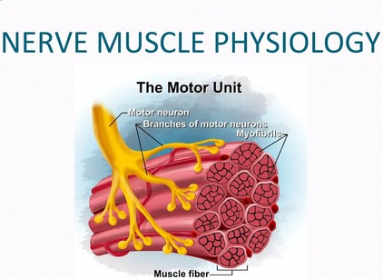
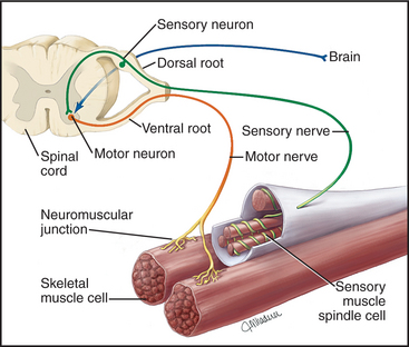
A motor neuron is a highly specialized, efferent nerve cell that transmits electrical command signals strictly away from the central nervous system (brain and spinal cord) to effector targets like muscles or glands, thereby initiating physical movement or chemical secretion.
The "Final Common Pathway"
Motor neurons are universally referred to in physiology as the final common pathway. This term emphasizes a profound physiological principle: all the impossibly complex neural computations happening in higher brain centers (e.g., motor planning in the cerebral cortex, coordination in the basal ganglia, and balance checking in the cerebellum) must ultimately converge onto these lower motor neurons.
It is only through the firing of a lower motor neuron that a skeletal muscle can be activated and a movement can physically occur. Regardless of whether a movement is voluntary (throwing a ball) or reflexive (pulling your hand away from a hot stove), the command signal ultimately travels down a lower motor neuron to its target muscle fibers. This makes the motor neuron a critical biological bottleneck and the ultimate determinant of muscle activity.
Clinical Example: Amyotrophic Lateral Sclerosis (ALS) is a devastating disease that specifically targets and destroys these motor neurons. Because they are the final common pathway, their destruction leads to complete, irreversible muscle paralysis, even though the patient's higher brain functions remain perfectly intact.

1. Motor Neuron Anatomy: Key Structural Components
The metabolic and genetic center of the neuron. It contains the nucleus, extensive rough endoplasmic reticulum (Nissl bodies), and other organelles. It synthesizes vital neurotransmitters and structural proteins, and it receives synaptic inputs directly from thousands of other neurons.
Branching, tree-like extensions protruding from the soma. These are the primary receptive (input) regions of the cell. They are studded with ligand-gated ion channels that receive chemical signals (neurotransmitters) from adjacent cells and convert them into graded electrical potentials (EPSPs and IPSPs).
A specialized, cone-shaped anatomical region where the axon originates from the soma. This is the critical "trigger zone". It possesses the highest density of voltage-gated Na⁺ channels in the entire cell. It acts as an organic calculator, mathematically integrating all incoming graded potentials; if the sum reaches the critical threshold, an action potential is violently generated here.
A single, microscopic, tube-like projection that transmits the action potential (the output signal) away from the cell body toward the target. Some axons (like those reaching down to your toes) can exceed a full meter in length!
A thick, fatty, insulating layer wrapped around many axons. It is formed by Schwann cells in the PNS and Oligodendrocytes in the CNS. It is absolutely crucial for dramatically increasing the speed of action potential conduction and preventing electrical signal leakage.
Microscopic, unmyelinated gaps evenly spaced along the myelin sheath. These exposed patches contain a massive concentration of voltage-gated Na⁺ and K⁺ channels. The action potential is chemically regenerated at these nodes, allowing the signal to "jump" lightning-fast from node to node in a process called saltatory conduction.
The highly branched, club-like ends of the axon that form physical synapses with other cells. They contain thousands of synaptic vesicles packed with neurotransmitters and are specialized for converting the electrical action potential back into a chemical signal.
2. Functional Zones: Relating Structure to Role
To understand the flow of neurological information, we map these anatomical structures into four distinct functional zones:
- Input Zone (Dendrites & Cell Body): Receives and chemically integrates incoming signals, translating them into graded potentials.
- Integration Zone (Axon Hillock): The decision-making center. It sums all graded potentials. If the net depolarization is strong enough to reach threshold, it fires the weapon (triggers an action potential).
- Conduction Zone (Axon): Propagates the "all-or-nothing" action potential without any loss of signal strength over immense distances, heavily facilitated by the myelin sheath's saltatory conduction.
- Output Zone (Axon Terminals): Converts the traveling electrical action potential into a chemical signal by dumping neurotransmitters onto the next cell.
III. Synaptic Transmission: The Communication Bridge
Synaptic transmission is the fundamental, microscopic process by which one neuron (the presynaptic neuron) communicates with another neuron (the postsynaptic neuron) or an effector cell (like a muscle). Most synapses in the human nervous system are chemical synapses, meaning they utilize chemical messengers known as neurotransmitters to physically bridge the gap between cells where electricity cannot cross.

Anatomy of a Chemical Synapse
- Presynaptic Terminal (Axon Terminal): The specialized emitting end of the incoming axon. It is densely packed with synaptic vesicles (bubbles full of neurotransmitters), abundant mitochondria to fuel the constant energy demand, and crucial voltage-gated Ca²⁺ channels.
- Synaptic Cleft: The microscopic, fluid-filled extracellular void (typically only 20-50 nanometers wide) that physically separates the sending and receiving membranes.
- Postsynaptic Membrane: The highly specialized receiving region of the target cell's membrane, heavily studded with specific neurotransmitter receptors.
The Step-by-Step Mechanism of Neurotransmission
1. Presynaptic Events: Neurotransmitter Release
This phase is the miracle of converting an electrical spark into a chemical flood:
- Action Potential Arrives: An action potential successfully propagates down the axon and violently depolarizes the presynaptic terminal membrane.
- Depolarization Opens Voltage-Gated Ca²⁺ Channels: The sudden shift in positive electrical charge forces specialized calcium channels in the terminal membrane to snap open.
- Ca²⁺ Influx: Driven by a steep electrochemical gradient (calcium is highly concentrated outside the cell), Ca²⁺ ions rapidly rush into the presynaptic terminal. This influx is the absolute, non-negotiable trigger for neurotransmitter release.
- Ca²⁺ Triggers Vesicle Fusion: The sudden spike in intracellular Ca²⁺ activates specific docking proteins (SNARE proteins). These proteins act like winches, pulling the synaptic vesicles down and forcing them to fuse with the presynaptic cell membrane.
- Neurotransmitter Release (Exocytosis): As the vesicles violently fuse with the membrane, they burst open, expelling their payload of neurotransmitters directly into the synaptic cleft.
Botulism & Tetanus Toxins
The deadly Botulinum toxin (used cosmetically as Botox) and Tetanus toxin work by destroying the SNARE proteins mentioned in Step 4. Without SNARE proteins, synaptic vesicles cannot fuse with the membrane. The nerve fires, calcium rushes in, but the neurotransmitters are permanently trapped inside the terminal. This completely severs communication to the muscle, resulting in flaccid paralysis (in Botulism) or rigid, spastic paralysis (in Tetanus).
2. Postsynaptic Events: Receptor Binding & Ion Channel Opening
Once dumped into the cleft, neurotransmitters diffuse rapidly across the microscopic gap and bind reversibly to their specific, lock-and-key receptors on the postsynaptic membrane.
- Ligand-Gated Ion Channels (Ionotropic): The receptor itself is a physical ion channel. Binding of the neurotransmitter acts as a key that instantly pops the channel open, allowing an immediate rush of ions and a rapid change in the postsynaptic membrane potential. This generates:
- Excitatory Postsynaptic Potential (EPSP): A localized depolarization (e.g., via Na⁺ influx), making the inside of the neuron more positive and therefore more likely to fire.
- Inhibitory Postsynaptic Potential (IPSP): A localized hyperpolarization (e.g., via Cl⁻ influx or K⁺ efflux), making the inside of the neuron more negative and effectively "shutting it down," making it less likely to fire.
- G-Protein Coupled Receptors (Metabotropic): The receptor activates an intracellular middle-man called a G-protein. This initiates a slower, highly complex, but much more widespread and long-lasting signaling cascade within the cell. This can lead to the production of "second messengers" (e.g., cAMP) that can physically alter the cell's DNA gene expression or modulate nearby ion channels from the inside.
3. Neurotransmitter Inactivation/Removal: Terminating the Signal
To ensure precise, crisp, and discrete signaling, the chemical message must be swiftly deleted from the cleft the moment the signal is delivered. If left unchecked, the muscle or nerve would seize up in a state of permanent, toxic overstimulation. This termination happens through three mechanisms:
- Enzymatic Degradation: Specific assassin enzymes localized in the synaptic cleft chemically chop up and destroy the neurotransmitter. Example: Acetylcholinesterase (AChE) brutally breaks down acetylcholine into harmless choline and acetate in milliseconds.
- Reuptake: Specialized, ATP-driven transporter proteins on the presynaptic terminal (or nearby glial cells) actively act like biological vacuum cleaners, pumping the intact neurotransmitter back into the sending cell to be recycled for the next firing. Example: This is the primary mechanism for monoamines like serotonin and dopamine.
Clinical Application: Selective Serotonin Reuptake Inhibitors (SSRIs) like Prozac block these vacuum pumps, intentionally leaving serotonin in the cleft longer to treat severe depression. - Diffusion: Some neurotransmitters simply float and diffuse away from the synaptic cleft into the surrounding extracellular fluid, where their concentration drops to ineffective levels.

IV. Generation and Conduction of the Action Potential
The motor neuron is constantly bombarded with thousands of chemical signals (both excitatory and inhibitory) from thousands of other neurons every second. The neuron must mathematically integrate these signals to decide whether to fire a massive, "all-or-nothing" action potential.
1. Integration: Spatial and Temporal Summation
A single tiny EPSP is overwhelmingly too weak to push a neuron to threshold. Therefore, the Axon Hillock adds them all up through summation:
- Spatial Summation: Multiple EPSPs or IPSPs arriving at completely different physical locations on the dendrites at the exact same time add together. (Analogy: Ten different people pushing a heavy car at the same time).
- Temporal Summation: Rapid, successive, machine-gun-like EPSPs from a single presynaptic neuron add up over time before the previous one has a chance to fade away. (Analogy: One person rhythmically pushing a swing over and over to build momentum).
If the absolute algebraic sum of all incoming excitatory (EPSPs) and inhibitory (IPSPs) signals reaches the critical Threshold Potential (typically around -55 mV), the Axon Hillock opens its gates and an action potential is irrevocably generated.
2. Phases of the Action Potential
Once generated, the action potential propagates down the axon without losing a fraction of its strength. It follows a highly stereotyped, predictable voltage curve:
| Phase | Voltage Level | Cellular Event (Ion Channel Status) |
|---|---|---|
| 1. Resting State | -70 mV | The membrane is highly polarized. All voltage-gated Na⁺ and K⁺ channels are tightly closed. The Resting Membrane Potential (RMP) is maintained primarily by K⁺ leak channels (potassium constantly trickling out) and the relentless, energy-burning Na⁺/K⁺-ATPase pump. |
| 2. Depolarization to Threshold | -70 mV to -55 mV | The summed EPSPs cause a localized, sluggish depolarization. If the internal charge reaches the -55 mV threshold, it triggers a catastrophic, positive feedback loop. |
| 3. Rising Phase (Depolarization) | -55 mV shooting up to +30 mV | At threshold, thousands of voltage-gated Na⁺ channels violently snap open. A massive, sudden influx of Na⁺ rushes into the cell, erasing the negative charge and making the inside of the membrane highly positive. |
| 4. Falling Phase (Repolarization) | +30 mV dropping back toward -70 mV | At the exact peak (+30 mV), an inactivation gate swings shut on the Na⁺ channels, instantly stopping the Na⁺ influx. Simultaneously, the sluggish voltage-gated K⁺ channels finally fully open. A massive efflux of K⁺ rushes out of the cell, rapidly repolarizing the membrane back to negative. |
| 5. Undershoot (Hyperpolarization) | Dips below -70 mV (e.g., -80 mV) | The voltage-gated K⁺ channels are slow to close, causing an excessive, prolonged efflux of K⁺. The membrane becomes briefly more negative than its resting state. During this time, the neuron enters its Relative Refractory Period, where only an abnormally massive stimulus can trigger another firing. (Note: The Absolute Refractory Period occurs during the rising and falling phases, where it is physically impossible for the cell to fire again, ensuring the signal only travels in one forward direction). |
| 6. Restoration | Returns to -70 mV | All voltage-gated channels finally slam shut. The Na⁺/K⁺-ATPase pump works continuously in the background, burning ATP to shovel Na⁺ back out and K⁺ back in, fully restoring the long-term chemical gradients. |
Local Anesthetics (Lidocaine)
When a dentist injects Lidocaine into your gums, the drug physically enters the nerve cell and plugs up the Voltage-Gated Na⁺ Channels from the inside. If Na⁺ cannot rush in during the Rising Phase, the action potential completely fails to generate. The pain signal is stopped dead in its tracks, and your brain never feels the drill.
V. Muscle Physiology: The Architecture of Contraction
While nerves specialize in communication, muscle tissue is purely specialized for mechanical contraction, generating kinetic force and bodily movement. We focus here on Skeletal Muscle.
A. Skeletal Muscle Micro-Structure
- Muscle Fiber (Cell): A single, highly elongated, cylindrical, multinucleated cell spanning the length of the muscle.
- Sarcolemma: The specialized, electrically excitable plasma membrane of a muscle fiber. It features deep, tube-like invaginations that dive into the center of the cell called T-tubules.
- Sarcoplasm: The unique cytoplasm of a muscle fiber. It is heavily packed with mitochondria (for ATP), glycogen (sugar storage), myoglobin (oxygen storage), and thousands of contractile rods called myofibrils.
- Myofibrils: Long, rod-like contractile organelles running parallel to the fiber, entirely composed of repeating microscopic units called sarcomeres.
- Sarcoplasmic Reticulum (SR): A highly specialized, web-like smooth endoplasmic reticulum that wraps around each and every myofibril like a sleeve. Its sole purpose is storing, releasing, and recapturing massive amounts of Ca²⁺ ions.
- The Triad: A critical anatomical junction consisting of one central T-tubule physically flanked on both sides by two enlarged sacs of the SR (terminal cisternae). This ensures the electrical signal diving down the T-tubule instantly triggers the adjacent SR.

B. The Sarcomere: The Ultimate Contractile Unit
The sarcomere is the fundamental, repeating contractile unit of a myofibril, extending from one Z-disc to the next Z-disc. Its highly organized geometry gives skeletal muscle its striated (striped) appearance.
The Filaments (The Machinery):
- Thick Filaments (Myosin): Composed entirely of the protein myosin. Each myosin molecule looks like a golf club, with a twisted tail and two globular, pivoting heads. The heads are the true engines; they contain an actin-binding site and an ATP-binding site (which functions as an ATPase enzyme to burn fuel).
- Thin Filaments (Actin Complex): Composed primarily of a twisted double-strand of actin pearls. Critically, the thin filament also houses two highly sensitive regulatory proteins that act as a lock-and-key system:
- Tropomyosin: A long, thread-like, rod-shaped protein that spirals around the actin. In a relaxed muscle, it physically covers and blocks the myosin-binding sites on the actin, preventing contraction.
- Troponin: A specialized complex of three proteins pinned to tropomyosin. The Troponin C (TnC) subunit acts as the ultimate lock; it is the specific component that strongly binds incoming Ca²⁺ ions, shifting the tropomyosin out of the way and initiating the violent contraction sequence.
The Bands and Zones (The Geography):
- A Band: The entire, dark length of the thick (myosin) filament. Crucial rule: Its physical length remains absolutely constant and never changes during contraction.
- I Band: The light region containing only thin (actin) filaments. It drastically shortens during contraction as the filaments slide inward.
- H Zone: The lighter, central region of the A band containing only thick filaments (no actin overlap). It completely disappears/shortens during maximum contraction.
- M Line: A dark, structural anchoring line running straight down the dead center of the H zone, holding the thick filaments in strict alignment.
- Z Disc (Z Line): The jagged, zig-zag boundary line that perfectly defines the absolute ends of a single sarcomere and anchors the thin filaments in place.

VI. The Neuromuscular Junction (NMJ): The Nerve-Muscle Interface
The neuromuscular junction (NMJ) is arguably the most important chemical synapse in the human body. It is the highly specialized, mandatory interface where a motor neuron's axon terminal docks with a skeletal muscle fiber.
Anatomy of the NMJ:
- Presynaptic Terminal: The swollen end of the motor neuron's axon, packed tight with thousands of synaptic vesicles heavily loaded with the neurotransmitter Acetylcholine (ACh).
- Synaptic Cleft: The microscopic physical gap between the nerve and the muscle. It is flooded with a highly active enzyme called Acetylcholinesterase (AChE).
- Motor End Plate: A highly specialized, crater-like region of the muscle's sarcolemma positioned directly under the nerve terminal. It features deep, accordion-like junctional folds densely packed with millions of specialized Nicotinic Acetylcholine Receptors (nAChRs).
End-Plate Potential (EPP): The Muscle's First Electrical Response
This is the explosive, step-by-step translation of a nerve thought into a muscle shock:
- ACh Release: An action potential traveling down the motor neuron opens terminal Ca²⁺ channels, triggering the massive exocytosis of ACh into the synaptic cleft.
- ACh Binding: ACh diffuses across the tiny cleft and instantly binds to the nAChR receptors waiting on the motor end plate.
- Channel Opening: The binding of exactly two ACh molecules to a receptor forces the central ion channel to pop open.
- Ion Movement: Na⁺ ions rapidly rush into the muscle fiber, while a smaller, slower stream of K⁺ ions moves out. The absolute net effect is a massive influx of positive charge into the muscle.
- Depolarization (EPP): This sudden net influx of positive Na⁺ ions causes a rapid, incredibly large, localized depolarization of the motor end plate, scientifically known as the End-Plate Potential (EPP). Important physiological note: Unlike the weak EPSPs in the brain, a single healthy EPP is immensely powerful and is always large enough to instantly trigger a full, unstoppable action potential in the adjacent sarcolemma (this is known as the safety factor).
- ACh Inactivation: In mere milliseconds, the ACh is violently ripped off the receptor and degraded by the enzyme acetylcholinesterase (AChE) suspended in the cleft, terminating the excitatory signal instantly and allowing the muscle fiber to repolarize for the next breath or step.
Myasthenia Gravis vs. Nerve Gas
- Myasthenia Gravis: An autoimmune disease where the body mistakenly produces antibodies that attack and destroy the nAChRs on the motor end plate. Even though the nerve releases plenty of ACh, there are no receptors left to catch it. The EPP fails to reach threshold, resulting in profound, progressive muscle weakness (drooping eyelids, inability to swallow).
- Sarin Nerve Gas & Organophosphates: These toxic chemicals permanently destroy the enzyme AChE in the synaptic cleft. ACh is released but is never destroyed. It violently overstimulates the receptors continuously, causing the muscles to undergo fatal, tetanic, spastic paralysis (the diaphragm locks up and the victim suffocates).
VII. Excitation-Contraction Coupling
This is the awe-inspiring physiological bridge by which an invisible electrical signal (the muscle action potential) is instantly converted into a violent mechanical event (muscle contraction).
- Muscle AP Propagation: The newly generated action potential travels like a wave of fire across the surface of the sarcolemma and rapidly dives deep down into the core of the cell via the T-tubules.
- DHPR Activation: As the electrical shock travels down the T-tubule, it hits and alters the physical shape of specialized voltage-sensitive sensor proteins in the membrane called Dihydropyridine Receptors (DHPRs).
- Mechanical Linkage to RyRs: In skeletal muscle, these DHPR sensors are physically, mechanically linked like a locked door handle to Ryanodine Receptors (RyRs). The RyRs are giant "plug" channels embedded directly in the membrane of the adjacent Sarcoplasmic Reticulum (SR).
- RyR Opening and Ca²⁺ Release: The voltage change forces the DHPR to literally pull the RyR plug open mechanically. This unplugs the SR, allowing billions of stored, highly pressurized Ca²⁺ ions to violently flood out of the SR and into the surrounding sarcoplasm (cytoplasm).
- Increase in Intracellular Ca²⁺: This sudden, massive spike in sarcoplasmic Ca²⁺ concentration is the absolute, immediate, non-negotiable chemical trigger for muscle contraction.

VIII. The Mechanism of Muscle Contraction: The "Sliding Filament Theory"
The Sliding Filament Theory universally proposes that muscle shortening (contraction) occurs strictly by the thick (myosin) and thin (actin) filaments sliding past one another, dragging the Z-discs closer together. The filaments themselves never actually shrink or change length; they merely increase their overlap.
1. Role of Ca²⁺: Unlocking the Binding Sites
Before the engines can engage, the track must be cleared. The Ca²⁺ ions released from the SR flood the sarcomere and bind immediately to the Troponin C subunit perched on the thin actin filaments. This binding causes a profound shape change in the entire troponin complex, which in turn violently tugs on the long, thread-like tropomyosin molecule. The movement of tropomyosin physically drags it away from the myosin-binding sites on the actin beads, exposing them completely to the waiting myosin heads.

2. The Cross-Bridge Cycle (Molecular Events): The Powerhouse
The cross-bridge cycle is a blindingly fast, repetitive, four-step biochemical engine sequence that physically causes the thin filaments to slide over the thick filaments.
Cross-Bridge Formation (The Latch)
The energized, upright ("cocked") myosin head—which is already tightly holding onto a spent ADP and inorganic phosphate (Pi) molecule from the previous cycle—possesses a massive chemical attraction (affinity) for the exposed actin filament. The moment the tropomyosin shifts to uncover the binding sites (thanks to calcium), the myosin head violently snaps upward and forms a strong, unbreakable physical link with the actin. This physical connection between thick and thin filaments is the famous "cross-bridge."
The Power Stroke (The Pull)
The instant physical formation of the cross-bridge chemically triggers the immediate release of the trapped inorganic phosphate (Pi) from the myosin head. This release unleashes the stored mechanical energy, causing the myosin head to forcefully pivot on its hinge, jerking from its high-energy 90° angle to a low-energy, bent 45° angle. This brutal pivoting movement is the power stroke. Because it is firmly attached like a grappling hook, the pivoting myosin head violently drags the entire thin actin filament a short microscopic distance (~10 nm) toward the dead center (M-line) of the sarcomere. Immediately following the pivot, the remaining ADP molecule is ejected, leaving the myosin head in a rigid, low-energy state, still tightly locked onto the actin.
Cross-Bridge Detachment (The Release)
Following the power stroke, the spent myosin head remains rigidly "stuck" to the actin in a low-energy configuration (known as the "rigor" state). The absolute only biochemical way for the myosin head to let go of the actin is for a brand new, fresh molecule of ATP to physically bind to the empty ATP-binding site on the back of the myosin head. The instant this new ATP binds, it causes an allosteric (shape) change that severely weakens the molecular bond between the myosin and the actin, reducing their chemical affinity to zero, and forcing the myosin head to abruptly detach.
Re-cocking of the Myosin Head (The Reset)
The completely detached myosin head, now carrying its fresh ATP, immediately acts as an aggressive enzyme (myosin ATPase). It instantly hydrolyzes (burns) the ATP, snapping it into ADP and inorganic phosphate (Pi). The explosive energy released from breaking this intense ATP chemical bond is captured entirely by the myosin hinge, physically forcing the head to snap backward, moving from its low-energy bent position back to its high-energy, upright, "cocked" position. It is now fully energized, reset, and coiled like a rat-trap, ready to violently repeat the entire cycle by latching onto another active site further down the actin filament (provided Ca²⁺ is still present in the sarcoplasm keeping the sites exposed).
Clinical Insight: Rigor Mortis
Why do bodies become stiff after death? Upon death, cellular respiration halts, and ATP production completely drops to zero. Simultaneously, cell membranes degrade, allowing calcium to leak into the sarcoplasm, triggering Step 1 (Cross-Bridge Formation) and Step 2 (The Power Stroke). However, because there is absolutely no new ATP being produced by the dead body, Step 3 (Detachment) cannot occur. Millions of myosin heads remain permanently, rigidly locked onto the actin filaments in the low-energy rigor state. The entire muscular system locks into a solid, unbreakable spasm known as rigor mortis, which only subsides days later when the muscle proteins literally begin to rot and decompose.
3. Sarcomere Shortening: The Ultimate Result
As millions of myosin heads asynchronously paddle through this cycle thousands of times per second, the results are profound across the geometry of the cell:
- The thin filaments actively slide inward, aggressively pulled past the stationary thick filaments toward the M-line.
- The Z-discs on either end of the sarcomere are violently dragged closer together, physically shortening the entire microscopic sarcomere unit.
- Under a microscope, the light I bands and the central H zone compress and shorten.
- The dark A band remains absolutely unchanged in length, proving the thick filaments do not shrink.
When hundreds of thousands of sarcomeres lined up in series shorten simultaneously, the entire gross muscle belly shortens, pulling the tendon and generating massive skeletal force.
IX. Muscle Relaxation
Muscle relaxation is not simply a passive fading out; it is a highly active, metabolically expensive, energy-requiring biochemical process. You must burn ATP to relax!
- Cessation of Motor Neuron Signal: The upper brain commands stop, the lower motor neuron halts its action potential firing, and no new ACh is exocytosed into the NMJ cleft.
- AChE Activity: The remaining ACh currently sitting in the synaptic cleft is rapidly and aggressively broken down into choline and acetate by the ever-present enzyme acetylcholinesterase, completely starving the nAChR receptors of their ligand.
- Repolarization of Sarcolemma: The muscle fiber nAChR channels slam shut, Na⁺ influx ceases, and the sarcolemma and deep T-tubules fully repolarize back to a negative resting state.
- Ca²⁺ Reuptake into SR (The Heavy Lifting): As the T-tubules repolarize, the DHPR voltage sensors return to normal, pushing the massive RyR calcium release channels on the SR closed. Simultaneously, thousands of active transport vacuums called SERCA pumps (Sarcoplasmic/Endoplasmic Reticulum Calcium ATPase) burn massive amounts of ATP to aggressively pump the escaped Ca²⁺ ions from the sarcoplasm backward, against their gradient, into the SR storage vaults.
- Tropomyosin Blocks Active Sites: As the ruthless SERCA pumps drop the ambient sarcoplasmic Ca²⁺ levels back to near zero, calcium physically detaches from the Troponin C receptor. The Troponin complex snaps back to its original, un-calcium-bound shape. This shape change allows the long tropomyosin rod to physically shift back over, snapping into place and completely re-covering and blocking the myosin-binding sites on the actin filament.
- Muscle Relaxes: With the binding sites completely shielded, no new cross-bridges can possibly form. The remaining attached myosin heads finish their current cycle, detach, and are physically barred from reattaching. The muscle fiber loses tension and passively lengthens back to its resting resting length due to gravity or the pull of an opposing antagonist muscle.
Malignant Hyperthermia
This is a rare, life-threatening genetic emergency triggered by the administration of certain general anesthetics (like halothane) or muscle relaxants (like succinylcholine) during surgery. The patient possesses a mutated, defective Ryanodine Receptor (RyR). When exposed to the anesthetic gas, the mutant RyR jams wide open, dumping an endless, unstoppable flood of calcium into the sarcoplasm. The muscles instantly go into a massive, full-body hyper-metabolic contraction. The SERCA pumps burn through the body's entire ATP supply trying to pump the calcium back in, generating highly lethal amounts of bodily heat (hyperthermia) and lactic acid. Without immediate administration of the antidote Dantrolene (which specifically acts to forcefully slam the RyR channels shut), the patient will rapidly die of a metabolic meltdown on the operating table.
X. List of References
This comprehensive synthesis of excitable tissue physiology is got from and aligns with the following gold-standard academic texts and resources universally utilized in medical and physiological education:
- Hall, J. E., & Guyton, A. C. (2015). Guyton and Hall Textbook of Medical Physiology (13th ed.). Philadelphia, PA: Elsevier. (Primary source for comprehensive AP mechanics and excitation-contraction coupling).
- Costanzo, L. S. (2018). Physiology (6th ed.). Philadelphia, PA: Elsevier. (Primary source for succinct, step-by-step molecular breakdowns of the cross-bridge cycle and NMJ pharmacology).
- Boron, W. F., & Boulpaep, E. L. (2016). Medical Physiology (3rd ed.). Philadelphia, PA: Elsevier. (Advanced reference for SNARE protein mechanics, spatial/temporal summation mathematical integration, and specific ion channel gating kinetics).
- Katzung, B. G., Masters, S. B., & Trevor, A. J. (2021). Basic & Clinical Pharmacology (15th ed.). McGraw-Hill Education. (Reference for applied clinical correlations, including Botox, local anesthetics, and Malignant Hyperthermia antidotes).
Nerve and Muscle Physiology Read More »
PHYSIOLOGY OF EXCITABLE TISSUES
PHYSIOLOGY OF EXCITABLE TISSUES
By the end of this exhaustive physiological guide, you will master:
- The fundamental definition of Excitability and the specialized cells that possess it.
- The electrochemical mechanisms maintaining the Resting Membrane Potential (RMP).
- The exact structural and functional gating mechanisms of Ion Channels.
- The step-by-step ionic basis of the Action Potential and signal propagation.
- The mechanisms of Inhibition (hyperpolarization, presynaptic, and pharmacological).
- The profound Clinical Significance of excitability, focusing heavily on electrolyte imbalances and neurological diseases.
1. Introduction to Excitability
Excitability refers to the fundamental ability of a living cell to respond to an environmental stimulus by generating a rapid, highly coordinated electrical signal known as an Action Potential. It is defined as a measurable physical and chemical change that occurs across the cell membrane when a stimulus is applied to a tissue. A stimulus is any external or internal agent (electrical, chemical, thermal, or mechanical) that produces this excitation.
The action potential is a transient, rapid, and self-propagating reversal of the electrical potential across the cell membrane. This electrical signal is the medium through which cells rapidly transmit information, either along the length of an individual cell (like a wire) or to other cells via specialized junctions (synapses). This property is crucial for rapid communication and coordination within the body, underpinning virtually every complex physiological function, from perception, thought, and memory to voluntary movement and visceral regulation.
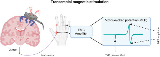
The Electrical Tripwire
Think of an excitable cell like a highly sensitive electrical tripwire or alarm system.
- The Resting State is the armed system, charged and waiting for a trigger.
- The Stimulus is the physical pressure that activates the tripwire.
- The Action Potential is the immediate, swift, and uniform "alarm bell" that rings loudly and clearly, sending its message undiminished through the entire system to orchestrate a massive, coordinated response.
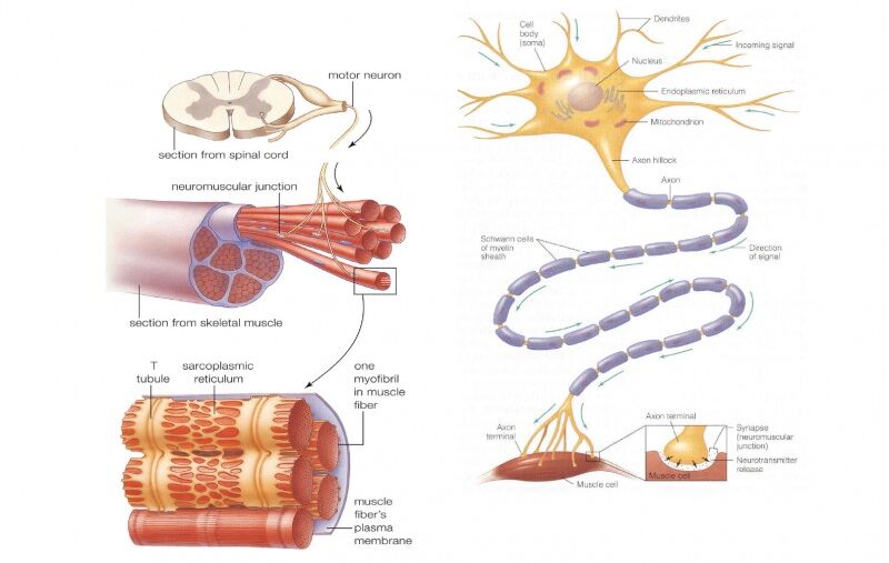
2. Types of Excitable Cells
While all living cells exhibit some degree of responsiveness to their environment, only a select group possess the highly specialized membrane proteins and machinery required to generate and propagate rapid electrical action potentials. These are the "excitable cells."
The Master Communicators
Expanded Role: Neurons are the fundamental functional units of the nervous system. Their primary function is the rapid transmission of electrical and chemical signals for sensory input, integration, motor output, cognition, and emotion.
Unique Features: They possess specialized anatomical structures: dendrites (the "antennae" to receive signals), a cell body (soma) (the metabolic center), and a long axon (the "transmission cable" to send signals), which is often insulated by a lipid-rich myelin sheath to drastically speed up conduction velocity.
The Effectors of Movement
Muscle cells are specialized for contraction, which generates physical force and movement. Their electrical excitability is the absolute prerequisite for this mechanical action (Excitation-Contraction Coupling).
- Skeletal Muscle Cells: Responsible for all voluntary movements (walking, speaking, breathing). When a somatic motor neuron sends an action potential, it triggers a massive muscle action potential across the sarcolemma, leading to instantaneous contraction.
- Cardiac Muscle Cells: Found only in the myocardium, responsible for the rhythmic and involuntary pumping of blood. They possess autorhythmicity (can generate their own pace) and have distinctively long action potentials featuring a "plateau phase" to ensure coordinated, non-tetanic contractions.
- Smooth Muscle Cells: Mediate involuntary movements in the walls of internal organs like the digestive tract, blood vessels, and urinary bladder. Their excitability is influenced heavily by stretch, local chemical mediators, and the autonomic nervous system.
The Secretory Responders
Expanded Role: Many glandular cells (e.g., in the adrenal medulla, pancreatic beta-cells) exhibit true excitability. They can respond to an electrical stimulus from a neuron or a chemical change (like high blood glucose) by generating their own electrical depolarization event.
Excitability Link: This electrical event is typically coupled directly to the release of their internal secretions (hormones, digestive enzymes). For example, adrenal medullary cells depolarize in response to a sympathetic neuronal signal, triggering massive Calcium (Ca²+) influx and the immediate exocytosis of epinephrine. This ensures precise, rapid control over systemic hormone release.
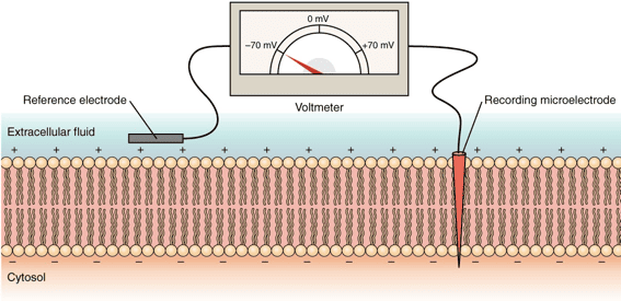
3. The Membrane Potential
The capacity of these cells to generate electrical signals rests entirely on the foundational concept of the Membrane Potential. This is the voltage difference across the cell's outer boundary (the lipid bilayer). It represents stored, potential electrical energy created by an uneven distribution of ions (electrically charged particles) between the Intracellular Fluid (ICF) and the Extracellular Fluid (ECF).
Resting Membrane Potential (RMP)
When an excitable cell is quiet, it maintains a stable, baseline electrical charge called the Resting Membrane Potential (RMP). In this state, the inside of the cell consistently holds a negative charge relative to the outside. Standard RMP values include:
- Neurons: Typically around -70 mV.
- Skeletal & Cardiac Muscle: Typically around -90 mV.
Creating and Maintaining the RMP
The RMP is not static; it is a highly dynamic state, constantly maintained by an active, energy-consuming interplay of three major factors:
- Ion Gradients (The Concentration Divide): The foundational requirement is the extreme difference in the concentrations of key ions: a massive concentration of Sodium (Na+) outside the cell and a massive concentration of Potassium (K+) inside the cell.
- Selective Permeability (The Leaky Gates): At rest, the cell membrane is 50 to 100 times more permeable to K+ than to Na+. This is because the membrane possesses many more open K+ "leak" channels than Na+ leak channels. Consequently, K+ constantly leaks out of the cell, carrying positive charge away and leaving the inside of the cell negative.
- Sodium-Potassium ATPase Pump (The Gradient Upholder): This ubiquitous active transporter continually burns cellular energy (ATP) to pump 3 Na+ ions OUT for every 2 K+ ions it pumps IN. This directly maintains the steep concentration gradients and, because it removes more positive charge than it brings in, it contributes a small amount to the RMP's negativity (acting as an electrogenic pump).
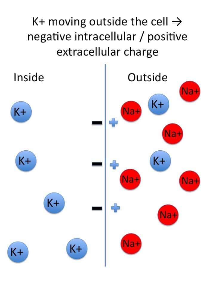
Deeper: Equilibrium Potential
The Nernst Equation
The Equilibrium Potential for a specific single ion is the exact membrane voltage at which there is no net movement of that ion across the membrane. At this specific voltage, the electrical force pulling the ion in one direction is perfectly, equally balanced by the chemical (concentration) force pushing it in the other direction. The Nernst Equation calculates this theoretical value:
Eion = (RT / zF) * ln([ion]out / [ion]in)
Physiological Context: The equilibrium potential for Potassium (EK) is approximately -90 mV. The equilibrium potential for Sodium (ENa) is approximately +60 mV. Because the resting membrane is highly permeable to Potassium and mostly impermeable to Sodium, the overall RMP (-70 mV) sits very close to the Potassium equilibrium potential!
4. Ion Channels: The Gates of Excitability
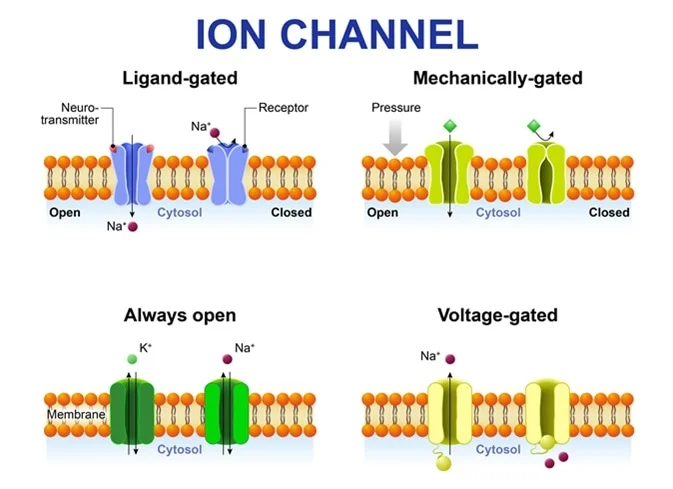
Ion channels are highly specialized, complex transmembrane proteins that form selective pores allowing specific ions to cross the otherwise impermeable lipid bilayer.
Types Relevant to Excitability:
1. Leak Channels (Non-gated)
These channels are essentially always open. They are instrumental in establishing the RMP, particularly the widespread K+ leak channels that allow the continuous efflux of positive charge.
2. Gated Channels (The Responsive Switches)
These channels have molecular "gates" that open or close only in response to a particular physiological trigger. They are the absolute essential machinery for generating action potentials.
| Type of Gated Channel | Trigger Mechanism | Physiological Example |
|---|---|---|
| Voltage-Gated Channels | Open or close in direct response to changes in the transmembrane voltage. They possess charged amino acids that physically move when the voltage changes. | Voltage-gated Na+ channels (driver of depolarization) and Voltage-gated K+ channels (driver of repolarization). |
| Ligand-Gated Channels (Chemically Gated) | Open or close only when a specific chemical messenger (a ligand), such as a neurotransmitter or hormone, binds to a receptor site on the channel. | The Nicotinic Acetylcholine Receptor at the neuromuscular junction. Binding of ACh opens the pore to let Na+ rush in. |
| Mechanically Gated Channels | Open or close when the membrane itself is physically deformed, stretched, or subjected to pressure. | Pacinian corpuscles in the skin (deep pressure receptors) or hair cells in the inner ear (responding to sound waves). |
5. Initiating the Response: Stimulus and Threshold
The Stimulus: A Call to Action
A stimulus is any detectable change (electrical, chemical, or mechanical) in the cell's environment that has the potential to alter its resting membrane potential. Stimuli can cause two types of local changes:
- Depolarization: A shift in membrane voltage where the inside of the cell becomes less negative (e.g., moving from -70 mV up to -50 mV). This is an excitatory shift.
- Hyperpolarization: A shift where the inside of the cell becomes more negative (e.g., moving from -70 mV down to -90 mV). This is an inhibitory shift.
Threshold: The Point of No Return
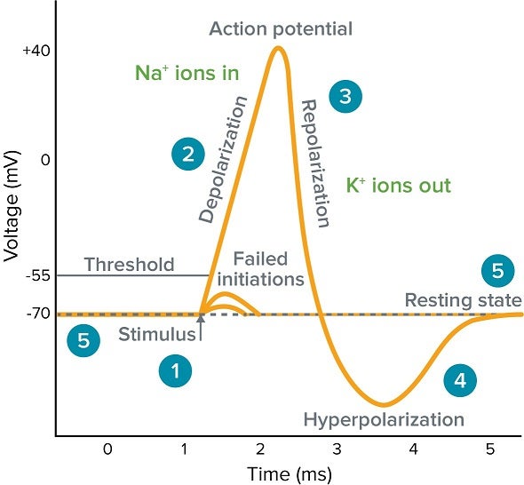
Threshold is the critical, specific voltage level that a depolarization must reach for an action potential to fire (typically around -55 mV in neurons). It operates on an "all-or-none" principle: if a stimulus causes a local depolarization that reaches this threshold, a full, unstoppable action potential fires. If the stimulus is weak and only depolarizes the cell to -60 mV, nothing happens, and the potential simply dissipates.
6. The Action Potential: Step-by-Step
The action potential is the primary electrical signal employed by excitable cells to swiftly transmit information across massive anatomical distances (like from the spinal cord to the toe). It stands as an "all-or-nothing" phenomenon: once initiated, it proceeds through its entire sequence with consistent strength, never diminishing over distance.
| Stage | Membrane Potential | Ionic Movement & Channel Status |
|---|---|---|
| 1. Resting State | -70 mV | All voltage-gated Na+ and K+ channels are CLOSED. The RMP is maintained by K+ leak channels and the active Na+/K+ pump. |
| 2. Depolarization to Threshold | -70 mV to -55 mV | A local stimulus causes a few voltage-gated Na+ channels to open. A small amount of Na+ trickles into the cell. If enough Na+ enters to push the voltage to the -55 mV threshold, the cell commits to firing. |
| 3. Rising Phase (Depolarization) | -55 mV to +30 mV | Threshold triggers a massive positive feedback loop. A vast number of voltage-gated Na+ channels rip open. A massive and swift surge of Na+ rushes into the cell, causing the inside to rapidly become positive (+30 mV). |
| 4. Repolarization Phase | +30 mV back down toward -70 mV | At the +30 mV peak, the voltage-gated Na+ channels inactivate (a physical "plug" blocks the pore), instantly stopping Na+ influx. Simultaneously, the slower voltage-gated K+ channels finally open fully. A massive outflow of K+ rapidly restores the membrane's negative internal charge. |
| 5. Afterhyperpolarization (Undershoot) | Dips below -70 mV (e.g., -80 mV) | The voltage-gated K+ channels are sluggish and close slowly. K+ continues to exit for a brief period, causing the membrane to become temporarily more negative than the baseline RMP. |
| 6. Return to Rest | Returns to -70 mV | The slow K+ channels finally close completely. The ever-active Na+/K+ pump and normal leak channels re-establish the original resting ion concentration gradients. |
Defining Features of Action Potentials
- All-or-Nothing: If the threshold is crossed, the action potential unfolds completely with the exact same magnitude and shape every time. If threshold is not reached, no action potential occurs.
- Non-Decremental: Action potentials are continuously re-generated along the membrane. A signal starting in the brain is just as strong when it reaches the foot.
7. Refractory Periods & Signal Propagation
Refractory Periods (The Reset Time)
A cell cannot fire continuously without resetting. This required downtime is called the refractory period.
- Absolute Refractory Period: Occurs during the entire rising phase and most of the repolarization phase. During this time, the voltage-gated Na+ channels are either already fully open or rigidly inactivated. Therefore, absolutely NO second stimulus, regardless of how massively strong it is, can trigger another action potential. Function: This ensures one-way propagation of the signal and prevents the signal from traveling backward.
- Relative Refractory Period: Occurs during the afterhyperpolarization phase. The Na+ channels have reset to their closed (but ready) state, but the cell is hyperpolarized (extra negative). A stimulus can provoke another action potential, but it must be significantly stronger than normal to overcome the hyperpolarization.
Propagation of Action Potentials: Spreading the Message
The massive electrical shift at one point on the membrane triggers the opening of voltage-gated Na+ channels in the immediately adjacent area. This process repeats endlessly, moving the signal down the length of the nerve or muscle fiber like a burning fuse.
Myelination: Enhancing Speed via Saltatory Conduction
Many nerve fibers are thickly insulated by a fatty lipid layer called the Myelin Sheath (produced by Schwann cells in the PNS and Oligodendrocytes in the CNS). Action potentials cannot form where myelin exists. Therefore, the signal must "jump" from one microscopic uninsulated gap (a Node of Ranvier) to the next. This rapid, jumping propagation is termed Saltatory Conduction and dramatically increases the signal's speed while saving vast amounts of cellular energy.
Factors Influencing Conduction Speed:
- Fiber Diameter: Larger diameter fibers conduct signals much more quickly because there is less internal electrical resistance.
- Myelination: Myelinated fibers transmit signals up to 50 times faster than unmyelinated fibers.
8. Inhibition of Excitability
Just as cells must generate signals to act, they desperately need ways to inhibit signals, ensuring precise motor control, emotional regulation, and preventing uncontrolled, chaotic firing (seizures).
Driving Further from Threshold
Inhibitory neurotransmitters (like GABA in the brain or Glycine in the spinal cord) bind to ligand-gated channels that selectively allow Cl− (Chloride) to enter the cell or K+ to leave. The outcome is an immediate increase in the negative charge inside the cell (e.g., dropping from -70 mV to -80 mV). This creates an Inhibitory Postsynaptic Potential (IPSP), making it significantly harder for any excitatory stimulus to push the cell to threshold.
Muting the Signal at its Source
An inhibitory neuron releases a neurotransmitter (e.g., GABA) directly onto the axon terminal of an excitatory neuron. This slightly reduces the electrical charge of the terminal. Consequently, when an action potential arrives at that terminal, fewer voltage-gated Calcium channels open, meaning fewer excitatory neurotransmitters are released into the synaptic cleft. This allows for brilliant fine-tuning and selective dampening of specific signals without shutting down the whole system.
Manipulating Channels with Drugs
A vast array of modern drugs and natural toxins work by directly interfering with ion channels to deliberately shut down excitability.
- Local Anesthetics (e.g., Lidocaine): Physically plug the intracellular pore of voltage-gated Na+ channels. Without Na+ influx, pain-sensing nerves cannot generate action potentials, resulting in numbness.
- Tetrodotoxin (TTX): A highly potent neurotoxin found in pufferfish. It permanently blocks the extracellular side of voltage-gated Na+ channels, causing rapid, fatal respiratory paralysis.
- Anti-epileptic Drugs (e.g., Phenytoin): Work by stabilizing Na+ channels in their inactivated state, preventing the excessively rapid, repetitive firing seen in seizures.
9. Clinical Significance of Excitability
An in-depth comprehension of cellular excitability is absolutely vital for understanding, diagnosing, and creating effective medical treatments for numerous devastating conditions affecting the nervous system and musculature.
Conditions of the Nervous System and Muscles:
- Epilepsy: Marked by episodes of abnormal, highly synchronized, and excessive electrical firing of large groups of neurons in the cerebral cortex, resulting in seizures. It represents a catastrophic failure of normal inhibitory mechanisms.
- Multiple Sclerosis (MS): A severe autoimmune disease where the myelin sheath insulating central nerve fibers is destroyed by the body's own immune system. This exposes the underlying axon, drastically slowing, weakening, or completely blocking action potential propagation. It leads to progressive muscle weakness, blindness, and sensory disturbances.
- Myasthenia Gravis: An autoimmune disease that generates antibodies to destroy Nicotinic acetylcholine receptors at the neuromuscular junction. This reduces the ability of motor nerve signals to adequately excite muscle cells, leading to profound, easily fatigued muscle weakness (ptosis, difficulty breathing).
- Cardiac Arrhythmias: Irregular heart rhythms stemming from abnormalities in the electrical excitability, automaticity, or conduction pathways of heart muscle cells. This leads to potentially fatal disruptions to the heart's synchronized pumping action (e.g., Ventricular Fibrillation).
10. The Critical Role of Electrolyte Imbalances
Because the entire system of excitability relies on precise concentration gradients of ions, systemic electrolyte imbalances alter the very foundation of the resting membrane potential and threshold, leading to severe clinical emergencies.
| Electrolyte Imbalance | Pathophysiological Mechanism & Cellular Effect | Clinical Consequences |
|---|---|---|
| Hyperkalemia (Elevated K+) |
High extracellular K+ prevents K+ from leaking out of the cell. The resting membrane potential becomes less negative (e.g., moves from -90mV to -70mV in the heart), putting it dangerously close to threshold. While this initially causes twitchy hyperexcitability, prolonged depolarization permanently inactivates voltage-gated Na+ channels, ultimately rendering cells completely inexcitable. | Lethal cardiac arrhythmias, characteristic "peaked T-waves" on an ECG, and eventual flaccid paralysis leading to cardiac arrest. |
| Hypokalemia (Low K+) |
Low extracellular K+ creates a steeper gradient, forcing too much K+ to leak out of the cell. The RMP becomes more negative (hyperpolarized, e.g., dropping to -100mV). This moves the cell much further away from the threshold, making it significantly harder to fire an action potential. | Profound muscle weakness, fatigue, diminished reflexes, and dangerous heart arrhythmias (prominent U-waves on ECG). |
| Hyponatremia / Hypernatremia (Sodium Imbalances) |
Because the rapid, massive influx of Na+ is the absolute primary driver of the depolarization spike, severe imbalances in ECF Na+ levels disrupt the amplitude (height) and speed of action potentials. Furthermore, severe sodium imbalances cause massive cellular swelling or shrinking due to osmotic shifts. | Confusion, lethargy, severe muscle twitching, seizures, coma, and brain damage. |
| Hypocalcemia (Low Ca²+) |
Paradoxical Effect: Calcium ions normally bind to the outside of the cell membrane, physically "shielding" Na+ channels. When calcium is low, this shield is removed. Voltage-gated Na+ channels become highly unstable and rip open at much lower, more negative voltages. The threshold is dangerously lowered. | Severe, involuntary muscle spasms, cramps, and tetany (e.g., Trousseau's sign and Chvostek's sign). |
| Hypercalcemia (High Ca²+) |
High extracellular calcium heavily coats the membrane, stabilizing the Na+ channels and making them incredibly stiff and hard to open. The threshold is raised, drastically decreasing neuronal excitability. | "Bones, stones, groans, and psychiatric overtones." Severe muscle weakness, lethargy, depressed reflexes, and reduced overall neurological function. |
References
- Guyton and Hall: Textbook of Medical Physiology. (Chapters on Membrane Potentials, Action Potentials, and Contraction of Skeletal Muscle).
- Katzung, B. G.: Basic & Clinical Pharmacology. (Chapters on Local Anesthetics and Drugs Acting on the Central Nervous System).
- Ganong, W. F.: Review of Medical Physiology. (Excitable Tissue: Nerve & Muscle).
- Costanzo, L. S.: Physiology. (Cellular Physiology: Resting Membrane Potential and Action Potentials).
PHYSIOLOGY OF EXCITABLE TISSUES Read More »
Body Fluids and Compartments
Body Fluid and Electrolyte Physiology
By the conclusion of this exhaustive master guide, you will be deeply conversant with:
- The precise composition, volume, and distribution of Total Body Water (TBW) and its sub-compartments.
- The fundamental principles of Solutes, Solvents, Diffusion, and Osmosis.
- The exact application of Starling Forces in governing capillary fluid exchange.
- The complex neurohormonal mechanisms regulating Volume and Osmolarity (RAAS, ADH, ANP).
- The clinical methodology for Measuring Fluid Compartments using the Indicator-Dilution method.
- The pathophysiology of fluid imbalances and the evidence-based application of Intravenous (IV) Fluid Therapy based on tonicity.
I. Solutes, Solvents, and the Principles of Fluid Movement
At the absolute heart of all physiological processes involving body fluids is the complex biochemical interaction between solutes and solvents, and their dynamic movement across semipermeable biological membranes.
1. Solutes and Solvents: The Basics
- Solution: A perfectly homogeneous mixture composed of two or more substances.
- Solvent: The dissolving medium; the substance present in the greatest amount.
- Solute: The substance(s) present in a lesser amount that gets dissolved by the solvent.
Water: The Universal Biological Solvent
The primary and overwhelmingly abundant solvent in all body fluids is WATER (H&sub2;O). Water possesses unique biochemical properties that make it the ideal medium for life:
- Polarity: Due to its bent shape and uneven electron distribution, water is a highly polar dipole. This allows it to dissolve a massive variety of other polar molecules and ionic compounds (like sodium chloride) by forming hydration shells around them.
- High Heat Capacity: Water can absorb or release vast amounts of heat with only a modest change in its own temperature, which is essential for regulating core body temperature.
- High Heat of Vaporization: Evaporating a small amount of water takes a large amount of heat, allowing for highly efficient cooling through sweating and insensible respiratory fluid loss.
2. Common Solutes in Body Fluids
Body fluids are not just water; they are highly complex "soups" containing a vast array of life-sustaining solutes:
- Electrolytes: Chemical compounds that dissociate into ions in water and can conduct an electrical current.
- Cations (positively charged): Sodium (Na+), Potassium (K+), Calcium (Ca²+), Magnesium (Mg²+).
- Anions (negatively charged): Chloride (Cl−), Bicarbonate (HCO&sub3;−), Phosphate (HPO&sub4;²−), Sulfate (SO&sub4;²−).
- Non-electrolytes: Substances with covalent bonds that do not dissociate in solution (they carry no electrical charge).
- Nutrients: Glucose, amino acids, fatty acids, vitamins.
- Metabolic Wastes: Urea, creatinine, uric acid, bilirubin.
- Proteins: Massive molecules like Albumin, globulins, and fibrinogen, which act as polyanions (carrying multiple negative charges).
- Gases: Dissolved Oxygen (O&sub2;) and Carbon Dioxide (CO&sub2;).
3. Simple Movement: Diffusion vs. Osmosis
The movement of these substances is primarily governed by passive, physical processes that strictly do not require the expenditure of cellular energy (ATP).
Diffusion
- Definition: The net movement of solute particles from an area of higher solute concentration to an area of lower solute concentration (moving "down" the concentration gradient).
- Mechanism: Driven purely by the inherent, random kinetic energy (Brownian motion) of molecules.
- Influencing Factors (Fick's Law): The rate of diffusion increases with a larger concentration gradient, higher temperature, smaller molecular size, a shorter travel distance, and a massively larger surface area (e.g., the alveoli in the lungs).
- Simple Diffusion: Lipid-soluble (non-polar) solutes pass directly through the phospholipid bilayer (e.g., O&sub2;, CO&sub2;, steroid hormones).
- Facilitated Diffusion: Water-soluble or charged solutes move down their gradient but require the help of specific membrane proteins, either channel proteins (for ions) or carrier proteins (for glucose).
Osmosis
- Definition: The net movement of water (the solvent) across a selectively permeable membrane from an area of higher water concentration (lower solute concentration) to an area of lower water concentration (higher solute concentration).
- Mechanism: Water molecules move down their own concentration gradient, largely through specialized protein channels called Aquaporins.
- Selectively Permeable Membrane: Crucial for osmosis. The membrane must allow water to pass freely but strictly restrict the passage of the osmotically active solutes.
- Osmotic Pressure: The exact amount of hydrostatic pressure that must be applied to prevent the inward flow of water across a semipermeable membrane. The higher the trapped solute concentration, the higher the osmotic "pull" or pressure.
Summary of Movement Principles:
Rule 1: Solutes move by Diffusion (from high solute to low solute).
Rule 2: Water moves by Osmosis (from low solute to high solute). In clinical terms: "Water follows solutes."
These passive movements are absolutely essential for delivering oxygen and nutrients to tissues, removing metabolic wastes, and maintaining precise cell volume and shape.
II. The Body Fluid Compartments
To truly appreciate the clinical dynamics of body fluids, we must map exactly where this fluid resides. Imagine the human body as a complex system of interconnected containers. This meticulous compartmentalization is the ultimate key to maintaining cellular and systemic homeostasis.
1. Total Body Water (TBW)
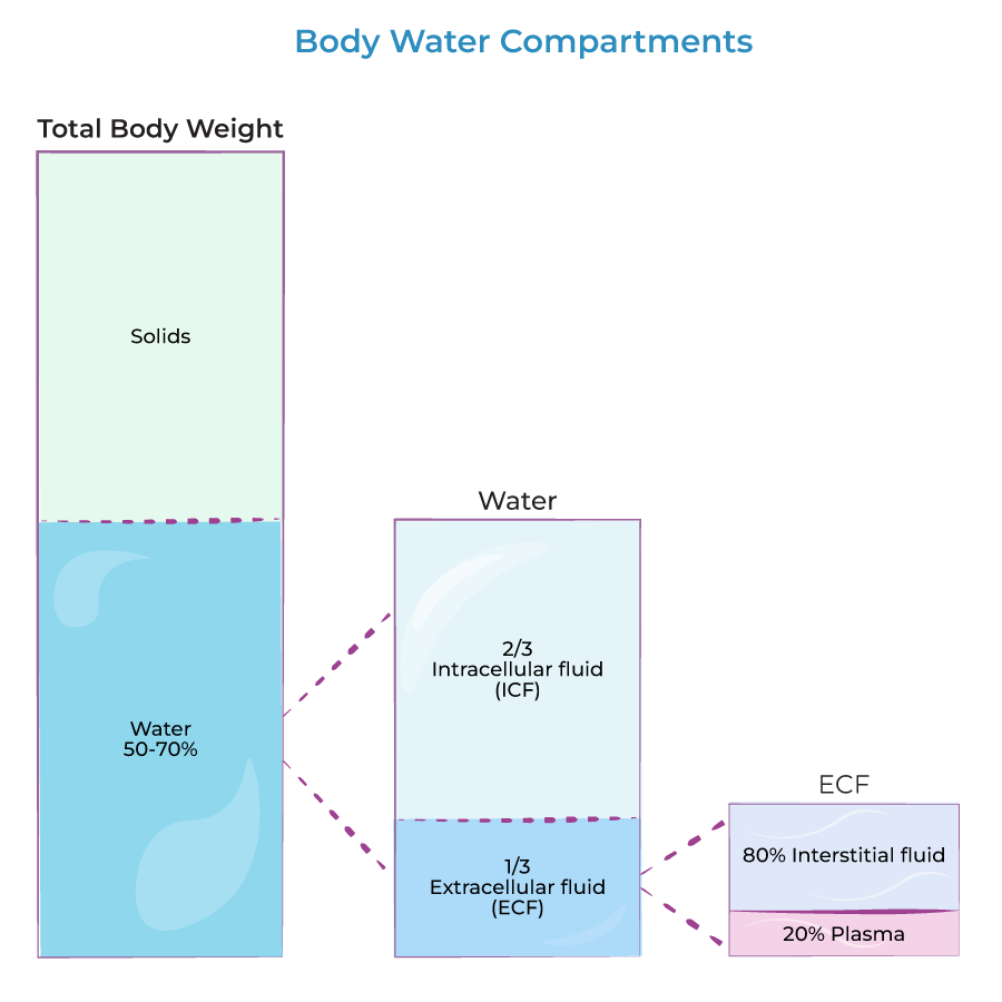
TBW refers to the absolute sum of all water contained within the body. It represents a massively significant proportion of human body mass.
- Standard Reference Volume: In a standard, healthy 70 kg (154 lb) adult male, TBW is roughly 42 Liters.
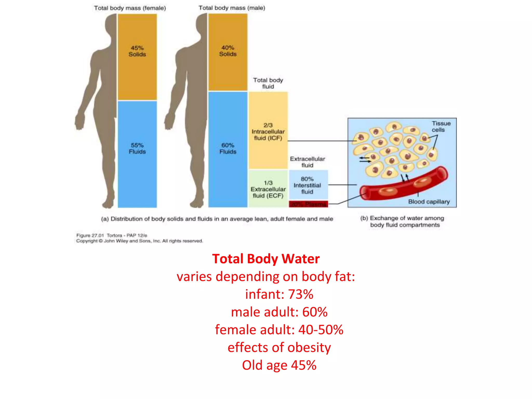
- Proportion & Variations: Approximately 60% of an adult male's body weight is water. However, this percentage fluctuates heavily based on physiological factors:
- Age: Premature infants can be up to 80% water. Full-term infants are ~70-75%. As humans age, muscle mass decreases and connective tissue/fat increases, meaning the elderly can drop to a dangerous 45-50% TBW (putting them at exceptionally high risk for rapid, fatal dehydration).
- Sex: Females generally have a slightly lower TBW percentage (~50-55%) than males. This is because women naturally have a higher physiological percentage of adipose tissue (fat).
- Body Fat Content: Fat cells (adipocytes) contain almost zero water. Therefore, individuals with higher body fat percentages (obesity) will have significantly lower TBW percentages relative to their total weight compared to highly muscular individuals.
The total 42 Liters of body water is not uniformly distributed but is strictly divided into two primary compartments by the cell membrane:
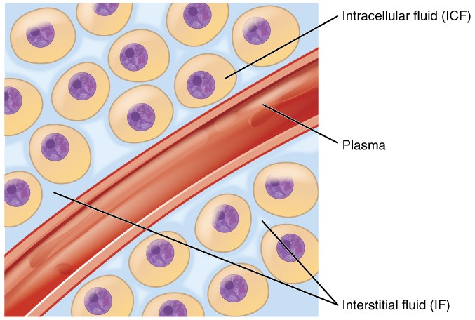
A. Intracellular Fluid (ICF)
The ICF is the fluid locked inside the trillions of cells in the body. It is the immediate, highly regulated aqueous environment where the vast majority of metabolic and biochemical activities occur.
- Proportion: The ICF constitutes the largest single fluid compartment, accounting for exactly two-thirds (2/3) of the TBW. In a 70 kg adult, this is roughly 28 Liters (40% of total body weight).
- Composition (The Cell's Internal Environment):
- Major Cations: Potassium (K+) is the undisputed king of the ICF (concentration ~140 mEq/L). Its high internal concentration is crucial for resting membrane potential, nerve impulse transmission, and muscle contraction. Magnesium (Mg²+) is the second most abundant, vital as a cofactor for all ATP-dependent enzymatic reactions.
- Major Anions: Phosphate (PO&sub4;³−) is a critical component of energy currency (ATP) and intracellular buffering. Proteins are heavily concentrated inside the cell, carrying strong negative charges, contributing to osmolarity, and acting as intracellular buffers.
- Low Concentrations: Sodium (Na+) is kept extremely low inside the cell (~14 mEq/L), as is Chloride (Cl−).
- Key Characteristics:
- Selective Permeability: The phospholipid bilayer plasma membrane is the ultimate barrier separating the ICF from the outside world.
- Osmotic Equilibrium: Despite having completely different chemical ingredients than the outside fluid, the total number of particles (osmolarity) of the ICF is normally in perfect, dynamic equilibrium with the outside.
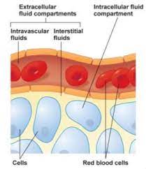
B. Extracellular Fluid (ECF)
The ECF is all the fluid found outside the cells. It acts as the body's vast "internal sea" that bathes, feeds, and cleanses all cells.
- Proportion: The ECF constitutes approximately one-third (1/3) of the TBW, which is roughly 14 Liters (20% of total body weight).
- Composition (The Body's Transport Medium):
- Major Cations: Sodium (Na+) dominates the ECF (~140 mEq/L). It is the absolute primary determinant of ECF osmolarity and overall fluid volume.
- Major Anions: Chloride (Cl−) (~100 mEq/L) and Bicarbonate (HCO&sub3;−) (~24 mEq/L), which acts as the body's primary acid-base buffer system.
- Other Components: A rich, circulating soup of glucose, amino acids, circulating hormones, oxygen, and metabolic waste (urea).
The 3 Sub-compartments of the ECF:
This is the literal "tissue fluid" filling the microscopic spaces between the cells. It is the largest component of the ECF, comprising about 80% of ECF volume (~10.5 Liters). Its ionic composition is nearly identical to blood plasma, but it lacks large proteins. The ISF is the critical "middle-man" medium for exchanging nutrients and gases between the blood capillaries and the cell membranes.
This is the pale-yellow fluid component of blood, circulating violently within the cardiovascular system. It accounts for 20% of ECF volume (~3.5 Liters). Its defining characteristic, compared to ISF, is a massive concentration of large plasma proteins (like Albumin). Plasma is the primary transport highway for red/white blood cells, nutrients, and waste.
A highly specialized, trapped component of the ECF, representing only 1-2% of body weight (approx. 1 to 2 Liters). It consists of fluids actively secreted by specific epithelial cells into distinct, enclosed body cavities. Examples include: Cerebrospinal Fluid (CSF), Intraocular Fluid (aqueous humor), Synovial Fluid in joints, Serous Fluids (pleural, pericardial, peritoneal fluid), and massive amounts of Gastrointestinal secretions.
III. Measurement of Fluid Compartments (The Indicator Dilution Method)
Physicians and physiologists cannot simply drain a human to measure fluid volumes. Instead, they use a highly accurate mathematical principle called the Indicator-Dilution Method.
The Principle: You inject a known mass (amount) of a non-toxic marker substance (indicator) into the blood. You allow it time to distribute evenly through a specific compartment. You then draw a blood sample and measure the final concentration.
Formula: Volume = Mass of Indicator Injected / Concentration of Indicator in Sample.
Choosing the Right Indicator:
The entire validity of the test relies on choosing an indicator that distributes only in the target compartment you wish to measure.
- Total Body Water (TBW): Measured using isotopes of water, like heavy water (D&sub2;O) or tritiated water (HTO), or Antipyrine. Because these are literally water molecules, they go everywhere water goes, crossing all membranes perfectly.
- Extracellular Fluid (ECF) Volume: Measured using Inulin, Mannitol, or radioactive Sodium/Thiosulfate. These molecules easily cross the capillary wall into the interstitial space but are physically too large to cross the cell membrane, so they remain trapped perfectly inside the ECF.
- Plasma Volume: Measured using Evans blue dye (T-1824) or Radioactive Iodine-125 tagged Albumin. These are massive molecules that bind instantly to plasma proteins. They are completely physically trapped inside the blood vessels and cannot even cross into the interstitial space.
Calculating the Hidden Compartments:
You cannot directly measure the Interstitial Fluid or the Intracellular Fluid because there is no chemical tracer that exclusively targets them without first going through plasma. Therefore, they are derived via simple subtraction:
- Interstitial Fluid (ISF) Volume = ECF Volume − Plasma Volume.
- Intracellular Fluid (ICF) Volume = Total Body Water (TBW) − ECF Volume.
IV. Fluid Movement Between Compartments and Regulatory Mechanisms
The precise, uninterrupted movement of water and solutes between the body's fluid compartments is the absolute cornerstone of physiological survival. This dynamic equilibrium is meticulously regulated by intense physical forces and membrane properties.
A. Fluid Movement Between Plasma and Interstitial Fluid (Capillary Exchange)
The exchange of massive amounts of fluid, nutrients, and waste between the blood plasma and the tissue cells (via the ISF) occurs primarily across the microscopically thin, porous walls of the capillaries. This movement is governed by Starling Forces.
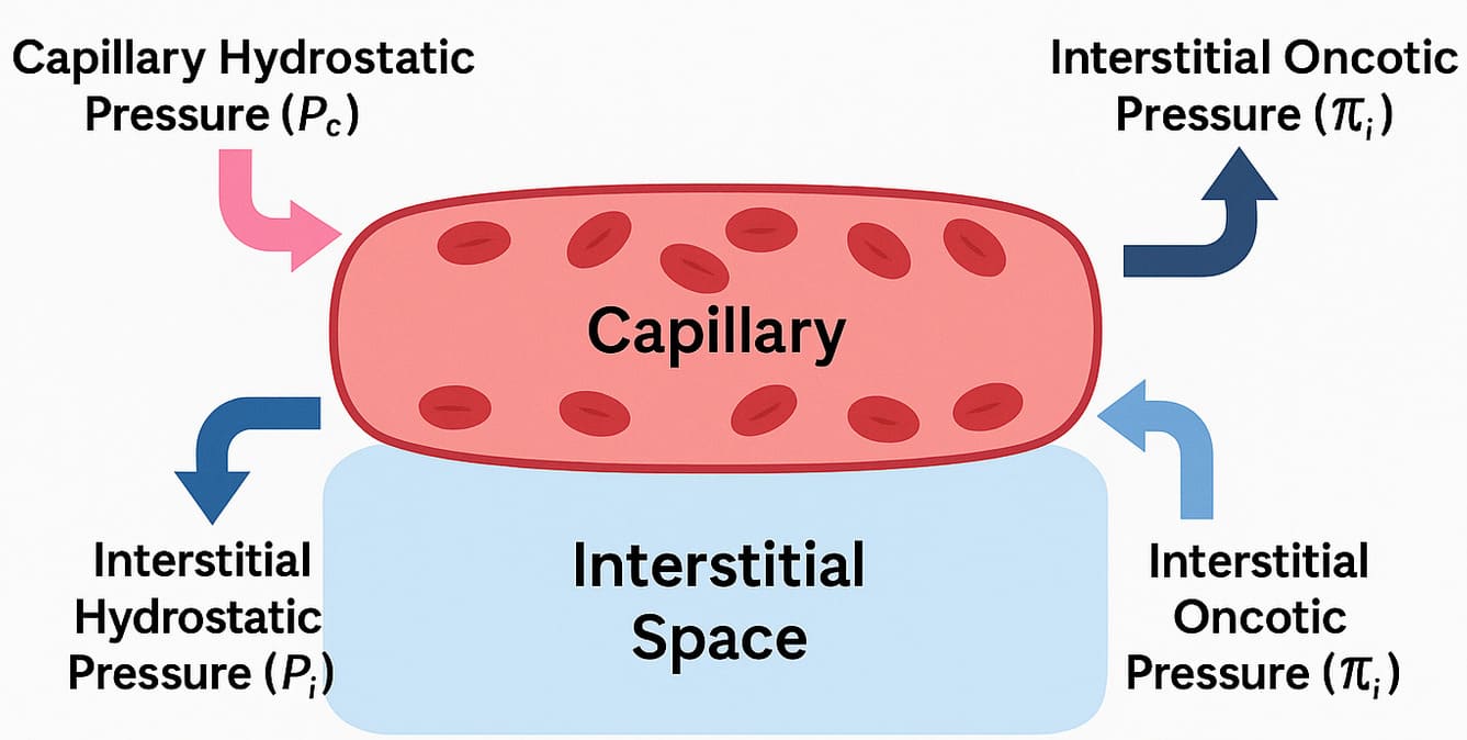
Starling Forces: The Four Drivers of Capillary Exchange
- Capillary Hydrostatic Pressure (Pc): This is the physical blood pressure exerted by the fluid within the capillaries, acting as the "pushing" force against the capillary wall.
- Effect: It violently forces fluid OUT of the capillary and into the interstitial space (Filtration).
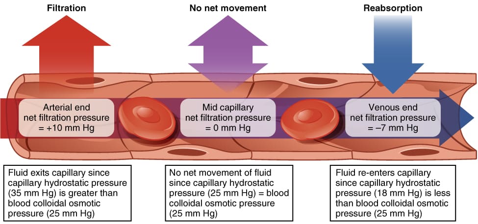
- Dynamics: Pc is highest at the arterial end of the capillary (~30-35 mmHg) due to heart pumping, and progressively drops as blood flows to the venous end (~10-15 mmHg).
- Interstitial Fluid Hydrostatic Pressure (Pif): The pressure exerted by the physical fluid already sitting in the interstitial space outside the capillary.
- Effect: It tends to push fluid BACK INTO the capillary.
- Dynamics: Pif is usually very low, often close to zero or even slightly negative (due to lymphatic suction), creating a slight vacuum effect.
- Capillary Oncotic (Colloid Osmotic) Pressure (πc): The specialized osmotic pressure exerted by the massive, non-diffusible plasma proteins (primarily Albumin) permanently trapped within the blood.
- Effect: Acts as a massive chemical sponge, PULLING fluid back into the capillary from the interstitial space (Reabsorption).
- Dynamics: πc remains highly constant along the entire length of the capillary (typically around 25-28 mmHg).
- Interstitial Fluid Oncotic Pressure (πif): The osmotic pull exerted by the tiny amount of proteins that manage to leak into the interstitial fluid.
- Effect: Tends to pull fluid OUT of the capillary.
- Dynamics: πif is normally very low (typically 2-8 mmHg) because healthy capillaries restrict protein leakage.
Net Filtration Pressure (NFP)
The absolute net movement of fluid is determined by a mathematical balance of these forces, expressed by the Starling equation:
NFP = (Pc - Pif) - (πc - πif)
- At the arterial end: NFP = (35 - 0) - (26 - 2) = +11 mmHg. A positive NFP means outward pressure wins, indicating robust Net Filtration (nutrient-rich fluid moves OUT to feed tissues).
- At the venous end: NFP = (15 - 0) - (26 - 2) = -9 mmHg. A negative NFP means the inward pull of albumin wins, indicating robust Net Reabsorption (waste-filled fluid moves IN to go back to the heart/kidneys).
The Vital Role of the Lymphatic System
Notice the math: +11 out, but only -9 back in. There is a continuous, slight physiological imbalance where filtration constantly exceeds reabsorption by about 2-3 Liters a day. This excess fluid, along with any escaped proteins, is swept up by the Lymphatic System. The lymphatics act as an emergency drainage sewer, returning this "lymph" fluid back into the venous circulation at the subclavian veins.
Clinical Pathology: If the lymph nodes are blocked (e.g., by a tumor, radiation, or parasitic elephantiasis), the fluid has nowhere to go, resulting in massive, disfiguring tissue swelling called Lymphedema.
B. Fluid Movement Between ECF and ICF (Across Cell Membranes)
While capillary walls are leaky, the cell membrane is highly restrictive. Exchange between the ISF and the ICF is driven exclusively by Osmosis. The cell membrane is highly permeable to water (via aquaporins) but highly impermeable to most solutes (like Sodium).
Osmolarity vs. Tonicity (Crucial Distinction)
- Osmolarity: A strict chemical measurement. It quantifies the total absolute concentration of ALL solute particles present in a solution, regardless of whether they can cross a cell membrane. Expressed as milliosmoles per liter (mOsm/L). Normal blood plasma is strictly regulated at 280-300 mOsm/L.
- Effective Osmoles: Solutes that CANNOT cross the cell membrane (like Na+, Cl−, Mannitol). Because they are trapped on one side, they exert a permanent osmotic "pull" on water.
- Ineffective Osmoles: Solutes that freely slip right through the cell membrane (like Urea and Ethanol). Because they move freely, they balance out instantly and cause zero permanent water shifting.
- Tonicity: A strictly biological, functional term. It describes the physical effect a fluid has on actual cell volume. Tonicity is determined SOLELY by the concentration of Effective (non-penetrating) osmoles.
The extracellular fluid has the exact same concentration of non-penetrating solutes as the inside of the cell. There is zero net movement of water. Cell volume remains perfectly stable and healthy.
The extracellular fluid is diluted (has fewer trapped solutes than the inside of the cell). Water aggressively rushes INTO the hyper-concentrated cells to dilute them. Cells swell massively, and may rupture (lyse). Can cause deadly cerebral edema.
The extracellular fluid is highly concentrated (has too much sodium/solutes). The strong ECF aggressively sucks water OUT of the cells. Cells shrink and shrivel violently (crenation). Can cause severe neurological damage and coma.
Active Transport's Essential Role: The Na+/K+ Pump
While water movement is passive, maintaining these osmotic gradients requires massive energy. Cells are packed with highly concentrated, trapped negative proteins that constantly try to suck water into the cell. To prevent all human cells from swelling and exploding, the Na+/K+ ATPase pump runs continuously. By burning ATP to forcefully kick 3 Na+ ions OUT of the cell for every 2 K+ ions it brings in, it creates a net outward osmotic pull that exactly counters the inward pull of the proteins. If a cell loses oxygen (hypoxia) and runs out of ATP, the pump fails, sodium rushes in, water follows, and the cell undergoes fatal swelling (hydropic degeneration).
V. Neurohormonal Regulation of Body Fluid Volume and Osmolarity
The human body uses incredibly sophisticated feedback loops involving the brain, heart, and kidneys to ensure fluid balance remains absolute.
A. Regulation of ECF Volume (Primarily Sodium Balance)
The overall volume of the blood and ECF is dictated entirely by its total Sodium (Na+) content. The golden rule of physiology is: "Where Sodium goes, water is forced to follow."
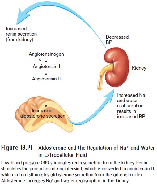
- Renin-Angiotensin-Aldosterone System (RAAS): The body's primary volume-preservation system.
- Trigger: The kidneys detect low blood pressure, low blood volume, or low sodium delivery.
- Action: Kidneys secrete the enzyme Renin into the blood. Renin converts circulating Angiotensinogen into Angiotensin I. As blood passes through the lungs, ACE (Angiotensin-Converting Enzyme) converts it into Angiotensin II.
- Result: Angiotensin II is the most potent vasoconstrictor in the body (instantly raising blood pressure). Furthermore, it forces the adrenal glands to secrete the steroid hormone Aldosterone. Aldosterone commands the kidneys to aggressively reabsorb sodium back into the blood. Water violently follows the sodium, restoring total ECF volume.
- Antidiuretic Hormone (ADH) / Vasopressin (Volume role): While primarily for osmolarity, if blood volume drops severely (e.g., a massive hemorrhage of >10% blood loss), pressure receptors (baroreceptors) in the aorta panic and trigger a massive release of ADH from the posterior pituitary to clamp down blood vessels and save all water in the kidneys.
- Atrial Natriuretic Peptide (ANP) & BNP: The "counter-regulatory" system. If ECF volume is too high, the massive volume stretches the muscular walls of the heart's atria and ventricles. The stretched heart muscle releases ANP and BNP. These hormones force the kidneys to excrete sodium (natriuresis) and excrete water (diuresis) directly into the urine, safely reducing blood volume and pressure.
- Sympathetic Nervous System (SNS): When activated by stress or low pressure, sympathetic nerves constrict renal blood vessels (dropping kidney filtration so less urine is made) and directly stimulate renin release.
B. Regulation of ECF Osmolarity (Primarily Water Balance)
ECF osmolarity is primarily determined by the concentration of solutes relative to water. The body alters osmolarity purely by holding onto or urinating out pure free water.
- ADH (Vasopressin) - The Primary Osmoregulator:
- Trigger: Microscopic osmoreceptors in the hypothalamus are exquisitely sensitive. An increase in plasma osmolarity of just 1% (meaning the blood is getting too salty/concentrated) violently stimulates the posterior pituitary to release ADH.
- Action: ADH travels to the kidneys and binds to V2 receptors, forcing the insertion of water channels (Aquaporin-2) into the collecting ducts.
- Result: Massive amounts of pure water are reabsorbed back into the blood, diluting the salty plasma back to a normal 285 mOsm/L, resulting in a tiny volume of highly concentrated, dark urine. Conversely, if you drink a gallon of water, blood osmolarity drops, ADH is shut off, and you urinate out gallons of clear, dilute water.
- The Thirst Mechanism: The vital behavioral component. The exact same hypothalamic osmoreceptors that trigger ADH also stimulate the conscious cerebral cortex, creating an overwhelming, agonizing sensation of thirst, compelling the organism to physically find and drink water to dilute the ECF.
VI. Clinical Pathophysiology: Fluid Imbalances
Disturbances in fluid regulation can have profound, rapidly life-threatening consequences.
| Pathology | Definition & Causes | Clinical Consequences |
|---|---|---|
| Hypovolemia (ECF Volume Deficit) |
A massive loss of isotonic fluid. Caused by acute traumatic hemorrhage, severe vomiting/diarrhea, or extensive third-degree burns (plasma weeping). | Decreased venous return, plummeting blood pressure, tachycardia, poor tissue perfusion, hypoxia, and progression to lethal Hypovolemic Shock. |
| Hypervolemia (ECF Volume Excess) |
Excessive accumulation of isotonic fluid. Caused by severe Heart Failure (failing pump), Renal Failure (can't excrete urine), or Cirrhosis. | Massive high blood pressure, distended jugular veins, severe peripheral pitting edema. If backed up into the lungs, it causes lethal Pulmonary Edema, destroying gas exchange. |
| Hyponatremia (Low Plasma Sodium) |
A disorder of water excess relative to sodium (Plasma Na+ < 135 mEq/L). Creates a dangerously hypotonic ECF. | Because the blood is dilute, water violently shifts INTO the concentrated brain cells. Causes lethal Cerebral Edema, leading to confusion, intractable seizures, brain herniation, and coma. |
| Hypernatremia (High Plasma Sodium) |
A disorder of absolute water deficit relative to sodium (Plasma Na+ > 145 mEq/L). Creates a highly hypertonic ECF. Caused by diabetes insipidus or extreme sweating without water replacement. | The salty blood violently sucks water OUT of the brain cells. Causes lethal Cellular Crenation (Shrinkage), leading to brain hemorrhage (tearing of bridging veins), intense lethargy, seizures, and death. |
| Edema (Third Spacing) |
Abnormal accumulation of excess interstitial fluid in the tissues. | Caused by disruption of Starling Forces: 1) Increased Pc (Heart Failure), 2) Decreased πc (Liver failure/malnutrition causing low albumin), 3) Increased capillary permeability (Inflammation/Sepsis), 4) Blocked lymphatics. |
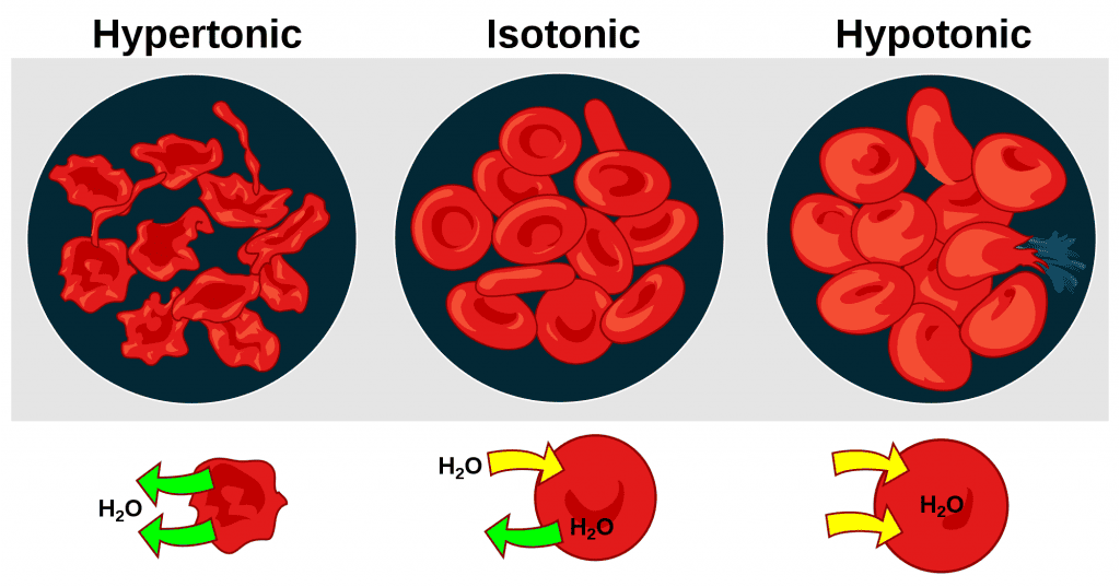
VII. Clinical Scenarios: Tonicity, Osmolarity, and Intravenous (IV) Fluid Therapy
The administration of IV fluids is the most common invasive procedure in medicine. Safe administration requires an absolute mastery of fluid tonicity and exactly how fluids distribute upon entering the vein.
General Principles of IV Fluid Distribution
- Initial Introduction: ALL IV fluids are introduced directly into the plasma compartment via a catheter.
- Subsequent Distribution: Where the fluid goes next depends entirely on the fluid's physical Tonicity.
- Therapeutic Goals: Isotonic fluids expand ECF volume; hypotonic fluids shift water into cells to rehydrate them; hypertonic fluids violently draw water out of cells to reduce brain swelling.
0.9% Normal Saline (NS) & Lactated Ringer's (LR)
- Composition: 0.9% NaCl contains 154 mEq/L Na+ and 154 mEq/L Cl−. LR contains physiological balances of Na+, Cl−, K+, Ca²+, and lactate (which the liver converts to bicarbonate buffer).
- Distribution: Because they are perfectly isotonic, they do not cause a single drop of water to shift into or out of the cells. They remain entirely locked within the Extracellular Fluid (ECF) compartment. Note: For every 1 Liter of NS infused, roughly 250 mL stays in the blood plasma, and 750 mL leaks into the interstitial space.
- Clinical Uses: Rapid, massive volume resuscitation.
- Hospital Scenario: A hypotensive trauma patient arriving via ambulance with severe acute blood loss is given a rapid IV pressure-infusion of 2 liters of warmed Lactated Ringer's to aggressively increase circulating blood volume and save them from shock.
0.45% Saline (Half-Normal) & D5W
- Composition: 0.45% NaCl has exactly half the sodium of normal blood. D5W (5% Dextrose in Water) is mathematically isosmotic in the IV bag (~252 mOsm/L). However, once infused, the body's cells rapidly metabolize and eat the glucose, leaving behind nothing but pure, unadulterated free water. Therefore, inside the body, D5W acts as a severely hypotonic solution.
- Distribution: Because the fluid is so dilute, water aggressively shifts from the ECF into the Intracellular Fluid (ICF).
- Clinical Uses: Treating severe hypernatremia (cellular dehydration) and providing maintenance free water.
- Hospital Scenario: An elderly patient found comatose in a hot apartment has severe hypernatremia (Na+ = 165). Their brain cells are shrunk. They receive a very slow, tightly controlled infusion of D5W or 0.45% Saline to gently push water back into their dehydrated brain cells. Warning: Infusing hypotonic fluids too fast will cause explosive brain swelling.
3% Hypertonic Saline & Mannitol
- Composition: 3% NaCl is massively, aggressively hypertonic (1026 mOsm/L). Mannitol is an osmotic diuretic sugar that cannot cross cell membranes.
- Distribution: These create a violent osmotic gradient in the blood that acts like a vacuum, aggressively sucking water OUT of the Intracellular Fluid (ICF) and expanding the blood volume. Cells shrink.
- Clinical Uses: Treating severe, symptomatic hyponatremia and aggressively reducing lethal cerebral edema.
- Hospital Scenario: A patient arrives with a traumatic brain injury and massive, life-threatening intracranial pressure (brain swelling). The neurosurgeon orders rapid, small boluses of 3% Saline or Mannitol. The hypertonic fluid pulls water directly out of the swollen brain tissue, saving the patient's life. Warning: Extreme caution is required. Reversing hyponatremia too quickly with 3% saline strips water from the brainstem, causing a horrific, irreversible paralyzing disease called Osmotic Demyelination Syndrome (Central Pontine Myelinolysis).
Albumin, Dextran, and PRBCs
- Colloids (e.g., 5% or 25% Albumin): Solutions containing massive protein molecules that physically cannot cross intact capillary membranes. They remain trapped within the intravascular plasma compartment, exerting a massive oncotic pull that sucks fluid from the interstitial space into the blood. Highly effective for rapid plasma volume expansion in severe shock or massive burn victims.
- Blood Products (PRBCs): Packed Red Blood Cells are isotonic. They expand the intravascular plasma compartment, but completely uniquely, they instantly increase the oxygen-carrying capacity of the blood to treat hemorrhagic shock.
Solutes, Solvents, and Simple Movement in Body Fluids
At the heart of all physiological processes involving fluids is the interaction between solutes and solvents, and their movement across various compartments.
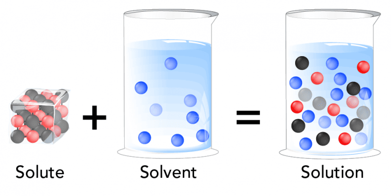
1. Solutes and Solvents: The Basics
- Solution: A homogeneous mixture composed of two or more substances.
- Solvent: The substance that is present in the greatest amount in a solution and does the dissolving.
- Solute: The substance(s) that are present in a lesser amount in a solution and get dissolved by the solvent.
What is the Solvent of Body Fluid?
The primary and overwhelmingly abundant solvent in all body fluids is WATER (H₂O).
Water's unique properties make it an ideal biological solvent:
- Polarity: Allows it to dissolve a wide variety of other polar molecules and ions.
- High Heat Capacity: Helps regulate body temperature.
- High Heat of Vaporization: Allows for cooling through sweating.
Common Solutes in Body Fluids:
Body fluids are complex solutions containing a vast array of solutes:
- Electrolytes: Ions that conduct electricity.
- Cations (positively charged): Sodium (Na⁺), Potassium (K⁺), Calcium (Ca²⁺), Magnesium (Mg²⁺).
- Anions (negatively charged): Chloride (Cl⁻), Bicarbonate (HCO₃⁻), Phosphate (HPO₄²⁻).
- Non-electrolytes:
- Nutrients: Glucose, amino acids, fatty acids, vitamins.
- Metabolic Wastes: Urea, creatinine, uric acid.
- Proteins: Albumin, globulins, fibrinogen.
- Gases: Oxygen (O₂), Carbon Dioxide (CO₂).
2. Simple Movement of Solutes and Solvents
The movement of substances is primarily governed by passive processes that do not require cellular energy (ATP).
A. Movement of Solutes: Diffusion
- Definition: The net movement of solute particles from an area of higher solute concentration to an area of lower solute concentration (down the concentration gradient).
- Mechanism: Driven by the inherent random kinetic energy of molecules.
- Factors Affecting Diffusion Rate: The rate is faster with a larger concentration gradient, higher temperature, smaller molecular size, shorter distance, and larger surface area.
- Types of Diffusion:
- Simple Diffusion: Solutes pass directly through the lipid bilayer (e.g., O₂, CO₂, fatty acids).
- Facilitated Diffusion: Solutes move with the help of membrane proteins (channels or carriers), still following the concentration gradient (e.g., glucose, ions).
B. Movement of Solvents: Osmosis
- Definition: The net movement of water (the solvent) across a selectively permeable membrane from an area of higher water concentration (lower solute) to an area of lower water concentration (higher solute).
- Mechanism: Water molecules move down their own concentration gradient.
- Selectively Permeable Membrane: Crucial for osmosis, as it allows water to pass but restricts most solutes.
- Osmotic Pressure: The pressure needed to prevent the inward flow of water across a semipermeable membrane. The higher the solute concentration, the higher the osmotic pressure.
Summary of Movement Principles:
- Solutes move by Diffusion: From high solute concentration to low solute concentration.
- Water (Solvent) moves by Osmosis: From high water concentration (low solute) to low water concentration (high solute).
These passive movements are essential for:
- Nutrient delivery and waste removal.
- Gas exchange in the lungs.
- Maintaining cell volume and shape.
- Fluid balance between intracellular and extracellular compartments.
Clinical Scenarios:
Basic Principle: Water follows solutes. Specifically, water moves from an area of lower effective solute concentration (higher water concentration) to an area of higher effective solute concentration (lower water concentration) across a semipermeable membrane.
Scenario 1: Blood Transfusion
- Product: Whole blood or packed red blood cells.
- Tonicity: Isotonic.
- Effect: Primarily increases the plasma volume. No significant shift of fluid between ECF and ICF. Also delivers oxygen-carrying capacity.
- Clinical Use: To replace blood loss or treat anemia.
Scenario 2: Intravenous (IV) Fluid Administration
1. Isotonic Solutions (e.g., Normal Saline - 0.9% NaCl, Lactated Ringer's - LR)
- Composition: 0.9% NaCl (NS) contains 154 mEq/L Na⁺ and 154 mEq/L Cl⁻. Lactated Ringer's (LR) contains Na⁺, Cl⁻, K⁺, Ca²⁺, and lactate. Both are effectively isotonic.
- Distribution: The fluid stays entirely within the ECF compartment, distributing between the plasma (~1/4) and interstitial fluid (~3/4).
- Clinical Uses: Volume expansion for dehydration, hypovolemic shock, hemorrhage.
- Hospital Scenario: A hypotensive car accident patient receives a rapid infusion of NS or LR to restore intravascular volume and blood pressure.
2. Hypotonic Solutions (e.g., 0.45% NaCl - Half Normal Saline, D5W - Dextrose 5% in Water)
- Composition: 0.45% NaCl has half the sodium of NS. D5W is initially isotonic, but the dextrose is rapidly metabolized, leaving free water.
- Distribution: Water moves from the ECF into the ICF compartment to equalize osmolality, hydrating the cells.
- Clinical Uses: To treat cellular dehydration (e.g., hypernatremia).
- Hospital Scenario: A patient with severe hypernatremia is given a slow infusion of Half Normal Saline to allow water to shift into their dehydrated brain cells.
3. Hypertonic Solutions (e.g., 3% NaCl - Hypertonic Saline, D5NS)
- Composition: 3% NaCl is very hypertonic (1026 mOsm/L). D5NS is initially hypertonic, then becomes isotonic as dextrose is metabolized.
- Distribution: Water moves out of the ICF and into the ECF compartment, causing cells to shrink.
- Clinical Uses: To treat severe symptomatic hyponatremia and to reduce cerebral edema.
- Hospital Scenario: A patient with traumatic brain injury and high intracranial pressure is given a slow infusion of 3% Hypertonic Saline to draw fluid out of the swollen brain cells.
4. Colloids (e.g., Albumin, Dextran, Hetastarch)
- Composition: Solutions containing large molecules (proteins, large sugars) that do not easily cross capillary membranes.
- Distribution: Due to their large size, they primarily remain within the intravascular space (plasma), exerting an oncotic pull that draws fluid from the interstitial space into the plasma.
- Clinical Uses: Rapid plasma volume expansion, especially in severe hypoalbuminemia or burns.
- Hospital Scenario: A patient with severe burns and plasma volume depletion is given an infusion of Albumin to rapidly restore intravascular volume.
Summary Table of IV Fluid Effects:
| IV Fluid Type | Effective Tonicity | Primary Distribution | Effect on ICF Cells |
|---|---|---|---|
| Isotonic (NS, LR) | Isotonic | ECF only (plasma & ISF) | No change |
| Hypotonic (0.45% NaCl, D5W) | Hypotonic | ECF & ICF | Swell |
| Hypertonic (3% NaCl) | Hypertonic | ECF (draws from ICF) | Shrink |
| Colloids (Albumin) | Effectively Hypertonic (oncotic) | Plasma only (draws from ISF) | No direct effect |
VIII. References & Recommended Reading
- Hall, J. E., & Guyton, A. C. (2015). Guyton and Hall Textbook of Medical Physiology (13th ed.). Elsevier. (Definitive text for Starling forces and fluid compartmentalization).
- Costanzo, L. S. (2018). Physiology (6th ed.). Elsevier. (Excellent, concise explanations of osmolarity vs. tonicity).
- Kasper, D. L., Fauci, A. S., Hauser, S. L., Longo, D. L., Jameson, J. L., & Loscalzo, J. (2015). Harrison's Principles of Internal Medicine (19th ed.). McGraw-Hill Education. (Gold standard for clinical pathophysiology of hyponatremia and IV fluid management).
- Mount, D. B. (2014). Fluid and Electrolyte Disturbances. In Harrison's Principles of Internal Medicine. McGraw-Hill.
Body Fluids and Compartments Read More »
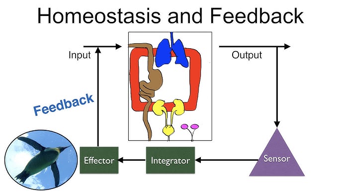
Homeostasis Physiology
Homeostasis
Imagine you're driving a car, aiming to maintain a constant speed of 60 mph. You press the gas going uphill and ease off going downhill. Your goal is to keep that speed constant despite external changes. That's essentially what your body does, constantly, for hundreds of variables.
Homeostasis (from Greek "homoios" meaning "similar" and "stasis" meaning "standing still") is the ability of an organism to maintain a relatively stable internal environment despite continuous changes in the external environment. It's not a static state, but a dynamic equilibrium where conditions fluctuate within narrow, acceptable limits around a set point.
Many physiologists translate this into the saying, “constantly changing to stay the same.” The ability of the human body to quickly adapt to any changes and to re-establish stability is the essence of homeostasis.
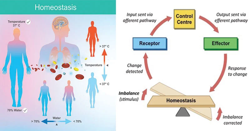
The Importance of Homeostasis
Survival itself depends on the body's ability to maintain this internal balance. Deviations outside the normal range can impair cell function, leading to disease or death.
Enzyme and Protein Function
Almost all biochemical reactions are catalyzed by enzymes (proteins), which are highly sensitive to their environment.
Impact of Imbalance: Deviations in temperature or pH can denature enzymes, altering their 3D shape and halting vital metabolic pathways.
Cellular Integrity and Volume
The cell membrane's selective permeability and active transport mechanisms are critical for maintaining appropriate solute concentrations.
Impact of Imbalance: Changes in extracellular fluid osmolarity can cause cells to swell and burst (lysis) or shrink and die (crenation). Disrupted ion gradients incapacitate nerve and muscle function.
Efficient Communication Systems
The nervous and endocrine systems require specific conditions to transmit signals effectively.
Impact of Imbalance: Improper electrolyte balance (Na⁺, K⁺, Ca²⁺) can lead to severe nerve and muscle dysfunction, including seizures, paralysis, and cardiac arrhythmias.
Energy Production (ATP)
Cells require a continuous supply of oxygen and nutrients, and efficient removal of waste, to produce ATP.
Impact of Imbalance: Oxygen deprivation (hypoxia) leads to a cellular energy crisis and buildup of lactic acid. Accumulation of wastes like CO₂ can become toxic and alter pH, leading to organ failure.
Immune System Function
Immune cells and proteins need stable conditions to effectively fight off pathogens without harming healthy tissues.
Impact of Imbalance: Uncontrolled fever can become detrimental to immune cells themselves. Chronic stress and elevated cortisol can suppress the immune system.
Examples of Homeostatically Regulated Variables
The body tightly regulates hundreds of variables to maintain this dynamic equilibrium. Key examples include:
- Body temperature
- Blood pressure
- Blood glucose levels
- Blood pH
- Oxygen and carbon dioxide levels
- Water balance
- Ion concentrations (Na⁺, K⁺, Ca²⁺)
Homeostasis is Maintained by Feedback Loops
The primary way the human body maintains homeostasis is with the use of feedback loops. A feedback loop is a mechanism that allows for continual assessment of the body’s physiology and a way to correct various elements if they should go out of balance. There are two types of feedback loops: negative and positive.
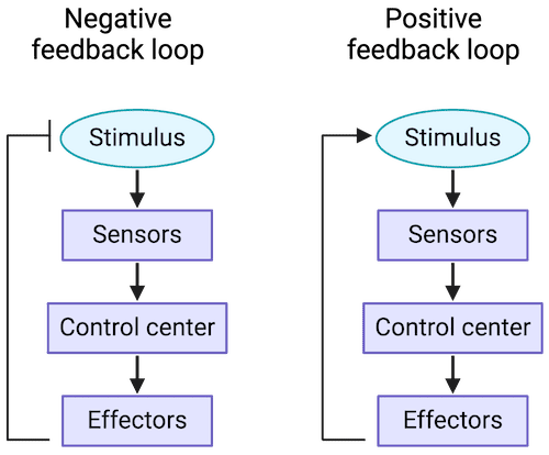
Negative Feedback Loop
The response opposes (or negates) the original stimulus. This is by far the most common type in the human body.
Positive Feedback Loop
The response augments (or intensifies) the original stimulus. The cycle repeats until it is broken. This type is very rare but critically important.
Parameters and Set Points
For any feedback loop, there is a parameter that is being monitored, and it has a set point, or a ‘normal range’ in which it exists when the body is in balance. The stimulus that starts the feedback loop is a change in that parameter that pushes it above or below its normal set point range.
| Table 1.1: Examples of Blood Parameters and Their Set Points | |
|---|---|
| Osmolarity of Blood | 295-310 mOsM |
| pH of Blood | 7.35-7.45 |
| Arterial PCO₂ | 35-46 mmHg |
| Arterial PO₂ | 80-100 mmHg |
| Glucose (fasting) | 70-100 mg/dL |
| Sodium (Na⁺) | 135-145 mM |
| Potassium (K⁺) | 3-5 mM |
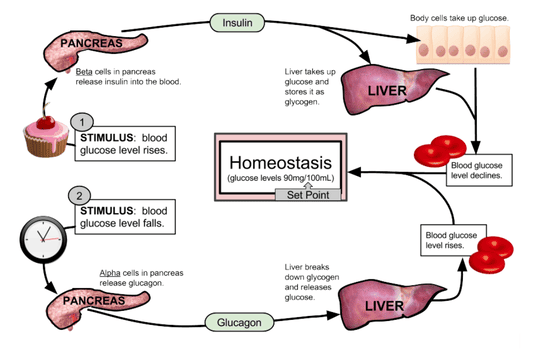
Example: Blood Glucose Regulation (Between Meals)
A person’s blood glucose (parameter) has a normal range (set point) of 70 to 100 mg/dL. If a person has not eaten in a while, their blood glucose decreases. If it goes below 70 mg/dL, the person will have hypoglycemia (low blood sugar). This decrease is the stimulus.
This decrease is detected by receptors in the pancreas, which responds by releasing the hormone glucagon into the bloodstream. Glucagon travels to the liver and stimulates hepatocytes (liver cells) to break down their glycogen stores and release glucose molecules into the blood. This increases blood glucose levels, opposing the original stimulus. Once glucose is restored to its normal range, the signal for glucagon release dissipates. This "off switch" is a key element of negative feedback.
The Nitty Gritty of the Feedback Loop
To describe feedback loops with consistent terms, we can identify seven general components that create the loop.
Homeostatic Control Mechanisms (The "Feedback Loops")
To maintain homeostasis, the body uses control systems, most of which involve feedback loops. These loops constantly monitor conditions, detect changes, and initiate responses to bring variables back to their set point.
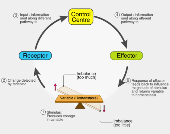
Every feedback loop has three basic components:
1. Receptor (Sensor)
Function: Monitors the environment and responds to changes (stimuli). It detects the deviation from the set point.
Action: Sends information (input) along an afferent pathway (e.g., nerve impulses) to the control center.
Example: Thermoreceptors in the skin and hypothalamus detect changes in body temperature.
2. Control Center (Integrator)
Function: Receives and analyzes the input from the receptor. It compares the input to the set point (the ideal value) and determines the appropriate response.
Action: Sends commands (output) along an efferent pathway (e.g., nerve impulses, hormones) to the effector.
Example: The hypothalamus in the brain acts as the body's thermostat, comparing current body temperature to the set point of ~37°C (98.6°F).
3. Effector
Function: Carries out the control center's response. It provides the means for the control center's output to affect the stimulus.
Action: Its action either reduces the stimulus (negative feedback) or enhances it (positive feedback).
Example: Sweat glands, blood vessels in the skin, and skeletal muscles (shivering) are effectors that help regulate body temperature.
The Communication Pathway
RECEPTOR
Afferent Pathway
CONTROL CENTER
Efferent Pathway
EFFECTOR
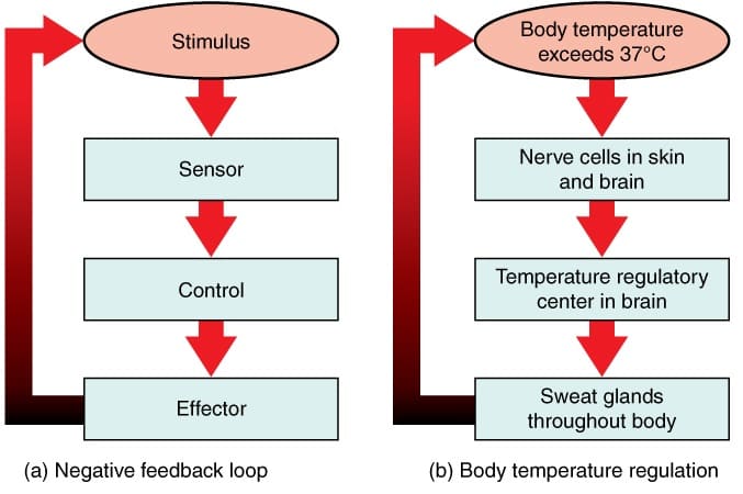
Types of Feedback Loops
Negative Feedback Loops
(Most Common and Essential for Homeostasis)
Mechanism: The output of the system shuts off or reduces the intensity of the original stimulus, bringing the variable back toward the set point. It works to counteract the change.
Goal: To prevent severe changes and maintain stability.
Analogy: A thermostat controlling a furnace. When the temperature drops, the furnace turns on. Once the temperature reaches the set point, the furnace turns off (negative feedback).
Specific Example: Increased Body Temperature
If a person has been digging in the garden on a hot day, their body temperature rises above its set point of about 98.6°F. This is the stimulus. Thermoreceptors in the skin detect this change and send afferent information to the hypothalamus (the integration center). The hypothalamus then sends efferent signals to the effector tissues: sweat glands and cutaneous blood vessels. The response is diaphoresis (sweating) and cutaneous vasodilation (widening of blood vessels in the skin). Evaporation of sweat and increased blood flow to the skin dissipate heat, causing body temperature to decrease back to its set point.
Other Physiological Examples:
- Blood Glucose Regulation: After a meal, high blood glucose stimulates the pancreas to release insulin. Insulin causes cells to take up glucose, lowering blood glucose levels.
- Blood Pressure Regulation: Baroreceptors detect high blood pressure, signal the brain, which then slows heart rate and dilates blood vessels to lower pressure.
Positive Feedback Loops
(Rare, but Important for Specific Events)
Mechanism: The output of the system enhances or exaggerates the original stimulus, driving the variable further away from the initial set point. This is often part of a process that needs to be completed quickly.
Goal: To amplify a process until a specific event is completed.
Analogy: A microphone picking up sound, which is amplified and fed back into the microphone, creating a loop of increasing volume.
Specific Example: Childbirth
When a baby is ready to be born, its head pushes down upon the cervix, increasing pressure. This stretch (the stimulus) is detected by mechanoreceptors, which send an afferent signal to the brain. The brain (integration center) signals the posterior pituitary to release the hormone oxytocin. Oxytocin (efferent pathway) travels in the blood to the uterus (effector tissue), causing its smooth muscle to contract more forcefully. This pushes the baby’s head harder against the cervix, intensifying the original stimulus and triggering more oxytocin release. This cycle repeats, with contractions becoming stronger and more frequent, until the baby is born, which breaks the loop.
Other Physiological Examples:
- Blood Clotting: Platelets at an injury site release chemicals that attract more platelets, rapidly forming a plug.
- Generation of an Action Potential: An initial depolarization opens some Na⁺ channels, causing more Na⁺ to enter, which opens even more Na⁺ channels, leading to a rapid, all-or-nothing spike.
Homeostasis Physiology Read More »
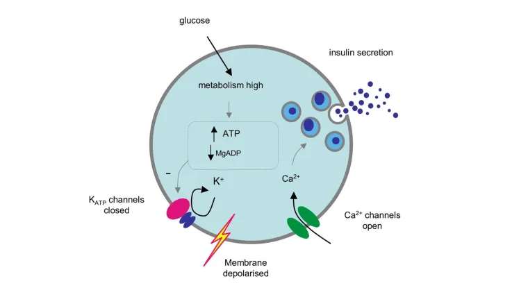
Physiology and Cell Physiology
Introduction to Basic Physiology & The Cell
Physiology is the rigorous scientific discipline dedicated to studying the functional activities and intricate mechanisms within a biological body. For example: why can the heart automatically beat, and how do electrical impulses coordinate this action? The word Physiology is derived from two ancient Greek words: physis, meaning "nature"; and logos, meaning "study". Thus, it is the study of the nature of living things.
1. Physiology Involves Process and Function
Words, names, and terms are of paramount importance in any scientific discipline because they carry highly precise meanings. Knowing and deeply understanding the relationships and etymological roots of these words will help tremendously in remembering and comprehending the vast amounts of information in a much deeper, more permanent way. This foundational knowledge will stay with you long after the course is over, enabling you to recognize important elements in other medical disciplines when you connect them to their deeper meanings.
Defining Physiology
The etymology (word origin) of the term Physiology traces back to 1560’s French, descending directly from the Latin physiologia, meaning “The study and description of natural objects, natural philosophy". This, in turn, is derived from the Greek physios meaning "nature, natural, physical"; and logia meaning "study". This gives us the fullest, most accurate meaning of Physiology as the "Science of the normal function of living things". When studying physiology, it is absolutely imperative that we also deeply understand the basic anatomy involved, as anatomy (structure) and physiology (function) are two sides of the same coin and go hand in hand.
Defining Anatomy
The etymology of the term Anatomy originates from Late 1300’s terminology in both Latin (anatomia) and Greek (anatome). These ancient words are derived from ana which means "up"; and tomos (or temnein) which means "to cut". Together this translates to "a cutting up", which is clearly referencing the act of physical dissection! In general clinical terms, anatomy is considered the “Study or knowledge of the structure (form) and organization of the human body“. While courses and textbooks for anatomy and physiology are often separated for convenience, they are inextricably connected to each other. A structure is designed specifically to support its function, and a function can only be carried out by an appropriately designed structure.
2. Etymology for the Language of Physiology
Another incredibly useful concept related to the importance of words in physiology (and anatomy) is mastering the etymology (origin of the word) of the vast array of scientific terms used in the healthcare field. Since the overwhelming majority of these words are derived from Latin and Greek, it is incredibly helpful to know the origins and ‘translations’ of these prefixes, roots, and suffixes. Becoming highly aware of the origins of words will greatly help students to:
- Understand exactly what a complex medical term means without memorizing it blindly.
- Assist you in instantly predicting what a brand-new, unseen term means when you first encounter it in a clinical setting.
The solution is hypertonic. Hyper means "above normal" or "excessive", and tonic refers to "strength" or "tension". Therefore, the solution is strong, excessively concentrated, and has a high osmotic pressure compared to another fluid.
The patient has hypoglycemia. Hypo is the direct opposite of hyper and means "below normal" or "deficient". The glyc portion refers to glucose (a type of sugar), and emia means "condition of the blood". Therefore, this statement translates perfectly to: the person has abnormally low blood sugar levels.
A marathon runner presents with hyponatremia. Hypo still means "below normal". The natr portion comes from natrium, which is the Latin word for sodium (this is exactly why the chemical symbol for sodium on the periodic table is Na), and emia still means "blood". Therefore, this diagnosis means the person has dangerously low sodium levels in their blood.
The bone marrow undergoes erythropoiesis. Erythro means "red", and poiesis means "to make" or "formation". Thus, this term describes the physiological process of producing new red blood cells.
Along the way in this physiology course, we will encounter many of these terms that, once we know the origin and meaning of, will help us figure out newer terms with absolute ease and familiarity. Anyone who has taken a medical terminology course will know the immense value of understanding the meaning of roots, prefixes, and suffixes.
Interactive Exercise: Decode the Term
Diagnosis: Pancytopenia. (Hint: there are 3 distinct root terms here: pan, cyto, and penia).
The Detailed Answer:
• Pan means "all" or "every" (like a pandemic, which affects all people).
• Cyto means "cell".
• Penia means "deficiency", "lack of", or "poverty".
Translation: A deficiency of ALL cellular components in the blood. A patient with pancytopenia has dangerously low levels of red blood cells (anemia), white blood cells (leukopenia), and platelets (thrombocytopenia), usually due to complete bone marrow failure.
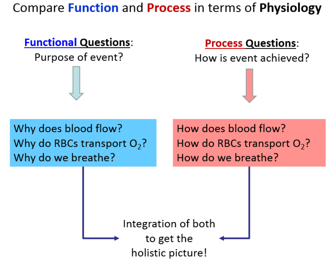
3. Compare Function and Process in Human Physiology
As we look to understand the central themes of physiology, an important concept is learning exactly how to ask questions about what’s occurring in the human body. In general, there are two basic, distinct approaches to physiology: 1) We can ask Functional Questions; and 2) We can ask Process Questions.
1. Functional Questions (The "Why")
These are strictly related to Why something occurs. They seek to understand the ultimate purpose or the evolutionary advantage of a mechanism. For example, what is the survival purpose of the heart beating? These can often be answered from a "big picture" perspective without requiring microscopic detail.
- Q: Why does blood flow?
A: To continuously transport vital nutrients, hormones, and gases (oxygen) around the body to sustain tissues, and to carry away toxic metabolic wastes (carbon dioxide, urea) to the excretory organs. - Q: Why do Red Blood Cells (RBCs) transport O₂?
A: To efficiently deliver oxygen to all the body tissues that desperately need it to perform aerobic cellular respiration and generate ATP (energy). - Q: Why do we breathe?
A: To extract essential oxygen (O₂) from the inhaled atmospheric air and to continuously release volatile carbon dioxide (CO₂) back out of the body, thus preventing fatal acid build-up in the blood.
2. Process Questions (The "How")
These are related to How something physically and chemically occurs. For example, how does the heart actually manage to beat? Often these issues must be answered in an extensively detailed, step-by-step, mechanistic manner.
- Q: How does blood flow?
A: The muscular ventricles of the heart contract (systole) to generate a massive high-pressure wave. Blood flows strictly down this pressure gradient, moving from the high-pressure aorta through the progressively lower-pressure arteries, capillaries, and veins, finally returning to the heart aided by skeletal muscle pumps and tissue fluid pressures. - Q: How do RBCs transport O₂?
A: Inside the red blood cells, the iron-containing heme portion of the hemoglobin molecule undergoes conformational changes. It exhibits a high chemical affinity for O₂ when the surrounding partial pressure of O₂ is high (such as in the pulmonary capillaries of the lungs, forcing O₂ to bind). Conversely, it exhibits a low affinity for O₂ when the surrounding partial pressure for O₂ is low (such as in metabolically active, oxygen-depleted muscle tissue, forcing the hemoglobin to drop the O₂ where it is needed). - Q: How do we breathe?
A: The diaphragm and external intercostal muscles (skeletal muscles of respiration) contract, pulling the rib cage up and the diaphragm down. This physical action dramatically increases the volume of the thoracic cavity. According to Boyle's Law, an increase in volume causes an inverse decrease in pressure. This creates a negative pressure gradient inside the lungs compared to the outside atmosphere, effectively vacuuming air down its pressure gradient into the lungs. Exhalation is the passive recoil of these same structures.
Things to Notice about Function and Process
Notice that the How part (the process) requires vastly more details and involves a strict ‘pathway’ approach. It is much more like sequential storytelling compared to the broader, less detailed functional aspects. The more arduous and demanding component of physiology is mastering these detailed processes. This is precisely the reason we need to take our time and fully, deeply understand the fundamentals before we delve into intricate, microscopic details.
What most students quickly recognize about physiology is that it is vastly more conceptual than anatomy because there is almost always a chemical or physical process to describe in a logical, step-by-step manner. There are usually two sides to the functions discussed in physiology. This is because at the very center of the human body is balance, which provides the equilibrium necessary to function properly. When we explain the mechanism of how we breathe in, we must simultaneously understand how we breathe out. When we explain how insulin lowers blood sugar, we must understand how glucagon raises it. Often, once you master one side of the physiological story, the other side falls into place much more easily.
4. Basic Functions of a Complex Organism
Holistically, we will examine Human Physiology as it relates to the foundational basics of how a multi-system, trillions-of-cells living organism functions as a single, perfectly coordinated entity. To be considered a living organism, the body must perform several critical basic functions. Below is an excessively detailed breakdown of these functions:
- Differentiation: The process by which unspecialized, generic cells (like stem cells) develop into highly specialized cells with distinct structures and functions. Extra Example: A single fertilized egg cell divides and its progeny differentiate into vastly different structures, such as conductive nerve cells, contractile muscle cells, and absorptive intestinal cells.
- Responsiveness (Excitability): The body's ability to detect and respond instantly to changes in its internal or external environment. Extra Example: If you touch a hot stove (external stimulus), sensory nerve endings detect the heat and trigger a withdrawal reflex. Internally, if blood calcium levels drop, the parathyroid glands detect this and release hormones to restore balance.
- Metabolism: The sum of all the intricate chemical processes that occur in the body. It consists of two phases: Catabolism (the breaking down of complex molecules into simpler ones to release energy, like digesting food) and Anabolism (the building up of complex molecules from simpler ones, requiring energy, like building new muscle protein).
- Growth and Repair: Growth refers to an increase in body size, either due to an increase in the size of existing cells (hypertrophy), an increase in the total number of cells (hyperplasia), or an increase in the amount of material surrounding cells. Repair is the ongoing process of replacing dead, damaged, or scraped-off cells (like skin cells constantly replacing themselves).
- Movement: Not just walking or running, but motion at every single level. This includes the coordinated action of the entire body, the movement of individual organs (like the stomach churning food or the gallbladder squeezing bile), single cells (like white blood cells physically crawling toward an infection), and even tiny organelles moving inside a cell.
- Excretion: The vital process of removing the toxic by-products of digestion and metabolism. If wastes like urea, carbon dioxide, or lactic acid are allowed to accumulate, they rapidly become fatal. The respiratory, digestive, and urinary systems all share this massive burden.
- Reproduction: The formation of new cells for tissue growth, repair, or replacement, OR the production of a completely new individual to ensure the continuation of the human species.
What we will find is that all of the diverse systems we will study in this course will contain many, if not all, of these basic functions deeply embedded within them.
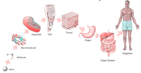
5. Levels of Organization & Body Systems
A body system (also called an organ system) is a highly integrated, complex collection of organs in the body that work seamlessly together to perform a specific, vital function. The truth is that all systems are intimately connected, relying heavily on one another, but it is highly useful for educational purposes to study them separately, even though they are not separate at all. With all of our body systems operating constantly and simultaneously, it is absolutely necessary to have an overarching regulatory system in place to maintain stability, harmony, and equilibrium across all these integrated systems. This unifying, central element in all of physiology is called Homeostasis.
The Six Levels of Structural Organization:
- Chemical Level: The most basic level. Includes atoms (like Carbon, Hydrogen, Oxygen, Nitrogen) combining to form essential molecules (like DNA, glucose, water).
- Cellular Level: Molecules combine to form cells, the basic structural and functional units of an organism. (e.g., Muscle cells, nerve cells, blood cells).
- Tissue Level: Groups of similar cells and their surrounding materials working together to perform a specific function. (The four basic types: Epithelial, Connective, Muscular, Nervous).
- Organ Level: Structures composed of two or more different types of tissues. They have specific functions and recognizable shapes. (e.g., The stomach, heart, liver, brain).
- System Level: Consists of related organs with a common function. (e.g., The digestive system includes the mouth, esophagus, stomach, intestines, liver, and pancreas working to extract nutrients).
- Organismal Level: The highest level. All the parts of the human body functioning together to constitute the total living organism.
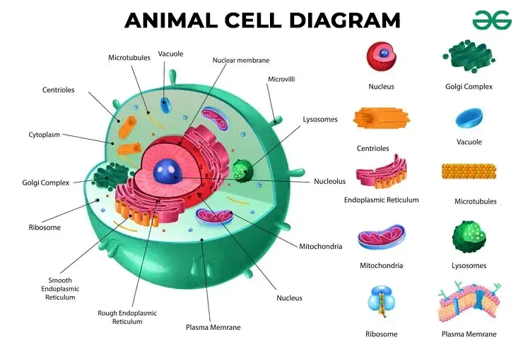
6. The Cell: The Fundamental Unit of Life
The cell is the absolute basic building block of all living things. Every single physiological process begins at the cellular level. To understand how the cell works, we can beautifully compare it to a bustling, highly organized city.
A. The Outer Boundary: The City Wall and Gates
The Cell (Plasma) Membrane
This is the outermost boundary of the cell, an incredibly thin, highly flexible, and selectively permeable (or semipermeable) barrier. It's primarily composed of a phospholipid bilayer, thickly populated with embedded proteins, carbohydrates, and cholesterol molecules.
- Phospholipid Bilayer: Two opposing layers of phospholipids. Each lipid molecule has a hydrophilic ("water-loving") polar head that faces the watery environments inside and outside the cell, and two hydrophobic ("water-fearing") non-polar fatty acid tails that face inward toward each other, forming the water-repelling core of the membrane.
- Proteins: These are crucial for nearly all membrane functions. Integral (transmembrane) proteins span the entire width of the membrane, forming open channels, carrier pumps, and signaling receptors. Peripheral proteins are loosely attached to the inner or outer surface, often involved in intracellular signaling cascades or anchoring the cytoskeleton.
- Cholesterol: Wedged deeply within the hydrophobic core between the fatty acid tails, cholesterol helps stabilize the membrane's fluidity. It prevents the membrane from freezing solid at low temperatures and stops it from becoming too loose and falling apart at high body temperatures.
- Glycocalyx (Carbohydrates): Chains of complex carbohydrates attached to proteins (forming glycoproteins) or attached to lipids (forming glycolipids) on the outer surface. This forms a unique, fuzzy "sugar coat" or cellular "ID tag." Extra Example: The ABO blood group markers on your red blood cells are actually part of the glycocalyx!
Physiological Functions of the Cell Membrane:
- Selective Permeability: Strictly controls exactly what enters and leaves the cell, maintaining perfect homeostasis. (Functions like the city's border control).
- Cell Recognition: The glycocalyx allows immune cells to recognize each other and identify foreign invaders.
- Communication/Signaling: Receptor proteins bind to chemical messengers like hormones and neurotransmitters, translating outside signals into internal actions.
- Cell Adhesion: Proteins allow cells to stick tightly together to form robust tissues (like skin).
- Protection: Provides a flexible physical barrier shielding internal components.
B. The Cell's Internal Environment: The City Hall and Workers
The Cytoplasm
The cytoplasm is literally everything inside the cell membrane but entirely outside the nucleus. It consists of three main elements:
- Cytosol: The jelly-like, semi-fluid, viscous portion where all organelles are suspended. It's mostly water heavily packed with dissolved solutes (ions, glucose, amino acids, ATP, fatty acids, etc.).
- Organelles: The highly specialized "little organs" with very specific, indispensable functions (discussed next).
- Inclusions: Temporary, non-functioning storage bodies. Extra Examples: massive glycogen granules in liver and muscle cells for energy storage, giant lipid droplets in fat cells (adipocytes), and melanin pigment granules in skin cells.
Physiological Functions of the Cytoplasm:
- Site of Many Metabolic Reactions: Crucial biochemical pathways, such as glycolysis (the first, anaerobic step of glucose breakdown to extract energy), occur entirely free-floating in the cytosol.
- Suspension of Organelles: Provides the perfect physical and chemical medium for all organelles to exist, interact, and function.
C. The Control Center: The City Hall/Mayor's Office
The Nucleus
Usually the largest and most prominent organelle, the nucleus is securely enclosed by a double-layered membrane called the nuclear envelope, which is punctured by nuclear pores that highly regulate the entry and exit of molecules. Inside, it contains:
- Chromatin: The relaxed, uncondensed, thread-like form of DNA (our genetic material) carefully wrapped around specialized structural proteins called histones. When the cell prepares to divide, this messy chromatin tightly condenses into distinct, visible structures called chromosomes.
- Nucleolus: A dense, dark, spherical body deeply embedded within the nucleus. It is the primary site of rapid ribosome synthesis.
Physiological Functions of the Nucleus:
- Genetic Control: Contains the cell's entire genetic blueprint (DNA), directing absolutely all cell activities by strictly controlling which proteins are synthesized. (The "master architectural plan" for the city).
- DNA Replication & Transcription: This is where DNA faithfully copies itself before cell division (mitosis) and where DNA's genetic code is safely transcribed into messenger RNA (mRNA), which then leaves the nucleus to give instructions to the factories.
- Ribosome Production: The nucleolus continuously synthesizes and assembles ribosomal RNA (rRNA) subunits.
D. Protein Synthesis and Processing: The Factories and Delivery Services
Ribosomes
Tiny, granular, non-membranous organelles made of ribosomal RNA (rRNA) and various proteins. They are the ultimate "protein factories" of the cell. They physically read the mRNA code coming from the nucleus to perfectly assemble amino acids into long proteins (a process called translation). They can exist as free ribosomes floating in the cytosol (making proteins for immediate use within the cell) or bound ribosomes securely attached to the Endoplasmic Reticulum (making proteins destined for export out of the cell or for embedding into membranes).
Endoplasmic Reticulum (ER)
An extensive, labyrinth-like network of interconnected membranous tubes and flattened sacs that extends massively throughout the cytoplasm, and is directly continuous with the outer membrane of the nuclear envelope.
- Rough Endoplasmic Reticulum (RER): Heavily studded with ribosomes, giving it a "rough" or sandpaper-like appearance under a microscope. Its primary function is to synthesize, fold, and modify proteins that are destined for secretion, insertion into cell membranes, or delivery to lysosomes. (e.g., adding sugar chains in a process called glycosylation). Extra Example: Pancreatic cells that produce massive amounts of digestive enzymes have incredibly large, active RERs.
- Smooth Endoplasmic Reticulum (SER): Completely lacks ribosomes. Its highly specialized functions include massive lipid and steroid hormone synthesis, intense detoxification of harmful drugs and poisons (making it extremely abundant in liver cells), and the storage and release of calcium ions. Extra Example: In skeletal muscle cells, a specialized SER called the sarcoplasmic reticulum stores the calcium absolutely crucial for triggering muscle contraction.
Golgi Apparatus (Golgi Complex)
A distinct stack of flattened, slightly curved membranous sacs called cisternae. It acts as the ultimate "Post Office" or "Packaging and Shipping Center" of the cell.
Physiological Functions of the Golgi Apparatus:
- Modification, Sorting, and Packaging: It receives vesicles containing raw proteins and lipids from the ER at its cis face, further modifies them (like adding or trimming sugar tags), sorts them based on their final destination, and packages them into new vesicles leaving from its trans face.
- Vesicle Formation: It actively forms various types of vesicles, including secretory vesicles (for exocytosis/dumping out of the cell), lysosomes (cellular stomachs), and transport vesicles that seamlessly deliver new structural components to patch the plasma membrane.
E. Energy Production: The Power Plant
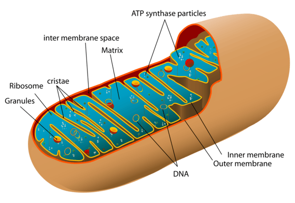
Mitochondria
Oval or bean-shaped organelles distinctly enclosed by a double membrane: a relatively smooth outer membrane and an inner membrane that is highly folded inward to form structures called cristae. These folds drastically increase the surface area available for chemical reactions. The dense, fluid-filled space within the inner membrane is called the matrix. Mitochondria also astonishingly possess their own unique, circular DNA (mtDNA) inherited exclusively from the mother.
Physiological Function (The "Powerhouses of the Cell"):
They are the primary site of aerobic cellular respiration. They utilize oxygen to meticulously break down and convert fuel molecules like glucose and fatty acids into ATP (adenosine triphosphate), the primary, usable energy currency of the entire cell. Extra Example: Cells that require massive, continuous amounts of energy, such as cardiac (heart) muscle cells and swimming sperm cells, have spectacularly high numbers of mitochondria compared to inactive cells.
F. Waste Management and Recycling: The Cleaning Crew
Lysosomes
Tiny, spherical membranous sacs packed tightly with incredibly powerful hydrolytic (digestive) enzymes that function best in an acidic environment. They act as the "Recycling Centers" and "Stomachs" of the cell.
- They aggressively break down ingested foreign substances and bacteria (phagocytosis).
- They digest worn-out, broken cellular organelles to recycle their raw materials (a process called autophagy).
- In dying or damaged cells, they can burst open to digest the entire cell (autolysis).
- Clinical Correlate: Tay-Sachs disease is a fatal genetic disorder where a specific lysosomal enzyme is missing. Toxic lipids build up uncontrollably in the brain because the lysosomes cannot break them down.
Peroxisomes
Smaller membranous sacs containing potent oxidative enzymes, most notably catalase and oxidase. They act as the highly specialized "Detoxification Squad".
- They aggressively neutralize highly reactive, harmful free radicals and detoxify toxins like alcohol (making them very abundant in kidney and liver cells).
- They heavily assist in the breakdown (beta-oxidation) of very long-chain fatty acids for use in energy production.
- During their reactions, they produce dangerous hydrogen peroxide (H₂O₂), but their built-in catalase enzyme instantly converts it safely into water and oxygen.
G. The Cell's Internal Support and Movement: The Infrastructure
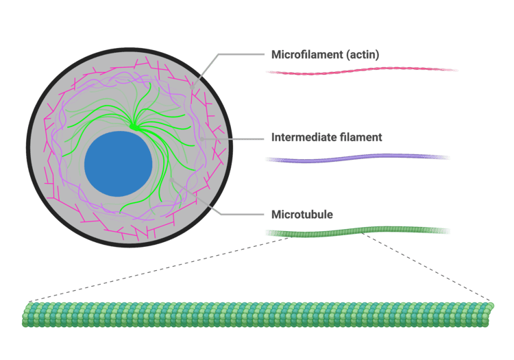
The Cytoskeleton
An intricate, dynamic network of protein filaments extending massively throughout the cytoplasm, providing cell shape, internal support, and physical highways for transport. It consists of three main types of filaments:
- Microfilaments (Actin): The thinnest fibers; deeply involved in cell movement, gross shape changes, cell division (cleavage furrow), and forming the contractile machinery of muscle cells.
- Intermediate Filaments: Tough, rope-like proteins that provide immense structural stability and fiercely resist mechanical pulling stress on the cell. Extra Example: Keratin in skin cells is an intermediate filament that makes skin tough and waterproof.
- Microtubules: The largest elements; hollow tubes made of tubulin. They determine the overall cell shape, form the "railroad tracks" for motor proteins to drag organelles across the cell, and are the primary structural core of cilia, flagella, and the mitotic spindle.
Centrosomes, Cilia, and Flagella
- Centrosomes and Centrioles: Located strategically near the nucleus, the centrosome contains two barrel-shaped centrioles positioned at right angles. It acts as the main Microtubule-Organizing Center (MTOC), generating and organizing the mitotic spindle required to pull chromosomes apart during cell division.
- Cilia and Flagella: Hair-like, motile cellular projections entirely made of microtubules.
- Cilia are short and numerous, beating in coordinated waves to move substances across the cell surface. Extra Example: Millions of cilia in your respiratory tract constantly sweep mucus and trapped dust up and out of your lungs.
- Flagella are much longer and usually singular, acting like a whip to propel the entire cell forward. Extra Example: The only human cell with a flagellum is the male sperm cell.
Summary Table of Organelles
| Organelle | Key Physiological Functions |
|---|---|
| Plasma Membrane | Selective barrier, cell recognition, intercellular communication, structural protection. |
| Nucleus | Ultimate genetic control, houses DNA, site of DNA replication and mRNA transcription. |
| Ribosomes | Direct protein synthesis via the translation of mRNA into amino acid chains. |
| Rough ER (RER) | Synthesis, folding, and intense modification of proteins destined for export or membrane insertion. |
| Smooth ER (SER) | Lipid and steroid synthesis, detoxification of drugs/poisons, massive intracellular Ca²⁺ storage. |
| Golgi Apparatus | Modifies, sorts, meticulously packages proteins and lipids into transport/secretory vesicles. |
| Mitochondria | Site of aerobic cellular respiration, massive ATP synthesis (the ultimate powerhouse). |
| Lysosomes | Intracellular digestion of debris, bacteria, and old organelles; primary cellular waste removal. |
| Peroxisomes | Detoxification (especially neutralizing free radicals and alcohol), extensive fatty acid breakdown. |
| Cytoskeleton | Maintains cell shape, structural support, internal transport tracks, enables cell motility. |
| Centrosomes | Organize the microtubule network and generate the mitotic spindle during cell division. |
| Cilia / Flagella | Cilia move external substances across the cell surface; Flagella physically propel the entire cell. |
7. Biological Membranes: The Fluid Mosaic Model
Biological membranes are highly dynamic, incredibly fluid structures that sharply define the boundaries of cells (plasma membrane) as well as the internal organelles. They are absolutely essential for maintaining cellular integrity, strictly regulating transport, facilitating communication, and housing vital enzymatic reactions. The most universally accepted scientific model describing membrane structure is the Fluid Mosaic Model.
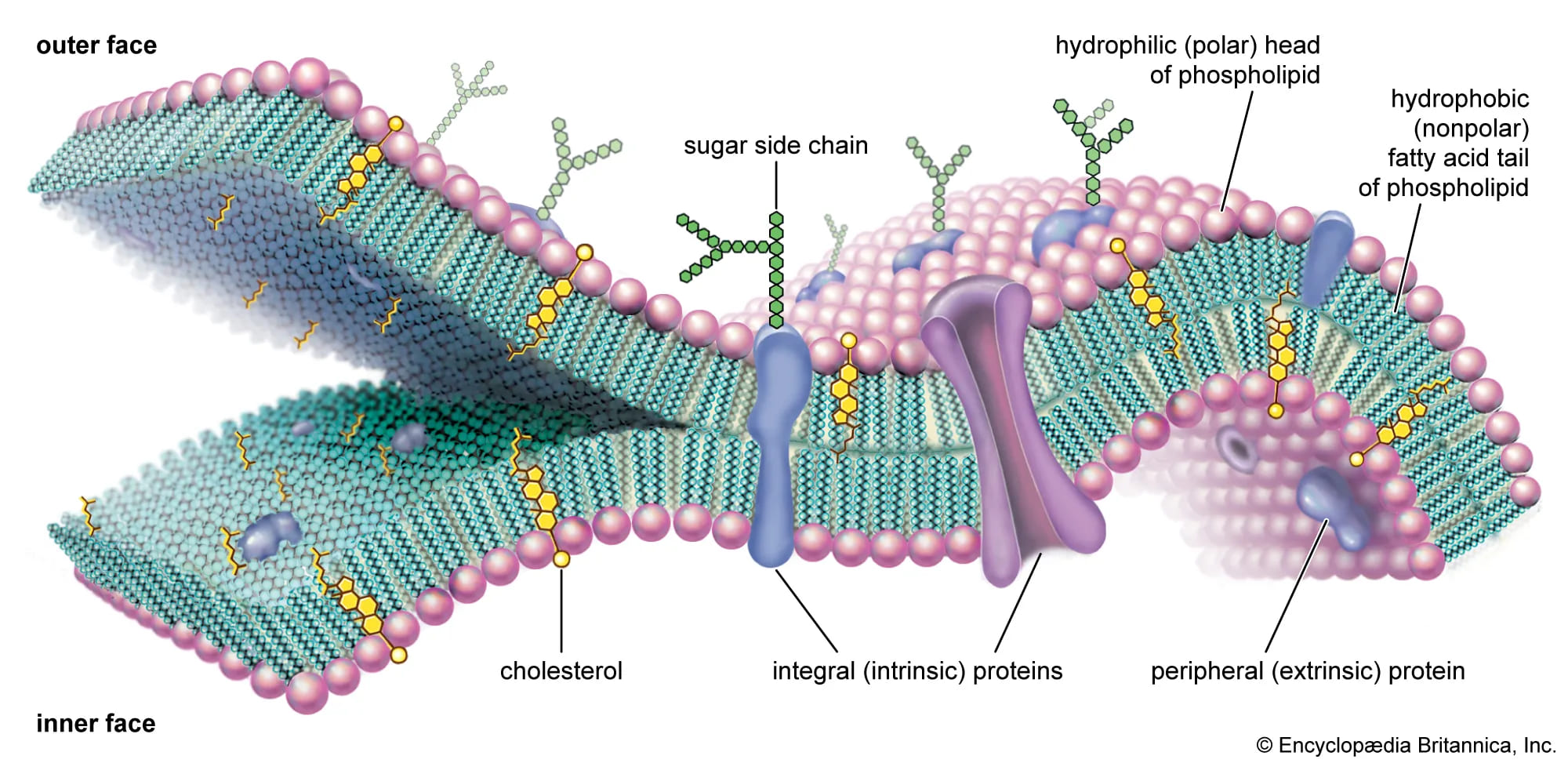
The Fluid Mosaic Model
Proposed brilliantly by Singer and Nicolson in 1972, this model describes the cell membrane as a highly fluid lipid bilayer where an array of proteins are embedded or attached, looking much like a complex, ever-shifting mosaic artwork.
- "Fluid": Refers to the constant, rapid movement of individual phospholipid molecules and proteins within the two-dimensional plane of the membrane. Lipids and many proteins are not locked in place; they constantly drift laterally, rotate on their axes, and flex their tails.
- "Mosaic": Refers to the diverse, scattered "patchwork" of different functional proteins and other structural molecules (like cholesterol and carbohydrates) permanently or temporarily embedded within the vast ocean of the lipid bilayer.
A. Lipids of the Cell Membrane
The central, primary structural framework of the membrane is the fluid lipid bilayer, which is predominantly made of phospholipids and cholesterol.
1. Phospholipids
Phospholipids are by far the most abundant lipids in the membrane. They possess a crucial chemical property: they are amphipathic. This means a single molecule contains both a highly hydrophilic (water-loving) polar head and two highly hydrophobic (water-fearing) non-polar fatty acid tails. In the watery environment of the body, they spontaneously and instantly self-assemble to form a bilayer. The hydrophobic tails hide inward, desperately avoiding water, while the hydrophilic heads proudly face the watery environments inside (intracellular fluid) and outside (extracellular fluid) the cell.
2. Cholesterol
Cholesterol molecules are tough, rigid, ring-shaped lipids inserted tightly between the phospholipids. They act as the ultimate membrane temperature buffer, perfectly regulating fluidity. At normal, warm body temperatures, cholesterol restricts movement, reducing excessive fluidity and making the membrane stronger and less leaky. Conversely, at dangerously low temperatures, cholesterol prevents the phospholipid tails from packing too tightly together, thus increasing fluidity and stopping the cell membrane from freezing solid.
Lipid Functions in the Cell Membrane:
- Forms the fundamental, self-healing bilayer structure.
- Provides a robust selectively permeable barrier, naturally allowing only small, uncharged, fat-soluble substances (O₂, CO₂, steroid hormones) to easily pass through directly.
- Acts as an impenetrable physical barrier for all water-soluble substances (glucose, salts, ions), which absolutely require physical assistance from embedded proteins to cross.
B. Membrane Proteins
While lipids form the barrier, proteins are the hardworking machines of the membrane, performing almost all of its specific, dynamic functions.
1. Integral (Transmembrane) Proteins
These are tightly bound proteins that fully span across the entire thickness of the membrane. They are deeply embedded in the hydrophobic core and can only be removed by completely destroying the bilayer. They primarily function as open channels, carrier transport proteins, active pumps, receptors, and enzymes.
2. Peripheral Proteins
These are loosely, temporarily bound to the membrane's inner or outer surface. They do not penetrate the hydrophobic core and are easily detached without harming the membrane. They often function as localized enzymes, signaling relays, or cytoskeletal anchors holding the membrane shape.
Functions of Membrane Proteins:
- Transport: Facilitating the precise movement of specific, rejected substances across the barrier (channels, carriers, ATP pumps).
- Enzymatic Activity: Catalyzing vital metabolic reactions directly at the membrane surface.
- Signal Transduction: Acting as sophisticated receptors catching chemical messengers (like adrenaline) and transmitting the order inside.
- Cell-Cell Recognition: Acting as strict identification tags (glycoproteins) so immune cells don't attack the body's own tissues.
- Intercellular Joining: Forming tight junctions and desmosomes to physically link neighboring cells together.
- Attachment to Cytoskeleton & ECM: Providing immense structural stability by tethering the cell's skeleton to the outside world.
C. Carbohydrates of the Cell Membrane
Carbohydrates are exclusively found on the external, outward-facing surface of the plasma membrane. They never face the inside. They are permanently attached to lipids (forming glycolipids) or to proteins (forming glycoproteins). This entire "sugar coat" covering the cell is called the glycocalyx, which serves as a highly specific, unique molecular signature for every different cell type in your body.
Functions of Membrane Carbohydrates (Glycocalyx):
- Cell-Cell Recognition: Crucial for distinguishing "self" tissues from "non-self" invaders (e.g., orchestrating immune responses, dictating ABO blood types, and organ transplant rejection).
- Cell Adhesion: The sticky sugars help cells firmly bind to one another in tissues.
- Receptors: Can uniquely act as receptors for specific hormones, or tragically, as hijacking points for bacterial toxins and viruses (like the flu or COVID-19).
- Protection: Provides a cushioning, protective physical barrier against mechanical damage and harsh enzymes.
Properties of the Cell Membrane
The specific composition and arrangement of lipids, proteins, and carbohydrates endow the cell membrane with its most essential, life-sustaining properties:
- Selectively Permeable (Semi-permeable): This is arguably its most important property. The membrane precisely and selfishly regulates exactly which substances can enter or leave the cell. The hydrophobic lipid core acts as the primary absolute barrier. Small, nonpolar molecules (O₂, CO₂) and highly lipid-soluble molecules pass directly and effortlessly. However, charged ions (Na⁺, K⁺) and large polar molecules (glucose) are utterly rejected and require specific, designated transport proteins to grant them entry.
- Fluidity: The membrane is absolutely not a rigid, solid shell; its molecular components are in constant, swirling motion. Fluidity is heavily influenced by body temperature, the amount of cholesterol (which acts as a stabilizing buffer), and the degree of saturation of the fatty acid tails (kinked, unsaturated tails increase fluidity). This property is absolutely essential for vesicles to fuse with the membrane, for the cell to divide, and for transport proteins to physically shift their shapes.
- Asymmetry: The two faces (the inner leaflet touching the cytoplasm, and the outer leaflet touching the blood/fluid) of the plasma membrane are completely structurally and functionally different. For example, carbohydrates are exclusively on the outer surface (glycocalyx), and specific lipids and signaling proteins are oriented strictly in one direction. This is vital for directional signaling, shape maintenance, and cell recognition.
- Self-Sealing Capability: Due to the powerful thermodynamic forces of hydrophobic interactions, if the membrane is punctured or torn, the lipids have a natural, spontaneous tendency to instantly re-seal themselves, completely preventing the fatal leakage of cytoplasmic contents. This is crucial for maintaining cell integrity during physical stress or when a needle enters a cell.
Summary of Key Membrane Functions:
- Protective Barrier: Encloses the cell's delicate contents, safely separating the intracellular from the chaotic extracellular environment.
- Selective Transport: Heavily regulates the passage of every single substance entering and exiting the cell.
- Cell-Cell Communication: Contains specific receptors for circulating hormones, drugs, and neurotransmitters.
- Cell Recognition & Adhesion: Facilitates cell identification by the immune system and the formation of solid tissues.
- Enzymatic Activity: Houses membrane-bound enzymes that rapidly catalyze specific biochemical reactions.
- Maintenance of Cell Shape: Provides profound structural support in conjunction with the inner cytoskeleton.
- Generates Membrane Potential: Crucial for the electrical firing of nerve and muscle cells.
- Endocytosis & Exocytosis: Manages the bulk, massive transport of large materials into and out of the cell.
8. Membrane Potential: The Electrical Voltage Across the Membrane
Before looking at exactly how things physically move across the membrane, it's absolutely essential to understand that there is a permanent electrical difference, or voltage, sitting directly across every single cell membrane in your body. This is called the membrane potential.
Membrane Potential is the exact measurable difference in electrical charge (or potential energy) between the inside and the outside of a cell. By strict scientific convention, the inside of the cell is always measured as being negative relative to the outside.
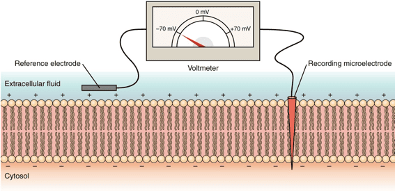
How is the Membrane Potential Established?
It is driven by three distinct, constantly working factors:
- Unequal Distribution of Ions: There are radically different concentrations of ions (charged particles) inside versus outside the cell.
- Outside the cell (Extracellular Fluid - ECF): Features a massively high concentration of Na⁺ (sodium) and Cl⁻ (chloride).
- Inside the cell (Intracellular Fluid - ICF): Features a massively high concentration of K⁺ (potassium) and huge, negatively charged proteins and phosphates (which are physically too large to ever leave the cell, trapping their negative charge inside).
- Selective Permeability of the Membrane: The cell membrane is not equally permeable to all ions. At rest, it is packed with specific "leak channels" that make it vastly more permeable to K⁺ than to Na⁺. Because K⁺ is highly concentrated inside, it constantly leaks out down its concentration gradient. Every time a positive K⁺ leaves, it leaves behind a negative charge, making the inside of the cell significantly more negative.
- Sodium-Potassium Pump (Na⁺/K⁺ ATPase): This relentless active transport pump burns massive amounts of ATP to constantly eject exactly 3 Na⁺ ions out of the cell for every 2 K⁺ ions it pumps back in. Because it pumps out more positive charges (3) than it brings in (2), this pump is inherently electrogenic and contributes directly and continuously to the negative charge inside the cell.
Resting Membrane Potential
In a resting (unstimulated) neuron or muscle cell, the steady-state electrical potential established and maintained perfectly by these factors is called the Resting Membrane Potential. It is typically measured at around -70 mV (millivolts), meaning the inside is 70 millivolts more negative than the outside.
Physiological Significance:
The resting membrane potential is absolutely not just a passive, resting state; it is a massive form of stored, coiled potential energy (like a stretched rubber band) crucial for:
- Excitability: It allows excitable cells (like neurons and heart muscle cells) to instantly open gates and generate rapid, explosive electrical signals (action potentials) for instant communication, thought, and muscle contraction.
- Secondary Active Transport: The immense energy stored in the steep Na⁺ and K⁺ ion gradients can be brilliantly harnessed by the cell to power the "uphill" transport of other essential substances (like glucose and amino acids) across the membrane.
Clinical Correlate: Hyperkalemia
If a patient's kidneys fail, potassium (K⁺) builds up in the blood outside the cell (a condition called hyperkalemia). This destroys the carefully maintained K⁺ gradient. The resting membrane potential is ruined, the heart muscle cells cannot reset their electrical charge, and the patient suffers a sudden, fatal cardiac arrhythmia (heart attack).
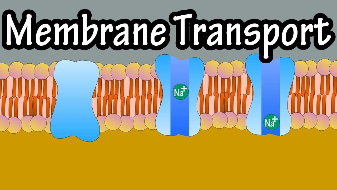
9. Membrane Transport
Membrane transport is the fundamental physiological process that meticulously governs the movement of all substances across biological membranes. It's essential for maintaining cellular homeostasis, acquiring vital nutrients, aggressively expelling toxic waste products, and facilitating cell-to-cell communication. Substances cross the membrane via two general, overarching mechanisms: Passive Transport and Active Transport.
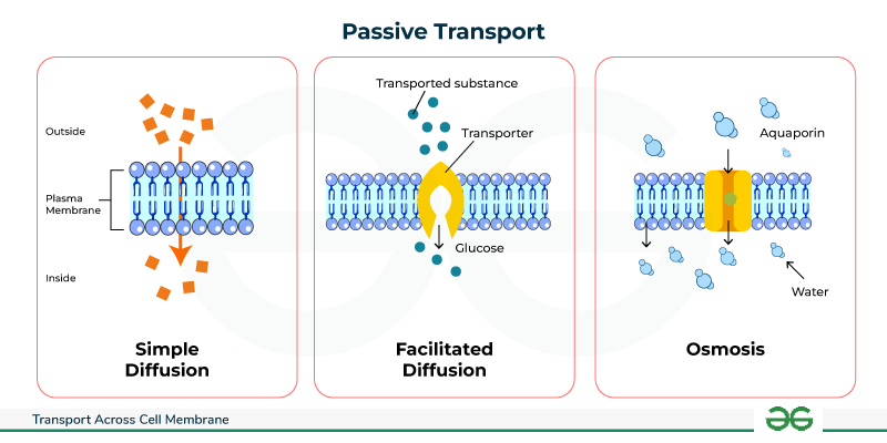
1. Passive Transport: Moving Downhill
Passive transport is the movement of substances across a cell membrane completely without the direct expenditure of cellular metabolic energy (ATP). This movement is always "downhill," meaning down the electrochemical or concentration gradient of the substance. The energy driving this movement comes purely from the inherent, random kinetic energy of the molecules themselves and the potential energy stored in the concentration gradient.
1.1. Simple Diffusion: Through the Lipid Bilayer
In simple diffusion, substances move directly and effortlessly through the lipid bilayer without any help from membrane proteins whatsoever.
- Highly Permeable: Small, nonpolar (lipophilic/fat-soluble) molecules like O₂, CO₂, and steroid hormones readily dissolve directly into the hydrophobic core and pass completely through to the other side.
- Moderately Permeable: Small, uncharged polar molecules like water and ethanol can pass to a very limited degree because they are small enough to slip through the lipid gaps.
- Impermeable: Large polar molecules (like glucose) and absolutely all charged ions (Na⁺, K⁺, Ca²⁺) cannot pass through the lipid core on their own under any circumstances.
The sole driving force is the concentration gradient. The random, chaotic molecular motion (kinetic energy) results in a net movement from an area of highly crowded concentration to an area of lower concentration, continuing unabated until perfect equilibrium is reached across the membrane.
Key Characteristics of Simple Diffusion:
- Absolutely no membrane proteins are involved.
- Does not exhibit saturation kinetics: The rate of diffusion increases linearly forever with the concentration gradient; there is no maximum transport rate (Vmax) because there are no proteins to get "full".
- According to Fick's Law, the rate is directly proportional to the gradient magnitude, lipid solubility, and membrane surface area, and inversely proportional to molecular size and the physical thickness of the membrane.
1.2. Facilitated Diffusion: Protein-Assisted Passage
This process uses specialized integral membrane proteins (channels or carriers) to beautifully facilitate the movement of specific, impermeable substances down their electrochemical gradient. It is still completely passive, as absolutely no ATP is directly consumed.
A. Channel Proteins (Pores)
These proteins form a hollow, water-filled pore directly across the membrane, allowing the incredibly rapid, single-file passage of specific ions or water molecules. Most channels are tightly gated, meaning they act like doors that only open or close in response to specific, required stimuli:
- Voltage-Gated Channels: Respond instantly to changes in electrical membrane potential (e.g., Na⁺ and K⁺ channels crucial in firing neurons).
- Ligand-Gated Channels: Respond only when a specific chemical messenger binds to them (e.g., neurotransmitter receptors at muscle synapses).
- Mechanically-Gated Channels: Respond directly to physical deformation or stretching of the membrane (e.g., touch receptors in your skin, or hearing receptors in the ear).
- Leak Channels: Are generally always randomly fluttering open and closed, heavily contributing to the resting membrane potential.
- Extra Examples: Ion channels (Na⁺, K⁺, Cl⁻, Ca²⁺) and aquaporins, which are highly specialized channels dedicated exclusively to letting massive amounts of water through instantly.
B. Carrier Proteins (Transporters)
These proteins work like revolving doors. They physically bind to a specific molecule on one side, undergo a massive conformational (shape) change, and then release the molecule safely on the other side. This physical shifting process makes it much slower than channel-mediated transport.
- Saturation Kinetics: Because there are a strictly finite number of carrier proteins on a cell, the transport rate has an absolute maximum speed limit (Vmax) that hits when every single carrier is occupied and working as fast as it can.
- Specificity: Carriers are incredibly highly specific for the exact molecule(s) they are designed to transport.
- Competition: Structurally similar, "imposter" molecules can compete for the exact same binding site, blocking the real molecule.
- Extra Examples: Glucose Transporters (GLUT proteins, which insulin commands to surface) and various amino acid transporters.
Key Characteristics of Facilitated Diffusion:
- Strictly involves specific integral membrane proteins (channels or carriers).
- Exhibits saturation kinetics (Vmax limit) due to the limited, physical number of transporters.
- Can be easily subject to chemical competition.
- Can be heavily regulated by the cell (e.g., by gating channels shut, or inserting/removing carriers from the membrane to control absorption).
1.3. Osmosis: The Grand Movement of Water
Osmosis is the incredibly powerful net movement of water across a selectively permeable membrane, strictly from an area of higher water concentration (which means lower solute concentration) directly to an area of lower water concentration (which means higher, saltier solute concentration). The driving force is the water potential gradient, determined entirely by the difference in non-penetrating solute concentration on either side of the membrane.
Osmotic pressure is the "pulling" or "sucking" force a solution with a higher solute concentration forcefully exerts to draw water toward it. Tonicity refers to the actual physical effect of a surrounding solution on the cell's total volume:
- Isotonic: The concentration is perfectly equal inside and outside. There is no net water movement; cell volume remains perfectly normal.
- Hypotonic: The outside fluid is dilute (watery). Water rushes rapidly into the cell, causing it to aggressively swell and potentially burst (a fatal event called lysis).
- Hypertonic: The outside fluid is extremely concentrated (salty). Water is sucked rapidly out of the cell, causing the cell to drastically shrivel and shrink (a process called crenation).
Clinical Correlate: Intravenous (IV) Fluids
When a patient arrives at the hospital dehydrated, you cannot simply inject pure, hypotonic tap water into their veins. Doing so would cause their red blood cells to rapidly absorb the water, swell, and violently explode (hemolysis), killing the patient. Instead, nurses administer 0.9% Normal Saline, which is perfectly isotonic to human blood plasma, safely rehydrating the body without destroying the cells.
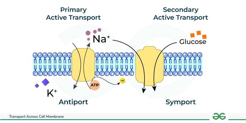
2. Active Transport: Against the Current, with Energy
Active transport is the strenuous, demanding process of actively moving substances across a cell membrane directly against their electrochemical or concentration gradient (i.e., dragging them from a region of lower concentration forcefully into a region of higher concentration). This distinctly "uphill" movement necessitates the direct or indirect expenditure of precious cellular metabolic energy, almost invariably derived from the violent hydrolysis of ATP.
2.1. Primary Active Transport: Direct ATP Expenditure
Primary active transporters are robust integral membrane proteins that function fundamentally as ATPases; they directly bind to and snap the phosphate off of ATP, utilizing that massive burst of released energy to physically power the forced movement of solutes. These heavy-duty transporters are often called "pumps."
- Na⁺/K⁺ ATPase (Sodium-Potassium Pump): Found aggressively pumping in virtually all animal cells, this absolutely vital, life-sustaining pump forcibly moves 3 Na⁺ ions entirely out of the cell and drags 2 K⁺ ions back into the cell for every single ATP molecule hydrolyzed. It is strictly electrogenic (creates a negative charge imbalance) and is undeniably fundamental for maintaining steep Na⁺/K⁺ gradients, establishing the baseline resting membrane potential, regulating water balance and cell volume, and driving secondary active transport.
- Ca²⁺ ATPases (e.g., SERCA, PMCA): These relentless pumps maintain the extremely, dangerously low intracellular Ca²⁺ concentration needed for survival. SERCA constantly pumps Ca²⁺ safely into the sarcoplasmic/endoplasmic reticulum for deep storage (which is absolutely crucial for allowing a muscle to relax after a contraction), while PMCA furiously pumps Ca²⁺ completely out of the cell membrane into the blood.
- H⁺/K⁺ ATPase (Gastric Proton Pump): Located almost exclusively in the specialized parietal cells of the stomach lining, this pump aggressively secretes H⁺ (acid protons) directly into the stomach lumen, creating the highly destructive, acidic environment (pH 1-2) necessary for dissolving food and activating digestion. Extra Example: This exact pump is the sole target of Proton Pump Inhibitor (PPI) drugs, like Omeprazole, which shut the pump down to cure severe stomach ulcers and acid reflux.
- ABC Transporters (ATP-Binding Cassette): A gigantic, ancient superfamily of transporters that move a vast, diverse array of substrates.
Extra Example 1: MDR1 (P-glycoprotein) is an ABC transporter notorious in oncology; it causes multidrug resistance in cancer cells by acting as a biological bouncer, actively catching and pumping expensive chemotherapy drugs right back out of the tumor cell before they can work.
Extra Example 2: The CFTR protein is a specialized Cl⁻ channel; a genetic mutation destroying this exact pump causes the thick, deadly mucus buildup characteristic of the disease Cystic Fibrosis.
2.2. Secondary Active Transport (Co-transport)
Secondary active transport is highly efficient because it does not directly hydrolyze ATP itself. Instead, it ingeniously acts like a waterwheel; it uses the immense potential kinetic energy stored in an existing, steep electrochemical gradient (typically the massive Na⁺ gradient that was previously created and paid for by the Na⁺/K⁺ pump) to power the transport of a completely second substance against its own gradient.
- Symporters (Cotransporters): Both the driving ion (e.g., Na⁺ rushing down its gradient) and the "freeloading" transported solute are physically dragged across the membrane in the exact same direction. Extra Example: The Na⁺-Glucose Symporter (SGLT) found heavily in the intestine and kidneys, which utilizes the rushing flow of Sodium to forcefully absorb essential Glucose from your food against a steep gradient, ensuring none is wasted in the feces or urine.
- Antiporters (Exchangers): The driving ion rushes in, providing energy to forcefully eject the transported solute in the exact opposite direction. Extra Example: The Na⁺-Ca²⁺ Exchanger (NCX), which is absolutely crucial for aggressively removing toxic excess Ca²⁺ from cardiac (heart) muscle cells after every single heartbeat, allowing the heart to relax. Also, the Na⁺-H⁺ Exchanger (NHE) which pumps acid out of the cell to regulate intracellular pH.
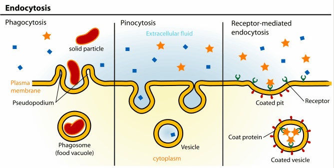
3. Vesicular Transport (Bulk Transport): For the Heavy Lifting
Vesicular transport is the macro-scale mechanism used exclusively for moving massively large molecules, whole macromolecules, and giant particulate matter (even whole bacteria) into or out of the cell. It involves the complex, energy-demanding physical formation, movement, and fusion of large membrane-bound sacs called vesicles. Because it physically reshapes the cell membrane, it absolutely always requires massive amounts of cellular energy (ATP).
3.1. Endocytosis: Bringing the Outside In
Endocytosis is the active process by which cells internalize large substances. The plasma membrane physically sinks inward (invaginates), wraps entirely around the material, and pinches off, forming a brand-new intracellular vesicle.
- Phagocytosis ("Cell Eating"): The aggressive, targeted ingestion of massive, solid particles like invading bacteria, dead tissue, or cellular debris by highly specialized immune sentinels (e.g., macrophages and neutrophils). The cell physically extends long "arms" called pseudopods to totally engulf the target, forming a giant internal death-chamber called a phagosome, which then merges with a lysosome for total destruction.
- Pinocytosis ("Cell Drinking"): The constant, non-specific, routine gulping of tiny droplets of extracellular fluid and whatever dissolved solutes happen to be floating in it. It is how cells sample their environment.
- Receptor-Mediated Endocytosis: An exquisitely, highly specific, targeted process. Specific extracellular ligands (e.g., LDL-cholesterol particles, iron-carrying transferrin, or even sneaky viruses) must perfectly bind to complementary, matching receptors on the surface. These loaded receptors then rapidly slide together and cluster into specialized depressions called clathrin-coated pits, which fold inward and are cleanly brought into the cell as a customized vesicle.
3.2. Exocytosis: Releasing to the Outside
Exocytosis is the reverse process by which cells actively release large substances. Intracellular, membrane-bound vesicles are driven by motor proteins to the edge of the cell, where they seamlessly fuse with the plasma membrane, violently dumping their contents to the outside world.
- Constitutive Secretion: A continuous, ongoing, unregulated baseline process operating in all cells. It is used to constantly deliver newly minted lipids and proteins to expand the plasma membrane, and to continuously secrete the structural components of the extracellular matrix (like collagen).
- Regulated Secretion: A highly controlled process that occurs exclusively in specialized secretory cells (e.g., brain neurons, pancreatic endocrine cells). Secretory vesicles containing highly potent products like neurotransmitters, digestive enzymes, or hormones are stockpiled safely near the membrane. They are strictly held back and only released in a massive burst in response to a very specific, targeted signal (almost always a sudden, sharp rise in intracellular Ca²⁺).
10. The "Why": Importance and Functions of Membrane Transport
The precise, unrelenting control over exactly what enters and exits a cell underlies the success of virtually every single physiological process in the entire human body.
- Maintenance of Cellular Homeostasis: Strict, life-or-death ion gradients (e.g., maintaining low intracellular Na⁺ and high K⁺) and exact internal pH are aggressively maintained at incredibly great energy cost to absolutely prevent cell death and ensure proper, flawless enzyme function.
- Nutrient Acquisition: Transport systems enable cells to highly efficiently scavenge, absorb, and aggressively concentrate essential, life-sustaining molecules like glucose, vitamins, and amino acids from the blood.
- Waste Removal: Active transporters continuously expel harmful, toxic metabolic waste products (like urea, lactic acid, and excess acid protons), totally preventing their lethal accumulation inside the cytoplasm.
- Generation of Electrical Signals: In complex neurons and muscle cells, the rapid, highly controlled movement of ions surging through gated channels generates action potentials, which are the fundamental electrical basis of every single thought, heartbeat, and physical movement.
- Cell-to-Cell Communication: Exocytosis violently releases neurotransmitters across synapses and dumps hormones into the blood, while endocytosis helps regulate receptor sensitivity by swallowing up overused receptors from the membrane.
- Regulation of Cell Volume: Constant ion pumping, most especially the tireless Na⁺/K⁺ ATPase, strictly controls internal intracellular osmolarity, absolutely preventing cells from fatally swelling up and bursting or aggressively shrinking and collapsing.
- Absorption and Reabsorption: Massive, highly coordinated transport processes in the Gastrointestinal (GI) tract and the nephrons of the kidneys are utterly essential for absorbing nutrients from food and perfectly regulating the entire body's water, total electrolyte, and systemic acid-base balance.
| Process | Energy Requirement | Gradient Direction | Transporter Requirement | What Specifically Moves? | Examples & Clinical Notes |
|---|---|---|---|---|---|
| Passive Processes (No ATP Burned) | |||||
| Simple Diffusion | None | Downhill (High to Low) | None (Straight through lipid) | Small, highly lipid-soluble molecules | O₂, CO₂, steroid hormones crossing tissues. |
| Facilitated Diffusion | None | Downhill (High to Low) | Yes (Channel or Carrier protein) | Ions, large glucose, amino acids | Glucose transporters (GLUT), Na⁺/K⁺ voltage channels. |
| Osmosis | None | Downhill (High water to Low water) | Yes (Aquaporins heavily aid) | Water molecules exclusively | Red blood cells expanding or shrinking in different tonic IV solutions. |
| Active Processes (Requires ATP) | |||||
| Primary Active Transport | Yes (Directly breaks ATP) | Uphill (Low to High) | Yes (A specialized Pump) | Ions against steep gradients | Na⁺/K⁺ pump, Ca²⁺ SERCA pump, H⁺ stomach acid pump. |
| Secondary Active Transport | No (Uses kinetic energy of an existing ion gradient) | Uphill (Low to High) | Yes (Symporter or Antiporter) | Ions, glucose, amino acids dragged along | Na⁺-glucose co-transporter (SGLT) in the kidney. |
| Vesicular Transport | Yes (Massive ATP used) | N/A (Bulk physical movement) | No (Uses membrane vesicles) | Large particles, whole bacteria, macromolecules, bulk fluids | Phagocytosis (macrophages eating bacteria), Exocytosis (releasing insulin). |
List of Academic References
- Hall, J. E., & Hall, M. E. (2020). Guyton and Hall Textbook of Medical Physiology (14th ed.). Elsevier. (Definitive source on membrane potentials, the Na⁺/K⁺ pump, and systemic physiology).
- Boron, W. F., & Boulpaep, E. L. (2016). Medical Physiology (3rd ed.). Elsevier. (Excellent deep dive into cellular organelles, secondary active transport, and molecular physiology).
- Alberts, B., Johnson, A., Lewis, J., et al. (2014). Molecular Biology of the Cell (6th ed.). Garland Science. (The absolute gold standard for the Fluid Mosaic Model, vesicular transport, and cytoskeletal structures).
- Costanzo, L. S. (2017). Physiology (6th ed.). Elsevier. (Highly recommended for clear, mechanistic explanations of functional vs. process physiology and simple diffusion mathematics).
- Chabner, L. (2020). The Language of Medicine (12th ed.). Saunders. (A premier resource for medical terminology, etymology, roots, and prefixes like pancytopenia).
Physiology and Cell Physiology Read More »
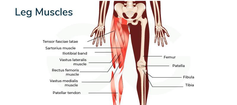
Muscles of the Lower Limb
Anatomy of the Lower Extremities: Muscles from the Pelvis to the Toe
By the end of this highly detailed anatomical master guide, you will be deeply conversant with:
- The Osteology (Bones) of the lower limb, including the intricate details of the bony pelvis, hip bone, and femur.
- The vital Ligaments of the Hip Joint that provide unmatched stability and posture control.
- The precise Origins, Insertions, Innervations, and Actions of every muscle group from the gluteal region down to the four layers of the foot.
- Clinical Correlations and Real-World Examples illustrating what happens when these bones, nerves, and muscles are injured.
Part 1: Osteology — The Bony Pelvis & The Hip Bone
The lower limb is engineered for two massive responsibilities: bearing the entire weight of the upper body and locomotion (movement). The hip joint is one of the most important joints in the body for complex movements like walking, running, or dancing. It relies on a strong bony foundation.
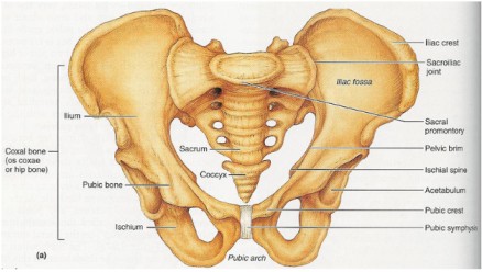
The Bony Pelvis
The bony pelvis is a massive, basin-shaped ring of bones connecting the flexible vertebral column to the femurs (thigh bones). It is formed by the sacrum, the coccyx (tailbone), and the two large hip bones (Os coxae).
Functional Example: The pelvis acts like a structural funnel. When you stand, the weight of your head, arms, and torso travels down your spine, hits the sacrum, splits in half across the pelvic ring, and is driven straight down into the heads of your two femurs.
The Hip Bone (Os Coxa)
Each large, irregularly shaped hip bone is actually a fusion of three primary bones. In children, these are separate bones joined by cartilage, but they completely fuse into a single solid bone by the end of puberty (around age 20-25).
The largest, most superior (top) part of the hip bone. It forms the prominent "wings" of the pelvis that you can feel when you put your hands on your hips.
- Iliac Crest: The palpable superior border. It serves as a major attachment site for abdominal muscles.
- Spines: It terminates anteriorly as the Anterior Superior Iliac Spine (ASIS) and posteriorly as the Posterior Superior Iliac Spine (PSIS). Below these are the AIIS and PIIS.
- Surfaces: The large, concave internal Iliac Fossa (houses the iliacus muscle); the rough outer Gluteal Surface (where the gluteal muscles attach); and the medial Auricular Surface (which articulates with the sacrum to form the sacroiliac joint).
- Notches: The Greater Sciatic Notch, a massive indentation allowing the thick sciatic nerve to exit the pelvis and enter the leg.
Forms the posteroinferior (lower-back) part of the hip bone. This is the heavy, durable bone you sit on.
- Ischial Tuberosity: The large, roughened "sitting bone." It supports your entire body weight when seated and is the origin point for the hamstring muscles.
- Ischial Spine: A pointed projection posterior to the acetabulum. It physically separates the Greater and Lesser Sciatic Notches.
- Ramus of the Ischium: A branch of bone that projects forward to join with the pubis.
Forms the anterior (front) part of the hip bone.
- Body of Pubis: The central, flat part that meets the other pubic bone at the midline cartilaginous joint called the Pubic Symphysis.
- Superior & Inferior Rami: Bars of bone that branch off the body to help form the acetabulum and the obturator foramen.
- Key Markings: Includes the Pubic Tubercle (where the inguinal ligament attaches) and the Obturator Crest for muscle attachments.
Key Landmarks of the Fused Hip Bone
- The Acetabulum: The deep, cup-shaped socket on the lateral surface of the hip bone, formed by the union of all three bones (Ilium, Ischium, Pubis). It articulates with the head of the femur.
- Lunate Surface: The crescent-shaped, smooth articular cartilage region.
- Acetabular Fossa: The rough, deep central depression.
- Acetabular Labrum: A raised ring of fibrocartilage that deepens the socket, gripping the femoral head like a suction cup for increased stability.
- Obturator Foramen: The massive opening created by the ischium and pubis. While it looks huge on a skeleton, in a living person it is almost completely closed by the tough obturator membrane, leaving only a tiny obturator canal for the obturator nerve and blood vessels to squeeze through into the thigh.
The Femur (Thigh Bone)
The femur is the longest, strongest, and heaviest bone in the human body. It is designed to transmit weight seamlessly from the hip bone down to the tibia.
- Proximal End: Features the highly spherical Head (which fits into the acetabulum). The head contains a tiny pit called the Fovea Capitis (attachment for the ligament of the head of the femur). Below the head is the constricted Neck. At the base of the neck are the massive Greater and Lesser Trochanters (leverage points for massive hip muscles). The Intertrochanteric Line (anterior) and Intertrochanteric Crest (posterior) connect the two trochanters.
- Shaft: A long, cylindrical body that bows slightly forward. It features a very prominent posterior ridge called the Linea Aspera, acting like a seam for the attachment of many powerful thigh muscles. Proximally, the linea aspera splits into the Pectineal Line and the Gluteal Tuberosity.
- Distal End: Flares out to form the knee joint with the massive Medial and Lateral Condyles. The deep posterior notch between them is the Intercondylar Fossa. The sides feature the Medial and Lateral Epicondyles (for knee ligament attachment), and the front features the smooth Patellar Surface (where the kneecap glides).
When an elderly patient with osteoporosis "breaks their hip," they usually have not broken the hip bone (os coxa). Instead, they have suffered a fracture across the Neck of the Femur. Because the blood vessels supplying the femoral head run exactly along this neck, a fracture here can sever the blood supply, leading to Avascular Necrosis (bone death) of the femoral head, often requiring a total hip replacement.
Part 2: Key Ligaments of the Hip Joint
Because the hip bears so much weight, its joint capsule is heavily reinforced by spiraling, thick ligaments. When you stand up straight, these ligaments twist tightly like wringing out a towel, pulling the femoral head deep into the socket and locking the joint in place. This allows you to stand for long periods using almost zero muscle energy!
- Iliofemoral Ligament (Y-ligament of Bigelow): The strongest ligament in the entire human body! Located anteriorly, it looks like an inverted 'Y'. Action: It prevents hyperextension of the hip when standing upright.
- Pubofemoral Ligament: Located anteroinferiorly (front and bottom). Action: It limits excessive abduction (spreading the legs too wide) and extension.
- Ischiofemoral Ligament: Located posteriorly (the back of the joint capsule). Action: It limits excessive internal rotation and adduction.
- Ligament of the Head of the Femur (Ligamentum Teres): Located entirely inside the joint cavity. It connects the fovea capitis of the femur to the acetabular fossa. It provides minimal physical stability but carries a tiny artery to feed the head of the femur in children.
- Transverse Acetabular Ligament: A strong band that bridges the gap (notch) at the bottom of the acetabulum, completing the full 360-degree socket ring.
Part 3: Muscles of the Lower Limb
The powerful muscles of the lower limb are uniquely designed for stability, locomotion, and maintaining an upright posture. We will cover them regionally, starting from the hip and working our way down to the toes.
1. The Hip Muscles: The Iliopsoas Group
The Iliopsoas is not one muscle, but a composite of two muscles that merge. It is the strongest hip flexor in the body.
- Psoas Major: Originates high up from the lumbar vertebrae of the spine.
- Iliacus: Originates from the broad iliac fossa of the pelvis.
- Insertion: The two muscle bellies merge and insert together onto the lesser trochanter of the femur.
- Main Actions: Flexes the hip. This is the primary muscle you use when doing high-knee sprints, lifting your leg to climb a tall stair, or doing sit-ups (because it pulls the torso toward the thighs).
Because the Psoas Major originates from the lumbar spine and travels all the way down into the thigh, infections from the spine (like spinal tuberculosis) can drain into the sheath of this muscle. This creates a painful pocket of pus called a Psoas Abscess. A patient will present with severe groin pain and will refuse to extend their hip, keeping their leg pulled up in a flexed posture to reduce the stretch on the infected muscle.
2. Muscles of the Gluteal Region (Buttocks)
These muscles are absolutely essential for hip movement, pelvic stability, and keeping the trunk upright. They are divided into a superficial layer and a deep layer.
Superficial Gluteal Muscles
The largest, thickest, and most superficial gluteal muscle. It forms the primary mass of the buttocks.
- Action: It is the main extensor of the thigh. It generates massive power.
- Functional Example: You barely use this muscle when walking normally on flat ground. However, you use it heavily when climbing steep stairs, running, or performing a heavy barbell squat (standing up from a seated position). It is also a lateral rotator.
Lies deep to the Maximus. A thick, fan-shaped muscle.
- Action: The main abductor and medial rotator of the thigh.
- Functional Example: Its most vital job is stabilizing the pelvis during walking. When you lift your right foot off the ground to take a step, your left Gluteus Medius contracts aggressively to keep your pelvis level, preventing your right hip from dropping towards the floor.
The smallest and deepest of the three gluteal muscles.
- Action: It works as an exact synergist with the Gluteus Medius to abduct and medially rotate the thigh, helping to stabilize the pelvis during the gait cycle.
A small muscle situated anterolaterally (front and side) near the hip pocket.
- Action: Flexes, abducts, and medially rotates the thigh.
- Functional Example: Its main job is to pull tight (tense) the massive band of connective tissue on the side of your leg called the Iliotibial (IT) tract. By pulling this tight, it acts like a brace to lock and stabilize the knee in extension.
If the Superior Gluteal Nerve (which innervates the Gluteus Medius and Minimus) is injured—perhaps due to a poorly placed intramuscular injection in the buttocks—the patient loses the ability to stabilize their pelvis. When they stand on the affected leg, their pelvis will visibly drop on the unsupported, healthy side. To compensate while walking, they will lean their torso heavily over the injured side, producing a very distinctive waddling walk known as the Trendelenburg Gait.
Deep Gluteal Muscles (Short External Rotators)
This group of six smaller muscles lies completely hidden deep to the bulky gluteus maximus. They act much like the "rotator cuff" of the shoulder. They collectively function as powerful lateral rotators of the thigh and physically hug the head of the femur, stabilizing it tightly inside the acetabulum.
- Piriformis: Pear-shaped muscle.
- Origin: Anterior surface of sacrum. Insertion: Superior border of greater trochanter. Innervation: Nerve to Piriformis (S1, S2).
- Actions: Laterally rotates, abducts (when hip is flexed), and extends the thigh.
- Superior Gemellus:
- Origin: Ischial spine. Insertion: Medial surface of greater trochanter. Innervation: Nerve to Obturator Internus.
- Actions: Laterally rotates and abducts the thigh.
- Obturator Internus:
- Origin: Pelvic surface of obturator membrane. Insertion: Medial surface of greater trochanter. Innervation: Nerve to Obturator Internus.
- Actions: Laterally rotates and abducts the thigh.
- Inferior Gemellus:
- Origin: Ischial tuberosity. Insertion: Medial surface of greater trochanter. Innervation: Nerve to Quadratus Femoris.
- Actions: Laterally rotates and abducts the thigh.
- Obturator Externus:
- Origin: External surface of obturator membrane. Insertion: Trochanteric fossa of femur. Innervation: Obturator Nerve.
- Actions: Laterally rotates and adducts the thigh.
- Quadratus Femoris: A flat, square-shaped muscle.
- Origin: Lateral border of ischial tuberosity. Insertion: Quadrate tubercle on intertrochanteric crest. Innervation: Nerve to Quadratus Femoris.
- Actions: A powerful lateral rotator and adductor of the thigh.
The massive Sciatic Nerve (the thickest nerve in the body) exits the pelvis and usually passes directly inferior to (underneath) the piriformis muscle. If a person overworks their glutes, the piriformis muscle can become tight, inflamed, or undergo spasms. This swollen muscle physically crushes the sciatic nerve against the pelvic bone. The patient will suffer from excruciating shooting pain, tingling, and numbness radiating from the buttock all the way down the back of the leg to the foot. This is called Sciatica caused by Piriformis Syndrome.
Summary Table of Gluteal Muscles
| Muscle | Origin | Insertion | Innervation | Main Actions |
|---|---|---|---|---|
| Gluteus Maximus | Ilium, sacrum, coccyx | IT tract, gluteal tuberosity | Inferior Gluteal N. | Extends & laterally rotates thigh |
| Gluteus Medius | External surface of ilium | Greater trochanter | Superior Gluteal N. | Abducts & medially rotates thigh; stabilizes pelvis |
| Gluteus Minimus | External surface of ilium | Greater trochanter | Superior Gluteal N. | Abducts & medially rotates thigh; stabilizes pelvis |
| Tensor Fasciae Latae | ASIS, iliac crest | IT tract | Superior Gluteal N. | Flexes, abducts, medially rotates thigh |
| Piriformis | Anterior sacrum | Greater trochanter | N. to Piriformis | Laterally rotates & abducts thigh |
| Obturator Internus | Obturator membrane | Greater trochanter | N. to Obturator Internus | Laterally rotates & abducts thigh |
| Gemelli (Sup & Inf) | Ischial spine/tuberosity | Greater trochanter | Varies | Laterally rotate & abduct thigh |
| Quadratus Femoris | Ischial tuberosity | Intertrochanteric crest | N. to Quadratus Femoris | Powerful lateral rotator of thigh |
Part 4: Muscles of the Thigh
The powerful muscles of the thigh are tightly packed and separated by thick walls of fascia (connective tissue) into three distinct compartments: Anterior, Medial, and Posterior. Each compartment has a primary nerve and a primary action.
1. Anterior Compartment of the Thigh (The Extensors)
Innervation: Femoral Nerve (L2, L3, L4).
Main Actions: Primarily extension of the knee (kicking a ball); some flexion of the hip.
- Quadriceps Femoris: The massive muscle on the front of your thigh. It is actually a group of four distinct muscle bellies that converge into a single, incredibly strong patellar tendon (which encases the kneecap). It is the powerful extensor of the knee (essential for standing up, jumping, and kicking).
- Rectus Femoris: Runs straight down the middle. Unique because it originates on the pelvis (AIIS), meaning it crosses two joints. It extends the knee AND flexes the hip.
- Vastus Lateralis: The large muscle on the outside of the thigh.
- Vastus Medialis: The teardrop-shaped muscle on the inside of the knee. (Crucial for patellar tracking; keeps the kneecap from dislocating).
- Vastus Intermedius: Hidden completely beneath the Rectus Femoris.
- Sartorius: The longest muscle in the human body. It runs diagonally like a seatbelt from the outer hip (ASIS) to the inner knee. It flexes, abducts, and laterally rotates the thigh, and flexes the knee. Functional Example: It is known as the "tailor's muscle" because it performs the exact movement required to sit cross-legged on the floor.
2. Medial Compartment of the Thigh (The Adductors)
Innervation: Mostly Obturator Nerve (L2, L3, L4).
Main Actions: Primarily adduction of the thigh (squeezing the knees together).
This group includes five muscles. Functional Example: These are the muscles horseback riders use heavily to grip the horse, or soccer players use to pass the ball with the inside of their foot. Straining these muscles causes a "groin pull."
- Pectineus
- Adductor Longus
- Adductor Brevis
- Adductor Magnus: The largest and most powerful. It is a hybrid muscle. It has an "adductor part" (innervated by the obturator nerve) and a "hamstring part" (innervated by the sciatic nerve) that helps extend the hip.
- Gracilis: A very long, delicate, strap-like muscle running down the extreme medial thigh.
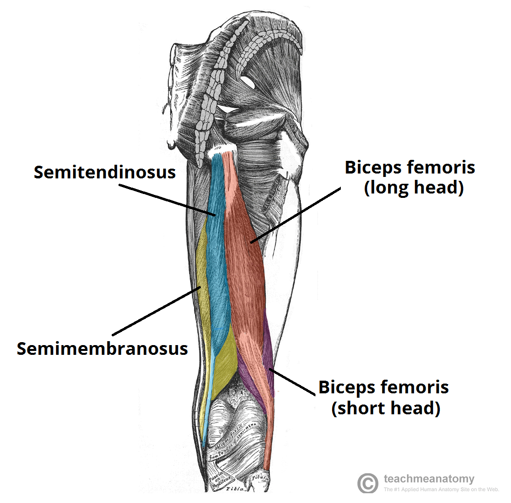
3. Posterior Compartment of the Thigh (The Hamstrings)
Innervation: Sciatic Nerve (Tibial portion), except for the short head of Biceps Femoris.
Main Actions: Primarily flexion of the knee (bending the knee backward) and extension of the hip.
- Biceps Femoris: The lateral (outer) hamstring muscle. As the name suggests, it has two heads (a long head from the ischial tuberosity and a short head from the linea aspera). It flexes the knee and laterally rotates the leg.
- Semitendinosus: A medial hamstring muscle. About half its length is a long, cord-like tendon. It flexes the knee and medially rotates the leg.
- Semimembranosus: A medial hamstring muscle lying directly deep to the Semitendinosus. It features a wide, flat, membranous tendon at its origin. It flexes the knee and medially rotates the leg.
Hamstring injuries are incredibly common in sprinters, hurdlers, and soccer players. These muscles cross two joints (hip and knee). During an explosive sprint, the hamstrings must eccentrically contract (brake) to stop the leg from violently snapping forward at the end of the swing phase. This massive, sudden tension often causes the muscle fibers to tear, usually near their origin at the Ischial Tuberosity, resulting in severe pain, bruising, and inability to extend the leg.
Summary Table of Thigh Muscles
| Muscle | Origin | Insertion | Innervation | Main Actions |
|---|---|---|---|---|
| ANTERIOR COMPARTMENT | ||||
| Rectus Femoris | AIIS | Patella & Tibial Tuberosity | Femoral N. | Extends knee, flexes hip |
| Vastus Lateralis | Greater trochanter, linea aspera | Patella & Tibial Tuberosity | Femoral N. | Extends knee |
| Vastus Medialis | Intertrochanteric line, linea aspera | Patella & Tibial Tuberosity | Femoral N. | Extends knee |
| Vastus Intermedius | Femoral shaft | Patella & Tibial Tuberosity | Femoral N. | Extends knee |
| Sartorius | ASIS | Medial tibia (Pes Anserinus) | Femoral N. | Flexes, abducts, lat. rotates thigh; flexes knee |
| MEDIAL COMPARTMENT | ||||
| Adductor Longus/Brevis/Magnus | Pubis, Ischial ramus | Femur (linea aspera) | Obturator N. (Magnus also Sciatic N.) | Adduct thigh; Magnus also extends thigh |
| Gracilis | Pubic symphysis | Medial tibia (Pes Anserinus) | Obturator N. | Adducts thigh, flexes knee |
| POSTERIOR COMPARTMENT (HAMSTRINGS) | ||||
| Biceps Femoris | Long: Ischial tuberosity; Short: Linea aspera | Head of fibula | Sciatic N. (Tibial & Common Fibular) | Flexes knee, extends hip, lat. rotates leg |
| Semitendinosus | Ischial tuberosity | Medial tibia (Pes Anserinus) | Sciatic N. (Tibial) | Flexes knee, extends hip, med. rotates leg |
| Semimembranosus | Ischial tuberosity | Medial condyle of tibia | Sciatic N. (Tibial) | Flexes knee, extends hip, med. rotates leg |
Part 5: Muscles of the Leg
The portion of the lower limb between the knee and the ankle is anatomically called the "leg" (crus). The muscles here are divided into four tightly packed compartments bounded by the tough interosseous membrane and unyielding fascial septa.
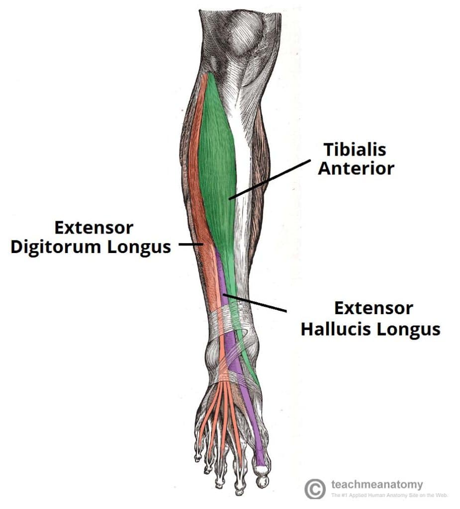
1. Anterior Compartment of the Leg
Innervation: Deep Fibular (Peroneal) Nerve (L4, L5, S1).
Main Actions: Primarily dorsiflexion of the ankle (pulling the toes up toward the shin) and extension of the toes.
- Tibialis Anterior: The thick muscle next to the shin bone. The main dorsiflexor and invertor of the foot. It prevents your toes from dragging on the ground when you swing your leg forward to walk.
- Extensor Digitorum Longus: Extends the lateral four toes.
- Extensor Hallucis Longus: Extends the great toe (hallux).
- Fibularis (Peroneus) Tertius: A small muscle that dorsiflexes and everts the foot.
If a patient fractures the head of their fibula, the Deep Fibular Nerve wrapping around the bone can be crushed. This paralyzes the entire anterior compartment. The patient completely loses the ability to dorsiflex the ankle. As they walk, their foot flops down limply (Foot Drop), and their toes drag on the ground. To compensate, they must lift their knee abnormally high on every step, producing a "Steppage Gait."
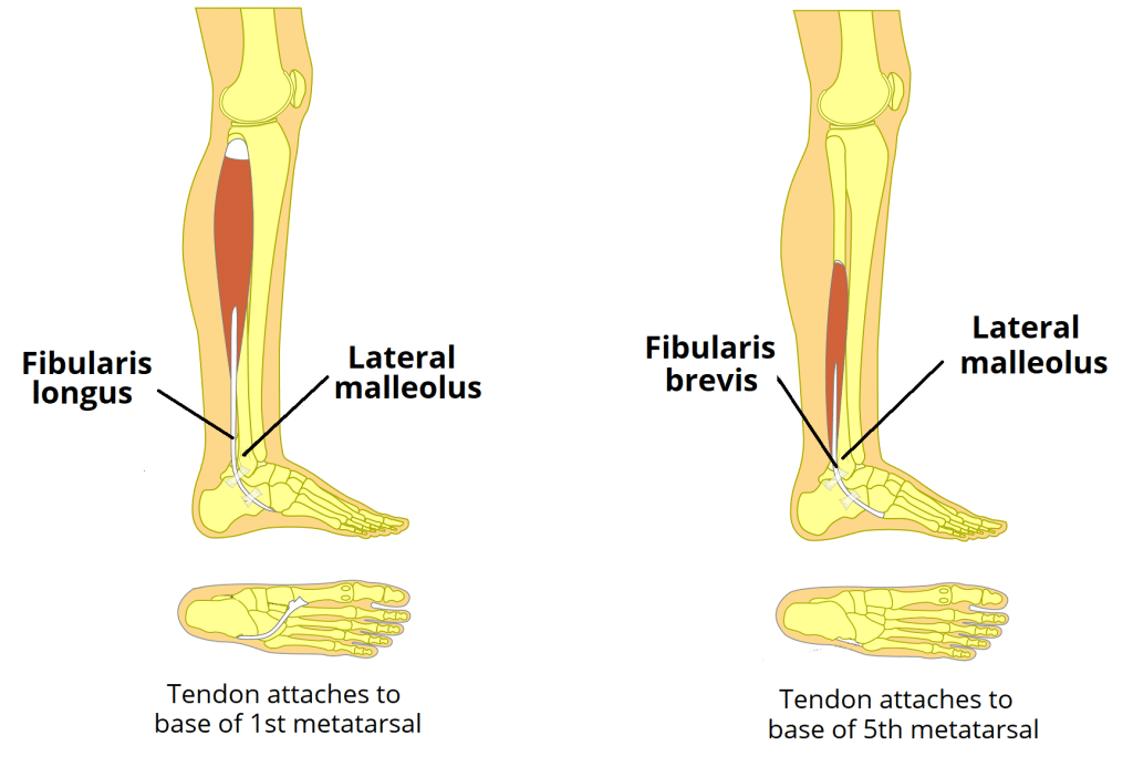
2. Lateral Compartment of the Leg
Innervation: Superficial Fibular (Peroneal) Nerve (L5, S1, S2).
Main Actions: Primarily eversion of the foot (turning the sole outward) and weak plantarflexion.
This compartment contains two muscles: Fibularis (Peroneus) Longus and Fibularis (Peroneus) Brevis. Functional Example: When you accidentally step on a rock and your ankle starts to roll inward (inversion sprain), these muscles fire aggressively, everting the foot to try and save your ankle ligaments from tearing.
3. Posterior Compartment of the Leg
Innervation: Tibial Nerve (L4-S2).
Main Actions: Primarily plantarflexion of the ankle (pointing toes down, standing on tiptoes), inversion of the foot, and flexion of the toes.
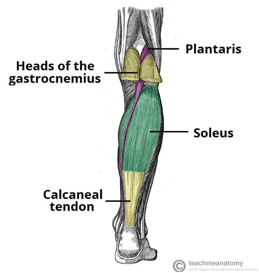
Superficial Group (The Calf Muscles)
This group forms the massive bulk of the calf. They all merge to insert onto the heel bone via the massive Calcaneal (Achilles) tendon.
- Gastrocnemius: The prominent, two-headed muscle that gives the calf its shape. Because it crosses the knee joint, it can both flex the knee and plantarflex the ankle. Highly active during explosive jumping and sprinting.
- Soleus: A massive, flat, powerful muscle lying deep to the gastrocnemius. It is a pure plantarflexor. Functional Example: It is packed with slow-twitch endurance fibers because it fires continuously all day long to keep you from falling forward when standing still.
- Plantaris: A tiny muscle belly with an incredibly long, thin tendon. Often mistaken for a nerve by medical students.
(Together, the Gastrocnemius and Soleus are known as the Triceps Surae).
Deep Group
These muscles lie buried beneath the soleus, plastered against the back of the tibia and fibula.
- Popliteus: A small diagonal muscle behind the knee. Its crucial job is to "unlock" the fully extended knee by laterally rotating the femur slightly, allowing flexion to begin.
- Flexor Digitorum Longus: Flexes the lateral four toes.
- Flexor Hallucis Longus: Flexes the great toe (provides the powerful "push-off" force at the end of a walking stride).
- Tibialis Posterior: The deepest muscle. It is the main invertor of the foot and provides massive dynamic support to the medial arch of the foot.
The tendons, artery, and nerve of the deep posterior compartment pass snugly behind the medial malleolus (inner ankle bone) on their way to the foot. You can remember their order from anterior to posterior using the classic phrase:
"Tom, Dick, And Very Nervous Harry"
Tibialis posterior, flexor Digitorum longus, posterior tibial Artery, Vein, tibial Nerve, flexor Hallucis longus.
Summary Table of Leg Muscles
| Muscle | Origin | Insertion | Innervation | Main Actions |
|---|---|---|---|---|
| ANTERIOR COMPARTMENT | ||||
| Tibialis Anterior | Lateral tibia | Medial cuneiform, 1st metatarsal | Deep Fibular N. | Main dorsiflexor; inverts foot |
| Extensor Digitorum Longus | Tibia, fibula | Distal phalanges of digits 2-5 | Deep Fibular N. | Extends lateral four toes |
| Extensor Hallucis Longus | Fibula | Distal phalanx of great toe | Deep Fibular N. | Extends great toe |
| LATERAL COMPARTMENT | ||||
| Fibularis (Peroneus) Longus | Head of fibula | 1st metatarsal, medial cuneiform | Superficial Fibular N. | Everts foot; plantarflexes ankle |
| Fibularis (Peroneus) Brevis | Lateral fibula | Base of 5th metatarsal | Superficial Fibular N. | Everts foot; plantarflexes ankle |
| POSTERIOR COMPARTMENT (SUPERFICIAL) | ||||
| Gastrocnemius | Femoral condyles | Calcaneus via Achilles tendon | Tibial N. | Plantarflexes ankle, flexes knee |
| Soleus | Tibia, fibula | Calcaneus via Achilles tendon | Tibial N. | Powerful plantarflexor |
| POSTERIOR COMPARTMENT (DEEP) | ||||
| Tibialis Posterior | Tibia, fibula | Navicular, cuneiforms, etc. | Tibial N. | Main invertor of foot |
| Flexor Digitorum Longus | Posterior tibia | Distal phalanges of digits 2-5 | Tibial N. | Flexes lateral four toes |
| Flexor Hallucis Longus | Posterior fibula | Distal phalanx of great toe | Tibial N. | Flexes great toe |
Part 6: Muscles of the Foot
The intrinsic muscles of the foot (meaning both origin and insertion are within the foot) are divided into dorsal (top) and plantar (sole) groups. They are responsible for fine motor control, spreading the toes, and providing dynamic, spring-like support to the arches of the foot when walking barefoot on uneven surfaces like sand.
1. Dorsal Muscles of the Foot
- Extensor Digitorum Brevis: Originates from the calcaneus (heel bone) and helps extend toes 2-4.
- Extensor Hallucis Brevis: Originates from the calcaneus and helps extend the great toe.
2. Plantar Muscles of the Foot (The Sole)
These muscles are incredibly complex and are organized into four distinct layers from superficial (closest to the skin) to deep (closest to the bones). They are covered by the thick, leathery Plantar Aponeurosis (fascia). They are primarily innervated by the Medial and Lateral Plantar Nerves (branches of the Tibial Nerve).
 Layer 1 (Superficial)
The first layer you see after stripping away the thick plantar fascia.
- Abductor Hallucis: Abducts and flexes the great toe. (Innervated by Medial Plantar N.)
- Flexor Digitorum Brevis: Flexes the lateral four toes at the PIP joints. (Innervated by Medial Plantar N.)
- Abductor Digiti Minimi: Abducts and flexes the little toe. (Innervated by Lateral Plantar N.)
 Layer 2
Contains two muscle groups directly associated with the long tendons coming from the leg.
- Quadratus Plantae: A square muscle that grabs onto the tendon of the Flexor Digitorum Longus (FDL). Because the FDL tendon comes in at a diagonal angle from the ankle, the Quadratus Plantae pulls on it to straighten its line of pull, ensuring the toes flex straight down. (Innervated by Lateral Plantar N.)
- Lumbricals (4): Worm-like muscles originating off the FDL tendons. They flex the MTP joints and extend the IP joints of the lateral four toes. (Innervated by Medial and Lateral Plantar Nerves).
 Layer 3
Focuses primarily on the great and little toes.
- Flexor Hallucis Brevis: Flexes the great toe at the MTP joint. Its split tendon houses the two tiny sesamoid bones of the foot. (Innervated by Medial Plantar N.)
- Adductor Hallucis: Has a large oblique head and a transverse head. It firmly adducts the great toe, helping maintain the transverse arch of the foot. (Innervated by Lateral Plantar N.)
- Flexor Digiti Minimi Brevis: Flexes the little toe. (Innervated by Lateral Plantar N.)
 Layer 4 (Deepest)
Buried deeply between the metatarsal bones.
- Plantar Interossei (3): Adduct toes 3-5.
Mnemonic: PAD (Plantar Adduct). (Innervated by Lateral Plantar N.) - Dorsal Interossei (4): Abduct toes 2-4.
Mnemonic: DAB (Dorsal Abduct). (Innervated by Lateral Plantar N.)
Although their fleshy muscle bellies are located way up in the leg, the massive tendons of the Tibialis Anterior and Fibularis Longus cross the ankle joint and insert deep into the bones of the foot. The Fibularis Longus tendon wraps completely under the sole of the foot to meet the Tibialis Anterior, creating a structural "stirrup" or "sling." Together, they provide immense dynamic support holding up the arches of your foot and preventing flat feet (pes planus).
Summary Table of Foot Muscles
| Layer/Group | Muscle | Origin | Insertion | Innervation | Main Actions |
|---|---|---|---|---|---|
| DORSAL MUSCLES | |||||
| Dorsal | Extensor Digitorum Brevis | Calcaneus | Extensor expansions 2-4 | Deep Fibular N. | Extends toes 2-4 |
| Dorsal | Extensor Hallucis Brevis | Calcaneus | Prox. phalanx of great toe | Deep Fibular N. | Extends great toe |
| PLANTAR MUSCLES | |||||
| Layer 1 | Abductor Hallucis | Calcaneus | Prox. phalanx of great toe | Medial Plantar N. | Abducts & flexes great toe |
| Layer 1 | Flexor Digitorum Brevis | Calcaneus | Middle phalanges 2-5 | Medial Plantar N. | Flexes toes 2-5 (PIP) |
| Layer 1 | Abductor Digiti Minimi | Calcaneus | Prox. phalanx of 5th toe | Lateral Plantar N. | Abducts & flexes 5th toe |
| Layer 2 | Quadratus Plantae | Calcaneus | Tendon of FDL | Lateral Plantar N. | Assists FDL in flexing |
| Layer 2 | Lumbricals (4) | Tendons of FDL | Extensor expansions 2-5 | Medial & Lateral Plantar N. | Flex MTPs, Extend IPs |
| Layer 3 | Flexor Hallucis Brevis | Cuboid, cuneiforms | Prox. phalanx of great toe | Medial Plantar N. | Flexes great toe |
| Layer 3 | Adductor Hallucis | Metatarsals 2-4 | Prox. phalanx of great toe | Lateral Plantar N. | Adducts great toe |
| Layer 3 | Flexor Digiti Minimi Brevis | Base of 5th metatarsal | Prox. phalanx of 5th toe | Lateral Plantar N. | Flexes little toe |
| Layer 4 | Plantar Interossei (3) | Metatarsals 3-5 | Prox. phalanges 3-5 | Lateral Plantar N. | Adduct toes (PAD) |
| Layer 4 | Dorsal Interossei (4) | Adjacent metatarsals | Prox. phalanges 2-4 | Lateral Plantar N. | Abduct toes (DAB) |
List of References
The anatomical details, functional biomechanics, and clinical correlations in this guide are synthesized based on universally accepted medical anatomy curricula and reference standards, including:
- Moore, K. L., Dalley, A. F., & Agur, A. M. R. Clinically Oriented Anatomy. 8th Edition. Lippincott Williams & Wilkins. (Core standard for clinical and regional anatomy).
- Standring, S. Gray's Anatomy: The Anatomical Basis of Clinical Practice. 42nd Edition. Elsevier. (Definitive reference for gross anatomical structures).
- Netter, F. H. Atlas of Human Anatomy. 7th Edition. Elsevier. (For visual correlations of muscle layers, bony landmarks, and fascial compartments).
- Snell, R. S. Clinical Anatomy by Regions. 9th Edition. Lippincott Williams & Wilkins.
- Drake, R., Vogl, A. W., & Mitchell, A. W. M. Gray's Anatomy for Students. 4th Edition. Elsevier.
Muscles of the Lower Limb Read More »
Muscles of the Upper Limb
Muscles of the Upper Limbs and The Brachial Plexus
The upper limb is a area designed for both power (like lifting heavy weights) and precision (like performing microsurgery or playing the violin). By the end of this guide, you will master:
- The complex "wiring" of the upper limb via the Brachial Plexus.
- The origins, insertions, and innervations of the Chest, Shoulder, Arm, Forearm, and Hand muscles.
- Real-world biomechanical examples of how these muscles function in daily life.
- The devastating clinical consequences (Pathology and Palsies) when these nerves or muscles are injured.
Part 1: The Brachial Plexus
Before we can study the muscles, we must understand how the brain communicates with them. The brachial plexus is a highly complex, interwoven network of nerves formed by the anterior rami (branches) of the lower four cervical nerves (C5, C6, C7, C8) and the first thoracic nerve (T1). It is exclusively responsible for the motor and sensory innervation of the entire upper limb.
To remember the anatomical progression of the plexus from the spine down to the arm, use the famous mnemonic:
"Real Texans Drink Cold Beer"
(Roots → Trunks → Divisions → Cords → Branches)
1. Roots (C5, C6, C7, C8, T1)
The five roots are the anterior primary rami of the spinal nerves. They emerge from the spinal cord and pass through a tight space in the neck between the anterior and middle scalene muscles. (Clinical tip: Tight neck muscles can compress these roots, causing numbness down the arm—a condition called Thoracic Outlet Syndrome).
- Key Branches from Roots:
- Dorsal Scapular Nerve (C5): Innervates the Rhomboids and Levator Scapulae (pulls the shoulder blades together).
- Long Thoracic Nerve (C5, C6, C7): (Mnemonic: "C5, 6, 7 keep your wings from heaven"). Innervates the Serratus Anterior.
2. Trunks (Superior, Middle, Inferior)
As the roots travel laterally over the first rib, they merge to form three distinct trunks:
- Upper (Superior) Trunk: Formed by the union of C5 and C6 roots.
- Middle Trunk: A direct, straight continuation of the C7 root.
- Lower (Inferior) Trunk: Formed by the union of C8 and T1 roots.
- Key Branches from Trunks:
- Suprascapular Nerve (C5, C6): Originates strictly from the Upper Trunk. It dives deep to innervate the Supraspinatus and Infraspinatus (key rotator cuff muscles).
- Nerve to Subclavius (C5, C6): Innervates the subclavius muscle.
3. Divisions (Anterior and Posterior)
As the trunks pass under the clavicle (collarbone), each of the three trunks splits into an Anterior and a Posterior division. This is a crucial evolutionary separation!
- The 3 Posterior Divisions: Combine to supply all the future extensors of the upper limb (triceps, wrist extensors).
- The 3 Anterior Divisions: Combine to supply all the future flexors of the upper limb (biceps, wrist flexors).
4. Cords (Lateral, Posterior, Medial)
The six divisions regroup in the axilla (armpit) to form three cords. They are named strictly for their physical position relative to the massive Axillary Artery.
- Lateral Cord (C5-C7): Formed by the anterior divisions of the upper and middle trunks. Gives off the Lateral Pectoral Nerve.
- Posterior Cord (C5-T1): Formed by the posterior divisions of all three trunks. Gives off the Upper & Lower Subscapular Nerves and the Thoracodorsal Nerve (which powers the mighty Latissimus Dorsi).
- Medial Cord (C8-T1): Formed by the anterior division of the lower trunk. Gives off the Medial Pectoral Nerve and Medial Cutaneous Nerves of the arm and forearm.
5. Branches (The 5 Major Terminal Nerves)
The cords finally split into the five major nerves that travel down the arm.
(Mnemonic: MARMU)
The "Popeye" Nerve.
- Motor: Powers the entire anterior arm compartment (Biceps Brachii, Brachialis, Coracobrachialis). Responsible for elbow flexion.
- Sensory: Pierces the muscle to become the lateral cutaneous nerve, supplying the skin of the lateral forearm.
The "Shoulder Pad" Nerve.
- Motor: Wraps around the surgical neck of the humerus to power the Deltoid and Teres Minor.
- Sensory: Skin over the lower deltoid (the "regimental badge area").
The "Great Extensor" Nerve.
- Motor: Powers EVERY single muscle in the posterior compartments of the arm and forearm (Triceps and all wrist/finger extensors).
- Sensory: Posterior skin of arm/forearm, and the dorsal aspect of the lateral 2.5 digits (the anatomical snuffbox area).
The "Laborer's Nerve".
- Motor: Powers most anterior forearm muscles (flexors/pronators), and the critical thenar (thumb) muscles for grasping.
- Sensory: Skin of the lateral palm and palmar aspect of the lateral 3.5 digits.
The "Musician's Fine-Tuning" Nerve.
- Motor: Powers only 1.5 muscles in the forearm (Flexor Carpi Ulnaris, medial half of FDP), but powers almost ALL the intrinsic fine-motor muscles of the hand (interossei/lumbricals).
- Sensory: Skin of the medial 1.5 digits (the pinky and half the ring finger).
Brachial Plexus Summary Table
| Level | Components | Key Nerve Branches | Clinical Notes |
|---|---|---|---|
| ROOTS | Anterior Rami of C5, C6, C7, C8, T1 | Dorsal Scapular N (C5): Rhomboids, Levator Scapulae Long Thoracic N (C5-C7): Serratus Anterior |
Emerge between Scalenes. Injury to Long Thoracic N. → Winged Scapula. |
| TRUNKS | Upper: C5 + C6 Middle: C7 Lower: C8 + T1 |
Suprascapular N (C5, C6): Supraspinatus, Infraspinatus N. to Subclavius (C5, C6): Subclavius |
Pass over 1st rib. Erb-Duchenne palsy is an upper trunk injury. |
| DIVISIONS | Each trunk divides into an Anterior & Posterior Division | No direct named branches. | Posterior divisions strictly supply extensors; Anterior supply flexors. |
| CORDS | Lateral: Ant. divisions of Upper & Middle Posterior: Post. divisions of all 3 Medial: Ant. division of Lower |
Lateral Pectoral N. Upper & Lower Subscapular N., Thoracodorsal N. Medial Pectoral N., Medial Cutaneous Nerves |
Named for physical position wrapping around the axillary artery. |
| BRANCHES | Terminal Nerves | Musculocutaneous N. Axillary N. Radial N. Median N. Ulnar N. |
Major nerves of the upper limb. Injuries lead to highly distinct motor & sensory deficits. |
Brachial Plexus Injuries and Clinical Correlates
Nerve injuries are devastating and present with highly specific, testable postures:
- Upper Plexus Injury (Erb-Duchenne Palsy): Affects C5-C6 roots. Usually caused by traumatic tearing, such as shoulder dystocia during a difficult childbirth or a motorcycle accident where the head and shoulder are violently pushed apart.
Result: The classic "Waiter's Tip" position. The arm hangs limply adducted (deltoid paralyzed), medially rotated (external rotators paralyzed), and the elbow is extended with the wrist flexed. - Lower Plexus Injury (Klumpke's Palsy): Affects C8-T1 roots. Caused by excessive, violent abduction of the arm (e.g., grabbing a tree branch to stop a fall from a height, or pulling a baby out by its arm).
Result: Affects intrinsic hand muscles, leading to a total "Claw Hand" of all digits. - Radial Nerve Injury (Wrist Drop): Commonly caused by mid-shaft humeral fractures (the nerve spirals tightly around the bone) or compression in the axilla ("Saturday night palsy" from passing out with an arm over a chair, or improperly fitted crutches).
Result: Absolute inability to extend the wrist and fingers. The hand hangs completely limp (Wrist Drop). - Median Nerve Injury (Carpal Tunnel Syndrome): Compression of the median nerve as it passes under the flexor retinaculum at the wrist (often due to repetitive typing or vibration).
Result: Causes numbness and "pins and needles" in the lateral 3.5 digits, and severe weakness/wasting of the fleshy base of the thumb (thenar atrophy), producing an "Ape Hand" deformity. - Ulnar Nerve Injury ("Claw Hand"): Injury usually occurs where it wraps around the medial epicondyle of the elbow (hitting your "funny bone") or at the wrist.
Result: Loss of intrinsic hand muscles leads to specific "clawing" (hyperextension of MCP and flexion of IP joints) of the 4th and 5th digits, and severe sensory loss over the medial pinky side of the hand.
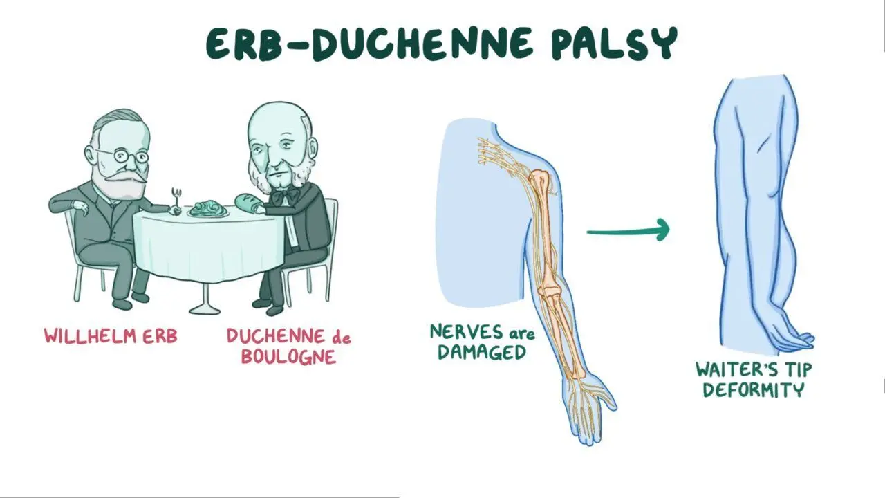
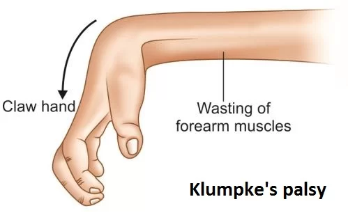
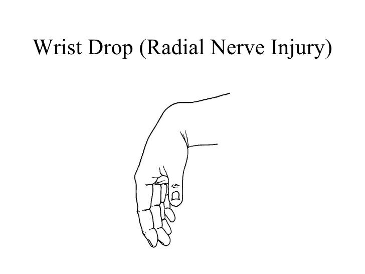
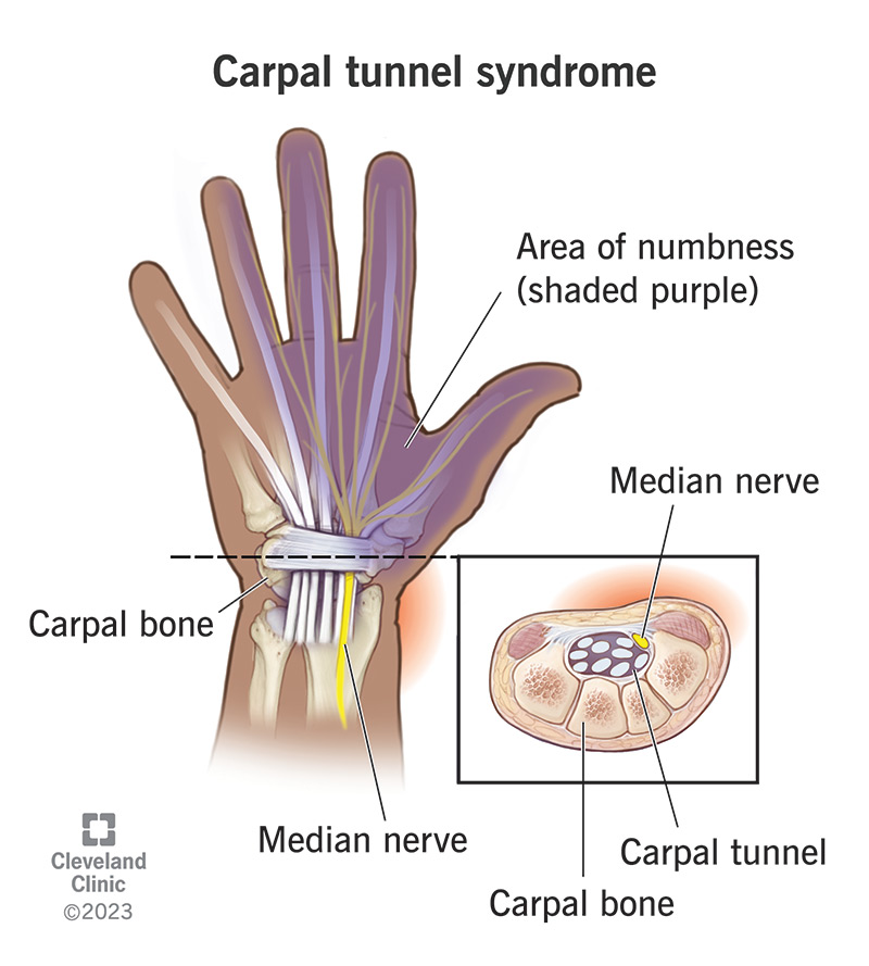
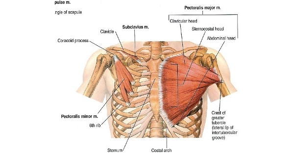
Part 2: Muscles of the Chest (Pectoral Region)
The muscles of the pectoral region serve a dual purpose: the superficial layers anchor the heavy upper limb to the axial skeleton of the thorax, while the deeper layers are the biological bellows responsible for the mechanics of breathing.
1. Superficial Muscles of the Pectoral Region
- a. Pectoralis Major: A massive, fan-shaped muscle covering the upper chest. It has two parts: a clavicular head and a larger sternocostal head.
Biomechanical Example: It is the ultimate hugging muscle. It is a powerful adductor and medial rotator of the arm. The clavicular head flexes the arm (e.g., throwing a bowling ball underhand), while the sternocostal head extends it from a flexed position (e.g., the downward pull in a butterfly swimming stroke or performing a push-up). - b. Pectoralis Minor: A thin, triangular muscle lying completely hidden deep to the Pectoralis Major.
Biomechanical Example: It depresses the shoulder and protracts the scapula (pulls it forward and downward). Think of the motion of reaching down to pick up a heavy suitcase. - c. Subclavius: A tiny, cylindrical muscle located directly inferior to the clavicle.
Function: It actively anchors and depresses the clavicle. More importantly, it acts as a soft biological cushion to protect the underlying subclavian vessels and brachial plexus if the collarbone fractures. - d. Serratus Anterior: Visually distinct, saw-toothed slips of muscle on the lateral thoracic wall.
Biomechanical Example: Known as the "Boxer's Muscle". It is the prime mover for aggressively protracting the scapula (the motion of throwing a punch or pushing a heavy stalled car). It is also essential for upwardly rotating the scapula, which is required to lift your arm entirely above your head. Paralysis (Long Thoracic N. injury) leads to the pathognomonic "winged scapula".
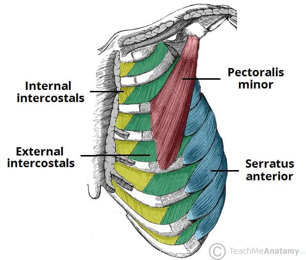
2. Deep Muscles of the Thorax (Associated with Respiration)
- a. Intercostal Muscles (External, Internal, Innermost): Three distinct layers of muscles woven tightly in the intercostal spaces between the ribs.
Function: The External Intercostals lift and elevate the ribs out and up, expanding chest volume for forced inspiration (taking a deep breath before diving into water). The Internal and Innermost Intercostals violently depress the ribs for forced expiration (blowing out all the candles on a birthday cake). - b. Transversus Thoracis: A thin, star-like muscle on the inner anterior thoracic wall (behind the sternum) that weakly depresses the ribs.
Summary Table of Chest Muscles
| Muscle | Origin | Insertion | Innervation | Main Actions |
|---|---|---|---|---|
| SUPERFICIAL PECTORAL MUSCLES | ||||
| Pectoralis Major | Clavicle, Sternum, Costal Cartilages 1-6 | Intertubercular groove of humerus (lateral lip) | Lat & Med Pectoral N. | Adducts & medially rotates arm; flexes & extends arm. |
| Pectoralis Minor | Ribs 3-5 | Coracoid process of scapula | Medial Pectoral N. | Depresses shoulder; protracts the scapula. |
| Subclavius | 1st rib | Inferior surface of clavicle | N. to Subclavius | Depresses & anchors the clavicle tightly. |
| Serratus Anterior | Ribs 1-9 | Medial border of scapula (anterior surface) | Long Thoracic N. | Protracts & upwardly rotates scapula (prevents winging). |
| DEEP THORACIC (RESPIRATORY) MUSCLES | ||||
| External Intercostals | Rib above | Rib below | Intercostal Nerves | Elevate ribs (forced, deep inspiration). |
| Internal Intercostals | Rib above | Rib below | Intercostal Nerves | Depress ribs (forced, active expiration). |
Part 3: Muscles of the Upper Limbs
The muscles of the upper limb enable a remarkable range of movements. The shoulder provides maximum 360-degree mobility, the elbow acts as a powerful hinge lever, the forearm provides rotational positioning, and the hand yields unparalleled dexterity. We divide them regionally.
1. Muscles of the Shoulder (Scapulohumeral Region)
These muscles act primarily on the incredibly shallow, highly mobile glenohumeral (shoulder) joint, providing both movement and vital dynamic stability.
- a. Deltoid: The massive, thick, triangular muscle forming the rounded, armored contour of the shoulder.
Biomechanical Example: Its three parts (anterior, middle, posterior) allow it to perform almost every action. The entire muscle is the absolute prime mover of arm abduction (lifting the arm out to the side like a bird flapping its wings) only after the first 15 degrees. The anterior fibers flex and medially rotate (reaching forward to shake a hand), while the posterior fibers extend and laterally rotate (pulling open a heavy vault door). - b. Rotator Cuff Muscles (SITS): A critical group of four muscles that tightly surround the shoulder joint like a biological cuff. Their tendons physically blend directly into the joint capsule, dynamically sucking the humerus into the shallow socket to prevent dislocation during movement. Remembered by the mnemonic SITS.
- Supraspinatus: Action: Initiates the very first 15 degrees of arm abduction (before the deltoid takes over). Clinical: It is the most commonly torn rotator cuff muscle because its tendon gets aggressively pinched (impingement syndrome) under the acromion bone during repetitive overhead lifting.
- Infraspinatus: Action: A powerful lateral rotator of the arm (e.g., the motion of winding up to pitch a baseball).
- Teres Minor: Action: Synergist to the infraspinatus; also laterally rotates the arm.
- Subscapularis: Action: The only rotator cuff muscle on the front of the scapula. It is a powerful medial rotator of the arm (e.g., the motion of tucking your shirt into the back of your pants).
- c. Teres Major: A thick, rounded muscle inferior to Teres Minor.
Biomechanical Example: Often colloquially called "Lat's Little Helper." It is NOT part of the rotator cuff. Its main actions are to powerfully extend, adduct, and medially rotate the arm. Think of the aggressive, downward pulling motion of chopping wood with an axe or performing a pull-up.
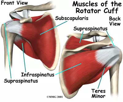
Summary Table of Shoulder Muscles
| Muscle | Origin | Insertion | Innervation | Main Actions |
|---|---|---|---|---|
| Deltoid | Clavicle, acromion, spine of scapula | Deltoid tuberosity of humerus | Axillary N. (C5, C6) | Abducts arm (>15°); flexes & medially rotates; extends & laterally rotates. |
| Supraspinatus | Supraspinous fossa of scapula | Greater tubercle of humerus (superior facet) | Suprascapular N. (C5, C6) | Initiates arm abduction (first 15°); stabilizes joint. |
| Infraspinatus | Infraspinous fossa of scapula | Greater tubercle of humerus (middle facet) | Suprascapular N. (C5, C6) | Laterally rotates arm; stabilizes joint. |
| Teres Minor | Lateral border of scapula | Greater tubercle of humerus (inferior facet) | Axillary N. (C5, C6) | Laterally rotates arm; stabilizes joint. |
| Subscapularis | Subscapular fossa (anterior scapula) | Lesser tubercle of humerus | Upper & Lower Subscapular N. | Medially rotates arm; provides massive anterior stability. |
| Teres Major | Inferior angle of scapula | Intertubercular groove of humerus (medial lip) | Lower Subscapular N. | Extends, adducts, medially rotates arm (woodchopper). |
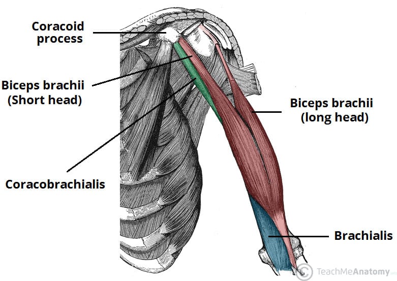
2. Muscles of the Arm (The Brachium)
The true "arm" (the segment between the shoulder and elbow) is cleanly divided into an anterior (flexor) and posterior (extensor) compartment by tough, fibrous intermuscular septa.
Anterior (Flexor) Compartment
Innervation: Musculocutaneous Nerve (C5, C6, C7)
Arterial Supply: Brachial artery
- Biceps Brachii: A prominent two-headed muscle (Long and Short head) that astonishingly doesn't attach to the humerus at all!
Biomechanical Example: Because it attaches to the radius, it is not just a flexor, but the most powerful supinator of the forearm. Think of aggressively driving a corkscrew into a wine bottle (supination) and then pulling the cork out (flexion). - Brachialis: Lies deep to the biceps.
Biomechanical Example: This is the absolute "workhorse" and primary flexor of the elbow. Because it attaches to the ulna (which doesn't rotate), it simply flexes the elbow powerfully regardless of hand position (e.g., doing heavy hammer curls or lifting a heavy mug of beer). - Coracobrachialis: The smallest of the three. It assists in weak flexion and adduction of the arm tightly to the body at the shoulder.
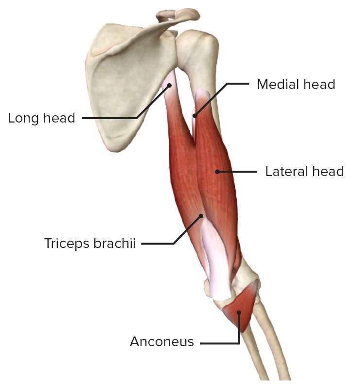
Posterior (Extensor) Compartment
Innervation: Radial Nerve (C6, C7, C8, T1)
Arterial Supply: Deep brachial artery (Profunda brachii)
- Triceps Brachii: The massive, sole muscle occupying the entire posterior arm, possessing three heads (long, lateral, medial).
Biomechanical Example: It is the powerful, absolute extensor of the forearm at the elbow. Think of the violent snapping motion of throwing a dart, hammering a nail, or doing a triceps push-down. The long head (crossing the shoulder joint) also assists in extending and pulling the arm backward. - Anconeus: A tiny, triangular muscle at the posterior elbow. It assists the triceps in locking out forearm extension and prevents the joint capsule from being pinched during movement.
Summary Table of Arm Muscles
| Muscle | Origin | Insertion | Innervation | Main Actions |
|---|---|---|---|---|
| ANTERIOR COMPARTMENT | ||||
| Biceps Brachii | Long head: Supraglenoid tubercle Short head: Coracoid process |
Radial tuberosity & bicipital aponeurosis | Musculocutaneous N. | Powerfully supinates forearm; flexes forearm. |
| Brachialis | Anterior, distal half of humerus | Coronoid process & tuberosity of ulna | Musculocutaneous N. | Primary, pure flexor of forearm (the workhorse). |
| Coracobrachialis | Coracoid process of scapula | Middle medial surface of humerus | Musculocutaneous N. | Flexes and adducts arm at the shoulder. |
| POSTERIOR COMPARTMENT | ||||
| Triceps Brachii | Long: Infraglenoid tubercle Lat/Med: Posterior surface of humerus |
Olecranon process of ulna | Radial N. | Powerful, primary extensor of forearm. |
| Anconeus | Lateral epicondyle of humerus | Lateral surface of olecranon & superior ulna | Radial N. | Assists triceps in extension; stabilizes elbow capsule. |
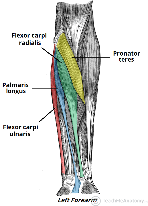
3. Muscles of the Forearm (The Antebrachium)
The numerous muscles of the forearm are complexly arranged in stacked layers. They are cleanly divided into an anterior (flexor/pronator) and posterior (extensor/supinator) compartment. Their long tendons pass through the wrist to control the hand like strings on a puppet.
Anterior (Flexor-Pronator) Compartment
Innervation: Mostly Median Nerve. Exception: Flexor Carpi Ulnaris & the medial half of FDP are powered by the Ulnar Nerve.
Main Actions: Flexion of the wrist and fingers; pronation (turning palm down) of the forearm.
- Superficial Layer (4 muscles originating from the Medial Epicondyle):
- Pronator Teres: Pronates (turns palm down) and weakly flexes the forearm.
- Flexor Carpi Radialis (FCR): Flexes and abducts the wrist.
- Palmaris Longus: Flexes the wrist. (Interesting fact: It is entirely absent in about 15% of the human population! When present, its long, useless tendon is frequently harvested by surgeons for tendon grafts).
- Flexor Carpi Ulnaris (FCU): Flexes and heavily adducts the wrist (ulnar deviation).
- Intermediate Layer (1 muscle):
- Flexor Digitorum Superficialis (FDS): Its tendons split in half. It specifically flexes the middle phalanges (PIP joints) of digits 2-5 (e.g., the specific curling finger motion used when playing a piano).
- Deep Layer (3 muscles):
- Flexor Digitorum Profundus (FDP): The deepest flexor. It passes entirely through the split tendons of the FDS to flex the absolute distal tips (DIP joints) of digits 2-5. Think of the aggressive, crushing fingertip grip needed by professional rock climbers.
- Flexor Pollicis Longus (FPL): Flexes the distal phalanx of the thumb (the primary muscle used when rapidly texting on a smartphone).
- Pronator Quadratus: A square muscle at the wrist. The primary, deep pronator of the forearm.
Posterior (Extensor-Supinator) Compartment
Innervation: Radial Nerve and its deep branch (Posterior Interosseous Nerve).
Main Actions: Extension of the wrist and fingers; supination (turning palm up) of the forearm.
- Superficial Layer: Includes the wrist extensors (ECRL, ECRB, ECU), finger extensors (Extensor Digitorum, Extensor Digiti Minimi), and the highly unique Brachioradialis.
Biomechanical Example: The Brachioradialis is the "drinking muscle". It sits in the extensor compartment and is innervated by the radial nerve, but it actually flexes the elbow when the arm is in a mid-pronated position (like bringing a glass of water to your mouth). - Deep Layer: Includes the Supinator muscle, and the three "outcropping" muscles of the thumb: Abductor Pollicis Longus (APL), Extensor Pollicis Brevis (EPB), and Extensor Pollicis Longus (EPL). These three tendons form the triangular depression on the back of the hand known as the Anatomical Snuffbox. Also includes the Extensor Indicis, which allows for independent extension of the index finger (pointing at something).
Summary Table of Forearm Muscles
| Muscle | Origin | Insertion | Innervation | Main Actions |
|---|---|---|---|---|
| ANTERIOR COMPARTMENT | ||||
| Pronator Teres | Medial epicondyle, coronoid process | Lateral surface of radius | Median N. | Pronates & flexes forearm. |
| Flexor Carpi Radialis | Medial epicondyle | Base of 2nd & 3rd metacarpals | Median N. | Flexes & abducts wrist (radial deviation). |
| Palmaris Longus | Medial epicondyle | Palmar aponeurosis | Median N. | Tenses palmar fascia; flexes wrist. |
| Flexor Carpi Ulnaris | Medial epicondyle, olecranon | Pisiform, hook of hamate, 5th metacarpal | Ulnar N. | Flexes & adducts wrist (ulnar deviation). |
| Flexor Digitorum Superficialis | Medial epicondyle, coronoid, anterior radius | Middle phalanges of digits 2-5 (splits) | Median N. | Flexes PIP joints and wrist. |
| Flexor Digitorum Profundus | Anterior ulna, interosseous membrane | Distal phalanges of digits 2-5 | Median N. (lat half), Ulnar N. (med half) | Flexes DIP joints (crushing grip). |
| Flexor Pollicis Longus | Anterior radius, interosseous membrane | Distal phalanx of thumb | Median N. (AIN) | Flexes distal thumb joint (texting). |
| Pronator Quadratus | Distal, anterior ulna | Distal, anterior radius | Median N. (AIN) | Primary pronator of forearm. |
| POSTERIOR COMPARTMENT | ||||
| Brachioradialis | Lateral supracondylar ridge of humerus | Styloid process of radius | Radial N. | Flexes forearm in mid-pronated position. |
| Extensor Carpi Radialis Longus | Lateral supracondylar ridge of humerus | Base of 2nd metacarpal | Radial N. | Extends & abducts wrist. |
| Extensor Carpi Ulnaris | Lateral epicondyle, posterior ulna | Base of 5th metacarpal | Radial N. (PIN) | Extends & adducts wrist. |
| Supinator | Lateral epicondyle, ulna crest | Proximal third of radius | Radial N. (Deep br.) | Primary supinator of forearm (driving a screw). |
4. Muscles of the Hand (The Intrinsic Controllers)
While the heavy lifting is done by the forearm muscles acting via long tendons, the intrinsic muscles of the hand (contained entirely within the hand itself) are exclusively responsible for fine motor control, delicate pinching, and exquisite dexterity.
a. Thenar Muscles (Ball of the Thumb)
This fleshy group acts strictly on the thumb (pollux). All are innervated by the Recurrent Branch of the Median Nerve, EXCEPT for the massive Adductor Pollicis.
- Abductor Pollicis Brevis (APB): Pulls the thumb away from the palm.
- Flexor Pollicis Brevis (FPB): Flexes the proximal joint of the thumb.
- Opponens Pollicis (OP): Swings the thumb across the palm to touch the other fingers (the evolutionary hallmark of human dexterity).
- Adductor Pollicis: The heavy muscle in the web space. It violently adducts the thumb (innervated by the Ulnar Nerve). Think of clamping a piece of paper tightly between your thumb and index finger.
b. Hypothenar Muscles (Ball of the Little Finger)
This group acts on the little finger (digiti minimi). All are innervated by the Deep Branch of the Ulnar Nerve.
- Abductor Digiti Minimi (ADM): Pulls the pinky away.
- Flexor Digiti Minimi Brevis (FDMB): Flexes the pinky.
- Opponens Digiti Minimi (ODM): Opposes the little finger (rotates it to meet the thumb, forming a "cup" in your palm to hold water).
c. Deep Intrinsic Muscles
- Lumbricals (4 muscles): Tiny, worm-shaped muscles that magically originate entirely from the moving tendons of the FDP, rather than bone!
Action: They flex the MCP joints and extend the IP joints. (This creates the "bye-bye" hand wave or the "tabletop" position). Lateral two = Median N., Medial two = Ulnar N. - Interossei (7 muscles): Muscles tightly packed between the metacarpal bones. All innervated by the Ulnar N.
Mnemonic Action: The 4 Dorsal Interossei Abduct the fingers (DAB - spreading fingers wide). The 3 Palmar Interossei Adduct the fingers (PAD - squeezing fingers together).
Summary Table of Hand Muscles
| Group | Muscle | Origin | Insertion | Innervation | Action |
|---|---|---|---|---|---|
| Thenar | Abductor Pollicis Brevis | Flexor retinaculum, scaphoid, trapezium | Proximal phalanx of thumb | Median N. (Recurrent br.) | Abducts thumb |
| Flexor Pollicis Brevis | Flexor retinaculum, trapezium | Proximal phalanx of thumb | Median N. (Recurrent br.) | Flexes thumb | |
| Opponens Pollicis | Flexor retinaculum, trapezium | 1st metacarpal (radial side) | Median N. (Recurrent br.) | Opposes thumb | |
| Adductor Pollicis | Capitate, 2nd & 3rd metacarpals | Proximal phalanx of thumb | Ulnar N. (Deep br.) | Adducts thumb (pinching paper) | |
| Hypothenar | Abductor Digiti Minimi | Pisiform bone | Proximal phalanx of digit 5 | Ulnar N. (Deep br.) | Abducts little finger |
| Flexor Digiti Minimi Brevis | Hook of hamate, flexor retinaculum | Proximal phalanx of digit 5 | Ulnar N. (Deep br.) | Flexes little finger | |
| Opponens Digiti Minimi | Hook of hamate, flexor retinaculum | 5th metacarpal (ulnar border) | Ulnar N. (Deep br.) | Opposes little finger (cupping palm) | |
| Deep Intrinsic | Lumbricals (4) | Tendons of Flexor Digitorum Profundus | Extensor expansions of digits 2-5 | Lat 2: Median N. Med 2: Ulnar N. |
Flex MCP joints, Extend IP joints (tabletop) |
| Dorsal Interossei (4) | Adjacent sides of two metacarpals (bipennate) | Proximal phalanges & extensor expansions | Ulnar N. (Deep br.) | Abduct fingers (DAB) | |
| Palmar Interossei (3) | Palmar surfaces of 2nd, 4th, 5th metacarpals | Proximal phalanges & extensor expansions | Ulnar N. (Deep br.) | Adduct fingers (PAD) |
References
The anatomical and biomechanical descriptions provided in this guide have been synthesized in accordance with standard international anatomical literature and surgical texts, including:
- Moore, K. L., Dalley, A. F., & Agur, A. M. R. (2018). Clinically Oriented Anatomy (8th ed.). Wolters Kluwer.
- Standring, J. (Ed.). (2015). Gray's Anatomy: The Anatomical Basis of Clinical Practice (41st ed.). Elsevier.
- Netter, F. H. (2018). Atlas of Human Anatomy (7th ed.). Elsevier.
- Drake, R., Vogl, A. W., & Mitchell, A. W. M. (2019). Gray's Anatomy for Students (4th ed.). Elsevier.
- Snell, R. S. (2011). Clinical Anatomy by Regions (9th ed.). Lippincott Williams & Wilkins.
Muscles of the Upper Limb Read More »
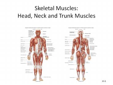
Muscles of the Head, Neck and Trunk
Muscles of the Axial Skeleton & The Cranial Nerves
The axial skeleton forms the central axis of the human body, consisting of the skull, vertebral column, and thoracic cage. The muscles attached to this axis are fundamental for life—they allow us to express emotion, eat, breathe, maintain our posture, and protect our vital organs. By the end of this exhaustive module, you will be able to:
- Identify and categorize the muscles of the head, face, neck, and torso based on their anatomical location.
- Understand the precise origin, insertion, and innervation of each major axial muscle.
- Relate muscular actions to activities of daily living (ADLs) and recognize clinical pathologies associated with muscular dysfunction.
- Master the 12 Cranial Nerves, including their functional types, assessment methods, and clinical relevance.
A. Muscles of the Head and Face
The muscles of the head are unique compared to the rest of the body. They can be broadly categorized into two highly specialized functional groups: the muscles of facial expression and the muscles of mastication (chewing). Understanding these groups is vital for neurological assessments and diagnosing conditions like strokes or nerve palsies.
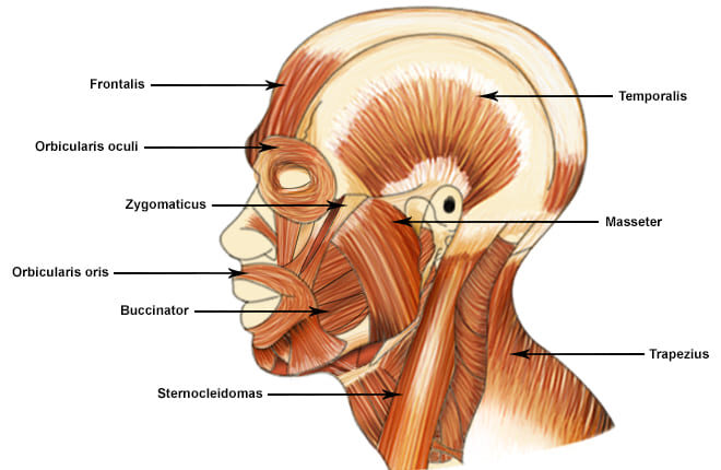
1. Muscles of Facial Expression
Unlike most skeletal muscles that connect bone to bone, these unique muscles frequently originate from the skull and insert directly into the skin or other muscles. This anatomical quirk allows them to tug on the skin, enabling us to show a vast, nuanced range of non-verbal emotions. They are all innervated by the Facial Nerve (Cranial Nerve VII).
A broad, continuous musculofibrous layer covering the top of the skull. It consists of two bellies connected by a broad tendinous sheet called the galea aponeurotica.
- Action: The Frontal belly raises the eyebrows and horizontally wrinkles the forehead skin (the classic expression of surprise or fright). The Occipital belly anchors and pulls the scalp posteriorly.
- Clinical/Daily Example: Raising your eyebrows when you hear shocking news. Tension in the galea aponeurotica is a common cause of tension headaches.
A complex, sphincter-like ring muscle encircling the entire orbit (eye socket).
- Action: Its primary action is to close the eye. The inner (palpebral) part closes it gently (blinking, sleeping), while the outer (orbital) part closes it forcefully (squinting, winking).
- Clinical/Daily Example: Squeezing your eyes shut when walking into a dust storm or facing bright sunlight. In Bell's Palsy (CN VII damage), the patient cannot close the eye, leading to severe dry eye (exposure keratitis).
The complex, multi-layered sphincter muscle encircling the mouth. It is the "kissing muscle."
- Action: It closes, compresses, and protrudes the lips. It shapes the lips during speech.
- Clinical/Daily Example: Puckering up for a kiss, playing a brass instrument like a trumpet, or drinking from a straw. Paralysis here causes drooling and slurred speech (dysarthria).
These diagonal muscles extend from the zygomatic bone (cheekbone) down to the corner of the mouth.
- Action: They are the primary "smiling" muscles, actively raising the lateral corners of the mouth upward and outward.
- Clinical/Daily Example: A genuine, joyful smile (often called a Duchenne smile) strongly engages the Zygomaticus Major alongside the Orbicularis Oculi.
A thin, flat, deep muscle forming the muscular wall of the cheek. Its name derives from the Latin word for "trumpeter."
- Action: It forcefully compresses the cheek against the teeth. It is vital for whistling, sucking, and holding food safely between the teeth during chewing so you don't bite your own cheek.
- Clinical/Daily Example: Infants rely heavily on the buccinator to suckle milk. Glassblowers and musicians use it to expel air forcefully.
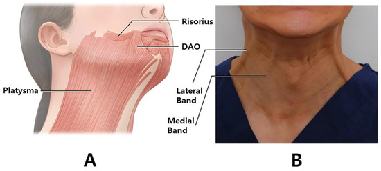 f. Platysma
A broad, superficial, paper-thin sheet of muscle in the anterior neck, originating from the chest fascia and inserting into the mandible.
- Action: It tenses the skin of the neck, depresses the mandible (opens the jaw slightly), and pulls the lower lip down and back.
- Clinical/Daily Example: The expression of sudden horror or extreme fright. It is also the muscle men instinctively tense when shaving the front of their neck to create a flat surface.
2. Muscles of Mastication (Chewing)
These four powerful pairs of muscles are responsible for moving the mandible (lower jaw) at the Temporomandibular Joint (TMJ) for chewing, grinding, and speaking. Unlike facial expression muscles, they are all heavily innervated by the Mandibular division of the Trigeminal Nerve (Cranial Nerve V3).
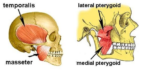
A thick, extremely powerful, quadrilateral muscle covering the lateral side of the jaw.
- Action: It is the prime elevator of the mandible (forcefully closes the jaw).
- Clinical/Daily Example: Biting down hard on a tough piece of steak. Bruxism (nighttime teeth grinding) causes the masseter to become severely hypertrophied (enlarged) and painful.
A large, fan-shaped muscle filling the temporal fossa on the side of the skull, passing deep to the zygomatic arch.
- Action: Elevates the mandible (closes jaw) and its posterior fibers actively retract the mandible (pull it backward).
- Clinical/Daily Example: If you place your fingers on your temples and clench your teeth, you will feel this muscle bulge. Tension here is a massive contributor to stress headaches.
Located deep inside the jaw, mirroring the masseter on the internal surface of the mandible.
- Action: Elevates the jaw synergistically with the masseter and assists in side-to-side (excursive) grinding movements.
- Clinical/Daily Example: The circular, side-to-side grinding motion used when chewing tough plant fibers or gum.
A short, thick, two-headed muscle located deep and horizontally in the jaw. It is the functional oddball of the group.
- Action: It protracts the mandible (pulls it forward), moves it side-to-side, and is the ONLY muscle of mastication that helps OPEN (depress) the jaw.
- Clinical/Daily Example: Thrusting your lower jaw forward to create an underbite, or opening your mouth wide to bite an apple. TMJ disorders often involve spasms of the lateral pterygoid.
Summary Table of Head & Face Muscles
| Muscle | Origin | Insertion | Action |
|---|---|---|---|
| FACIAL EXPRESSION (Innervation: CN VII - Facial Nerve) | |||
| Occipitofrontalis | Galea aponeurotica (Frontal); Occipital bone (Occipital) | Skin of eyebrows; Galea aponeurotica | Raises eyebrows, wrinkles forehead, pulls scalp backward |
| Orbicularis Oculi | Frontal and maxillary bones (medial orbit) | Tissue of eyelid | Closes eye forcefully or gently, squints, blinks |
| Orbicularis Oris | Maxilla and mandible (indirectly via surrounding muscles) | Skin and muscle at angles of mouth | Closes and protrudes lips (puckering, kissing, whistling) |
| Zygomaticus Major/Minor | Zygomatic bone (Cheekbone) | Skin and muscle at angle of mouth | Raises lateral corners of mouth (smiling, laughing) |
| Buccinator | Molar region of maxilla and mandible | Orbicularis oris | Compresses cheek against teeth (whistling, sucking, chewing) |
| Platysma | Fascia of chest (pectoralis and deltoid) | Base of mandible; skin at corner of mouth | Tenses skin of neck, depresses mandible, expresses horror |
| MASTICATION (Innervation: CN V3 - Mandibular Branch of Trigeminal) | |||
| Masseter | Zygomatic arch | Angle and lateral ramus of mandible | Prime elevator of mandible (closes jaw forcefully) |
| Temporalis | Temporal fossa | Coronoid process of mandible | Elevates and retracts mandible |
| Medial Pterygoid | Sphenoid and palatine bones | Medial surface of ramus of mandible | Elevates mandible, assists in side-to-side grinding |
| Lateral Pterygoid | Sphenoid bone (Greater wing and lateral plate) | Condylar process of mandible; TMJ capsule | Protracts and depresses (OPENS) jaw, side-to-side movement |
B. Muscles of the Neck
The muscles of the neck are highly diverse. They are responsible for moving the heavy head, stabilizing the fragile cervical spine, assisting in breathing (respiration), and facilitating the complex internal mechanics of swallowing (deglutition) and speech (phonation). They are categorized here based on anatomical location and their functional primary actions.
1. Superficial Anterior Neck Muscles
- Sternocleidomastoid (SCM)
A prominent, large, two-headed muscle on each anterolateral side of the neck. It is a major anatomical landmark dividing the neck into anterior and posterior triangles.
Action: When acting alone (unilaterally), it rotates the head to the opposite side and laterally flexes it to the same side. When both act together (bilaterally), they powerfully flex the neck (pulling the chin to the chest).
Clinical Example: Torticollis (Wryneck) is a condition where the SCM is in chronic spasm, forcing the patient's head to be tilted and rotated painfully. It is also an accessory muscle of breathing during respiratory distress.
2. Suprahyoid Muscles (Above the Hyoid Bone)
These muscles are located superior to the hyoid bone, forming the physical floor of the mouth. They are primarily responsible for elevating the hyoid bone and widening the esophagus during the initial, voluntary phase of swallowing and speaking.
- a. Digastric: A unique two-bellied muscle (anterior and posterior bellies) connected by a central tendon. It acts to elevate the hyoid during swallowing, or if the hyoid is fixed, it strongly depresses the mandible (opens the mouth wide, like yawning).
- b. Mylohyoid: A flat, triangular muscle that physically forms the sling-like floor of the mouth. It elevates the hyoid and forces the tongue upward to push food back into the throat.
- c. Geniohyoid: A narrow muscle located deep to the mylohyoid. It elevates and strongly protracts (pulls forward) the hyoid bone.
- d. Stylohyoid: A slender muscle running parallel to the posterior digastric belly. It elevates and retracts (pulls backward) the hyoid bone, elongating the floor of the mouth.
3. Infrahyoid Muscles (Strap Muscles - Below the Hyoid)
These "strap-like" muscles are located inferior to the hyoid bone. Their main physiological job is to depress the hyoid bone and larynx, returning them to their resting positions *after* the act of swallowing or speaking is complete. They act like biological bungee cords.
- a. Sternohyoid: The most superficial strap muscle. It depresses the hyoid bone and larynx after elevation.
- b. Omohyoid: A two-bellied muscle (superior and inferior bellies) that originates all the way from the scapula. It acts to depress and retract the hyoid.
- c. Sternothyroid: Located deep to the sternohyoid. It specifically depresses the larynx (voice box) and the hyoid bone, crucial for altering vocal pitch (e.g., singing low notes).
- d. Thyrohyoid: Acts to depress the hyoid bone, but uniquely, if the hyoid is fixed, it actually elevates the larynx to help close the airway during swallowing to prevent choking.
4. Deep Lateral Neck Muscles (Scalenes)
- Scalene Group (Anterior, Middle, and Posterior)
Located deep on the sides of the neck, attached to the cervical vertebrae and the top ribs.
Action: Their primary action is lateral flexion of the neck. However, because they attach to the first two ribs, they act as major accessory muscles of inspiration by elevating the rib cage.
Clinical Example: In severe asthma or COPD, you will see a patient's neck muscles visibly straining (Scalene retractions) as they fight to pull the rib cage open for air. Furthermore, tightness here can compress the brachial plexus nerves, leading to Thoracic Outlet Syndrome (numbness/tingling in the arm).
Summary Table of Neck Muscles
| Muscle | Origin | Insertion | Innervation | Action |
|---|---|---|---|---|
| Sternocleidomastoid | Manubrium of sternum & medial Clavicle | Mastoid process of temporal bone | Spinal Accessory (CN XI), C2-C3 | Unilateral: Rotates head to opposite side, flexes same side. Bilateral: Flexes neck. |
| Digastric | Mandible (anterior) & Mastoid process (posterior) | Hyoid bone (via fascial sling) | CN V3 (anterior) & CN VII (posterior) | Elevates hyoid during swallowing; depresses mandible to open mouth wide. |
| Mylohyoid | Medial surface of Mandible | Hyoid bone & median raphe | CN V3 (Mandibular nerve) | Forms mouth floor; elevates hyoid & floor of mouth during swallowing. |
| Sternohyoid | Manubrium & medial Clavicle | Inferior border of Hyoid bone | Ansa cervicalis (C1-C3) | Depresses hyoid and larynx after swallowing. |
| Omohyoid | Superior border of Scapula | Inferior border of Hyoid bone | Ansa cervicalis (C1-C3) | Depresses and retracts hyoid. |
| Sternothyroid | Posterior surface of Manubrium | Thyroid cartilage of Larynx | Ansa cervicalis (C1-C3) | Depresses larynx and hyoid (lowers vocal pitch). |
| Thyrohyoid | Thyroid cartilage of Larynx | Inferior border of Hyoid bone | C1 spinal nerve (via CN XII) | Depresses hyoid; if hyoid is fixed, elevates larynx. |
| Scalenes (Ant, Mid, Post) | Transverse processes of Cervical vertebrae (C2-C7) | First & Second ribs | Cervical spinal nerves | Laterally flexes neck; elevates ribs 1 & 2 for forced inspiration. |
C. Muscles of the Torso (Trunk)
The muscles of the trunk form the vital core cylinder of the body. They are absolutely critical for maintaining bipedal posture, protecting soft internal organs without bone coverage (the abdomen), facilitating the pressure changes needed for respiration, and enabling a highly dynamic range of physical movements.
1. Muscles of the Back
These complex, heavily layered muscles move and stabilize the vertebral column, head, and the shoulder girdle.
- a. Superficial Back Muscles (The Appendicular Movers):
Though located on the back, these primarily act to move the upper limbs.- Trapezius: A massive kite-shaped muscle covering the upper back. It moves the scapula (shoulder blade). Example: Shrugging your shoulders (elevation), pulling shoulders back for posture (retraction).
- Latissimus Dorsi: The widest muscle in the human body. Example: The prime mover for a swimmer pulling through the water, or doing a pull-up (extends, adducts, and medially rotates the arm).
- Rhomboids & Levator Scapulae: Deeper muscles that retract and elevate the scapula, keeping the shoulders squared.
- b. Intermediate Back Muscles (The Respiratory Assistants):
Thin muscles that attach to the ribs to assist in the mechanics of breathing.- Serratus Posterior Superior: Elevates ribs to help with inspiration.
- Serratus Posterior Inferior: Depresses ribs to assist in forced expiration.
- c. Deep (Intrinsic) Back Muscles (The Postural Core):
Responsible for the posture and movement of the vertebral column itself.- Erector Spinae Group: A massive column of muscle flanking the spine (consisting of the Iliocostalis, Longissimus, and Spinalis). It is the prime mover of back extension. Clinical Example: Spraining this muscle group during a heavy deadlift or improper lifting technique is the most common cause of lower back pain.
- Transversospinalis Group: Deeper still, these short muscles connect individual vertebrae, stabilizing the spine and providing fine rotational movements.
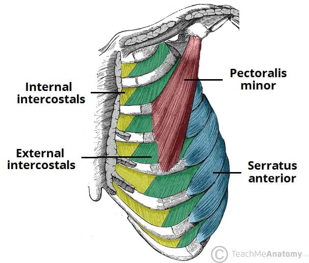
2. Muscles of the Thorax (Chest Wall)
These muscles are primarily dedicated to altering the volume of the thoracic cavity, making the mechanics of breathing (ventilation) possible.
- a. Intercostal Muscles: Located *between* the ribs.
- External Intercostals: Their fibers run down and inward. They elevate the ribs like a bucket handle to expand the chest for inspiration.
- Internal and Innermost Intercostals: Their fibers run down and outward. They forcefully depress the ribs, shrinking the chest cavity for forced expiration (like blowing out a candle).
- b. Diaphragm:
The absolute primary muscle of respiration. It is a large, dome-shaped muscular and tendinous sheet that perfectly separates the thoracic and abdominal cavities.
Action: When it contracts, the dome flattens, dropping inferiorly. This dramatically increases thoracic volume and decreases thoracic pressure, sucking air into the lungs.
Clinical Example: Hiccups are involuntary, spasmodic contractions of the diaphragm. A spinal cord injury at or above C3 can cause respiratory paralysis because the Phrenic nerve (C3, C4, C5 "keeps the diaphragm alive") is severed.
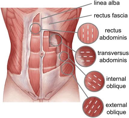
3. Muscles of the Abdominal Wall
Because the abdomen lacks skeletal protection (like ribs), it relies entirely on a muscular corset. These muscles protect viscera, flex the trunk, and perform the Valsalva maneuver (compressing the abdomen to increase intra-abdominal pressure for defecation, urination, vomiting, and childbirth).
The famous vertical "six-pack" muscle running straight down the front. It is segmented by connective tissue bands called tendinous intersections.
- Action: It is the powerful primary flexor of the vertebral column.
- Daily Example: Performing a sit-up or a crunch.
Three distinct layers of flat muscles wrapping the sides of the abdomen like plywood for immense structural integrity.
- External Oblique: Outermost; fibers run down and in (like hands in pockets). Rotates the trunk.
- Internal Oblique: Middle; fibers run up and in. Flexes and rotates the trunk.
- Transversus Abdominis: Deepest; fibers run horizontally. Acts like a tight weightlifting belt to suck in the gut and compress abdominal contents.
A deep, thick, square-shaped muscle forming the posterior abdominal wall next to the spine.
- Action: Unilaterally, it laterally flexes the trunk (bends you sideways) and elevates the hip ("hip hiking").
- Clinical Example: Extremely tight QL muscles are a frequent, hidden cause of severe chronic lower back pain and pelvic tilting.
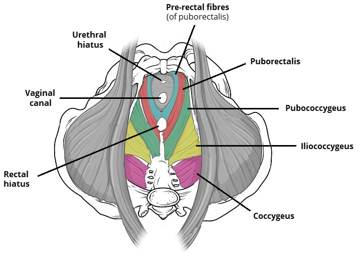
4. Pelvic Floor Muscles (Pelvic Diaphragm)
These muscles close the inferior outlet (the bottom hole) of the bony pelvis. They act as a supportive hammock.
- Levator Ani Group & Coccygeus:
This broad, highly important, funnel-shaped muscle group forms the major part of the pelvic floor.
Action: It physically supports all the heavy pelvic organs (bladder, uterus, rectum) and constantly resists increases in intra-abdominal pressure (when you cough or sneeze). It contains the sphincters that control urinary and fecal continence.
Clinical Example: Weakness in the Levator Ani (often due to trauma from vaginal childbirth) leads to urinary incontinence (leaking urine when coughing) or pelvic organ prolapse (where the uterus drops down). Medical professionals prescribe "Kegel exercises" to specifically strengthen this muscle group.
Summary Table of Torso Muscles
| Muscle | Origin | Insertion | Innervation | Main Actions |
|---|---|---|---|---|
| BACK MUSCLES | ||||
| Trapezius | Occipital bone, Ligamentum nuchae, C7-T12 spinous processes | Clavicle, acromion, spine of scapula | Spinal Accessory (CN XI), C3-C4 | Elevates, retracts, depresses, and upwardly rotates the scapula |
| Latissimus Dorsi | T7-L5 spinous processes, iliac crest, thoracolumbar fascia | Intertubercular groove of humerus | Thoracodorsal Nerve (C6-C8) | Prime mover of arm extension; adducts and medially rotates arm |
| Erector Spinae Group | Iliac crest, sacrum, lumbar/thoracic spinous processes | Ribs, thoracic/cervical vertebrae, mastoid process | Dorsal rami of spinal nerves | Prime mover of back extension; laterally flexes vertebral column |
| THORACIC MUSCLES | ||||
| External Intercostals | Inferior border of rib above | Superior border of rib below | Intercostal nerves (T1-T11) | Elevate ribs (widens chest for inspiration) |
| Internal Intercostals | Superior border of rib below | Inferior border of rib above | Intercostal nerves (T1-T11) | Depress ribs (shrinks chest for forced expiration) |
| Diaphragm | Xiphoid process, costal cartilages, lumbar vertebrae | Central tendon | Phrenic Nerves (C3-C5) | Primary muscle of inspiration; flattens to expand thoracic volume |
| ABDOMINAL & PELVIC MUSCLES | ||||
| Rectus Abdominis | Pubic crest and symphysis | Xiphoid process, costal cartilages 5-7 | Intercostal nerves (T7-T12) | Powerfully flexes vertebral column, compresses abdomen |
| External Oblique | Lower 8 ribs (Ribs 5-12) | Linea alba, pubic tubercle, iliac crest | Intercostal nerves (T7-T12) | Flexes vertebral column, rotates trunk to opposite side |
| Internal Oblique | Thoracolumbar fascia, iliac crest, inguinal ligament | Linea alba, pubic crest, last 3 ribs (10-12) | Intercostal (T7-T12), Iliohypo/inguinal (L1) | Flexes vertebral column, rotates trunk to same side |
| Transversus Abdominis | Thoracolumbar fascia, iliac crest, inner cartilages of ribs 7-12 | Linea alba, pubic crest | Intercostal (T7-T12), Iliohypo/inguinal (L1) | Compresses abdominal contents (increases intra-abdominal pressure) |
| Quadratus Lumborum | Iliac crest and iliolumbar ligament | Last rib (Rib 12), transverse processes of L1-L4 | Lumbar Plexus (T12-L4) | Laterally flexes vertebral column; maintains upright posture |
| Levator Ani Group | Posterior body of pubis, ischial spine | Coccyx, walls of pelvic organs (prostate/vagina/rectum) | Pudendal Nerve (S2-S4), Direct S3-S4 branches | Supports pelvic organs, maintains fecal and urinary continence |
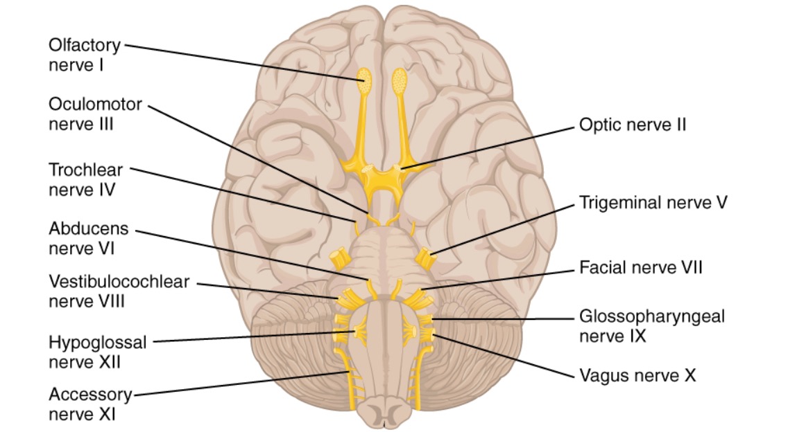
Reference: The 12 Cranial Nerves
The cranial nerves are an absolute cornerstone of neurological anatomy. They are a set of 12 paired nerves that arise directly from the brain and brainstem, bypassing the spinal cord entirely. They are uniquely responsible for conveying special sensory (vision, hearing), general sensory, and complex motor information to and from the head and neck region, as well as controlling vast parasympathetic visceral functions.
Crucial Mnemonics for Memorization
Medical students rely heavily on mnemonics to rapidly recall the order and functional type of the 12 cranial nerves.
- For Nerve Names (I through XII):
"Oh Oh Oh To Touch And Feel A Girls Vagina Ah Heaven"
(Olfactory, Optic, Oculomotor, Trochlear, Trigeminal, Abducens, Facial, Auditory/Vestibulocochlear, Glossopharyngeal, Vagus, Accessory, Hypoglossal). - For Functional Type (S=Sensory, M=Motor, B=Both):
"Some Say Marry Money, But My Brother Says Big Brains Matter More"
Deep Dive: The 12 Cranial Nerves
- I. Olfactory Nerve (Sensory)
Function: Dedicated solely to the special sense of smell (olfaction). It bypasses the thalamus and goes directly to the olfactory cortex.
Clinical Test & Pathology: Ask the patient to identify non-irritating, common scents (e.g., coffee, soap, vanilla) with their eyes closed and one nostril blocked at a time. Total loss of smell is called Anosmia, common in severe head trauma (shearing of olfactory bulbs) or early signs of neurodegenerative diseases like Parkinson's. - II. Optic Nerve (Sensory)
Function: Transmits the special sense of vision from the retina of the eye to the visual cortex in the occipital lobe.
Clinical Test & Pathology: Test visual acuity utilizing a Snellen chart. Test visual fields by confrontation (testing peripheral vision). Examine the optic disc using an ophthalmoscope. Damage can result in partial or total blindness (anopsia). - III. Oculomotor Nerve (Motor & Parasympathetic)
Function: Controls the majority of extraocular eye muscles (superior, inferior, medial recti, and inferior oblique) to move the eye up, down, and medially. It raises the upper eyelid (levator palpebrae). It also provides parasympathetic control to constrict the pupil (sphincter pupillae).
Clinical Test & Pathology: Ask patient to track a finger making a wide "H-pattern" in the air. Shine a light in the eye to check for the pupillary light reflex. Damage presents as a "down and out" deviated eye, severe eyelid drooping (ptosis), and a blown, unreactive dilated pupil. - IV. Trochlear Nerve (Motor)
Function: Controls exactly one muscle: the superior oblique muscle, which utilizes a pulley-like tendon (trochlea) to move the eye downward and inward.
Clinical Test & Pathology: Ask the patient to look down and toward their nose. Damage causes the eye to drift upward, resulting in severe vertical double vision (diplopia), causing the patient to chronically tilt their head to compensate. - V. Trigeminal Nerve (Both - Sensory & Motor)
Function: The massive sensory nerve for the entire face (touch, pain, temperature) via three branches: Ophthalmic (V1), Maxillary (V2), and Mandibular (V3). It provides Motor innervation specifically to the muscles of mastication.
Clinical Test & Pathology: Test facial sensation across all three zones lightly with a cotton wisp and a sharp pin. Ask the patient to forcefully clench their jaw and palpate the masseter and temporalis muscles for equal strength. Trigeminal Neuralgia (Tic Douloureux) is a pathology involving excruciating, lightning-shock pain across the face. - VI. Abducens Nerve (Motor)
Function: Controls exactly one muscle: the lateral rectus muscle, which moves the eye laterally (it literally "abducts" the eye away from the midline).
Clinical Test & Pathology: Ask the patient to look far to the side (laterally). Damage paralyzes the lateral rectus, so the eye gets pulled inward (medially) by the unopposed medial rectus, resulting in a cross-eyed appearance and horizontal double vision. - VII. Facial Nerve (Both - Sensory, Motor, & Parasympathetic)
Function: Motor control for all muscles of facial expression. Sensory for the special sense of taste from the anterior two-thirds of the tongue. Parasympathetic control of tearing (lacrimal glands) and salivation (submandibular/sublingual glands).
Clinical Test & Pathology: Ask the patient to smile broadly, frown, puff out their cheeks, and raise their eyebrows tightly. Bell's Palsy is a severe acute unilateral paralysis of this nerve, resulting in a completely sagging face on one side, inability to close the eye, and drooling. - VIII. Vestibulocochlear Nerve (Sensory)
Function: Splits into two distinct parts: the cochlear nerve handles the special sense of hearing, and the vestibular nerve handles balance, equilibrium, and spatial orientation.
Clinical Test & Pathology: Test hearing using the whisper test, and tuning forks (Rinne and Weber tests) to differentiate conductive vs. sensorineural hearing loss. Test balance with the Romberg test. Pathologies here cause profound deafness, ringing in the ears (tinnitus), or severe, debilitating dizziness (vertigo). - IX. Glossopharyngeal Nerve (Both - Sensory, Motor, & Parasympathetic)
Function: Motor function for swallowing (stylopharyngeus muscle). Sensory for taste from the posterior one-third of the tongue, and general sensation from the pharynx, tonsils, and middle ear. It also monitors blood pressure via the carotid sinus. Parasympathetic control of the parotid salivary gland.
Clinical Test & Pathology: Lightly touch the back of the throat with a cotton swab to check the gag reflex. Ask the patient to say "ahhh" and watch for symmetrical uvula elevation. Damage causes loss of gag reflex and difficulty swallowing (dysphagia). - X. Vagus Nerve (Both - Sensory, Motor, & Parasympathetic)
Function: The name means "the wanderer." It is the massive parasympathetic superhighway of the body, providing motor innervation to the heart (slowing it down), lungs, and most of the abdominal viscera (stimulating digestion). It also provides motor control to the pharynx and larynx for swallowing and speaking.
Clinical Test & Pathology: Assessed simultaneously with CN IX (gag reflex, swallowing). Listen to the patient's voice; a damaged Vagus nerve will paralyze the vocal cords, leading to profound hoarseness and severe difficulty swallowing. - XI. Accessory Nerve (Motor)
Function: A unique nerve with both cranial and spinal roots. It strictly controls two massive neck/back muscles: the trapezius and the sternocleidomastoid (SCM).
Clinical Test & Pathology: Ask the patient to forcefully shrug their shoulders upward against the examiner's hands (testing Trapezius). Ask them to turn their head forcefully to the side against resistance (testing SCM). Damage causes a dropped, sagging shoulder and weakness turning the head. - XII. Hypoglossal Nerve (Motor)
Function: Controls almost all the intrinsic and extrinsic muscles of the tongue, allowing for complex maneuvers required for speech and manipulating food.
Clinical Test & Pathology: Ask the patient to stick their tongue straight out. If the nerve is damaged on one side, the healthy side pushes unopposed, causing the tongue to visibly deviate toward the paralyzed side. Long-term damage will also show atrophy (wasting away) and fasciculations (twitching) of the tongue muscle.
List of References
- Tortora, G. J., & Derrickson, B. H. (2017). Principles of Anatomy and Physiology (15th ed.). Wiley. (Core foundational anatomy and muscular attachments).
- Moore, K. L., Dalley, A. F., & Agur, A. M. R. (2018). Clinically Oriented Anatomy (8th ed.). Lippincott Williams & Wilkins. (Clinical correlations, facial expression pathologies, and cranial nerve assessments).
- Netter, F. H. (2018). Atlas of Human Anatomy (7th ed.). Elsevier. (Visual spatial relationships of the deep neck, back, and pelvic floor musculature).
- Guyton, A. C., & Hall, J. E. (2020). Textbook of Medical Physiology (14th ed.). Elsevier. (Physiological mechanisms of mastication, deglutition, and respiration).
- Bickley, L. S., Szilagyi, P. G., & Hoffman, R. M. (2020). Bates' Guide to Physical Examination and History Taking (13th ed.). Wolters Kluwer. (Standardized clinical testing protocols for the 12 Cranial Nerves).
Muscles of the Head, Neck and Trunk Read More »