Poliomyelitis Lecture nOTES
Poliomyelitis
Introduction to Poliomyelitis
Poliomyelitis, commonly known as polio, is an acute, highly contagious viral infectious disease that has historically caused widespread fear due to its potential to cause irreversible flaccid paralysis and death, particularly in children under 5 years of age. While massive progress has been made toward global eradication, understanding poliomyelitis remains critical for surveillance, immunization, and nursing care.
The word poliomyelitis derives from the Greek polios (gray), myelon (marrow, referring to the spinal cord), and -itis (inflammation)—literally meaning “inflammation of the gray matter of the spinal cord.”
- Nature of the Disease: Polio is an acute, highly contagious enteroviral infection transmitted predominantly via the fecal–oral route.
- Causative Agent: Caused by the poliovirus (serotypes 1, 2, and 3).
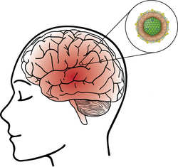
- Primary Target: Although the virus initially replicates in the gastrointestinal tract, severe clinical disease results from invasion and destruction of motor neurons in the anterior horn of the spinal cord and the brainstem.
- Clinical Spectrum: Infection ranges from asymptomatic carriage (~90–95% of infections) and mild febrile illness to aseptic meningitis and irreversible paralytic disease (<1% of infections).
- Historical Context: Before effective vaccines were introduced in the mid-20th century, polio epidemics struck regularly worldwide, earning the name “infantile paralysis” due to its predominance in young children.
- Impact: Paralytic polio causes permanent muscle weakness, asymmetric flaccid paralysis, muscle atrophy, skeletal deformities, late-onset Post-Polio Syndrome, and death when respiratory muscles are affected.
The Causative Agent: Poliovirus
The agent responsible for poliomyelitis is the poliovirus (PV), a strictly human pathogen.
Classification and Viral Structure
- Family: Picornaviridae (pico = small, rna = RNA virus).
- Genus: Enterovirus (enteron = intestine), reflecting its primary site of replication and fecal excretion.
- Viral Structure: A small (~30 nm), non-enveloped icosahedral RNA virus. Because it lacks a lipid envelope, poliovirus resists lipid solvents, detergents, common disinfectants, and gastric acid, enabling prolonged environmental survival and efficient fecal–oral transmission.
- Genomic Material: Single-stranded, positive-sense RNA genome.
Serotypes (Immunological Types)
There are three antigenically distinct serotypes of wild poliovirus (WPV): Types 1, 2, and 3. Immunity to one serotype does not protect against the other two; therefore, immunization must cover all three serotypes.
- Wild Poliovirus Type 1 (WPV1): Historically the most frequent cause of paralytic polio and the only wild serotype still circulating. It remains endemic in two countries (Afghanistan and Pakistan) and is responsible for all ongoing WPV outbreaks.
- Wild Poliovirus Type 2 (WPV2): Last naturally occurring case was confirmed in India in 1999, and WPV2 was certified globally eradicated in September 2015. To eliminate Type 2 vaccine-associated paralytic polio (VAPP) and circulating vaccine-derived poliovirus (cVDPV2), the Type 2 component was removed from routine oral polio vaccine during the synchronized global switch from trivalent OPV (tOPV) to bivalent OPV (bOPV, Types 1 and 3) in April 2016.
- Wild Poliovirus Type 3 (WPV3): Last naturally occurring case occurred in Nigeria in 2012, and WPV3 was certified globally eradicated in October 2019. Routine immunization programs in OPV-using countries continue to use bivalent OPV (bOPV, containing Types 1 and 3) alongside Inactivated Polio Vaccine (IPV) until global bOPV cessation occurs following WPV1 eradication.
Epidemiology of Polio
A. Mode of Transmission
Poliovirus is highly contagious (household secondary attack rates can exceed 90% in unimmunized contacts) and spreads via:
- Fecal–Oral Route (Predominant): Infected individuals shed poliovirus in feces for weeks, even when asymptomatic. Contamination of water, food, or hands occurs due to:
- Poor Sanitation: Inadequate latrine facilities or sewage contaminating drinking water sources.
- Inadequate Hand Hygiene: Failure to wash hands after defecation, diaper changing, or before preparing food.
- Contaminated Food and Water: Ingestion of unboiled water or food handled by an infected carrier.
- Oral–Oral Route (Less Common): Spread via respiratory droplets from coughing or sneezing during early pharyngeal replication, especially in crowded living conditions.
- Incubation Period: Typically 7–14 days (range 3–35 days).
- Period of Infectivity: Most contagious 7–10 days before and after symptom onset; virus is shed in throat secretions for ~1–2 weeks and in stool for 3–6 weeks or longer (and potentially months/years in patients with primary B-cell immunodeficiencies).
B. Reservoirs
- Humans Only: Humans are the sole natural reservoir for poliovirus; there is no animal reservoir or insect vector. This biological fact makes complete global eradication achievable.
C. Historical Global Prevalence
- Widespread Before Vaccination: Prior to Salk’s IPV (1955) and Sabin’s OPV (early 1960s), polio was endemic worldwide, peaking in summer and autumn in temperate climates.
- Sanitation Paradox: Improved sanitation delayed natural infant exposure until later childhood (after maternal antibodies waned), paradoxically increasing rates of severe paralytic epidemics and requiring widespread use of negative-pressure “iron lungs.”
D. Current Geographical Distribution and cVDPV
- Dramatic Reduction: Since the Global Polio Eradication Initiative (GPEI) began in 1988, global polio cases have dropped by >99.9%. The WHO African Region was certified free of indigenous wild poliovirus in August 2020.
- Remaining WPV1 Endemic Countries: Only Afghanistan and Pakistan remain endemic for WPV1, though occasional importations and positive environmental detections remain a global threat requiring vigilant surveillance.
- Circulating Vaccine-Derived Poliovirus (cVDPV): In under-immunized communities with poor sanitation, attenuated OPV strains (most commonly Type 2, cVDPV2) can circulate person-to-person for prolonged periods, mutate, and regain neurovirulence, causing paralysis outbreaks indistinguishable from wild polio.
E. Silent Transmission by Asymptomatic Carriers
- The “Iceberg” Phenomenon: Because ~90–95% of infections are asymptomatic and <1% cause paralysis, 100 to 1,000+ silent infections occur for every visible paralytic case.
- Public Health Impact: Asymptomatic shedders silently propagate transmission, requiring both mass immunization and environmental wastewater/sewage surveillance alongside Acute Flaccid Paralysis (AFP) reporting.
Pathophysiology of Poliovirus Infection
A. Viral Entry and Initial Replication
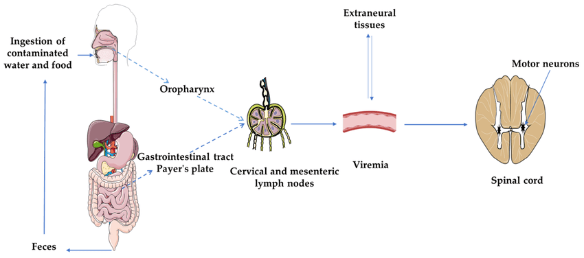
- Entry: Poliovirus enters through the mouth via contaminated food, water, hands, or droplets.
- Primary Replication Sites: The virus binds to the cell-surface receptor CD155 (poliovirus receptor) and replicates first in lymphoid tissues of the oropharynx (tonsils) and then in the small intestine (Peyer’s patches) and mesenteric lymph nodes.
- Viremia (Minor and Major):
- Minor Viremia: Virus enters the bloodstream transiently; in ~95–99% of hosts, neutralizing antibodies and cell-mediated immunity halt progression here, producing asymptomatic or mild abortive illness.
- Major Viremia: In ~1–5% of infected individuals, continued replication in deep lymph nodes and reticuloendothelial tissues generates sustained high-level viremia, enabling CNS entry.
B. Invasion of the Central Nervous System (CNS)
- Routes to the CNS: Poliovirus crosses the blood–brain barrier during major viremia or travels upward along peripheral motor nerves via retrograde axonal transport.
- Selective Motor Neuron Tropism: Poliovirus specifically infects and destroys somatic motor neurons in:
- Anterior Horn Cells (AHC) of the Spinal Cord: Lower motor neurons controlling skeletal limb, trunk, and diaphragmatic muscles.
- Brainstem Motor Nuclei (“Bulbar” Centers): Cranial nerve nuclei (especially CN V, VII, IX, X, XI, XII) controlling facial movement, swallowing, speech, and vital medullary respiratory/vasomotor centers.
- Lytic Neuronal Destruction: Intracellular viral replication kills motor neurons (lytic infection), provoking perivascular inflammation, edema, and microglial neuronophagia. Because destroyed motor neurons cannot regenerate, denervated skeletal muscle fibers undergo permanent lower motor neuron flaccid paralysis and neurogenic atrophy.
C. Clinical Forms of Polio Infection
- Asymptomatic (Inapparent) Infection (90–95%): No clinical symptoms; viral replication is limited to the GI tract/minor viremia, but fecal shedding perpetuates silent community transmission.
- Abortive Polio / Minor Illness (4–8%): Brief, self-limiting viral illness lasting 2–3 days without CNS signs—fever, malaise, headache, sore throat, nausea, vomiting, and abdominal pain. Indistinguishable from other common viral infections.
- Non-Paralytic Polio / Aseptic Meningitis (1–2%): Poliovirus invades the meninges without destroying motor neurons. Alongside abortive symptoms, the patient develops nuchal rigidity (stiff neck), back stiffness, Kernig/Brudzinski signs, muscle tenderness, and irritability. CSF shows lymphocytic pleocytosis with normal glucose. Full recovery typically occurs within 2–10 days.
- Paralytic Polio (<1%): Severe CNS motor neuron destruction producing sudden asymmetric flaccid paralysis. Often follows a biphasic course (initial minor illness → brief 1–3 day remission → sudden return of high fever, severe myalgia/meningismus, and rapid paralysis).
Clinical Manifestations of Paralytic Polio
A. General Signs and Symptoms of Acute Paralytic Polio
- Biphasic Fever: An initial mild fever that subsides briefly, followed by a sudden second spike coinciding with neurological deterioration.
- Meningeal & Musculoskeletal Pain: Severe headache, nuchal and spinal rigidity (e.g., positive tripod sign when sitting), and exquisite muscle pain, spasms, and tenderness to touch.
- Rapid Onset of Characteristic Paralysis: Progresses over hours to 3–4 days and stabilizes once fever resolves:
- Flaccid (Hypotonic): Limp, floppy muscles with diminished or absent deep tendon reflexes (areflexia).
- Asymmetric: Patchy involvement affecting one limb or one side more than the other.
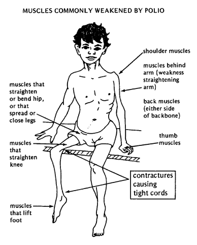
- Proximal > Distal: Large proximal muscle groups (thighs, shoulders) are usually weaker than distal hands/feet.
- Lower Limbs > Upper Limbs: Legs are affected roughly twice as often as arms.
- Intact Sensation: Crucially, sensory perception remains completely intact (distinguishing polio from transverse myelitis and Guillain–Barré syndrome).
B. Patterns of Paralysis
- Spinal Polio (Most Common, ~79% of paralytic cases): Destruction of spinal anterior horn cells causing asymmetric flaccid paralysis of the legs, arms, abdominal wall, and—if C3–C5 (phrenic nerve) or thoracic segments are involved—diaphragmatic and intercostal respiratory failure.
- Bulbar Polio (~2%, Most Fatal): Destruction of cranial nerve and medullary motor nuclei in the brainstem (“bulb”), causing:
- Dysphagia & Pooling of Secretions: Pharyngeal/palatal paralysis causing nasal regurgitation, wet voice, and high risk of fatal aspiration.
- Dysphonia / Aphonia & Facial Weakness: Nasal or absent voice, weak cough, and asymmetric facial palsy.
- Central Cardiorespiratory Failure: Damage to medullary respiratory and vasomotor centers causes irregular shallow breathing (Cheyne–Stokes/apnea), labile blood pressure, tachycardia/arrhythmias, and sudden cardiac arrest.
- Bulbospinal Polio (~19%): Combined destruction of both spinal anterior horn cells and brainstem nuclei, causing simultaneous limb paralysis, respiratory muscle paralysis, and swallowing failure.
C. Outcome of Paralysis
- Partial Recovery: Once acute inflammation subsides, mildly injured neurons recover and surviving axons sprout collateral branches, yielding gradual strength gains over 6–12 months; however, completely destroyed neurons leave permanent flaccid paralysis.
- Mortality: Overall case-fatality among paralytic cases is ~5–10%, rising to 25–75% in bulbar or respiratory-muscle paralysis without mechanical ventilation.
Critical Nursing Precaution: Provocation Poliomyelitis
Avoid unnecessary intramuscular (IM) injections during the acute febrile illness or in any child presenting with suspected non-paralytic polio. Trauma from IM injections during poliovirus viremia increases blood–brain barrier permeability and triggers localized severe paralysis in the injected limb (provocation poliomyelitis).
Diagnosis of Poliomyelitis
A. Clinical Suspicion and AFP Surveillance
- Acute Flaccid Paralysis (AFP) Case Definition: Any child aged <15 years presenting with sudden onset of floppy/flaccid paralysis (or any person of any age suspected of polio by a clinician) must be reported immediately and investigated under national AFP surveillance.
- Key Clinical Clues: Fever at onset, rapid asymmetric flaccid paralysis within 24–72 hours, absent deep tendon reflexes, and preserved sensation.
B. Laboratory Confirmation (Gold Standard)
- Viral Isolation and RT-PCR (Gold Standard):
- Stool Specimens (Primary): Collect two stool samples (8–10 g each, thumb-sized) 24–48 hours apart within 14 days of paralysis onset, transported immediately under reverse cold chain (2–8°C) to a WHO-accredited polio laboratory (e.g., Uganda Virus Research Institute [UVRI]).
- Throat Swabs & CSF: Throat swabs are helpful only in the first few days; CSF culture has low yield.
- Environmental Surveillance: Sewage/wastewater testing detects silent poliovirus circulation in communities.
- Testing & Sequencing: RT-PCR and cell culture (L20B/RD cells) isolate the virus, followed by intratypic differentiation and genetic sequencing to distinguish WPV1 from Sabin vaccine virus or cVDPV.
- Serology: Paired acute and convalescent serum samples (3–4 weeks apart) showing a ≥4-fold rise in neutralizing antibody titer (or positive IgM); less useful than stool RT-PCR because it cannot distinguish wild virus from recent vaccination.
C. Cerebrospinal Fluid (CSF) Analysis
- Findings: Clear CSF under normal/slightly raised pressure; early pleocytosis (20–300 WBCs/μL, initially neutrophils shifting within days to lymphocytes), normal glucose, and normal-to-mildly elevated protein.
- Diagnostic Value: Excludes bacterial meningitis (low glucose, high neutrophils) and Guillain–Barré syndrome (albuminocytologic dissociation: high protein with normal WBC count).
D. Differential Diagnosis for Acute Flaccid Paralysis (AFP)
- Guillain–Barré Syndrome (GBS): Usually symmetric ascending paralysis, afebrile onset, sensory paresthesias, and CSF albuminocytologic dissociation.
- Transverse Myelitis: Symmetric paralysis below a distinct spinal sensory level with early bladder/bowel paralysis.
- Traumatic Injection Neuritis (Sciatic Nerve Injury): Paralysis localized to one leg immediately after an unsafe gluteal IM injection.
- Other Conditions: Non-polio enteroviruses (EV-D68, EV-A71), West Nile virus, botulism, tick paralysis, myasthenia gravis, and hypokalemic periodic paralysis.
Management of Acute Polio Infection
There is no specific antiviral drug or cure for poliovirus infection, and antibiotics have no effect on the virus. Management is entirely supportive, aimed at preserving life (especially airway and breathing), easing pain, and preventing permanent skeletal deformities.
A. Supportive Management Principles
- Isolate patient using standard and enteric (contact/stool) precautions (plus droplet precautions in the first week); disinfect feces and soiled linens with chlorine/bleach because alcohol gels alone are poorly effective against non-enveloped poliovirus.
- Avoid IM injections and strenuous physical exertion during the acute febrile phase (exercise during early viremia increases paralysis severity).
B. Specific Supportive Care Strategies
- Complete Bed Rest & Close Monitoring: Maintain strict bed rest on a firm mattress with a footboard; monitor vital signs, cough strength, swallowing ability, and respiratory capacity (counting single-breath count/vital capacity) every 2–4 hours to detect impending respiratory or bulbar failure early.
- Pain & Muscle Spasm Relief:
- Moist Heat Therapy: Apply warm moist packs (Sister Kenny method) to tender, spasming muscles to relieve pain and prevent contractures.
- Analgesics: Administer oral/rectal paracetamol or NSAIDs; use opioids or respiratory-depressant sedatives with extreme caution (avoid unless the patient is mechanically ventilated, as they can mask hypoxia and precipitate respiratory arrest).
- Respiratory & Bulbar Airway Support: Position patients with bulbar paralysis slightly head-down/semi-prone or lateral to drain secretions, keep suction ready, initiate humidified oxygen, and arrange urgent endotracheal intubation, tracheostomy, and positive-pressure mechanical ventilation if diaphragm/intercostal paralysis or apnea develops.
- Nutrition & Hydration: Provide IV fluids during acute vomiting/febrile illness; use soft nasogastric or gastrostomy feeding when dysphagia poses an aspiration risk.
- Bladder & Bowel Management: Perform intermittent or indwelling aseptic urinary catheterization for transient bladder paralysis/retention; administer stool softeners and adequate fluids for paralytic constipation.
- Early Positioning & Rehabilitation: Maintain neutral anatomical alignment (neutral footboards to prevent equinus foot drop, pillows/rolls under knees and hips); begin gentle passive range-of-motion (ROM) exercises only after acute fever and muscle tenderness subside, progressing to active physiotherapy, orthotic braces/splints, assistive devices, and orthopedic tendon/joint surgery later if contractures occur.
C. Pharmacology: Pharmacokinetics and Pharmacodynamics
| Agent / Drug Class | Pharmacodynamics (Mechanism & Action) | Pharmacokinetics (ADME & Nursing Notes) |
|---|---|---|
| Oral Polio Vaccine (bOPV / nOPV2 — Live Attenuated) | Live attenuated polioviruses replicate harmlessly in the oropharyngeal and intestinal mucosa, inducing robust local secretory IgA (mucosal immunity) as well as systemic IgG—blocking both paralytic disease and fecal–oral viral shedding/transmission. | Kinetics: Given PO (2 drops); replicates in GI tract for 1–4 weeks and is shed in feces (contributing to passive contact immunity). Store frozen long-term or at 2–8°C for up to 6 months; check Vaccine Vial Monitor (VVM) before every dose. Nursing Note: If child vomits immediately, repeat dose. Contraindicated in severe primary immunodeficiency. |
| Inactivated Polio Vaccine (IPV — Killed Vaccine) | Contains formalin-inactivated WPV1, 2, and 3; stimulates systemic B-cells to produce high levels of circulating serum IgG neutralizing antibodies that prevent viremia and CNS paralysis (minimal intestinal IgA). | Kinetics: Administered 0.5 mL IM into outer aspect of right thigh (2.5 cm from PCV site) or SC; zero risk of VAPP/cVDPV. Store at 2–8°C (never freeze—freezing destroys potency). |
| Paracetamol (Acetaminophen — Analgesic/Antipyretic) | Inhibits central prostaglandin synthesis (COX inhibition in the CNS) and acts on the hypothalamic heat-regulating center to relieve myalgia, headache, and biphasic fever without depressing respiration or causing GI bleeding. | ADME: Rapid oral/rectal absorption; peak 30–60 min; minimal protein binding; metabolized in liver (glucuronidation/sulfation; toxic NAPQI in overdose); renal excretion (t1/2 2–3 hrs). Preferred over aspirin in children (avoids Reye syndrome). |
| Ibuprofen (NSAID — Anti-inflammatory Analgesic) | Reversibly inhibits COX-1 and COX-2 peripherally and centrally, decreasing prostaglandin-mediated muscle tenderness, meningeal pain, and fever. | ADME: ~80–100% oral absorption; >99% albumin-bound; hepatic CYP2C9 metabolism; renal excretion (t1/2 ~2 hrs). Give with food/milk; ensure adequate hydration to protect kidneys. |
| Lactulose / Bisacodyl (Bowel Care for Paralytic Constipation) | Lactulose acts osmotically in the colon (fermented by gut flora into organic acids that draw water and soften stool); bisacodyl stimulates colonic myenteric plexus peristalsis. | ADME: Lactulose is minimally absorbed (<3%), metabolized by colonic bacteria, and excreted in feces; onset 24–48 hrs. Monitor stool frequency and hydration. |
Post-Polio Syndrome (PPS)
Years after surviving acute paralytic polio, many patients develop a progressive late neurological disorder called Post-Polio Syndrome (PPS).
A. Definition and Onset
- Late-Onset Neurological Sequela: Occurs 15–40+ years after acute paralytic poliomyelitis (affecting ~25–40% of polio survivors). It is not a reactivation or recurrence of live poliovirus infection (patients are non-infectious).
- Progressive Course: Characterized by new, slowly progressive weakness in muscles previously affected or apparently spared during the original illness.
B. Characteristic Symptoms
- New Progressive Muscle Weakness: Asymmetric weakness in previously affected or seemingly recovered limbs.
- Overwhelming Fatigue: Debilitating physical and mental exhaustion (“polio wall”) unimproved by brief rest.
- Muscle & Joint Pain: Chronic aching, cramping, or degenerative joint pain from altered biomechanics.
- New Muscle Atrophy & Fasciculations: Visible loss of muscle bulk and muscle twitching.
- Cold Intolerance, Sleep Apnea, & Bulbar Decline: Sensitivity to cold, nocturnal hypoventilation, and new dysphagia or dyspnea in survivors of bulbar polio.
C. Pathophysiology
- Compensatory Motor Unit Enlargement: After acute polio destroyed motor neurons, surviving adjacent motor neurons sprouted extra terminal axons to re-innervate orphaned muscle fibers, restoring functional strength.
- Overuse Degeneration & Aging: Decades of chronic metabolic overload cause premature pruning and degeneration of those distal nerve sprouts (combined with normal age-related neuron loss), re-denervating muscle fibers and causing new weakness and fatigue.
D. Diagnosis and Management
- Diagnosis: A clinical diagnosis of exclusion requiring prior paralytic polio, ≥15 years of neurological stability, new progressive weakness/fatigability lasting ≥1 year, and exclusion of hypothyroidism, radiculopathy, ALS, or myasthenia gravis.
- Management: Energy conservation and activity pacing (avoid exercising to exhaustion), non-fatiguing submaximal physical therapy, updated lightweight orthotics/mobility aids, pain control, weight control, and non-invasive nocturnal ventilation (BiPAP/CPAP) if respiratory insufficiency develops.
Complications of Poliomyelitis
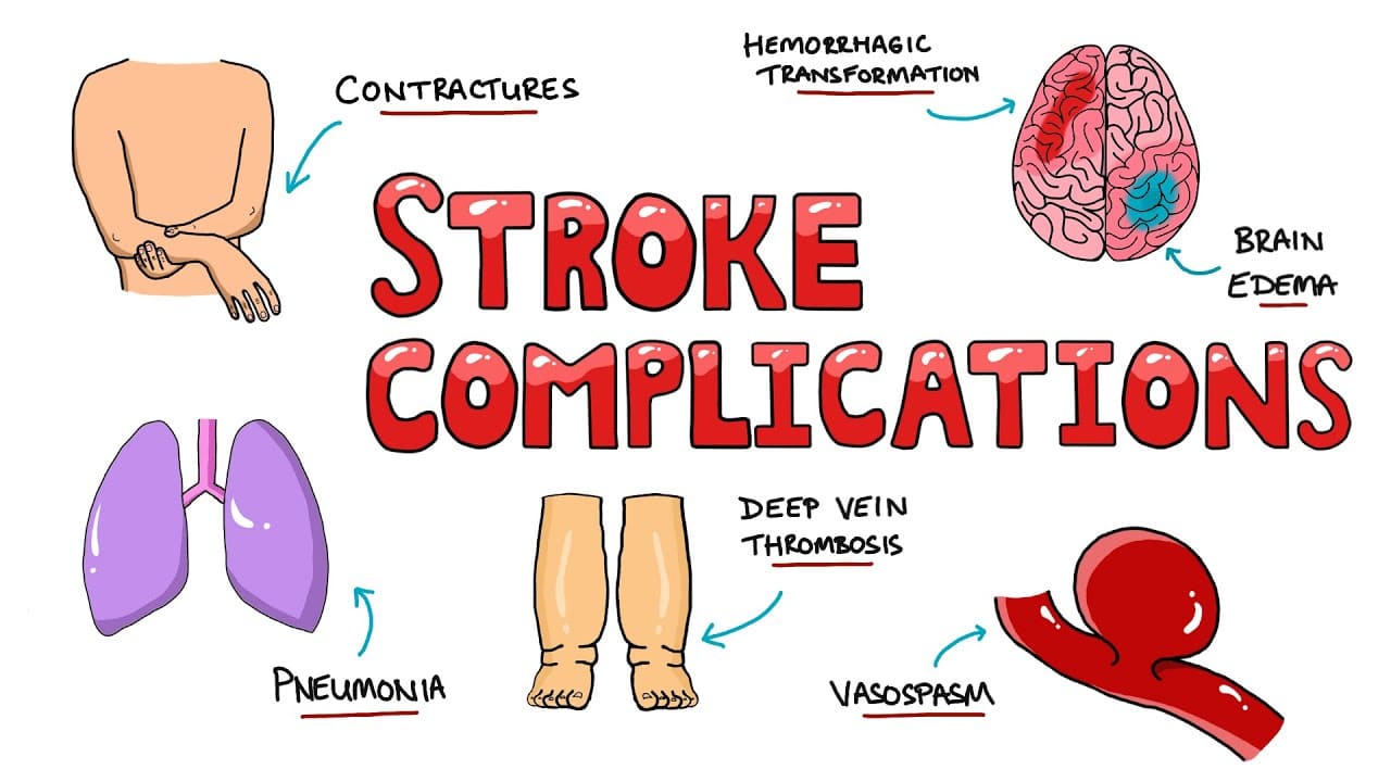
- Respiratory Complications: Acute diaphragmatic/intercostal paralysis, respiratory failure, atelectasis, and aspiration pneumonia.
- Cardiovascular Complications: Myocarditis, autonomic labile hypertension, arrhythmias, and deep vein thrombosis (DVT)/pulmonary embolism from prolonged immobilization.
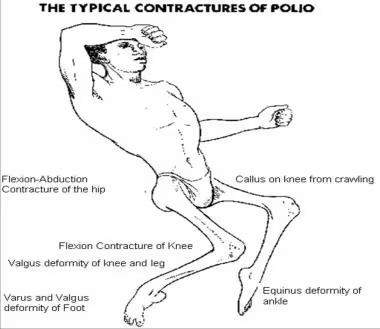
- Musculoskeletal Deformities: Permanent flaccid paralysis, severe muscle wasting, joint contractures (equinus foot drop, flexion contractures), limb-length discrepancy, and paralytic scoliosis/kyphoscoliosis.
- Renal & Metabolic Complications: Urinary retention, recurrent UTIs, and hypercalciuria/renal calculi (kidney stones) from prolonged immobilization bone resorption.
- Long-Term & Psychological Impact: Pressure injuries, Post-Polio Syndrome, chronic disability, social stigma, and depression.
Prevention of Polio: The Role of Vaccination
Because there is no cure for polio, vaccination is the single most effective cornerstone of prevention and global eradication.
A. Importance of Vaccination
- Only Effective Protection: Induces neutralizing antibodies before wild or vaccine-derived poliovirus can reach the CNS.
- Herd Immunity: High population coverage (≥80–90%) interrupts person-to-person transmission, protecting immunocompromised and newborn infants.
- Eradication Prerequisite: Essential to permanently eliminate WPV1 and stop cVDPV outbreaks.
B. Types of Polio Vaccines (IPV, OPV, and nOPV2)
| Feature | Inactivated Poliovirus Vaccine (IPV — Salk) | Oral Poliovirus Vaccine (OPV — Sabin: bOPV / nOPV2) |
|---|---|---|
| Composition | Killed/inactivated poliovirus of all 3 serotypes (1, 2, 3) | Live attenuated (weakened) poliovirus: bOPV (Types 1 & 3 routine), nOPV2 (genetically stabilized Type 2 for outbreaks) |
| Route & Dose | 0.5 mL Intramuscular (or fractional ID) | 2 drops Orally (PO) |
| Immunity Induced | Strong systemic IgG immunity (protects individual from paralysis); minimal intestinal IgA | Strong intestinal mucosal IgA + systemic IgG (blocks gut infection and shedding) |
| Herd Spread | Does not shed vaccine virus to contacts | Shed in stool; passively immunizes close contacts in low-sanitation settings |
| Safety Risks | Zero risk of VAPP or cVDPV; safe in immunocompromised patients | Rare VAPP (~1 in 2.7 million first doses) and cVDPV in under-immunized areas (nOPV2 markedly lowers cVDPV2 reversion risk) |
| Cold Chain | 2–8°C (never freeze) | Heat-sensitive; store frozen or 2–8°C short-term; check VVM |
C. Uganda Routine Polio Immunization Schedule (UNEPI)
Under the Uganda National Expanded Programme on Immunization (UNEPI), children receive combined OPV and IPV to achieve both intestinal mucosal protection and durable systemic Type 2 immunity:
| Age / Visit | Vaccine | Dose & Route | Site & Notes |
|---|---|---|---|
| At Birth (or within first 2 weeks) | OPV-0 (Birth dose) | 2 drops PO | Mouth (primer dose; do not count toward the 3 primary doses if given late) |
| 6 Weeks | OPV-1 + IPV-1 | OPV: 2 drops PO IPV: 0.5 mL IM | IPV injected into outer aspect of right upper thigh, at least 2.5 cm away from PCV-1 site |
| 10 Weeks | OPV-2 | 2 drops PO | Mouth |
| 14 Weeks | OPV-3 + IPV-2 | OPV: 2 drops PO IPV: 0.5 mL IM | IPV injected into outer aspect of right upper thigh, at least 2.5 cm away from PCV-3 site |
D. Global Polio Eradication Strategy (GPEI)
- High Routine & Campaign Coverage: Sustaining high routine OPV + IPV uptake alongside Supplementary Immunization Activities (SIAs) to eliminate zero-dose children.
- Synchronized OPV Phasing: Following the 2016 tOPV-to-bOPV switch, deploying nOPV2 for cVDPV2 outbreaks and planning global bOPV withdrawal after WPV1 eradication.
- Outbreak Response: Rapid, high-coverage immunization campaigns (nOPV2/bOPV/fIPV) around any detected AFP case or positive environmental sample.
- Sensitive Surveillance: Active AFP surveillance (<15 years) and sewage/environmental testing.
- Laboratory Containment & All-IPV Transition: Strict biosafety containment of poliovirus stocks and eventual universal transition to IPV.
Nursing Management and Care Plan (NANDA-I 2024–2026)
Nursing Care Plan for Poliomyelitis
The nursing care plan below applies the Nurses Revision Uganda Nursing Process standard and NANDA-I 2024–2026 diagnoses across acute paralytic polio and convalescent/PPS care, separating three-part actual diagnoses (PES) from two-part risk diagnoses, with SMART future-tense outcomes and past-tense evaluation.
| Assessment | Nursing Diagnosis (NANDA-I 2024–2026) | Planning (Goals & Expected Outcomes) | Nursing Interventions | Rationale | Evaluation |
|---|---|---|---|---|---|
Subjective: Patient reports sudden shortness of breath and inability to cough strongly or swallow saliva. Objective: Shallow rapid respirations (RR 34/min), weak cough, nasal voice, pooled pharyngeal secretions, falling single-breath count, SpO2 89%. | Ineffective breathing pattern (and Ineffective airway clearance) related to diaphragmatic/intercostal muscle paralysis and bulbar pharyngeal weakness as evidenced by shallow tachypnea (34/min), weak cough, pooled secretions, and SpO2 89%. | Short-Term Goal (within 1 hour): Patient will maintain patent airway and adequate oxygenation (SpO2 ≥95%; ABGs within normal limits). Expected Outcomes: – Secretions will remain cleared without aspiration. – Ventilatory support will be initiated promptly before severe hypoxia occurs. | – Monitor respiratory rate, depth, single-breath count, SpO2, and swallowing reflex every 1–2 hours. – Position in lateral/semi-prone postural drainage (head slightly lowered if bulbar secretions pool), suction gently, keep NPO, and give humidified oxygen. – Avoid respiratory-depressant sedatives/opioids unless intubated; prepare emergency intubation/mechanical ventilation/tracheostomy equipment at bedside. | – Serial respiratory checks detect ascending spinal/bulbar respiratory failure before sudden arrest. – Lateral drainage and NPO status prevent fatal aspiration pneumonia. – Timely mechanical ventilation prevents hypoxic brain injury and death. | Goal Met: Within 1 hour of lateral drainage, suctioning, and prompt ventilatory support, SpO2 stabilized at 97% with clear airway. |
Subjective: Child cries when limbs or back are touched, reporting severe aching calf and back pain (8/10). Objective: Guarding, stiff neck/back (tripod sign), muscle tenderness and spasms, fever 38.7°C. | Acute pain related to anterior horn/meningeal inflammation and painful reflex muscle spasms as evidenced by pain rating 8/10, guarding, crying on touch, and nuchal/back rigidity. | Short-Term Goal (within 24 hours): Patient will verbalize or demonstrate pain relief (pain score ≤3/10) and rest comfortably. Expected Outcomes: – Muscle spasms and tenderness will subside. – Patient will tolerate gentle neutral positioning. | – Handle paralyzed/tender limbs gently by supporting joints (never grasping sore muscle bellies directly) during bed rest. – Apply warm moist packs (Sister Kenny method) to tender spastic muscles for 15–20 minutes several times daily. – Administer prescribed oral/rectal paracetamol or ibuprofen (strictly avoiding IM injections). | – Supporting joints avoids compressing inflamed, hyperalgesic muscle bellies. – Moist heat relaxes painful spasms and improves local blood flow. – Oral/rectal non-opioid analgesia relieves pain without depressing respiration or risking provocation paralysis. | Goal Met: Within 24 hours of moist heat packs and paracetamol, pain dropped to 2/10 and the child rested peacefully. |
Subjective: Patient states, “My right leg suddenly went completely limp this morning and I cannot stand.” Objective: Asymmetric flaccid paralysis of right lower limb (power 0/5), absent patellar/ankle reflexes, preserved pinprick sensation, risk of foot drop. | Impaired physical mobility related to irreversible viral destruction of spinal anterior horn motor neurons as evidenced by right lower-limb flaccid paralysis (0/5) and areflexia. | Intermediate Goal (within 21 days): Affected limb will remain free from joint contractures and patient will begin safe assistive rehabilitation. Expected Outcomes: – Ankle and knee will maintain full passive range of motion without equinus foot drop. – Patient will utilize splints/orthotics and mobility aids appropriately. | – Maintain neutral functional body alignment on a firm mattress using a footboard (90° ankle dorsiflexion), trochanter rolls, and small knee supports. – Wait until acute fever and muscle tenderness resolve before initiating gentle passive ROM exercises (progressing to active physiotherapy later). – Collaborate with physiotherapy/orthotics for lightweight braces, crutches, or wheelchair training (and energy pacing in PPS). | – Neutral alignment prevents permanent equinus deformity and flexion contractures while unopposed muscles recover. – Delaying vigorous exercise during acute viremia prevents worsening neuronal injury. – Early orthotic support maximizes long-term independence. | Goal Met: At 21-day review, ankle and knee maintained full passive ROM with zero foot drop, and the patient began fitting for a leg brace. |
Subjective: Patient reports suprapubic fullness and inability to pass urine since previous evening. Objective: Palpable bladder distension above symphysis pubis, zero urine output for 10 hours, no bowel movement for 4 days. | Urinary retention (and Risk for constipation) related to transient autonomic/spinal bladder and bowel innervation impairment and immobility as evidenced by palpable bladder distension and 10-hour anuria. | Short-Term Goal (within 2 hours) & Intermediate (within 5 days): Bladder will empty smoothly (≥0.5 mL/kg/hr) and patient will establish regular soft bowel movements every 1–2 days. Expected Outcomes: – Bladder distension will resolve immediately without UTI. – Bowel sounds and soft stool passage will resume. | – Perform strict aseptic intermittent or indwelling urinary catheterization as prescribed and monitor hourly output and urine clarity. – Encourage high-fiber nutrition (once swallowing safe) and adequate hydration; administer prescribed lactulose/stool softeners. – Remove catheter promptly once spontaneous bladder tone returns. | – Timely catheterization relieves painful distension and prevents hydronephrosis/UTI. – Adequate hydration and osmotic laxatives prevent fecal impaction and immobilization-related renal stones. | Goal Met: Catheterization drained 550 mL clear urine immediately, spontaneous voiding returned on Day 5, and soft stools passed regularly. |
Subjective: Patient with bulbar dysphagia coughs when sipping water; family worried about weight loss. Objective: Weak gag/swallow reflex, pooled saliva, febrile catabolism; unable to take oral diet safely. | Risk for aspiration (and Risk for inadequate nutritional intake) related to bulbar cranial nerve weakness (dysphagia) and impaired cough/gag reflexes. | Short-Term Goal (throughout acute bulbar phase): Patient will maintain adequate caloric/fluid intake without aspiration or pneumonia. Expected Outcomes: – Lungs will remain clear to auscultation. – Body weight and hydration markers will remain stable. | – Assess gag and swallowing reflexes before offering any oral liquids; keep NPO if impaired. – Administer IV fluids initially and transition to slow nasogastric tube (NGT) high-protein feeds with head of bed elevated 30–45°. – Check NGT placement and gastric residuals prior to every feed; monitor lung sounds and temperature. | – Withholding oral intake until bulbar function recovers eliminates food/fluid aspiration. – Enteral NGT feeding meets elevated febrile/healing caloric demands safely. | Goal Met: Patient tolerated NGT feeding without vomiting or aspiration pneumonia, maintaining clear lung fields and stable weight. |
Subjective: Patient immobilized on bed rest due to bilateral lower-limb paralysis. Objective: Continuous pressure over sacrum and heels; dampness from diaphoresis/catheter; zero voluntary leg movement. | Risk for adult pressure injury (or Risk for child pressure injury) related to prolonged immobility, flaccid paralysis, and moisture exposure. | Long-Term Goal (throughout hospitalization): Patient’s skin over all bony prominences will remain intact and free from redness or breakdown. Expected Outcomes: – Sacrum, heels, and trochanters will remain pink, warm, and intact. – Turning schedule will be maintained consistently. | – Inspect skin over sacrum, heels, malleoli, and occiput every shift; use pressure-relieving mattress and heel-offloading pillows. – Reposition patient gently every 2 hours (avoiding friction/shear) once acute pain stabilizes. – Keep linen clean, dry, and wrinkle-free; apply barrier cream if moisture present. | – Offloading pressure every 2 hours prevents capillary ischemia and tissue necrosis over bony prominences. – Dry, wrinkle-free surfaces prevent maceration and shear. | Goal Met: Throughout the entire hospital stay, skin over all bony prominences remained completely intact. |
Subjective: Acute AFP child’s family lives in a crowded household with 4 unvaccinated younger children sharing one pit latrine. Objective: Patient shedding live poliovirus in stool; household contacts unimmunized; improper stool disposal. | Risk for infection (community fecal–oral transmission) related to prolonged fecal shedding of poliovirus, inadequate sanitation/hand hygiene, and unvaccinated household contacts. | Short-Term Goal (within 24 hours): Zero secondary poliovirus infections will occur among ward contacts or household members. Expected Outcomes: – Two stool samples will be collected and dispatched under cold chain within 48 hours for AFP surveillance. – Family and staff will practice chlorine disinfection and handwashing; contacts will receive catch-up OPV/IPV. | – Collect two stool specimens (24–48 hrs apart, within 14 days of paralysis onset) and notify District Surveillance Officer immediately. – Enforce enteric/contact isolation; disinfect stool/bedpans/soiled linen with freshly prepared chlorine/bleach solution and wash hands thoroughly with soap and running water. – Coordinate rapid immunization of unimmunized household/community contacts per MoH outbreak protocol. | – Timely stool collection enables definitive RT-PCR confirmation and rapid outbreak containment. – Poliovirus is non-enveloped and resists alcohol rubs; soap washing and chlorine reliably inactivate fecal virus. | Goal Met: Two cold-chain stool samples reached UVRI within 48 hours, enteric chlorine protocols were maintained, and household contacts were vaccinated. |
Subjective: Mother states, “I skipped the 10- and 14-week clinic visits because I feared vaccine fever and thought birth drops were enough.” Objective: Child Health Card shows only OPV-0 received; mother unaware of IPV-1/IPV-2, safe stool disposal, or provocation polio danger from unsafe IM injections. | Inadequate health knowledge (formerly Deficient knowledge) related to misconceptions about vaccine side effects and unfamiliarity with the UNEPI multi-dose schedule as evidenced by incomplete immunization card and expressed beliefs. | Short-Term Goal (before discharge): Mother will accurately explain the UNEPI polio schedule, hygiene prevention, and AFP warning signs. Expected Outcomes: – Mother will schedule catch-up OPV and IPV doses on the Child Health Card. – Mother will verbalize handwashing with soap and safe stool disposal practices. | – Address vaccine fears empathetically; explain clearly why OPV-0 alone is insufficient and why all OPV (birth, 6, 10, 14 wks) plus IPV-1 (6 wks) and IPV-2 (14 wks) doses are needed for full immunity. – Teach handwashing with soap after latrine/diaper care, boiling drinking water, and avoiding unnecessary IM injections during febrile viral illnesses. – Verify understanding via teach-back and record follow-up clinic dates clearly. | – Empathic counseling corrects fear-based vaccine hesitancy and secures full series completion. – Safe water/hygiene breaks fecal–oral spread, while teach-back ensures lasting retention. | Goal Met: Prior to discharge, the mother scheduled catch-up IPV/OPV visits and accurately taught back hygiene rules and immunization dates. |