Nurses Revision

nursesrevision@gmail.com

Uterine Relaxants

Uterine Relaxants

Uterine Relaxants / Tocolytics

Uterine relaxants, clinically referred to as tocolytics, are pharmacological agents used to inhibit uterine contractions and delay preterm labor.

The term "tocolysis" is derived from the Greek words tokos (childbirth) and lysis (loosening/dissolving).

Clinical Goals of Using Tocolytics

The objective of tocolytic therapy is rarely to prevent preterm birth indefinitely. Instead, the focus is on short-term prolongation of pregnancy (around 48 hours to 7 days) to allow for critical interventions that improve neonatal outcomes.

  1. The "48-Hour Window" for Corticosteroids: The most significant goal is to delay delivery long enough to administer a full course of antenatal corticosteroids (e.g., Betamethasone or Dexamethasone). These steroids require approximately 48 hours to achieve maximum effect in stimulating fetal surfactant production, which significantly reduces the risk of Respiratory Distress Syndrome (RDS), intraventricular hemorrhage, and necrotizing enterocolitis in the neonate.
  2. In Utero Transport: Delaying delivery provides time to safely transfer the pregnant person to a tertiary care facility with a Neonatal Intensive Care Unit (NICU) equipped to handle highly premature infants.
  3. Magnesium Sulfate for Neuroprotection: For pregnancies less than 32 weeks gestation, tocolysis allows time to administer Magnesium Sulfate for fetal neuroprotection, which reduces the risk and severity of cerebral palsy.
Key Consideration: Tocolytics are generally indicated between 24 and 34 weeks of gestation when the benefits of delaying delivery outweigh the risks of continuing the pregnancy.

Classification and Mechanisms of Action

Tocolytic agents are categorized by their pharmacological class. Each works through a different way to reduce the availability of intracellular calcium or decrease the sensitivity of uterine myofibrils to calcium, thereby inhibiting contractions.

Class Primary Medication Mechanism of Action
Beta-Adrenergic Agonists Terbutaline (Brethine) Stimulates β2-receptors in uterine smooth muscle. This increases intracellular cyclic adenosine monophosphate (cAMP), which leads to muscle relaxation.
Calcium Channel Blockers (CCBs) Nifedipine (Procardia) Inhibits the influx of extracellular calcium ions into the uterine smooth muscle cells (myometrium). Less calcium means the muscles cannot contract effectively.
Nonsteroidal Anti-inflammatory Drugs (NSAIDs) Indomethacin (Indocin) Blocks the enzyme cyclooxygenase (COX), which inhibits the synthesis of prostaglandins. Prostaglandins are potent stimulators of uterine contractions and cervical ripening.
Magnesium Sulfate Magnesium Sulfate Acts as a calcium antagonist. It competes with calcium for entry into the cell and displaces calcium from the sarcoplasmic reticulum, effectively "quieting" the electrical activity of the muscle.
Clinical Note on Preference: Currently, Nifedipine and Indomethacin are often preferred first-line agents due to their oral/rectal ease of administration and generally more favorable maternal side-effect profiles compared to older treatments like Terbutaline or high-dose Magnesium Sulfate.

Terbutaline (Brethine)

1. Classification

  • Therapeutic Class: Tocolytic (Uterine Relaxant).
  • Pharmacologic Class: Beta2-Adrenergic Agonist (Sympathomimetic).
  • Legal Classification: Prescription Only (Schedule VI in various jurisdictions).

2. Available Forms and Precise Dosages

  • Subcutaneous (SC) Injection:
    • Dosage: 0.25 mg administered every 20 minutes to 3 hours.
    • Note: Primarily used for short-term "rescue" tocolysis or to delay delivery for maternal transport/steroid administration.
  • Intravenous (IV) Infusion:
    • Dosage: Start at 2.5–5 mcg/min; increase by 2.5 mcg/min every 20 minutes.
    • Maximum Dose: Generally capped at 25 mcg/min depending on maternal heart rate and uterine response.
  • Oral (PO):
    • Note: No longer recommended for long-term maintenance tocolysis due to FDA Black Box warnings regarding cardiac risks.
  • 3. Indications and Contraindications

    Indications

    • Preterm Labor: Management of preterm labor in pregnancies between 20 and 34 weeks gestation.
    • Uterine Hyperstimulation: Treatment of uterine tachysystole (with or without fetal heart rate changes).

    Contraindications

    • Maternal Cardiac Disease: Pre-existing structural heart disease or arrhythmias.
    • Uncontrolled Hyperthyroidism: Risk of thyroid storm or severe tachycardia.
    • Poorly Controlled Diabetes: Risk of severe hyperglycemia and ketoacidosis.
    • Gestational Age >34 Weeks: Risk-benefit ratio shifts toward delivery.
    • Cervical Dilation >4 cm: Advanced labor where tocolysis is unlikely to be effective.

    4. Side Effects and Adverse Effects

    Maternal Effects

    • Tachycardia: Significant increase in heart rate due to cross-stimulation of Beta1 receptors.
    • Palpitations and Tremors: Common peripheral nervous system side effects.
    • Hypokalemia: Intracellular shifting of potassium; requires monitoring of serum levels.
    • Hyperglycemia: Stimulation of glycogenolysis in the liver.
    • Pulmonary Edema: A severe adverse effect, often associated with fluid overload or concurrent corticosteroid use.
    • Hypotension: Resulting from peripheral vasodilation.

    Fetal/Neonatal Effects

    • Fetal Tachycardia: Direct result of the drug crossing the placenta.
    • Neonatal Hypoglycemia: Reactive hyperinsulinemia in the neonate following maternal hyperglycemia.

    5. Drug Interactions

    • Beta-Blockers: Antagonize the effects of Terbutaline, rendering it ineffective.
    • Corticosteroids: Significantly increases the risk of maternal pulmonary edema.
    • Diuretics: Can exacerbate Terbutaline-induced hypokalemia.

    6. Nursing Care and Administration

    Assessments

    • Vital Signs: Monitor maternal pulse and blood pressure every 15 minutes during IV titration; hold dose if maternal HR >120 bpm.
    • Fetal Heart Rate (FHR): Continuous electronic monitoring to detect tachycardia or distress.
    • Lung Sounds: Auscultate every 4 hours to check for crackles (early sign of pulmonary edema).
    • I/O Monitoring: Strict fluid intake/output tracking; limit fluid intake to 1,500–2,500 mL/24 hours to prevent fluid overload.

    Administration Guidelines

    • IV Setup: Administer via a secondary infusion pump for precise titration.
    • Patient Positioning: Maintain the patient in a lateral recumbent position to maximize placental perfusion and minimize hypotension.
    • Laboratory Review: Monitor blood glucose and potassium levels frequently during prolonged administration.
    • Patient Education: Instruct the patient to report chest pain, shortness of breath, or "racing" heart immediately.

    Salbutamol (Albuterol)

    1. Classification

    • Therapeutic Class: Tocolytic (Uterine Relaxant); Bronchodilator.
    • Pharmacologic Class: Short-Acting Beta2-Adrenergic Agonist (SABA).
    • Legal Classification: Prescription Only (Schedule VI).

    2. Available Forms and Precise Dosages

  • Intravenous (IV) Infusion:
    • Standard Concentration: Often 5 mg diluted in 500 mL of 5% Glucose or 0.9% Normal Saline.
    • Titration: Initial rate of 10 mcg/min (1.2 mL/min of the standard solution).
    • Increments: Increase by 10 mcg/min every 10–20 minutes until uterine contractions cease or side effects become intolerable.
    • Maximum Dose: capped at 45–60 mcg/min.
  • Subcutaneous (SC)/Intramuscular (IM) Injection:
    • Dosage: 100–500 mcg every 4 hours, primarily used for acute stabilization or transport.
  • Oral (PO):
    • Note: Similar to Terbutaline, oral use for maintenance tocolysis is generally avoided due to poor efficacy and high maternal cardiac risk.
  • 3. Indications and Contraindications

    Indications

    • Preterm Labor (Uncomplicated): Short-term (up to 48 hours) management to allow for corticosteroid administration or maternal transfer.
    • External Cephalic Version: To relax the uterus during attempts to turn a breech fetus.
    • Uterine Hypertonus: Emergency management of tetanic contractions during labor.

    Contraindications

    • Cardiac Disease: Pre-existing ischemic heart disease, severe hypertension, or hypertrophic cardiomyopathy.
    • Antepartum Hemorrhage: Conditions such as placenta previa or abruption where delay of delivery may endanger the mother/fetus.
    • Eclampsia/Severe Pre-eclampsia: The cardiovascular strain of Salbutamol can worsen maternal status.
    • Intrauterine Infection: Tocolysis is contraindicated in the presence of chorioamnionitis.
    • Fetal Compromise: Evidence of fetal distress or intrauterine growth restriction (IUGR).

    4. Side Effects and Adverse Effects

    Maternal Effects

    • Reflex Tachycardia: Compulsory monitoring required; hold dose if HR exceeds 130–140 bpm.
    • Hypotension: Due to Beta2-mediated vasodilation of peripheral blood vessels.
    • Tremors and Anxiety: Stimulation of skeletal muscle receptors leads to fine tremors and nervousness.
    • Hypokalemia: Shifts potassium into cells, potentially causing arrhythmias.
    • Metabolic Acidosis/Hyperglycemia: Enhanced glycogenolysis; particularly dangerous in diabetic patients.
    • Chest Pain: May indicate myocardial ischemia in susceptible patients.

    Fetal/Neonatal Effects

    • Fetal Tachycardia: Crosses the placenta directly; fetal HR often mirrors maternal HR increases.
    • Hyperinsulinism: Fetal response to maternal hyperglycemia, leading to neonatal hypoglycemia post-delivery.

    5. Drug Interactions

    • Beta-Adrenergic Blockers: Negate the tocolytic effect (e.g., Propranolol).
    • Corticosteroids (Dexamethasone/Betamethasone): Synergy in causing pulmonary edema and severe hyperglycemia.
    • Non-Potassium Sparing Diuretics: Potentiates the risk of life-threatening hypokalemia.

    6. Nursing Care and Administration Protocols

    Assessments

    • Maternal Heart Rate: Monitor continuously or every 10 minutes during titration; withhold if pulse >140 bpm.
    • Fluid Balance: Maintain strict intake/output records; fluid restriction is often implemented (e.g., <2,000 mL/day).
    • Respiratory Status: Observe for dyspnea, chest tightness, or productive cough (indicators of pulmonary edema).
    • Blood Chemistry: Frequent monitoring of blood glucose and serum potassium.

    Administration Guidelines

    • IV Delivery: Use an infusion pump and a dedicated IV line; never bolus Salbutamol for tocolysis.
    • Dilution: Ensure compatibility with carrier fluids (avoid highly concentrated dextrose if the patient is diabetic).
    • Post-Treatment Monitoring: Continue monitoring for at least 12 hours after the infusion stops for rebound effects or pulmonary complications.
    • Patient Education: Reassure the patient that tremors and palpitations are expected side effects but to report chest pain immediately.

    Nifedipine (Calcium Channel Blocker)

    Legal and Medical Classifications

    • Pharmacological Class: Calcium Channel Blocker (Dihydropyridine derivative). It inhibits the influx of calcium ions through "slow channels" into vascular smooth muscle and myocardium.
    • Therapeutic Class: Tocolytic / Antihypertensive. In obstetrics, it is used off-label to delay preterm labor by relaxing uterine smooth muscle.
    • Pregnancy Category: Category C. Human studies are limited, but it is widely used as a first-line tocolytic due to a more favorable side-effect profile compared to beta-agonists.

    Available Forms and Precise Dosages

    • Oral Immediate-Release (IR) Capsules: Preferred for rapid onset during the initial "loading" phase of tocolysis.
    • Oral Extended-Release (ER/XL) Tablets: Used for maintenance therapy to provide stable plasma concentrations.
    • Loading Dose: 10–20 mg orally every 20 minutes for up to 3 doses. This rapid titration aims to achieve therapeutic levels quickly to stop contractions.
    • Maintenance Dose: 10–20 mg orally every 4–8 hours. The total daily dose should not exceed 120 mg to minimize systemic cardiovascular risks.

    Indications and Contraindications

    • Indications: Management of preterm labor (24–34 weeks gestation). It is used to delay delivery for 48 hours to allow for corticosteroid administration (fetal lung maturation) and GBS prophylaxis.
    • Contraindications (Maternal): Hypersensitivity to nifedipine or other dihydropyridines.
    • Contraindications (Cardiovascular): Severe hypotension (BP < 90/60 mmHg) or preload-dependent cardiac states (e.g., aortic stenosis), as vasodilation may cause critical drops in cardiac output.
    • Contraindications (Obstetric): Intrauterine infection (chorioamnionitis), fetal distress, or premature rupture of membranes (PROM) with signs of infection, where delaying delivery is unsafe.

    Side Effects and Adverse Effects

    • Maternal Hypotension: Peripheral vasodilation reduces systemic vascular resistance, which can lead to lightheadedness or syncope.
    • Reflex Tachycardia: A compensatory mechanism where the heart rate increases in response to a drop in blood pressure caused by vasodilation.
    • Facial Flushing and Headache: Resulting from the dilation of cutaneous and cerebral blood vessels.
    • Peripheral Edema: Increased capillary hydrostatic pressure caused by precapillary vasodilation leads to fluid extravasation into tissues.
    • Fetal Effects: Generally considered safe with minimal impact on fetal heart rate, though severe maternal hypotension can theoretically lead to decreased uteroplacental perfusion and fetal hypoxia.

    Drug Interactions

    • Magnesium Sulfate: Concurrent use is controversial and requires extreme caution. Both are calcium antagonists and can synergistically cause profound hypotension and neuromuscular blockade.
    • Beta-Blockers: May increase the risk of congestive heart failure or severe hypotension by suppressing compensatory tachycardia.
    • Grapefruit Juice: Inhibits CYP3A4 metabolism of nifedipine, significantly increasing plasma concentrations and the risk of toxicity.
    • Antihypertensives: Enhanced hypotensive effect when combined with other vasodilators or diuretics.

    Nursing Care and Administration

    • Blood Pressure Monitoring: Assess BP and pulse immediately before each dose and every 15–30 minutes during the loading phase. Hold medication if BP is < 90/60 or if significant tachycardia is present.
    • Administration Route: Capsules should be swallowed whole. Sublingual administration is strictly contraindicated in obstetrics due to the risk of unpredictable, precipitous drops in blood pressure.
    • Patient Positioning: Maintain the patient in a left lateral recumbent position to maximize uteroplacental perfusion and minimize orthostatic hypotension.
    • Fluid Balance: Monitor Intake and Output (I&O) and assess for signs of pulmonary edema, especially if the patient is receiving concurrent IV fluids or steroids.
    • Fetal Assessment: Continuous electronic fetal monitoring (EFM) is required during the loading phase to ensure fetal well-being remains stable as maternal hemodynamics shift.
    • Patient Education: Instruct the patient to rise slowly from a sitting or lying position to prevent falls from orthostatic hypotension.

    Indomethacin (NSAID)

    Classification

    • Therapeutic Class: Antipyretic, analgesic, nonsteroidal anti-inflammatory drug (NSAID).
    • Pharmacologic Class: Nonselective cyclooxygenase (COX) inhibitor.
    • Pregnancy Category: Category B (first and second trimester); Category D (third trimester/after 30 weeks gestation).

    Available Forms and Dosage

    • Oral (Capsules): 25 mg and 50 mg.
    • Rectal (Suppositories): 50 mg.
    • Loading Dose: 50 mg to 100 mg administered orally or rectally.
    • Maintenance Dose: 25 mg to 50 mg every 6 hours for a maximum of 48 hours.
    • Dosage Limitation: Treatment is strictly limited to 48 hours to prevent premature closure of the fetal ductus arteriosus.
    • Gestational Age Limit: Typically not administered after 32 weeks gestation due to increased fetal risks.

    Indications

    • Preterm Labor Suppression: Used as a first-line or second-line tocolytic, particularly for early preterm labor (less than 30–32 weeks).
    • Polyhydramnios Management: Reduces fetal urine production to lower amniotic fluid volume.

    Contraindications

    • Maternal Asthma: Risk of bronchospasm (aspirin-sensitive asthma triad).
    • Active Peptic Ulcer Disease: Risk of gastrointestinal hemorrhage or perforation.
    • Coagulation Disorders: Interference with platelet aggregation increases bleeding risk.
    • Renal Impairment: Reduces renal blood flow and glomerular filtration rate.
    • Gestational Age >32 Weeks: High risk for fetal ductus arteriosus constriction.
    • Suspected Fetal Heart Defect: Especially ductal-dependent lesions.

    Side Effects and Adverse Effects

    Maternal

    • Gastrointestinal Distress: Nausea, vomiting, and dyspepsia due to inhibition of protective prostaglandins in the gastric mucosa.
    • Platelet Dysfunction: Increased bleeding time because COX-1 inhibition prevents thromboxane A2 production.
    • Peripheral Edema: Prostaglandin inhibition in the kidneys leads to sodium and water retention.

    Fetal

    • Premature Closure of Ductus Arteriosus: Inhibition of PGE2 (which keeps the ductus patent) causes constriction, potentially leading to pulmonary hypertension.
    • Oligohydramnios: Reduced fetal renal blood flow leads to decreased urine output, lowering amniotic fluid volume.
    • Necrotizing Enterocolitis (NEC): Possible reduction in fetal mesenteric blood flow.
    • Intraventricular Hemorrhage (IVH): Related to alterations in fetal cerebral blood flow and platelet function.

    Drug Interactions

    • Anticoagulants/Antiplatelets: Potentiates bleeding risks.
    • ACE Inhibitors/Diuretics: Increases risk of renal failure due to combined effects on renal hemodynamics.
    • Lithium: May increase lithium levels to toxic ranges by decreasing renal clearance.

    Nursing Care and Assessments

    Administration

    • Administer with Food/Milk: Reduces gastric irritation and risk of ulceration.
    • Rectal Administration: Consider if the patient is experiencing significant nausea or vomiting.
    • Adherence to 48-Hour Limit: Meticulous tracking of start time to prevent prolonged fetal exposure.

    Maternal Assessments

    • Gastrointestinal Screening: Monitor for epigastric pain or occult blood in stool.
    • Respiratory Assessment: Check for wheezing or shortness of breath, especially in patients with a history of allergies.
    • Renal Function Monitoring: Track Intake and Output (I&O) and serum creatinine if therapy is repeated.

    Fetal Assessments

    • Ultrasound Evaluation: Assessment of amniotic fluid index (AFI) before and during therapy to detect oligohydramnios.
    • Fetal Echocardiogram: Indicated if therapy exceeds 48 hours to monitor for ductal constriction (evidenced by increased flow velocity).
    • Continuous Fetal Monitoring: Assessing for non-reassuring heart rate patterns related to decreased placental or fetal perfusion.

    Magnesium Sulfate (MgSO4)

    Legal and Medical Classifications

    • Legal Classification: Class B Controlled Drug (Prescription Only Medication).
    • Medical Classification: Anticonvulsant, Tocolytic, Electrolyte Replenisher, and Osmotic Laxative.

    Available Forms and Strengths

    • Form: Sterile aqueous solution for intravenous (IV) or intramuscular (IM) injection.
    • Common Strength: 50% solution (5 g/10 ml), 20% solution (2 g/10 ml), or 10% solution (1 g/10 ml).

    Indications

    • Preeclampsia and Eclampsia: Used primarily to prevent and control seizures. It acts as a CNS depressant and blocks neuromuscular transmission.
    • Fetal Neuroprotection: Administered in anticipated preterm birth (usually <32 weeks) to reduce the risk of cerebral palsy.
    • Tocolysis: Utilized as a secondary agent to inhibit uterine contractions by competing with calcium at the cellular level, thereby decreasing myometrial contractility.
    • Hypomagnesemia: Correction of magnesium deficiency.
    • Severe Asthma: Bronchodilation via smooth muscle relaxation.

    Dosage and Administration Protocols

    Loading Dose (Total 14 g)

    • Intravenous (IV) Component: 4 g of MgSO4 administered slowly over 15–20 minutes.
      • Preparation: Draw 8 ml of 50% MgSO4 (4 g) into a 20 ml syringe. Add 12 ml of water for injection to create a 20% solution (4 g in 20 ml).
    • Intramuscular (IM) Component: 10 g administered immediately following the IV dose.
      • Preparation: Draw 10 ml of 50% MgSO4 (undiluted 5 g) into each of two 20 ml syringes. Add 1 ml of 2% Lignocaine to each to minimize injection site pain.
      • Administration: Deep IM injection into each buttock (Z-track method recommended).

    Maintenance Dose

    • Dosage: 5 g of 50% MgSO4 every 4 hours.
    • Preparation: 10 ml of 50% MgSO4 plus 1 ml of 2% Lignocaine.
    • Duration: Continued for 24 hours after the last seizure or 24 hours post-delivery.
    • Breakthrough Seizures: If a seizure occurs before the next scheduled dose, an additional 2 g may be given slowly IV.

    Contraindications

    • Myasthenia Gravis: Magnesium inhibits acetylcholine release at the neuromuscular junction, which can precipitate a fatal myasthenic crisis.
    • Renal Impairment: Magnesium is excreted solely by the kidneys; impaired clearance leads to rapid toxic accumulation.
    • Myocardial Damage/Heart Block: Magnesium affects cardiac conduction and can exacerbate existing heart blocks.
    • Hypocalcemia: High magnesium levels further suppress calcium, risking tetany or cardiac arrest.

    Side Effects and Adverse Effects

    Maternal Effects

    • Flushing and Diaphoresis: Result of peripheral vasodilation.
    • Nausea and Vomiting: Gastric irritation and CNS effects.
    • Muscle Weakness: Inhibition of neuromuscular transmission.
    • Hypotension: Due to smooth muscle relaxation in the vascular walls.

    Fetal/Neonatal Effects

    • Neonatal Hypotonia: "Floppy baby" syndrome due to neuromuscular blockade crossing the placenta.
    • Respiratory Depression: Reduced drive in the neonate if maternal levels are high.
    • Reduced Fetal Heart Rate (FHR) Variability: CNS depressant effect on the fetus.

    Toxicity (Hypermagnesemia)

    • Loss of Deep Tendon Reflexes (DTRs): Occurs at 7–10 mEq/L; magnesium interferes with the release of acetylcholine at the motor endplate.
    • Respiratory Depression: Occurs at >10–12 mEq/L; paralysis of respiratory muscles.
    • Cardiac Arrest: Occurs at >25 mEq/L; profound electrical conduction interference.

    Drug Interactions

    • Calcium Channel Blockers (e.g., Nifedipine): Synergistic effect can lead to severe hypotension and neuromuscular blockade.
    • Neuromuscular Blockers: Potentiates the effect of agents like vecuronium.
    • Digitalis: Magnesium toxicity can be masked or cardiac arrhythmias exacerbated.

    Nursing Care and Assessments

    Pre-Administration Assessment

    • Verify presence of Patellar Reflex (Knee Jerk): If absent, the drug must be withheld as this is the first sign of toxicity.
    • Respiratory Rate (RR): Must be >12–16 breaths/min.
    • Urinary Output: Must be >30 ml/hr (or 100 ml/4 hours) to ensure adequate drug excretion.

    Monitoring during Therapy

    • Continuous Fetal Monitoring: Assessing for loss of variability or bradycardia.
    • Strict Intake/Output: Use of a Foley catheter to monitor renal clearance accurately.
    • Bed Rest/Safety: Side rails up and seizure precautions due to sedation and muscle weakness.

    Management of Toxicity

    • Antagonist: Calcium Gluconate (10% solution).
    • Dose: 1 g (10 ml) given slowly IV over 3 minutes.
    • Mechanism: Calcium directly antagonizes the neuromuscular and cardiac effects of magnesium.

    Uterine Relaxants Read More »

    Drugs used in labor

    Drugs used in Labor

    Drugs used in Labour

    Drugs used in labour can be grouped according to the effect they have on the uterus.

    1. Uterine Stimulants/Uterine Mortility drugs. (Oxytocics)
    2. Uterine relaxants (Tocolytics)

    Uterine Stimulants/Uterine Motility Drugs(Oxytocics)

    Uterine motility drugs stimulate uterine contractions to assist labor (oxytocics) or induce abortion (abortifacients)

    Oxytocics

    Oxytocics stimulate contraction of the uterus, much like the action of the hypothalamic hormone oxytocin, which is stored in the posterior pituitary. These drugs include

    • Ergonovine (Ergotrate)
    • Methylergonovine (Methergine)
    • Oxytocin (Pitocin, Syntocinon).

    Oxytocin

    Legal class; class B controlled drugs
    Medical class; oxytocic drugs
    Form; sterile solution for injection
    Strength; 10 IV per ampule.

    Indications of Oxytocin
    1.  Induction of labor
    2.  Cases of inter-uterine fetal death.
    3.  Hypotonic uterine contractions
    4.  Mothers with hypertension
    5.  After delivery to control bleeding
    6. Pre-eclampsia and eclampsia
    7.  Congestive cardiac failure
    8.  Post term
    9. Prevent PPH
    10. Incomplete or missed abortion.
    11. Active management of third stage of labor.

    Contraindications of Oxytocin
    •  Hypertonic uterine
    •  Fetal and maternal distress
    •  Multiple pregnancy
    •  Trial of labor
    •  Mal presentation like breech, brow
    •  Cephalo pelvic disproportion
    •  Low blood pressure
    Dose
    1. Induction/argumentation of labour;  5 I.U into 500mls of solution for infusion, initially, 5 drops per min.
    2. Preventing of PPH after delivery of the placenta; Slow I.V, 5 I.U, increase rate during 3rd stage.

    Route
    •  Intramuscular
    • Intravenously when mixed with normal saline or dextrose

    Side Effects
    •  Dizziness
    • Nausea and vomiting
    • Rashes
    • Fetal brandy cardia
    • Hypotension

    Adverse Effects
    •  Lead to ruptured uterus
    • Hypotension
    • Tachycardia
    • Intra uterine fetal anoxia and hypoxia to the fetus leading to birth asphyxia.
    Pharmacokinetics
    •  Absorption is immediate following IV injections
    • Drug is distributed throughout the extracellular fluid. Some amount enters the fetal circulation.
    • It is metabolized rapidly in kidney and liver in small amount and are excreted in urine

    Abortifacients

    Abortifacients are used to evacuate uterine contents via intense uterine contractions. These drugs include;

    • Misoprostol
    • Carboprost (Hemabate)
    • Dinoprostone (Cervidil, Prepidil Gel, Prostin E2)
    • Mifepristone ( Mifeprex). 

    Misoprostol

    Legal class; class B controlled drugs
    Medical class; oxytocic drugs/ cervical, rippening agent
    Form; tablet
    Strength; 200mcg/ 100mcg tablet

    Indications
    •  Induction of labour
    • Control post partum hemorrhage due to uterine atony
    • Before cervical dilatation
    • Intra-uterine fetal death
    • Gastric and deudenal ulcerations
    Contraindications
    •  Mal presentation
    • Placeta previa grade 3 and 4
    • Multiparous mothers
    • Cephalo pelvic disproportion
    • Hypersensitivity to misoprostol
    Dose
    • Induction of labour; 100mcg vaginally every after 12hrs
    • NSAID ulcerations; 200mcg 4 times a day
    Route
    • Sublingually
    • Rectally
    • Vaginally
    Side Effects
    • Headache
    • Dizziness
    • Fever
    • Shivering
    • Vomiting
    • Uterine rupture
    • Fetal distress.
    • Constipation
    Pharmacokinetics.

    Absorbed in the GIT and distributed widely through out the body metabolised in the liver and is excreted in urine.

    DINOPROSTOL

    Available preparation – 3mg tab
    Available brand – Prostin

    Pharmacokinetics

    Following vaginal insertion, it diffused slowly into the maternal blood.
    There is also some local absorption into the uterus through the cervix
    It is distributed widely in the molter, metabolized in the lungs, liver, kidney, spleen and other maternal tissues and excreted in urine with small amount in faeces.

    Indications
    • Induction of labor
    • Missed abortion
    Contraindications
    •  Active cardiac diseases
    • Multiple pregnancy
    • Hypersentivity to dinoprostol
    • Untreated pelvic infection
    • Caesarian section

    Dose :  3 mg vaginally

    Side Effects
    •  Abdominal pain
    • Nausea and vomiting
    • Hypotension
    • Shivering
    • Back pain
    • Rapid cervical dilatation

    SYNTOMETRINE

    Legal class; class B controlled drugs
    Medical class; oxytocic drug
    Form; sterile solution for injection
    Strength; combination of ergometrine and Pitocin ( ergometrine 0.5 mg + Pitocin 5 IU) –It exists in an ampules of 1 mill

    Dose
    • I ml as single dose but can be repeated where necessary if bleeding is not controlled
    Route
    •    Intramuscular
    •    Intravenous
    Indications
    1.  Give to multi gravidas after delivery
    2.   Mothers with a history of post partum hemorrhage
    3. Multiple or twin delivery because of large placental site
    4. Mothers with heavy lochia
    5. Abortion when fundal height is less than 12 weeks
    Contraindications

    Mothers with cardiac disease, pre eclampsia, eclampsia and hypertension.

    Adverse Effects
    •  Retained placenta
    •  IUFD in undiagnosed second twin
    • Lead to retained 2nd twin
    • Uterine rapture if given in abortion, above 20 weeks of gestation products of conception are not fully out.
    • Causes hypoxia and anoxia
    Side Effects
    • Nausea and vomiting
    • Headache
    • Hypotension
    • Dyspnea
    • Muscle pain

    ERGOMETRINE

    Legal class; class B controlled drugs
    Medical class; oxytocic drug
    Form; tablet and sterile solution
    Strength/dosage; tabs 0.25 to 0.5mg tab
    Injection 200mcg/ml
    0.5mg/ml
    EFFECTS
    It causes sudden prolonged intermittent uterine contraction
    INDICATION
    Contra indications
    Side effects
    Dangers
    (Are the same as for syntometrine)

    Drugs used in Labor Read More »

    Fertility Drugs/ Gonadotropin Drugs drugs

    Fertility Drugs/Gonadotropin Drugs

    Fertility Drugs

    Fertility drugs are drugs that stimulate the female reproductive system.

    Examples of Fertility drugs;

    • Cetrorelix (Cetrotide)
    • Chorionic gonadotropin (Chorex, Profasi, Pregnyl).
    • Chorionic gonadotropin alpha (Ovidrel).
    • Clomiphene (Clomid)
    • Menotropins
      (Pergonal, Humegon).

    Therapeutic Actions and Indications.

    Women without primary ovarian failure who cannot get pregnant after 1 year of trying may be candidates for the use of fertility drugs. Fertility drugs work either directly to stimulate follicles and ovulation or stimulate the hypothalamus to increase FSH and LH levels, leading to ovarian follicular development and maturation of ova.

    Indications

    1. Given in sequence with human chorionic gonadotropin (HCG) to maintain the follicle and hormone production, these drugs are used to treat infertility in women with functioning ovaries whose partners are fertile.
    2. Fertility drugs also may be used to stimulate multiple follicle development for the harvesting of ova for in vitro fertilization.
    3. Menotropins also stimulate spermatogenesis in men with low sperm counts and otherwise normally functioning testes.
    4. Cetrorelix inhibits premature LH surges in women undergoing controlled ovarian stimulation by acting as a GnRH antagonist.
    5. Chorionic gonadotropin is used to stimulate ovulation by acting like GnRH and affecting FSH and LH release.

    Contraindications of fertility drugs

    1. Allergy to fertility drug:  Prevent hypersensitivity.
    2.  Primary ovarian failure: These drugs only work to stimulate functioning ovaries
    3. Thyroid or adrenal dysfunction. Drugs have effects on the hypothalamic-pituitary axis.
    4. Ovarian cysts: Can be stimulated by the drugs and can become larger
    5. Pregnancy: Due to the potential for serious fetal effects
    6. Idiopathic uterine bleeding: Can represent an underlying problem that could be exacerbated by the stimulatory effects of these drugs.
    7. Lactation: Risk of adverse effects on the baby
    8. Thromboembolic disease. Increased risk of thrombus formation
    9. Women with respiratory diseases: Alterations in fluid volume and blood flow can overtax the respiratory system.

    Adverse effects of fertility drugs

    • Greatly increased risk of multiple births and birth defects
    • Ovarian overstimulation: abdominal plain, distention, ascites, pleural effusion
    • Headache
    • Fluid retention
    • Nausea
    • Bloating
    • Uterine bleeding
    • Ovarian enlargement
    • Gynecomastia
    • Febrile reactions possibly due to stimulation of progesterone release.

    Fertility Drugs

    DrugIndicationDose
    Clomifene Anovulatory infertility50mg daily for 5 days, starting within 5 days of onset of menstruation (preferably on the second day) or at any time if cycles have ceased
    Bromocriptine Hyper prolactanaemic, infertility, Suppression of lactation, Hypogonadism, Galactorrhoea syndrome, Benign breast diseaseInitially 1.25mg at bed time increased gradually to the usual dose of 2.5mg 3 times a day with food increased if necessary to a max. dose 30mgdaily

    Nursing Diagnosis

    • Acute pain related to headache, fluid retention, or GI upset
    • Sexual dysfunction related to alterations in normal hormone control
    • Disturbed body image related to drug treatment and diagnosis
    • Deficient Knowledge regarding drug therapy
    •  Risk for Impaired Tissue Perfusion (Cardiopulmonary, Peripheral) related to increased risk for thrombus formation
    •  Situational Low Self-Esteem related to the need for fertility drugs.

    Fertility Drugs/Gonadotropin Drugs Read More »

    Estrogen Receptor Modulators

    Estrogen Receptor Modulators

    Estrogen Receptor Modulators

    Estrogen Receptor Modulators are agents that either stimulate or block specific estrogen receptor sites.

    They are used to stimulate specific estrogen receptors to achieve therapeutic effects of increased bone mass without stimulating the endometrium and causing other less desirable effects i.e. these drugs stimulate the estrogen receptors in the body so as to produce estrogen as needed by the body.

    Examples of Estrogen Receptor Modulators.

    Two available estrogen receptor modulators are raloxifene (Evista) and toremifene (Fareston).

    Raloxifene

    Dose : 60 mg/day Orally.

    Indications : Used therapeutically to stimulate specific estrogen receptor sites, which results in an increase in bone mineral density without stimulating the endometrium in women; reduces risk of invasive breast cancer in  postmenopausal women with osteoporosis who are at
    high risk for invasive breast cancer

    Toremifene

    Dose : 60 mg/day orally until disease progression occurs.

    Indications: Used as an antineoplastic agent because of its effects on estrogen receptor sites for treatment of advanced breast cancer in postmenopausal women with estrogen receptor–positive and estrogen
    receptor–unknown tumors

    Contraindications of Estrogen Receptor Modulators
    • Allergy to estrogen receptor modulators.
    • Contraindicated in pregnancy and lactation because of potential effects on the fetus or neonate. 
    •  History of venous thrombosis or smoking. Increased risk of blood clot formation if smoking and estrogen are combined.
    Adverse Effects of Estrogen Receptor Modulators
    • Raloxifene has been associated with GI upset, nausea, and vomiting.
    • Changes in fluid balance may cause headache, dizziness, visual changes, and mental changes.
    • Specific estrogen receptor stimulation may cause hot flashes, skin rash, edema, and vaginal bleeding.
    Clinically Important Drug–Drug Interactions
    •  Cholestyramine: reduced raloxifene absorption
    • Highly protein-bound drugs (e.g. diazepam, ibuprofen, indomethacin, naproxen): interference on binding sites
    • Warfarin: decreased prothrombin time if taken with raloxifene

    Nursing Considerations

    1.  Assess for the mentioned cautions and contraindications (e.g. drug allergies, cardiovascular diseases, metabolic bone disease, history of thromboembolism, etc.) to prevent any complications.
    2. Perform a thorough physical assessment (e.g. bowel sounds, skin assessment, vital signs, mental status, etc.) to establish baseline data before drug therapy begins, to determine effectiveness of
      therapy, and to evaluate for occurrence of any adverse effects associated with drug therapy.
    3. Assist with pelvic and breast examinations. Ensure specimen collection for Pap smear and obtain a history of patient’s menstrual cycle to provide baseline data and to monitor for any adverse
      effects that could occur.
    4. Arrange for ophthalmic examination especially for patients who are wearing contact lenses because hormonal changes can alter the fluid in the eye and curvature of the cornea, which can
      change the fit of contact lenses and alter visual acuity.
    5. Monitor laboratory test results (e.g. urinalysis, renal and hepatic function tests, etc.) to determine possible need for a reduction in dose and evaluate for toxicity.

    Nursing Diagnoses
    •  Ineffective tissue perfusion related to changes in the blood vessels brought about by drug therapy and risk of thromboemboli
    • Excess fluid volume related to fluid retention
    • Acute pain related to systemic side effects of gastrointestinal (GI) pain and headache


    Implementation with Rationale
    These are vital nursing interventions done in patients who are taking female sex hormones and estrogen receptor modulators:

    •  Administer drug with food to prevent GI upset.
    • Provide analgesic for relief of headache as appropriate.
    • Provide small, frequent meals to assist with nausea and vomiting.
    • Monitor for swelling and changes in vision or fit of contact lenses to monitor for fluid retention and fluid changes.
    • Provide comfort measures to help patient tolerate drug effects.
    • Provide safety measures (e.g. adequate lighting, raised side rails, etc.) to prevent injuries.
    • Educate client on drug therapy to promote understanding and compliance.


    Evaluation

    Here are aspects of care that should be evaluated to determine effectiveness of drug therapy:

    •  Monitor patient response to therapy (palliation of signs and symptoms of menopause, prevention of pregnancy, decreased risk factors for coronary artery disease, and palliation of certain cancers).
    • Monitor for adverse effects (e.g. GI upset, edema, changes in secondary sex characteristics, headaches, thromboembolic episodes, and breakthrough bleeding).
    • Evaluate patient understanding on drug therapy by asking patient to name the drug, its indication, and adverse effects to watch for.
    • Monitor patient compliance to drug therapy

    Estrogen Receptor Modulators Read More »

    Fertility Drugs/ Gonadotropin Drugs drugs

    Gonadotropin drugs

    GONADOTROPINS

    Gonadotropins are fertility medications given by injection that contain follicle-stimulating hormone (FSH) alone or combined with luteinizing hormone (LH).

    Gonadotropins are hormones that stimulate the gonads, which are the sex organs in the body

    Gonadotropins are produced by the pituitary gland, which is a small gland located at the base of the brain. The release of gonadotropins is regulated by the hypothalamus.


    In females, the gonads are the ovaries, and in males, they are the testes.

    Gonadotropins are a class of medications used to treat infertility and disorders associated with reproductive functions.

    Types of Gonadotropins

    There are two main types of gonadotropins:

    1. Follicle-stimulating hormone (FSH): This hormone stimulates the growth and development of follicles in the ovaries of females and sperm production in the testes of males.

    Females

    Males

    – Normal Ovarian Function: FSH is useful for the development and maturation of follicles in the ovaries, which contain the eggs. This ensures regular ovulation and fertility.

    – Estrogen Production: FSH stimulates the production of estrogen by the growing follicles. Estrogen is for the development of female secondary sexual characteristics, menstrual cycle regulation, and overall reproductive health.

    – Improved Egg Quality: FSH contributes to the development of healthy eggs, increasing the chances of successful fertilization and pregnancy.

    – Fertility Treatment: FSH is a key component of fertility treatments like in vitro fertilization (IVF) to stimulate multiple egg production.

    – Sperm Production: FSH is essential for the production of sperm in the testes. It stimulates the Sertoli cells, which are responsible for nourishing and supporting sperm development.

    – Improved Sperm Quality: FSH contributes to the production of healthy, motile sperm, increasing the chances of fertilization.

    2. Luteinizing hormone (LH): This hormone triggers ovulation in females and testosterone production in males.

    Females

    Males

    – Ovulation: LH triggers the release of the mature egg from the follicle (ovulation), which is essential for fertilization.

    – Corpus Luteum Formation: After ovulation, LH stimulates the formation of the corpus luteum, which produces progesterone. Progesterone is for maintaining the uterine lining for potential pregnancy.

    – Hormonal Balance: LH plays a role in regulating the production of estrogen and progesterone, contributing to hormonal balance in the female body.

    – Fertility Treatment: LH is used in fertility treatments to trigger ovulation and support the development of the corpus luteum.

    – Testosterone Production: LH stimulates the Leydig cells in the testes to produce testosterone. Testosterone is essential for male sexual development, sperm production, and overall health.

    – Secondary Sexual Characteristics: LH-driven testosterone production is responsible for the development of male secondary sexual characteristics like facial hair, muscle mass, and deepening of the voice.

    – Libido and Sexual Function: Testosterone, produced under the influence of LH, plays a crucial role in libido and sexual function.

    GONADOTROPIN DRUGS (Fertility Drugs)

    Gonadotropin Drugs/Fertility drugs are agents that stimulate the female reproductive system.

    Fertility drugs are medications used to help women who are having trouble getting pregnant. They work by stimulating the ovaries to produce more eggs, increasing the chances of conception.

    Indications for Fertility Drugs:

    1. Treatment of infertility in women with functioning ovaries whose partners are fertile: This is a broad category encompassing various causes of infertility, including:

    • Anovulation: When a woman doesn’t ovulate regularly, fertility drugs can stimulate ovulation and increase the chances of pregnancy.
    • Polycystic Ovarian Syndrome (PCOS): PCOS often causes irregular ovulation. Fertility drugs can help regulate ovulation and improve fertility.
    • Endometriosis: This condition can affect ovulation and egg quality. Fertility drugs can help stimulate ovulation and improve chances of conception.
    • Premature Ovarian Failure: In some cases, women experience premature ovarian failure, leading to low egg reserves. Fertility drugs can help stimulate limited egg production.
    • Unexplained Infertility: When the cause of infertility is unknown, fertility drugs can be used to stimulate ovulation and see if it improves chances of pregnancy.

    2. Used to stimulate multiple follicle development for harvesting of ova for in vitro fertilization (IVF): This is a crucial aspect of IVF, where multiple eggs are needed for fertilization and embryo transfer.

    3. Menotropins are used to stimulate spermatogenesis in men with low sperm counts and otherwise normally functioning testes: While not directly related to female fertility, this highlights the broader application of fertility drugs in both men and women.

    Contraindications for Fertility Drugs:
    1. Allergy to fertility drug: Prevent hypersensitivity reactions.
    2. Primary ovarian failure: These drugs only work to stimulate functioning ovaries.
    3. Ovarian cysts: Can be stimulated by the drugs and can become larger.
    4. Pregnancy: Due to the potential for serious fetal effects.
    5. Idiopathic uterine bleeding: Can represent an underlying problem that could be exacerbated by the stimulatory effects of these drugs.
    6. Lactation: Risk of adverse effects on the baby.
    7. Thromboembolic disease: Increased risk of thrombus formation.
    8. Women with respiratory diseases: Alterations in fluid volume and blood flow can overtax the respiratory system.
    Adverse Effects:
    • Greatly increased risk of multiple births and birth defects.
    • Ovarian overstimulation: abdominal pain, distention, ascites, pleural effusion.
    • Others: headache, fluid retention, nausea, bloating, uterine bleeding, ovarian enlargement, gynecomastia, and febrile reactions possibly due to stimulation of progesterone release.
    • Fluid retention is a common side effect of fertility medications, because;

      Hormonal Changes: Fertility drugs increase estrogen levels, which can lead to fluid retention. Estrogen promotes sodium retention in the body, and sodium attracts water, causing fluid buildup.

      Increased Blood Flow: Fertility drugs increase blood flow to the ovaries and uterus, which can lead to fluid buildup in the pelvic area.

    Drugs used in treatment of infertility

    Name

    Clinical uses and dosage

    Contraindications

    Clomifene


    • Available in tablet form of 50mg

    • Brand name Clomid

    Infertility due to failure to ovulate.

    Given 50 mg daily × 5/7

    Starting from the 5th day of the cycle ,

    Increase to 100mg ×5/7

    From day 5-10 if no response.

    Pregnancy.

    Bromocriptine


    • Available in tablet form of 2.5mg

    Female infertility associated with hyperprolactinemia

    Dosage 1.25 – 2.5mg

    Bid × 3-7 days with food.

    Inhibition of lactation 2.5mg bid with meals × 14 days.


    Severe ischemic heart disease

    Uncontrolled hypertension

    Pregnancy

    Breast feeding.

    FEMALE REPRODUCTIVE SYSTEM DRUGS

    Drugs that affect the female reproductive system typically include hormones and hormonal-like agents.

    These drug types include;

    1.  Female Sex Hormones
    2. Estrogen Receptor Modulators
    3. Fertility Drugs/gonadotropins
    4. Drugs used in labor
    5. Abortifacients
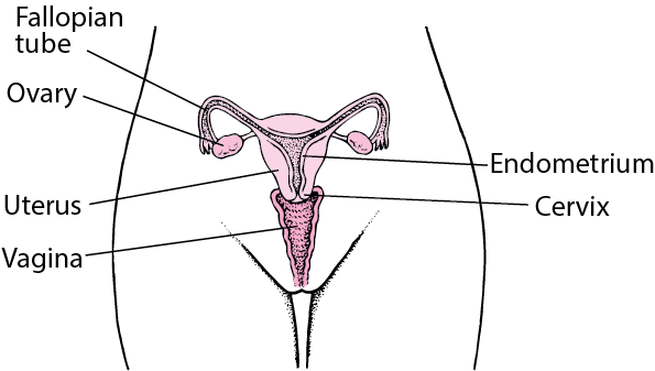
    gonadotropin sites

    Gonadotropin Sites of Action

    Female Sex Hormones

    The female sex hormones can be used to replace hormones that are missing or to act on the control mechanisms of the endocrine system to decrease the release of endogenous hormones.
    Drugs that act like estrogen, particularly at specific estrogen receptors, are also used to stimulate the effects of estrogen in the body with fewer of the adverse effects.

    Female sex hormones include;

    • Estrogens 
    • Progestins

    Estrogens.

    This hormone is naturally produced by the ovaries, placenta and adrenal glands. It stimulates the development of female sex characteristics, prepares the body for pregnancy, affects the release of FSH and LH, and is responsible for proliferation of the endometrial lining.

    Low estrogen in the body is responsible for the signs and symptoms of menopause, in the uterus, vagina, breast and cervix.

    Other Functions of estrogen include;

    1. Breast development.
    2. Increase cholesterol in bile, to prevent damaging effects of bile salts.
    3. Increases fat storage, such as in breast tissue.
    4. Maintains bone mineral density.
    5. Maintains muscle strength.
    6. Prevents atherosclerosis, by increasing HDL concentration and lowering LDL.
    7. Estrogen is responsible for maintaining libido, memory, and mental health. 
    8. It stimulates ovulation, maintains the uterine walls and is important in vaginal lubrication.
    Indications of Estrogen Therapy.
    • Estrogens are used for hormone replacement therapy (HRT) when ovarian activity is blocked or absent.
    • Is used to control the signs and symptoms of menopause.
    • They can also be used in therapy for prostate cancer and inoperable breast cancer, also as palliative care.
    • Treatment of female hypogonadism(when the body produces little or no hormones).
    • Treat ovarian failure.
    • Oral contraceptives (estrogen and progestin)
    • Morning after pill (emergency pills)
    • Endometriosis
    • Dysmenorrhea, used with progestin.

    Progestin/Progesterone.

    This promotes maintenance of pregnancy and it is called a pregnancy hormone.

    Its functions include;

    1. Transforms proliferative endometrium into secretory endometrium.
    2. Prevents follicle maturation, ovulation and uterine contractions.
    3. Used in contraceptives. It inhibits release of GnRH, FSH and LH, hence follicle development and ovulation are prevented.
    Indications of Progestin.
    • Used as a contraceptive.
    • Maintains pregnancy and development of secondary sex characteristics.
    • Use to treat primary and secondary amenorrhea, and functional uterine bleeding.
    • Treatment of acne and premenstrual dysphoric disorder (PMDD).
    • For the relief of signs and symptoms of menopause .
    Contraindications of Female Sex hormones.

    Estrogen

    • Known allergies
    • Idiopathic vaginal bleeding.
    • Breast Cancer(Estrogen dependant cancer)
    • CVA since it increases clotting factor prodn.
    • Hepatic dysfunction.
    • Pregnancy.
    • Lactation.

    Progestin/Progesterone

    • PID
    • STD
    • Endometriosis
    • Renal and hepatic disorders.
    • Epilepsy.
    • Asthma.
    • Migraine headaches
    • Cardiac Dysfunction —potential excerbation.
    Adverse Effects.
    • Corneal Changes.
    • Photosensitivity.
    • Peripheral edema.
    • Chloasma ( patches on the face)
    • Hepatic adenoma.
    • Nausea
    • Vomiting.
    • Abdominal cramps.
    • Bloating.
    • Withdraw bleeding.
    • Changes in menstrual flow.

    Important aspects/issues to remember.

    1.  Women receiving any of these drugs should receive an annual medical examination, including
      breast examination and Pap smear, to monitor for adverse effects and underlying medical
      conditions.
    2. Women taking estrogen should be advised not to smoke because of the increased risk of
      thrombotic events.
    3. Women who are receiving these drugs for fertility programs should receive a great deal of psychological support and comfort measures to cope with the many adverse effects associated
      with these drugs. The risk of multiple births should be explained.
    4. Drugs are used in treatment of specific cancers in males and they should be advised about the
      possibility of estrogenic effects.
    5. Not indicated during pregnancy or lactation because of potential for adverse effects on the fetus
      or neonate.
    Examples of female sex hormones and dosages.

    Estrogen

    1. Estradiol, 1–2 mg/day orally or  1–5 mg IM every 3–4 weeks or  2–4 g intravaginal cream daily.
    2. Estrogens, conjugated (C.E.S., Premarin), 0.3–1.25 mg/day orally.
    3. Estropipate (Ortho-Est, Ogen), 0.625–5 mg/day orally.

    Progestin/Progesterone.

    1. Etonogestrel (Implanon) 68 mg implanted sub dermally for up to 3 yr, replaced or changed when needed.
    2. Medroxyprogesterone (Provera) 5–10 mg/day PO for 5–10 days for amenorrhea or 400–1000 mg/week IM for cancer therapy or 150 mg of deep IM every 3 months (13 weeks) for contraception.
    Clinically important Drug Interactions

    Estrogen

    •  Barbiturates, rifampin, tetracyclines, phenytoin: decreased serum estrogen levels
    • Corticosteroids: increased therapeutic and toxic effects of corticosteroids.
    • Nicotine: Increased risk of thrombi and emboli
    • Grapefruit juice: inhibition of metabolism of estradiols
    • St. John’s wort: can affect metabolism of estrogens and can make estrogen-containing
      contraceptives less effective.

    Progestins

    •  Barbiturates, carbamazepine, phenytoin, griseofulvin, penicillin, tetracyclines, rifampin: reduced
      effectiveness of progestins
    • St. John’s wort: can affect the metabolism of progestins and can make progestin-containing
      contraceptives less effective..

    Gonadotropin drugs Read More »

    pneumonia in children

    Pneumonia in Children

    Pediatric Pneumonia Lecture Notes
    Pediatric Pneumonia

    Pneumonia remains a leading cause of morbidity and mortality in children worldwide, especially in developing countries. Its epidemiology and etiology differ significantly from adults, largely due to variations in immune system maturity, exposure patterns, and anatomical differences.

    Pneumonia is an acute inflammatory condition of the lung parenchyma caused by an infection.

    • lung parenchyma is the the functional tissue of the lungs, specifically the alveoli and bronchioles.

    This inflammation leads to the filling of the alveolar spaces with exudate, cells, and fluid, a process known as consolidation. This consolidation impairs gas exchange, leading to symptoms such as cough, fever, chills, and difficulty breathing.

    In simpler terms, pneumonia is an infection that inflames the air sacs in one or both lungs. The air sacs may fill with fluid or pus (purulent material), causing cough with phlegm or pus, fever, chills, and trouble breathing.

    Classifications of Pneumonia

    Pneumonia can be classified in various ways, each providing a different lens through which to understand its cause, presentation, and management.

    A. By Etiology (Cause of Infection):

    This classification focuses on the specific microorganism responsible for the infection.

    1. Bacterial Pneumonia: The most common type, often more severe than viral pneumonia.
      • Common Pathogens:
        • Streptococcus pneumoniae (Pneumococcus): The most frequent cause of community-acquired bacterial pneumonia.
        • Haemophilus influenzae.
        • Staphylococcus aureus (including MRSA).
        • Klebsiella pneumoniae.
        • Mycoplasma pneumoniae (often called "walking pneumonia" due to milder symptoms).
        • Chlamydophila pneumoniae.
        • Legionella pneumophila (Legionnaires' disease).
    2. Viral Pneumonia: Often milder than bacterial pneumonia but can be severe, especially in infants, elderly, and immunocompromised individuals.
      • Common Pathogens:
        • Influenza viruses (Types A and B).
        • Respiratory Syncytial Virus (RSV).
        • Adenoviruses.
        • Parainfluenza viruses.
        • Human Metapneumovirus.
        • Coronaviruses (e.g., SARS-CoV, MERS-CoV, SARS-CoV-2).
    3. Fungal Pneumonia: Less common, usually affecting individuals with weakened immune systems or those exposed to large amounts of fungi in the environment.
      • Common Pathogens:
        • Pneumocystis jirovecii (PCP pneumonia, common in HIV/AIDS patients).
        • Histoplasma capsulatum (Histoplasmosis).
        • Coccidioides immitis (Coccidioidomycosis or Valley Fever).
        • Blastomyces dermatitidis (Blastomycosis).
        • Aspergillus species.
    4. Parasitic Pneumonia: Rare, caused by parasites, usually seen in immunocompromised individuals or those who have traveled to endemic areas.
      • Common Pathogens:
        • Toxoplasma gondii.
        • Strongyloides stercoralis.
    5. Aspiration Pneumonia: Occurs when foreign material (e.g., food, liquid, vomit, stomach contents) is inhaled into the lungs, leading to inflammation and often secondary bacterial infection.
      • Causes: Impaired swallowing mechanisms, altered consciousness, gastroesophageal reflux.
    6. Chemical Pneumonia (Pneumonitis): Lung inflammation caused by inhaling irritating chemicals or toxic gases, rather than an infectious agent. This is not an infection but can predispose to one.
      • Causes: Inhalation of smoke, noxious fumes, or gastric acid.
    B. By Anatomical Location (Area of Lung Affected):

    This classification describes the pattern of lung involvement as seen on chest imaging.

    1. Lobar Pneumonia: Affects a large, continuous area of an entire lobe of a lung. Often caused by Streptococcus pneumoniae.
      • Appearance: Typically seen as a dense, homogeneous consolidation on chest X-ray.
    2. Bronchopneumonia (or Lobular Pneumonia): Characterized by patchy consolidation centered around the bronchi and bronchioles, often affecting multiple lobes. More common in infants, young children, and the elderly.
      • Appearance: Patchy infiltrates on chest X-ray, often bilateral and basal.
    3. Interstitial Pneumonia: Involves the interstitial spaces of the lung (the tissue between the alveoli and capillaries), rather than primarily the air sacs. More commonly associated with viral or atypical bacterial infections.
      • Appearance: Reticular or reticulonodular patterns on chest X-ray.
    4. Miliary Pneumonia: A form of pneumonia characterized by the wide dissemination of an infectious agent (Mycobacterium tuberculosis) throughout the lung tissue in small, discrete lesions resembling millet seeds.
      • Appearance: Fine, diffuse nodular infiltrates throughout both lungs on chest X-ray.
    C. By Duration:

    This classification refers to the time course of the illness.

    1. Acute Pneumonia: Rapid onset and progression of symptoms, typically resolving within days to a few weeks with appropriate treatment. Most common form.
    2. Chronic Pneumonia: Persistent symptoms and radiological findings lasting for weeks to months, or even longer. Often associated with specific pathogens (e.g., Mycobacterium tuberculosis, fungi) or underlying conditions.
    D. By Clinical Grounds / Acquisition Setting:

    This is one of the most clinically relevant classifications, as it guides initial empiric treatment decisions.

    1. Community-Acquired Pneumonia (CAP): Pneumonia acquired outside of hospitals or long-term care facilities.
      • Common Pathogens: Streptococcus pneumoniae, Mycoplasma pneumoniae, Chlamydophila pneumoniae, Haemophilus influenzae, influenza virus.
    2. Hospital-Acquired Pneumonia (HAP) / Nosocomial Pneumonia: Pneumonia that develops 48 hours or more after hospital admission and was not incubating at the time of admission.
      • Common Pathogens: Often more virulent and antibiotic-resistant bacteria, such as Pseudomonas aeruginosa, Staphylococcus aureus (MRSA), Klebsiella species, Escherichia coli.
    3. Ventilator-Associated Pneumonia (VAP): A subtype of HAP that develops in patients who have been mechanically ventilated for more than 48 hours.
      • Common Pathogens: Similar to HAP, often highly resistant organisms.
    Etiology of Pneumonia

    The etiology refers to the specific agents or organisms responsible for causing pneumonia. As discussed in Objective 1, these can be broadly categorized.

    A. Common Bacterial Pathogens:

    These are the most frequent causes of pneumonia, especially bacterial pneumonia.

    1. Streptococcus pneumoniae (Pneumococcus):
      • Description: The leading cause of community-acquired bacterial pneumonia (CAP) in all age groups, particularly in adults.
      • Characteristics: Gram-positive coccus, typically arranged in pairs (diplococci). Has a polysaccharide capsule that protects it from phagocytosis.
      • Risk Factors: Old age, chronic lung disease, recent viral infection, immunocompromised status.
    2. Haemophilus influenzae:
      • Description: A common cause of both CAP and HAP, especially in individuals with chronic obstructive pulmonary disease (COPD) or other underlying lung conditions.
      • Characteristics: Gram-negative coccobacillus.
      • Risk Factors: COPD, cystic fibrosis, alcoholism.
    3. Staphylococcus aureus:
      • Description: Can cause severe pneumonia, often seen as HAP or as a complication of viral infections (e.g., influenza). Methicillin-resistant S. aureus (MRSA) is a significant concern, especially in VAP and HCAP.
      • Characteristics: Gram-positive coccus, often arranged in clusters. Produces various toxins.
      • Risk Factors: Recent influenza, injection drug use, skin/soft tissue infection, hospitalization, surgical procedures.
    4. Klebsiella pneumoniae:
      • Description: A common cause of HAP and, less frequently, severe CAP, particularly in individuals with alcoholism or diabetes. Known for causing "currant jelly" sputum.
      • Characteristics: Gram-negative rod, often encapsulated.
      • Risk Factors: Alcoholism, diabetes, chronic lung disease, hospitalization.
    5. Pseudomonas aeruginosa:
      • Description: A significant cause of HAP and VAP, particularly in immunocompromised patients, those with cystic fibrosis, or prolonged hospital stays. Difficult to treat due to antibiotic resistance.
      • Characteristics: Gram-negative rod.
      • Risk Factors: Cystic fibrosis, bronchiectasis, mechanical ventilation, broad-spectrum antibiotic use, immunocompromised state.
    6. Mycoplasma pneumoniae:
      • Description: A common cause of "atypical pneumonia" or "walking pneumonia" in young adults and school-aged children. Causes milder, but prolonged, symptoms.
      • Characteristics: Lacks a cell wall, making it resistant to many common antibiotics (e.g., penicillin).
    7. Chlamydophila pneumoniae:
      • Description: Another cause of atypical pneumonia, often with milder symptoms.
      • Characteristics: Obligate intracellular bacterium.
    8. Legionella pneumophila:
      • Description: Causes Legionnaires' disease, a severe form of pneumonia often associated with contaminated water sources (e.g., air conditioning systems, hot tubs).
      • Characteristics: Gram-negative rod, fastidious growth requirements.
    B. Common Viral Pathogens:

    Viruses are a very common cause of pneumonia, especially in children. They can also predispose to secondary bacterial infections.

    1. Influenza Viruses (A and B): Seasonal epidemics cause widespread respiratory illness, including primary viral pneumonia and often secondary bacterial pneumonia.
    2. Respiratory Syncytial Virus (RSV): The most common cause of lower respiratory tract infections in infants and young children, often leading to bronchiolitis and pneumonia.
    3. Adenoviruses: Can cause a range of respiratory illnesses, including pneumonia, particularly in children and immunocompromised individuals.
    4. Parainfluenza Viruses: Common cause of croup, but can also cause bronchiolitis and pneumonia, especially in children.
    5. Coronaviruses (e.g., SARS-CoV-2): Various coronaviruses can cause respiratory infections, with SARS-CoV-2 (COVID-19) being a notable cause of severe viral pneumonia and acute respiratory distress syndrome (ARDS).
    C. Common Fungal Pathogens:

    More prevalent in immunocompromised individuals or specific geographic regions.

    1. Pneumocystis jirovecii: Causes Pneumocystis pneumonia (PCP), a common and severe opportunistic infection in individuals with HIV/AIDS.
    2. Endemic Fungi (e.g., Histoplasma capsulatum, Coccidioides immitis, Blastomyces dermatitidis): Found in specific geographic areas. Exposure to spores can lead to pneumonia, especially in immunocompromised individuals.
    3. Aspergillus species: Can cause invasive aspergillosis, a severe pneumonia, primarily in severely immunocompromised patients (e.g., transplant recipients, leukemia patients).
    D. Aspiration of Gastric Contents/Foreign Material:

    Not an infectious agent itself, but the aspiration of acidic gastric contents or other foreign material can cause a severe chemical pneumonitis, which then often becomes secondarily infected by oral flora (anaerobic bacteria).

    Pathogenesis of Pneumonia

    Pathogenesis describes the sequence of events that leads to the development of pneumonia, from initial exposure to clinical symptoms.

    A. Normal Host Defenses:

    The respiratory tract has several protective mechanisms to prevent infection:

    1. Upper Airway Filtration: Nasal hairs, turbinates, and mucous membranes filter out large particles.
    2. Epiglottis and Cough Reflex: Protect the lower airways from aspiration.
    3. Mucociliary Escalator: Ciliated epithelial cells line the trachea and bronchi, moving mucus (which traps pathogens) upwards for expectoration or swallowing.
    4. Alveolar Macrophages: Phagocytic cells in the alveoli that engulf and destroy pathogens and debris.
    5. Humoral and Cellular Immunity: Antibodies (IgA, IgG) and T lymphocytes provide specific immunity.
    Mechanisms of Pathogen Entry:

    Pneumonia develops when pathogens overcome or bypass these host defenses.

    1. Aspiration (Most Common): Microaspiration of oropharyngeal secretions containing pathogens is the most frequent route. This happens constantly in small amounts, but typically the host defenses clear them. Impaired consciousness, dysphagia, or presence of a nasogastric tube increases the risk of significant aspiration.
    2. Inhalation: Airborne pathogens (e.g., viruses, Mycoplasma, Legionella, fungi) can be inhaled directly into the lower respiratory tract.
    3. Hematogenous Spread: Pathogens from a distant site of infection (e.g., endocarditis, IV drug use, abdominal sepsis) can travel through the bloodstream to the lungs.
    4. Direct Spread: Less common, but can occur from contiguous infected sites (e.g., empyema spreading to lung, trauma).
    Pathophysiology:

    Once pathogens reach the lower respiratory tract and evade local defenses, a series of events leads to inflammation and consolidation:

    1. Colonization and Multiplication: Pathogens colonize the alveoli and/or terminal bronchioles and begin to multiply.
    2. Immune Response and Inflammation:
      • Alveolar Macrophages: Are typically the first line of defense. If overwhelmed, they release cytokines (e.g., TNF-alpha, IL-1, IL-6, IL-8).
      • Neutrophil Recruitment: These cytokines attract neutrophils from the bloodstream into the alveolar spaces.
      • Increased Vascular Permeability: The inflammatory response causes vasodilation and increased permeability of the alveolar-capillary membrane.
    3. Fluid Exudation and Consolidation:
      • Plasma fluid, red blood cells, and fibrin leak into the alveolar spaces.
      • Neutrophils and bacteria fill the alveoli.
      • This mixture of fluid, cells, and debris leads to the characteristic consolidation seen in pneumonia, where the lung tissue becomes dense and airless.
    4. Impaired Gas Exchange:
      • The consolidated alveoli can no longer participate in gas exchange.
      • This leads to ventilation-perfusion mismatch (areas are perfused but not ventilated), resulting in hypoxemia (low blood oxygen).
      • The increased work of breathing due to decreased lung compliance and airway obstruction can also lead to hypercapnia (high blood carbon dioxide) in severe cases.
    5. Tissue Damage: The inflammatory process and release of bacterial toxins can cause damage to the alveolar and bronchial epithelial cells, impairing mucociliary function and further propagating inflammation.
    6. Resolution: With effective immune response and/or antibiotic treatment, the inflammation subsides, macrophages clear cellular debris, and the exudate is reabsorbed, allowing the lung to return to normal function.
    Etiology of Pediatric Pneumonia (Causative Agents)

    The pathogens responsible for pneumonia vary significantly by age group.

    A. Neonates (Birth to 1 Month):
  • Pneumonia in neonates is often acquired perinatally (from the mother during birth) or nosocomially (in the hospital).
  • Bacterial:
    • Group B Streptococcus (GBS): Common cause of early-onset neonatal sepsis and pneumonia.
    • Gram-negative enteric bacilli: Escherichia coli, Klebsiella pneumoniae.
    • Listeria monocytogenes.
  • Viral: Less common primary cause, but can be involved (e.g., Herpes Simplex Virus - HSV).
  • B. Infants (1 Month to 6 Months):
  • Transition period, with a mix of perinatal pathogens and increasing community-acquired pathogens.
  • Bacterial:
    • Streptococcus pneumoniae (pneumococcus): Increasingly common.
    • Haemophilus influenzae (non-typeable or type b if unvaccinated).
    • Staphylococcus aureus: Can cause severe disease.
  • Atypical Bacteria:
    • Chlamydia trachomatis: Can cause afebrile pneumonia, often associated with conjunctivitis, transmitted from mother during birth. Presents at 2-12 weeks of age.
    • Bordetella pertussis (whooping cough): Can cause severe pneumonia, especially in unvaccinated infants.
  • Viral (Most Common Overall):
    • Respiratory Syncytial Virus (RSV): The leading cause of bronchiolitis and pneumonia in infants.
    • Parainfluenza viruses: (Types 1, 2, 3).
    • Adenovirus: Can cause severe and prolonged disease.
    • Influenza viruses: (A and B).
    • Human Metapneumovirus.
  • C. Preschool Children (6 Months to 5 Years):
  • Viral (Still Most Common):
    • RSV, Influenza, Parainfluenza, Adenovirus, Human Metapneumovirus, Rhinovirus.
  • Bacterial:
    • Streptococcus pneumoniae (Pneumococcus): Remains the most frequent bacterial cause.
    • Haemophilus influenzae (non-typeable).
    • Staphylococcus aureus (including MRSA).
    • Streptococcus pyogenes (Group A Strep): Less common but can cause severe pneumonia.
  • Atypical Bacteria:
    • Mycoplasma pneumoniae: Becomes more common in this age group, though classically associated with school-aged children.
  • D. School-Aged Children and Adolescents (> 5 Years):
  • The spectrum of pathogens begins to resemble that of adults.
  • Atypical Bacteria (Increasingly Common):
    • Mycoplasma pneumoniae: The most common cause of "atypical pneumonia" or "walking pneumonia."
    • Chlamydophila pneumoniae.
  • Bacterial:
    • Streptococcus pneumoniae.
    • Staphylococcus aureus (including MRSA).
    • Haemophilus influenzae.
    • Streptococcus pyogenes.
  • Viral:
    • Influenza A and B.
    • Adenovirus.
  • E. Less Common but Important Causes (Across Age Groups):
  • Tuberculosis (Mycobacterium tuberculosis): Consider in endemic areas or with risk factors.
  • Fungal Pneumonia: (e.g., Pneumocystis jirovecii pneumonia - PCP) primarily in immunocompromised children.
  • Aspiration Pneumonia: In children with feeding difficulties, GERD, or neurological impairment.
  • Clinical Presentation of Pneumonia in Children

    Recognize age-specific manifestations and indicators of severity to ensure timely intervention.

    I. General Signs and Symptoms of Pneumonia in Children
  • Cough: May be dry, moist, or productive (though young children rarely expectorate sputum). Can sometimes be the only prominent symptom.
  • Tachypnea (Increased Respiratory Rate): Often the most sensitive and specific sign of pneumonia in children, especially in infants. Defined as:
    • < 2 months: ≥ 60 breaths/min
    • 2-11 months: ≥ 50 breaths/min
    • 1-5 years: ≥ 40 breaths/min
    • 5 years: ≥ 20 breaths/min
  • Fever: Present in many cases, but can be absent, especially in neonates, young infants, or immunocompromised children.
  • Dyspnea (Difficulty Breathing): Manifested as increased work of breathing.
  • Lethargy / Irritability: Non-specific signs of illness in children.
  • Poor Feeding / Decreased Oral Intake: Common in infants and young children.
  • Chest Pain: More common in older children, often pleuritic (sharp, worse with breathing).
  • Abdominal Pain: Can be referred pain from diaphragmatic irritation, especially in lower lobe pneumonia.
  • II. Age-Specific Clinical Manifestations
    A. Neonates (Birth to 1 Month):
  • Pneumonia in neonates is often subtle and non-specific, making diagnosis challenging.
  • Non-specific Signs:
    • Respiratory Distress: Tachypnea (often the earliest sign), grunting, nasal flaring, retractions (subcostal, intercostal, suprasternal).
    • Apnea: Pauses in breathing, especially in premature infants.
    • Cyanosis (bluish discoloration) or pallor.
    • Lethargy, irritability, hypotonia.
    • Poor feeding, vomiting.
    • Temperature instability (hypothermia is common, fever less so).
    • Jaundice.
  • Physical Exam: May reveal decreased breath sounds, crackles (rales), or wheezing.
  • B. Infants (1 Month to 1 Year):
  • More overt signs of respiratory illness are typically present.
  • Key Signs:
    • Tachypnea: Always a critical sign.
    • Retractions: Subcostal, intercostal, suprasternal, supraclavicular.
    • Nasal Flaring.
    • Grunting: Short, low-pitched sounds during expiration, attempting to increase end-expiratory pressure.
    • Cough: Can be prominent, may be paroxysmal, especially with Pertussis or viral causes like RSV.
    • Fever.
    • Poor feeding, decreased activity.
    • Wheezing (more common with viral pneumonia/bronchiolitis).
  • Physical Exam: Crackles, decreased breath sounds, dullness to percussion (if consolidation is significant).
  • C. Toddlers and Preschoolers (1 Year to 5 Years):
  • Similar to infants, but with more verbal communication of symptoms.
  • Key Signs:
    • Tachypnea.
    • Cough: Often harsh and persistent.
    • Fever.
    • Dyspnea, increased work of breathing.
    • Lethargy, irritability, decreased playfulness.
    • Decreased appetite.
    • Abdominal pain: Can be a presenting complaint, particularly with lower lobe pneumonia irritating the diaphragm.
  • Physical Exam: Crackles, rhonchi, decreased breath sounds, dullness to percussion.
  • D. School-Aged Children and Adolescents (> 5 Years):
  • Clinical presentation begins to resemble adult pneumonia.
  • Key Signs:
    • Cough: Can be productive with sputum, especially in bacterial pneumonia.
    • Fever and Chills.
    • Dyspnea / Shortness of Breath.
    • Pleuritic Chest Pain: Sharp pain worsened by breathing or coughing.
    • Headache, malaise, myalgia.
    • Abdominal pain.
    • "Atypical" Pneumonia (e.g., Mycoplasma pneumoniae): Often presents with more insidious onset, low-grade fever, persistent dry cough, headache, and malaise, sometimes called "walking pneumonia."
  • Physical Exam: Crackles, egophony, decreased breath sounds, dullness to percussion.
  • III. Indicators of Severe Pneumonia / Respiratory Distress in Children

    Rapid recognition of these signs is critical for determining the need for hospitalization and intensive care.

  • Inability to Feed/Drink: Especially in infants and young children.
  • Severe Respiratory Distress:
    • Severe Tachypnea (respiratory rate significantly above age-appropriate limits).
    • Severe Retractions (all types, especially supraclavicular, tracheal tug).
    • Grunting.
    • Nasal Flaring.
    • Central Cyanosis: Bluish discoloration of the tongue, lips, and nail beds, indicating hypoxemia.
    • Head Bobbing: Especially in infants.
  • Altered Mental Status: Lethargy, extreme irritability, difficult to arouse, confusion.
  • Hypoxemia: SpO2 < 90% (or lower, depending on altitude and clinical context) on room air.
  • Signs of Dehydration.
  • Signs of Shock: Tachycardia, poor perfusion, hypotension (a late sign in children).
  • Diagnostic Approaches for Pneumonia in Children
    Clinical Assessment (The Most Important Step):
  • History:
    • Onset and duration of symptoms (fever, cough, respiratory distress, feeding difficulties).
    • Exposure history (sick contacts, daycare, travel).
    • Vaccination status.
    • Risk factors (prematurity, underlying medical conditions).
    • Medication history.
  • Physical Examination:
    • General Appearance: Alertness, activity level, signs of distress.
    • Vital Signs: Respiratory rate (most sensitive sign of pneumonia), heart rate, temperature, blood pressure.
    • Respiratory Examination:
      • Inspection: Work of breathing (retractions, nasal flaring, grunting), cyanosis, symmetry of chest movement.
      • Palpation: Tactile fremitus (may be increased over consolidation, but difficult in young children).
      • Percussion: Dullness over consolidated areas or pleural effusion.
      • Auscultation:
        • Crackles (rales): Suggestive of alveolar inflammation/fluid.
        • Bronchial breath sounds: Over consolidated lung tissue.
        • Wheezing: More common in viral causes or with underlying reactive airway disease.
        • Decreased or absent breath sounds: May indicate consolidation or pleural effusion.
    • Other Systems: Assess for dehydration, cardiac involvement, neurological status.
  • Pulse Oximetry:
    • Essential non-invasive test in all children suspected of having pneumonia.
    • Measures oxygen saturation (SpO2). Hypoxemia (SpO2 < 90-92% on room air) is a strong indicator of severity and often guides hospitalization and oxygen therapy.
    Chest Radiography (CXR):
  • Indications:
    • Typically not recommended for routine diagnosis of uncomplicated community-acquired pneumonia in children who can be managed as outpatients and whose diagnosis is clear clinically.
    • Recommended for:
      • Children with severe pneumonia.
      • Uncertain diagnosis, or if differential diagnoses like foreign body aspiration are considered.
      • Failure to respond to initial empiric therapy.
      • Suspicion of complications (e.g., pleural effusion, empyema, abscess).
      • Recurrent pneumonia.
  • Findings:
    • Lobar Consolidation: Suggests bacterial pneumonia.
    • Interstitial Infiltrates: More characteristic of viral or atypical pneumonia.
    • Bronchial Wall Thickening/Peribronchial Cuffing: Common in viral infections.
    • Pleural Effusion, Empyema, Pneumothorax: Indicate complications.
    • Hyperinflation: Common in viral bronchiolitis.
  • Limitations:
    • Cannot reliably distinguish between bacterial and viral pneumonia.
    • Poor correlation between radiological findings and clinical severity.
    • Radiation exposure.
  • Laboratory Tests:
  • Blood Cultures:
    • Generally NOT recommended for routine CAP in outpatient settings.
    • Consider for: Hospitalized children with severe pneumonia, immunocompromised children, suspicion of bacteremia. Low yield (typically < 1-2%).
  • Complete Blood Count (CBC) with Differential:
    • Not routinely recommended for uncomplicated CAP.
    • May show leukocytosis with neutrophilia in bacterial infection, or lymphocytosis in viral infection, but findings can overlap and are not definitive.
  • Inflammatory Markers (e.g., C-reactive protein (CRP), Procalcitonin):
    • May be elevated in bacterial infections, but also in severe viral infections.
    • Not routinely used for initial diagnosis but can sometimes aid in differentiating bacterial from viral, or monitoring response to treatment.
  • Viral Diagnostics (e.g., Nasopharyngeal Swabs for PCR):
    • Recommended for: All hospitalized infants and young children with suspected viral pneumonia/bronchiolitis (e.g., RSV, influenza, adenovirus, parainfluenza).
    • Important for infection control, cohorting patients, and avoiding unnecessary antibiotic use.
    • Does not rule out bacterial co-infection.
  • Sputum Culture:
    • Difficult to obtain in young children, often contaminated by upper airway flora. Not routinely recommended.
  • Pleural Fluid Analysis:
    • If pleural effusion is present, diagnostic thoracentesis may be performed to identify the pathogen and guide treatment for empyema.
  • Tuberculin Skin Test (TST) / Interferon-Gamma Release Assay (IGRA):
    • Consider in children with persistent or recurrent pneumonia, or risk factors for tuberculosis.
  • Serology for Atypical Pathogens (e.g., Mycoplasma pneumoniae, Chlamydia pneumoniae):
    • Can be useful for retrospective diagnosis, but acute and convalescent titers are needed, so not helpful for acute management.
  • Differential Diagnoses

    Many conditions can mimic pneumonia in children due to similar respiratory symptoms.

    1. Upper Respiratory Tract Infection (URI) / Common Cold: Often presents with cough, rhinorrhea, low-grade fever. Absence of tachypnea and significant work of breathing usually differentiates it from pneumonia.
    2. Bronchiolitis: Common in infants < 2 years, primarily caused by RSV. Presents with cough, rhinorrhea, tachypnea, prominent wheezing, and crackles. Often difficult to distinguish clinically from viral pneumonia, and they can coexist.
    3. Asthma Exacerbation / Reactive Airway Disease: Wheezing, cough, dyspnea. History of recurrent episodes or triggers may point to asthma.
    4. Foreign Body Aspiration: Sudden onset of choking, coughing, dyspnea, particularly in toddlers. Can lead to unilateral wheezing or recurrent localized pneumonia. A high index of suspicion is needed. CXR may show unilateral hyperinflation or atelectasis.
    5. Croup (Laryngotracheobronchitis): "Barking" cough, inspiratory stridor, hoarseness, typically worse at night. Primarily affects the upper airway.
    6. Pertussis (Whooping Cough): Prolonged paroxysmal cough, often followed by a "whooping" sound and post-tussive emesis. Can cause severe pneumonia in infants.
    7. Heart Failure: Tachypnea, cough, poor feeding, hepatomegaly, often in infants with congenital heart disease. CXR may show cardiomegaly and pulmonary edema.
    8. Pulmonary Edema: Can result from fluid overload, acute kidney injury, or cardiac dysfunction.
    9. Pleural Effusion (without underlying pneumonia): Can cause dyspnea and decreased breath sounds, but usually related to other causes (e.g., malignancy, autoimmune disease).
    10. Tuberculosis: Consider in endemic areas or with risk factors, especially for persistent cough, failure to thrive, or abnormal CXR.
    Medical Management for Pediatric Pneumonia

    Aims: The medical management of pediatric pneumonia aims to eradicate the causative pathogen, alleviate symptoms, prevent complications, and provide supportive care tailored to the child's age and severity of illness.

    I. General Principles of Management
    1. Assessment of Severity: The initial step is to assess the severity of pneumonia to determine the appropriate level of care (outpatient vs. inpatient, general ward vs. ICU). Key indicators include respiratory distress (tachypnea, retractions, grunting, nasal flaring), hypoxemia (SpO2 < 90-92%), inability to feed, lethargy, and signs of dehydration.
    2. Empiric Antibiotic Therapy:
      • Rationale: While viral etiologies are common, bacterial pneumonia can be severe and life-threatening. Clinical signs often overlap, and rapid viral testing may not be immediately available. Therefore, empiric antibiotic treatment is crucial, especially in moderate to severe cases, to cover likely bacterial pathogens.
      • De-escalation: Once a pathogen is identified (e.g., strong evidence of viral infection) or if the child rapidly improves, antibiotics may be discontinued or narrowed.
    3. Supportive Care: This is the cornerstone of management for all types of pneumonia (viral and bacterial) and focuses on maintaining oxygenation, hydration, nutrition, and comfort.
    II. Specific Management Strategies
    A. Antimicrobial Therapy (Based on your provided text and general guidelines):

    The choice of antibiotic depends on the child's age, severity of illness, local resistance patterns, and immunization status.

    1. For Infants under 2 months with Severe Pneumonia (Hospitalized):
      • First-line combination: Ampicillin (150-200 mg/kg/day in divided doses IV) plus Gentamycin (5-6 mg/kg/day IV).
        • Rationale: Covers common neonatal pathogens like Group B Streptococcus and Gram-negative enteric bacilli.
      • Alternative if Penicillin Not Available/Suitable: Cefotaxime (IV).
      • If Condition Does Not Improve/Suspicion of S. aureus: Add Cloxacillin (IV) to cover Staphylococcus aureus.
      • Duration: Typically 10 days, but can be individualized based on clinical response and pathogen.
    2. For Older Children 2 months to 5 years (Hospitalized with Severe Pneumonia):
      • First-line: Ceftriaxone (IV, 50-100 mg/kg/day once daily) or Ampicillin plus Gentamycin.
        • Rationale: Ceftriaxone provides broad-spectrum coverage against Streptococcus pneumoniae and Haemophilus influenzae. Ampicillin + Gentamycin is an alternative.
      • Consideration for Atypical Pathogens (e.g., Mycoplasma): If atypical pneumonia is suspected (e.g., persistent cough, gradual onset, older child), a macrolide (e.g., Azithromycin) may be added or used alone depending on clinical suspicion and local guidelines.
      • Duration: Typically 7-10 days.
    3. For Children with Non-Serious Pneumonia (Outpatient Management):
      • First-line: Amoxicillin (oral, 80-90 mg/kg/day divided twice daily).
        • Rationale: Effective against Streptococcus pneumoniae, the most common bacterial cause in this age group, and has a good safety profile.
      • Alternative if Amoxicillin Allergy or Suspected Atypical Pathogen: Macrolide (e.g., Azithromycin, Erythromycin) may be considered.
      • Duration: Typically 5-7 days for uncomplicated cases.
    B. Symptomatic Management and Supportive Care:
    1. Fever Management:
      • Paracetamol (Acetaminophen): Administer for fever (and pain) as per weight-based dosing.
      • Tepid Sponging: Can be used as an adjunctive measure if the child is uncomfortable or has very high fever, but should not be the sole method of fever reduction and can cause discomfort.
      • Goal: Improve comfort and reduce metabolic demands, not necessarily to normalize temperature.
    2. Respiratory Support:
      • Positioning: Nurse patient in a semi-sitting up position or with the head elevated to aid breathing and improve lung expansion.
      • Airway Clearance:
        • Nasal Irrigation: With 0.9% sodium chloride to clear nasal passages, especially important in neonates and infants who are obligate nasal breathers.
        • Assisted Coughing/Suctioning: If the child is unable to clear secretions effectively. Suctioning should be performed gently and only when necessary to avoid trauma or laryngospasm.
        • Chest Physiotherapy (CPT) / Chest Exercises: Can be helpful, especially in cases with significant secretions or atelectasis, but evidence for routine use in uncomplicated pneumonia is mixed.
      • Monitoring for Increased Respiratory Distress: Continuous assessment of respiratory rate, work of breathing, and oxygen saturation is paramount.
      • Bronchodilators: Administer bronchodilators (e.g., inhaled salbutamol) if there is evidence of bronchospasm or significant wheezing, especially in children with a history of asthma or bronchiolitis.
      • Oxygen Therapy:
        • Indication: Administer oxygen where hypoxemia (SpO2 < 90-92% on room air) or cyanosis has occurred.
        • Delivery Methods: Nasal cannula, oxygen mask, high-flow nasal cannula (HFNC) for more severe cases.
        • Goal: Maintain SpO2 > 90-92% (or higher, depending on clinical scenario).
    3. Fluid and Nutritional Support:
      • Hydration: Promote adequate rehydration.
        • Oral Fluids: Encourage frequent sips of oral fluids (water, breast milk, rehydration solutions) as tolerated.
        • Intravenous (IV) Fluids: In children with severe respiratory difficulty, vomiting, or inability to take oral fluids, place an IV line and give fluids cautiously. Typically, start with 70-80% of normal maintenance fluids to avoid fluid overload, which can worsen pulmonary edema. Resume oral fluids as soon as possible.
      • Nutrition:
        • Breastfeeding on Demand: For infants, if they are able to suck effectively and without severe respiratory distress. Breast milk provides vital antibodies and nutrients.
        • Well-Balanced Nutrition: For older children. If oral intake is poor due to dyspnea or fatigue, nasogastric tube (NGT) feeding may be necessary to ensure adequate caloric and fluid intake.
    C. General Care and Monitoring:
    1. Observations: Regular and frequent monitoring of respiratory rate, temperature, heart rate, and oxygen saturation is essential to assess response to treatment and detect deterioration.
    2. Hygiene: Maintain good personal and environmental hygiene to prevent further infections and transmission.
    3. Keep Patient Warm and Dry: Ensure comfortable body temperature and clean, dry clothing/bedding.
    4. Change Position: Regularly change the patient's position to prevent skin breakdown, promote lung expansion, and facilitate secretion drainage.
    5. Rest: Provide adequate rest periods to conserve the child's energy.
    6. Pain Management: Treat any associated pain (e.g., pleuritic chest pain) with analgesics like paracetamol or ibuprofen.
    Nursing Diagnoses for Pediatric Pneumonia

    These diagnoses guide the nurse in identifying patient needs and planning individualized care.

    1. Ineffective Airway Clearance related to increased tracheobronchial secretions, ineffective cough (especially in young children), and inflammation, as evidenced by adventitious breath sounds (crackles, rhonchi), ineffective or absent cough, nasal flaring, tachypnea, dyspnea, pallor/cyanosis, poor feeding.
    2. Impaired Gas Exchange related to alveolar-capillary membrane changes (inflammation, exudate), ventilation-perfusion mismatch, as evidenced by tachypnea, dyspnea, hypoxemia (SpO2 < 90-92%), cyanosis, restlessness/irritability/lethargy, abnormal blood gases.
    3. Ineffective Breathing Pattern related to inflammation, pain (pleuritic), and fatigue, as evidenced by tachypnea, dyspnea, use of accessory muscles, shallow respirations, retractions, grunting.
    4. Risk for inadequate Fluid Volume related to fever, increased insensible fluid loss (tachypnea), decreased oral intake, and vomiting, as evidenced by dry mucous membranes, decreased urine output, poor skin turgor, sunken fontanelles (infants), absent tears.
    5. Inadequate protein energy intake related to anorexia, dyspnea, fatigue, increased metabolic needs, and difficult feeding, as evidenced by reported inadequate intake, weight loss/poor weight gain, refusal to eat/drink, fatigue during feeding.
    6. Hyperthermia related to infectious process and increased metabolic rate, as evidenced by elevated body temperature, flushed skin, tachycardia, tachypnea, irritability.
    7. Acute Pain related to inflammation of lung parenchyma/pleura or generalized body aches, as evidenced by verbal reports of pain (older child), grimacing, guarding, restlessness, crying, irritability, withdrawal.
    8. Activity Intolerance related to imbalance between oxygen supply and demand, generalized weakness, and fatigue, as evidenced by verbal reports of fatigue (older child), decreased play/activity, exertional dyspnea, abnormal heart rate/blood pressure response to activity.
    9. Excessive Anxiety (Child/Parent) related to dyspnea, threat to health status, hospitalization, unfamiliar environment, and fear of unknown outcomes, as evidenced by restlessness, crying, apprehension, irritability, verbalization of concerns.
    10. Inadequate Health Knowledge (Parents) related to disease process, treatment regimen, home care, and prevention, as evidenced by questions, inaccurate follow-through of instructions, verbalization of concerns.
    Specific Nursing Interventions for Pediatric Pneumonia

    These interventions are tailored to the child's age and developmental stage, focusing on gentle, non-threatening approaches.

    A. For Ineffective Airway Clearance / Impaired Gas Exchange / Ineffective Breathing Pattern:
    Intervention Detail/Rationale
    1. Continuous Respiratory Assessment Monitor respiratory rate, depth, rhythm, effort (retractions, nasal flaring, grunting), breath sounds, SpO2 (continuous pulse oximetry is often used), skin color (for cyanosis) every 1-4 hours or more frequently as needed.
    2. Positioning Place the child in a semi-Fowler's position (head of bed elevated 30-45 degrees) or position of comfort to promote lung expansion. Avoid positions that might impede breathing.
    3. Airway Management
    • Nasal Care: Perform nasal saline irrigation and gentle suctioning, especially before feeds and sleep, for infants and young children to clear nasal passages.
    • Encourage Coughing: For older children, encourage deep breathing and effective coughing. For younger children, provide chest physiotherapy (percussion, vibration) as prescribed, followed by suctioning or assisted coughing if appropriate, to mobilize secretions.
    • Suctioning: Perform gentle nasopharyngeal or oropharyngeal suctioning only when necessary to remove visible secretions that the child cannot clear. Use appropriate catheter size and technique to avoid trauma.
    4. Oxygen Therapy
    • Administer warmed, humidified oxygen via nasal cannula, mask, hood, or tent as prescribed, to maintain SpO2 > 90-92%.
    • Monitor oxygen flow rate and ensure patency of delivery device.
    • Minimize crying and agitation to conserve oxygen.
    5. Administer Medications Give bronchodilators, antibiotics, and other prescribed respiratory medications (e.g., corticosteroids) on time and monitor for effectiveness and side effects.
    6. Maintain Hydration Ensure adequate hydration to thin secretions. (See Fluid and Nutrition section).
    B. For Risk for Inadequate Fluid Volume / Inadequate protein energy intake:
    Intervention Detail/Rationale
    1. Monitor Fluid Balance Strictly monitor intake (oral, IV, NGT) and output (urine, stools, emesis). Assess for signs of dehydration (e.g., dry mucous membranes, sunken fontanelles, poor skin turgor, decreased urine output).
    2. Promote Hydration
    • Oral: Offer small, frequent amounts of preferred clear fluids (e.g., Pedialyte, water, diluted juice). For infants, encourage frequent, shorter breastfeeds or formula feeds if tolerated.
    • IV Fluids: Administer IV fluids as prescribed, monitoring for signs of overhydration (e.g., crackles, edema).
    3. Optimize Nutrition
    • Small, Frequent Meals: Offer small, frequent, nutrient-dense meals or snacks.
    • Rest Before Feeds: Allow rest periods before feeding to conserve energy.
    • NGT/OGT Feeding: If the child has significant respiratory distress, is unable to feed orally, or is losing weight, administer feeds via nasogastric or orogastric tube as prescribed.
    • Consult Dietitian: For specialized nutritional assessment and planning.
    C. For Hyperthermia / Acute Pain:
    Intervention Detail/Rationale
    1. Monitor Temperature Assess temperature regularly.
    2. Fever Management
    • Administer antipyretics (e.g., Paracetamol, Ibuprofen) as prescribed, ensuring correct dose based on weight.
    • Remove excessive clothing, use lightweight blankets.
    • Tepid sponging may be used if the child is uncomfortable, but avoid chilling.
    3. Pain Assessment Use age-appropriate pain scales (e.g., FLACC scale for non-verbal children, Faces Pain Scale for older children).
    4. Pain Management
    • Administer analgesics (e.g., Paracetamol, Ibuprofen) as prescribed.
    • Utilize non-pharmacological methods: comfort positioning, distraction (toys, stories, music), parental presence, gentle touch.
    D. For Activity Intolerance:
    Intervention Detail/Rationale
    1. Balance Rest and Activity Organize care to allow for uninterrupted rest periods.
    2. Encourage Age-Appropriate Activity Gradually increase activity as tolerated, monitoring for signs of fatigue or respiratory distress.
    3. Assist with ADLs Provide assistance with activities of daily living as needed to conserve energy.
    E. For Excessive Anxiety (Child/Parent):
    Intervention Detail/Rationale
    1. Child
    • Provide a calm, reassuring presence.
    • Use age-appropriate language to explain procedures.
    • Allow comfort items (e.g., blanket, toy) and parental presence.
    • Use distraction techniques during procedures.
    2. Parents
    • Provide clear, consistent information about the child's condition, treatment plan, and prognosis.
    • Answer questions honestly and empathetically.
    • Encourage participation in care, as appropriate.
    • Address their fears and concerns, and provide emotional support.
    • Refer to social work or spiritual care if needed.
    F. For Inadequate Health Knowledge (Parents):
    Intervention Detail/Rationale
    1. Assess Learning Needs Determine what parents already know and what information they need.
    2. Educate on
    • Disease Process: What pneumonia is, what to expect during recovery.
    • Medications: Name, purpose, dose, frequency, side effects, importance of completing full antibiotic course.
    • Home Care: How to manage fever, cough, recognize worsening symptoms, return precautions (when to seek medical attention).
    • Nutrition and Hydration: Importance of maintaining intake, encouraging small, frequent feeds.
    • Prevention: Hand hygiene, avoiding sick contacts, importance of immunizations (influenza, PCV, Hib).
    • Follow-up: Importance of follow-up appointments.
    3. Teach-Back Method Have parents demonstrate or verbalize understanding of key information.
    4. Provide Written Materials For reinforcement.
    Prevention Strategies for Pediatric Pneumonia

    Effective prevention can significantly reduce the global burden of this disease.

    I. Vaccination

    Vaccines are one of the most effective tools in preventing severe pneumonia and its complications in children.

    1. Pneumococcal Conjugate Vaccine (PCV):
      • Targets: Streptococcus pneumoniae, the leading bacterial cause of pneumonia, meningitis, and sepsis in children.
      • Impact: Dramatically reduced the incidence of invasive pneumococcal disease and pneumonia in vaccinated children and, through herd immunity, in unvaccinated individuals.
      • Recommendation: Universal vaccination for infants, typically administered in a series of doses (e.g., PCV13, PCV15, PCV20 depending on national guidelines).
    2. Haemophilus influenzae type b (Hib) Vaccine:
      • Targets: Haemophilus influenzae type b, another significant bacterial cause of pneumonia, meningitis, and epiglottitis.
      • Impact: Led to a near elimination of invasive Hib disease in vaccinated populations.
      • Recommendation: Universal vaccination for infants, typically administered in a series of doses.
    3. Influenza (Flu) Vaccine:
      • Targets: Seasonal influenza viruses (Type A and B), which can directly cause viral pneumonia or predispose to secondary bacterial pneumonia.
      • Impact: Reduces the risk of influenza illness, hospitalizations, and deaths.
      • Recommendation: Annual vaccination for all children 6 months of age and older, especially those with underlying chronic conditions.
    4. Measles, Mumps, Rubella (MMR) Vaccine:
      • Targets: Measles virus, which can cause severe pneumonia directly and also predispose to secondary bacterial pneumonia due to its immunosuppressive effects.
      • Impact: Significantly reduced measles-associated pneumonia and mortality.
      • Recommendation: Universal vaccination for children.
    5. Pertussis (Whooping Cough) Vaccine (DTaP/Tdap):
      • Targets: Bordetella pertussis, which can cause severe pneumonia, especially in unvaccinated infants.
      • Impact: Reduces the incidence and severity of pertussis.
      • Recommendation: Universal vaccination for infants and booster doses for older children/adolescents. Tdap is also recommended for pregnant women to provide passive immunity to newborns.
    6. Respiratory Syncytial Virus (RSV) Immunization (Passive):
      • Targets: RSV, the leading cause of bronchiolitis and pneumonia in infants.
      • Palivizumab (Synagis): A monoclonal antibody given monthly during RSV season to high-risk infants (e.g., premature infants, those with chronic lung disease, significant congenital heart disease).
      • Newer options: Maternal RSV vaccine and longer-acting monoclonal antibodies are emerging.
      • Impact: Reduces the severity and hospitalization rates due to RSV in vulnerable infants.
    II. Improved Nutrition

    Malnutrition significantly impairs the immune system, making children more susceptible to infections, including pneumonia, and increasing the severity of illness.

    1. Exclusive Breastfeeding: For the first 6 months of life, breast milk provides essential antibodies and immune factors that protect infants from respiratory infections.
    2. Appropriate Complementary Feeding: After 6 months, introduce nutritious, age-appropriate complementary foods alongside continued breastfeeding up to 2 years and beyond.
    3. Adequate Overall Nutrition: Ensure children receive a balanced diet rich in vitamins and minerals to support a robust immune system. Addressing micronutrient deficiencies (e.g., Vitamin A, Zinc) can also be important.
    III. Environmental and Hygiene Measures

    Reducing exposure to pathogens and irritants is critical for preventing pneumonia.

    1. Improved Indoor Air Quality:
      • Reduce Exposure to Indoor Air Pollution: Promote the use of clean cooking fuels and improved cooking stoves to reduce exposure to biomass fuel smoke.
      • Avoid Tobacco Smoke Exposure: Strict avoidance of passive (secondhand) smoke exposure from parents/caregivers, as it irritates airways, impairs ciliary function, and increases susceptibility to respiratory infections.
    2. Good Hand Hygiene:
      • Frequent Handwashing: Educate children and caregivers on the importance of frequent and thorough handwashing with soap and water, especially after coughing/sneezing, before eating, and after using the toilet.
    3. Reduce Crowding: Minimizing overcrowding, especially in daycare settings or households, can reduce the transmission of respiratory pathogens.
    4. Clean Water and Sanitation: Access to clean water and adequate sanitation can indirectly prevent infections that weaken the immune system.
    IV. Health Promotion and Access to Care
    1. Early Recognition and Treatment of Illnesses: Promptly seek medical attention for respiratory symptoms to prevent progression to severe pneumonia.
    2. Management of Underlying Conditions: Effectively manage chronic conditions like asthma, cystic fibrosis, and congenital heart disease, which predispose children to pneumonia.
    3. HIV Prevention and Treatment: In regions with high HIV prevalence, preventing mother-to-child transmission and ensuring access to antiretroviral therapy for children with HIV are crucial, as HIV-positive children are at much higher risk of severe and recurrent pneumonia.
    4. Community Health Programs: Implement and support community-based health programs that promote child health, provide education, and improve access to primary healthcare services, especially in underserved areas.
    5. Antibiotic Stewardship: While a treatment strategy, responsible antibiotic use also plays a role in prevention by limiting the development of antibiotic-resistant bacteria, which could make future pneumonia harder to treat.

    Pneumonia in Children Read More »

    Asthma in children

    Asthma in Children

    Paediatric Asthma Lecture Notes
    Paediatric Asthma

    Asthma is a chronic reversible inflammatory disease of the airways characterized by an obstruction of airflow.

    Asthma can be defined as:

    • A chronic inflammatory disorder of the airways.
    • Characterized by airway hyperresponsiveness (AHR), leading to recurrent episodes of wheezing, breathlessness, chest tightness, and coughing.
    • These episodes are associated with widespread, but variable, airflow obstruction within the lung that is often reversible spontaneously or with treatment.

    In simpler terms, a child with asthma has airways that are always a bit "twitchy" or sensitive (inflammatory), making them overreact to various triggers. When they react, the airways narrow, causing the typical asthma symptoms. This narrowing is usually temporary and can be relieved.

    • Inflammation causes recurrent typical characteristics of recurrent episodes of wheezing(occurs during expiration), breathlessness, chest tightness, and coughing, which respond to treatment with bronchodilators.
    • Many inflammatory mediators play a role; mast cells, eosinophils, T-lymphocytes, macrophages, neutrophils, and epithelial cells.
    • No precise cause but genetic and triggers are associations
    Pathophysiology in Children

    The pathophysiology of asthma involves a complex interplay of genetic predisposition, environmental exposures, and immunological responses that lead to characteristic changes in the airways.

  • Airway Inflammation: This is the central and most important feature of asthma. The airways of children with asthma are chronically inflamed, even when they are asymptomatic.
    • Immune Cells Involved:
      • Eosinophils: Key inflammatory cells, recruited to the airways, releasing mediators that damage epithelial cells and contribute to bronchoconstriction.
      • Mast Cells: Reside in the airway mucosa; when activated by allergens or other stimuli, they release potent bronchoconstrictive and inflammatory mediators (e.g., histamine, leukotrienes, prostaglandins).
      • T-lymphocytes (Th2 cells): Predominantly involved in allergic asthma, producing cytokines (e.g., IL-4, IL-5, IL-13) that promote B-cell production of IgE, eosinophil differentiation and survival, and mucus production.
      • Macrophages & Neutrophils: Also contribute to the inflammatory process, especially in severe asthma or in asthma triggered by viral infections.
    • Structural Changes: Chronic inflammation can lead to remodeling of the airway wall over time, including:
      • Epithelial damage/shedding: Increases airway sensitivity.
      • Subepithelial fibrosis: Thickening of the basement membrane.
      • Smooth muscle hypertrophy and hyperplasia: Increase in the size and number of smooth muscle cells, contributing to greater airway narrowing.
      • Mucus gland hyperplasia and hypersecretion: Leads to excessive, tenacious mucus production that can plug airways.
      • Angiogenesis: Formation of new blood vessels, contributing to airway edema.
  • Airway Hyperresponsiveness (AHR):
    • This refers to the exaggerated bronchoconstrictor response of the airways to various stimuli that would cause little or no effect in healthy individuals.
    • It's a consequence of the underlying inflammation and structural changes. The smooth muscle cells contract more easily and forcefully.
    • Common stimuli include allergens, irritants (smoke, fumes), cold air, exercise, viral infections, and certain chemicals.
  • Reversible Airflow Obstruction: During an asthma exacerbation, several factors lead to narrowing of the airways:
    • Bronchoconstriction: Contraction of the airway smooth muscle, rapidly reducing the airway lumen.
    • Airway Edema: Swelling of the airway walls due to inflammation and increased vascular permeability.
    • Increased Mucus Production and Plugging: Thick, tenacious mucus can further block smaller airways.
    • This obstruction causes characteristic symptoms like wheezing (due to air trying to pass through narrowed airways), shortness of breath, and cough.
    • The reversibility (either spontaneously or with bronchodilator medication) is a hallmark feature distinguishing asthma from other obstructive lung diseases.
  • Summary

    The pathophysiology in asthma is reversible and airway inflammation leads to airway narrowing.

    • Trigger Factor. When a person is exposed to a trigger, it causes airway inflammation and mast cells are activated.
    • Activation. When the mast cells are activated, it releases several chemicals called mediators. These chemicals perpetuate the inflammatory response, causing increased blood flow, vasoconstriction, hypersecretion of mucus, the attraction of white blood cells to the area, airway muscle constriction and bronchoconstriction.
    • Narrow Breathing Passages. Acute bronchoconstriction due to allergens results from a release of mediators from mast cells that directly contract the airway.
    • Asthma features: As asthma becomes more persistent, the inflammation progresses and other factors may be involved in the airflow limitation, Signs include wheezing, cough, dyspnea, chest tightness. etc.
    Asthma Phenotypes in Children

    It's important to recognize that asthma isn't a single disease but rather a syndrome with different presentations, especially in children:

    1. Early-Onset (Viral-Induced) Wheezing/Asthma:
      • Often triggered by viral respiratory infections (e.g., RSV, rhinovirus) in infancy and early childhood.
      • May not involve significant allergic sensitization.
      • Many children with viral-induced wheezing "grow out of it" by school age, but a subset will go on to develop persistent asthma.
      • This phenotype is often characterized by neutrophilic inflammation.
    2. Allergic (Atopic) Asthma:
      • The most common phenotype in older children and adults.
      • Strong association with atopy (a genetic predisposition to develop allergic reactions), often coexisting with eczema and allergic rhinitis.
      • Triggered by exposure to common allergens (e.g., dust mites, pollen, pet dander).
      • Characterized by eosinophilic inflammation and IgE-mediated responses.
      • Often persists into adulthood.
    3. Other Phenotypes: Less common but include exercise-induced bronchoconstriction, occupational asthma, and severe asthma that is difficult to control.
    Asthma Severity Classification (Levels of Asthma)

    The Global Initiative for Asthma (GINA) guidelines, widely used internationally, classify asthma into categories based on symptom frequency, nocturnal awakenings, reliever use, and interference with normal activity. Lung function measurements (FEV1 and FEV1/FVC ratio) are also considered for older children capable of performing spirometry.

    1. Intermittent Asthma: Asthma is considered intermittent if without treatment any of the following are true:
      • Daytime symptoms: ≤ 2 days per week.
      • Nighttime awakenings: ≤ 2 times per month.
      • Reliever (SABA) use: ≤ 2 days per week.
      • Interference with normal activity: None.
      • Exacerbations: Infrequent, usually mild.
      • Lung Function (for children > 5 years capable of spirometry):
        • FEV1 > 80% predicted.
        • FEV1/FVC: Normal.
      • Recommendation: No daily controller medication is typically needed, but a short-acting beta-agonist (SABA) is used for quick relief of symptoms.
    2. Mild Persistent Asthma: Asthma is considered mild persistent if without treatment any of the following are true:
      • Daytime symptoms: > 2 days per week but not daily.
      • Nighttime awakenings: 3-4 times per month.
      • Reliever (SABA) use: > 2 days per week but not daily.
      • Interference with normal activity: Minor limitation.
      • Exacerbations: May affect activity.
      • Lung Function (for children > 5 years):
        • FEV1 > 80% predicted.
        • FEV1/FVC: Normal.
      • Recommendation: Requires daily low-dose inhaled corticosteroid (ICS) or a leukotriene receptor antagonist (LTRA) as a controller medication, in addition to SABA for quick relief.
    3. Moderate Persistent Asthma: Asthma is considered moderate persistent if without treatment any of the following are true:
      • Daytime symptoms: Daily.
      • Nighttime awakenings: > 1 time per week but not nightly.
      • Reliever (SABA) use: Daily.
      • Interference with normal activity: Some limitation.
      • Exacerbations: May require oral corticosteroids.
      • Lung Function (for children > 5 years):
        • FEV1 60-80% predicted.
        • FEV1/FVC: Reduced by 5%.
      • Recommendation: Requires daily low-to-medium dose ICS plus a long-acting beta-agonist (LABA), or medium-dose ICS, in addition to SABA for quick relief.
    4. Severe Persistent Asthma: Asthma is considered severe persistent if without treatment any of the following are true:
      • Daytime symptoms: Continual.
      • Nighttime awakenings: Often nightly.
      • Reliever (SABA) use: Several times per day.
      • Interference with normal activity: Extreme limitation.
      • Exacerbations: Frequent, may require oral corticosteroids, hospitalizations.
      • Lung Function (for children > 5 years):
        • FEV1 < 60% predicted.
        • FEV1/FVC: Reduced by > 5%.
      • Recommendation: Requires daily high-dose ICS plus LABA and, potentially, oral corticosteroids, or other advanced therapies (e.g., biologics), in addition to SABA for quick relief.
    Risk Factors for Developing Asthma

    These are factors that increase a child's susceptibility to developing asthma. They often represent a combination of genetic predisposition and early-life environmental exposures.

    1. Genetic Predisposition/Family History:
      • Atopy: The strongest identifiable risk factor. Atopy is a genetic tendency to develop allergic diseases (asthma, allergic rhinitis, eczema). Children with a personal history of atopic dermatitis (eczema) or allergic rhinitis are at significantly higher risk for asthma.
      • Parental Asthma: Children with one asthmatic parent have a 2-3 fold increased risk of developing asthma; if both parents have asthma, the risk is even higher (up to 6-fold). This highlights the strong hereditary component.
    2. Environmental Exposures in Early Life:
      • Exposure to Tobacco Smoke:
        • Maternal Smoking during Pregnancy: Increases the risk of wheezing and asthma in offspring, potentially due to altered lung development.
        • Secondhand Smoke Exposure (Passive Smoking): A well-established risk factor for developing asthma and a major trigger for exacerbations. It irritates airways, impairs lung growth, and increases susceptibility to respiratory infections.
      • Early Life Viral Respiratory Infections:
        • Respiratory Syncytial Virus (RSV) and Rhinovirus: Severe infections, especially in infancy, are strongly associated with recurrent wheezing and an increased risk of developing persistent asthma, particularly in genetically susceptible individuals.
        • The link is complex; these infections might unmask underlying airway hyperresponsiveness or contribute to airway remodeling.
      • Allergen Exposure:
        • Early sensitization to perennial indoor allergens: (e.g., house dust mites, pet dander from cats/dogs, cockroaches) can contribute to the development of allergic asthma, especially in genetically predisposed children.
        • The "hygiene hypothesis" suggests that reduced exposure to certain microbes in early life might shift the immune system towards an allergic (Th2) response.
      • Air Pollution: Exposure to outdoor air pollutants (e.g., particulate matter, ozone, nitrogen dioxide from traffic) can increase the risk of asthma development and exacerbations.
    3. Other Factors:
      • Low Birth Weight/Prematurity: Premature infants, especially those with bronchopulmonary dysplasia (BPD), have a higher risk of developing recurrent wheezing and asthma-like symptoms.
      • Obesity: Growing evidence suggests a link between childhood obesity and an increased risk of developing asthma, particularly non-allergic phenotypes.
      • Gastroesophageal Reflux Disease (GERD): While GERD can be a trigger for existing asthma, severe or chronic GERD in infancy may also be a risk factor for developing respiratory symptoms.
      • Sex: Before puberty, boys are more likely to have asthma than girls. This trend often reverses after puberty.
    Triggers for Asthma Exacerbations

    Triggers are specific stimuli that can cause airways to narrow and provoke asthma symptoms in a child who already has asthma. Identifying and avoiding these triggers is a cornerstone of asthma management.

  • Allergens:
    • Indoor Allergens:
      • House Dust Mites: Found in bedding, carpets, upholstered furniture.
      • Pet Dander: From cats, dogs, birds, rodents.
      • Cockroach Allergens: Found in droppings and body parts, especially in urban environments.
      • Molds: Indoors (damp areas like bathrooms) and outdoors.
    • Outdoor Allergens:
      • Pollen: From trees, grasses, weeds (seasonal).
  • Irritants:
    • Tobacco Smoke: Both secondhand and thirdhand smoke (residue on surfaces).
    • Air Pollution: Outdoor pollutants (ozone, particulate matter, sulfur dioxide, nitrogen dioxide).
    • Strong Odors/Fumes: Perfumes, cleaning products, paint fumes, deodorizers, cooking odors.
    • Chemical Sprays: Hair spray, aerosols.
    • Wood Smoke/Fireplace Smoke.
    • Dust: General household dust (distinct from dust mite allergen).
  • Respiratory Infections:
    • Viral Infections: The most common trigger for asthma exacerbations in children, especially in infants and preschoolers. Viruses like rhinovirus (common cold), RSV, influenza, and parainfluenza can cause significant airway inflammation and trigger wheezing episodes.
    • Bacterial Infections: Less common as direct triggers, but can sometimes lead to exacerbations.
  • Exercise:
    • Exercise-Induced Bronchoconstriction (EIB): Occurs when airways narrow during or after physical activity, often exacerbated by cold, dry air. It is a common manifestation of asthma, not a separate condition, but can also occur in non-asthmatic individuals.
  • Weather Changes / Meteorological Factors:
    • Cold Air: Can directly irritate and narrow airways.
    • Changes in Temperature or Humidity.
    • Thunderstorms: Can worsen asthma, possibly by increasing airborne allergen levels (e.g., pollen fragments).
  • Emotional Factors / Stress:
    • Strong Emotions: Crying, laughing, anger, anxiety, stress can sometimes trigger or worsen asthma symptoms, likely through vagal nerve stimulation or changes in breathing patterns.
  • Gastroesophageal Reflux Disease (GERD):
    • Acid reflux into the esophagus can indirectly trigger bronchoconstriction through vagal reflexes or microaspiration into the airways.
  • Certain Medications:
    • Nonsteroidal Anti-Inflammatory Drugs (NSAIDs): (e.g., ibuprofen, aspirin) can trigger asthma in a small subset of sensitive individuals (aspirin-exacerbated respiratory disease, AERD).
    • Beta-blockers: (even eye drops) can worsen asthma by causing bronchoconstriction.
  • Clinical Presentation of Asthma in Children

    The clinical presentation of asthma in children is highly variable, influenced by the child's age, the severity of the asthma, and the specific triggers involved. It's often referred to as "the great masquerader" because its symptoms can overlap with other common childhood respiratory illnesses.

    I. Cardinal Symptoms of Asthma

    Regardless of age, asthma is primarily characterized by a constellation of recurrent respiratory symptoms, often worse at night or in the early morning, or in response to exercise or other triggers.

    1. Wheezing:
      • A high-pitched, whistling sound produced by air passing through narrowed airways, usually heard on exhalation but can be heard on inhalation in severe cases.
      • It's the most recognized symptom, but its absence does not rule out asthma, especially in young children or during a severe attack (where airflow might be too limited to produce a sound – "silent chest").
    2. Cough:
      • Can be dry, persistent, hacking, or can produce sputum (though less common in young children).
      • Often worse at night, with exercise, or after exposure to triggers.
      • Sometimes, cough is the only symptom, leading to a diagnosis of "cough-variant asthma."
    3. Shortness of Breath (Dyspnea):
      • Difficulty breathing, often described by older children as feeling "winded" or "out of breath."
      • In younger children, this may manifest as rapid breathing (tachypnea) or increased work of breathing.
    4. Chest Tightness:
      • A constricting sensation in the chest, often described by older children as feeling like "an elephant sitting on my chest" or "a band squeezing my chest."
      • Younger children may rub their chest or be irritable.
    Age-Specific Presentations

    The way these cardinal symptoms manifest and are described can differ significantly between infants/toddlers and older children/adolescents.

    A. Infants and Young Children (typically < 5-6 years old):

    Diagnosing asthma in this age group is challenging because:

    • Their airways are smaller and more prone to obstruction.
    • They often have frequent viral infections that cause wheezing, and many "outgrow" this viral-induced wheezing.
    • They cannot verbally describe symptoms.
    • Objective lung function tests are difficult to perform.

    Common Manifestations:

    • Recurrent episodes of wheezing and coughing, often following a viral infection (e.g., "always getting colds that go to their chest").
    • Persistent cough, especially at night or with activity.
    • Increased work of breathing:
      • Tachypnea (rapid breathing).
      • Nasal flaring.
      • Retractions: Sucking in of skin between ribs (intercostal), below ribs (subcostal), or above clavicles (supraclavicular/substernal).
      • Grunting: A short, low sound heard at the end of exhalation, indicating partial closure of the glottis to maintain lung volume.
      • Head bobbing (in severe cases).
    • Feeding difficulties: Interruptions in feeding due to breathlessness.
    • Irritability and restlessness: Due to hypoxemia and respiratory distress.
    • Fatigue or lethargy: In severe cases.
    • Prolonged expiratory phase.
    B. Older Children and Adolescents (typically > 5-6 years old):

    In this age group, symptoms become more similar to adult asthma and they are better able to communicate their symptoms.

    • Classic Symptoms: Recurrent wheezing, coughing, shortness of breath, chest tightness.
    • Exercise-Induced Symptoms: Cough, wheezing, or shortness of breath that starts during or shortly after physical activity. This is a very common presentation in this age group.
    • Nocturnal Symptoms: Symptoms that wake them from sleep (cough, wheezing, dyspnea).
    • Seasonal Patterns: Symptoms worsening during specific seasons (e.g., pollen season).
    • Symptoms after exposure to specific triggers: (e.g., pets, dust, smoke).
    • Decreased activity or avoidance of sports due to breathlessness.
    • Poor performance in school (due to nocturnal symptoms or exacerbations).
    Asthma Exacerbations (Asthma Attacks)

    An asthma exacerbation is an acute or subacute episode of progressively worsening shortness of breath, cough, wheezing, or chest tightness, or a combination of these symptoms.

  • Signs of a Mild-to-Moderate Exacerbation:
    • Increased respiratory rate.
    • Use of accessory muscles (mild).
    • Audible wheezing.
    • Cough.
    • Children may be anxious.
    • Able to speak in full sentences.
    • Oxygen saturation (SpO2) often > 92-94%.
    • Peak Expiratory Flow (PEF) or FEV1: 50-80% of personal best or predicted.
  • Signs of a Severe Exacerbation (Requires urgent medical attention):
    • Severe dyspnea, child struggles to breathe.
    • Speech limited to single words or phrases.
    • Use of accessory muscles (prominent retractions, sternocleidomastoid use).
    • Loud wheezing, or absent wheezing ("silent chest" - very ominous sign indicating severe airflow obstruction).
    • Cyanosis (bluish discoloration of lips, nail beds) - a late sign of hypoxemia.
    • Confusion, drowsiness, altered consciousness (ominous signs).
    • Tachycardia and possibly bradycardia (in very severe cases).
    • SpO2 < 92%.
    • PEF or FEV1: < 50% of personal best or predicted.
  • Status Asthmaticus: A severe, life-threatening asthma exacerbation that is refractory to standard bronchodilator and corticosteroid therapy. This is a medical emergency requiring aggressive management.
  • Diagnostic Approaches

    The diagnosis of asthma is largely clinical, based on a recurring pattern of respiratory symptoms and response to asthma medications.

    A. Clinical History (The most important component):

    A detailed history should be obtained from the child (if old enough) and caregivers, focusing on:

    1. Symptom Characteristics:
      • Recurrent episodes of wheezing, coughing, shortness of breath, chest tightness.
      • Timing: Worse at night, in the early morning, or seasonally.
      • Triggers: What provokes symptoms (e.g., exercise, cold air, allergens, viral infections, strong odors, emotional stress).
      • Response to Medications: Improvement with bronchodilators (e.g., albuterol/salbutamol).
    2. Family History:
      • Parental history of asthma, allergies, eczema.
      • Siblings with asthma.
    3. Personal History:
      • History of atopic dermatitis (eczema), allergic rhinitis (hay fever).
      • History of viral-induced wheezing in infancy.
      • Recurrent pneumonia or bronchitis.
      • Hospitalizations or emergency department visits for respiratory symptoms.
      • Environmental exposures (tobacco smoke, pets, mold).
    4. Impact on Daily Life:
      • School absences.
      • Limitations on physical activity or sports.
      • Sleep disturbances.
    B. Physical Examination:

    Often normal between exacerbations, but during an exacerbation, findings may include:

    1. Audible Wheezing: On auscultation (inspiration, expiration, or both). Absence of wheezing (silent chest) can be an ominous sign of severe obstruction.
    2. Increased Work of Breathing: Tachypnea, retractions (intercostal, subcostal, supraclavicular), nasal flaring, prolonged expiratory phase.
    3. Cyanosis: Bluish discoloration of lips/nail beds (a late sign of severe hypoxemia).
    4. Tachycardia: Increased heart rate.
    5. Hyperinflation: Barrel chest, especially in chronic, poorly controlled asthma.
    6. Allergic Stigmata: Nasal crease, allergic shiners (dark circles under eyes), pale/boggy nasal mucosa (suggesting allergic rhinitis).
    C. Objective Tests (When feasible):
    1. Spirometry with Bronchodilator Reversibility (for children typically ≥ 5-6 years old):
      • Gold standard for diagnosis and monitoring in cooperative children.
      • Procedure: Measures forced expiratory volume in 1 second (FEV1) and forced vital capacity (FVC).
      • Asthma Findings: Obstructive pattern (reduced FEV1, reduced FEV1/FVC ratio).
      • Reversibility: A significant improvement in FEV1 (usually ≥ 12% increase) after administration of a short-acting bronchodilator (e.g., albuterol) confirms reversible airflow obstruction, a hallmark of asthma.
    2. Peak Expiratory Flow (PEF) Monitoring:
      • Measures the maximum speed of exhalation.
      • Can be used at home for daily monitoring of lung function in older children (>5-6 years) to detect worsening asthma and guide management.
      • Less sensitive than spirometry and effort-dependent, but useful for identifying personal best and variability.
    3. Bronchial Provocation Tests (e.g., Methacholine Challenge):
      • Used when asthma is suspected but spirometry is normal and reversibility is absent.
      • Patient inhales increasing doses of a bronchoconstricting agent (e.g., methacholine). A significant drop in FEV1 indicates airway hyperresponsiveness.
      • Usually performed in specialized centers.
    4. Allergy Testing (Skin Prick Test or Specific IgE Blood Test):
      • Identifies specific allergens that trigger symptoms, helping with avoidance strategies.
      • Positive tests support a diagnosis of allergic asthma but do not, by themselves, diagnose asthma.
    5. Fractional Exhaled Nitric Oxide (FeNO):
      • Measures the level of nitric oxide in exhaled breath, which is often elevated in eosinophilic airway inflammation (a type of asthma inflammation).
      • Can be useful as an adjunctive tool in diagnosis and for monitoring response to inhaled corticosteroids.
    6. Therapeutic Trial:
      • In young children (< 5 years) where objective tests are difficult, a diagnosis can sometimes be made based on a significant improvement in symptoms (e.g., reduction in wheezing episodes, cough, improved activity) with a trial of asthma controller medication (e.g., low-dose inhaled corticosteroid).
    Challenges in Diagnosing Asthma in Young Children (<5 years)
    • Non-specific Symptoms: Cough and wheezing are common with viral infections.
    • Difficulty with Objective Tests: Cannot perform spirometry or PEF.
    • "Transient Early Wheezers": Many infants wheeze with viral infections but do not develop chronic asthma.
    • Predictive Indices: The Asthma Predictive Index (API) uses a combination of major (parental asthma, eczema, allergic sensitization) and minor (other allergic conditions, wheezing unrelated to colds) criteria to predict which wheezing infants are more likely to develop persistent asthma.
    Differential Diagnoses for Pediatric Asthma

    It's crucial to rule out other conditions that can cause similar respiratory symptoms.

    1. Infections:
      • Bronchiolitis: (Especially in infants, usually RSV-related).
      • Viral Tracheobronchitis (Croup): Inspiratory stridor, barking cough.
      • Pneumonia: Fever, localized crackles/rhonchi, infiltrates on chest X-ray.
      • Pertussis (Whooping Cough): Paroxysms of coughing followed by inspiratory "whoop."
    2. Upper Airway Obstruction:
      • Foreign Body Aspiration: Sudden onset of coughing, choking, unilateral wheezing. Always consider in any child with new onset or unexplained unilateral wheezing.
      • Laryngomalacia/Tracheomalacia: Stridor, often worse when crying or feeding.
      • Vocal Cord Dysfunction: Paradoxical vocal cord movement leading to inspiratory obstruction.
      • Enlarged Adenoids/Tonsils: Can cause noisy breathing and obstructive sleep apnea.
    3. Congenital/Structural Abnormalities:
      • Cystic Fibrosis (CF): Chronic cough, recurrent infections, failure to thrive, steatorrhea.
      • Congenital Heart Disease: Symptoms of heart failure (tachypnea, poor feeding, sweating with feeds).
      • Tracheoesophageal Fistula/H-type fistula: Recurrent aspiration, coughing with feeds.
      • Bronchopulmonary Dysplasia (BPD): History of prematurity and chronic lung disease.
      • Airway Malformations: Tracheal stenosis, vascular rings.
    4. Gastrointestinal Issues:
      • Gastroesophageal Reflux Disease (GERD): Reflux leading to chronic cough or aspiration.
    5. Immunodeficiency:
      • Recurrent infections, failure to thrive.
    6. Other:
      • Alpha-1 Antitrypsin Deficiency: Rare, but can cause early-onset emphysema.
      • Primary Ciliary Dyskinesia: Chronic sinusitis, bronchiectasis, situs inversus.
    Medical Management Strategies for Pediatric Asthma

    The goal of asthma management in children is to achieve and maintain good asthma control, which means:

    • Minimizing chronic symptoms: Day and night.
    • Preventing severe exacerbations: Reducing emergency room visits and hospitalizations.
    • Maintaining normal (or near-normal) lung function.
    • Maintaining normal activity levels: Including participation in sports and play.
    • Avoiding adverse effects from asthma medications.

    Asthma management is guided by a stepwise approach, where treatment is "stepped up" if control is not achieved and "stepped down" when control is maintained for a period. This approach is personalized and outlined in the child's Asthma Action Plan.

    Key Components of Asthma Management
    1. Patient and Family Education: This is paramount.
      • Understanding asthma (what it is, triggers, goals of treatment).
      • Proper use of inhalers and devices (spacers are critical for children).
      • Recognizing worsening symptoms and knowing when to seek help.
      • Adherence to medication regimens.
      • Development of a personalized Asthma Action Plan.
    2. Environmental Control and Trigger Avoidance:
      • Identifying and reducing exposure to known allergens (dust mites, pet dander, mold, pollen).
      • Eliminating exposure to tobacco smoke (e.g., parental smoking cessation).
      • Avoiding irritants (strong odors, air pollution).
      • Managing co-morbid conditions (e.g., allergic rhinitis, GERD).
    3. Pharmacological Therapy: Medications are generally divided into two main categories:
      • Controller Medications (Preventive): Taken daily, long-term, to reduce airway inflammation and prevent symptoms.
      • Reliever Medications (Quick-Relief): Taken as needed to rapidly open airways and relieve acute symptoms during an exacerbation.
    Pharmacological Therapy: Controller Medications

    These medications are the cornerstone of long-term asthma control, addressing the underlying inflammation.

    1. Inhaled Corticosteroids (ICS):
      • Mechanism: Anti-inflammatory agents that reduce airway inflammation, mucus production, and airway hyperresponsiveness. They are the most effective long-term controller medication for persistent asthma.
      • Examples: Fluticasone, Budesonide, Mometasone, Beclomethasone, Ciclesonide.
      • Delivery: Via metered-dose inhaler (MDI) with a spacer/valved holding chamber (VHC) or nebulizer.
      • Dosing: Taken daily. Doses are categorized as low, medium, or high, based on age and specific product.
      • Side Effects: Generally well-tolerated. Local side effects (oral candidiasis/thrush, dysphonia) can be minimized by using a spacer and rinsing the mouth after use. Systemic effects (e.g., growth suppression) are minimal at recommended doses and outweighed by the benefits of asthma control.
    2. Long-Acting Beta2-Agonists (LABA):
      • Mechanism: Bronchodilators that provide long-lasting (up to 12 hours) relaxation of airway smooth muscle.
      • Examples: Salmeterol, Formoterol.
      • Important Note: LABAs should NEVER be used alone in asthma. They must always be used in combination with an ICS, typically in a single inhaler device (e.g., Fluticasone/Salmeterol, Budesonide/Formoterol). This is because while they relax muscles, they do not treat the underlying inflammation, and monotherapy can lead to worsened outcomes.
      • Role: Added to ICS therapy when asthma is not well-controlled on ICS alone (e.g., moderate persistent asthma).
    3. Leukotriene Receptor Antagonists (LTRAs):
      • Mechanism: Block the action of leukotrienes, inflammatory mediators that contribute to bronchoconstriction, mucus secretion, and airway inflammation.
      • Example: Montelukast (oral tablet/granules).
      • Role: Can be used as an alternative or add-on therapy for mild persistent asthma, especially if there's an allergic component or exercise-induced bronchoconstriction. Also helpful for co-morbid allergic rhinitis. Generally less potent than ICS.
    4. Other Controller Medications (for severe/uncontrolled asthma, used by specialists):
      • Systemic Corticosteroids: Oral prednisone/prednisolone are used for short bursts during severe exacerbations but are not for long-term daily control due to significant systemic side effects. Long-term oral corticosteroids are reserved for the most severe, refractory cases.
      • Immunomodulators/Biologics: (e.g., Omalizumab, Mepolizumab, Reslizumab, Benralizumab) are monoclonal antibodies targeting specific inflammatory pathways (e.g., IgE, IL-5) for children with severe, persistent allergic or eosinophilic asthma not controlled by standard therapy.
      • Cromolyn Sodium/Nedocromil: Mast cell stabilizers, rarely used now due to less efficacy compared to ICS.
    Pharmacological Therapy: Reliever Medications (Quick-Relief)

    These medications provide rapid relief of acute symptoms and are used on an as-needed basis.

    1. Short-Acting Beta2-Agonists (SABAs):
      • Mechanism: Rapidly relax airway smooth muscle, leading to bronchodilation within minutes.
      • Examples: Albuterol (Salbutamol outside the US), Levalbuterol.
      • Delivery: Via MDI with a spacer/VHC or nebulizer.
      • Role: Used for acute symptom relief (wheezing, cough, shortness of breath) during an asthma attack or before exercise (for EIB).
      • Important Note: Frequent SABA use (>2 days/week, not including pre-exercise use) indicates poorly controlled asthma and signals a need to step up controller therapy.
    2. Systemic Corticosteroids (Oral/IV):
      • Mechanism: Powerful anti-inflammatory agents.
      • Role: Used for short courses (e.g., 3-5 days) during moderate to severe asthma exacerbations to reduce airway inflammation and prevent progression to severe lung damage. They are not quick-relief in the same way as SABAs but are critical for resolving inflammation during attacks.
    Stepwise Approach to Management (Simplified)

    This is a general guide, with specific dosages and choices tailored to the individual child.

    • Step 1: Intermittent Asthma: SABA as needed.
    • Step 2: Mild Persistent Asthma: Low-dose ICS daily OR LTRA daily. SABA as needed.
    • Step 3: Moderate Persistent Asthma: Medium-dose ICS daily OR Low-dose ICS + LABA daily. SABA as needed.
    • Step 4: Moderate-Severe Persistent Asthma: Medium-dose ICS + LABA daily OR High-dose ICS daily. SABA as needed.
    • Step 5-6: Severe Persistent Asthma: High-dose ICS + LABA daily, possibly with additional therapies (e.g., LTRA, biologics, oral corticosteroids). SABA as needed.
    Practically,

    General Principles: Stepwise approach based on symptom control. Inhaled route preferred. Use spacers for children/poor technique.

    Reliever Therapy (For symptom relief): Short-Acting Beta2-Agonists (SABA) - e.g., Salbutamol inhaler 100-200mcg (1-2 puffs) PRN.

    Controller Therapy (Regular prevention - based on severity step):

    • Step 1 (Intermittent): SABA PRN only.
    • Step 2 (Mild Persistent): Low-dose Inhaled Corticosteroid (ICS) - e.g., Beclomethasone 100-200mcg BID. Plus SABA PRN.
    • Step 3 (Moderate Persistent): Low-dose ICS + Long-Acting Beta2-Agonist (LABA) - e.g., Salmeterol/Fluticasone or Budesonide/Formoterol combination inhaler OR Medium/High-dose ICS. Plus SABA PRN. (UCG suggests high-dose ICS first). Consider adding Aminophylline 200mg BID (adults - less preferred now).
    • Step 4 (Severe Persistent): High-dose ICS + LABA +/- other controllers (e.g., LTRA, Theophylline, Tiotropium). Consider regular low-dose oral Prednisolone (specialist). Plus SABA PRN.
    Acute Asthma Attack Management:
    • Mild/Moderate (Outpatient/HC3): Salbutamol inhaler (via spacer) 2-10 puffs OR Nebulized Salbutamol 2.5-5mg. Repeat Q20-30min PRN for 1 hour. Oral Prednisolone 1mg/kg (max 50mg) daily for 3-5 days.
    • Severe (Referral/HC4/Hospital): Oxygen (aim SpO2 >94%). High-dose Salbutamol (nebulized or MDI+spacer, repeated frequently). Add Ipratropium Bromide nebulized (250-500mcg) Q20-30min initially. Systemic Corticosteroids (Oral Prednisolone or IV Hydrocortisone 100mg Q6H). Consider IV Aminophylline (loading + infusion - use with caution, specialist input).
    • Life-Threatening (Hospital/ICU): As for Severe, plus consider IV Magnesium Sulphate, potential need for intubation/ventilation.

    Rescue Course Oral Steroids: Short course (3-5 days) of Prednisolone can be used at any step for exacerbations.

    Nursing Diagnoses for Pediatric Asthma

    Nursing diagnoses provide a framework for individualized care based on the child's response to their health condition. Here are some key nursing diagnoses relevant to pediatric asthma:

    1. Ineffective Airway Clearance related to bronchoconstriction, increased mucus production, and airway inflammation, as evidenced by wheezing, cough, dyspnea, abnormal breath sounds, and use of accessory muscles.
      Rationale: Directly addresses the primary physiological impairment in asthma.
    2. Impaired Gas Exchange related to altered oxygen supply (bronchoconstriction, mucous plugging) and alveolar-capillary membrane changes (inflammation) as evidenced by hypoxemia, tachypnea, restlessness, and abnormal blood gas values.
      Rationale: Focuses on the consequence of compromised airway clearance on oxygenation and ventilation.
    3. Ineffective Breathing Pattern related to bronchoconstriction, anxiety, and fear, as evidenced by tachypnea, dyspnea, nasal flaring, retractions, and prolonged expiratory phase.
      Rationale: Addresses the altered mechanics of breathing often seen during an exacerbation.
    4. Activity Intolerance related to imbalance between oxygen supply and demand, and fatigue secondary to increased work of breathing, as evidenced by verbal reports of fatigue, shortness of breath on exertion, and reluctance to participate in age-appropriate activities.
      Rationale: Highlights the impact of asthma on the child's ability to engage in normal life.
    5. Excessive Anxiety (Child and/or Parent) related to acute illness, fear of suffocation, potential for serious complications, and insufficient knowledge of disease process/management, as evidenced by restlessness, irritability, crying, verbalization of concerns, and difficulty sleeping.
      Rationale: Recognizes the emotional toll of a chronic illness and acute exacerbations.
    6. Inadequate Health Knowledge (Child and/or Parent) regarding disease process, triggers, medication regimen, and emergency management, as evidenced by verbalized questions, inaccurate follow-through of instructions, and recurrent exacerbations.
      Rationale: Addresses the critical need for education in managing a chronic condition effectively.
    7. Risk for Ineffective Therapeutic Regimen Management related to complexity of medication schedule, lack of resources, cultural beliefs, or insufficient support systems.
      Rationale: Proactive diagnosis to identify potential barriers to adherence.
    8. Risk for Infection related to compromised respiratory system and altered immune response (especially if on oral steroids).
      Rationale: Children with asthma are often more susceptible to respiratory infections, which are also common triggers.
    Specific Nursing Interventions for Pediatric Asthma
    A. During an Acute Exacerbation:
    Intervention Detail/Rationale
    1. Assess Respiratory Status Frequently
    • Monitor respiratory rate, effort, depth, and rhythm.
    • Auscultate lung sounds for wheezing, diminished breath sounds.
    • Assess for use of accessory muscles, nasal flaring, retractions.
    • Monitor oxygen saturation (SpO2) via pulse oximetry.
    • Assess level of consciousness, restlessness, and anxiety.
    • Evaluate skin color and capillary refill.
    2. Administer Medications as Ordered
    • Bronchodilators (SABAs): Administer via MDI with spacer/VHC or nebulizer. Ensure proper technique and assess response (decreased wheezing, improved SpO2, reduced work of breathing). Monitor for side effects (tachycardia, tremors).
    • Corticosteroids (Oral/IV): Administer as prescribed to reduce inflammation.
    3. Maintain Patent Airway and Optimize Breathing
    • Position child upright or in a position of comfort (e.g., tripod position) to facilitate breathing.
    • Provide supplemental oxygen as ordered to maintain SpO2 > 92-95%.
    • Encourage slow, deep breathing (if age-appropriate).
    • Encourage effective coughing to clear secretions.
    4. Reduce Anxiety (Child and Parents)
    • Maintain a calm environment.
    • Stay with the child, providing reassurance.
    • Explain procedures and what to expect in simple, age-appropriate language.
    • Involve parents in care as much as possible, providing clear updates.
    5. Monitor for Worsening Status
    • Be vigilant for signs of respiratory failure (decreased level of consciousness, cyanosis, bradycardia, absent breath sounds/wheezing, exhaustion).
    • Prepare for potential intubation and mechanical ventilation in severe cases.
    B. For Long-Term Management and Education (Critical Role):
    Intervention Detail/Rationale
    1. Educate on Asthma Pathophysiology and Triggers
    • Explain what asthma is in simple terms (inflammation, bronchoconstriction, mucus).
    • Help identify specific triggers for the child and discuss avoidance strategies (e.g., dust mite control, pet dander reduction, smoking cessation for parents).
    • Emphasize the importance of flu and pneumonia vaccines.
    2. Medication Education
    • Purpose: Differentiate between controller (preventive, daily) and reliever (rescue, as needed) medications.
    • Administration Technique: Demonstrate and have child/parent return-demonstrate proper use of MDIs with spacers, nebulizers, and dry powder inhalers. Emphasize rinsing mouth after ICS.
    • Adherence: Discuss the importance of daily controller medication use even when feeling well.
    • Side Effects: Explain potential side effects and how to manage them.
    3. Asthma Action Plan (AAP) Teaching
    • Review the individualized AAP with child and parents.
    • Ensure understanding of "Green," "Yellow," and "Red" zones, and the corresponding actions.
    • Teach how to recognize early warning signs of an exacerbation.
    • Instruct on when to use reliever medications and when to seek emergency care.
    • If applicable, teach how to use a peak flow meter and interpret readings.
    4. Promote Self-Management Skills
    • Encourage older children to participate in their own care and decision-making.
    • Develop problem-solving skills for managing symptoms at school, during activities, etc.
    5. Support and Resources
    • Provide emotional support and validate fears/concerns.
    • Refer to support groups, asthma camps, and community resources.
    • Advocate for the child's needs at school (e.g., medication administration, reduced physical activity during exacerbations).
    6. Nutritional Support and Hydration
    • Encourage adequate fluid intake to thin secretions (during exacerbations and generally).
    • Address any concerns related to appetite or feeding difficulties.
    7. Monitor Growth and Development
    • Regularly assess growth parameters, especially in children on long-term ICS, although significant growth suppression is rare at therapeutic doses.
    • Monitor for psychosocial impacts of chronic illness.

    Asthma in Children Read More »

    Pericarditis

    Pericarditis

    Pericarditis Lecture Notes
    PERICARDITIS
    Introduction

    Pericarditis is the inflammation of the pericardium, a double-layered sac that encloses the heart and the roots of the great vessels (aorta, pulmonary artery, vena cavae). This sac provides protection, lubrication, and helps to anchor the heart within the chest cavity. When inflamed, the layers of the pericardium can rub against each other, causing characteristic pain and other symptoms.


    The Pericardium

    The pericardium is a thin, two-layered, fluid-filled sac that covers the outer surface of the heart.(normal volume of the fluid is around 50ml)

    • It also prevents the heart from over-expanding when blood volume increases, which keeps the heart functioning efficiently.
    • It shields the heart from infection or malignancy and contains the heart in the chest wall.
    Etiology (Causes) of Pericarditis

    Pericarditis can be caused by various factors, with idiopathic (unknown cause) being the most common, often suspected to be viral in origin.

    Infections:
    • Viral: Most common cause of acute pericarditis (e.g., coxsackievirus, echovirus, influenza, HIV).
    • Bacterial: Less common but more severe (e.g., tuberculosis, staphylococcal, streptococcal).
    • Fungal and Parasitic: Rare, typically in immunocompromised individuals.
    Other Causes:
    • Autoimmune Diseases: Systemic inflammatory conditions like Systemic Lupus Erythematosus (SLE), rheumatoid arthritis, scleroderma, and inflammatory bowel disease.
    • Myocardial Infarction (Heart Attack):
      • Early Post-MI Pericarditis: Occurs within a few days of a heart attack due to inflammation from myocardial necrosis.
      • Dressler's Syndrome (Post-cardiac Injury Syndrome): An autoimmune reaction occurring weeks to months after a heart attack, cardiac surgery, or trauma.
    • Uremia: Occurs in patients with kidney failure due to the buildup of toxins (uremic pericarditis).
    • Malignancy: Cancer spreading to the pericardium (e.g., lung cancer, breast cancer, lymphoma).
    • Trauma: Injury to the chest or heart, including iatrogenic (due to medical procedures).
    • Radiation Therapy: Can lead to acute or chronic pericarditis.
    • Drugs: Certain medications (e.g., procainamide, hydralazine, isoniazid) can induce drug-induced lupus-like syndromes with pericardial involvement.
    • Metabolic Disorders: Hypothyroidism (myxedema).
    According to Culprit
    Infectious Pericarditis

    Infections are a common cause, particularly viral, leading to acute pericarditis. Other pathogens are less frequent but can cause more severe disease.

  • Viral: This is the most common cause of acute pericarditis. Viruses directly infect and inflame the pericardium.
    • Common culprits: Coxsackievirus B (most frequent), Adenovirus, Echovirus, Influenza virus (A and B), Parvovirus B19, Herpesviruses (CMV, EBV, VZV), HIV.
    • Mechanism: Direct viral invasion and replication within pericardial cells, triggering an inflammatory response.
  • Bacterial: Less common in developed countries due to widespread antibiotic use, but can be severe, often leading to purulent (pus-filled) pericarditis.
    • Pyogenic (Pus-forming) Bacteria: Staphylococcus aureus, Streptococcus pneumoniae (Pneumococci), other Streptococci.
    • Routes of Infection: Hematogenous spread (from bloodstream, e.g., septicemia), direct extension from adjacent infections (e.g., pneumonia, empyema), or direct inoculation (e.g., cardiac surgery, trauma).
    • Tuberculosis (TB): A significant cause in endemic areas. Tuberculous pericarditis can lead to chronic, constrictive pericarditis.
  • Fungal: Rare, typically seen in immunocompromised individuals.
    • Examples: Histoplasma capsulatum, Candida species, Aspergillus.
  • Parasitic: Extremely rare in most regions, but important in specific geographic areas.
    • Example: Toxoplasma gondii, Entamoeba histolytica (amoebic pericarditis), Echinococcus (hydatid cyst).
  • Non-Infectious Pericarditis

    A significant proportion of pericarditis cases are not caused by direct infection but rather by systemic conditions, injury, or other inflammatory processes.

  • Autoimmune/Inflammatory Diseases: Conditions where the immune system mistakenly attacks the body's own tissues.
    • Systemic Lupus Erythematosus (SLE): Pericarditis is a common manifestation of lupus.
    • Rheumatoid Arthritis (RA): Less common, but can cause pericardial involvement.
    • Scleroderma (Systemic Sclerosis): Can lead to pericardial effusion and thickening.
    • Ankylosing Spondylitis: A chronic inflammatory disease primarily affecting the spine, but can have cardiac manifestations.
    • Inflammatory Bowel Disease (IBD): (Crohn's disease, Ulcerative colitis) can have extra-intestinal manifestations, including pericarditis.
    • Rheumatic Fever: An inflammatory disease that can develop as a complication of untreated streptococcal infection, affecting the heart (rheumatic carditis), joints, brain, and skin. Pericarditis is one component of carditis.
  • Post-Cardiac Injury Syndromes: Inflammatory reactions following damage to the heart or pericardium.
    • Dressler's Syndrome (Post-Myocardial Infarction Syndrome): An immune-mediated inflammation of the pericardium that occurs weeks to months after a myocardial infarction (heart attack).
    • Post-Pericardiotomy Syndrome (PPS): Occurs after cardiac surgery (e.g., bypass surgery, valve replacement, pacemaker insertion) due to inflammation from surgical trauma.
  • Trauma: Direct chest trauma (e.g., blunt force, penetrating injuries) can cause pericardial injury and inflammation.
  • Metabolic Disorders:
    • Uremia: Occurs in patients with severe kidney failure (end-stage renal disease) due to the accumulation of metabolic toxins that irritate the pericardium. It typically does not respond to anti-inflammatory drugs and requires dialysis.
    • Myxedema (Severe Hypothyroidism): Can lead to pericardial effusion due to increased capillary permeability and fluid retention.
  • Malignancy (Cancer):
    • Metastatic Cancer: Cancer cells can spread to the pericardium from primary tumors (e.g., lung cancer, breast cancer, lymphoma, leukemia, melanoma). This often leads to malignant pericardial effusion.
    • Primary Pericardial Tumors: Very rare (e.g., mesothelioma).
  • Radiation-Induced Pericarditis: Can occur as a complication of radiation therapy to the chest for cancer treatment (e.g., breast cancer, Hodgkin's lymphoma). Can manifest acutely or years after treatment.
  • Acute Myocardial Infarction (MI): Early pericarditis can occur in the first few days after a transmural (ST-elevation) MI due to inflammation over the necrotic myocardial tissue.
  • Aortic Dissection: If an aortic dissection extends into the pericardial sac, it can cause hemopericardium (blood in the pericardial sac) and acute pericarditis-like pain. This is a medical emergency.
  • Drug-Induced Pericarditis: Certain medications can trigger a lupus-like syndrome or direct pericardial inflammation.
    • Examples: Isoniazid, Procainamide, Hydralazine, Phenytoin, Minoxidil, Cyclosporine, Anthracyclines (some chemotherapy drugs).
  • Idiopathic Pericarditis: When no specific cause can be identified despite thorough investigation, it is termed idiopathic. This is the most common diagnosis for acute pericarditis, often presumed to be viral.
  • Pathophysiology of Pericarditis

    The acute inflammatory response in pericardium can produce either serous or purulent fluid, or a dense fibrinous material. In viral pericarditis, the pericardial fluid is most commonly serous, is of low volume, and resolves spontaneously.

    Neoplastic, tuberculous, and purulent pericarditis may be associated with large effusions that are exudative, hemorrhagic, and leukocyte filled.

    Gradual accumulation of large fluid volumes in the pericardium, even up to 250 mL, may not result in significant clinical signs.

    Clinical Manifestations of Pericarditis

    Beck's triad is a collection of three medical signs associated with acute cardiac tamponade. The signs are:

    • Low arterial blood pressure
    • Distended neck veins
    • Distant, muffled heart sounds.

    Chest pain symptoms associated with pericarditis can be described as:

    • Sharp and stabbing chest pain (caused by the heart rubbing against the pericardium). May increase with coughing, deep breathing or lying flat.
    • Can be relieved by sitting up and leaning forward.
    • You may also feel the need to bend over or hold your chest to breathe more comfortably.
    Other clinical features include;

    The symptoms of pericarditis can range from mild to severe and may mimic other cardiac conditions. The classic symptoms include:

  • Chest Pain:
    • Character: Typically sharp, stabbing, or pleuritic (worsens with deep breath, cough, or swallowing). Can also be dull, aching, or pressure-like.
    • Location: Usually substernal (behind the breastbone) or precordial (over the heart), often radiating to the left shoulder, neck, trapezius ridge (shoulder blade area), or back.
    • Aggravating Factors: Worsens with lying flat (supine position), deep inspiration, coughing, swallowing, and sometimes with movement.
    • Relieving Factors: Often eased by sitting up and leaning forward. This position reduces pressure on the inflamed pericardium.
  • Pericardial Friction Rub: A characteristic scratching, grating, or squeaking sound heard during auscultation of the heart, caused by the inflamed pericardial layers rubbing against each other. It is best heard with the diaphragm of the stethoscope over the left sternal border, with the patient leaning forward and exhaling. This is a highly specific sign.
  • Dyspnoea (Shortness of Breath): May be due to pleuritic chest pain limiting deep breaths, or in severe cases, due to pericardial effusion leading to cardiac tamponade.
  • Low-Grade Fever: Common, especially in infectious causes.
  • Fatigue and Malaise: Generalized symptoms due to the inflammatory process.
  • Palpitations: Can occur if the inflammation irritates the heart muscle or conductive system.
  • Cough: May be present due to irritation of the airways or associated pleural inflammation.
  • Anxiety: Often results from the frightening nature of chest pain and other symptoms.
  • Cardinal Signs and Symptoms of Pericarditis (Mnemonics)

    Remember “Friction” (as previously noted) and also consider the more comprehensive "PERICARDITIS" mnemonic for key features:

    • Friction rub pericardial (sounds like a grating, scratching sound), Fever
    • Radiating substernal pain to left shoulder, neck or back
    • Increased pain when in supine position (leaning forward relieves pain)
    • Chest pain that is stabbing (will feel like a heart attack)
    • Trouble breathing when lying down (supine position)
    • Inspiration or coughing makes pain worse
    • Overall feels very sick and weak
    • Noticeable ST segment elevation on ECG (often widespread concave up)
    P.E.R.I.C.A.R.D.I.T.I.S. Mnemonic:
    • Pleuritic chest pain (worsens with breathing)
    • ECG changes (widespread ST elevation, PR depression)
    • Rub (pericardial friction rub)
    • Increased pain with supine position
    • Cough, fever, malaise (flu-like symptoms)
    • Autoimmune disease history
    • Radiation to trapezius ridge (classic finding)
    • Difficulty breathing (dyspnoea)
    • Increased pain with inspiration
    • Treatment with NSAIDs (often effective)
    • Idiopathic or Infectious cause (viral most common)
    • Sitting up and leaning forward relieves pain
    Types of Pericarditis

    Pericarditis is classified based on its temporal course and characteristics:

  • Acute Pericarditis:
    • Onset: Sudden and rapid.
    • Duration: Typically resolves within 3 weeks.
    • Characteristics: Often associated with severe chest pain and a pericardial friction rub. Usually self-limiting, but can recur.
    • Common Causes: Viral infections, idiopathic.
  • Incessant Pericarditis:
    • Duration: Lasts for more than 4-6 weeks but less than 3 months, with continuous presence of symptoms and signs without remission.
    • Characteristics: Symptoms persist despite initial treatment, indicating ongoing inflammation.
  • Recurrent Pericarditis:
    • Onset: Occurs after a symptom-free interval of at least 4-6 weeks following an acute episode.
    • Characteristics: Can be very distressing for patients, with repeated episodes of chest pain and inflammation. Often requires long-term management.
    • Causes: Often idiopathic, but can be associated with autoimmune conditions.
  • Chronic Pericarditis:
    • Duration: Develops slowly and lasts for more than 3 months.
    • Characteristics: Can lead to pericardial thickening and fibrosis, potentially progressing to more serious conditions like constrictive pericarditis. Symptoms may be less acute but persistent.
  • Constrictive Pericarditis:
    • Nature: A serious complication of chronic pericarditis where the pericardium becomes thick, rigid, and fibrotic.
    • Mechanism: This hardened sac restricts the heart's ability to expand and fill with blood properly during diastole.
    • Consequences: Leads to impaired cardiac filling, elevated venous pressures, and symptoms of right-sided heart failure (e.g., severe edema, ascites, jugular venous distension).
  • Investigations for Pericarditis

    Diagnosing pericarditis involves a combination of clinical assessment, specific tests to confirm inflammation, identify the cause, and assess for complications.

    Medical History and Physical Exam:
  • History: Detailed inquiry about chest pain characteristics (onset, location, radiation, aggravating/relieving factors), fever, recent infections, autoimmune conditions, trauma, medications, and travel history.
  • Physical Exam:
    • Pericardial Friction Rub: The hallmark sign, a scratching or squeaking sound best heard with the diaphragm of the stethoscope over the left sternal border, with the patient leaning forward and holding their breath in expiration.
    • Signs of Pericardial Effusion/Tamponade: Muffled heart sounds, pulsus paradoxus, jugular venous distension, hypotension (late signs).
    • Signs of Systemic Disease: Rash, joint swelling (suggesting autoimmune disease).
  • Diagnostic Tests
  • Electrocardiography (ECG):
    • Classic Findings: Widespread ST-segment elevation (concave upwards) in most leads (unlike MI, which is localized and convex), and PR-segment depression (especially in leads II, aVF, V5, V6). These changes reflect inflammation of the epicardium.
    • Evolution: ECG changes typically evolve over days to weeks, from ST elevation to T-wave inversion, then normalization.
  • Echocardiography (Echo):
    • Purpose: The most important imaging test. It is essential for assessing for pericardial effusion (fluid around the heart) and its hemodynamic significance (e.g., signs of cardiac tamponade).
    • Information Provided: Can visualize the pericardium, quantify effusion size, assess cardiac chamber size and function, and identify signs of cardiac tamponade (e.g., right ventricular diastolic collapse, paradoxical septal motion).
  • Cardiac CT scan/MRI:
    • Cardiac Computed Tomography (CT): Useful for visualizing pericardial thickening, calcification (in constrictive pericarditis), and large effusions. Can help differentiate pericardial disease from myocardial disease.
    • Cardiovascular Magnetic Resonance Imaging (MRI): Provides excellent soft tissue characterization. It is the gold standard for detecting pericardial inflammation, edema, and fibrosis. Can also differentiate constrictive pericarditis from restrictive cardiomyopathy.
  • Blood Tests:
    • Inflammatory Markers: C-reactive protein (CRP) and Erythcyte Sedimentation Rate (ESR) are usually elevated.
    • Cardiac Biomarkers: Troponin (I or T) may be mildly elevated in myopericarditis. CK-MB and Myoglobin may be checked.
    • Infectious Workup: Viral Serology, Bacterial Cultures (blood/fluid), TB Tests (PPD, IGRAs, AFB stains).
    • Autoimmune Markers: ANA, RF, Anti-dsDNA if autoimmune disease is suspected.
    • Renal Function Tests: BUN and Creatinine to assess for uremia.
  • Radionuclide Scanning (e.g., PET scan): May be used in complex cases to detect areas of active inflammation or malignancy, particularly if other tests are inconclusive.
  • Pericardiocentesis and Pericardial Biopsy:
    • Pericardiocentesis: A procedure to drain fluid from the pericardial sac. Indicated for large effusions, signs of cardiac tamponade, or for diagnostic purposes.
    • Pericardial Biopsy: Rarely performed, but may be considered in cases of chronic or recurrent pericarditis with an unknown etiology.
  • Nursing Interventions and Management of Pericarditis

    Nursing care for patients with pericarditis focuses on pain management, monitoring for complications, providing emotional support, and patient education.

    General Principles of Management
  • Goal: Relieve pain, reduce inflammation, prevent complications (e.g., cardiac tamponade, constrictive pericarditis), and treat the underlying cause.
  • Setting: Mild cases may be managed outpatient, while moderate to severe cases, or those with complications, require hospitalization.
  • Management for Mild Pericarditis

    Patients with mild, uncomplicated pericarditis often respond well to conservative measures and oral medications.

  • Pain Assessment and Management:
    • Assess Patient’s Pain: Characterize the pain (sharp, stabbing, dull), location, radiation, and aggravating/relieving factors. Use a pain scale (e.g., 0-10) to quantify severity. Pericarditis pain can be excruciatingly painful.
    • Positioning for Pain Relief: Keep patient in a high Fowler’s position (sitting upright) or encourage leaning forward. Avoid a supine (lying flat) position, as it exacerbates pericardial pain by increasing pressure on the inflamed pericardium.
  • Monitoring for Complications (e.g., Cardiac Tamponade):
    • Constant Vigilance: Cardiac tamponade is a life-threatening complication that requires immediate recognition and intervention.
    • Key Signs to Monitor (Beck's Triad): Muffled or Distant Heart Sounds, Jugular Venous Distension (JVD) with Clear Lungs, Hypotension.
    • Other Signs: Pulsus Paradoxus, Tachycardia, narrowed pulse pressure, decreased urine output, cool extremities, altered mental status.
  • Administer Medications as Prescribed by Physician:
    • High-dose Aspirin: Often used, especially for post-MI pericarditis.
    • NSAIDs (e.g., Ibuprofen, Indomethacin): The cornerstone of treatment for acute pericarditis. Administer with food/milk. Monitor for GI bleeding.
    • Colchicine: An anti-inflammatory agent increasingly used as first-line therapy or in combination with NSAIDs. Do not take with grapefruit juice.
    • Corticosteroids (e.g., Prednisone): Reserved for patients who do not respond to NSAIDs/Colchicine or have specific etiologies. Taper slowly.
    • IV Antibiotics: Administered if bacterial pericarditis is diagnosed or strongly suspected.
  • Management for Moderate to Severe Pericarditis / Hospitalized Patients

    These patients require more intensive monitoring and often invasive procedures.

    • Comprehensive Assessment: Establish good rapport, take detailed history, and perform continuous observations of vital signs.
    • Pain Management Intensified: Continue positioning for relief, monitor pain levels continuously, and administer stronger analgesics (e.g., morphine) if needed.
    • Intensive Cardiac Monitoring: Hourly assessment for cardiac tamponade signs and continuous ECG monitoring.
    • Fluid Balance and Hemodynamic Support: Careful maintenance of I&O, daily weight checks, oxygen administration to maintain SpO2 >90%, and IV antihypertensives if needed.
    • Medication Administration and Monitoring: Administer meds with food to reduce GI side effects and ensure timely antibiotics if bacterial.
    • Patient Education and Psychological Support: Discuss disease process, reduce anxiety, prepare for procedures, educate on post-surgical care and activity progression, and teach warning signs for home.
    • Bowel and Bladder Care: Provide bedside commode and assist with bathing to conserve energy.
    • Monitoring for Specific Complications: Closely monitor for persistent cough, vomiting, or systolic BP >180 mmHg.
    Specific Nursing Interventions (Procedure Table)
    Intervention Category Action & Rationale/Detail
    Pain Management and Comfort Assess pain level regularly using a standardized scale. Evaluate effectiveness of analgesics within 30-60 mins. Administer meds promptly. Position patient in high Fowler's or leaning forward (avoid supine). Provide non-pharmacological relief (guided imagery, distraction).
    Vital Signs and Hemodynamic Monitoring Monitor vitals frequently. Continuously monitor ECG for ST-T changes. Assess for signs of cardiac tamponade (muffled sounds, JVD, hypotension, pulsus paradoxus) every 4-8 hours or PRN. Monitor for signs of decreased cardiac output. Administer O2 to maintain SpO2 > 90%.
    Medication Administration and Monitoring Administer NSAIDs/Corticosteroids with food/milk to minimize GI irritation. Educate on side effects. Monitor for adverse effects (GI bleeding, hyperglycemia, diarrhea). Ensure timely antibiotic administration if prescribed.
    Fluid Balance and Nutritional Support Maintain accurate I&O records. Monitor daily weights. Encourage oral fluids unless contraindicated. Provide easily digestible diet. Assist with feeding if fatigued.
    Activity and Rest Ensure bed rest during acute phase (until fever/pain/rub resolve). Assist with ADLs. Provide bedside commode to reduce straining. Educate on gradual return to activity.
    Patient Education and Psychological Support Explain disease process and treatment. Reassure that pain is likely not an MI. Build rapport. Provide psychological support. Explain procedures (e.g., pericardiocentesis). Educate on warning signs of recurrence or complications. Discuss medication adherence.
    Monitoring for Other Complications Monitor for persistent cough, vomiting, or systolic BP >180 mmHg. Assess for signs of chronic/constrictive pericarditis (persistent JVD, ascites, edema).
    Nursing Diagnoses for Pericarditis
    Acute Pain
    • Related to: Inflammatory process of the pericardium.
    • As evidenced by: Verbalization of severe chest pain ("10 out of 10", sharp, stabbing), facial grimacing, guarding, restlessness, increased HR/BP, pain exacerbated by breathing/coughing/lying supine, pain relieved by leaning forward.
    Rationale: The hallmark of acute pericarditis is severe, often pleuritic, chest pain caused by the inflammation and irritation of the pericardial layers. This pain significantly impacts comfort and can trigger sympathetic responses.
    Hyperthermia
    • Related to: Inflammatory process (e.g., infection, autoimmune response).
    • As evidenced by: Body temp > 38.0°C, flushed skin, warm to touch, increased HR/RR, sweating/chills, malaise.
    Rationale: Inflammation, particularly if infectious, often leads to a systemic febrile response as the body attempts to combat the underlying cause and inflammatory mediators are released.
    Decreased Cardiac Output
    • Related to: Impaired ventricular filling due to pericardial inflammation and/or effusion.
    • As evidenced by: Fatigue, weakness, inability to perform ADLs, shortness of breath, tachycardia, hypotension, weak pulses, cool skin, delayed capillary refill, decreased urine output, altered mental status, abnormal hemodynamics.
    Rationale: Inflammation of the pericardium can lead to fluid accumulation (effusion) or thickening/constriction, both of which can impede the heart's ability to fill adequately, thereby reducing the amount of blood pumped out to the body.
    Activity Intolerance
    • Related to: Acute chest pain, decreased cardiac output, and systemic inflammation.
    • As evidenced by: Verbalization of fatigue/weakness after exertion, dyspnea on exertion, disinterest in ADLs, need for increased rest, changes in vitals with activity.
    Rationale: The pain associated with pericarditis makes movement difficult, and the systemic inflammatory response, coupled with potentially decreased cardiac output, reduces the patient's physiological reserve for physical activity.
    Excessive Anxiety
    • Related to: Chest pain of unknown etiology, fear of serious cardiac event (e.g., heart attack), threat to health status.
    • As evidenced by: Verbalization of fear/worry, increased HR/RR, restlessness, crying, sleep disturbances, questioning prognosis.
    Rationale: Chest pain is often associated with myocardial infarction, leading to significant anxiety for patients. The uncertainty of the diagnosis, the severity of symptoms, and the potential for complications can further exacerbate anxiety.
    Risk for Ineffective Health Management
    • Related to: Insufficient knowledge of the disease process, treatment regimen, and potential for recurrence.
    Rationale: Patients need comprehensive education on their condition, medications, symptom recognition, and activity modifications to prevent recurrence and manage the disease effectively post-discharge.
    Risk for Fluid Volume Deficit
    • Related to: Fever-induced diaphoresis, nausea/vomiting impacting oral intake, aggressive diuretic therapy.
    Rationale: While fluid overload is a concern with effusions, certain interventions or symptoms can lead to dehydration, necessitating careful fluid balance monitoring.
    Risk for Impaired Gas Exchange
    • Related to: Decreased lung expansion due to large pericardial effusion, reduced cardiac output impacting pulmonary perfusion.
    Rationale: A very large effusion can restrict lung expansion, and severe cardiac compromise can lead to ventilation-perfusion mismatch.
    Risk for Infection (post-procedural)
    • Related to: Invasive procedures (e.g., pericardiocentesis, pericardiectomy).
    • As evidenced by: Surgical incision/puncture site, disruption of skin integrity, invasive lines.
    Rationale: Any break in skin integrity or invasive procedure introduces a risk of localized or systemic infection.
    Complications of Pericarditis

    While most cases of acute pericarditis are benign and self-limiting, complications can occur, ranging from mild to life-threatening.

    • Pericardial Effusion: Accumulation of excess fluid within the pericardial sac. Can range from small to large and rapidly accumulating.
    • Cardiac Tamponade: A medical emergency where a large or rapidly accumulating effusion compresses the heart, restricting filling. Leads to decreased cardiac output, hypotension, and shock. Requires urgent drainage.
    • Recurrent Pericarditis: Episodes recur after a symptom-free interval. Often requires long-term anti-inflammatory therapy.
    • Chronic Pericarditis: Persists > 3 months. Can lead to thickening/fibrosis.
    • Constrictive Pericarditis: Severe, long-term complication where the pericardium becomes thick, rigid, and fibrotic, preventing proper filling. Causes right-sided heart failure symptoms. Treatment often requires pericardiectomy.
    • Myocarditis (Myopericarditis): Inflammation of the heart muscle occurring concurrently. Can lead to myocardial dysfunction and arrhythmias.
    • Fatal Hemorrhage: Rare but catastrophic, associated with trauma, iatrogenic injury, or vessel rupture.
    • Stroke/Paraplegia/Abdominal Ischemia: Severe complications specifically associated with Aortic Dissection if it involves great vessels or spinal/abdominal blood supply. If dissection causes hemopericardium, it mimics pericarditis but requires different emergency management.

    Pericarditis Read More »

    Rheumatic Heart Disease

    Rheumatic Heart Disease

    Rheumatic Fever and Rheumatic Heart Disease
    Rheumatic Fever and Rheumatic Heart Disease

    To understand Rheumatic Heart Disease (RHD), we must first understand its precursor: Acute Rheumatic Fever (ARF). These two conditions are intimately linked in a cause-and-effect relationship.

    Acute Rheumatic Fever (ARF)

    Acute Rheumatic Fever is a post-streptococcal, systemic inflammatory disease that can affect the heart, joints, brain, and skin.

    • It is an autoimmune reaction that occurs as a delayed, non-suppurative (non-pus-forming) sequela of an untreated or inadequately treated Group A Streptococcus (GAS) pharyngeal infection (strep throat).
    • It typically manifests 2 to 4 weeks after the initial strep throat infection.
    • ARF is considered a reversible condition in its acute phase, meaning the inflammatory manifestations can resolve. However, the cardiac involvement can lead to permanent damage.
    Key Characteristics:
    • Inflammatory: Involves inflammation of various connective tissues throughout the body.
    • Systemic: Can affect multiple organ systems.
    • Autoimmune: The body's immune system mistakenly attacks its own tissues.
    • Delayed: Occurs after the initial infection has resolved.
    • Preventable: Proper treatment of strep throat can prevent ARF.
    Rheumatic Heart Disease (RHD)

    Rheumatic Heart Disease is the chronic, permanent cardiac damage that results from one or more episodes of Acute Rheumatic Fever.

    • It is the most serious complication of ARF, leading to progressive fibrosis, scarring, and deformation of the heart valves, particularly the mitral and aortic valves.
    • RHD is the leading cause of acquired heart disease in children and young adults in many parts of the world, especially in low and middle-income countries.
    Key Characteristics:
    • Chronic: Long-lasting and progressive.
    • Permanent Damage: Involves irreversible changes to heart structures.
    • Valvular Heart Disease: Primarily affects the heart valves, leading to stenosis (narrowing), regurgitation (leakage), or both.
    • Cumulative: Each episode of ARF can add further damage to the heart.
    III. The Relationship Between ARF and RHD
    • Think of ARF as the "acute attack" caused by the immune system's reaction to strep.
    • RHD is the "scar" left on the heart by that attack.
    • Not everyone who gets ARF will develop RHD, but everyone who has RHD first had ARF.
    • The severity and recurrence of ARF episodes determine the extent of cardiac damage, leading to RHD.
    In summary:
    • Acute Rheumatic Fever (ARF) is the acute inflammatory immune response following a strep throat infection.
    • Rheumatic Heart Disease (RHD) is the long-term, permanent heart damage (especially to the valves) that can result from one or more episodes of ARF.

    ‘rheumatism licks the joint, but bites the whole heart’.

    Etiology (The Cause) of Acute Rheumatic Fever
    • The Sole Trigger: Acute Rheumatic Fever is exclusively caused by a preceding infection with Group A Streptococcus (GAS), specifically Streptococcus pyogenes.
    • Location of Infection: The GAS infection must be a pharyngeal (throat) infection (i.e., strep throat). Skin infections with GAS (e.g., impetigo) generally do not lead to ARF, though they can cause acute glomerulonephritis.
    • Untreated or Inadequately Treated: ARF develops when a GAS pharyngitis is either not treated with antibiotics or not treated sufficiently to eradicate the bacteria. Prompt and appropriate antibiotic treatment of strep throat can effectively prevent ARF.
    • Specific Strains: While all GAS strains can cause strep throat, only certain "rheumatogenic" strains are associated with ARF. These strains often have specific M-protein types that are particularly potent in eliciting the autoimmune response.
    Pathophysiology of Acute Rheumatic Fever

    The development of ARF is a classic example of an autoimmune disease triggered by an infection, primarily through a process called molecular mimicry.

    1. Initial GAS Infection and Immune Response:
      • When Streptococcus pyogenes infects the pharynx, the body's immune system mounts a response to fight the bacteria.
      • Key bacterial components, particularly the M protein (a virulence factor on the surface of GAS), are recognized as foreign antigens.
      • The immune system produces antibodies against these GAS antigens.
    2. Molecular Mimicry:
      • The crucial step in ARF pathogenesis is that some of the bacterial antigens, especially specific epitopes (molecular parts) of the M protein, share structural similarities with proteins found in human tissues. This phenomenon is called molecular mimicry.
      • These human proteins are often found in the heart (myosin, tropomyosin, valvular glycoproteins), joints (collagen), brain (neuronal antigens in basal ganglia), and skin.
    3. Cross-Reactivity and Autoimmune Attack:
      • Due to molecular mimicry, the antibodies and T-lymphocytes (a type of white blood cell) produced by the immune system to fight the GAS infection cross-react with these structurally similar human tissues.
      • The immune system mistakenly identifies these healthy human tissues as foreign invaders and launches an autoimmune attack against them.
    4. Inflammation and Tissue Damage:
      • This autoimmune attack leads to widespread inflammation in various parts of the body.
      • The specific manifestations depend on which tissues are targeted by the cross-reactive immune response:
        • Heart (Carditis): Inflammation of the heart muscle (myocarditis), pericardium (pericarditis), and endocardium (endocarditis), particularly the heart valves. This is the most serious manifestation and can lead to permanent damage (RHD).
        • Joints (Arthritis): Inflammation of the large joints (e.g., knees, ankles, elbows, wrists). Typically migratory polyarthritis.
        • Brain (Sydenham Chorea): Inflammation in the basal ganglia, leading to involuntary movements.
        • Skin (Erythema Marginatum, Subcutaneous Nodules): Inflammatory skin lesions and subcutaneous nodules.
    5. Aschoff Bodies:
      • A characteristic pathological finding in the heart in ARF is the Aschoff body. These are granulomatous lesions consisting of swollen collagen fibers, inflammatory cells (lymphocytes, plasma cells), and characteristic multinucleated giant cells called Anitschkow cells (or "caterpillar cells").
      • Aschoff bodies are considered pathognomonic for ARF and contribute to the inflammation and damage within the myocardium and valves.
    Clinical Manifestations of Acute Rheumatic Fever

    The symptoms of ARF appear 2-4 weeks after an untreated or inadequately treated GAS pharyngeal infection. The manifestations can be widespread and affect various organ systems.

    A. Major Manifestations (The "JONES" Criteria Mnemonic):

    These are the most common and significant clinical signs of ARF.

    1. J - Joints (Polyarthritis):
      • Migratory Polyarthritis: The most common major manifestation, affecting about 75% of patients.
      • Typically affects large joints (knees, ankles, elbows, wrists).
      • The inflammation moves from one joint to another over hours to days (migratory).
      • Extremely painful but responds dramatically and quickly to NSAIDs.
      • Self-limiting and non-deforming; does not cause permanent joint damage.
    2. O - Myocarditis (Carditis):
      • Pancarditis: Inflammation of all three layers of the heart (pericardium, myocardium, endocardium).
      • Occurs in about 50-60% of cases and is the only manifestation that can lead to permanent heart damage (RHD).
      • Signs/Symptoms:
        • New or changing heart murmur: Especially mitral regurgitation (most common) or aortic regurgitation.
        • Pericarditis: Pericardial friction rub, chest pain, distant heart sounds.
        • Cardiomegaly: Enlarged heart on chest X-ray.
        • Congestive Heart Failure: Tachycardia, dyspnea, orthopnea, crackles, peripheral edema (in severe cases), gallop rhythm.
        • Tachycardia out of proportion to fever.
    3. N - Nodules (Subcutaneous Nodules):
      • Rare: Occurs in <5% of cases, usually in severe ARF.
      • Description: Small, firm, painless, mobile nodules (0.5-2 cm) over bony prominences (e.g., elbows, knees, knuckles, scalp, vertebrae).
      • Appear late in the course of ARF.
    4. E - Erythema Marginatum:
      • Rare: Occurs in <5% of cases.
      • Description: A distinctive, non-pruritic (non-itchy) rash.
      • Characterized by pink or red macular lesions with clear centers and serpiginous (snake-like) or wavy borders.
      • Typically found on the trunk and proximal extremities, but never on the face.
      • Often evanescent (fades quickly) and exacerbated by heat.
    5. S - Sydenham Chorea (St. Vitus' Dance):
      • Late Manifestation: Can appear months after the initial strep infection, sometimes as the only major manifestation.
      • Description: A neurological disorder characterized by abrupt, involuntary, purposeless movements (chorea), muscular weakness, and emotional lability.
      • Typically affects the face, hands, and feet.
      • Self-limiting (usually resolves within weeks to months) but can be very distressing.
      • Worsens with stress and disappears during sleep.
    B. Minor Manifestations:

    These are less specific but contribute to the diagnostic picture.

    1. Clinical Findings:
      • Fever: Usually >38.0°C (100.4°F).
      • Arthralgia: Joint pain without objective signs of inflammation (i.e., no redness, swelling). If polyarthritis is present, arthralgia cannot be used as a minor criterion.
    2. Laboratory Findings (Inflammatory Markers):
      • Elevated Erythrocyte Sedimentation Rate (ESR): A non-specific marker of inflammation. (>60mm/hr)
      • Elevated C-Reactive Protein (CRP): Another non-specific marker of inflammation. (above 3mg/dl)
      • Leukocytosis
    3. Electrocardiographic (ECG) Findings:
      • Prolonged PR interval: Indicates delayed conduction through the AV node, suggestive of carditis, but not specific for ARF. (Must be absent of other causes like first-degree AV block).
    Minor criteria mnemonic:
    • C – CRP Increased
    • A – Arthralgia (Joint pain)
    • F – Fever (> 38.5 degrees Celicius)
    • E – Elevated ESR (>60mm/hr)
    • P – Prolonged PR Interval
    • A – Anamnesis (suggestive of rheumatism)
    • L – Leukocytosis
    II. Diagnostic Criteria: Modified Jones Criteria (2015)

    The diagnosis is primarily clinical, relying on a set of criteria known as the Jones Criteria, which combine major and minor clinical manifestations with evidence of a preceding Group A Streptococcus (GAS) infection.

    The diagnosis of initial ARF requires:

    1. Evidence of a Preceding Group A Streptococcus (GAS) Infection
      • PLUS
    2. Specific Combination of Major and Minor Manifestations
    A. Evidence of Preceding GAS Infection:
    • Must be present for diagnosis!
    • Positive throat culture for GAS.
    • Positive rapid streptococcal antigen test.
    • Elevated or rising streptococcal antibody titers (e.g., Antistreptolysin O [ASO] titer, Anti-DNase B titer) – most reliable evidence, especially if symptoms are delayed.
    B. Combination of Manifestations:
    • For Populations with Low Risk of ARF (e.g., most developed countries):
      • 2 Major Criteria
      • OR
      • 1 Major and 2 Minor Criteria
    • For Populations with Moderate-to-High Risk of ARF (e.g., many developing countries):
      • 2 Major Criteria
      • OR
      • 1 Major and 2 Minor Criteria
      • Note: In these populations, a lower threshold for minor criteria is often accepted. For example, specific ranges for ESR/CRP might be used, and monoarthralgia (pain in one joint) might be considered a minor criterion if polyarthralgia is not present.
    Important Exclusions:
    • A prolonged PR interval on ECG can be considered a minor criterion unless carditis is already a major criterion.
    • Arthralgia cannot be used as a minor criterion if arthritis is a major criterion.
    Cardiac Manifestations and Progression to Rheumatic Heart Disease (RHD)

    Cardiac involvement, or rheumatic carditis, is the most serious manifestation of Acute Rheumatic Fever (ARF) because it is the only one that can lead to permanent disability and death. When the inflammation from ARF leaves lasting structural damage to the heart, particularly the valves, it is then diagnosed as Rheumatic Heart Disease (RHD).

    I. Cardiac Manifestations During Acute Rheumatic Fever (Rheumatic Carditis)

    Rheumatic carditis is an inflammatory process that can affect any of the three layers of the heart (pancarditis).

    1. Endocarditis (Valvulitis):
      • This is the most common and clinically significant form of carditis in ARF.
      • Affected Valves: The mitral valve is most frequently involved (70-80% of cases), often leading to mitral regurgitation. The aortic valve is the second most common (30-50% of cases), leading to aortic regurgitation. The tricuspid and pulmonary valves are rarely affected in isolation.
      • Pathology: Inflammation of the valvular endothelium leads to swelling, loss of continuity, and the formation of small, sterile vegetations (verrucae) along the lines of closure. These verrucae are composed of fibrin and platelets and contribute to valve dysfunction.
      • Clinical Signs: New or changing heart murmurs are the hallmark.
        • Mitral Regurgitation: A high-pitched, blowing holosystolic murmur heard best at the apex, radiating to the axilla.
        • Aortic Regurgitation: A high-pitched, decrescendo diastolic murmur heard best at the left sternal border.
    2. Myocarditis:
      • Inflammation of the heart muscle itself.
      • Pathology: Characterized by the presence of Aschoff bodies (histopathological hallmark of ARF) in the interstitial tissue, along with diffuse inflammatory infiltrates. This inflammation can weaken the heart muscle.
      • Clinical Signs:
        • Tachycardia: Especially tachycardia out of proportion to fever.
        • Cardiomegaly: Enlarged heart on chest X-ray.
        • Symptoms of Heart Failure: Dyspnea, fatigue, orthopnea, peripheral edema (in severe cases), gallop rhythm.
        • ECG changes: Prolonged PR interval (first-degree AV block) is common but not specific.
    3. Pericarditis:
      • Inflammation of the pericardial sac surrounding the heart.
      • Pathology: Accumulation of fluid (pericardial effusion) or fibrin deposits.
      • Clinical Signs:
        • Pericardial friction rub: A characteristic grating sound heard on auscultation.
        • Chest pain: Often sharp, pleuritic, and worse with inspiration or lying flat.
        • Distant heart sounds: If a significant effusion is present.
        • Signs of tamponade: (rare in ARF but possible with large effusions).
    II. Progression to Rheumatic Heart Disease (RHD)

    Rheumatic Heart Disease develops as a chronic sequel of rheumatic carditis. The acute inflammation of ARF resolves, but the damage inflicted on the heart valves becomes permanent and often progressive.

    1. Healing and Scarring:
      • After the acute inflammatory phase of ARF subsides, the damaged heart valves undergo a process of healing that involves fibrosis, calcification, and retraction.
      • The sterile verrucae on the valve leaflets become fibrosed.
    2. Valvular Deformities and Dysfunction:
      • This scarring and architectural distortion lead to two main types of valvular dysfunction:
        • Stenosis: Narrowing of the valve opening, impeding forward blood flow. This often develops years after the initial ARF episode.
        • Regurgitation (Insufficiency): Incomplete closure of the valve, allowing backward blood flow (leakage). This can be present acutely during ARF or develop chronically.
      • Over time, these dysfunctions place increased workload on the heart chambers, leading to hypertrophy, dilation, and eventually heart failure.
    3. Most Commonly Affected Valves in RHD:
      • Mitral Stenosis: The most common form of RHD, occurring due to fusion of the commissures, thickening and shortening of chordae tendineae, and calcification. This typically manifests 5-10 years or more after the initial ARF.
        • Auscultation: Diastolic rumble at the apex, opening snap.
      • Mitral Regurgitation: Can be present acutely with carditis or persist chronically due to leaflet damage and annular dilation.
      • Aortic Stenosis: Less common than mitral stenosis, often coexisting with aortic regurgitation.
      • Aortic Regurgitation: Can persist from the acute phase or develop chronically.
      • Mixed Valvular Disease: It is common to have a combination of stenosis and regurgitation affecting multiple valves (e.g., mitral stenosis and regurgitation, often with aortic involvement).
    4. Factors Influencing Progression:
      • Severity of initial carditis: More severe acute carditis increases the risk of RHD.
      • Recurrent episodes of ARF: Each subsequent ARF episode further damages the valves, accelerating the progression to severe RHD. This is why secondary prophylaxis is so critical.
      • Age at first attack: Younger age at first ARF episode is associated with a higher risk of developing RHD and more severe RHD.
      • Genetic predisposition.
    5. Clinical Consequences of RHD:
      • Heart Failure: Due to chronic valvular overload and myocardial dysfunction.
      • Arrhythmias: Atrial fibrillation is common with mitral valve disease.
      • Embolic Events: Due to clot formation in dilated atria (especially with atrial fibrillation) or on damaged valves.
      • Infective Endocarditis: Damaged valves are more susceptible to bacterial colonization.
      • Pulmonary Hypertension: Particularly with severe mitral stenosis.
    Diagnostic Approaches for Rheumatic Heart Disease (RHD)

    The key is to identify the characteristic valvular changes caused by previous ARF.

    Clinical Assessment
  • History:
    • Previous history of ARF: This is a crucial indicator, though many patients with RHD may not recall a documented ARF episode.
    • History of recurrent sore throats: Especially in childhood, indicative of potential past GAS infections.
    • Symptoms of valvular heart disease:
      • Dyspnea (shortness of breath): Especially on exertion, a primary symptom of heart failure due to valvular dysfunction.
      • Fatigue, weakness.
      • Palpitations: Due to arrhythmias (e.g., atrial fibrillation in mitral valve disease).
      • Chest pain.
      • Syncope (fainting).
      • Edema: Peripheral or pulmonary edema (signs of heart failure).
      • Symptoms of stroke or transient ischemic attack: Due to embolic events from damaged valves or atrial fibrillation.
  • Physical Examination:
    • Cardiac Auscultation: This is paramount. The presence of characteristic heart murmurs is often the first clue.
      • Mitral Stenosis: Low-pitched diastolic rumble at the apex, often with an opening snap. Loud S1.
      • Mitral Regurgitation: Holosystolic murmur at the apex, radiating to the axilla.
      • Aortic Stenosis: Systolic ejection murmur at the right upper sternal border, radiating to the carotids.
      • Aortic Regurgitation: High-pitched, decrescendo diastolic murmur at the left sternal border.
    • Signs of Heart Failure: Tachycardia, tachypnea, crackles in lungs, elevated jugular venous pressure (JVP), hepatomegaly, peripheral edema.
    • Peripheral Signs of Valvular Disease: (e.g., water-hammer pulse in severe aortic regurgitation).
  • Imaging Studies (Primary Diagnostic Tools)
  • Echocardiography (Echo):
    • The gold standard for diagnosing and assessing the severity of RHD.
    • Transthoracic Echocardiography (TTE): A non-invasive ultrasound of the heart that provides detailed images of heart chambers, valves, and blood flow.
    • What it reveals:
      • Valvular Morphology: Leaflet thickening, calcification, commissural fusion (especially in mitral stenosis), chordal thickening and fusion, subvalvular apparatus abnormalities.
      • Valvular Function: Presence and severity of stenosis (measured by pressure gradients, valve area) and regurgitation (measured by jet size, regurgitant volume).
      • Chamber Dimensions and Function: Left atrial and ventricular enlargement, ventricular hypertrophy, systolic and diastolic dysfunction.
      • Pulmonary Artery Pressure: Indication of pulmonary hypertension.
    • Doppler Echocardiography: Crucial for assessing blood flow dynamics across the valves and quantifying the severity of stenosis and regurgitation.
    • Importance: Can detect subclinical RHD (valvular changes without overt symptoms), allowing for early intervention and secondary prophylaxis.
  • Chest X-ray (CXR):
    • Can show cardiomegaly (enlarged heart silhouette), which may suggest significant valvular disease or heart failure.
    • May show signs of pulmonary congestion or pulmonary edema in cases of left-sided heart failure (e.g., mitral stenosis, mitral regurgitation).
    • Calcification of heart valves may occasionally be visible.
    • Limited utility for definitive diagnosis of specific valvular lesions but provides useful contextual information.
  • Electrocardiography (ECG):
  • Not diagnostic for RHD itself, but can show changes associated with valvular heart disease and its complications.
  • Findings may include:
    • Left atrial enlargement: Often seen in mitral stenosis or regurgitation.
    • Left ventricular hypertrophy: In response to pressure or volume overload (e.g., aortic stenosis, aortic regurgitation).
    • Right ventricular hypertrophy: With significant pulmonary hypertension.
    • Arrhythmias: Atrial fibrillation is common, particularly with mitral stenosis and left atrial enlargement.
    • Conduction abnormalities.
  • Medical and Surgical Management Strategies

    Aims of Management:

    1. Treat the acute inflammatory process of ARF.
    2. Prevent recurrences of ARF, which cause further cardiac damage.
    3. Manage the complications of established RHD (heart failure, arrhythmias).
    4. Correct the structural damage to the heart valves through surgical intervention when necessary.
    I. MANAGEMENT OF ACUTE RHEUMATIC FEVER (ARF)

    The focus during ARF is on eradicating the GAS infection, suppressing the inflammatory response, and providing supportive care.

    1. Eradication of Group A Streptococcus (GAS) Infection (Primary Prophylaxis):
      • Goal: To eliminate any remaining GAS bacteria from the throat to prevent further antigenic stimulation.
      • Antibiotic of choice: Penicillin.
        • Benzathine Penicillin G: Single intramuscular injection (1.2 million units for adults/children >27kg, 600,000 units for children <27kg). This is preferred due to excellent compliance.
        • Oral Penicillin V: 250 mg 2-3 times daily for 10 days. Requires strict adherence.
      • Allergy to Penicillin: Erythromycin or a first-generation cephalosporin can be used.
    2. Anti-inflammatory Therapy:
      • Goal: To suppress the acute inflammatory manifestations and alleviate symptoms.
      • Aspirin:
        • Primary treatment for arthritis and fever. High doses (e.g., 50-75 mg/kg/day in divided doses) are used.
        • Rapidly relieves joint pain within 24-48 hours.
        • Continued for 2-6 weeks, with gradual tapering as inflammatory markers (ESR, CRP) normalize.
      • Corticosteroids (Prednisone):
        • Indicated for moderate-to-severe carditis (e.g., with cardiomegaly, heart failure, or significant pericardial effusion).
        • High doses (e.g., 1-2 mg/kg/day) for 2-4 weeks, followed by a gradual taper over several weeks.
        • Provides more potent anti-inflammatory effect and can prevent progression of severe carditis.
      • NSAIDs (non-steroidal anti-inflammatory drugs): Can be used for mild arthritis if aspirin is contraindicated or not tolerated.
    3. Supportive Care:
      • Bed Rest: Recommended for patients with carditis, ranging from strict bed rest for severe carditis and heart failure to reduced activity for mild carditis or arthritis only. Activity is gradually increased as symptoms resolve.
      • Management of Heart Failure: Diuretics (to reduce fluid overload), ACE inhibitors (to reduce afterload), and rarely digoxin for severe systolic dysfunction.
      • Management of Sydenham Chorea: Sedatives (e.g., benzodiazepines) or anticonvulsants (e.g., valproic acid, carbamazepine) may be needed for severe chorea.
    4. Secondary Prophylaxis (Prevention of Recurrent ARF):
      • Crucial for preventing progression to RHD or worsening existing RHD.
      • Continuous antibiotic administration to prevent any future GAS infections.
      • Drug of choice: Benzathine Penicillin G (1.2 million units IM every 3-4 weeks). This is the most effective due to guaranteed compliance.
      • Oral Penicillin V: Twice daily if IM injections are refused or not feasible, but compliance is a major issue.
      • Duration of Secondary Prophylaxis:
        • ARF without carditis: 5 years or until age 21 (whichever is longer).
        • ARF with carditis but no residual heart disease: 10 years or until age 21 (whichever is longer).
        • ARF with residual heart disease (RHD): At least 10 years or until age 40 (whichever is longer); often lifelong.
    II. MANAGEMENT OF RHEUMATIC HEART DISEASE (RHD)

    Once RHD is established, management focuses on secondary prophylaxis (as above), managing complications, and surgical correction of severe valvular lesions.

    1. Medical Management:
      • Secondary Prophylaxis: Continues to be the cornerstone to prevent further damage.
      • Heart Failure Management:
        • Diuretics: To manage fluid retention and congestion.
        • ACE Inhibitors/ARBs: To reduce afterload and improve cardiac function.
        • Beta-blockers: For heart rate control and symptom management in select cases.
        • Digoxin: For rate control in atrial fibrillation or in severe systolic heart failure.
      • Arrhythmia Management:
        • Atrial Fibrillation: Common with mitral valve disease. Requires rate control (beta-blockers, calcium channel blockers, digoxin) and anticoagulation (warfarin or DOACs) to prevent embolic stroke.
      • Infective Endocarditis Prophylaxis: Generally no longer recommended for most RHD patients, unless they have prosthetic valves or a history of infective endocarditis. Consult current guidelines.
      • Regular Follow-up: With a cardiologist, including serial echocardiograms to monitor the progression of valvular disease and cardiac function.
    2. Surgical Management (Valve Repair or Replacement):
      • Indication: Reserved for severe RHD when valvular dysfunction leads to significant symptoms, hemodynamic compromise, or progressive heart enlargement despite optimal medical therapy.
      • Types of Procedures:
        • Valve Repair: Preferred option if feasible, especially for mitral regurgitation or less severe mitral stenosis. Techniques include commissurotomy (surgical or balloon), annuloplasty (repair of the valve ring), or chordal repair.
          • Percutaneous Balloon Valvuloplasty: A less invasive option for suitable cases of mitral stenosis.
        • Valve Replacement: If repair is not possible or inadequate.
          • Mechanical Valves: Durable, but require lifelong anticoagulation (warfarin).
          • Bioprosthetic Valves (Tissue Valves): Do not require lifelong anticoagulation, but are less durable and may require re-replacement in 10-15 years, especially in younger patients.
      • Timing of Surgery: Crucial to balance the risks of surgery against the benefits of preventing irreversible myocardial damage. Guidelines consider symptoms, severity of regurgitation/stenosis, and left ventricular function.
    III. Patient Education and Lifestyle Modifications:
    • Understanding the Disease: Educate patients and families about ARF and RHD, the importance of prophylaxis, and signs/symptoms of complications.
    • Adherence to Medications: Emphasize the critical importance of continuous secondary prophylaxis and other prescribed medications.
    • Healthy Lifestyle: Balanced diet, regular exercise (as tolerated), smoking cessation.
    • Family Planning: Women with RHD need counseling regarding pregnancy, as it can worsen their cardiac condition.
    Nursing Diagnoses for Acute Rheumatic Fever (ARF)
    • Acute Pain related to inflammatory process in joints (arthritis) and/or pericardium (pericarditis).
    • Activity Intolerance related to cardiac inflammation (carditis), joint pain, and/or fatigue.
    • Risk for Decreased Cardiac Output related to myocardial inflammation (myocarditis) and valvular dysfunction.
    • Impaired Physical Mobility related to painful joints (arthritis) and prescribed bed rest.
    • Excessive Anxiety related to illness, hospitalization, painful procedures, and uncertain prognosis.
    • Inadequate health Knowledge regarding disease process, treatment regimen, and importance of secondary prophylaxis.
    • Risk for Injury related to involuntary movements (Sydenham chorea).
    • Disrupted Body Image related to skin rash (Erythema marginatum) or prolonged illness.
    • Inadequate protein energy intake related to fever, decreased appetite, and increased metabolic demands.
    NURSING INTERVENTIONS FOR ACUTE RHEUMATIC FEVER (ARF)
    1. For Acute Pain
    Intervention Detail
    Assess pain regularly Use age-appropriate pain scales.
    Administer analgesics as ordered Aspirin or NSAIDs for arthritis; corticosteroids for severe carditis.
    Non-pharmacological comfort measures Positioning, warm/cold compresses (as tolerated), distraction, gentle handling of affected joints.
    Provide environment Provide a quiet, restful environment.
    2. For Activity Intolerance & Impaired Physical Mobility
    Intervention Detail
    Implement prescribed bed rest or activity restrictions Explain the rationale to the child/family.
    Assist with ADLs Provide help with hygiene, feeding, and toileting as needed.
    Reposition frequently To prevent skin breakdown and promote comfort.
    Monitor Monitor vital signs and signs of fatigue during activity.
    Gradual Increase Gradually increase activity as tolerated and as ordered by the physician, once acute phase subsides.
    3. For Risk for Decreased Cardiac Output
    Intervention Detail
    Monitor vital signs frequently Especially heart rate, rhythm, and blood pressure.
    Assess for signs of heart failure Tachycardia, tachypnea, crackles, peripheral edema, weight gain, gallop rhythm, decreased urine output.
    Administer cardiac medications Diuretics, ACE inhibitors, or other cardiac medications as ordered.
    Monitor fluid balance Monitor I&O and daily weights.
    Positioning Elevate head of bed: To ease breathing. Maintain strict bed rest as indicated for severe carditis.
    Diagnostics Prepare for and assist with diagnostic tests: ECG, echocardiogram.
    4. For Excessive Anxiety
    Intervention Detail
    Explain procedures Provide age-appropriate explanations of procedures and treatments.
    Emotional support Encourage expression of feelings.
    Family involvement Facilitate family presence and involvement in care.
    Play therapy Provide opportunities for play and diversion. Refer to child life specialists if available.
    5. For Inadequate Health Knowledge
    Intervention Detail
    Educate child and family about ARF Cause, manifestations, and prognosis.
    Emphasize secondary antibiotic prophylaxis Explain the medication, dosage, schedule, and duration. Reinforce that prophylaxis prevents recurrence and further heart damage.
    Teach signs/symptoms Teach signs/symptoms of recurrent strep throat or ARF exacerbation.
    Resources Provide written instructions and resources. Ensure understanding of medication administration.
    6. For Risk for Injury (Sydenham Chorea)
    Intervention Detail
    Provide a safe environment Pad side rails, remove sharp objects, ensure clear pathways.
    Assist with ADLs Assist with feeding and dressing as needed.
    Administer medications Administer medications (e.g., sedatives) as prescribed.
    Environment Maintain calm, quiet environment to minimize exacerbation of movements.
    Nursing Diagnoses for Rheumatic Heart Disease (RHD)

    Once RHD is established, nursing diagnoses shift to chronic management.

    • Decreased Cardiac Output related to valvular stenosis and/or regurgitation, and impaired ventricular function.
    • Activity Intolerance related to decreased cardiac reserve, dyspnea, and fatigue.
    • Risk for Infection (Infective Endocarditis) related to damaged heart valves.
    • Risk for Impaired Gas Exchange related to pulmonary congestion (e.g., in mitral stenosis).
    • Risk for Ineffective Cerebral Tissue Perfusion related to potential embolic events (e.g., with atrial fibrillation).
    • Inadequate health Knowledge regarding chronic disease management, medication adherence, and signs of worsening condition.
    • Ineffective Health Management related to complexity of treatment regimen and financial constraints.
    NURSING INTERVENTIONS FOR RHEUMATIC HEART DISEASE (RHD)
    1. For Decreased Cardiac Output & Activity Intolerance
    Intervention Detail
    Monitor cardiac status Vital signs, heart sounds (murmurs), signs of heart failure (edema, crackles, dyspnea).
    Administer cardiac medications Diuretics, ACE inhibitors, beta-blockers, digoxin as ordered.
    Monitor fluid balance I&O, daily weights, assess for edema. Assess nutritional status with consideration for fluid restrictions.
    Education Educate on energy conservation techniques.
    Assist with activity progression Encourage balance between rest and activity.
    2. For Risk for Infection (Infective Endocarditis)
    Intervention Detail
    Oral hygiene Educate patient/family on meticulous oral hygiene.
    Teach signs and symptoms Teach signs and symptoms of infective endocarditis: Persistent fever, chills, new or changing murmur.
    Antibiotic prophylaxis Reinforce need for antibiotic prophylaxis for certain dental/medical procedures IF indicated by current guidelines (e.g., prosthetic valves, history of IE).
    3. For Risk for Ineffective Cerebral Tissue Perfusion (and other embolic events)
    Intervention Detail
    Anticoagulation therapy Educate about the importance of anticoagulation therapy (e.g., warfarin) if prescribed: Emphasize strict adherence, regular monitoring (INR), and dietary considerations.
    Teach signs/symptoms Teach signs/symptoms of bleeding and clotting. Assess for signs of stroke or transient ischemic attack.
    4. For Inadequate Health Knowledge & Ineffective Health Management
    Intervention Detail
    Reinforce understanding Reinforce understanding of RHD, its progression, and management. Reiterate the lifelong importance of secondary prophylaxis and other medications.
    Lifestyle Education Educate on diet (e.g., low sodium for heart failure) and appropriate exercise.
    Follow-ups Stress the need for regular cardiology follow-ups and echocardiograms.
    Surgical options Discuss surgical options if indicated: Prepare patient/family for procedures, recovery, and post-operative care.
    Social Support Address potential financial barriers and refer to social services if needed. Provide emotional support and counseling: Chronic illness can be overwhelming.

    Rheumatic Heart Disease Read More »

    Sickle Cell Disease

    Sickle Cell Disease

    Sickle Cell Disease/Sickle Cell Anaemia

    Sickle cell disease is an inherited red-blood cell disorder which causes the body to produce abnormally shaped red blood cells.

    Sickle cell disease is inherited as an autosomal recessive trait. Normal Hb A gets replaced with Abnormal Hb S.

    Children with this disorder have atypical haemoglobin  molecules called haemoglobin S which can distort red blood cells into a sickle or crescent shape.

    sickle cell normal and abnormal

    Red blood cells with normal hemoglobin are smooth, disk-shaped, and flexible, like doughnuts without holes. They can move through the blood vessels easily.

    Cells with sickle cell hemoglobin are stiff and sticky. When they lose their oxygen, they form into the shape of a sickle or crescent, like the letter C.

    These cells stick together and can’t easily move through the blood vessels. This can block small blood vessels and the movement of healthy, normal oxygen-carrying blood. The blockage can cause pain

    Classification of sickle cell disease

    Disease is broadly classified into;

    1.  Sickle Cell Anaemia (Homozygous): Are patients whose Red blood cells only contain abnormal beta chains leading to HbSS (SS). These patients are said to have sickle-cell anaemia and they have S+S of Sickle cell disease. Individuals with sickle cell anaemia inherit two copies of the faulty haemoglobin gene, one from each parent. This is denoted as HbSS or SS. Other names: HbSS, SS disease, Haemoglobin S.

    2. Sickle Cell Trait (Heterozygous): Patients whose Red blood cells contain a mixture of normal beta chains of HbA and abnormal beta chains of HbS. Thus patients have both HbA and HbS (HbAS). Individuals with sickle cell trait inherit one copy of the normal haemoglobin gene and one copy of the faulty haemoglobin gene. This is denoted as HbAS. People with sickle cell trait are usually asymptomatic, meaning they don’t experience the typical symptoms of SCD. They are carriers of the faulty gene and can pass it on to their children.

    To understand Homozygous and Heterozygous,

    SCD (Sickle Cell Disease): Think of this as a house built with a faulty instruction manual. The manual has instructions for building strong, healthy red blood cells (the “bricks” of your blood), but the instructions are messed up. This leads to problems with the shape and function of red blood cells, causing sickle cell disease.

    Autosomal: This refers to the chromosomes that determine most of your traits, except for sex (male or female). Imagine these chromosomes like the foundation of your house.

    Heterozygous: You have two copies of each autosomal chromosome, one from each parent. Imagine you received an instruction manual with good instructions from your mom and a manual with a faulty set from your dad. This means you have a good copy and a faulty copy of the gene that causes sickle cell disease. You are a “carrier” of the faulty gene, but you don’t have SCD.

    Homozygous: You received the same instruction manual from both parents. There are two possibilities:

    • Homozygous dominant: You received two good instruction manuals (from both parents). Your house is built strong and healthy, you don’t have SCD.
    • Homozygous recessive: You received two faulty instruction manuals (from both parents). Your house has serious problems, you have SCD.

    Recessive: A recessive gene only causes a disease when you have two faulty copies (like in the homozygous recessive case). Think of it as needing two faulty instruction manuals to build a house with problems.

    Dominant: A dominant gene always causes a disease, even if you only have one faulty copy (like in the heterozygous case). Imagine the faulty instruction manual overrides the good one.

    Summary:

    • SCD: A faulty instruction manual leads to problems with red blood cells.
    • Autosomal: The chromosomes that determine most traits (the house’s foundation).
    • Heterozygous: You have one good and one faulty copy of a gene (one good and one faulty instruction manual).
    • Homozygous: You have two identical copies of a gene (two good or two faulty instruction manuals).
    • Recessive: You need two faulty copies to express the disease (two faulty instruction manuals to build a bad house).
    • Dominant: You only need one faulty copy to express the disease (one faulty instruction manual is enough to build a bad house).
    • Red Blood Cells: These cells carry oxygen throughout the body.
    • Haemoglobin: A protein within red blood cells that binds to oxygen.
    • Haemoglobin Gene: A gene located on chromosome 11 that provides instructions for making haemoglobin.

    Possibility of Sickle cell Disease

    Problems in sickle cell disease begin around 5 to 6 months of age. Sickle-cell disease occurs when a person inherits two abnormal copies of the haemoglobin gene, one from each parent. This gene occurs in chromosome 11.

    Type of GeneNormalTraitDisease
    One Parent with Trait50%50%0%
    Both Parents with Trait25%50%25%
    One Parent with Disease50%50%50%
    Both Parents have Disease0%0%100%

    Cause of Sickle Cell Disease

    • It is caused by a defect in beta chains where a given amino acid is replaced by another (Substitution of valine for glutamic acid) at position 6 of the chain.
    • This change creates abnormal hemoglobin called HbS.

    Sickle cell disease is caused by a genetic mutation in the gene that produces haemoglobin, a protein in red blood cells that carries oxygen.

    • Normal Haemoglobin: Normal haemoglobin is made up of two alpha chains and two beta chains, denoted as HbA.
    • Sickle Cell Haemoglobin: In sickle cell disease, there’s a single point mutation in the beta chain of haemoglobin, replacing a glutamic acid with valine, at position 6 of the chain.This mutated haemoglobin is called HbS.

    Pathophysiology of Sickle Cell Disease.

    Normally each haemoglobin molecule consists of four molecules of haem folate into one molecule of globin.

    But in sickle cell disease this is altered and cells become sickle shaped, glutamine is replaced by valine. The sickle cells elongate under conditions of lower oxygen concentration, Acidosis takes place and dehydration.

    When red blood cells (RBCs) containing homozygous HbS are exposed to deoxygenated conditions, the sickling process begins. This distorts the membranes of red blood cells. The cell becomes easily entangled leading to blood viscosity, vessel occlusion and tissue necrosis.

    These cells fail to return to normal shape when normal oxygen tension is restored. As a result, these rigid blood cells are unable to deform as they pass through narrow capillaries, leading to vessel occlusion and ischemia. The actual anaemia of the illness is caused by haemolysis, the destruction of the red cells, because of their shape.

    Although the bone marrow attempts to compensate by creating new red cells, it does not match the rate of destruction. Healthy red blood cells usually function for 90–120 days, but sickled cells only last 10–20 days. Increased sequestration of Red blood cells in the spleen also cause anaemia

    Clinical Presentation of SCD 

    Children are rarely symptomatic until late in the first years of life related to increased amounts of fetal haemoglobin being cleared from blood. The severity of symptoms can vary from person to person. Sickle-cell disease may lead to various acute and chronic complications, several of which have a high mortality rate.

    • Painful swelling of hands and feet (Hand-foot syndrome): This is a common presentation in children, caused by vaso-occlusive crisis in the small blood vessels of the hands and feet.
    • Pain crisis (sickle crisis): This is a major complication characterized by intense pain due to blocked blood flow to a specific area of the body and can last for days or weeks. Pain in the chest, abdomen, limbs, and joints.
    • Anaemia: A consistent feature, as the lifespan of sickle red blood cells is shortened. This leads to fatigue, weakness, and paleness.
    • Jaundice: Caused by the breakdown of red blood cells, leading to a yellowish discoloration of the skin and eyes.
    • Haemoglobin levels: Usually low, ranging from 6 g/dL to 9 g/dL, indicating the severity of anaemia.
    • Shortness of breath: Caused by complications like pneumonia, acute chest syndrome, and pulmonary hypertension.
    • Fatigue and weakness: A common symptom due to the low oxygen levels caused by anaemia.
    • Priapism: A painful erection lasting for hours or days, caused by blocked blood flow in the penis. If not promptly treated, it can lead to impotence.
    • Abdominal swelling and pain: Often associated with spleen enlargement (splenomegaly) or blockages in the blood vessels supplying the intestines.
    • Unusual headache: May be a sign of stroke, as sickled cells can block blood flow to the brain.
    • Loss of appetite: A common symptom associated with anaemia and pain.
    • Irritability: Can be a response to pain, fatigue, or other symptoms.
    • Bossing of the bones of the skull: Indicates active erythropoiesis (red blood cell production) to compensate for the loss of sickle cells.
    • Intercurrent infections: Patients with sickle cell disease are more susceptible to infections like pneumonia, acute respiratory infections, and malaria, often complicated by severe anaemia.
    • Splenomegaly: Enlarged spleen, common in younger children, but often shrinks in older children due to splenic infarction.
    • Growth retardation: Can occur due to chronic illness, pain, and infections.
    • Stroke: A serious complication resulting from blocked blood flow to the brain, leading to brain damage.

    Newborns: May present with jaundice, delayed cord clamping, and possible failure to thrive.

    Children:

    • Dactylitis (Hand-foot Syndrome): Painful swelling of hands and feet due to vaso-occlusive crisis.
    • Splenomegaly: Often present in young children, but can be absent in older children due to splenic infarction (damage).
    • Delayed growth and development are common due to recurrent infections and pain crises.
    • Delayed puberty: Can be a feature, especially in males.

    Adults:

    • Chronic pain is a defining feature, often with unpredictable patterns.
    • Pulmonary complications: Pulmonary hypertension, acute chest syndrome, and pneumonia are frequent issues.
    • Osteonecrosis: Damage to bone due to lack of blood flow.
    • Avascular necrosis: Can affect bones, especially hips and shoulders.
    • Chronic kidney disease: Can develop over time due to repeated damage to the kidneys.
    Chronic Symptoms:
    • Jaundice: Yellowing of the skin and whites of the eyes due to the breakdown of red blood cells.
    • Gallstones: Formation of stones in the gallbladder, often caused by a build-up of bilirubin from red blood cell breakdown.
    • Progressive kidney impairment: Damaged blood vessels in the kidneys can lead to reduced kidney function over time.
    • Growth retardation: Slower growth of long bones and skeletal deformities, particularly in the spine, can occur.
    • Delayed puberty: The chronic illness can delay the onset of puberty.
    • Chronic painful leg ulcers: Related to chronic anaemia and poor blood flow to the extremities.
    • Decreased lifespan: While advancements in medical care have improved life expectancy, individuals with sickle cell disease still have a shortened lifespan compared to the general population.
    • Altered body structures: These include “bossing” of the skull (abnormal thickening of the skull bones), as well as septic necrosis (bone death due to infection) in the femur (thigh bone) and head of the humerus (upper arm bone).

    Sickle-cell crisis

    Sickle cell crisis is pain that can begin suddenly and lasts several hours to several days.

    The terms “sickle-cell crisis” or “sickling crisis” may be used to describe several independent acute conditions occurring in patients with Sickle Cell Disease. It happens when sickled red blood cells block small blood vessels that carry blood to bones. Children may present with pain in the back, knees, legs, arms, chest or stomach. The pain can be throbbing, sharp, dull or stabbing.

    Types of Sickle Cell Crisis.

    (i)  Vaso-occlusive Crisis: This is the most common form of crisis. Small blood vessels are occluded by the sickle cells causing distal ischemia and infarction, leading to pain, swelling, and inflammation.

    • Symptoms: Intense pain in the bones, joints, abdomen, chest, or head. Other symptoms may include fever, fatigue, and shortness of breath.
    • Extremities.  Bone destruction leading to osteoporosis or ischaemic necrosis.
    • Foot and hand syndrome due to aseptic infarction of metacarpals and metatarsals causing swelling and pains often this is seen in infants and toddlers.
    • Triggers: Dehydration, infection, cold weather, high altitude, and strenuous physical activity.
    • Treatment: Pain management with analgesics, intravenous fluids, and blood transfusions in severe cases.

    (ii)  Splenic sequestration Crisis:  Large amounts of blood become pooled to the spleen, leading to a decrease in blood volume and blood pressure. The spleen becomes massively enlarged.

    • Symptoms: Abdominal pain, swelling, fever, and shock. Great decrease in Red blood cells mass occurs within hours. Signs of circulatory collapse develop rapidly.
    • This is the most frequent cause of death in infants with sickle cell disease.
    • Treatment: Immediate medical attention with intravenous fluids, blood transfusions, and sometimes splenectomy.

    (iii) Aplastic Crisis:  The bone marrow ceases to produce RBCs. A sudden drop in red blood cell production, leading to severe anaemia and worsening of symptoms. There will be low blood cell circulation in blood hence anaemia.

    • Cause: Usually triggered by viral infections like parvovirus B19. Folic acid deficiency and Ingestion of bone marrow toxins (eg, phenylbutazone).
    • Symptoms: Fatigue, weakness, pallor, and shortness of breath.
    • Treatment: Blood transfusions to increase red blood cell count.

    (iv) Haemolytic CrisisHemolytic crisis occurs when large numbers of red blood cells are destroyed over a short time. The loss of red blood cells occurs much faster than the body can produce new red blood cells.

    • Cause: Often triggered by infections.
    • Symptoms: Fatigue, pallor, jaundice, and dark urine.
    • Treatment: Blood transfusions and treatment of underlying infections.

    Causes of hemolysis include:

    • A lack of certain proteins inside red blood cells
    • Autoimmune diseases
    • Certain infections
    • Defects in the haemoglobin molecules inside red blood cells
    • Defects of the proteins that make up the internal framework of red blood cells
    • Side effects of certain medicines
    • Reactions to blood transfusions.

    (v)  Acute chest syndrome. This occurs in the chest, when sickled red blood cells block blood flow to the lungs, leading to inflammation and damage. This can be life-threatening. It often occurs suddenly, when the body is under stress from infection, fever, or dehydration. 

    • Symptoms: Chest pain, fever, shortness of breath, cough, and rapid breathing.
    • Treatment: Oxygen therapy, antibiotics, pain management, and sometimes mechanical ventilation.

    Precipitating Factors of Sickle Cell Crisis

    Sickle cell crises are painful episodes that occur when sickle red blood cells block blood flow in the body. These crises can be triggered by various factors, including:

    Environmental and Physiological Factors:

    • Dehydration: Lack of fluids can thicken the blood, making it harder for sickle cells to flow through small blood vessels.
    • Infection: Infections can increase the body’s demand for oxygen, putting stress on already compromised red blood cells.
    • Trauma: Injury, including even minor cuts or bruises, can lead to localized blood clotting and trigger a crisis.
    • Extreme Temperature Fluctuations: Both extreme heat and cold can constrict blood vessels and lead to blockage.
    • High Altitude: The thinner air at high altitudes can lead to oxygen deprivation, increasing the likelihood of sickling.
    • Hypoxia: Low oxygen levels in the blood, from any cause, can trigger sickling.
    • Acidosis: Increased acidity in the blood can also contribute to sickling.

    Lifestyle and Emotional Factors:

    • Strenuous Physical Exercise: Intense physical activity can increase the body’s demand for oxygen and contribute to sickling.
    • Extreme Fatigue: Prolonged exhaustion weakens the body’s ability to fight off crises.
    • Extreme Exertion: Similar to intense exercise, any extreme physical effort can trigger a crisis.
    • Emotional Stress: Stress hormones can constrict blood vessels and increase the likelihood of sickling.

    Other Contributing Factors:

    • Pregnancy: The increased blood volume and hormonal changes during pregnancy can make women more susceptible to crises.
    • Asthma: The inflammatory response in asthma can trigger sickle cell crises.
    • Anxiety: Similar to stress, anxiety can constrict blood vessels and increase the risk of a crisis.
    • Dehydration.
    • Infection.
    • Trauma.
    • Strainous Physical exercises.
    • Extreme fatigue.
    • Extreme exertion
    • Severe cold that constricts peripheral vessels
    • Fever Excessive exercise
    • Hypoxia.
    • Acidosis.
    • Extreme temperature
    • High attitude
    • Emotional stress
    • Pregnancy
    • Asthma
    • Anxiety
    • Abrupt changes in temperature
    Diagnosis and Investigations sickle (2) (1)

    Diagnosis and Investigations:

    • Family history: A strong family history of sickle cell disease is a big indicator.
    • Full blood count and peripheral film: The blood test may show leukocytosis (increased white blood cell count) due to bacterial infection and reveal the presence of sickle cells.
    • Haemoglobin estimation: Will reveals a low haemoglobin level (6-8 g/dL) with a high reticulocyte count (10-20%), indicating the body’s attempt to compensate for the loss of red blood cells.
    • Sickling test: This simple test, done by finger or heel prick, observes a drop of blood under a microscope after removing oxygen. Sickle-shaped cells are indicative of the disease. However, it doesn’t distinguish between the trait and the disease or other sickle haemoglobin opathies.
    • Haemoglobin electrophoresis: This more definitive test involves separating different types of haemoglobin through an electric current. It identifies the presence and amount of HbS (sickle haemoglobin), providing a definitive diagnosis for both the trait and the disease.
    • Sickledex test: A rapid screening test for detecting the presence of HbS in the blood.
    • Peripheral blood smear: Examines a blood sample under a microscope to identify sickle cells and reticulocytes.
    • Urinalysis: Analyzes urine for signs of kidney damage.
    • Liver and renal function tests: Assess the function of the liver and kidneys.
    • Chest radiography: Used to diagnose Acute Chest Syndrome.
    • Abdominal ultrasound: Can help detect problems in the abdomen, such as a mesenteric crisis (blockage of blood vessels in the intestines).
    • Sickling test (emergency screening): Can be performed before surgery to identify individuals with sickle cell disease.
    Differential Diagnosis
    • Acute anaemia
    • Carotid-Cavernous Fistula (CCF)
    • haemoglobin  C Disease
    • Hemolytic Anaemia
    • Osteomyelitis in Emergency Medicine
    • Pulmonary Embolism (PE)
    • Rheumatoid Arthritis Hand Imaging
    • Septic Arthritis

    Management of Sickle Cell Disease.

    Management is according to the type of crisis .

    Aims of Management

    • Avoiding pain episodes.
    • Relieving symptoms.
    • Preventing complications.
    1. Acute painful attacks require supportive therapy with intravenous fluids, oxygen, antibiotics and adequate analgesia.
    2. Crises can be extremely painful and usually require narcotic analgesia. Morphine is the drug of choice. Milder pain can sometimes be relieved by codeine, paracetamol and NSAIDs.
    3. Oxygen Therapy: Supplementary oxygen is provided to address hypoxia and alleviate symptoms.
    4. Prophylaxis is with penicillin twice daily, up to 5 years of age due to the immature immune system that makes them more prone to early childhood illnesses is recommended and vaccination with polyvalent pneumococcal and Haemophilus influenzae type B vaccine .
    5. Hydration: Drinking plenty of fluids is essential to prevent dehydration and improve blood flow.
    6. Blood Transfusions: Regular transfusions are used to increase haemoglobin levels and reduce the frequency of crises. Transfusions should be given for heart failure, strokes, acute chest syndrome, acute splenic sequestration and aplastic crises.
    7. Anaemia Transfusions should only be given for clear indications.
    8. Patients with steady state anaemia, those having minor surgery or having painful episodes without complications should not be transfused.
    9. Transfusion and splenectomy may be life-saving for young children with splenic sequestration. A full compatibility screen should always be performed.
    10. Folic acid 5 mg daily for life is recommended.
    11. Hydroxycarbamide (hydroxyurea)starting dose 20 mg/kg is the first drug which has been widely used as therapy for sickle cell anaemia. It acts by increasing Hb F concentrations but the reduction in neutrophils may also help. Hydroxycarbamide has been shown in trials to reduce the episodes of pain, the acute chest syndrome and the need for blood transfusions.
    12. Malaria prevention: Since they are more vulnerable to malaria, because the most common cause of painful crises in malaria countries is infection with malaria. It has therefore been recommended that people with sickle-cell disease living in malarial countries should receive anti-malarial chemoprophylaxis monthly for life i.e sulfadoxine pyrimethamine.
    13. Pain management
    14. Home management
    • Paracetamol 1 g every 8 hours
    • Child: 10-15 mg/kg 6-8 hourly
    • And/or ibuprofen Child: 5-10 mg/kg 8 hourly.
    • Adults 400-600 mg 6-8 hourly.
    • And/or diclofenac 50 mg 8 hourly
    • Children only >9 years and >35 kg: 2 mg/kg in 3 divided doses.
    • If pain not controlled, add:
    • Codeine 30-60 mg every 6 hours (only in patients >12 years).
    • Or tramadol 50-100 mg every 6-8 hours (only in  patients >12 years)
    • Or Oral morphine at 0.2-0.4 mg/kg every 4 hours and re-assess pain level.
    • If pain still not controlled, refer to hospital

    • At the hospital; 

    • Morphine oral: Child and Adult: 0.3-0.6 mg/kg per dose and re-assess

    • Morphine Intravenously.

    • Child: 0.1-0.2 mg/kg per dose

    • Adult: 5-10 mg dose and re-assess

    • Use of laxative: bisacodyl 2.5 mg to 5 mg orally to prevent constipation due to morphine intake.

    Cure

    • The only therapy approved by the FDA that may be able to cure SCD is a bone marrow or stem cell transplant.
    • Bone marrow or stem cell transplants are very risky and can have serious side effects, including death. For the transplant to work, the bone marrow must be a close match. Usually, the best donor is a brother or sister.

     Lifestyle Modifications:

    • Regular Exercise: Moderate exercise, when tolerated, can improve cardiovascular health and reduce the risk of complications.
    • Stress Management: Techniques like relaxation, meditation, and yoga can help manage stress levels and reduce the risk of crises.
    • Healthy Diet: A nutritious diet rich in fruits, vegetables, and whole grains can support overall health.
    • Avoidance of Extreme Temperatures: Extreme heat and cold can trigger crises.
    • Altitude Management: Individuals should avoid high altitudes to minimize the risk of hypoxia.

    Surgery:

    • Bone Marrow Transplant: This is a potential cure, but it is a high-risk procedure with limited availability.
    • Other Surgical Interventions: Surgical procedures may be necessary to correct bone deformities or treat complications like leg ulcers.

    Support and Counseling:

    • Genetic Counselling: Provides information about the inheritance of sickle cell disease and family planning options.
    • Psychosocial Support: Provides emotional and practical support to help individuals cope with the challenges of living with sickle cell disease.
    • Patient Education: Empowers individuals to manage their condition effectively by providing information on symptoms, triggers, and treatment options.

    Prevention of Sickle cell crisis.

    1. Hydration:

    • Drink plenty of water: Staying well-hydrated is crucial for maintaining adequate blood flow and preventing sickling.
    • Carry a water bottle and sip water regularly throughout the day.
    • Avoid dehydration, especially during exercise, hot weather, or travel.

    2. Temperature Management:

    • Avoid extreme temperatures: Both excessive heat and cold can trigger sickle cell crises.
    • Stay in air-conditioned environments during hot weather.
    • Dress in layers to adjust to temperature changes.
    • Be aware of the risk of hypothermia during cold weather.

    3. Altitude Management:

    • Avoid high altitudes: Low oxygen levels at high altitudes can worsen sickle cell symptoms.

    4. Oxygen Management:

    • Avoid situations with low oxygen levels: Avoid intense physical exertion, especially in hot, humid, or high-altitude environments.
    • Use proper breathing techniques during exercise.

    5. Infection Prevention:

    • Vaccination: Receive all recommended vaccinations, including the pneumococcal vaccine, to protect against infections.
    • Wash your hands frequently with soap and water.
    • Use hand sanitizer when soap and water are unavailable.
    • Avoid close contact with sick individuals.
    • Practice safe food handling and preparation to prevent foodborne illness.

    6. Routine Medical Care:

    • Yearly visits to an eye doctor: Regular eye exams are crucial to monitor for signs of retinopathy, a serious complication of sickle cell disease.
    • Regular checkups with a haematologist: Follow your doctor’s recommendations for regular blood tests and monitoring.
    • Early intervention: Seek medical attention promptly for any unusual symptoms or signs of a sickle cell crisis.

    7. Stress Management:

    • Practice stress-reducing techniques: Stress can trigger sickle cell crises.
    • Engage in activities you enjoy, like meditation, yoga, or spending time in nature.
    • Seek counselling or therapy if you’re struggling to manage stress.

    8. Lifestyle Modifications:

    • Maintain a healthy weight: Obesity can worsen sickle cell symptoms.
    • Eat a balanced diet rich in fruits, vegetables, and whole grains.
    • Avoid smoking and excessive alcohol consumption.
    • Get regular exercise, but consult your doctor about safe levels.

    9. Advocacy and Support:

    • Join a sickle cell support group: Connect with other individuals living with sickle cell disease and share experiences and resources.
    Nursing Diagnosis
    1. Acute pain related to tissue hypoxia due to agglutination of sickled cells within blood vessels evidenced by patient verbalization.
    2. Risk for infection related to lowered immunity.
    3. Impaired Gas Exchange related to decreased oxygen-carrying capacity of the blood, reduced RBC life span/premature destruction, abnormal RBC structure; sensitivity to low oxygen tension (strenuous exercise, increase in altitude) as evidenced by difficulty in breathing.
    4. Ineffective Tissue Perfusion related to vaso-occlusive nature of sickling as evidenced by changes in vital signs: diminished peripheral pulses/capillary refill, general pallor or decreased mentation, restlessness.
    5. Risk for Deficient Fluid Volume related to increased fluid needs, e.g., hypermetabolic state/fever, inflammatory processes.
    6. Acute Pain related to Intravascular sickling with localized stasis, occlusion, and infarction/necrosis as evidenced by  generalized pain, described as throbbing,  or severe ; affecting peripheral extremities, bones, joints, back, abdomen, or head (headaches)
    7. Risk for Impaired Skin Integrity related to impaired circulation (venous stasis and vaso-occlusion)

    Prevention Of Sickle Cell Disease

    • Genetic counselling is important to prevent passing on the trait or disease to children for those wanting to have them.
    • Premarital counselling is encouraged. Early recognition/screening of children with low Hb.

    Complications of Sickle Cell anaemia

    1. Stroke. Issues in circulation will result to blockages, therefore predisposing the patient to develop thrombolytic strokes
    2. Acute chest syndrome. This is characterized by chest pain, fever and difficulty breathing requiring emergency medical treatment
    3. Pulmonary hypertension. This type of anaemia can cause build-up of unnecessary lung pressure due to problems with circulation as a result of erythrocyte clumping
    4. Organ damage. Due to the chronic inability of the red blood cells to provide essential oxygen for normal organ function, patients with sickle cell anaemia may develop organ failure, which can be fatal.
    5. Blindness. One of the potential complications of having abnormal red blood cells circulating in the body is damage to smaller blood vessels, particularly the eye. This in turn will cause eye damage and eventually blindness.
    6. Leg ulcers. Poor wound healing and rampant skin breakdown can be observed for patients suffering from sickle cell anaemia.
    7. Gallstones. The build of bilirubin caused by the metabolism of the abnormal erythrocytes will result to gall stones that will block the flow of bile.
    8. Priapism. This is a condition wherein men with Sickle cell anaemia will present with painful and long-lasting erections due to the blockages of the tiny blood vessels of the penis.
    9. Pregnancy complications. Sickle cell anaemia increases the risk of high blood pressure and the presence of clots that will interfere with the normal development of the fetus.

    NURSING CARE PLAN FOR A PATIENT WITH SICKLE CELL CRISIS

    Assessment

    Diagnosis

    Goals/Expected Outcomes

    Intervention

    Rationale

    Evaluation

    Cyanosis, breathlessness at a rate of 28 breaths/min, restlessness, and SpO2 of 80%.

    Impaired gaseous exchange related to increased viscosity of blood evidenced by cyanosis, breathlessness, restlessness, and SpO2 of 80%.

    – Establish adequate gaseous exchange within 2 hours.

    – Improve SpO2 by 10% within the first 30 minutes.

    – Establish a normal breathing pattern without assisted respiration within 1 hour.

    – Restore normal skin color in 30 minutes.

    – Establish an intravenous line and administer fluids (normal saline 500 mL every 6 hours for 24 hours).

    – Encourage fluid intake by mouth.

    – Start a fluid input and output chart.

    – Assess the need for more fluids after 24 hours.

    – Take vital signs every 30 minutes for 2 hours, paying attention to breathing and SpO2, then adjust according to findings.

    – Administer oxygen 3 L/min for 1 hour using a face mask.

    – Establishing IV access and administering fluids help to reduce blood viscosity and improve circulation.

    – Encouraging oral fluid intake promotes hydration.

    – Fluid balance chart helps to monitor fluid status.

    – Regular assessment ensures timely adjustments in fluid therapy.

    – Oxygen therapy increases oxygen saturation in the blood.

    – Patient is resting.

    – Normal breathing pattern restored, rate 20 breaths/min.

    – SpO2 improved to 98% on room air.

    – Normal skin colour restored, lips look pink.

    Patient verbalizing throbbing pain in the legs and joints, rating score of 8 on the pain scale.

    Acute pain related to intravascular sickling with localized stasis evidenced by patient verbalizing throbbing pain in the legs and joints.

    – Relieve pain within 4 hours.

    – Improve venous patency

    –  Improve circulatory flow.

    – Administer analgesia (pethidine 50 mg single dose, then tramadol 50 mg every 8 hours for 3 days as prescribed and document).

    – Continue intravenous fluids as above and monitor pain hourly.

    – Analgesics provide comfort and relieve restlessness.

    – IV fluids maintain normal circulatory flow.

    – Patient reports pain relief after 4 hours, score 2 on the pain scale.

    Reduced haemoglobin  levels of 5 g/L according to laboratory results, swelling of the lower limbs and joints.

    Altered tissue perfusion related to decreased red blood cells as evidenced by reduced haemoglobin levels of 5 g/L, swelling of the lower limbs and joints.

    – Restore normal tissue perfusion within 24 hours.

    – Establish normal tissue perfusion.

    – Transfuse with units of packed cells 5 mL/kg/h as prescribed.

    – Continue with fluid balance chart.

    – Apply a warm compress to the affected areas.

    – Elevate the affected limbs.

    – Blood transfusion increases haemoglobin levels.

    – Fluid balance chart monitors fluid status.

    – Warm compresses promote vasodilation and circulation to hypoxic areas.

    – Elevation reduces swelling and promotes venous return.

    – Increased haemoglobin  levels of 7 g/dL as seen in post-transfusion lab report.

    – Swelling has subsided, and the patient is able to move the limb.

    Fever, hypermetabolic state, dehydration symptoms (dry mucous membranes, poor skin turgor).

    Risk for fluid volume deficit related to increased fluid needs due to hypermetabolic state or fever.

    – Maintain adequate hydration.

    – Prevent fluid volume deficit.

    – Monitor vital signs and fluid status regularly.

    – Encourage oral fluid intake and administer IV fluids as needed.

    – Educate the patient on the importance of fluid intake.

    – Regular monitoring detects early signs of fluid deficit.

    – Ensuring adequate hydration prevents complications.

    – Fluid balance is maintained, and signs of dehydration are absent.

    Presence of venous stasis, vaso-occlusion, decreased mobility, and risk of skin breakdown.

    Risk for impaired skin integrity related to impaired circulation due to venous stasis and vaso-occlusion, and decreased mobility.

    – Prevent skin breakdown.

    – Maintain skin integrity.

    – Assess skin regularly for signs of breakdown.

    – Reposition the patient every 2 hours.

    – Provide skin care and keep the skin clean and dry.

    – Use pressure-relieving devices as needed.

    – Regular assessment and repositioning prevent pressure ulcers.

    – Good skin care promotes skin integrity.

    – Skin remains intact without signs of breakdown.


    Sickle Cell Disease Read More »

    Want notes in PDF? Join our classes!!

    Send us a message on WhatsApp
    0726113908

    Scroll to Top
    Enable Notifications OK No thanks