Module Unit Description: This unit equips students with knowledge and understanding of ethical standards of nursing, infection prevention and control, and skills in basic nursing care, bed making, vital observations, and patient hygiene.
Learning Outcomes for this Unit:
By the end of this unit, the student shall be able to:
Apply ethical standards in nursing.
Take vital observations from patients.
Carry out basic nursing care, prevent and control infections.
Differentiate normal from abnormal anatomy.
Carry out effective disinfection and sterilisation.
Topic: Introduction to Nursing
Introduction
Nursing has been called the oldest of the arts and the youngest of the professions.
The term ‘nurse’ evolved from the Latin word nutrix which means ‘nourishing’ and the word nursing comes also from the Latin word nutrix meaning to ‘nourish’ or ‘cherish’. Nourish means to ‘supply that which is necessary for life’.
Today nursing emerged as a learned profession that is both a science and art. Science is the observation, identification, description, experimental investigation and theoretical explanation of natural phenomena (it is a body of knowledge). Art is the application of knowledge and skill to individualized action.
History of Nursing
Nursing originated with the desire to nurture, nourish, to provide comfort, care and assurance to the sick children, the ill family and eventually entire tribes. Long before hospitals existed, caregiving was an instinctive, family centered activity passed down through generations, utilizing herbal remedies and holistic comfort measures.
The First Documented Visiting Nurse
The 1st known nurse is deaconess Phoebe mentioned in Romans 16:1, who was sent to Rome by St Paul as the visiting nurse to take care of the sick, both women and men during the early years of the Christian church. By entering homes to provide care, Phoebe laid the earliest foundations for what we now recognize as community and public health nursing.
Before the foundation of modern nursing, nuns and the military often provided nursing like services. During the Crusades and medieval periods, military religious orders, such as the Knights Hospitaller, built hospitals to care for sick and injured pilgrims. The Christian churches have been long term patrons of nursing and influential in the development of the ethics of modern nursing, establishing the concept of nursing as an act of charity and selfless service.
Elsewhere, other nursing traditions developed such as in Islam. During the time of Prophet Muhammad, women like Rufaidah bint Sa'ad served as the first female Muslim nurses, setting up tents during battles to treat the wounded, which mirrored early military nursing.
Early nursing was not recognized and respected but the declaration of Christianity as accepted religion in the Roman Empire drove an expansion of the provision of care which led to its recognition. The church mandated the construction of hospitals (xenodochia) in every cathedral city, shifting nursing from isolated family care into a publicly recognized, institutional duty.
History of Nursing in Uganda and the World
The Florence Nightingale Era
The mid 19th century marked a revolutionary turning point for the profession. In 1852, Florence Nightingale started nursing in hospital setting due to wars and prevailing unemployment for the women in UK. She recognized that the marginalized status of women could be elevated through structured, noble work.
In 1853, Nightingale, founder of modern nursing, was associated to the beginning of nursing because she was instrumental in establishing sanitary conditions and reducing mortality rates during the Crimean war at the barracks hospital in Turkey from 42.7% to 22% in 6 months. Armed with statistics and a profound understanding of environmental hygiene, she introduced handwashing, clean linens, and proper ventilation. Florence Nightingale believed that nursing was started in 1810s before that there was poor knowledge of medical and surgical infection and prevention. Surgery was confined to emergency amputation and this had a terrible mortality rate due to poor conditions, often referred to as the "Dark Age of Nursing" where hospitals were places of death rather than healing.
In 1855, she put her theory of nursing and hospital experience into writing so that her system could be continued and therefore Nightingale introduced reforms that changed the care of the sick throughout the world. Her famous text, "Notes on Nursing: What it is, and What it is Not," became the cornerstone of nursing education.
Educational Revolution: In 1860s, she opened the Nightingale training school for nurses in London at St Thomas hospital. She was able to train them with the books and notes she wrote on nursing and hospitals during her experience. This established nursing as a formal, respectable career rather than a menial task.
Global Expansion: This inspired the opening of the US schools based on her model and all countries adopted the Nightingale format, ensuring that formalized nursing education spread globally.
The Birth of Nursing in Uganda
As the Nightingale model spread globally, it arrived on the African continent through medical missions. Helped by missionaries, nursing found its way into Africa and to Uganda by Lady Catherine Cook.
Historical Context of Lady Catherine Cook:
Lady Catherine Cook (formerly Katherine Timpson) arrived in Uganda in 1897 alongside Dr. Sir Albert Cook under the Church Missionary Society. Together, they established Mengo Hospital, the very first hospital in East Africa. Recognizing the devastatingly high maternal and infant mortality rates in the region, Lady Catherine Cook took the initiative to start the first formal nursing and midwifery training school in Uganda in the 1920s. Her pioneering efforts transitioned Ugandan nursing from informal care to a highly respected, structured profession, mirroring the Nightingale reforms adapted for the African context.
Gender Roles, Devotion, and Professionalization
Nursing as a Calling
Note: Florence Nightingale felt that she was leading a religious movement therefore a nurse must be dedicated in a religious way as it is a calling. She demanded strict moral discipline, absolute dedication, and continuous self sacrifice from her students. She inspired such a spirit of devotion up to now in her followers. Because she targeted respectable young women to elevate the profession's status, the group was entirely female and so the general public has thought of nursing as a woman’s work ever since.
The Complex History of Male Nurses
Despite the modern stereotype of nursing being female dominated, men have a deep historical root in the profession, dating back to ancient military and religious orders. However, a darker period existed. Male nurses were 1st documented in practicing primitive nursing during the 17th century. It was during this time in history that men and women provided nursing care while serving punishment. During plagues and times of high mortality, courts would often force criminals and people of ill repute to perform the hazardous, undesirable work of nursing the highly contagious, drastically lowering the public perception of the role before Nightingale's era.
The Push for Official Registration
While Nightingale established the educational foundation, the legal and professional regulation of nursing was championed by another visionary. Mrs. Bedford Fenwick realized that there was a distinct knowledgeable body she believed that she could turn into profession. Ethel Gordon Fenwick (Mrs. Bedford Fenwick) campaigned tirelessly for the State Registration of Nurses, arguing that nursing was not just a religious calling or a domestic duty, but a distinct scientific discipline requiring statutory regulation.
She also believed that those who trained a qualified standard would allow nursing to evolve as a profession, protecting the public from untrained practitioners. Thus up to today, worldwide one must undergo prescribed syllabus of theory and practical education to be recognized as a nurse, complete with national licensure exams, professional councils (such as the Uganda Nurses and Midwives Council), and continuous professional development.
NIGHTINGALE’S PLEDGE
''I solemnly pledge myself before God
And in the presence of this assembly
To pass my life in purity
And to practice my profession faithfully
I will abstain from whatever is
Deleterious and mischievous
And will not take or knowingly
Administer any harmful drug.
I will do all in my power to maintain
And elevate the standard of my profession
And will hold in confidence. All personal
Matter committed to my keeping
And all family affairs coming to my
Knowledge in practice of my calling
With loyalty I will endeavour to aid
The physician in his work
And devote myself to the welfare
Of those committed to my care.''
Definition of Some Terms
Here are some key terms you will encounter in nursing:
Nursing: Is defined as the unique function of the nurse to care and nurture the individual, sick or well in the performance of those activities contributing to health or its recovery or peaceful death, that s/he would perform unaided if s/he had the necessary strength, will or knowledge to do so. (international council of nurses, 1973)
Nurse: Is a person who is qualified in the art and science of nursing and meets certain prescribed standards of education and clinical competence. Or Is a trained person to look after the sick or well individuals to perform those activities they cannot do on their own.
Health: Is a dynamic state in which an individual adapts to internal and external environment so that there is a state of physical, emotional intellectual, social and spiritual well-being. Or Is a state of physical, mental, spiritual, emotional, economical and social well-being and not merely in the absence of disease or other disorders (infirmities.)
Ethics: Is a code of moral principles that govern proper conduct of a profession. The ethics serve to protect the rights of human beings.
Etiquette: These are rules set to govern a specific profession and they vary from one profession to another.
Illness: Is a state in which a person’s physical, emotional, intellectual, social, developmental or spiritual functioning is diminished or impaired compared with that person’s previous experiences.
Disease: Any deviation from or interruption of the normal function or structure of any part, organ or system of the body manifesting with a characteristic set of signs and symptoms.
Profession: Is an occupation with normal principles that are devoted to the human and social welfare. The service is based on specialized knowledge and skills developed in a scientific and learned manner.
Hospital: Is an organized institution which promotes the comfort and the health of the patients.
Ethical Standards & Principles of Professional Ethics and Etiquette
Ethical standards or principles are higher than those standards made by law. For example, to steal is wrong by law and it’s punishable by law. To tell lies is not wrong by law but is wrong by the ethical standards of behavior. The following are the ethical standards of principles:
Discipline
Intelligent obedience
Punctuality
Tactful understanding and patience
Respect for persons
Respect for autonomy that individuals are able to act for themselves to the level of their capability
Respect for freedom
Respect for beneficence
Respect for non-maleficience
Respect for veracity truth telling
Respect for justice fair and equal treatment
Respect for rights
Respect for fidelity fulfilling promises
Confidentiality protecting privileged information
High sense of responsibility
Ethics of Nurses
Nursing as other professions has its standard of right behaviours that all nurses must adhere to. Some of the nurses’ ethics are as follows;
The fundamental responsibility of a nurse is a three fold:
To conserve life
To alleviate suffering
To promote health
The nurse must at all times maintain the highest standard of nursing care and of professional code.
A nurse must maintain his/her knowledge and skills at constantly high level.
Religious beliefs of patient must be respected.
Nurses must recognize not only their responsibility but also the limitations of their professional functions.
Nurses must hold confidence in all personal information entrusted to them.
The nurse is under the obligation to carry out physicians’ order intelligently with loyalty and to refuse to participate in unethical procedures e.g. abortion, mercy killing etc.
A nurse is entitled to just remuneration and accepts only such compensation as the contract, actual or implied provides.
Nurses should no permit their names to be used in connection with advertisement of products or any other form of self advertisement e.g. going in public with a uniform.
A nurse co-operates with and maintains harmonious relationships with members of other professions and with his/her professional colleagues.
A nurse should participate and share responsibility(ies) with other citizens and other health professions in promoting efforts to meet the health needs of the public, local, district, national, international component.
Roles of a Nurse
Nurses work as a team which comprises of nurses, doctors, occupation therapists, social workers, physiotherapists, nutritionists and many others. The following are some of the roles of a nurse:
Care giver: Care giving encompasses the physical psychological, developmental, cultural and spiritual needs.
Patient’s advocate and protector: The nurse must represent the client’s/patient’s needs and wishes to other health professionals e.g. client’s wishes for information to the physician.
Communicator: A nurse should identify patient’s problems and then communicate these verbally or in writing to other members of the health team.
Teacher: As a teacher, the nurse helps patients/clients, their relatives, colleagues and the community to learn about their health and the healthcare procedures they need to perform to restore or maintain their health.
Counselor: The nurse counsels health individual with normal adjustments, difficulties and focuses on helping the person to develop new attitudes, feelings and behaviours by encouraging the client to look at alternative behaviours, recognize the choices and develop a sense of control.
Nurse educator: Some nurses take up teaching of nursing as their profession for example as tutors, clinical instructors, lecturers and professors. They maintain their clinical skills and facilitate the development of nursing skills in students.
Manager: Management in nursing is the co-ordination and facilitation of nursing services; nurses are involved in the management of the nursing care by communication i.e. Directly with hospitalized patients, Within the nursing team, Within the wider health team (including doctors and paramedical staff).
Decision maker: The nurse observes the patient continuously and makes decision regarding nursing diagnosis of the patients and the steps of the nursing process.
Rehabilitator: In the physical medical department, the nurse helps patients in rehabilitation. This is also done in psychiatric department.
Characteristics of a Professional Nurse
Good physical and mental health.
Truthful and efficient in technical competence.
Cleanliness, tidy, neat and well groomed.
Confidence in others and her/himself.
Intelligence.
Open minded, co-operative, responsible and able to develop good interpersonal relations.
Leadership quality.
Positive attitude.
Self-belief towards human care and cure.
Conveys co-operative attitude towards co-workers.
Activities/Functions of a Nurse
Some of the functions of a nurse include the following:
Receiving of patients in out patient department and giving them guidance.
Admission of patients on wards, ensuring comfort and reassurance to them.
Perform duties such as bed making, dump dusting etc.
Administer medications to the patients and monitoring the side effects.
Taking of vital observations i.e. pulse, respirations, blood pressure, oxygen saturation and level of consciousness and record them to the patient’s charts.
Co-ordinates patients with special services such as physiotherapy, radiotherapy psycho-social support etc.
It is also the duty of a nurse to co-ordinate patients to the special clinics like diabetic, cardiac, T.B, skin, cancer institute etc.
Provides health education, immunization both in the units and out reaches.
Reinforces and repeats doctor’s explanations to the patients in layman’s language (local language or in simple terms.)
Knows the number of the patients at her/his unit and their conditions.
Keeps the ward/unit inventory on daily basis, weekly, monthly and annually.
Makes reports about his/her unit per shift.
Qualities/Standards of a Good Nurse
Punctuality: This is vital for smooth running of the hospital and speedy recovery of the patients, so a nurse is required to be punctual while performing all duties.
Confidentiality: A nurse is to ensure that the patient’s diagnosis, problems and condition are not discussed with outsiders who are not involved in the patient’s health care. The information should only be released to the relatives and friends with the patients consent.
Fidelity: Obligation to remain faithful to ones commitments.
Empathetic: Awareness of and insight into feelings, emotions and behavior of another person and their meaning and significance.
Resourcefulness and initiative: The nurse should be able to act immediately during emergency by using her/his common sense, knowledge and with ability to use the available resources or equipment for the benefit of the patients. S/he should execute nursing care with in her/his professional level of responsibility.
Alert and observant: It is the power to see, hear and appreciate what is being done and act accordingly and intelligently.
Tactifulness (creativeness): A nurse must be careful to say and to do the right thing with greatest consideration for the other person’s feelings.
Faithfulness: The nurse should remain true or loyal to the patients always while executing her duty. Also to the colleagues and any other thing entrusted to her.
Loyalty: A nurse must be loyal to her patient colleagues, superiors for the good of the patient.
Truthfulness and genuineness: A nurse must be honest in word and deed to her patients, fellow workers, with self and the entire community. This is the most important, vital virtue and of special value to nursing profession. She should also be able to admit her mistakes whether discovered by herself or by someone else.
Speed and gentility: The nurse should always act fast and in a responsible and polite manner while carrying out her/his procedures especially during the emergencies.
Accuracy (in decision making): The nurse should be correct and precise in whatever she does because the life of the patient is in her hands.
High sense of responsibility: to promote health, restores health and alleviates suffering.
Respectful: The nurse should show respect to self, patient, seniors, juniors and all people in authority.
Courteous: It costs nothing to be polite and considerate to others. S/he should be straight forward in all s/he does.
Integrity: S/he should adhere to moral principles of the profession and be honest to the patients/clients.
Justice: All individuals will have equal and fair access to health care, resources available according to an individual’s need.
Caring: It is the obligation of the nurse to give service of care to the sick person as her calling meeting the patient’s physical, spiritual and psychological needs.
Co-operative: The nurse should have a sense of working with others, so as to be able to give adequate and quality care to the patients and entire community.
Accountable: A nurse must be responsible for any action done either to the patients or for the hospital.
Responsiveness: S/he should be able to react quickly to the situation at hand e.g in emergencies.
Being considerate: A nurse should be thoughtful or kind to the patients when rendering health services to them.
Poise: S/he should be composed or show dignity of manner while carrying out her/his duties.
Intelligent: The nurse should show high sense of knowledge during performance of the procedures to the patients.
Control of emotions: A nurse should be good tempered and able to control or cope with emotions such as anger, irritation, love or hatred. The nurse needs to develop emotional maturity in order to manage the problems and different behaviours of the patients, caretakers and fellow colleagues.
Tolerance and understanding: A nurse must realize that the patients are physically, emotionally, psychologically sick and worried about their health, disease, homes and family. Therefore human understanding, sympathy together with technical knowledge and efficiency are foundation on which a true profession nurse must build her career.
Cleanliness: Personal and environmental cleanliness and tidiness are essential to quick recovery of the patients and the nurse herself. Apart from other infection control methods, orderliness plays a role in the prevention of disease and infections.
N.B: Nurses learn about professional values both from formal institutions and from informal observation of practicing nursing staff and gradually incorporates professional values into their personal value system. Some of the values are non-moral and others are moral. Example of non-moral values include the following:
Hairstyle
Uniform
Colours
Fashions of shoes etc.
There are two principles under-minding ethical practices in nursing and health care i.e. beneficence (the obligation to do good) and non-maleficience (obligation to do no harm). The two are related but distinct and if the distinction is recognized, it helps to guide moral conduct of a nurse.
AIMS OF A NURSE
To help save life.
To help prevent further suffering.
To help prevent disease and improve the health of the fellow men.
To assist the individual by performing those activities or duties which he would if able to and knowledgeable by himself.
Liberal Meaning of the word ‘Nurse’ (Acronym)
N Nobility/Knowledgeable
U Usefulness/Understanding
R Responsibility
S Simplicity/Sympathy
E Efficiency/Equanimity
REQUIREMENTS OF NURSING
Interest: As a nurse/health worker to be, one should show interest in people and the profession.
Instinct of parental love and care: Since the nurse is the care giver to the patient, s/he must show love and care for the sick individuals during their stay in the hospital.
Liking of people: so that service is not offered coldly but with warmth and tolerance that makes it easy for social interaction.
Empathy: is called for rather than sympathy.
Physical fitness: nursing involves physical work that is quite heavy and in an environment of many infections.
Trustworthy: a nurse should be truthful and dedicated to her/his work at all times.
Intelligence and adequate education: so as to cope with the scientific terms used in the medical profession (knowledge in technique, drugs skills etc.)
Integrity and self respect: must be maintained in all circumstances with faithful assurance.
Professional Code of Conduct
Is the way how one must behave towards his/her clients/patients, institution and the entire community which is acceptable professionally and publically. The code of conduct is as follows:
Self: Report any conduct that endangers client/patients. Stay informed of current nursing practices, theory and issues and make judgement based on facts.
Client/patient: Provide clients/patients with accurate information about care and conduct nursing in a manner that ensures clients’ safety and well being.
Professional: Maintain ethical standards in practice. Encourage other professional peers to follow the same ethical standards. Report colleagues with unethical behaviours.
Employment institution: Follow practices and procedures defined by the institution.
Community/society: Maintain ethical conduct in the care of all clients in all settings. Every health worker must conduct him/herself in a manner that is acceptable professionally and publically at all times.
Code of conduct and ethics for health workers (Extracted from relevant Act)
Part IV.
Article 29. Code of conduct: This part of the act shall constitute a code of conduct and shall be observed by all health workers.
Article 30. Responsibility to patients:
A health worker shall hold the health, safety and interest of the patient to be first consideration and shall render due respect to each patient at all times and in all circumstances.
Ensure that no action or omission on your part or sphere of responsibility is detrimental (endangers) the interest or condition or safety of the patient.
A nurse shall provide a patient with relevant, clear and accurate information about his/her health and the management for her/his condition.
Treatment and other forms of medical intervention to a patient who has capacity to consent shall not be undertaken without the patient’s full free and informed consent except in emergencies when such intervention may be done in the best of the patient. Incase of minor or other incompetent patients, consent shall be obtained from apparent/relative/guardian or the head of the hospital.
The nurse shall respect the confidentiality information relating to the patient and his family; such information shall not be disclosed to anyone without the patient’s consent or appropriate guardian, except where it is the best interest of the patient.
A health worker who attends to a person held in detention shall do so in the interest of the detainee and strict confidentiality must be observed just as with other patients.
A health worker shall not take, ask or accept any bribe from the patient or relatives.
Maximum care shall be taken not to compromise the confidentiality and interest of the patient when carrying out an examination or supplying a report at the request of an authorized person.
A health worker shall no abandon a patient under his/her care.
Article 31. Responsibility to the community:
The nurse should ensure that no action or omission on her/his part or sphere of responsibility is detrimental (endangers) the interest or condition or safety of the public.
A health shall promote the provision of effective health services and shall notify the health team and other authorities whenever he/she becomes aware of the hazard to the community e.g. outbreak of cholera, dysentery, bola etc.
Article 32. Responsibility to health unit/institution (place of work):
The health worker shall abide by the rules and regulations governing the place of work and shall confirm to the expectations of the health unit, and strive to fulfill the mission of the institution.
Article 33. Responsibility to law, profession and self:
A health worker shall observe law; uphold the dignity of his/her profession and accepted ethical principles.
A health worker shall not engage in activities that discredit his/her profession or delivery of health services and shall expose without fear or favour all those who engage in illegal or unethical conduct and practice e.g. stealing, poor dressing code etc.
The health worker shall respect the confidentiality of information relating to the patient and his/her family, such information shall not be disclosed to anyone without the patient’s or appropriate guardian’s written consent except where it is required by law.
A health worker shall keep a high standard of professional knowledge and skills in order to maintain a high standard of professional competence through continuing medical education program.
A health worker shall not directly or indirectly advertise his/her professional skills or allow him/her to be advertised directly or indirectly and shall not entice patients from his /her colleagues except h/she shall notify the public of the services available in the health facilities.
A health worker shall not indulge in dangerous life styles such as alcoholism, drug addiction, that discredit the profession.
The health worker shall no support or become associated with cults or unscientific practices professing to contribute to heath care.
A health worker shall be registered with his/her relevant professional council to be a member of the national association.
Nurses shall acknowledge any limitation in their knowledge and competence and decline any duty or responsibility unless able to perform them in a safe and skilled manner.
Article 34. Responsibility to colleagues:
A health worker shall co-operate with his/her professional colleagues, recognize and respect each others expertise in the interest of providing the best possible holistic care as a health team.
Revision Questions for Introduction to Ethical Standards:
In your own words, explain the meaning of the word 'nurse'.
Describe the key differences between nursing as a science and nursing as an art.
Who is considered the first known nurse mentioned in historical texts, and what was her role?
Briefly explain how nursing training started in Uganda.
What are the three main responsibilities of a nurse according to the ethical principles?
List at least five ethical standards or principles that guide a nurse's behavior.
Why is confidentiality important in nursing, and when can patient information be shared?
Give two examples of negligent actions that can result in harm to a patient.
What is the purpose of patient rights?
Explain the difference between criminal law and civil law in the context of nursing practice.
References (from Curriculum for CN-1111)
Uganda Catholic Medical Bureau (2015) Nursing and Midwifery procedure manual 2nd Edition Print Innovations and Publishers Ltd. Uganda
Nettina .S,M (2014) Lippincott Manual of Nursing Practice 10th Edition, Wolters Kluwer, Philadelphia, Newyork
Gupta, L.C., Sahu,U.C. and Gupta P.(2007):Practical Nursing Procedures. 3rd edition. JAYPEE brothers, New Delhi.
Craveni, R. Hirnle, C. and Henshaw, M.C. (2017). Fundamentals of Nursing Human Health and Function. 8th Edition. Wolters Kluwer
Hill, R., Hall, H and Glew, P. (2017). Fundamentals of Nursing and Midwifery, A person-Centered Approach to care. Wolters Kluwer
Rosdah I, BC and Kowalkski, TM (2017) Text book for Basic Nursing 11th Edition Wolters Kluwer.
Samson .R. (2009) Leadership and Management in Nursing Practice and Education 1st Edition Jaypee Brothers Medical Publishers India.
Taylor.C.R (2015) Fundamentals of Nursing, The Art and Science of person – centred nursing care, 8th Edition Wolters Kluwer, Health/Lippincott Williams and Wilkins.
Timby, K.B (2017) Fundamental Nursing Skills and concept 11th Edition Wolters Kluwers, Lippincotts Williams and Wilkins.
Lynn, P. (2015) Tyler's Clinical nursing skills, A Nursing Process Approach 4th Edition Wolters Kluwers, China
Gupta, D.S. (2005) Nursing Interventions for the critically ill 1st Edition Jaypee Brothers Medical Publishers Ltd. India.
Uganda Catholic Medical Buraeu (2010) Nursing and Midwifery Procedure Manual. 1st Ed. Print Innovations and Publishers Ltd., Uganda.
Carter, J. P. (2012) Lippincott's Textbook for nursing Assistant. 3rd Edition. Walters Kluwers. Lippingcotts Williams and Wilkins
Jensen, S. (2015) Nursing Health Assessment; A host Practice Approach. 2nd Edition. Wlaters Kluwer,
Gupta, D.S. (2005) Nursing Interventions for the Critically Ill. 1st Edition. Jaypee Brothers Medical Publishers Ltd. India.
UCMB. (2015) Nursing and Midwifery Procedure Manual. 2nd Edition. Print Innovation and Publishers Ltd. Kampala. Uganda.
Karesh, P. (2012) First Aid for Nurses. 1st Edition. Jaypee Brothers Publishers Ltd. India.
Molley, S. (2007) Nursing Process; A Clinical Guide. 2nd Edition. Jaypee Brothers Medical Publishers Ltd. India.
Carter, J.P. (2016) Lippincott's Textbook for Nursing Assistants. 4th Edition. Wolters Kluwer, Lippincotts Williams and Wilkins.
Rahim,A. (2017). Principles and practices of community medicine. 2nd Edition. JAYPEE Brothers Medical Publishers Ltd. New Delhi
Cherie Rector, (2017),Community & Public Health Nursing: Promoting The Public's Health 9e Lippincott Williams and Wilkins
Gail A. Harkness, Rosanna Demarco (2016) Community and Public Health Nursing 2nd edition, Lippincott Williams and Wilkins
Basavanthapp, B.T and Vasundhra, M.K (2008), Community Health Nursing, 2nd edition. JAYPEE Brothers Medical Publishers Ltd. New Delhi
Kamalam, S. (2017), Essentails in Community Health Nursing Practice 3rd edition. JAYPEE Brothers Publishers Ltd. New Delhi
James F. McKenzie, PhD, MPH, MCHES, MEd,and Robert R. Pinger, PhD, (2018) An Introduction to Community & Public Health, 9th edition, Jones and Bartlett Publishers. Sandburg, Massachusetts.
Maurer, F.A, Smith, C.M (2005), Community /Public health Nursing Practice, 3rd edition ELSEVIER SAUNDERS, USA
МОН, (2013) Occupational Safety and Health Training Manual, 1st Edition
МОН, (2008), Policy for Mainstreaming Occupational Health & Safety In The Health Service Sector.
Wooding, N. Teddy, N. Florence, N. (2012) Primary Health Care in East Africa. 1st Edition. Fountain Publishers. Kampala. Uganda.
Quick Quiz
Foundations Nursing Day 1 Quiz
FON - mobile-friendly and focused practice.
Privacy: Your details are used only for quiz tracking and certificates.
This module unit is intended to provide students with the opportunity to learn techniques and approaches of providing nursing care for conditions related to surgical attention. The content in this unit includes, introduction to surgical nursing, common surgical conditions, pre- and post-operative management, natural body defense mechanisms and specific surgical conditions.
Learning Outcomes:
Identify the common surgical conditions
Manage common surgical infections among pre-and post-operative patients
Identify surgical cases for referral
Apply infection prevention and control measures in the management of surgical conditions
INTRODUCTION TO SURGICAL NURSING
Definition of Surgery:
Surgery is a specialized branch of medicine that involves the diagnosis, treatment, and management of diseases, injuries, or deformities through physical intervention, typically by cutting, manipulating, or repairing tissues and organs. It is performed using a combination of manual and instrumental techniques.
HISTORICAL BACKGROUND OF SURGERY:
The history of surgery is a testament to humanity's continuous efforts to heal and improve health, evolving from rudimentary practices to highly sophisticated procedures.
Ancient Surgery: Early surgical practices were often rudimentary, driven by necessity, and based on empirical observations. Operations, though often crude, included trepanation (drilling holes in the skull), setting fractures, and wound care. Without understanding anatomy, physiology, or microbiology, these procedures were associated with immense pain and high mortality rates. Anesthesia was non-existent or relied on natural sedatives like opium or alcohol. Aseptic techniques were unknown, leading to rampant infections and septicemia. Practitioners were often individuals with practical skills, rather than formally trained medical professionals.
Transition and Early Modern Surgery: Significant advancements began in the 19th century with the revolutionary discoveries of anesthesia and antiseptic/aseptic techniques.
Anesthesia: The introduction of ether (by William Morton in 1846) and chloroform dramatically changed surgery by allowing patients to undergo painful procedures without consciousness or pain. This extended the duration and complexity of operations possible.
Antiseptic and Aseptic Techniques: Joseph Lister's work in the mid-19th century, applying Louis Pasteur's germ theory, led to the use of carbolic acid as an antiseptic. This drastically reduced post-operative infections and mortality. Aseptic techniques, emphasizing sterile environments, instruments, and surgical attire, further minimized contamination.
Modern Surgery: The 20th and 21st centuries have witnessed exponential growth in surgical capabilities, driven by:
Specialization: Emergence of distinct surgical specialties (e.g., cardiothoracic, neurosurgery, orthopedics).
Technological Advancements: Development of advanced imaging (X-rays, CT, MRI), minimally invasive techniques (laparoscopy, endoscopy), robotics, laser surgery, and microsurgery.
Improved Diagnostics and Pre-operative Care: Better understanding of patient physiology, improved diagnostic tools, and meticulous pre-operative preparation have significantly enhanced patient outcomes.
Post-operative Care: Advances in critical care, pain management, infection control, and rehabilitation have revolutionized recovery.
Formal Training and Research: Establishment of rigorous surgical training programs and continuous research contribute to evidence-based practices and innovation.
Implication of Surgery to Patients: From the patient's perspective, surgery has evolved from a terrifying last resort to a precise and often life-saving or quality-of-life-improving intervention. However, it still carries significant physical and psychological implications:
Physical Implications: Pain, risk of infection, bleeding, scarring, potential for complications related to anesthesia, and recovery time.
Psychological Implications: Anxiety, fear (of the unknown, pain, death, disfigurement), body image changes, loss of independence, and emotional distress.
Healthcare providers, especially nurses, must demonstrate empathy, provide comprehensive information, manage expectations, and offer psychological support to help patients navigate these implications.
TERMS USED IN SURGERY
Abscess: A localized collection of pus.
Adenoma: A benign epithelial tumour of glandular origin.
Aneurysm: Dilation of an artery/vein.
Colitis: Inflammation of the colon.
Dysplasia: Abnormal development or growth of tissue organs or cells.
Empyema: A collection of pus in a body cavity.
Cutaneous: Relating to or existing on or affecting the skin.
Gangrenous: Localized death and decomposition of body tissue, resulting from either obstructed circulation or bacterial infection.
Haematoma: A solid swelling of clotted blood within the tissues.
Haemorrhage: A heavy bleeding from a ruptured blood vessel.
Necrosis: Death of most or all of the cells in an organ or tissue due to disease, injury or failure of the blood supply and hence tissue death (ischemia).
Sepsis: The presence of pus-forming bacteria or their toxins in the blood or tissues.
Slough: A piece of dead soft tissue. Or a necrotic tissue separated from the living structure.
Stoma: Surgical opening/artificial opening made in an organ, especially an opening in the colon (colostomy) or ileum (ileostomy) made via the abdomen.
Suture: The fine thread or other material used surgically to close a wound or join tissues, an immovable joint (especially between the bones of the skull).
Thrombus: A blood clot that forms in a blood vessel and remains at the site of formation.
Infection: Is the invasion of the body tissue by pathogenic microorganisms.
Disinfectant: Is a chemical substance that is used for rendering only inanimate objects free from disease causing microorganisms with the exception of their spores. They include, phenol, chlorine.
Anti-septic solution: Is a substance that is used on a person’s skin to inhibit the growth and activity of micro-organisms, but not necessarily destroying them.
Contamination: Is the process by which something is rendered unclear or unsterile.
Carriers: Are people or animals that show no symptoms of illness but have pathogens on or in their bodies that can be transferred to others.
Disinfection: Is the elimination of virtually all pathogenic microorganism on inanimate objects with the exception of their spores, i.e., reducing the level of microbial contamination to an acceptably safe level.
COMMON SUFFIXES USED IN SURGERY
Angio: relating to blood vessels e.g. angiograms, contrast imaging of an artery
Antegrade: going in the direction of flow e.g. antegrade pyelogram; injection of contrast medium under imaging control into the renal pelvis percutaneously
Chole: related to the ability tree or bill e.g. cholelithiasis; gall stones
Cele: a cavity containing gas or fluid e.g. hydrocele, lymphocele, galactocele
Ectasia: related to dilation of the ducts e.g. sialectasia; dilation of salivary gland ducts
Ectomy: cutting something out e.g. gastrectomy
Gram: an imaging technique using radio-opaque contrast medium e.g. cholangiogram; to visualize the bile ducts
Lith: stone e.g. pyelolithotomy; removal of a stone from the renal pelvis by opening the renal pelvis
Oscopy: the inspection of a cavity, tube or organ with an instrument e.g. cystoscopy inspection of the bladder
Ostomy: opening something into another cavity or to the outside e.g colostomy; an opening of the colon on to the skin
Oma: denotes tumour/ neoplasm
Pyelo: relating to the pelvis of the kidney e.g. pyelogram; contrast imaging showing the renal pelvis
Otomy: making an opening in something e.g. laparotomy; exploring the abdomen
Per: going through a structure e.g. percutaneous; going through the skin
Plasty: refashioning something to alter functioning e.g. angio-plasty; to widen an obstruction in an artery
Retrograde: going in a reverse direction against the flow e.g. endoscopic retrograde, cholangiopancreatogram (ERC)
itis: denotes inflammation
rrhage: excessive flow
pnea: relates to breathing
rrhoea: means discharge
plegia: means paralysis
scopy: means examining
galy: relates to enlargement of an organ/structure
logy: study of
ase: related to enzyme
trans: going across a structure e.g percutaneous transluminal angioplasty
GENERAL CAUSES OF DISEASES
The study of causes of diseases is referred to as etiology.
THE GENERAL CAUSES INCLUDE:
Congenital: It’s when an individual is born with a disease in any of the organs due to damage in early weeks of development while in the uterus.
Hereditary: This is whereby an individual inherits (is passed on) the disease from the ancestors via genes e.g. sickle cell disease.
Traumatic: These include gunshots, surgical operations, excessive heat, or cold, corrosive chemicals, poisonous gases and electricity.
Mechanical: Those are any agencies that cause obstruction to the normal passages e.g. GIT, RT and blood vessels.
Deficiency: These are due to the absence of diet substances necessary for normal health, growth and replacement e.g. Kwashiorkor, Marasmus, Rickets, etc.
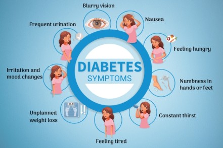
Metabolic disorders: Is the inability to deal with certain results of food. It may result in accumulation of unwanted chemical in the blood which may lead to trouble e.g. excess sugar in the blood which leads to diabetes mellitus.
Tumours: These are over growth of cells which have undergone changes that makes them multiply themselves. This can be benign or malignant.
Hypersensitivity: Some people are hypersensitive to small amounts of certain proteins and if exposed to them, they react. Hypersensitivity can be;
An allergy
Anaphylaxis.
Degenerative diseases: The ageing process usually results in various conditions e.g. osteo-arthritis, stroke etc.
Psychological factors: This can be an important cause of disease e.g. stress, anxiety, disappointments etc.
Aims of Surgery:
Surgery is performed with various objectives, often categorized by the primary goal of the intervention:
Diagnostic Purpose: To obtain tissue samples (e.g., biopsy) or to explore the body to confirm or determine the cause of a disease or condition.
Curative Purpose: To remove diseased tissue or an organ, repair damaged structures, or correct a deformity to cure a disease (e.g., appendectomy for appendicitis, tumor excision).
Palliative Purpose: To relieve symptoms or improve quality of life when a cure is not possible (e.g., tumor debulking to reduce pressure, colostomy for bowel obstruction).
Preventive Purpose (Prophylactic): To prevent the occurrence of a disease or complication in an at-risk individual (e.g., prophylactic mastectomy for high-risk breast cancer, removal of a precancerous polyp).
Reconstructive/Restorative Purpose: To repair or restore damaged tissue or organs, often after injury or disease (e.g., skin graft for burns, joint replacement).
Cosmetic Purpose: To improve physical appearance (e.g., rhinoplasty, facelift), though this often overlaps with reconstructive surgery.
Types and Classification of Surgery:
Surgery can be classified based on urgency, invasiveness, and purpose.
Classification by Urgency:
Emergency Surgery: Performed immediately to save a life, preserve function, or restore a vital body part (e.g., severe bleeding, ruptured appendix).
Urgent Surgery: Performed within 24-48 hours to address a condition that requires prompt intervention but is not immediately life-threatening (e.g., acute cholecystitis, kidney stones with obstruction).
Planned or Elective Surgery: Scheduled in advance, often to correct a non-life-threatening condition, improve quality of life, or for cosmetic reasons (e.g., cataract removal, hernia repair). This allows for thorough pre-operative assessment and patient preparation.
Classification by Extent/Magnitude:
Major Surgery: Involves significant risk, often requires general anesthesia, extensive tissue manipulation, and typically involves a longer hospital stay (e.g., open-heart surgery, organ transplantation).
Minor Surgery: Involves minimal risk, often performed under local or regional anesthesia, limited tissue manipulation, and may be done in an outpatient setting (e.g., removal of a skin lesion, carpal tunnel release).
Other Classifications:
Multistage Surgery: Procedures performed in several separate operations to achieve a complete outcome, often due to the complexity of the condition or the patient's recovery needs (e.g., reconstructive surgery after severe trauma).
Invasiveness:
Open Surgery: Involves a large incision to access the surgical site.
Minimally Invasive Surgery: Performed through small incisions using specialized instruments and cameras (e.g., laparoscopic surgery, robotic surgery, endoscopic surgery).
Principles of Surgery:
Fundamental principles guide surgical practice to ensure patient safety and optimal outcomes.
Safe Administration of Anesthesia: Ensuring the patient's physiological stability and comfort throughout the procedure, minimizing risks associated with anesthetic agents.
Asepsis and Infection Control: Strict adherence to sterile techniques to prevent surgical site infections, including meticulous hand hygiene, sterile draping, and instrument sterilization.
Hemostasis (Control of Bleeding): Meticulous control of bleeding to maintain patient's circulatory volume and provide a clear surgical field.
Gentle Tissue Handling: Minimizing trauma to tissues to promote healing and reduce post-operative pain and complications.
Accurate Anatomical Dissection: Precise identification and manipulation of anatomical structures to avoid damage to vital organs and achieve the surgical objective.
Prevention/Treatment of Circulatory Failure: Maintaining adequate fluid balance, blood pressure, and tissue perfusion throughout the perioperative period.
Quick and Effective Wound Healing: Employing proper surgical closure techniques, providing optimal wound care, and managing factors that can impair healing.
Prevention/Treatment of Complications: Proactive identification and management of potential complications such as DVT, pulmonary embolism, respiratory compromise, and organ dysfunction.
Restoration of Function: The ultimate goal of many surgeries, aiming to return the affected body part or system to its normal or near-normal function.
Patient Safety and Advocacy: Prioritizing patient well-being, verifying correct patient and site, and advocating for the patient throughout the surgical journey.
Patient's Concept of Disease:
A patient's concept of their disease significantly influences their acceptance of surgical intervention, adherence to pre- and post-operative instructions, and overall recovery. This concept is shaped by a multitude of factors, including:
Personal Beliefs and Experiences: Prior experiences with illness, surgery, or healthcare, as well as personal beliefs about health and illness, can heavily influence a patient's understanding and emotional response to a new diagnosis.
Cultural Background: Cultural beliefs about the causation of disease (e.g., spiritual, supernatural, environmental), traditional healing practices, and societal roles can affect how a patient perceives their illness and the proposed surgical treatment.
Socioeconomic Status: Access to information, educational background, and financial stability can impact a patient's ability to understand complex medical information and comply with treatment plans.
Emotional State: Feelings of fear, anxiety, depression, or denial can distort a patient's perception of their illness and their capacity to process information about their condition.
Information Received: The clarity, completeness, and manner in which information about the disease and surgery is conveyed by healthcare professionals plays a crucial role. Misinformation or lack of understanding can lead to mistrust or non-adherence.
Support Systems: The presence or absence of family and social support can influence a patient's emotional well-being and ability to cope with their illness and recovery.
Perceived Severity and Impact: How serious the patient perceives their condition to be, and its anticipated impact on their life, livelihood, and family, will shape their perspective.
Nurses play a critical role in assessing and understanding the patient's concept of disease, clarifying misconceptions, providing culturally sensitive care, and offering appropriate emotional and educational support.
Factors Affecting the Success of Surgical Care:
The success of surgical care is multifaceted, extending beyond the technical proficiency of the surgeon. It encompasses a complex interplay of patient-related, disease-related, surgical team-related, and systemic factors.
Patient-Related Factors:
Overall Health Status and Comorbidities: Pre-existing conditions (e.g., cardiovascular disease, diabetes, renal impairment, malnutrition, obesity) can significantly impact surgical risk, recovery, and susceptibility to complications.
Age: Extremes of age (very young or very old) often present unique physiological challenges and increased risks.
Nutritional Status: Poor nutrition can impair wound healing, immune function, and overall recovery.
Psychological State: High levels of anxiety, stress, or depression can negatively affect pain perception, immune response, and patient cooperation.
Compliance with Pre/Post-operative Instructions: Adherence to dietary restrictions, medication regimens, and post-operative rehabilitation is crucial for optimal outcomes.
Lifestyle Factors: Smoking, alcohol consumption, and substance abuse can increase surgical risks and hinder recovery.
Disease-Related Factors:
Severity and Stage of Disease: Advanced disease or critical conditions generally carry higher surgical risks and potentially less favorable outcomes.
Type and Location of Pathology: The nature of the condition and its anatomical location can influence surgical complexity and potential for complications.
Presence of Infection: Active infection at the surgical site or systemically can increase the risk of complications and delay healing.
Surgical Team and Process Factors:
Surgeon's Expertise and Experience: The skill, experience, and specialization of the surgical team directly influence the technical success of the procedure.
Anesthesia Management: Safe and effective administration of anesthesia, tailored to the patient's needs and the procedure, is vital.
Aseptic Technique and Infection Control: Strict adherence to sterilization and aseptic practices minimizes the risk of surgical site infections.
Pre-operative Assessment and Optimization: Thorough evaluation of the patient before surgery to identify and mitigate risks.
Intra-operative Management: Meticulous surgical technique, proper hemostasis, and efficient management of the surgical environment.
Post-operative Care: Comprehensive nursing care, pain management, early mobilization, and monitoring for complications.
Team Communication and Coordination: Effective communication among all members of the multidisciplinary surgical team (surgeon, anesthesiologist, nurses, technicians) is paramount for seamless and safe care.
Systemic and Environmental Factors:
Availability of Resources: Access to appropriate equipment, technology, blood products, and medical supplies.
Hospital Infrastructure and Policies: Quality of facilities, staffing levels, and adherence to evidence-based protocols.
Access to Follow-up Care: Availability of timely and appropriate post-discharge care, including rehabilitation and specialist consultations.
Socioeconomic Support Systems: Patient's access to social support, transportation, and home care resources after discharge.
ANESTHESIA
Introduction
Anesthesia is a critical component of modern surgical practice, derived from the Greek word "anaisthesia," meaning "without sensation." It is a pharmacologically induced and reversible state characterized by controlled and temporary loss of sensation, consciousness, or both, enabling painful medical procedures or surgical operations to be performed without the patient experiencing pain, touch, pressure, or temperature. The primary aim is to ensure patient comfort, safety, and cooperation during invasive procedures.
Medications that cause anesthesia are called anesthetics. These agents work by interfering with the transmission of nerve signals to the brain, thereby preventing the processing of painful stimuli. When the anesthetic effect wears off, nerve signals resume normal function, and sensation returns.
How Anesthetics Work
Anesthetics exert their effects by interacting with various components of the nervous system, primarily by altering the flow of ions across nerve cell membranes, which in turn inhibits the generation and propagation of electrical signals (nerve impulses). Specifically:
They can block voltage-gated sodium channels in nerve axons, preventing the initiation and conduction of action potentials (nerve signals).
They can enhance the activity of inhibitory neurotransmitters (like GABA) or suppress excitatory neurotransmitters, leading to a general depression of central nervous system activity.
The precise mechanisms vary depending on the class of anesthetic (e.g., local anesthetics directly block nerve conduction, while general anesthetics primarily affect the brain and spinal cord).
By stopping these nerve signals from reaching the brain, anesthetics allow medical procedures to be carried out without the patient experiencing pain or awareness. As the anesthetic is metabolized and eliminated from the body, its effects dissipate, allowing nerve signals to function normally and sensation to return.
Types of Anesthesia
Anesthesia is categorized based on the extent of the body affected and the level of consciousness maintained:
Local Anesthesia: This involves numbing a small, specific area of the body by injecting an anesthetic agent directly into the tissues around the nerves supplying that area. The patient remains fully conscious but does not feel pain in the numbed region. It is typically used for minor procedures (e.g., dental procedures, suturing a small cut, skin biopsies).
Regional Anesthesia: This type of anesthesia blocks sensation in a larger region of the body, such as an entire limb or the lower half of the body, by injecting anesthetic near a cluster of nerves (nerve block) or into the spinal canal. The patient typically remains conscious but may be sedated.
Spinal Anesthesia: Anesthetic is injected into the cerebrospinal fluid (CSF) in the subarachnoid space surrounding the spinal cord. This rapidly produces profound numbness and muscle relaxation in the lower body, used for lower abdominal, pelvic, or lower limb surgeries.
Epidural Anesthesia: Anesthetic is injected into the epidural space (outside the dura mater, the outermost membrane covering the spinal cord). A catheter can be left in place to allow for continuous or repeated administration of medication, providing prolonged pain relief. Commonly used for childbirth (labor analgesia) and surgeries of the lower body.
Peripheral Nerve Blocks: Anesthetic is injected near specific nerves or nerve plexuses (networks of nerves) to numb a particular limb or area (e.g., brachial plexus block for arm surgery, femoral nerve block for leg surgery).
General Anesthesia: This induces a state of controlled, reversible unconsciousness, where the patient is completely unaware and experiences no pain or memory of the procedure. It involves a combination of medications administered intravenously and/or by inhalation. The patient's vital functions (breathing, heart rate) are carefully monitored and supported, often requiring mechanical ventilation. It is used for major surgeries or procedures that require the patient to be completely still and unaware.
Sedation: This involves administering medications to depress the central nervous system, producing a state of reduced awareness, relaxation, and sometimes amnesia, but the patient remains able to respond to verbal commands or light tactile stimulation. It is used for uncomfortable or anxiety-provoking procedures that do not require full general anesthesia (e.g., colonoscopy, some dental procedures, minor orthopaedic reductions). Levels can range from minimal (anxiolysis) to deep sedation.
Route of Administration of Anesthetics
The method of delivery depends on the type of anesthesia and the specific agent used:
Inhalation: Volatile liquid anesthetics (e.g., Sevoflurane, Isoflurane) are vaporized and delivered as gases through a mask or endotracheal tube into the patient's respiratory system for general anesthesia. Nitrous oxide is also given via inhalation.
Intravenous (IV): Many anesthetic agents (e.g., Propofol, Ketamine, Midazolam, Fentanyl) are administered directly into the bloodstream through a vein, used for induction of general anesthesia, maintenance of anesthesia, or for sedation.
Local Infiltration: Anesthetic is injected directly into the tissue surrounding the surgical site (e.g., Lidocaine for suturing a wound).
Regional Injection: Anesthetic is injected near specific nerves or into the epidural or subarachnoid space (e.g., Bupivacaine for spinal or epidural blocks).
Topical/Transdermal: Applied to the skin or mucous membranes (e.g., lidocaine cream for numbing skin before an injection, sprays for throat numbing).
Side Effects of Anesthetics
While generally safe, anesthetics can cause various side effects, most of which are temporary and manageable:
Gastrointestinal: Feeling sick (nausea) or vomiting, which can be managed with antiemetics.
Neurological: Dizziness and feeling faint, headache (especially after spinal or epidural anesthesia), confusion or disorientation (particularly in older adults).
Temperature Regulation: Feeling cold and shivering (post-anesthesia shivering is common).
Local Reactions: Bruising and soreness at the injection site (for local or regional blocks).
Skin Reactions: Itchiness (especially with opioid use).
Throat Irritation: Sore throat or hoarseness (after endotracheal intubation for general anesthesia).
Muscle Aches: Generalized muscle pain from muscle relaxants used during general anesthesia.
Complications and Risks of Anesthesia
Serious complications are rare but can occur and are usually discussed during the pre-operative consent process:
Allergic Reactions: Severe allergic reactions (anaphylaxis) to anesthetic agents, though rare, can be life-threatening.
Cardiovascular Complications: Hypotension (low blood pressure), arrhythmias (irregular heartbeats), myocardial infarction (heart attack), or stroke, particularly in patients with pre-existing cardiac conditions.
Respiratory Complications: Respiratory depression, aspiration of gastric contents into the lungs (pneumonitis), bronchospasm, or laryngospasm.
Nerve Damage: Temporary or, rarely, permanent nerve damage (causing prolonged numbness, weakness, or paralysis) due to direct trauma from injection or compression.
Malignant Hyperthermia: A rare, life-threatening genetic condition triggered by certain general anesthetics, leading to a rapid rise in body temperature and muscle rigidity.
Awareness During Anesthesia: A rare occurrence where a patient gains some level of consciousness during general anesthesia.
Death: Extremely rare, but possible, particularly in patients with severe underlying health conditions.
Nurse’s Role in Surgical Diagnosis
The nurse plays a pivotal and continuous role in the diagnostic phase of surgical care. While the surgeon makes the definitive diagnosis, the nurse's observations, assessments, and data collection are crucial for accurate, timely, and holistic diagnosis, contributing significantly to the patient's care plan.
Taking a Comprehensive Patient History: The nurse often conducts the initial and ongoing patient assessments, collecting subjective data through detailed history taking. This includes the chief complaint, history of present illness, past medical and surgical history, family history, social history (e.g., smoking, alcohol, substance use), medication history, allergies, and review of systems. Proper documentation of this history is essential for the medical team.
Performing Physical Assessments and Documenting Observations: The nurse regularly performs physical assessments (e.g., vital signs, head-to-toe assessment, focused assessment on the affected area). Accurately recording and monitoring these objective observations (e.g., temperature, pulse, respiration, blood pressure, pain level, wound characteristics, changes in patient condition) provides critical data points for diagnostic reasoning.
Assisting with Diagnostic Procedures and Examinations: The nurse prepares the patient and the environment for physical examinations and various diagnostic tests. This includes setting up equipment (e.g., for physical exam, wound assessment), ensuring patient comfort and privacy, providing explanations of procedures, and assisting the physician as needed during examinations or specimen collection.
Carrying Out Ordered Investigations and Other Orders: The nurse is responsible for ensuring that prescribed diagnostic tests (e.g., blood tests, imaging studies like X-rays, CT scans, MRI, ECG) are performed correctly and that specimens are collected and transported appropriately. This includes preparing the patient for the test (e.g., NPO status, contrast dye administration), verifying orders, and ensuring patient safety during the process.
Patient Education and Preparation: Explaining the purpose of diagnostic tests and procedures to the patient, ensuring understanding and compliance.
Recognizing and Reporting Changes: Constantly observing the patient for changes in symptoms, physical signs, or responses to interventions, and promptly reporting significant findings to the medical team.
Advocacy: Advocating for the patient's needs and ensuring that all necessary diagnostic steps are taken to arrive at an accurate diagnosis.
Identification of a Patient (Patient Safety and Verification)
Correct patient identification is a fundamental and non-negotiable principle in healthcare, especially in the surgical setting, to prevent errors and ensure patient safety. Errors in patient identification can lead to wrong-patient, wrong-site, or wrong-procedure surgeries, medication errors, and incorrect test results.
Using Multiple Identifiers: Patients should be identified by at least two unique identifiers, never by room or bed number alone. Acceptable identifiers include:
Patient's full name (first and last).
Date of birth.
Medical record number.
Active Patient Participation: Whenever possible, the patient should be actively involved in the identification process by stating their name and date of birth. Nurses should be aware that some patients may respond "yes" when another patient's name is called due to confusion, hearing impairment, or a desire to be cooperative; therefore, asking open-ended questions like "Can you please state your full name and date of birth?" is crucial.
Addressing Bed Swaps: Nurses must be vigilant about potential bed or room changes without their knowledge. Re-identification of the patient should occur at every significant interaction, including medication administration, before procedures, and before transport.
Site Marking for Surgical Procedures: For procedures involving laterality (e.g., right vs. left limb), multiple structures (e.g., specific finger), or levels (e.g., spinal surgery), the surgical site should be clearly and unambiguously marked by the surgeon with indelible ink while the patient is awake and involved in the process. This is a critical component of the "Universal Protocol" for preventing wrong-site, wrong-procedure, wrong-person surgery.
"Time Out" Procedure: Immediately before the start of any invasive procedure, a "time out" (or pause for cause) is performed by the entire surgical team. During this time-out, the team collectively confirms:
The correct patient.
The correct site.
The correct procedure.
Availability of correct implants/equipment.
This final verification step is a crucial safety barrier.
Wristbands/Identification Bands: Patients should wear identification wristbands with their unique identifiers throughout their hospital stay. These should be checked against medical records before any intervention.
DECONTAMINATION
Decontamination is a crucial initial step in the reprocessing of reusable medical instruments and equipment. It refers to the process of physically or chemically removing or neutralizing harmful substances, particularly pathogenic microorganisms, from objects or surfaces to render them safe for subsequent handling, cleaning, and sterilization. The goal of decontamination is to protect healthcare workers from exposure to potentially infectious materials and to prevent cross-contamination.
Principles of Decontamination:
Risk Reduction: Reduces the bioburden (number of microorganisms) on instruments, making them safer to handle for staff involved in cleaning and sterilization.
Immediate Action: Should occur as soon as possible after use to prevent drying of organic matter (blood, tissue), which makes cleaning more difficult.
Personal Protective Equipment (PPE): Healthcare workers involved in decontamination must wear appropriate PPE, including gloves, gowns, masks, and eye protection, to prevent exposure to contaminants.
Methods of Decontamination:
Manual Cleaning (Pre-cleaning):
Initial step often done at the point of use or in a designated decontamination area.
Involves rinsing instruments with cool water to remove gross contamination, followed by scrubbing with brushes using enzymatic detergents or neutral pH detergents.
This step is critical as sterilization cannot compensate for inadequate cleaning.
Automated Cleaning:
Ultrasonic Cleaners: Use high-frequency sound waves to create cavitation bubbles that dislodge debris from instruments, especially in hard-to-reach areas.
Washer-Disinfectors: Automated machines that clean and thermally disinfect instruments, rendering them safe for handling before sterilization. They often include pre-rinse, wash, rinse, and thermal disinfection cycles.
Chemical Decontamination:
Use of chemical solutions to kill or inactivate microorganisms. Often used for heat-sensitive instruments or for surface disinfection.
Examples include glutaraldehyde or hydrogen peroxide solutions, but these are typically for high-level disinfection rather than full sterilization.
Importance in Surgical Nursing:
Nurses are often responsible for the initial decontamination at the point of use (e.g., wiping instruments during surgery) and ensuring proper transport of soiled instruments to the central sterile supply department.
Understanding decontamination processes is vital for preventing surgical site infections and maintaining a safe environment for both patients and staff.
STERILIZATION
Sterilization is the process by which all forms of microbial life, including bacteria, viruses, fungi, and highly resistant bacterial spores, are completely destroyed or removed from an object or surface. It represents the highest level of microbial killing and is essential for any medical device or instrument that will come into contact with sterile body tissues or the bloodstream during surgical procedures. The aim is to prevent healthcare-associated infections (HAIs).
Key Principles of Sterilization:
"All or Nothing": An item is either sterile or not sterile; there are no degrees of sterility.
Packaging Integrity: Sterile items must remain in intact, undamaged packaging until the point of use to maintain sterility.
Time-Related or Event-Related Sterility: Sterility is maintained until the package is opened, damaged, or expires, depending on the storage conditions and packaging.
Cleaning First: Sterilization cannot effectively occur if instruments are not thoroughly cleaned and decontaminated beforehand.
Common Methods of Sterilization:
Steam Sterilization (Autoclaving):
The most common, reliable, and cost-effective method for heat- and moisture-stable items.
Uses saturated steam under pressure at high temperatures (e.g., 121°C or 132°C) for a specific duration.
Works by denaturing and coagulating proteins within microorganisms.
Dry Heat Sterilization:
Used for materials that can be damaged by moisture (e.g., powders, oils, heat-stable glassware).
Involves exposure to high temperatures (e.g., 160°C to 170°C) for longer periods than steam sterilization.
Works by oxidation of cell components.
Ethylene Oxide (EtO) Sterilization:
Used for heat- and moisture-sensitive medical devices.
A colorless, flammable gas that kills microorganisms by alkylation of proteins and nucleic acids.
Requires a lengthy aeration time to dissipate residual EtO, which is toxic and carcinogenic.
Hydrogen Peroxide Gas Plasma Sterilization (e.g., Sterrad):
A low-temperature sterilization method suitable for heat- and moisture-sensitive instruments.
Uses hydrogen peroxide vapor in a plasma state, which generates reactive free radicals that destroy microorganisms.
Faster cycle times and safer than EtO as it produces non-toxic byproducts (water and oxygen).
A liquid chemical sterilant used for immersible, heat-sensitive instruments, often used for flexible endoscopes.
Rapidly destroys microorganisms by oxidation.
Monitoring Sterilization:
Sterilization processes are monitored using various indicators to ensure effectiveness:
Mechanical Indicators: Gauges and displays on the sterilizer that show temperature, pressure, and exposure time.
Chemical Indicators: Tapes, strips, or packages that change color when exposed to specific sterilization conditions (e.g., heat, steam, EtO). They indicate that the item has been processed, but not necessarily that it is sterile.
Biological Indicators: Vials containing highly resistant bacterial spores (e.g., Geobacillus stearothermophilus for steam, Bacillus atrophaeus for EtO/dry heat). These are the only indicators that directly monitor the lethality of the sterilization process by demonstrating whether the most resistant organisms have been killed.
Role of the Nurse in Sterilization:
Understanding the principles of sterility and aseptic technique.
Checking the integrity of sterile packaging before opening.
Maintaining a sterile field during surgical procedures.
Properly handling and storing sterile supplies.
Advocating for correct sterilization practices within the healthcare setting.
CONSENT IN SURGICAL NURSING (INFORMED CONSENT)
Informed consent is a cornerstone of ethical and legal medical practice, particularly in surgical nursing. It is a process by which a patient, or their legally authorized representative, grants voluntary permission for a medical procedure or treatment only after receiving and comprehending all relevant information about it. This ensures patient autonomy and protects their right to self-determination regarding their healthcare decisions.
Key Elements of Valid Informed Consent:
Disclosure of Information: The healthcare provider (typically the physician or surgeon performing the procedure) must provide the patient with comprehensive information, including:
The nature of the proposed procedure or treatment.
The purpose of the procedure (what it aims to achieve).
The expected benefits of the procedure.
The potential risks, common side effects, and serious complications associated with the procedure (including those related to anesthesia).
Available alternative treatments, including their risks and benefits.
The consequences of not undergoing the proposed procedure.
Patient Understanding: The patient must be able to comprehend the information provided. The information should be presented in a language and manner understandable to the patient, avoiding medical jargon. The provider should assess the patient's understanding by asking open-ended questions.
Voluntariness: The patient's decision to consent or refuse treatment must be made freely, without any form of coercion, manipulation, or undue pressure from healthcare providers, family, or others.
Competence/Capacity: The patient must have the mental capacity to make healthcare decisions. This means they must be able to understand the information, appreciate the consequences of their decision, and communicate their choice. If a patient is deemed incompetent (e.g., due to severe cognitive impairment, unconsciousness), a legally appointed surrogate decision-maker (e.g., power of attorney for healthcare, legal guardian, next of kin in a hierarchical order defined by law) will provide consent on their behalf.
Role of the Nurse in Informed Consent:
While the responsibility for obtaining informed consent rests with the physician performing the procedure, nurses play a crucial and multifaceted role in the informed consent process:
Reinforcing Information and Clarifying: Nurses often reinforce the information provided by the physician, clarify any misunderstandings the patient may have, and answer questions within their scope of practice. They should not provide new information that changes the scope of the consent.
Assessing Patient Understanding: Nurses are frequently present during the consent discussion or review the consent form with the patient. They can assess the patient's comprehension and identify if the patient has further questions or appears to be unduly influenced.
Witnessing the Signature: Nurses often witness the patient's signature on the consent form. By witnessing, the nurse is verifying that the patient signed the form and that, to their knowledge, the patient appeared to be competent and voluntarily signed. It does not imply that the nurse provided all the information or explained the procedure.
Advocating for the Patient: If a nurse believes the patient does not understand the information, is being coerced, or is not competent, they have an ethical responsibility to advocate for the patient. This may involve notifying the physician, nursing supervisor, or ethics committee.
Documentation: Accurately documenting the informed consent process, including who provided information, when it was discussed, and any patient questions or concerns, is essential.
Ensuring Valid Consent Before Procedures: Before any surgical procedure, the nurse is responsible for verifying that a valid informed consent form is present in the patient's chart and that it is complete and signed.
Informed consent is an ongoing process, not a one-time event, and applies to changes in treatment plans or additional procedures that may arise during the course of care.
Apnea means "without breathing." It is a pause in breathing that lasts longer than the normal brief pauses that occur during sleep.
Types of Apnea by Duration:
Type
Definition
Significance
Brief pause
Less than 10 seconds
Normal, especially in sleep
Apnea
20 seconds or more (preterm) OR 15 seconds or more (full-term)
Abnormal; requires evaluation
Any pause with color change
Any duration with cyanosis, pallor, or bradycardia
Always abnormal
Infant Apnea (Apnea of Infancy):
An unexplained episode of cessation of breathing for:
20 seconds or more in a premature infant
15 seconds or more in a full-term infant
ANY duration if associated with:
Bradycardia (slow heart rate less than 100 bpm)
Cyanosis (blue color)
Pallor (pale color)
Marked hypotonia (floppy, limp baby)
Points for Attention
Apnea is a SYMPTOM, not a disease. It always has an underlying cause that must be identified and treated. Do not just treat the apnea; hunt for the etiology!
3.2 Types of Apnea
A. Central Apnea
Definition: Apnea caused by failure of the brain to send signals to the breathing muscles.
Mechanism:
BRAIN (respiratory center) ➔ NO SIGNAL SENT ➔ Breathing muscles don't contract ➔ NO BREATHING
Characteristics:
No respiratory effort (chest doesn't move)
No airflow through nose/mouth
May see brief body movements
Often associated with bradycardia
Common Cause
Explanation
Common in
Immature CNS
Brainstem respiratory centers not fully developed
Premature infants
Head trauma
Brain injury affecting respiratory centers
Accidents, non-accidental injury
Seizures
Seizure activity suppresses breathing
Epilepsy, febrile seizures
Sepsis
Infection affects brain function
Any age; especially neonates
Toxins/Drugs
Medications suppress respiratory drive
Opioids, sedatives, anesthesia
Hypoglycemia
Low blood sugar affects brain
Diabetic mothers' babies, sepsis
Intracranial hemorrhage
Bleeding in brain
Premature infants, trauma
Congenital CNS malformations
Brain structure abnormalities
Present from birth
B. Obstructive Apnea
Definition: Apnea caused by blockage of the airway, despite the brain sending signals to breathe.
Mechanism:
BRAIN sends signal ➔ Breathing muscles contract ➔ AIRWAY IS BLOCKED ➔ NO AIRFLOW despite effort
Characteristics:
Respiratory effort present (chest moves, but no air passes)
May see paradoxical breathing (chest in, belly out)
Often associated with snoring or noisy breathing
Common during sleep
Common Cause
Explanation
Common in
Adenotonsillar hypertrophy
Enlarged tonsils and adenoids block airway
2-8 years old
Obstructive Sleep Apnea (OSA)
Repeated airway collapse during sleep
Overweight children; enlarged tonsils
Laryngomalacia
Floppy larynx collapses during inspiration
Infants; improves with age
Foreign body
Object lodged in airway
Toddlers (exploring age)
Nasal congestion
Blocked nose prevents breathing
Infants (obligate nose breathers)
Micrognathia/Pierre Robin
Small jaw pushes tongue backward
Newborns
Choanal atresia
Back of nose is blocked
Newborns (cannot breathe when feeding)
Secretions
Mucus or blood blocks airway
Post-surgery; infections
Vocal cord paralysis
Vocal cords don't open properly
Birth trauma; surgery
C. Mixed Apnea
Definition: Combination of both central and obstructive components.
Characteristics:
Starts as central apnea (no effort), followed by obstructive apnea (effort but no airflow)
OR starts as obstructive, then becomes central
Most common type in premature infants
Common Causes:
Premature infants with nasal congestion
Infants with gastroesophageal reflux (GERD)
Sedated patients with adenotonsillar hypertrophy
Infants with upper respiratory infections
3.3 Epidemiology (Who Gets Apnea?)
A. Apnea of Prematurity (AOP)
Incidence: Very common in premature infants
Less than 28 weeks gestation: Approx 80-90%
28-32 weeks: Approx 50%
32-35 weeks: Approx 20%
More than 35 weeks: Rare
Usually resolves by 36-40 weeks corrected gestational age.
Before 28 weeks: May persist longer; associated with prolonged hospitalization.
B. Apnea in Full-Term Infants
Very rare.
If present, ALWAYS indicates underlying disease. Requires thorough investigation.
C. Obstructive Sleep Apnea (OSA)
Prevalence: 1-5% of children.
Gender: Boys greater than Girls (ratio 3:1 to 5:1).
Peak age: 2-8 years (when tonsils are largest relative to airway).
Risk factors: Obesity, Down syndrome, craniofacial abnormalities, neuromuscular disorders.
Higher incidence in children with: Cerebral palsy, Muscular dystrophy, Spinal muscular atrophy, Congenital myopathies.
3.4 Etiology & Risk Factors
A. Central Apnea Causes
Immature Central Nervous System:
Why preemies are at risk: Respiratory center in brainstem not fully developed. Chemoreceptors (sensors for O2 and CO2) immature.
Response to hypoxia is paradoxical: preemies may STOP breathing instead of breathing faster.
Risk decreases as baby matures; usually resolves by term equivalent age.
Central Nervous System Injury:
Birth asphyxia: Lack of oxygen during delivery damages brain.
Intraventricular hemorrhage (IVH): Bleeding in brain ventricles (common in preemies).
Hypoxic-ischemic encephalopathy (HIE): Brain injury from oxygen deprivation.
Head trauma: Accidental or non-accidental (child abuse).
Meningitis/Encephalitis: Infection of brain or its coverings.
Metabolic Disturbances:
Hypoglycemia (low blood sugar), Hypocalcemia (low calcium), Hyponatremia (low sodium), Severe acidosis (too much acid in blood), Hyperbilirubinemia (very high bilirubin leading to kernicterus).
Seizures:
Post-ictal apnea (after seizure). Seizure activity itself can suppress breathing. Subtle seizures in neonates may present only as apnea.
Sepsis (Overwhelming infection), Meningitis (Infection of brain coverings), Necrotizing enterocolitis / NEC (Gut infection in preemies).
B. Obstructive Apnea Causes
Adenotonsillar Hypertrophy: Most common cause of OSA in children. Tonsils and adenoids grow rapidly between 2-8 years. Can completely block airway during sleep when muscles relax. Treatment: Tonsillectomy and adenoidectomy (T&A).
Obesity: Fat deposits around neck and airway. Increased risk of airway collapse during sleep. Growing problem globally.
Craniofacial Abnormalities:
Down syndrome: Midface hypoplasia, macroglossia, hypotonia.
Pierre Robin sequence: Micrognathia (small jaw), glossoptosis (tongue falls back), cleft palate.
Craniosynostosis: Premature fusion of skull bones.
Laryngomalacia: Floppy tissues above vocal cords collapse inward during inspiration. Most common cause of stridor in infants. Usually improves by 12-18 months.
Gastroesophageal Reflux (GERD): Stomach contents reflux into esophagus. Can trigger laryngeal reflex causing apnea. May cause micro-aspiration.
Allergies and Infections: Allergic rhinitis causes nasal congestion. Upper respiratory infections cause swelling. Both worsen obstructive apnea.
❓ Clinical Application Question
A mother brings her 4-year-old son to the clinic. She reports he snores very loudly at night, sometimes seemingly pausing his breath before gasping for air. He is also struggling with daytime sleepiness and bedwetting. Given his age, what is the most likely structural cause of his symptoms, and what type of apnea is this?
Answer: He is presenting with classic symptoms of Obstructive Sleep Apnea (OSA). Given his age (4 years old), the most likely structural cause is Adenotonsillar Hypertrophy (enlarged tonsils and adenoids).
3.5 Clinical Manifestations (Signs & Symptoms)
During Apnea Episode:
Sign
Description
Significance
Cessation of breathing
No chest movement for more than 15-20 seconds
Primary sign
Cyanosis
Blue color of lips, tongue, skin
Severe hypoxia
Pallor
Pale, ashen color
Poor perfusion
Bradycardia
Heart rate less than 100 bpm (infants)
Hypoxia affecting heart
Hypotonia
Limp, floppy baby
Severe hypoxia affecting brain
Changes in respiratory depth
Shallow breathing before pause
Warning sign
Between Episodes (Especially in OSA):
Sign
Description
Significance
Snoring
Loud, habitual snoring
Suggests airway obstruction
Restless sleep
Frequent position changes
Trying to find position to breathe
Sweating
Excessive sweating during sleep
Working hard to breathe
Mouth breathing
Always breathing through mouth
Nasal obstruction
Enuresis (bedwetting)
Especially if previously dry
Sleep disruption affects bladder
Morning headaches
Waking with headache
CO2 retention during night
Daytime sleepiness
Difficulty waking, napping
Poor quality sleep
Behavioral problems
Irritability, poor concentration
Sleep deprivation
Failure to thrive
Poor weight gain
Energy spent on breathing; poor feeding
In Infants with Apnea of Prematurity:
Episodes occur during sleep (especially active/REM sleep).
May be triggered by: Handling (nursing procedures), Feeding, Temperature changes, Position changes.
Usually self-resolves with tactile stimulation. May require bag-mask ventilation if severe.
Seizures: Hypoxia triggers seizure activity leading to further brain injury.
Death: Severe, prolonged apnea. Association with SIDS is controversial.
Chronic Complications (Especially OSA):
Failure to thrive: Energy expenditure; poor feeding leading to growth delay.
Cor pulmonale & Pulmonary hypertension: Chronic hypoxia causes blood vessel constriction placing a massive strain on the right heart, leading to right heart failure.
Intellectual & Behavioral difficulties: Chronic sleep disruption and hypoxia leads to learning difficulties and ADHD-like symptoms.
Systemic hypertension & Metabolic syndrome: Sympathetic activation from frequent arousals increases cardiovascular, obesity, and diabetes risks.
3.7 Differential Diagnosis
When evaluating a child with apnea, consider these conditions:
Condition
Key Distinguishing Features
Bacteremia/Sepsis
Fever or hypothermia, lethargy, poor feeding, hypotension, poor perfusion
Congenital Heart Disease
Cyanosis from birth, murmur, poor feeding
Brief Resolved Unexplained Events (BRUE)
Brief event in infant less than 1 year; now well; no explanation found
Bronchiolitis
Upper respiratory symptoms first; wheezing; RSV season
Bronchopulmonary Dysplasia
History of prematurity; chronic oxygen need
Childhood Sleep Apnea (OSA)
Snoring; daytime sleepiness; enlarged tonsils
Influenza
Seasonal; fever, cough, myalgia; severe cases have apnea
Laryngomalacia
Inspiratory stridor since birth; worse when supine
Opioid Toxicity
History of opioid exposure; pinpoint pupils; respiratory depression
Pediatric Asthma
Wheezing; triggers; reversible with bronchodilators
Pediatric Status Epilepticus
Rhythmic movements; eye deviation; post-ictal state
Gastroesophageal Reflux
Spitting up; irritability after feeds; Sandifer syndrome
Apparent Life-Threatening Event (ALTE)
Combination of apnea, color change, choking, gagging
3.8 Investigations (Diagnostic Tests)
A. For All Infants with Apnea:
Continuous Monitoring: Cardiorespiratory monitor (detects apnea/bradycardia) and Pulse oximeter. Nursing role: Ensure proper lead placement, respond to alarms, document episodes.
Surgical Management (Tonsillectomy and Adenoidectomy / T&A): FIRST LINE for children. 80-90% success rate. Nursing care post-op: Monitor for bleeding (first 24 hours and days 5-10), pain management, hydration, monitor airway.
Positive Airway Pressure (CPAP/BiPAP): Used if T&A is contraindicated/failed, or for neuromuscular disorders.
D. Specific Management: 3. Central Apnea from Other Causes
Sepsis: Antibiotics; supportive care.
Seizures: Anticonvulsants; EEG monitoring.
Head trauma: Neurosurgical consultation; ICP monitoring.
Metabolic: Correct electrolytes, glucose.
Medication-induced: Reverse agent (e.g., naloxone for opioids).
3.10 NURSING MANAGEMENT & CARE PLAN
Nursing Assessment:
A. Respiratory: Frequency, depth, pattern, duration of episodes, associated signs, triggers, response to stimulation.
B. Cardiovascular: Heart rate, blood pressure, perfusion.
C. Neurological: Level of consciousness, muscle tone, seizures, reflexes.
D. General: Temperature, feeding tolerance, growth, hydration.
E. Psychosocial: Parental anxiety, family understanding, home environment.
Nursing Care Plan for Infant with Apnea
DIAGNOSIS 1: Ineffective Breathing Pattern (Related to: Immature respiratory center OR airway obstruction)
Nursing Intervention
Rationale
Place infant on cardiorespiratory monitor with apnea alarm
Detects apnea and bradycardia immediately
Apply pulse oximeter
Monitors oxygen saturation continuously
Position infant prone (for AOP) or side-lying
Reduces apnea frequency; maintains airway
Maintain neutral thermal environment
Cold stress triggers apnea
Minimize handling and cluster care activities
Handling triggers apnea in preemies
Provide tactile stimulation during apnea episodes
Triggers breathing reflex
Have bag-valve-mask and suction ready at bedside
For emergency resuscitation
Avoid vagal stimulation (no rectal temps)
Vagal response causes bradycardia
DIAGNOSIS 2: Risk for Aspiration (Related to: GERD, impaired swallowing, or altered level of consciousness)
Nursing Intervention
Rationale
Position infant with head elevated 30 degrees during/after feeds
Reduces reflux
Feed slowly with frequent burping
Reduces stomach distension
Consider thickened feeds if GERD present
Reduces reflux episodes
Monitor for signs of aspiration (coughing, choking, color change)
Early detection
Hold infant upright for 20-30 minutes after feeding
Allows stomach emptying
DIAGNOSIS 3: Risk for Injury (Hypoxic Brain Injury) (Related to: Prolonged apnea episodes causing hypoxia)
Nursing Intervention
Rationale
Respond immediately to monitor alarms
Delays increase hypoxia duration
Stimulate infant at first sign of apnea
May abort episode
Provide bag-mask ventilation if no response to stimulation
Ensures oxygenation
Document all episodes (time, duration, associated signs, response)
Tracks pattern and severity
Ensure caffeine administered on time
Maintains therapeutic level
DIAGNOSIS 4: Anxiety (Parents) (Related to: Fear of infant dying, uncertainty, guilt)
Explain apnea condition, causes, and prognosis in simple terms.
Demonstrate and teach home monitoring equipment.
Encourage parents to verbalize fears and concerns.
Teach CPR and emergency procedures.
DIAGNOSIS 5: Deficient Knowledge (Parents)
Teach medication administration (caffeine).
Teach use of home apnea monitor and recognition of emergency signs.
Teach safe sleep practices (Reduces SIDS risk).
Schedule and emphasize follow-up appointments.
Home Apnea Monitoring
When is it needed? Apnea of prematurity not fully resolved at discharge, Infants with ALTE, Certain high-risk conditions.
Equipment: Cardiorespiratory monitor with event recording, Pulse oximeter.
Parent Education: How to place leads, respond to alarms, stimulate infant, call for help, CPR, battery backups, keeping a log.
Duration: Usually until infant is apnea-free for several weeks (typically until 43-44 weeks post-menstrual age for preemies).
3.11 Prognosis
Apnea of Prematurity: Usually resolves by 36-40 weeks corrected gestational age. Generally excellent prognosis if properly managed. Caffeine use is associated with better neurodevelopmental outcomes.
Obstructive Sleep Apnea: Excellent with appropriate treatment. 80-90% cured after tonsillectomy and adenoidectomy. Untreated OSA leads to significant morbidity.
Central Apnea from Other Causes: Depends entirely on underlying cause (e.g., Sepsis is good if treated promptly; Brain injury is variable).
3.12 Mnemonics
🧠 "APNEA" - Assessment Priorities
Alarm response (monitor alarms)
Position (prone for preemies)
No vagal stimulation
Equipment ready (bag-mask)
Assess and document episodes
🧠 "CAFFEINE" - Benefits in AOP
Central respiratory stimulation
Apnea frequency reduced
Few side effects
Flexible dosing (once daily)
Easy to administer
Improved neurodevelopment
Neuroprotective effects
Excellent safety profile
Nursing care plan for a pediatric patient with Apnea
Assessment
Nursing Diagnosis
Goals/Expected Outcomes
Interventions
Rationale
Evaluation
1. Child presents with episodes of apnea lasting more than 20 seconds, cyanosis, and bradycardia (heart rate < 100 bpm).
Ineffective Breathing Pattern related to immature respiratory control as evidenced by episodes of apnea, cyanosis, and bradycardia.
The child will maintain effective breathing patterns with no episodes of apnea, and oxygen saturation will remain above 95%.
– Continuously monitor the child’s respiratory rate, effort, and oxygen saturation using a cardiorespiratory monitor.
– Position the child in a supine or side-lying position with the head slightly elevated to facilitate airway patency.
– Administer oxygen as prescribed to maintain adequate oxygenation during and after apneic episodes.
– Stimulate the child gently (e.g., rub the back or flick the soles) during apneic episodes to prompt breathing.
– Prepare for possible resuscitation if apnea persists despite stimulation.
Continuous monitoring helps detect apneic episodes and guide interventions.
Proper positioning promotes airway patency and reduces the risk of obstructive apnea.
Administering oxygen improves oxygenation during apneic episodes.
Gentle stimulation often restarts breathing in infants with apnea.
Resuscitation may be necessary in severe cases to restore breathing.
The child maintains a normal breathing pattern, with no further episodes of apnea, and oxygen saturation remains within the target range.
2. Child exhibits signs of fatigue and decreased responsiveness between apneic episodes.
Activity Intolerance related to recurrent apneic episodes as evidenced by fatigue and decreased responsiveness.
The child will exhibit improved activity tolerance with increased periods of alertness and responsiveness.
– Allow for rest periods between feedings and activities to reduce fatigue.
– Monitor the child’s energy levels and responsiveness closely, adjusting activity levels as needed.
– Educate parents on the importance of providing a calm, low-stimulation environment to promote rest.
– Provide small, frequent feedings to minimize energy expenditure during feeding.
Rest periods help conserve the child’s energy and prevent excessive fatigue.
Close monitoring allows for timely adjustments to activity levels based on the child’s energy reserves.
A calm environment reduces stress and supports the child’s recovery.
Small, frequent feedings reduce the effort required during feeding, conserving energy.
The child demonstrates improved activity tolerance, with increased alertness and responsiveness between rest periods.
3. Parents express anxiety about the child’s condition and fear of apneic episodes occurring at home.
Anxiety related to fear of apneic episodes and uncertainty about the child’s condition as evidenced by parental verbalization of concern.
The parents will verbalize understanding of the child’s condition and demonstrate confidence in managing apneic episodes at home.
– Provide clear, concise information to the parents about apnea, including causes, signs, and interventions.
– Teach parents how to monitor the child’s breathing and how to respond to apneic episodes at home, including the use of home monitoring equipment if prescribed.
– Offer emotional support and reassurance, acknowledging the parents’ feelings and concerns.
– Encourage parents to ask questions and participate in the child’s care to increase their confidence.
Educating parents helps reduce anxiety by providing them with the knowledge and skills needed to manage the child’s condition.
Hands-on teaching and use of monitoring equipment empower parents to respond effectively to apneic episodes.
Emotional support reassures parents and validates their concerns.
Involving parents in care increases their confidence and sense of control.
The parents verbalize understanding of the child’s condition, demonstrate correct management of apneic episodes, and express increased confidence in caring for their child at home.
4. Child is at risk for impaired gas exchange due to recurrent apneic episodes.
Risk for Impaired Gas Exchange related to apneic episodes and immature respiratory control.
The child will maintain adequate gas exchange as evidenced by normal oxygen saturation levels and absence of cyanosis.
– Monitor oxygen saturation and signs of respiratory distress continuously, intervening promptly during apneic episodes.
– Administer supplemental oxygen as needed to maintain target oxygen saturation levels.
– Provide continuous positive airway pressure (CPAP) or mechanical ventilation if prescribed to support the child’s respiratory efforts.
– Monitor arterial blood gases (ABGs) or transcutaneous CO2 levels if indicated to assess gas exchange.
Continuous monitoring allows for prompt intervention during episodes of impaired gas exchange.
Supplemental oxygen supports adequate oxygenation during apneic episodes.
CPAP or mechanical ventilation provides respiratory support in cases of severe or persistent apnea.
Monitoring ABGs or CO2 levels provides information on the child’s gas exchange status, guiding treatment.
5. Child is at risk for infection due to immature immune system and potential for aspiration during apneic episodes.
Risk for Infection related to immature immune system and potential aspiration.
The child will remain free from infection as evidenced by normal temperature, white blood cell count, and absence of signs of infection.
– Practice strict hand hygiene and aseptic technique during all care and procedures.
– Monitor for signs of infection, including fever, increased WBC count, and changes in respiratory status.
– Provide prophylactic antibiotics if prescribed, especially in cases of suspected aspiration.
– Educate parents on infection prevention measures, including proper feeding techniques to minimize the risk of aspiration.
Strict hand hygiene and aseptic technique reduce the risk of introducing pathogens.
Early detection and treatment of infection are crucial to prevent complications.
Prophylactic antibiotics may reduce the risk of infection following aspiration events.
Parental education ensures adherence to infection prevention practices at home.
Quick Quiz
Apnea Quiz
Paediatrics - mobile-friendly and focused practice.
Privacy: Your details are used only for quiz tracking and certificates.
Diabetes Mellitus (DM), commonly referred to as diabetes, is a group of metabolic disorders in which there are high blood sugar levels over a prolonged period.
Insulin is the hormone secreted by β-cells of the pancreas; it helps to incorporate glucose into cells for metabolism.
In insulin deficiency, blood glucose level rises leading to excretion of sugar in the urine called Glycosuria.
Glucose loss is accompanied by increased loss of water in the urine causing Polyuria; hence Hyperglycemia, Glycosuria and Polyuria are the three cardinal clinical features of diabetes mellitus.
Diabetes mellitus is the most common prevalent endocrine disorder; it affects nearly 2% of the world population
Diabetes can be primary or secondary or idiopathic.
Image showing the pancreas where Insulin is produced by the body.
Pathophysiology of Diabetes(Simplified)
Diabetes results from two main issues: the pancreas not making enough insulin or the body’s cells not responding properly to the insulin produced.
1. Insufficient Insulin:Pancreas fails to produce adequate insulin.
Impaired insulin function disrupts blood sugar regulation, leading to hyperglycemia (elevated blood glucose levels).
2. Consequences of Hyperglycemia:
Excess glucose is expelled through urine, causing glycosuria.
High glucose in the glomerular filtrate attracts water, resulting in polyuria (excessive urination).
Loss of water triggers an intense feeling of thirst (polydipsia).
3. Cellular Deprivation and Compensatory Responses:
Despite high blood glucose, cells remain deprived.
Body responds with increased appetite, leading to overeating (polyphagia), worsening the condition.
4. Gluconeogenesis:
Body initiates gluconeogenesis to create glucose from proteins and fats.
5. Ketone Body Accumulation:
Excessive glucose from fats produces abundant ketone bodies, causing ketonemia (increased ketones in blood).
6. Acidosis and Respiratory Response:
Accumulated ketones reduce blood pH, leading to acidosis.
Body responds with rapid and deep breathing (Kussmaul respirations) to decrease acidity.
7. Potential Life-Threatening Complication:
Prolonged acidic state may lead to ketoacidosis, a severe medical/pediatric emergency.
Types of Diabetes Mellitus
There are three main types of diabetes mellitus and one unspecified;
Type 1 Diabetes Mellitus
Type 1 Diabetes Mellitus (T1DM) is marked by the pancreas’s failure to produce sufficient insulin, a vital hormone in blood sugar regulation. Formerly known as “insulin-dependent diabetes mellitus” (IDDM) or “juvenile diabetes,” Its cause is unknown.
Insulin Deficiency and Beta Cell Loss:T1DM is characterized by the loss of insulin-producing beta cells in the pancreatic islets. This leads to a deficiency in insulin, disrupting the body’s ability to regulate blood sugar.
Immune-Mediated or Idiopathic Classification: T1DM can be classified as immune-mediated or idiopathic. The majority of cases involve immune-mediated processes, where autoimmune attacks by T cells lead to beta cell loss. Onset can occur in children or adults, though historically labeled “juvenile diabetes” due to its prevalence in children.
Associated Complications: Complications may include impaired response to low blood sugar, infections, gastroparesis (causing erratic carbohydrate absorption), and endocrinopathies like Addison’s disease.
Genetic and Environmental Factors: T1DM has a genetic component, with specific HLA genotypes influencing susceptibility. Environmental triggers, such as viral infections or dietary factors (e.g., gliadin in gluten), can prompt diabetes onset, especially in genetically predisposed individuals.
Autoimmune Attack and Viral Influence: An autoimmune attack on pancreatic islets, often triggered by viral infections, is a key contributor. T1DM is more likely to manifest in childhood or early adulthood, with a sudden onset.
Management and Risks: Insulin and a comprehensive diet are crucial for managing T1DM. Patients face an increased risk of coma if concurrent infections like pyelonephritis or gastroenteritis are not promptly addressed.
Type 2 Diabetes Mellitus
The pathophysiology of type 2 diabetes mellitus is characterized by peripheral insulin resistance,impaired regulation of hepatic glucose production, and declining β-cell function, eventually leading to β -cell failure
Reduced insulin secretion and absorption leads to high glucose content in the blood.
Insulin Resistance and Reduced Secretion:T2DM is characterized by insulin resistance, where body tissues have a diminished response to insulin. This resistance is sometimes accompanied by a relative reduction in insulin secretion.
Insulin Receptor Dysfunction:The defective responsiveness of body tissues to insulin involves the insulin receptor, though, specific defects remain unknown. Diabetes cases with known defects are categorized separately.
Prevalence and Early Stage Abnormality:T2DM constitutes the majority, accounting for up to 90% of all diabetes mellitus cases. In the early stage, the primary abnormality is reduced insulin sensitivity, reversible by measures and medications improving insulin sensitivity or reducing liver glucose production.
Contributing Factors:Lifestyle factors, genetics, obesity (BMI > 30), lack of physical activity, poor diet, stress, and urbanization contribute to T2DM. Insulin resistance, overeating, inactivity, and obesity play roles in the etiology.
Dietary Management and Weight Loss: Management often involves adherence to a low-energy diet to facilitate weight loss. Lifestyle modifications, including dietary changes, exercise, and stress reduction, play roles in controlling T2DM.
Gestational Diabetes
Gestational diabetes mellitus (GDM) resembles type 2 DM in several aspects, involving a combination of relatively inadequate insulin secretion and responsiveness. It occurs in about 2–10% of all pregnancies and may improve or disappear after delivery.
Occurrence and Post-Delivery Transition:GDM shares similarities with type 2 DM, involving inadequate insulin secretion and responsiveness. It affects about 2–10% of pregnancies and may improve or vanish after childbirth.
Post-Pregnancy Diabetes Risk:Post-pregnancy, 5–10% of women with a history of gestational diabetes develop diabetes mellitus, often type 2. However, after pregnancy approximately 5–10% of women with gestational diabetes are found to have diabetes mellitus, most commonly type 2.
Temporary Nature and Health Impacts :While temporary during pregnancy, untreated GDM poses risks to both the mother and the fetus. Raised plasma glucose levels during pregnancy may lead to the birth of babies with increased birth weight, skeletal muscle malformations, and increased mortality risk. Risks associated with untreated GDM in newborns include macrosomia (high birth weight), congenital heart and central nervous system abnormalities, and skeletal muscle malformations. Elevated insulin levels in the fetal blood may hinder surfactant production, leading to respiratory distress syndrome.
Complications and Perinatal Risks:Complications may arise, such as high blood bilirubin levels due to red blood cell destruction. Severe cases can result in perinatal death, often attributed to poor placental perfusion caused by vascular impairment, leading to macrosomia and shoulder dystocia.
Management and Treatment: Gestational diabetes is fully treatable, but requires careful medical supervision throughout the pregnancy. Management may include dietary changes, blood glucose monitoring, and in some cases, insulin may be required.
Unspecified Diabetes Mellitus:
Maturity Onset Diabetes of the Young (MODY):
Maturity onset diabetes of the young (MODY) is an autosomal dominant inherited form of diabetes, due to one of several single-gene mutations causing defects in insulin production.
It is significantly less common than the three main types.
The name of this disease refers to early hypotheses as to its nature.
Being due to a defective gene, this disease varies in age at presentation and in severity according to the specific gene defect; thus there are at least 13 subtypes of MODY.
People with MODY often can control it without using insulin.
Others:
Prediabetes: Prediabetes indicates a condition that occurs when a person’s blood glucose levels are higher than normal but not high enough for a diagnosis of type 2 DM. Many people who later develop type 2 DM spend many years in a state of prediabetes.
“Type 3 Diabetes”:“Type 3 diabetes” has been suggested as a term for Alzheimer’s disease as the underlying processes may involve insulin resistance by the brain.
Aetiological Classification of Diabetes Mellitus:
Primary Diabetes Mellitus (Idiopathic):
Type 1 Diabetes (IDDM): β-cell destruction, usually leading to insulin deficiency.
Type 2 Diabetes (NIDDM): May range from insulin resistance with relative insulin deficiency to a predominantly insulin secretory defect with insulin resistance.
Secondary Diabetes Mellitus:Due to Other Underlying Diseases/Conditions:
Diseases of the pancreas, such as pancreatitis, pancreatic cancer, cystic fibrosis, or hemochromatosis, can destroy the gland leading to reduced insulin production.
Endocrine disorders (insulin antagonism) like Cushing’s syndrome, acromegaly, and hyperthyroidism.
Liver diseases like hepatitis, cirrhosis, are associated with glucose intolerance.
Gestational Diabetes Mellitus (Pregnancy-induced Diabetes Mellitus): Occurs during pregnancy and may resolve after delivery.
Predisposing Causes of Primary Diabetes Mellitus:
Age: 80% of cases occur after 50 years. DM is commonly a disease of middle-aged and elderly people.
Sex: Young males are more affected than females. In middle age, females are more affected.
Heredity: DM follows the family line in occurrence. 5% of patients have a familial history.
Autoimmunity: The body produces cells against insulin production.
Infections: Viral infections and staphylococci are associated with the causation of IDDM.
Obesity: The majority of NIDDM cases are obese.
Lifestyle Factors: Overeating with underactivity is associated with a high risk of incidence.
Other Predisposing Factors:
Sedentary lifestyle.
Poor dietary habits.
Metabolic syndrome.
Hypertension.
Ethnicity (some ethnic groups are more predisposed).
Gestational diabetes (increases the risk later in life).
Certain medications (e.g., glucocorticoids).
Previous gestational diabetes.
Clinical Features of Diabetes Mellitus
In mild cases, there may be no obvious signs, and the condition is detected accidentally during routine examination. However, in severe cases, especially in young children and young adults, pronounced symptoms may include:
PolyuriaDue to osmotic activity preventing water reabsorption in the renal tubule.
Polydipsia (increased thirst) follows polyuria, leading to dehydration due to constant loss of fluids and electrolytes.
Polyphagia with Weight Loss: Weight loss occurs due to the breakdown of fat and proteins caused by cellular glucose deficiency.
Weakness or Fatigue/Lassitude: Resulting from cells not receiving enough glucose.
Nocturnal Enuresis: Due to renal glucose exceeding the threshold. Nocturnal enuresis is when there is involuntary urination at night while asleep.
Glycosuria: This is when there is excessive amounts of glucose in urine.
Peripheral Neuropathy/Paresthesia: Nerve damage caused by chronically high blood sugar, leading to loss of sensation and numbness in the legs. In severe cases, symptoms include digestive issues, bladder problems, and difficulty controlling heart rate. Paresthesia is a symptom of neuropathy, since neuropathy is an umbrella term for any disease that affects the nerves.
Vulvovaginitis: Irritation of the genitalia caused by the local deposition of glucose. May be severe and disturb sleep.
Ketoacidosis: A serious complication involving excess blood acids (ketones). Symptoms include blurry vision, headache, fatigue, slow healing of cuts, and itchy skin.
Diabetic dermadromes. Skin rashes associated with diabetes with cutaneous eruptions in patients with long standing diabetic disease.
Vision Changes: Prolonged high blood glucose can cause glucose absorption in the lens of the eye, leading to changes in its shape and resulting in vision changes.
Comparison of type 1 and 2 diabetes
Type 1
Type 2
Sudden
Mostly children
Thin or normal
Ketoacidosis common
Antibodies usually present
Insulin low or absent totally
In identical twins is approximately 50%
Prevalence approximately 10%
Gradual
Mostly adults
Often obese
Rare
Absent
Normal, decreased or increased
Is approximately 90%
Prevalence approximately 90%
Diagnosis of Diabetes Mellitus:
Diabetes mellitus, characterized by recurrent or persistent high blood sugar, is diagnosed by demonstrating any one of the following criteria:
According to the current definition, two fasting glucose measurements above 126 mg/dl (7.0 mmol/l) are considered diagnostic for diabetes mellitus.
2. Plasma Glucose Two Hours After Oral Glucose Load: ≥ 11.1 mmol/l (200 mg/dl) two hours after a 75 g oral glucose load, as in a glucose tolerance test.
People with plasma glucose at or above 7.8 mmol/l (140 mg/dl) but not exceeding 11.1 mmol/l (200 mg/dl) two hours after the oral glucose load are considered to have impaired glucose tolerance.
3. Symptoms of High Blood Sugar and Casual Plasma Glucose:≥ 11.1 mmol/l (200 mg/dl)
Presence of symptoms along with casual plasma glucose above 11.1 mmol/l (200 mg/dl) indicates diabetes.
Note:
According to the World Health Organization, individuals with fasting glucose levels between 6.1 to 6.9 mmol/l (110 to 125 mg/dl) are considered to have impaired fasting glucose.
Glycated hemoglobin is considered superior to fasting glucose in determining cardiovascular disease risks and risks of death from any cause.
Important Information:
Two fasting glucose measurements above 126 mg/dl (7.0 mmol/l) are diagnostic for diabetes.
Impaired glucose tolerance, especially with plasma glucose levels between 7.8 mmol/l (140 mg/dl) and 11.1 mmol/l (200 mg/dl) after oral glucose, is a significant risk factor for progressing to diabetes and cardiovascular disease.
4. Other Diagnostic Investigations for Diabetes Mellitus:
Glucosuria:
Method: Detect glucose in urine using a test strip (uristicks).
Purpose: To identify the presence of glucose in the urine, indicating possible diabetes.
Ketonuria:
Method: Detect ketone bodies in urine.
Purpose: To identify the presence of ketones, which may indicate diabetic ketoacidosis.
Fasting Blood Sugar (FBS):
Method: Measure glucose concentration in blood samples obtained after at least 8 hours of the last meal.
Purpose: Assess baseline blood sugar levels after an overnight fast.
Random Blood Sugar (RBS):
Method: Measure glucose concentration in blood samples obtained at any time, regardless of the time of the last meal.
Purpose: Provide a snapshot of current blood sugar levels.
Oral Glucose Tolerance Test (OGTT):
Method: The patient fasts overnight, then ingests 75gm (5 tablespoons) of glucose with 300 ml of water. Blood samples are drawn at 1, 2, and 3 hours after glucose intake.
Purpose: A more accurate test for glucose utilization, especially if fasting glucose is borderline. It helps identify abnormal glucose metabolism over time.
Additional Information:
Normally, blood glucose should return to fasting levels (4.5 mmol or 80 mg/100 ml) after 2.5 hours of taking a meal.
Treatment and Nursing Management of Diabetes mellitus
Diabetes mellitus is a chronic disease, for which there is no known cureexcept in very specific situations.
Management concentrates on keeping blood sugar levels as close to normal, without causing low blood sugar.
This management is dependent on the type of diabetes mellitus and aims to:
Control diabetes and prevent complication
To bring down blood sugar levels
To help the patient comply to treatment
Management (non-pharmacological)
Control traditional Cardiovascular risk factors such as smoking, taking alcohol, management of dyslipidemia, intensive BP control and antiplatelet therapy.
Complication monitoringi.e. annual eye examination, annual microalbuminuria detection, feet examination, BP monitoring and lipid profile.
Patient’s education:Teach the patient on self-monitoring of blood glucose using glucometer and/or uristicks, moving with a diabetic card, keeping sugary food in the bag, method of insulin administration and consequences neglecting treatment
Patients should also be taught to prevent themselves from injury.
Diet – For type 1 the goal is to regulate insulin administration with a balanced diet – In most cases, high carbohydrate, low fat and low cholesterol diet is appropriate – Diet and insulin must be fixed to avoid fluctuation in blood glucose. Vitamins and minerals must be supplemented – Small frequent meals should be served to avoid peaks of hyperglycemia and no meal should be delayed. Snacks should be added to the main meals i.e in the middle of morning, early afternoon and before bed time. – Food should be palatable with high fibre food like legumes, burley, oat. Low salt in diet is advised (6g per day) – Avoid fried food, sweetened beverage, bakery products, honey and fine sugar
Type 2 DM patients need caloric restriction: Diet restriction must be combined with life style modification – Artificial sweeteners: e.g. Aspartame, saccharin, sucralose, and acesulfame are safe for use in all people with diabetes – Nutritive sweeteners: e.g. fructose and sorbitol, there use is increasing though they cause acute diarrhea in some patient.
Activity: Exercise improves insulin resistance and achieving glycemic control. – Exercise should start slowly for patients with limited activity. – Patients with Cardiovascular diseases should be evaluated before starting any exercise – Avoid exercises on an empty stomach, when blood sugar levels are low or high. – Heavy exercises like mental lifting are dangerous because it triggers hypoglycemia
Methods of treatment of diabetes > Diet > Diet + oral hypoglycemic agents > Diet and insulin
INSULIN THERAPY
Insulin is indicated for most patients of IDDM and IIDM who do not respond to oral hypoglycemic drugs. Doses are adjusted for individual patient needs to meet target glycemic control
Unmodified/soluble/rapid acting insulin:Dose 40 – 100 IU SC daily in 3 divided doses before meals, 40-80 IU for child. This is a clear solution and acts in half an hour and reaches peak in 2-6 hours, repeated injections are needed. This insulin can be used to control postprandialhyperglycemia and emergency ketoacidosis i.e,
Ultra short acting-Lispro: (Monomeric) absorbed to the circulation very rapidly and acts in 2-3 hours
Aspart: (Mono- and dimeric) absorbed to the circulation very rapidly
Short acting-Regular: (Hexameric) absorbed rapidly but slower than lispro and aspart, includes novolin R, humulin R.
Modified (deport) preparations: these are cloudy preparations/turbid suspensions made by either adding zinc for lente preparations or protamine (protein) for isophane preparations. They are used for maintenance treatment of type 1DM
Semi lente/prompt zinc; this is short acting and contains zinc microcrystals in acetate buffer. It is not used for IV because of buffer acetate
Lente insulin; Intermediate acting and acts in 12 hours e.g. humulin L. Dose: Adults 10-20 IU twice daily SC, Child: 5 – 10 IU twice daily
Ultralente; Long acting and acts 24-36 hours eg Ultratard.
Insulin analogues; mixture of modified and unmodified acts in 12 hours i.e,
Hypoglycemia: Patients should be aware of symptoms of hypoglycemia. Oral administration of 10-15 gm glucose. IV dextrose in patients with lost consciousness or/and 1 gm glucagon IM if IV access is not available
Skin rash at injection site: Use more purified insulin preparation.
Lipodystrophies (increase in fat mass) at injection site: rotate the site of injection
Insulin resistance
Allergy
Weight gain
Avoid using propranolol or other B-blockers in diabetics because they mask hypoglycemic symptoms.
Drugs that increase the blood glucose concentration should be avoided e.g Dioxide, Thiazide diuretics, Streptozotocin, Phenytoin. Corticosteroids, Oral contraceptives.
How to measure the insulin needed.
A good starting dose is 0.6 U/kg/day. The total dose should be divided to;
45% for basal insulin
55% for prandial insulin
The prandial dose is divided to
25% pre-breakfast
15% pre-lunch
15% pre-supper
Example: For a 50 kg patient The total dose = 0.6 x 50 = 30 U/day = 13.5 U for basal insulin (45% of dose) Administered in one or two doses = 16.5 U for prandial insulin (55% of dose) The 16.5 U are divided to: = 7.5 U pre-breakfast (25%) = 4.5 U pre-lunch (15%) = 4.5 U pre-supper (15%) The initial regimen should be modified Most Type 1 patients require 0.5-1.0 IU/kg/day
Medications for Type 2 Diabetes:
Anti-diabetic medications (hypoglycemics) are important for managing diabetes by lowering blood sugar levels. Various classes of these medications exist, some administered orally (e.g., metformin) and others via injection (e.g., GLP-1 agonists). It’s important to note that insulin is the primary treatment for Type 1 diabetes.
Sulphonylureas: Stimulate insulin secretion and release by the pancreas’ beta cells.
Examples include glibenclamide and chlorpropamide.
Biguanides:Increase glucose uptake by body cells and decrease glucose production by the liver.
Metformin (Glucophage) is a commonly recommended first-line treatment for Type 2 diabetes, showing evidence of decreased mortality.
Alpha-Glucosidase Inhibitors:Inhibit the enzyme hindering glucose uptake by cells.
Primarily used in Type 1 diabetes and in Type 2 when oral medications are ineffective.
Blood Pressure Management:
Given the serious cardiovascular risks associated with diabetes, maintaining blood pressure is crucial.
Target blood pressure levels are recommended below 130/80 mmHg, though evidence supports a range between 140/90 mmHg to 160/100 mmHg.
Angiotensin-converting enzyme inhibitors (ACEIs) are effective, while angiotensin receptor blockers (ARBs) may not be as beneficial in diabetes.
Aspirin is recommended for those with cardiovascular problems; however, routine use hasn’t proven beneficial in uncomplicated diabetes.
Surgery:
Weight loss surgery is effective in managing obesity and Type 2 diabetes.
Many individuals can maintain normal blood sugar levels with minimal or no medications post-surgery, reducing long-term mortality.
Short-term mortality risk from surgery is less than 1%, and eligibility criteria based on body mass index cutoffs are still unclear.
Pancreas transplant considerations are rare, usually for individuals with severe complications of Type 1 diabetes, including end-stage kidney disease.
Support:
In most healthcare systems, care often occurs outside hospitals unless complications arise.
Home telehealth support is an effective management strategy, particularly in cases of complications, challenging blood sugar control, or research projects.
Sites for Insulin Administration
Prevention of Diabetes:
Type 1 Diabetes:Unfortunately, there is currently no known preventive measure for Type 1 diabetes. It is primarily considered an autoimmune condition where the body’s immune system mistakenly attacks and destroys the insulin-producing beta cells in the pancreas.
Type 2 Diabetes:Prevention strategies for Type 2 diabetes focus on lifestyle modifications and healthy habits.
Maintaining a Healthy Diet:
Emphasize a balanced and nutritious diet rich in fruits, vegetables, whole grains, and lean proteins.
Limit the intake of processed foods, sugary beverages, and foods high in saturated and trans fats.
Control portion sizes to avoid overeating.
Regular Physical Exercise:
Engage in regular physical activity, such as walking, jogging, swimming, or cycling.
Aim for at least 150 minutes of moderate-intensity exercise per week.
Include strength training exercises to improve muscle strength and overall fitness.
Maintaining a Normal Body Weight:
Achieve and maintain a healthy body weight through a combination of a balanced diet and regular exercise.
Weight loss is particularly beneficial for those at risk or diagnosed with prediabetes.
Avoiding Tobacco Use:
Quitting or avoiding tobacco products is essential, as smoking is a significant risk factor for Type 2 diabetes.
Smoking cessation has numerous health benefits and contributes to overall well-being.
Control of Blood Pressure:
Regular monitoring and management of blood pressure are crucial.
Adopt a heart-healthy lifestyle, including a low-sodium diet, regular exercise, and stress management.
Proper Foot Care:
Individuals with diabetes need to pay special attention to foot care.
Regularly inspect feet for any cuts, sores, or signs of infection.
Choose comfortable, well-fitting shoes and avoid walking barefoot.
Avoiding Smoking:
In addition to its association with Type 2 diabetes, smoking is a risk factor for various cardiovascular and respiratory diseases.
Quitting smoking contributes significantly to overall health and reduces diabetes risk.
Additional Measures:
Regular health check-ups and screenings for diabetes risk factors.
Monitoring and managing stress levels through relaxation techniques and mindfulness.
Adequate sleep is essential for overall health and may play a role in diabetes prevention.
Limiting alcohol consumption, as excessive drinking can contribute to weight gain and affect blood sugar levels.
Complications of diabetes mellitus
Cardiomyopathy: The major long-term complications relate to damage to blood vessels. Diabetes doubles the risk of cardiovascular disease and about 75% of deaths in diabetics are due to coronary artery disease. Other “macrovascular” diseases are stroke, and peripheral artery disease.
Retinopathy: The primary complications of diabetes due to damage in small blood vessels include damage to the eyes, kidneys, and nerves. Damage to the eyes, known as diabetic retinopathy, is caused by damage to the blood vessels in the retina of the eye, and can result in gradual vision loss and blindness. Diabetes also increases the risk of having glaucoma, cataracts, and other eye problems. It is recommended that diabetics visit an eye doctor once a year.
Nephropathy: Damage to the kidneys, known as diabetic nephropathy, can lead to tissue scarring, urine protein loss, and eventually chronic kidney disease, sometimes requiring dialysis or kidney transplantation.
Neuropathy: Damage to the nerves of the body, known as diabetic neuropathy, is the most common complication of diabetes. The symptoms can include numbness, tingling, pain, and altered pain sensation, which can lead to damage to the skin.
Diabetic foot: Diabetes-related foot problems (such as diabetic foot ulcers) may occur, and can be difficult to treat, occasionally requiring amputation. Additionally, proximal diabetic neuropathy causes painful muscle atrophy and weakness.
Falls: There is a link between cognitive deficit and diabetes. Compared to those without diabetes, those with the disease have a 1.2 to 1.5-fold greater rate of decline in cognitive function. Being diabetic, especially when on insulin, increases the risk of falls in older people.
Other Complications:
Eye; Retinopathy leading to impaired vision, premature cataract, recurrent styles
Urinary system; renal failure, nephritic syndrome and pyelonephritis due to diabetes nephropathy
Genital tract; erectile dysfunction, loss of libido in men and menstrual irregularities, recurrent abortion, purulent vaginitis, infertility in females
Nervous system; Neuropathy resulting in tingling and numbness in the feet, stroke.
Skin; Staphylococcal skin infections e.g boils carbuncles, non healing ulcer and mucocutaneous candidiasis
Respiratory system; pneumonia, lung abscess and tuberculosis
Diabetic Emergencies
Hypoglycemia:
Low blood sugar (hypoglycemia) is a common occurrence in individuals with type 1 and type 2 Diabetes Mellitus (DM). While most cases are mild and not deemed medical emergencies, the effects can range from mild to severe.
Symptom Category
Mild Symptoms
Moderate Symptoms
Physical Signs
Common Signs
– Feelings of unease
– Confusion
– Drunkenness
– Sweating
– Changes in behavior (e.g., aggressiveness)
– Rapid breathing
– Trembling
– Seizures
– Sweating
– Increased appetite
– Unconsciousness (rarely, in severe cases)
– Cold and pale skin (although not definitive)
Management
Self-treatment with sugary foods or drinks.
Immediate attention with intravenous glucose or glucagon injections for severe cases
Hyperosmolar Hyperglycemic State:
More common in type 2 DM, hyperosmolar hyperglycemic state is mainly the result of dehydration. This state is characterized by significantly elevated blood sugar levels.
Hospitalization is often necessary.
Treatment involves fluid replacement, insulin administration, and correction of electrolyte imbalances.
Close monitoring of vital signs, blood glucose, and electrolytes.
Diabetic Ketoacidosis (DKA):
Diabetic Ketoacidosis (DKA) stands as a severe acute complication of Diabetes Mellitus where the body produces excess blood acids (ketones), posing a significant risk of death and morbidity, particularly with delayed treatment. The prognosis is notably worse in extreme age groups, with mortality rates ranging from 5-10%, but advancements in therapy have reduced mortality to over 2%.
Pathophysiology:
DKA arises from insulin deficiency and the action of counter-regulatory hormones, leading to hyperglycemia and glycosuria. The absence of insulinforces the body to use fats instead of glucose, resulting in ketosis and metabolic acidosis. Vomiting, insensible water losses, and electrolyte abnormalities further exacerbate the condition, with dehydration potentially leading to acute renal failure.
Correction of acidosis & hyperglycemia via Insulin administration.
Correction of electrolyte imbalances.
Treatment of underlying causes.
Monitoring for complications.
Fluid Replacement:
Hypovolemic shock: Administer 0.9% saline, Ringer’s lactate or plasma expander.
Dehydration without shock: Administer 0.9% Saline, adjusting to avoid rapid shifts in serum osmolality.
Insulin Therapy:
Start infusing regular insulin at 0.1U/kg/hour.
Adjust fluid composition as glucose decreases.
Continue insulin infusion until acidosis is cleared.
Correction of Electrolyte Balance:
Administer potassium supplementation to IV fluid.
Adjust based on serum potassium levels.
Monitoring:
Use a flow chart for fluid balance & Lab measures.
Measure serum glucose and electrolytes regularly.
Neurological & mental status examination.
Complications:
Cerebral Edema
Intracranial thrombosis or infarction.
Acute tubular necrosis.
Peripheral edema.
Nursing diagnosis for diabetes — 5 sample care plans
These five care plans cover the nursing diagnoses for diabetes you’ll see most often in clinical paperwork. NANDA-I 2026-aligned interventions, and the standard student template (diagnosis ➔ defining characteristics / risk factors ➔ goals ➔ interventions with rationales ➔ evaluation).
Care Plan #1 — Risk for Unstable Blood Glucose Level
This is one of the most frequently used nursing diagnoses for diabetes on the floor, especially for newly diagnosed patients or anyone whose regimen has just changed.
Scenario. A 52-year-old patient is newly diagnosed with type 2 diabetes during a hospital admission for cellulitis. A1C 9.4%. Fingerstick on admission 268 mg/dL. The patient says they haven’t checked sugars at home and don’t know how to use the meter the provider sent home with them.
Diagnosis (NANDA-I 2024–2026)
Risk for Unstable Blood Glucose Level related to new T2DM diagnosis, insufficient diabetes self-management knowledge, and acute illness.
(Risk-for diagnoses use “related to” only — no “as evidenced by.”)
Goals
By the end of the shift, the patient verbalizes the steps for checking a fingerstick glucose and demonstrates the technique with the bedside meter.
Pre-discharge, the patient identifies their personalized A1C and glucose targets and the three earliest signs of hypoglycemia.
Pre-discharge, the patient verbalizes when to call the provider (glucose < 70 or > 300 with symptoms, illness, ketones).
Interventions and rationales
No.
Intervention
Rationale
1
Monitor fingerstick glucose per facility schedule (typically AC and HS); document trends.
Frequent monitoring catches both hyper- and hypoglycemia before clinical decompensation.
2
Administer insulin and oral agents as prescribed; verify dose, type, and timing using the Five Rights and double-check per policy.
Insulin is a high-alert medication. Dosing errors are a leading source of inpatient adverse events.
3
Provide a structured 1:1 teach-back session covering meter use, target range, insulin pen technique, and the 15-15 rule.
Active demonstration with teach-back is more durable than verbal instruction alone.
4
Coordinate meal trays with mealtime insulin within the facility’s recommended window (often 15 minutes pre / post tray delivery).
Mismatched timing increases hypo- and hyperglycemia risk.
5
Refer to inpatient diabetes educator / DSMES program before discharge.
Structured DSMES is associated with improved A1C, self-management behaviors, and care engagement.
6
Coordinate post-discharge primary care / endocrinology follow-up within 1–2 weeks.
Early follow-up improves medication titration and reduces readmission.
Evaluation
Did the patient demonstrate accurate self-monitoring before discharge?
Did glucose stay within the individualized target range during hospitalization?
Did the patient verbalize hypoglycemia symptoms and rescue steps?
Care Plan #2 — Deficient Knowledge: Diabetes Self-Management
Scenario. A 67-year-old patient with T2DM is being discharged on basal-bolus insulin for the first time after years of metformin alone. They tell you, “I don’t really get when to take the long-acting versus the fast-acting. And I’m scared of needles.” Spouse expresses willingness to help but also has limited knowledge.
Diagnosis (NANDA-I 2024–2026)
Deficient Knowledge: Diabetes Self-Management related to new medication regimen, low prior exposure, and expressed fear of injection, as evidenced by patient stating, “I don’t get when to take which one” and reluctance to handle insulin pen during demonstration.
Goals
By the end of teaching session, the patient demonstrates correct insulin pen technique using a saline trainer.
Pre-discharge, the patient verbalizes the difference between basal and bolus insulin and when to use each.
Pre-discharge, the patient and the spouse together demonstrate fingerstick technique and verbalize what to do for a reading < 70 mg/dL.
Interventions and rationales
No.
Intervention
Rationale
1
Assess current knowledge, literacy level, cultural / language preferences, and learning style before teaching.
Tailored education improves retention and reduces non-adherence.
Teach-back is an AHRQ-recommended technique linked to better outcomes in low-health-literacy populations.
3
Provide written materials at a 5th–6th grade reading level with visuals. Avoid medical jargon.
Roughly half of U.S. adults have low health literacy; visuals improve comprehension.
4
Demonstrate insulin pen priming, dial-up, injection sites (rotation), and disposal. Use saline pen first if needle anxiety is present.
Building competence reduces fear and increases adherence.
5
Include the spouse / caregiver in every teaching encounter.
Shared knowledge reduces medication errors at home.
6
Connect to outpatient DSMES program; provide written instructions for follow-up appointment.
DSMES is associated with improved A1C and self-management behaviors.
7
Screen for diabetes distress with a validated tool (e.g., Diabetes Distress Scale).
High distress predicts non-adherence and depression.
Evaluation
Did the patient and spouse demonstrate competence with insulin pen and meter?
Did the patient verbalize basal vs. bolus differences in their own words?
Was the DSMES referral placed and confirmed before discharge?
Care Plan #3 — Risk for Infection
Scenario. A 58-year-old patient with T2DM is post-op day 2 from a colectomy. Glucose has been running 220–280 mg/dL on sliding scale. WBC trending up from 11 to 14. Surgical site is dry and intact, but the IV site shows mild erythema.
Diagnosis (NANDA-I 2024–2026)
Risk for Infection related to hyperglycemia-impaired immune function, surgical wound, and indwelling vascular and urinary devices.
Goals
The patient maintains glucose within the inpatient target range. Per ADA 2026: for most noncritically ill hospitalized patients, 100–180 mg/dL when achievable without significant hypoglycemia; for most critically ill / ICU patients, 140–180 mg/dL. Initiate or intensify therapy for persistent hyperglycemia ≥ 180 mg/dL confirmed on two occasions in 24 hours.
The patient is free from signs of new infection (no fever, no purulent drainage, no rising WBC) by hospital day 5.
The patient verbalizes three signs of infection to report after discharge.
Interventions and rationales
No.
Intervention
Rationale
1
Monitor temperature, HR, BP, and WBC each shift; escalate per sepsis screening criteria.
People with diabetes have a higher risk of severe sepsis and can present atypically (blunted fever response).
2
Target glucose 100–180 mg/dL for most noncritically ill inpatients when achievable safely; 140–180 mg/dL for critically ill / ICU patients. Notify the provider for persistent values ≥ 180 mg/dL confirmed on two occasions in 24 hours and anticipate regimen intensification — basal +/- prandial + correction insulin. Sliding-scale-only is discouraged for sustained hyperglycemia.
Hyperglycemia impairs neutrophil function and wound healing; tighter targets without survival benefit increase hypoglycemia risk.
3
Assess surgical site, IV / central line, and Foley insertion sites every shift. Remove lines as soon as clinically appropriate.
Each indwelling device is a portal of entry.
4
Use sterile technique for dressing changes; perform hand hygiene before and after every patient contact.
Standard infection-prevention bundle.
5
Encourage incentive spirometry, early mobility, and adequate nutrition / protein intake.
Reduces post-op pulmonary infection and supports wound healing.
6
Educate the patient on signs of incision infection — redness, warmth, swelling, drainage, fever, or new pain — and when to call.
Empowers early outpatient detection.
Evaluation
Were glucose values within the inpatient target range during the shift?
No new signs of infection at any monitored site?
Did the patient teach back three signs of infection to report after discharge?
Care Plan #4 — Risk for Impaired Skin Integrity (Diabetic Foot)
Scenario. A 71-year-old patient with T2DM x 15 years presents to clinic with bilateral plantar calluses. Reports occasional “numbness like a sock is on.” Monofilament screening: cannot feel the 10-g filament at 6 of 10 plantar sites bilaterally. Pulses palpable but diminished. No active ulcer today.
Diagnosis (NANDA-I 2024–2026)
Risk for Impaired Skin Integrity related to peripheral sensory neuropathy, diminished protective sensation, and chronic hyperglycemia.
(Note: if an actual ulcer or breakdown is present, switch to Impaired Skin Integrity or Impaired Tissue Integrity depending on depth — Wagner grade ≥ 2 typically requires the tissue-integrity label.)
Goals
The patient verbalizes a daily foot-inspection routine and demonstrates use of a mirror to view plantar surfaces by the end of the visit.
The patient remains free of new foot ulcers, blisters, or calluses requiring intervention at the 3-month follow-up.
The patient identifies three specific situations requiring same-day call to the provider (any open lesion, redness, drainage, or sudden change in sensation).
Interventions and rationales
No.
Intervention
Rationale
1
At routine diabetes visits, remove shoes and socks for visual inspection (skin, nails, deformities, footwear). Perform comprehensive foot exam (visual + 10-g monofilament + 128-Hz tuning fork + pulses + ABI if indicated) at least annually, and more often for high-risk feet.
ADA recommends at-least-annual comprehensive foot screening; high-risk patients (neuropathy, prior ulcer, deformity, PAD, loss of protective sensation) need more frequent surveillance.
2
Teach daily self-inspection: top, bottom, between toes; use a mirror or family member if the patient cannot see plantar surfaces.
Catches early breakdown before progression.
3
Reinforce footwear teaching: well-fitting, closed-toe shoes; never barefoot; no hot water for soaks; check shoes for foreign objects before wearing.
Protective sensation loss means the patient may not feel a pebble, blister, or burn.
4
Refer to podiatry for high-risk feet, deformities, callus debridement, or therapeutic footwear.
Specialist care reduces amputation risk.
5
Coordinate glycemic management referral if A1C is uncontrolled.
Improved glycemic control slows neuropathy progression.
6
Document the patient’s monofilament map and update at each visit.
Allows trending of sensory loss over time.
Evaluation
Did the patient demonstrate daily inspection technique?
Are the feet free of new lesions, calluses, or open wounds at follow-up?
Was a podiatry / vascular referral placed when indicated?
Care Plan #5 — Overweight
Scenario. A 44-year-old patient with T2DM, BMI 34, sedentary office job. A1C 8.2%. Reports that meals are usually fast food on the road; “I know I should eat better but I don’t have time.” Spouse cooks dinner. No prior dietitian referral.
Diagnosis (NANDA-I 2024–2026)
Overweight related to energy intake exceeding energy expenditure, sedentary lifestyle, and reliance on calorie-dense convenience foods, as evidenced by BMI 34, A1C 8.2%, and patient-reported fast-food meal pattern.
(NANDA-I uses Overweight and Obesity as distinct diagnoses by BMI category. Verify the precise label, definition, and defining characteristics against your licensed NANDA-I 2024–2026 text. If your facility’s text uses an alternate label such as “Imbalanced Nutrition” or a health-management diagnosis, swap accordingly.)
Goals
The patient identifies two realistic dietary changes they’re willing to try this week (e.g., swap one fast-food meal for a packed lunch, switch sugary drinks to water or unsweetened).
The patient verbalizes one weekly activity goal aligned with the ADA recommendation of ≥ 150 minutes / week of moderate activity.
A1C decreases by ≥ 0.5% at the 3-month follow-up.
Interventions and rationales
No.
Intervention
Rationale
1
Refer to a registered dietitian nutritionist for individualized medical nutrition therapy.
MNT can meaningfully improve glycemic outcomes and should be individualized by an RDN. Medicare covers MNT for diabetes.
2
Use motivational interviewing — explore what the patient values and is willing to change. Avoid prescriptive advice.
Patient-led goal-setting outperforms top-down instruction in sustained behavior change.
3
Teach plate method or carb-counting basics, whichever fits the patient’s literacy and preference.
Both approaches are ADA-endorsed and adaptable.
4
Discuss medication options for weight management when lifestyle alone is insufficient (GLP-1 RA, GLP-1 / GIP, SGLT2 inhibitor — provider-led).
Newer agents offer cardio-renal protection plus weight reduction.
5
Encourage gradual activity — start with 10-minute walks if the patient is sedentary.
Small consistent increases are more sustainable than large abrupt changes.
6
Screen for emotional eating, food insecurity, and barriers (work schedule, neighborhood food access).
Social determinants drive nutrition behavior; an MNT plan that ignores them won’t stick.
Evaluation
Did the patient identify and attempt the dietary changes?
Is the patient meeting the activity goal at follow-up?
Has A1C decreased at the 3-month visit?
Hypoglycemia rescue (15-15 rule)
Conscious patient, able to swallow safely:
Give 15 grams of fast-acting carbohydrate — examples: 4 oz juice, 4 oz regular soda, 3–4 glucose tablets, 1 tablespoon honey, 1 tube glucose gel.
Wait 15 minutes and recheck blood glucose.
If still < 70 mg/dL, repeat steps 1–2.
Once back in range, give a complex carb + protein snack if the next meal is more than an hour away.
Severe hypoglycemia (unable to swallow safely or altered mental status):
Do not give oral glucose — aspiration risk.
Administer glucagon 1 mg IM / SC (Glucagon Emergency Kit) or 3 mg intranasal (Baqsimi) per facility protocol or family / school training.
In the hospital: D50W 25 g IV per provider order.
Place patient in recovery position, monitor airway, and call rapid response / 911 if outside hospital.
Special note for alpha-glucosidase inhibitors: Patients on acarbose or miglitol must rescue with glucose (dextrose) specifically — sucrose absorption is delayed.
Sick day rules
When a person with diabetes is sick (cold, flu, GI illness, infection), glucose can swing dramatically. Teach:
Do not stop insulin — even if not eating. Basal insulin almost always continues. Adjust per provider sick-day plan.
Check glucose every 3–4 hours (or per provider).
Check ketones (urine or blood) if T1DM and glucose > 250 mg/dL.
Hydrate — sip clear fluids; if unable to eat, take regular (non-diet) clear fluids in small amounts to provide some carbohydrate.
Know when to call: persistent vomiting, glucose > 300, ketones moderate or large, signs of DKA (Kussmaul respirations, fruity breath, abdominal pain), altered mental status.
If the patient takes an SGLT2 inhibitor (Jardiance, Farxiga, Invokana, etc.): follow the provider’s sick-day plan for temporary hold during acute illness, dehydration, or poor oral intake — and before scheduled procedures. Watch for euglycemic DKA: nausea, vomiting, abdominal pain, rapid breathing, or general malaise warrant evaluation even if the glucose isn’t very high. ADA convention is to stop most SGLT2 inhibitors ~3 days before scheduled surgery.
Every patient with diabetes should have a written sick-day plan personalized by their provider.
Diabetic foot care checklist
Daily — by the patient:
Wash with warm (not hot) water; pat dry, especially between toes
Inspect tops, bottoms, sides, and between toes (mirror or helper if needed)
Moisturize tops and bottoms (not between toes — fungal risk)
Wear clean, dry socks
Inspect shoes for foreign objects, rough seams, or wear before putting them on
Never go barefoot, not even at home
Periodically:
Trim toenails straight across; file rough edges
Have a podiatrist trim if vision, dexterity, or sensation are limited
Replace shoes when they show wear
At routine diabetes visits:
Remove shoes and socks
Visually inspect skin, nails, deformities, footwear, and pulses as clinically indicated
At least annually (and more often for high-risk feet — neuropathy, PAD, deformity, prior ulcer, loss of protective sensation):
Comprehensive foot exam with 10-g monofilament, 128-Hz tuning fork (vibration), and full vascular and neurologic assessment
Refer to podiatry for: prior ulcer, deformity (hammer toes, Charcot foot), peripheral artery disease, loss of protective sensation on monofilament screening, or any active foot lesion.
Quick Quiz
DM Type 2 Quiz
Medical Nursing - mobile-friendly and focused practice.
Privacy: Your details are used only for quiz tracking and certificates.
Nurses and midwives have a professional responsibility to know and understand practical knowledge since it is the backbone of nursing and it highly impacts the clinical practice.
SCENARIO: TAKING OBSERVATIONS
At this station, there is patient on four (4) hourly observations.
INSTRUCTIONS:
Prepare the tray.
Take the temperature, pulse, respiration and blood pressure.
Record the findings on the observation chart.
Move to the next station when the bell rings.
OSPE/OSCE PRACTICAL GUIDE
STATION: CHECKLIST FOR TAKING VITAL OBSERVATIONS
STUDENT’S NSIN…………………………………………………………………………….
EXAMINER…………………………DATE………………………………….
S/No
AREAS TO BE ASSESSED
SCORE
DONE
PARTLY
DONE
NOT DONE
TOTAL
Explain the procedure
1
Inspect the axilla and dry with a swab
2
Remove the thermometer, dry and shake with a flick of the wrist until the mercury falls below 35oC, inspect it for cracks
2
Position the thermometer in the axilla with the tip pointing towards the patient’s head for 3 minutes
2
Ask the patient to place the hand over the chest, while using the wrist of the same hand to take the pulse, continue taking the respirations when hand is still on the wrist.
4
After three minutes, remove the thermometer read, wipe.
At this station, there are instruments prepared on a tray.
INSTRUCTIONS:
Name the instruments one by one.
State their use.
Speak loudly for the examiner to hear you.
Move to the next station when the bell rings.
OSPE/OSCE PRACTICAL GUIDE
STATION : CHECKLIST FOR IDENTIFICATION OF INSTRUMENTS
STUDENT’S NSIN…………………………………………………………………………….
EXAMINER…………………………DATE………………………………….
S/No
AREAS TO BE ASSESSED
SCORE
DONE
PARTLY
DONE
NOT DONE
TOTAL
Wash hands
2
Identify each instrument by naming
Cusco’s vaginal speculum
1
Dressing forceps
1
Sponge holing forceps
1
Uterine sound
1
Mouth gag
1
Airway piece
1
Cord scissor
1
Straight artery forceps
1
Explain the use of each of the instruments
Used during vaginal examination to view the cervix and walls of the vagina
1
Used for dressing wounds
1
Holding sponge/cotton swabs during mopping of blood
1
Measure the length of the uterus
1
Open mouth wide during oral care
1
Keep airway open
1
Cutting the umbilical cord
1
Arresting haemorrhage
1
Wash hands
2
TOTAL
20
COMMENTS
………………………………………………………………………………………………………
OSPE/OSCE PRACTICAL GUIDE
SCENARIO: MAKING ADMISSION BED
At this station, there is a need to make an admission bed.
INSTRUCTIONS
The trolley is already set
Make an admission bed.
Move to the next station when the bell rings.
OSPE/OSCE PRACTICAL GUIDE
STATION: MAKING ADMISSION BED
STUDENT’S NSIN…………………………………………………………………………….
EXAMINER…………………………DATE………………………………….
S/No
AREAS TO BE ASSESSED
SCORE
DONE
PARTLY
DONE
NOT DONE
TOTAL
Places 2 chairs at the foot of the bed and arranges linen on the chairs.
½
Checks the springs
½
Turns the mattress systematically
½
Puts on long mackintosh
1
Puts on bottom sheet and metres corners
1
Puts draw mackintosh and draw sheet
1
Places admission sheet over draw sheet
1½
Another admission sheet is put before the top sheet
1½
Puts the top sheet, metres corners at the bottom and folds the top.
1
Puts the blanket, metres cornes and bed cover, tucks the bottom, metres corners but does not tuck in the sides
1
Clears away
½
TOTAL
10
COMMENTS
………………………………………………………………………………………
………………………………………………………………………………………
OSPE/OSCE PRACTICAL GUIDE
SCENARIO: MAKING POST OPERATIVE BED
At this station you are to prepare a trolley for making post operative bed, and make the bed.
INSTRUCTIONS:
Prepare a trolley for post operative bed.
Make the post operative bed.
Move to the next station when the bell rings.
OSPE/OSCE PRACTICAL GUIDE
STATION: CHECKLIST FOR MAKING A POST OPERATIVE BED
CANDIDATES NUMBER………………………………………………………………….
EXAMINER……………………………………..DATE………………………………….
S/No
AREAS TO BE ASSESSED
SCORE
DONE
PARTLY
DONE
NOT DONE
TOTAL
Wash hands
1
Put chairs at the foot of the bed with the back of chairs opposite to each other
1
Move locker from the bed
1
Pull the bed away from the wall
1
Turn the mattress, check the springs
1
Straighten the mattress cover
1
Place the long mackintosh
1
Place the bottom sheet
1
Tack the sheet well
1
Put on the draw mackintosh and the draw sheet
2
Put on top sheet
1
Put on blankets and bed covers
2
Fold both sides of the bed linen into a neat pack which can easily be removed when lifting the patient on to the bed
4
Place a small mackintosh and draw sheet across the top of the bed and tack it in
1
Clear away
1
TOTAL
20
COMMENTS
………………………………………………………………………………………………………
OSPE/OSCE PRACTICAL GUIDE
SCENARIO: GIVING A BED PAN
At this station, there is abed ridden patient who needs to empty the bowel.
INSTRUCTIONS
Give a bed pan.
Move to the next station when the bell rings.
OSPE/OSCE PRACTICAL GUIDE
STATION: GIVING A BED PAN
STUDENT’S NSIN…………………………………………………………………………….
EXAMINER…………………………DATE………………………………….
S/No
AREAS TO BE ASSESSED
SCORE
DONE
PARTLY
DONE
NOT DONE
TOTAL
Explains the procedure to the patient
1
Screens the bed
1
Warms the bed pan using warm water
½
Gently slips the bed pan under the patient’s buttocks while the second nurse helps lift the patient
2
Give a toilet paper to the patient to clean herself if she can or helps the patient to clean
1½
Carefully remove the bed pan and cover it
1½
Offer the patient water to wash hands
1
Leave the patient comfortable
½
Clear the trolley and sluice the bed pan
1
TOTAL
10
COMMENTS
………………………………………………………………………………………
………………………………………………………………………………………
OSPE/OSCE PRACTICAL GUIDE
SCENARIO: PREPARATION OF A TROLLEY FOR BED BATH
At this station, there is abed ridden patient who needs to bed bathed.
INSTRUCTIONS
Prepare the trolley and present it to the examiner.
Move to the next station when the bell rings.
OSPE/OSCE PRACTICAL GUIDE
STATION: PREPARATION OF A TROLLEY FOR BED BATH
STUDENT’S NSIN…………………………………………………………………………….
EXAMINER…………………………DATE………………………………….
S/No
AREAS TO BE ASSESSED
SCORE
DONE
PARTLY
DONE
NOT DONE
TOTAL
Washes hands and cleans the trolley
½
Top shelf
Bath basin
Jug with hot water
Jug with cold water
2 flannel
Tray containing
Soap in a soap dish
Nail brush and nail cutter
Tooth brush and paste
Comb
Roll of toilet paper
Glove
½
½
½
½
½
½
½
½
½
½
Bottom shelf
2 bath towels
1 pair of sheet
1 bucket for used water
1 receiver
½
½
½
½
Bed side
Dirty linen container
Screen
Two chairs
Hand washing equipment
Bed pan and urinal
½
½
½
½
½
TOTAL
10
COMMENTS
………………………………………………………………………………………
………………………………………………………………………………………
OSPE/OSCE PRACTICAL GUIDE
STATION:
SCENARIO: BED BATH
At this station, there is a dependent patient in bed and needs to be bed bathed.
INSTRUCTIONS:
The equipments are ready prepared.
Carry out bed bath as the examiner observes and scores you.
Move to the next station when the bell rings.
OSPE/OSCE PRACTICAL GUIDE
STATION: CHECKLIST FOR BED BATH
EXAMINER:……………………………………DATE:……………………….
CANDIDATE NUMBER: …………………………………………………………………………….
S/No
KEY AREAS TO ASSESS
SCORE
DONE
PARTLY
DONE
NOT DONE
TOTAL
Explain the procedure to the patient and provide privacy
1
Offer a bed pan or urinal if required
1
Strip the bed to the top sheet and remove the patient’s gown
1
Wash and dry each part of the body separately uncovering only the part to be washed in the order of face, neck, arm, chest and abdomen and change water whenever necessary.
4
Wash each leg separately and wash the feet with water over the basin, dry them and cut the nails.
2
Turn the patient to the sides and wash the back starting from the neck to the buttocks and dry, paying special attention in between the folds.
2
Treat pressure areas
2
Turn the patient on the back, change the water and wash genitalia with another flannel.
2
Make up the bed with a clean linen
1
Dress up the patient
1
Clean the patient’s mouth
1
Comb the hair and make the patient comfortable
1
Clear away the equipments and report any abnormality observed
1
TOTAL
20
COMMENTS
………………………………………………………………………………………
………………………………………………………………………………………
OSPE/OSCE PRACTICAL GUIDE
SCENARIO: PREPARATION OF A TRAY FOR ORAL CARE
At this station, there is a patient who is on routine oral care.
INSTRUCTIONS
Prepare the tray for oral care and present it to the examiner.
Speak loudly for the examiner to hear you.
Move to the next station when the bell rings.
OSPE/OSCE PRACTICAL GUIDE
STATION: PREPARATION OF A TRAY FOR ORAL CARE
STUDENT’S NSIN…………………………………………………………………………….
EXAMINER…………………………DATE………………………………….
S/No
AREAS TO BE ASSESSED
SCORE
DONE
PARTLY
DONE
NOT DONE
TOTAL
Washes hands and cleans the tray
1
Prepares the equipment necessary onto the tray
Small/cap mackintosh and face towel-to protect the patient’s clothes
A pair of artery forceps-for holding the swab while cleaning
A pair of dissecting forceps-to pick swabs and squeeze of excess solution
A mouth gag-for opening the mouth incase of unconscious patients
Tongue depressor-to prevent tongue from falling backward
Tongue clip-to hold the tongue from falling backward
Solution of sodium bicarbonate-for cleaning the mouth
A gallipot of gauze rolled swabs-for cleaning
2 kidney dishes,-1 for used instruments and 1 for used swabs
Glycerine borax or vassiline-for lubricating the lips
1
1
1
1
1
1
1
1
1
1
TOTAL
10
COMMENTS
………………………………………………………………………………………
………………………………………………………………………………………
OSPE/OSCE PRACTICAL GUIDE
SCENARIO: ORAL CARE OF AN UNCONSCIOUS PATIENT
At this station, there is an unconscious patient for oral or mouth care
INSTRUCTIONS
Prepare a tray for mouth care.
Carry out the procedure of mouth wash on the patient.
Move to the next station when the bell rings.
OSPE/OSCE PRACTICAL GUIDE
STATION: CHECKLIST FOR ORAL CARE AN UNCONSCIOUS PATIENT
STUDENT’S NSIN…………………………………………………………………………….
EXAMINER…………………………DATE………………………………….
S/No
AREAS TO BE ASSESSED
SCORE
DONE
PARTLY
DONE
NOT DONE
TOTAL
Prepare a tray for mouth care
2
Screen the bed and wash hands
1
Position the patient in a lateral position and protect the clothes with towel
1
Remove the dentures if he/she has them
1
Insert the mouth gag, leave in position to keep mouth open
2
Inspect the mouth, note and report any abnormality
2
Grip a swab firmly with artery forceps, dip in cleaning solution, press against the gallipot to prevent dripping
2
Clean inner and outer surface of the teeth from the root to the crown. Clean the gums, inside the cheeks and tongue. Change swabs as often as needed. Avoid touching the soft palate.
4
Rinse the mouth with mouth wash
2
Wipe the lips with dabbing movement and apply lubricant
2
Leave the patient comfortable
1
TOTAL
20
COMMENTS
………………………………………………………………………………………
………………………………………………………………………………………
OSPE/OSCE PRACTICAL GUIDE
SCENARIO: TREATING PRESSURE AREAS
At this station, there is a bed ridden patient awaiting treatment of pressure areas.
INSTRUCTIONS:
Prepare the requirements.
Treat all the pressure areas.
Move to the next station when the bell rings.
OSPE/OSCE PRACTICAL GUIDE
STATION: CHECKLIST FOR TREATMENT OF PRESSURE AREAS
STUDENT’S NSIN…………………………………………………………………………….
EXAMINER…………………………DATE………………………………….
S/No
AREAS TO BE ASSESSED
SCORE
DONE
PARTLY
DONE
NOT DONE
TOTAL
Explain the procedure to the patient
2
Screen the bed
1
Pour warm water in the basin
1
Protect the bed linen from soiling with mackintosh and towel
1
Carefully assess the condition of the skin. If it is not broken wash it with soap and water using a flannel
4
Massage the area with soapy hand
2
Using flannel, rinse each and pant it dry
2
Apply a little Vaseline and massage onto the skin
2
Change or straighten the bed linen and live the patient comfortable
2
Thank the patient and clear away
1
Record the procedure and observation in patient’s chart
2
TOTAL
20
COMMENTS
………………………………………………………………………………………………………
………………………………………………………………………………………………………
SCENARIO: TEPID SPONGING
At this station, there is a patient in bed with hyperpyrexia, and needs tepid sponging.
INSTRUCTIONS:
The equipments are ready prepared.
Carry out tepid sponging as the examiner observes and scores you.
Move to the next station when the bell rings.
STATION: CHECKLIST FOR TEPID SPONGING
EXAMINER:……………………………………DATE:……………………….
CANDIDATE NUMBER: …………………………………………………………………………….
S/No
KEY AREAS TO ASSESS
SCORE
DONE
PARTLY
DONE
NOT DONE
TOTAL
Follow the general rules
1
Take the temperature and chart
1
Strip the bed to the top sheet
1
Sponge the face and dry. Apply cold compress on the forehead
1
Place the face flannel wrung out in cold water in the axilla, and the groin and change when necessary
2
Expose the arms and sponge, using long slow sweeping movements, pour water over the hands and change compress over the forehead.
3
Expose the chest and abdomen, and with a face flannel in each hand sponge the chest and abdomen together using long slow sweeping movements. Cover the patient before starting the next part.
3
Change the water in the bowl, sponge the legs and pour water over the feet
2
Remove the compress from the forehead and face flannels from the axilla and groins
1
Turn the patient gently the side, sponge the back using face flannels, long sweeping movements and then dry.
2
Remake the bed using clean linen and leave the patient comfortable
1
Give the patient a cold drink
1
Clear away the equipments
1
TOTAL
20
COMMENTS
………………………………………………………………………………………
………………………………………………………………………………………
OSPE/OSCE PRACTICAL GUIDE
SCENARIO: MANAGEMENT OF SECOND STAGE OF LABOUR
At this station there is a model representing a mother in 2nd stage of labour.
Requirements are already prepared.
INSTRUCTIONS:
Prepare yourself for the delivery
Conduct the delivery of the baby
Move to the next station when the bell rings.
OSPE/OSCE PRACTICAL GUIDE
STATION: CHECKLIST FOR MANAGEMENT OF SECOND STAGE OF LABOUR
Ensure privacy and explain to the mother that she is ready to push
1
Empty the bladder
1
Position the mother in a dorsal position with legs flexed and confirm second (2nd) stage
1
Check fetal heart every after contraction
1
Wash hands and put on sterile gloves
1
Drape the mother
1
Encourage mother to push with every contraction
1
Maintain flexion of the head
1
At crowning perform a episiotomy
1
Deliver the head by aiding extension
1
Clear the airway by use of bulb syringe
1
Feel for the cord around the neck. If loose slip it over the head, if tight clamp and cut it
1
Deliver the anterior shoulder by downward traction
1
Deliver the posterior shoulder by upward traction
1
Deliver the body by lateral flexion towards mother’s abdomen
1
Note time, score the baby, clamp and cut the cord, congratulate the mother
1
Show the baby’s face and sex to the mother
1
Wrap the baby in sterile towel, put on mother’s breast if condition is good and no contraindication
1
Put an identification band on the baby’s hand
1
Put end of cord in a receiver between mother’s legs
1
TOTAL
20
COMMENTS
………………………………………………………………………………………………………………………
OSPE/OSCE PRACTICAL GUIDE
SCENARIO: ADMINSTRATION OF ORAL MEDICINE
At this station there is a mentally sick patient who is to receive Haloperidol tablet 5mgs three times a day.
INSTRUCTIONS:
Prepare a tray for drug administration.
Administer the prescribed medicine to the patient.
Move to the next station when the bell rings.
OSPE/OSCE PRACTICAL GUIDE
STATION: CHECKLIST FOR ADMINISTRATION OF ORAL MEDICINE
CANDIDATES NUMBER………………………………………………………………….
EXAMINER……………………………………..DATE………………………………….
S/No
AREAS TO BE ASSESSED
SCORE
DONE
PARTLY
DONE
NOT DONE
TOTAL
Explain the procedure to the patient
2
Wash hands and dry
1
Verify the order from the patients chart
2
Confirm the identity of the patient by calling the patients name
2
Check the room or bed number before giving the drug
2
Assess the patient’s condition including the level of consciousness
2
Check the label, expiry date on the bottle/container
2
Check the dose on the prescription, get the dose on a spoon, and administer with water or milk to aid swallowing. Confirm that the drug has been swallowed
4
Sign the medicine list and leave the patient comfortable
2
Wash the medicine cups and return to their proper place
1
TOTAL
20
COMMENTS
………………………………………………………………………………………
………………………………………………………………………………………
OSPE/OSCE PRACTICAL GUIDE
SCENARIO: ADMINISTRATION OF A DRUG BY I.M
At this station, there is a patient in bed on P.P.F 0.8mg o.d.
INSTRUCTIONS:
The tray is already set.
Administer the injection.
Move to the next station when the bell rings.
OSPE/OSCE PRACTICAL GUIDE
STATION: CHECKLIST FOR ADMINISTRATION OF A DRUG BY I.M
EXAMINER:……………………………………DATE:……………………….
CANDIDATE NUMBER: …………………………………………………………………………….
S/No
KEY AREAS TO ASSESS
SCORE
DONE
PARTLY
DONE
NOT DONE
TOTAL
Follow the general rules
1
Wash hands
1
Read the prescription carefully and check the drug with the other Nurse including the amount to be given.
1
Assemble syringe and needle
1
Check the drug for label and expiry date
1
Break open or remove the top of the rubber cup
1
Reconstitute powdered drug according to the instructions on the bottle.
2
Draw up the prescribed dose of the drug
2
Expel the air
1
Choose the site for injection, clean the skin and draw it tightly and introduce the needle at an angle of 90o.
2
Withdraw the piston to make sure that the needle is not in the blood vessel
2
If no blood is seen in the syringe, continue to give the injection.
2
Withdraw the needle while pressing firmly round it with a swab.
1
Thank the patient and leave him/her comfortable
1
Record the drug and clear away.
1
TOTAL
20
COMMENTS
………………………………………………………………………………………
………………………………………………………………………………………
OSPE/OSCE PRACTICAL GUIDE
SCENARIO: URINE TESTING
At this station there is urine sample for testing.
Requirements needed are prepared.
INSTRUCTIONS:
Test the urine for colour, deposits, smell, specific gravity, glucose and proteins.
Record your findings on the piece of paper provided.
Move to the next station when the bell rings.
OSPE/OSCE PRACTICAL GUIDE
STATION: CHECKLIST FOR URINE TESTING
CANDIDATES NUMBER………………………………………………………………….
EXAMINER……………………………………..DATE………………………………….
S/No
AREAS TO BE ASSESSED
SCORE
DONE
PARTLY
DONE
NOT DONE
TOTAL
Note the appearance
2
Note the amount
1
Note the colour
1
Put enough urine in the glass container
2
Float the urinometer in the urine in the glass container
4
Dip the uristix in the urine compare the colour change with the one on the scale on the container
6
Record your findings on the paper
2
Wash hands
2
TOTAL
20
COMMENTS
………………………………………………………………………………………
………………………………………………………………………………………
OSPE/OSCE PRACTICAL GUIDE
SCENARIO: DRESSING A CLEAN WOUND
At this station, there is a patient with a clean wound which has to be dressed.
INSTRUCTIONS:
The requirements are already prepared.
Dress the wound.
Move to the next station when the bell rings.
OSPE/OSCE PRACTICAL GUIDE
STATION: CHECKLIST FOR DRESSING A CLEAN WOUND
STUDENT’S NSIN…………………………………………………………………………….
EXAMINER…………………………DATE………………………………….
S/No
AREAS TO BE ASSESSED
SCORE
DONE
PARTLY
DONE
NOT DONE
TOTAL
Explain the procedure to the patient
1
Position the part, put on dressing mackintosh and towel
1
Loosen the strapping
½
Wash hands
½
Open the dressing pack and arrange the instruments
1
Pour the lotion and add other missing requirements like swabs
1
Using clean gloves and dissecting forceps remove the loosened dressing and discard the gloves in a receiver and put used instruments in a receiver
4
Wash hands with soap and water and dry them using sterile hand towel
1
Put on sterile gloves and spread the dressing towel
1
Using dressing forceps, clean the wound from inside out, until clean
4
Place used instruments in a receiver
½
Apply the dressing
11/2
Apply strapping or bandage
1
Make patient comfortable, clear away and wash hands
Expose the abdomen from the sternum to the level of symphysis pubis
2
Take position at the foot of the bed and observe for signs of pregnancy:-
Size and shape of abdomen
Enlargement of the abdomen
Striae gravidarum, fetal movements
Linea nigra
Hyper pigmentation
2
Palpation of the abdomen
Light palpation for tenderness
Deep palpation for organomegally
2
Fundal height estimation
2
Deep pelvic palpation
Turn and face the foot of the mother. Palpate the lower pole to determine presentation, size of the presenting part and attitude
2
Fundal palpation
Turn and face the mother’s face, palpate the abdomen what is in the fundus and the lie
2
Lateral palpation
Support the right hand side of the abdomen with the left hand.
Palpate left side of the abdomen from the lower pole towards the upper pole to determine what is on the side of the abdomen
Palpate the right side of the abdomen in the same way
2
Note the irregular nodules which indicate the fetal limbs, and the long continuous curved mass which indicates the fetal back
2
Auscultation – listen
1
Share the findings with the mother
1
TOTAL
20
COMMENTS……………………………………………………………………………
OSPE/OSCE PRACTICAL GUIDE
SCENARIO: HEALTH EDUCATION ON DANGERS OF DRUG ABUSE
At this station, a group of community members have gathered for Health Education.
INSTRUCTIONS:
Give Health Education on the dangers of drug abuse.
Talk loudly for the examiner to hear and score you.
Move to the next station when the bell rings.
OSPE/OSCE PRACTICAL GUIDE
STATION: CHECKLIST FOR HEALTH EDUCATION ON DANGERS OF DRUG ABUSE
EXAMINER:……………………………………DATE:……………………….
CANDIDATE NUMBER: …………………………………………………………………………….
S/No
KEY AREAS TO ASSESS
SCORE
DONE
PARTLY
DONE
NOT DONE
TOTAL
Follow the general rules
1
Introduce yourself to the community members
2
Introduce the topic and asses clients knowledge
2
Define drug abuse
1
State the dangers of drug abuse
Loss of respect
Loss of job
theft
suicidal tendency
crime etc
4
Ask the community members to ask questions.
2
Answer the question.
2
Ask the members questions to evaluate the understanding of the community members
2
Summary of the talk
2
Thank the community members, give the date of the next Health Education talk
2
TOTAL
20
COMMENTS
………………………………………………………………………………………
………………………………………………………………………………………
OSPE/OSCE PRACTICAL GUIDE
SCENARIO: HEALTH EDUCATION ABOUT PREVENTIVE MEASURES OF HIV
At this station, there are mother who have come for antenatal care and needs to be health educated about preventive measures for HIV infection.
INSTRUCTIONS
Health educate the mothers and talk loudly as the examiner scores you.
Move to the next station when the bell rings.
OSPE/OSCE PRACTICAL GUIDE
STATION: HEALTH EDUCATION ABOUT PREVENTIVE
MEASURES FOR HIV INFECTION
STUDENT’S NSIN…………………………………………………………………………….
EXAMINER…………………………DATE………………………………….
S/No
AREAS TO BE ASSESSED
SCORE
DONE
PARTLY
DONE
NOT DONE
TOTAL
Arranges room and teaching charts
½
Introduces self
½
Introduces topic correctly
½
Asks mother what they know about HIV/AIDs and preventive measures
½
Explains content to mothers correctly e.g:-
Definition
Causes
Information about voluntary HIV testing
Preventive measures
Do not share sharp instruments
Abstinence
Faithfulness
Avoid unscreened blood transfusion
For infected mothers, use of the PMTCT
½
½
1
1
½
½
½
½
½
Ask mothers for any question
½
Checks understanding by asking mothers questions about the topic
½
Summarizes the topic
½
Thanks mothers for attending and makes another appointment day and a topic
½
TOTAL
10
COMMENTS
………………………………………………………………………………………
………………………………………………………………………………………
OSPE/OSCE PRACTICAL GUIDE
SCENARIO: COLOSTOMY CARE
At this station, there is a patient in bed.
INSTRUCTIONS:
The equipments are ready prepared.
Carry out colostomy care as the examiner observes and scores you.
Move to the next station when the bell rings.
OSPE/OSCE PRACTICAL GUIDE
STATION: CHECKLIST FOR COLOSTOMY CARE
EXAMINER:……………………………………DATE:……………………….
CANDIDATE NUMBER: …………………………………………………………………………….
S/No
KEY AREAS TO ASSESS
SCORE
DONE
PARTLY
DONE
NOT DONE
TOTAL
Explain the procedure to the patient
1
Provide privacy
1
Position the patient and turn down the bed clothes to expose the stoma
2
Wash hands and put on gloves
1
Remove the soiled bag gently taking care not to pull the skin
3
Wash the area around the stoma with soapy water and dry well
3
Apply a barrier cream
1
Re measure the stoma to make sure that the bag fits correctly and cut the correct size of circle in the stoma adhesive, using measuring guide.
3
Apply a clean bag as instructed
3
Clear away and leave the patient comfortable
1
Wash and dry hands
1
TOTAL
20
COMMENTS
………………………………………………………………………………………
………………………………………………………………………………………
OSPE/OSCE PRACTICAL GUIDE
SCENARIO: VULVA SWABBING/TOILET
At this station, there is a patient in bed who needs vulva swabbing.
INSTRUCTIONS:
The equipments are ready prepared.
Carry out vulva swabbing as the examiner observes and scores you.
Move to the next station when the bell rings.
OSPE/OSCE PRACTICAL GUIDE
STATION: CHECKLIST FOR VULVA SWABBING/TOILET
EXAMINER:……………………………………DATE:……………………….
CANDIDATE NUMBER: …………………………………………………………………………….
S/No
KEY AREAS TO ASSESS
SCORE
DONE
PARTLY
DONE
NOT DONE
TOTAL
Explain the procedure and provide privacy
1
Strip the bed to the top sheet
1
Place the draw mackintosh and towel under the patient’s buttocks
1
Place the patient in a dorsal position with the knees flexed and then abducted apart and fold back the top sheet
2
Wash, dry hands and put on sterile gloves
1
Drape the patient to protect the abdomen and thighs
2
Using the left hand, swab the vulva using a fresh swab for each part in the following order:-
Left labia majora
Right labia majora
Left labia minora
Right labia minora
Vagina introitius using right hand
1
1
1
1
2
Dry the vulva, put in position vulva pad if required
2
Turn the patient on the side, swab and dry the perineum
2
Clear away and leave the patient comfortable
1
Thank the patient and report any abnormality
1
TOTAL
20
COMMENTS
………………………………………………………………………………………
………………………………………………………………………………………
OSPE/OSCE PRACTICAL GUIDE
SCENARIO: EXAMINATION OF ANAEMIA
At this station, there is a patient in bed who needs to be assessed for anaemia
INSTRUCTIONS:
Examine the patient for anaemia, speak loudly as the examiner scores you.
Move to the next station when the bell rings.
OSPE/OSCE PRACTICAL GUIDE
STATION: CHECKLIST FOR EXAMINATION OF ANAEMIA
EXAMINER:……………………………………DATE:……………………….
CANDIDATE NUMBER: …………………………………………………………………………….
S/No
KEY AREAS TO ASSESS
SCORE
DONE
PARTLY
DONE
NOT DONE
TOTAL
Explain the procedure and provide privacy
1
Position the patient
1
Wash hands
1
Ask the patient to look up, open the lower eyelid and check for the:-
Paleness of the conjunctiva
2
Ask the patient to open the mouth and check for the paleness of the:-
Tongue
Gums
2
2
Straighten the arms and check for:-
Palmer paller
Capillary refill time (>3s is very slow) of the finger nails
1
2
on the lower limbs, check for
Paleness of the sole
Capillary refill time of the toes at the nail bed
1
2
Check the mucus membranes of the vagina (if female)
2
Give appropriate feedback and share the finding with the patient.
1
Advise the patient appropriately
1
Documents and thank the patient
1
TOTAL
20
COMMENTS
………………………………………………………………………………………
………………………………………………………………………………………
OSPE/OSCE PRACTICAL GUIDE
SCENARIO: CARE OF THE CORD
At this station, there is a newly born baby (doll) whose cord requires to be cared for.
INSTRUCTIONS
Carry out the care of the cord while examiner scores you.
Move to the next station when the bell rings.
OSPE/OSCE PRACTICAL GUIDE
STATION: CARE OF THE CORD
STUDENT’S NSIN…………………………………………………………………………….
EXAMINER…………………………DATE………………………………….
S/No
AREAS TO BE ASSESSED
SCORE
DONE
PARTLY
DONE
NOT DONE
TOTAL
Explain the procedure to the mother
½
Position the baby (lying flat on the back)
½
puts on sterile gloves
½
Inspects the cord for any sign of infection or bleeding
1
Holds the cord with the swabs and clean the base of the cord in a single circular movement using the once and discard
2½
Cleans the cord from the base upward with swab, discard and leave the cord to dry
1
Leave the baby comfortable and show the mother how to care for the cord.
1
Gives the baby to the mother and thank her
½
Clears away and record the findings and any abnormalities
1
TOTAL
10
COMMENTS
………………………………………………………………………………………
………………………………………………………………………………………
OSPE/OSCE PRACTICAL GUIDE
SCENARIO: BANDAGING THE RIGHT EYE
At this station, there is a patient with an injury on the right eye and needs bandaging. The tray is ready.
INSTRUCTIONS
Bandage the right eye
Move to the next station when the bell rings.
OSPE/OSCE PRACTICAL GUIDE
STATION: BANDAGING THE RIGHT EYE
STUDENT’S NSIN…………………………………………………………………………….
EXAMINER…………………………DATE………………………………….
S/No
AREAS TO BE ASSESSED
SCORE
DONE
PARTLY
DONE
NOT DONE
TOTAL
Explains the procedure to the patient and ensure privacy
1
Stands facing the patient who has asked to hold the eye pad in place till it is bandaged
1
Begins from the right side to the normal across the forehead and around the head in a fixing turn
2
From the back of the head the bandage comes under the ear, across the eye, covering the nasal side of the pad and straight over the lead and down the back.
2
The next turn comes under the ear, overlaps as it crosses the head and comes round to the front.
1
The pin should be in the centre of the forehead
1
Thanks, then leaves the patient comfortable and records the procedure.
1
Another admission sheet is put before the top sheet
1½
TOTAL
10
COMMENTS
………………………………………………………………………………………
………………………………………………………………………………………
OSPE/OSCE PRACTICAL GUIDE
SCENARIO: ADMINISTRATION OF ORAL DRUG
At this station, there is a patient suffering from schizophrenic illness, put on tablet Trifluoperazine 15mg b.d.
INSTRUCTIONS
Give the drug as prescribed.
Speak loudly for the examiner to hear you.
Move to the next station when the bell rings.
OSPE/OSCE PRACTICAL GUIDE
STATION: ADMINISTRATION OF A DRUG ORALLY
STUDENT’S NSIN…………………………………………………………………………….
EXAMINER…………………………DATE………………………………….
S/No
AREAS TO BE ASSESSED
SCORE
DONE
PARTLY
DONE
NOT DONE
TOTAL
Greet s the patient and explains the procedure
½
Washes hands and brings medicine tray at the patient’s bedside
½
Reads the prescription and checks with the label on the medicine bottle
1
Reads the label again to check name of the drug, strength and expiry date.
1
Uses spoon to pick the required dose and put them into a medicine cup
1
Re-reads the label before placing the bottle back to the trolley/tray and covers it
1
Asks for the patient’s name again, checks with prescription and assess the general condition before giving the drug.
2
Stays with the patient until patient swallows the drug and notes any immediate reactions.
1
Thank the patient
1
Document
1
TOTAL
10
COMMENTS
………………………………………………………………………………………
………………………………………………………………………………………
OSPE/OSCE PRACTICAL GUIDE
SCENARIO: PREPARATION OF A TRAY FOR PASSING A NASOGASTRIC TUBE
At this station, there is a patient who needs a Nasogastric tube for feeding.
INSTRUCTIONS
Prepare the tray for passing a nasogastric tube and present it to the examiner.
Move to the next station when the bell rings.
OSPE/OSCE PRACTICAL GUIDE
STATION: PREPARATION OF A TRAY FOR PASSING A NG TUBE
STUDENT’S NSIN…………………………………………………………………………….
EXAMINER…………………………DATE………………………………….
S/No
AREAS TO BE ASSESSED
SCORE
DONE
PARTLY
DONE
NOT DONE
TOTAL
Washes hands and cleans the tray
1
Prepares the equipment necessary onto the tray
Ryles tube (Nasogastric tube) in a bowl with spigot
1
2 kidney dishes
1
Lubricant
1
Gauze pieces or cotton in a gallipot
1
Adhensive plaster and scissors
1
10-20ml syringe and 5ml syringe
1
Gallipot with clean water (warm)
½
Glass of water and a jar of feed
1
Mackintosh cap and towel
1
Pair of disposable gloves
½
TOTAL
10
COMMENTS
………………………………………………………………………………………
………………………………………………………………………………………
OSPE/OSCE PRACTICAL GUIDE
SCENARIO: TAKING PATIENT’S PARTICULARS
At this station, there is an out-patient whose particulars are to be taken.
INSTRUCTIONS
Take the patient’s particulars
Speak loudly as the examiner scores you
Move to the next station when the bell rings.
OSPE/OSCE PRACTICAL GUIDE
STATION: TAKING PATIENT’S PARTICULARS
STUDENT’S NSIN…………………………………………………………………………….
EXAMINER…………………………DATE………………………………….
S/No
AREAS TO BE ASSESSED
SCORE
DONE
PARTLY
DONE
NOT DONE
TOTAL
Creates a rapport
½
Explains the procedure to the patient
½
Makes the patient comfortable
½
Asks for:
Name
½
Age
½
Address
½
Tribe
½
Religion
½
Occupation
½
Next of kin
1
Relation with next of kin
1
Marital status
½
Presenting complaints
1
Records the above information
½
Thanks the patient
½
Directs the patient where to go
1
TOTAL
10
COMMENTS
………………………………………………………………………………………
………………………………………………………………………………………
OSPE/OSCE PRACTICAL GUIDE
SCENARIO: APPLICATION OF TETRACYCLINE EYE OINTMENT
At this station, there is an out-patient seated on a chair with an eye problem, apply tetracycline eye ointment.
INSTRUCTIONS
Apply tetracycline eye ointment
Move to the next station when the bell rings.
OSPE/OSCE PRACTICAL GUIDE
STATION: APPLICATION OF TETRACYCLINE EYE OITMENT
STUDENT’S NSIN…………………………………………………………………………….
EXAMINER…………………………DATE………………………………….
S/No
AREAS TO BE ASSESSED
SCORE
DONE
PARTLY
DONE
NOT DONE
TOTAL
Explains the procedure to the patient and provides privacy
1
Prepares the tray and brings it to the bed side
1
Position the patient in a sitting up position
1
Washes hands and puts on glove
1
Places a folded swab on the lower lid
1
Draws up the upper lid
1
Places the nosal of the eye ointment 1cm away from the lower lid
1
Presses eye ointment horizontally from within outward on a lower lid
1
Wipes off any surplus ointment gently using a sterile swab
½
Thanks the patient and clear away
½
Records the findings
1
TOTAL
10
COMMENTS
………………………………………………………………………………………
………………………………………………………………………………………
OSPE/OSCE PRACTICAL GUIDE
SCENARIO: CHANGING BOTTOM SHEET FROM SIDE TO SIDE
At this station, there is a patient in the bed with a soiled bottom sheet which needs to be changed.
INSTRUCTIONS
Change the bottom sheet from side to side.
Move to the next station when the bell rings.
OSPE/OSCE PRACTICAL GUIDE
STATION: CHANGING BOTTOM SHEET FROM SIDE TO SIDE
STUDENT’S NSIN…………………………………………………………………………….
EXAMINER…………………………DATE………………………………….
S/No
AREAS TO BE ASSESSED
SCORE
DONE
PARTLY
DONE
NOT DONE
TOTAL
Creates a Rapport and explains the procedure
½
Provides privacy
½
Places two chairs at the foot of the bed
½
Gently loosens the beddings off the patient’s bed with the help of the assistant
½
Removes the bed cover and blanket and places them on the chairs at the foot of the bed
1
Removes the pillows and places them on the chairs
1
Gently positions the patient for turning
Places one hand over the chest
Places one leg over the other
½
½
Gently rolls the patient to the side
1
Rolls the dirty linen towards the patient
½
Rolls the clean linen (sheet, draw mackintosh and sheet) from one side to the other i.e towards the patient and completely makes that side
1½
Turns back the patient to the other side and gently removes the dirty lines
1
Straighten and remakes the bed, leaves the patient comfortable and thanks the patient
1
TOTAL
10
COMMENTS
………………………………………………………………………………………
………………………………………………………………………………………
OSPE/OSCE PRACTICAL GUIDE
Scenario: COUNSELLING AND INITIATING THE HIV POSTIVE PREGNANT MOTHER ON ARVS
Examiner’s name ………………..…date………
School code……………………………………………………candidate’s No…………………
NO.
AREAS TOBE ASSESSED
SCORE
DONE
PARTIALLY DONE
NOT DONE
TOTAL
1
Creates rapport with the mother
½
2
Ensures mother’s confidentiality
1
3
Reassures the mother that she is not the first or last.
½
4
Asks the mother if she has the married, the husband should have a test with other family member.
½
5
Informs the mother that there is an ART clinic within the hospital.
1
6
Counsels the mother to start treatment.
1
7
When she agrees, starts her on TDF+3TC+EFV as preferred first line treatment regimen.
1
8
Tells her to select time for taking for taking at least 7pm or 8pm without failing
1
9
Informs her to move with her ARVS even if she is going for a visit to maintain adherence.
Scenario: ACTIVE MANAGEMENT OF THIRD STAGE OF LABOUR
Examiner’s name …………………………………………………………………..…date………
School code……………………………………………………candidate’s No…
NO.
AREAS TOBE ASSESSED
SCORE
DONE
PARTIALLY DONE
NOT DONE
TOTAL
1
Defines third stage of labour correctly
½
2
Palpates the abdomen to exclude second twin
½
3
Gives Oxytocin 10 IU intramuscularly.
½
4
Extends the cord clamp to the vulva for easy holding.
½
5
Changes the gloves or rinses in the lotion
1
6
Puts the left hand on the funds of the uterus.
½
7
With the first contraction, turns the palm of the left hand facing the fundus applying counter traction over the pubic bone.
1
8
Right hand grasps the cord clamp,then applies a steady downward and outward traction until the placenta is seen at the vulva, then applies upward traction to receive the placenta in cupped hands.
1
9
Rolls the membranes, prevent from breaking then deliver the membranes using up ward and down ward movement.
½
Notes the time of delivery of the placenta
½
10
Rub the fundus to promote contraction of the uterus.
½
11
Carry out quick look for completeness of the membranes and puts in the receiver.
½
12
Cleans the vulva at the same time inspecting for tears, lacerations or extension of episiotomy, cervix and vaginal as well.
1
13
Puts a sterile pad in position, leaves the mother comfortable.
At this station there is a mother who is in first stage of labour, you are asked to set all the requirements needed for vulva swabbing and present to the examiner.
Instructions:
Perform the task
Speak loud for the examiner to hear
When the bell rings move to the next station.
Examiner’s name …………………………………………………………………..…date………………………………..
School code……………………………………………………candidate’s No…………………………………………..
NO.
AREAS TOBE ASSESSED
SCORE
DONE
PARTIALLY DONE
NOT DONE
TOTAL
1.
Disinfects the trolley and puts a sterile towel.
1
2
TOP SHELF
Sterile swabs in a Gallipot- for vulva swabbing
Sterile pad- to be put after the procedure
Antiseptic lotion in a Gallipot- for vulva swabbing
A pair of sterile gloves- for protection
Sterile drape and towel-for providing sterile surface
Receiver for used swabs
Sterile hand towel- for drying hands
1
1
½
1
1
1
½
3
BOTTOM SHELF
Small mackintosh and towel- for protecting the linens
1
Antiseptic lotion in a bottle- for vulva swabbing.
At this station there is a mother admitted in maternity ward in first stage of labour ward and senior midwife has ordered you to carry out vaginal examination to confirm the cervical dilatation.
Instructions:
Carry out vaginal examination, the requirements are already set.
Speak loud for examiner to hear.
Move to the next station when the bell rings
OSPE/OSCE PRACTICAL GUIDE
EXAMINER’S CHECKLIST.
Scenario: ANTENANTAL HISTORY TAKING
Examiner’s name……………………..…date………………………………..
School code…………………………………candidate’s No…….
NO.
AREAS TOBE ASSESSED
SCORE
DONE
PARTIALLY DONE
NOT DONE
TOTAL
1
Creates rapport
½
2
Offers sits to the mother
½
3
Takes the following histories:
Demographic data
1
Family history
1
Medical history
1
Past obstetric history
1
Present obstetric history
1
4
Calculates the EDD using LNMP as 15/Feb./2016 and reporting day as
At this station there is a patient presented in the health center two with severe bleeding on the left lower limb after having a serious cut during a fight.
Instructions:
Prepare and arrest the bleeding.
Move to the next station when the bell rings.
OSPE/OSCE PRACTICAL GUIDE
Scenario: ASSESSMENT OF DEHYDRATION
Examiner’s name …………………………………………………………………..…date………………………………..
School code……………………………………………………candidate’s No…………………………………………..
NO.
AREAS TOBE ASSESSED
SCORE
DONE
PARTIALLY DONE
NOT DONE
TOTAL
1.
Creates rapport and explains the procedure
1
2.
Requests the mother and inspects the child’s general condition
½
3.
Assess for the following signs from head to toe:-
Depressed fontanelles
Sunken eyes and absence of ears on crying
Irritability.
Dry lips and mucus membrane
Dry skin
Slow return of the skin on pinching
Thirsty as the child wants to crasp the cup and also drinks eagerly.
At this station there is a mother with a one year old child who has reported in health center two with history of severe diarrhorea and vomiting for two days.
Instructions:
As an in charge of health center two assess this child for signs of dehydration and report to the examiner your findings.
Move to the next station when the bell rings.
OSPE/OSCE PRACTICAL GUIDE
Scenario: PREPARING A COMPLETE TROLLEY FOR WOUND DRESSING.
Examiner’s name …………………………………………………………………..…date………………………………..
School code……………………………………………………candidate’s No…………………………………………..
NO.
AREAS TOBE ASSESSED
SCORE
DONE
PARTIALLY DONE
NOT DONE
TOTAL
1.
Disinfects the trolley and lays a sterile towel
1
2.
Picks sterile instruments methodically and puts on the top shelf.
Scenario: IDENTIFICATION OF INSTRUMENTS WITH THEIR USES.
Instructions:
Identify the instruments correctly with their functions
Speak loud for examiner to hear
Move to the next station
OSPE/OSCE PRACTICAL GUIDE
Scenario: MAKING A HOSPITAL BED
Examiner’s name …………………….…date………………………………..
School code………………candidate’s No…………………………………………..
NO.
AREAS TOBE ASSESSED
SCORE
DONE
PARTIALLY DONE
NOT DONE
TOTAL
1
Washes hands and requests for an assistant.
½
2
Brings the trolley near the bed side and puts two chairs at the bottom of the bed.
½
3
Screens and extend the bed away from the wall
½
4
Turns the mattress to check for firmness of the spring and straightens the mattress cover working from top to bottom of the bed.
½
5
Puts the long mackintosh and meters the corners to make an envelope then tucks in from top to bottom
1
6
Puts the bottom bed sheet and meters the corners to make an envelope then tucks in from top to bottom
1
7
Puts a draw mackintosh across the bed at the level of the buttocks and tucks on both sides
½
8
Puts a draw sheet on the draw mackintosh and also tucks in on both sides.
½
9
Puts the top bed sheet and meters the corners of the bottom to make an envelope then tucks in
1
10
Puts the blanket and meters the corners of the bottom to make an envelope then tucks in from top to bottom
1
11
Puts the counter pane and meters the bottom, the folds together with the blanket and top sheet up to the middle way and tucks in on both sides
1
12
Puts a pillow in a pillow case and places at the top ensuring that the open part doesn’t face the door.
1
13
Takes the bed back to the wall, clears away and washes hands.
1
TOTAL
10
Examiner’s comments……………………………………………
OSPE/OSCE PRACTICAL GUIDE
SCENARIO: MAKING A HOSPITAL BED
At this station, all the requirements for bed making are already set for you. Make an un occupied bed (hospital bed) while observing the rules of bed making.
Instructions:
Perform the task.
Move to the next station when the bell rings.
OSPE/OSCE PRACTICAL GUIDE
Scenario: DUMP DUSTING
Examiner’s name ……………………………..…date………………………………..
School code…………candidate’s No…………………………………………..
NO.
AREAS TOBE ASSESSED
SCORE
DONE
PARTIALLY DONE
NOT DONE
TOTAL
1
Puts on an apron and Washes hands and
1
2
Puts on clean gloves
1
3
Pours water in one basin and mix with soap to make soapy water and another basin with clean water.
1
4
Using a flannel ,dumps it in soapy water and dusts the top surface of the locker from far to nearby side
1
5
Rinses the towel and again dusts using clean water and dries up using a dry flannel.
1
6
Moves to the inner part following the same steps like in 2 and 3 above
1
7
Move to the lower parts and follow the same steps like in 2 and 3 above
1
8
Changes water whenever dirty
1
10
Clears away and washes hands
1
TOTAL
10
Examiner’s comments…………………………………………………………
OSPE/OSCE PRACTICAL GUIDE
SCENARIO: DUMP DUSTING A LOCKER
At this station, all the requirements for dump dusting are already set for you.
Instructions:
Carry out dump dusting.
Speak loud for examiner to hear
Move to the next station when the bell rings.
OSPE/OSCE PRACTICAL GUIDE
Scenario: SURGICAL HAND WASHING
Examiner’s name …………………………………..…date………………………………..
School code………………………candidate’s No…………………………………………..
NO.
AREAS TOBE ASSESSED
SCORE
DONE
PARTIALLY DONE
NOT DONE
TOTAL
1
Wets the hands and applies soap thoroughly to form foam.
1
2
Scrubs the left palm over the right palm down- up movement at least five times.
1
3
Scrubs the left dorsum over the right palm in the same manner like in 2 above and vice versa
1
4
Scrubs the left dorsum over the right palm with fingers interlocked and vice versa
1
5
Scrubs the left palm over the right with the fingers interlaced
1
6
Does the rotational rubbing of the left thumb and vice versa.
1
7
Scrubs the tips of the left fingers over the right palm and vice versa.
1
8
Rinses the hands thoroughly up to the point below the elbow joint methodically
1
9
Turns off the tap using the elbow but not the hand
1
10
Using a sterile hand towel, dries the hands methodically and discards it in a right place then remains with the hands up.
At this station you are provided with a model of the pelvis.
Instructions:
Name all its bones and joints correctly.
Speak loud for examiner to hear.
Move to the next station when the bell rings.
OSPE/OSCE PRACTICAL GUIDE
Scenario: PUTTING ON SURGICAL GLOVES.
Examiner’s name …………………………………………………………………..…date………………………………..
School code……………………………………………………candidate’s No…………………………………………..
NO.
AREAS TOBE ASSESSED
SCORE
DONE
PARTIALLY DONE
NOT DONE
TOTAL
1
Washes hands and
½
2
Identifies the correct size of the gloves and opens it on a sterile surface.
1
3
Carries out surgical hand washing methodically.
2
4
Opens the inner pack of the gloves, using the left hand picks the inner surface of the glove to dress the right hand without touching the sterile surface.
2
5
Using the dressed hand now, dresses the left hand while touching the sterile surface only.
2
6
Fixes the gloves correctly to fit the fingers
½
7
Keeps the hand above the level of the waist.
½
8
Removes the gloves methodically and discards them in a right place.
Hyperaldosteronism refers to excessive levels of aldosterone.
Aldosteronism refers to an abnormal excess of aldosterone, a hormone produced by the adrenal glands. Aldosterone plays a big role in regulating sodium and water balance in the body, thereby influencing blood pressure.
Aldosterone is a major mineralocorticoid hormone produced by the adrenal gland, in the zona glomerulosa, which is the outermost layer of the adrenal cortex. Aldosterone plays an important role in the regulation of sodium and water in the body, thereby maintaining and having an effect on blood pressure.
It is a type under ALDOSTERONISM, so therefore, let’s start from the very beginning.
Types of Aldosteronism
Aldosteronism is broadly classified into two categories:
1. Primary Hyperaldosteronism (Conn’s Syndrome):
This condition is characterized by excessive aldosterone production due to a problem within the adrenal glands themselves. This leads to sodium retention, potassium loss, and ultimately, a combination of hypokalemia (low potassium) and hypertension.
a) Causes:
Adrenal Adenoma (Conn’s Syndrome): This is the most common cause of primary hyperaldosteronism, accounting for approximately 60% of cases. It involves a benign tumor in the adrenal gland, leading to overproduction of aldosterone.
b) Clinical Presentation:
Hypertension: This is the most common symptom, often resistant to traditional antihypertensive medications.
Hypokalemia (<3.5 mmol/L): This is a characteristic feature, often leading to muscle weakness, fatigue, and even cramps or tetany (involuntary muscle contractions).
Nocturia: Frequent urination at night due to increased fluid retention.
Metabolic Alkalosis: The excess aldosterone can cause an imbalance in the body’s pH, leading to metabolic alkalosis.
Other Symptoms: Headaches, polydipsia (excessive thirst), and muscle weakness.
c) Diagnosis:
Elevated Serum Aldosterone: Measurement of aldosterone levels in the blood is the primary diagnostic tool.
Low Plasma Renin Activity: As aldosterone secretion is independent of renin in this case, renin levels are typically low.
Salt Loading Test: This test involves a high-salt diet followed by measurement of aldosterone levels. In primary aldosteronism, aldosterone levels remain elevated despite salt loading.
Renin-Aldosterone Stimulation Test: This test involves stimulating the renin-angiotensin system and assessing the response of aldosterone levels.
Imaging Studies: CT scan and MRI can be used to visualize the adrenal glands and identify any tumors.
d) Treatment and Management:
Surgical Removal (Adrenalectomy): This is the definitive treatment for adrenal adenomas, aiming to remove the tumor and restore normal aldosterone levels.
Medical Management:
Aldosterone Antagonists: Spironolactone (100-400mg daily) and eplerenone are effective in blocking the action of aldosterone and correcting hypokalemia.
Calcium Channel Blockers: Nefidipine can be used to control hypertension.
Steroid Replacement (Post-Surgery): Following adrenalectomy, patients may require lifelong steroid replacement therapy to prevent adrenal insufficiency. This may include medications such as:
Hydrocortisone (Cortef)
Cortisone acetate (Cortate)
Prednisone (Deltasone)
Prednisolone (Prelone)
Triamcinolone (Kenalog)
Betamethasone (Celestone)
Fludrocortisone (Florinef)
Fluid Management: Maintaining adequate fluid intake is important, especially following surgery.
Blood Sugar Monitoring: Regular monitoring of blood sugar is recommended due to potential effects on glucose metabolism.
2. Secondary Hyperaldosteronism:
This condition occurs when there is an increase in aldosterone production as a result of factors outside the adrenal glands. It is essentially a compensatory mechanism triggered by other conditions that lead to increased renin activity.
a) Common Causes:
Renovascular Hypertension: Narrowing of the renal arteries, leading to reduced blood flow to the kidneys and activating the renin-angiotensin-aldosterone system.
Heart Failure: The heart’s inability to effectively pump blood can lead to reduced blood flow to the kidneys, triggering renin release.
Cirrhosis: Liver disease can impair the synthesis of renin, causing a compensatory increase in aldosterone.
Nephrotic Syndrome: This condition involves protein loss in urine, which can activate the renin-angiotensin-aldosterone system.
Malnutrition: Prolonged malnutrition can lead to a decrease in circulating sodium, triggering the renin-angiotensin-aldosterone system.
Pregnancy: During pregnancy, there is a natural increase in aldosterone levels.
b) Treatment:
Treatment for secondary hyperaldosteronism focuses on addressing the underlying cause:
Angiotensin-Converting Enzyme (ACE) Inhibitors: Captopril, enalapril, etc., are effective in blocking the production of Angiotensin II, which in turn reduces aldosterone levels.
Angiotensin II Receptor Blockers (ARBs): Losartan, etc., block the action of Angiotensin II, lowering blood pressure and aldosterone levels.
Spironolactone: Can be used to directly block the action of aldosterone.
Complications of Aldosteronism:
High Blood Pressure Complications: Persistent hypertension can lead to:
Heart attack
Heart failure
Stroke
Kidney disease or failure
Hypokalemia (Low Blood Potassium): Can cause:
Arrhythmias (irregular heartbeats)
Muscle cramps
Weakness
Fatigue
Paralysis
Other Complications:
Metabolic alkalosis
Kidney stones
Bone loss
Diabetes
Nursing Care Plan: Hyperaldosteronism
Patient Data:A patient diagnosed with hyperaldosteronism presents with hypertension, muscle weakness, fatigue, polyuria, polydipsia, and hypokalemia. Lab results show elevated aldosterone levels, low potassium levels, and metabolic alkalosis.
Assessment
Nursing Diagnosis
Goals/Expected Outcomes
Nursing Interventions
Rationale
Evaluation
Patient presents with persistent hypertension, headache, blurred vision, and increased blood pressure readings.
Decreased Cardiac Output related to hypertension and electrolyte imbalance as evidenced by elevated BP (e.g., 160/100 mmHg), palpitations, and headache.
– Patient’s blood pressure will be maintained within normal limits.
– Patient will verbalize understanding of hypertension management.
– Patient will adhere to prescribed antihypertensive medications.
1. Monitor blood pressure, heart rate, and signs of hypertensive crisis.
2. Administer prescribed antihypertensive medications (e.g., spironolactone, calcium channel blockers).
3. Educate the patient on lifestyle modifications (low-sodium diet, weight control).
4. Monitor for complications like left ventricular hypertrophy and heart failure.
5. Prepare the patient for surgical adrenalectomy if indicated.
1. Prevents complications from sustained hypertension.
2. Spironolactone blocks aldosterone effects and helps control BP.
3. Lifestyle changes enhance BP control and prevent worsening of symptoms.
4. Early detection prevents cardiac complications.
5. Surgery may be necessary for aldosterone-secreting tumors (Conn’s syndrome).
– Patient maintains stable BP without complications.
– Patient verbalizes adherence to lifestyle and medication regimen.
Patient has hypokalemia as evidenced by muscle weakness, fatigue, leg cramps, and ECG changes.
Impaired water- electrolyte Imbalance related to excessive aldosterone secretion as evidenced by serum potassium <3.5 mEq/L and muscle weakness.
– Patient’s potassium levels will return to normal (3.5–5.0 mEq/L).
– Patient will demonstrate knowledge of potassium-rich dietary sources.
– Patient will remain free from cardiac arrhythmias.
1. Monitor serum potassium levels and ECG for arrhythmias.
2. Administer potassium supplements as prescribed.
2. Increases understanding and reduces fear of the unknown.
3. Promotes coping and psychological well-being.
4. Helps patient make informed treatment decisions.
5. Helps reduce stress and its physiological effects.
– Patient verbalizes reduced anxiety.
– Patient demonstrates understanding of condition.
– Patient actively participates in treatment.
Patient reports difficulty engaging in daily activities due to muscle weakness and fatigue.
Activity Intolerance related to hypokalemia-induced muscle weakness as evidenced by patient reporting fatigue and inability to perform normal activities.
– Patient will report improved energy levels.
– Patient will tolerate activities of daily living without excessive fatigue.
– Patient will participate in gradual activity progression.
1. Assess muscle strength, fatigue levels, and ability to perform daily activities.
2. Encourage rest periods between activities.
3. Provide a potassium-rich diet and encourage adherence to medications.
4. Assist with activities as needed but encourage independence.
5. Monitor for muscle cramps, arrhythmias, and weakness progression.
1. Identifies severity of fatigue and weakness.
2. Prevents overexertion and worsening of symptoms.
Status Epilepticus (SE) is a neurological emergency characterized by prolonged or repetitive seizure activity without full recovery of consciousness between seizures.
Historically, the definition was fixed at 30 minutes, but more recent understanding emphasizes the need for earlier intervention due to the risk of neuronal injury and treatment refractoriness.
Time-Based Definition:
The most widely accepted operational definition, particularly for convulsive SE (CSE), defines it as a seizure lasting longer than 5 minutes, or two or more seizures occurring within a 5-minute period without return to baseline consciousness between them.
This "5-minute rule" is crucial for prompt clinical intervention. It is an operational definition, meaning it's designed to prompt action, not necessarily to reflect the exact pathophysiological threshold for neuronal damage.
For non-convulsive SE (NCSE), the time threshold is generally considered 10 minutes or more of continuous or intermittent non-convulsive seizure activity.
Physiological/Pathophysiological Definition:
This refers to the point at which prolonged seizure activity leads to a failure of the normal mechanisms that terminate seizures, and continuous seizure activity results in long-term neuronal injury.
T1 (Clinical Stage): The first time point (e.g., 5 minutes for convulsive SE) at which continuous seizure activity is likely to be prolonged. This is the point at which treatment should be initiated.
T2 (Neuronal Injury Stage): The second time point (e.g., 30 minutes for convulsive SE) at which continuous seizure activity may lead to long-term neuronal injury, neuronal death, and/or alteration of neuronal networks (epileptogenesis) and may become resistant to treatment.
II. Classification of Status Epilepticus
SE can be broadly classified based on its clinical presentation and electrographic features:
Convulsive Status Epilepticus (CSE):
Generalized Convulsive SE: Involves bilateral tonic-clonic motor activity. This is the most common and easily recognizable form of SE and carries the highest risk of systemic complications and mortality. It typically presents as continuous generalized tonic-clonic seizures or a series of such seizures without regaining consciousness.
Focal Convulsive SE (or Epilepsia Partialis Continua - EPC): Characterized by continuous or repetitive focal motor activity (e.g., rhythmic jerking of a limb or facial twitching), which may remain localized or secondarily generalize. Consciousness may be preserved or impaired depending on the area of the brain involved.
Non-Convulsive Status Epilepticus (NCSE):
Characterized by continuous or fluctuating mental status changes and/or behavioral alterations due to ongoing non-convulsive seizure activity, without prominent motor manifestations. Diagnosis often requires Electroencephalography (EEG).
Generalized NCSE (e.g., Absence Status, Atypical Absence Status): Presents as prolonged periods of unresponsiveness, confusion, staring, or subtle automatisms (e.g., lip-smacking). Common in patients with generalized epilepsy syndromes.
Focal NCSE (e.g., Complex Partial Status): Can manifest with a wide range of symptoms, including confusion, aphasia, memory disturbances, bizarre behavior, or subtle focal neurological deficits. Often challenging to diagnose clinically without EEG.
Subtle SE: A severe form of CSE where the prominent motor activity has subsided due to treatment or exhaustion, but continuous electrographic seizure activity persists, often with only minimal motor signs (e.g., subtle eye deviation, twitching of fingers). This is a particularly dangerous form as the ongoing brain injury may be missed without EEG monitoring.
This is considered as a complication of grand mal epilepsy rather than a certain type of epilepsy. It is both a medical and psychiatric emergency. This condition is life threatening and getting treatment started fast is vital.
Pathophysiology of Status Epilepticus
The pathophysiology of Status Epilepticus (SE) involves an interplay between excitatory and inhibitory neurotransmission, leading to a failure of normal seizure-terminating mechanisms.
I. Failure of Seizure Termination Mechanisms:
Normally, a seizure is a self-limiting event. This self-termination is largely mediated by:
GABAergic Inhibition: Gamma-aminobutyric acid (GABA) is the primary inhibitory neurotransmitter in the brain. When GABA binds to GABA-A receptors, it causes chloride influx, hyperpolarizing the neuron and making it less likely to fire. During normal seizures, there is an increase in GABA release and an upregulation of GABA-A receptor sensitivity to terminate the seizure.
Ion Channel Modulation: As a seizure progresses, voltage-gated sodium channels in neurons undergo inactivation, reducing their ability to fire repeatedly. Potassium channels also open, leading to outward potassium current and neuronal hyperpolarization.
Adenosine and Endocannabinoids: These neuromodulators also contribute to seizure termination by decreasing neuronal excitability.
In SE, these normal termination mechanisms fail or become overwhelmed:
GABA-A Receptor Dysfunction:
Internalization: Prolonged seizure activity leads to the internalization (endocytosis) of GABA-A receptors from the neuronal cell surface. This means fewer GABA-A receptors are available on the membrane to bind GABA, thus reducing inhibitory tone.
Subunit Changes: There may be a shift in GABA-A receptor subunit composition, resulting in receptors that are less sensitive to GABA and benzodiazepines (a common first-line treatment for SE).
Reduced GABA Synthesis/Release: In some cases, there may be reduced synthesis or release of GABA.
Enhanced Excitatory Neurotransmission:
Glutamate Hypersensitivity: Glutamate is the primary excitatory neurotransmitter. During SE, there is a sustained release of glutamate, which binds to N-methyl-D-aspartate (NMDA) and alpha-amino-3-hydroxy-5-methyl-4-isoxazolepropionic acid (AMPA) receptors, leading to excessive calcium and sodium influx into neurons.
NMDA Receptor Upregulation/Desensitization Failure: Unlike GABA-A receptors, NMDA receptors may be upregulated or fail to desensitize effectively during prolonged seizures, perpetuating excitotoxicity.
II. Stages of Pathophysiological Progression:
The pathophysiology of SE is often described in stages, highlighting the progressive nature of the failure of compensatory mechanisms and the increasing difficulty of treatment:
Early Stage (Compensated Stage, 0-30 minutes):
Seizure Onset: Initial compensatory mechanisms (GABA release, ion channel changes) are working but are overwhelmed by the underlying pathology (e.g., acute brain injury, electrolyte imbalance).
Systemic Compensation: The body's autonomic nervous system responds to the increased neuronal activity. This includes increased heart rate, blood pressure, cardiac output, cerebral blood flow, and glucose utilization. Respiratory rate increases to maintain oxygenation.
Drug Responsiveness: At this stage, seizures are generally responsive to first-line antiseizure medications, particularly benzodiazepines, which act on GABA-A receptors.
Late Stage (Decompensated Stage, >30 minutes):
Failure of Autoregulation: The initial systemic compensatory mechanisms begin to fail.
Cerebral Edema & Ischemia: While initially cerebral blood flow increases, eventually, due to sustained metabolic demand, systemic hypotension, and increased intracranial pressure (from cerebral edema), cerebral blood flow becomes insufficient, leading to ischemia and hypoxia.
Systemic Complications: Persistent muscle contractions (in CSE) lead to hyperthermia, lactic acidosis, rhabdomyolysis, respiratory failure, cardiac arrhythmias, and acute kidney injury.
Neuronal Damage: Sustained excitotoxicity (due to excessive glutamate and calcium influx) leads to:
Apoptosis and Necrosis: Neuronal cell death.
Blood-Brain Barrier Breakdown: Can exacerbate cerebral edema and inflammation.
Changes in Gene Expression: Leading to long-term alterations in neuronal excitability and increased risk of future seizures (epileptogenesis).
Drug Refractoriness: Due to the internalization and subunit changes of GABA-A receptors, the brain becomes less responsive to benzodiazepines. Other antiseizure medications (which may work via different mechanisms, e.g., sodium channel blockade) may also become less effective.
Causes/Etiologies and Risk Factors for Status Epilepticus
Status Epilepticus (SE) can be caused by a wide variety of underlying conditions, ranging from acute brain injuries and systemic illnesses to chronic neurological disorders.
Common Etiologies of Status Epilepticus:
The causes of SE can be broadly categorized into acute, remote/chronic, and progressive, though many cases are multifactorial. The prevalence of different etiologies varies with age.
A. Acute Symptomatic Causes (Most Common in Adults with First-onset SE):
These are acute insults to the brain or severe systemic disturbances that directly trigger SE. They often carry a worse short-term prognosis.
Acute Cerebrovascular Events:
Stroke (Ischemic or Hemorrhagic): This is the most common cause in older adults.
Subarachnoid Hemorrhage, Subdural Hematoma, Epidural Hematoma: Bleeding within or around the brain.
Central Nervous System (CNS) Infections:
Meningitis, Encephalitis, Brain Abscess: Inflammation or infection of the brain and its coverings.
B. Remote/Chronic Symptomatic Causes (Often in Patients with Known Epilepsy):
These are pre-existing conditions that lower the seizure threshold. SE occurs either due to a breakthrough seizure or withdrawal of anti-seizure medication.
Prior Brain Injury: Remote Stroke, Old TBI, Prior CNS Infection: Scars from previous insults can be epileptogenic.
Genetic Epilepsy Syndromes: Certain genetic syndromes (e.g., Dravet syndrome, Lennox-Gastaut syndrome) are associated with a high risk of SE, particularly in children.
C. Idiopathic/Cryptogenic:
In a significant percentage of cases, especially in children, no specific cause can be identified despite thorough investigation.
Risk Factors for Status Epilepticus:
Risk factors predispose an individual to developing SE, either by lowering their seizure threshold or increasing the likelihood of prolonged seizures.
History of Epilepsy: This is the most significant risk factor. Patients with established epilepsy are at higher risk, especially if their seizures are poorly controlled.
Non-compliance with Anti-Seizure Medications (ASMs): Abrupt discontinuation or irregular intake of prescribed ASMs is a very common and preventable cause of SE.
Subtherapeutic ASM Levels: Due to poor absorption, drug interactions, or increased metabolism.
Age: SE has a bimodal distribution, with the highest incidence in very young children (infants and toddlers) and the elderly.
Children: Febrile seizures (prolonged febrile seizures can evolve into SE), CNS infections, hypoxic-ischemic encephalopathy, and genetic syndromes.
Elderly: Acute stroke, neurodegenerative diseases, and systemic metabolic derangements are more common.
Previous History of Status Epilepticus: Having experienced SE in the past significantly increases the risk of future episodes.
Alcoholism/Drug Abuse: Alcohol withdrawal is a major risk factor, as are intoxications with certain proconvulsant drugs.
Systemic Illnesses: Severe medical conditions (e.g., sepsis, multi-organ failure) can create metabolic environments conducive to SE.
Genetic Predisposition: Certain genetic factors can influence seizure susceptibility and the likelihood of developing SE.
Clinical Manifestations of Status Epilepticus
The presentation depends on whether it's convulsive or non-convulsive SE, its focal or generalized origin, and the duration of the activity.
Convulsive Status Epilepticus (CSE) Manifestations:
CSE is the most easily recognizable and typically presents with prominent motor activity.
Generalized Convulsive Status Epilepticus (GCSE):
Continuous or Repetitive Generalized Tonic-Clonic Seizures: This is the classic presentation.
Tonic Phase: Sustained muscle contraction (stiffening) of the limbs, trunk, and face. The patient may arch their back, clench their jaw, and emit a cry or groan as air is forced past the vocal cords. Respiratory effort may cease (apnea), leading to cyanosis. Pupils are often dilated and unreactive.
Clonic Phase: Rhythmic, jerky movements of the limbs and body, typically symmetrical. Breathing may be labored, and frothing at the mouth (sometimes blood-tinged if the tongue or cheek is bitten) may occur.
Lack of Recovery: The defining feature is the absence of a return to baseline consciousness between individual seizures if they are repetitive, or the continuous nature of a single tonic-clonic seizure beyond 5 minutes.
Autonomic Symptoms: Profound autonomic activation is common, especially early on:
Hyperthermia: Elevated body temperature due to sustained muscle activity.
Increased Secretions: Salivation, sweating.
Pupil Dilation: Non-reactive pupils.
Postictal Period (if seizures eventually terminate):
Profound confusion, somnolence, headache, muscle aches, and sometimes transient focal neurological deficits (Todd's paralysis).
Focal Convulsive Status Epilepticus (FCSE) / Epilepsia Partialis Continua (EPC):
Localized Motor Activity: Continuous or repetitive rhythmic jerking movements affecting only one part of the body (e.g., one limb, one side of the face, a finger). The patient may remain conscious or have impaired awareness depending on whether the seizure spreads.
Spread (Secondary Generalization): The focal motor activity can sometimes spread to involve the entire body, becoming secondarily generalized tonic-clonic SE.
Aura-like Symptoms (if conscious): Patients may report sensory phenomena (e.g., tingling, visual changes), autonomic symptoms (e.g., epigastric rising sensation), or psychological symptoms (e.g., fear, déjà vu) preceding or accompanying the motor activity if consciousness is preserved.
Non-Convulsive Status Epilepticus (NCSE) Manifestations:
NCSE is often more challenging to diagnose clinically as it lacks the overt motor manifestations of CSE. It presents primarily as altered mental status or behavioral changes. A high index of suspicion and prompt EEG are often required for diagnosis.
Generalized Non-Convulsive SE (e.g., Absence Status):
Altered Consciousness: Prolonged periods of staring, blank expression, unresponsiveness, or reduced interaction with the environment.
Subtle Motor Automatisms: May include slight eyelid fluttering, repetitive swallowing, lip-smacking, or minor head nodding. These are often brief and easily missed.
Confusion/Disorientation: After resolution, patients may be confused or disoriented for a period.
Amnesia: Patients may have no recollection of the event.
Common in Patients with Generalized Epilepsy: Often seen in those with absence epilepsy.
Focal Non-Convulsive SE (e.g., Complex Partial Status):
Fluctuating or Sustained Altered Mental Status: Can range from mild confusion to coma.
Behavioral Changes: Bizarre, agitated, or withdrawn behavior. Wandering, picking at clothes.
Sensory Symptoms: Persistent paresthesias, visual distortions, olfactory or gustatory hallucinations.
Subtle Motor Signs: Twitching, eye deviation, or posturing that may be very minor.
III. Subtle Status Epilepticus:
This is a critical form, particularly in critically ill patients, those who have received initial treatment for CSE but remain comatose, or those with severe brain injury.
Minimal Motor Activity: Overt convulsive movements have ceased (due to exhaustion or partial treatment), but continuous electrographic seizure activity persists on EEG.
Clinical Signs: May only involve very subtle movements such as:
Rhythmic eye twitching or deviation.
Nystagmoid eye movements.
Facial twitching (e.g., corners of the mouth).
Subtle finger or toe movements.
Chewing or swallowing movements.
Coma, unresponsiveness, or profound encephalopathy.
High Risk: This form carries a very poor prognosis and is often difficult to diagnose without continuous EEG monitoring. It represents ongoing brain injury despite the lack of obvious outward signs.
IV. Warning Signs of Impending SE:
Frequent or Clustering Seizures: Patients with epilepsy who experience an unusually high frequency of seizures over a short period may be at risk for progressing to SE.
Prolonged Postictal State: A postictal period that is unusually long or severe after a typical seizure may indicate evolving SE or an underlying acute cause.
Diagnostic Approach to Status Epilepticus
The overriding principle is expediency, as delays in diagnosis and treatment worsen outcomes.
I. Rapid Clinical Recognition and Initial Assessment:
Time is Brain: The immediate priority is to recognize that SE is occurring based on the time-based definition (seizure > 5 minutes, or recurrent seizures without regaining consciousness).
History (if available):
Witness Accounts: Crucial for describing seizure semiology (movements, progression, duration, consciousness level), prior seizure history, presence of pre-existing epilepsy, medication adherence, recent illness, trauma, drug/alcohol use, and comorbidities.
Emergency Personnel: Information from first responders about seizure duration and initial response to pre-hospital treatment.
Signs of Trauma: Head trauma, tongue lacerations, limb injuries.
Signs of Systemic Illness: Rash, nuchal rigidity (meningitis), signs of liver/kidney disease.
Neurological Examination:
Assess level of consciousness (Glasgow Coma Scale), pupillary response, cranial nerve function, motor response (asymmetric weakness, posturing), and reflexes. Look for signs of Todd's paralysis after generalized seizures.
II. Concurrent Emergency Interventions (often initiated before definitive diagnosis):
Renal Function Tests: Blood Urea Nitrogen (BUN), Creatinine.
Liver Function Tests: AST, ALT, Bilirubin (to rule out hepatic encephalopathy or assess liver injury).
Arterial Blood Gas (ABG): To assess for metabolic acidosis (lactic acidosis is common), hypoxia, and hypercapnia.
Full Blood Count (FBC): Look for signs of infection (leukocytosis), anemia, or thrombocytopenia.
Toxicology Screen: For illicit drugs, alcohol, and potentially proconvulsant medications.
Antiepileptic Drug (AED) Levels: If the patient is on chronic AEDs, check plasma levels to assess for subtherapeutic levels or non-adherence.
Serum Creatine Kinase (CK): Elevated in rhabdomyolysis due to prolonged muscle activity.
Lactate: Often elevated in convulsive seizures.
IV. Electroencephalography (EEG): The Gold Standard for Diagnosis:
Confirms SE: EEG definitively identifies continuous epileptiform activity. It is essential for diagnosing non-convulsive SE (NCSE), subtle SE, and for differentiating SE from non-epileptic seizures or other encephalopathies.
Monitors Response to Treatment: Helps to determine if seizure activity has truly ceased, especially in patients who remain comatose after treatment.
Prognostic Value: Certain EEG patterns (e.g., burst suppression) can indicate severity and help predict outcome.
Types of EEG:
Routine EEG: A standard recording (typically 20-30 minutes), often used as an initial assessment, but may miss intermittent activity.
Continuous EEG (cEEG): Crucial in patients with altered mental status, unexplained coma, or when there is suspicion of NCSE or subtle SE. It allows for prolonged monitoring to capture seizure activity that might otherwise be missed.
V. Neuroimaging (Urgent, guided by clinical suspicion):
Used to identify structural lesions or acute processes that are causing SE.
Non-contrast Head CT:
Often the first neuroimaging study performed, especially in the emergency setting.
Rapidly identifies acute intracranial hemorrhage (stroke, trauma), large tumors, hydrocephalus, or signs of acute cerebral edema.
May be normal in many cases of SE, especially those due to metabolic causes or remote lesions.
Brain MRI with and without contrast:
More sensitive than CT for detecting subtle structural lesions (e.g., cortical dysplasia, small tumors, encephalitis, remote ischemic injury) that may be the underlying cause of SE.
Often performed after initial stabilization and if CT is non-diagnostic.
Can show transient changes (e.g., T2/FLAIR hyperintensities) in cortical regions actively involved in prolonged seizure activity.
VI. Lumbar Puncture (LP):
Considered if there is suspicion of CNS infection (meningitis, encephalitis) or autoimmune encephalitis, especially if fever, nuchal rigidity, or altered mental status are prominent and other causes are ruled out.
Should be performed after neuroimaging has excluded intracranial mass lesions or signs of increased intracranial pressure that would contraindicate LP.
VII. Other Investigations (as indicated):
Cardiac Monitoring: To detect arrhythmias.
Chest X-ray: To assess for aspiration pneumonia or pulmonary edema.
Electrocardiogram (ECG): To rule out cardiac causes of syncope or evaluate for effects of electrolyte imbalances.
Emergency Management and Treatment of Status Epilepticus
The emergency management of Status Epilepticus (SE) follows a structured, time-dependent approach often referred to as a "SE protocol."
Aims of Management
The goal is to terminate seizure activity as quickly as possible, identify and treat the underlying cause, and prevent complications.
I. General Principles of SE Management: The "Time is Brain" Approach
B. 5-20 Minutes: First-Line Pharmacological Therapy (Benzodiazepines)
Mechanism: Benzodiazepines enhance GABAergic inhibition by increasing the frequency of chloride channel opening, leading to hyperpolarization and reduced neuronal excitability.
Administration:
Lorazepam (Ativan): 0.1 mg/kg IV (max 4 mg) infused over 2-5 minutes. Can be repeated once in 5-10 minutes if seizures persist. Preferred IV benzodiazepine due to longer duration of action compared to diazepam.
Diazepam (Valium): 0.15-0.2 mg/kg IV (max 10 mg) at 5 mg/min. Can be repeated once. Shorter duration of action than lorazepam, so often followed by a longer-acting AED.
Midazolam (Versed): 0.2 mg/kg IM (max 10 mg) or intranasal/buccal (0.2-0.5 mg/kg, max 10 mg). Useful in pre-hospital or when IV access is not yet established.
Side Effects: Respiratory depression, sedation, hypotension.
C. 20-40 Minutes: Second-Line Pharmacological Therapy (Non-Benzodiazepine ASDs)
If seizures persist despite two doses of benzodiazepines. These drugs load slowly but provide sustained seizure control.
Mechanism: Varies by drug (e.g., sodium channel blockade, modulation of neurotransmitters).
Options:
Levetiracetam (Keppra): 1000-3000 mg IV (typically 20-60 mg/kg, max 4500 mg) infused over 10-20 minutes. Minimal drug interactions, generally well-tolerated.
Fosphenytoin (Cerebyx): 15-20 mg PE (phenytoin equivalents)/kg IV at 100-150 mg PE/min. Prodrug converted to phenytoin. Less risk of local irritation and hypotension than phenytoin.
Valproate Sodium (Depakote): 20-40 mg/kg IV infused over 15-30 minutes. Contraindicated in liver disease or urea cycle disorders.
Lacosamide (Vimpat): 200-400 mg IV infused over 15-30 minutes.
Monitor: Cardiac rhythm (especially with fosphenytoin/phenytoin), blood pressure, respiratory status.
D. > 40 Minutes: Refractory Status Epilepticus (RSE) - Third-Line Therapy
SE persisting despite adequate doses of benzodiazepines and a second-line ASD. This constitutes a medical emergency requiring aggressive management in an Intensive Care Unit (ICU).
Goals: Induce burst suppression pattern on continuous EEG and maintain it for 12-24 hours.
Options (Continuous IV Infusions):
Midazolam (Versed): Continuous IV infusion, titrate to EEG burst suppression.
Propofol (Diprivan): Continuous IV infusion, titrate to EEG burst suppression. Requires intubation and mechanical ventilation due to profound respiratory depression. Risk of Propofol Infusion Syndrome with prolonged high doses.
Pentobarbital/Phenobarbital: Continuous IV infusion, titrate to EEG burst suppression. Long half-life, significant hypotension, respiratory depression, and prolonged sedation.
Continuous EEG Monitoring: Absolutely essential for titrating anesthetic agents and confirming cessation of electrographic seizures.
Intubation and Mechanical Ventilation: Almost always required for RSE management.
Vasopressors: Often needed to maintain blood pressure due to vasodilatory effects of anesthetic agents.
Consider Underlying Etiology: Intensive search for and treatment of reversible causes of RSE (e.g., autoimmune encephalitis, severe metabolic derangement, missed infection).
E. Super-Refractory Status Epilepticus (SRSE):
SE that continues for 24 hours or more after starting anesthetic therapy, or recurs after anesthetic withdrawal.
Management often involves further escalation of therapies, including ketamine, inhaled anesthetics, therapeutic hypothermia, immunotherapy (for suspected autoimmune causes), ketogenic diet, magnesium, or even surgical interventions (e.g., vagal nerve stimulator, resective surgery) in specific cases.
III. Management of Complications:
Hyperthermia: Cooling blankets, antipyretics.
Rhabdomyolysis/AKI: Aggressive IV fluids, monitor renal function.
Cerebral Edema: Osmotic agents (mannitol, hypertonic saline) if indicated.
Nursing Diagnoses (Acute/Immediate Concerns)
Risk for Ineffective Airway Clearance related to decreased level of consciousness, excessive secretions, tongue obstruction, and inability to clear airway as evidenced by noisy breathing, gurgling, snoring, or cyanosis.
Risk for Ineffective Breathing Pattern related to neuromuscular impairment, central nervous system depression from seizure activity and/or antiepileptic medications, as evidenced by bradypnea, tachypnea, irregular breathing, shallow respirations, or apnea.
Risk for Injury (Physical Trauma) related to uncontrolled motor activity during seizure, falls, or environmental hazards, as evidenced by potential for head trauma, lacerations, fractures, or aspiration.
Risk for Decreased Cardiac Output related to altered electrical activity of the heart, increased metabolic demands, and/or adverse effects of antiepileptic medications, as evidenced by tachycardia, bradycardia, hypotension, or arrhythmias.
Risk for Inadequate Fluid Volume related to hyperthermia, inadequate oral intake, increased metabolic rate, and medication effects, as evidenced by altered skin turgor, dry mucous membranes, changes in urine output, or electrolyte imbalances.
II. Secondary Nursing Diagnoses (Monitoring & Prevention):
Acute Confusion / Impaired Thought Processes related to ongoing seizure activity, postictal state, cerebral edema, and/or adverse effects of medications, as evidenced by disorientation, altered attention span, memory deficits, or impaired decision-making.
Risk for Ineffective Cerebral Tissue Perfusion related to cerebral edema, increased intracranial pressure, systemic hypotension, or prolonged cerebral vasoconstriction, as evidenced by changes in neurological status, pupillary response, or motor function.
Hyperthermia related to increased metabolic rate, sustained muscular activity, and hypothalamic dysfunction, as evidenced by elevated body temperature.
Risk for Inadequate protein energy nutritional intake related to hypermetabolic state, decreased level of consciousness, and prolonged NPO status, as evidenced by potential for weight loss, muscle wasting, or inadequate caloric intake.
Risk for Impaired Skin Integrity related to immobility, incontinence, hyperthermia, and prolonged pressure, as evidenced by potential for pressure ulcers, rashes, or skin breakdown.
Excessive Anxiety (Patient/Family) related to the life-threatening nature of the condition, uncertain prognosis, lack of control, and complex medical environment, as evidenced by expressed concerns, restlessness, agitation, or questions about care.
Specific Nursing Interventions for a Patient with Status Epilepticus
Nursing interventions for a patient with Status Epilepticus (SE) are immediate, systematic, and continuous, reflecting the urgency and complexity of the condition..
I. Immediate & Life-Sustaining Interventions (First 5-20 Minutes):
Stay calm and speak calmly if you are to give instructions or when reassuring bystanders
Remove the person from danger or vice versa if the patient is safe, don’t move them.
Note the time the seizure starts and continue checking if it does not stop in 5 minutes, call for an ambulance.
Loosen ties, necklaces or any cloth around the neck that may make it hard to breathe
Support the head with a soft flat material under like a folded jacket so as to protect it from injury during jerking
Clear space to and minimise any form of crowdness such that the patient receives fresh air.
As soon as the fit stops, Make the patient lie down in a lateral position so as to ensure he does not choke on his own saliva
Check that breathing is returning to normal if their breathing sounds difficult after the seizure has stopped call for an ambulance
Check gently to see that nothing is blocking their airway such as false teeth.
Stay with the patient until when the patient is fully awake
After recovery, reorient the patient and reassure incase he is embarrassed
Airway Adjuncts: Insert a nasopharyngeal or oropharyngeal airway if feasible and tolerated, but never force anything into the mouth during a tonic-clonic seizure.
Prepare for Intubation: Have intubation equipment (laryngoscope, endotracheal tubes, Ambu bag, suction) at the bedside, anticipating the need for advanced airway management if respiratory compromise occurs or prolonged SE develops.
2. Ensure Patient Safety & Prevent Injury:
Environmental Safety: Pad side rails, remove restrictive clothing, move furniture or objects away from the patient.
Protection during Seizure: Gently support the head; do not restrain limbs during convulsive activity.
Monitoring Environment: Maintain constant observation, either directly or via continuous monitoring.
3. Establish Intravenous Access & Administer Medications:
IV Access: Insert two large-bore IV catheters immediately for rapid fluid and medication administration.
First-Line Medications: Administer prescribed benzodiazepines (e.g., lorazepam IV, diazepam IV/PR, midazolam IM/intranasal/buccal) as ordered, observing for therapeutic effects and adverse reactions (especially respiratory depression).
Second-Line Medications: Prepare and administer prescribed non-benzodiazepine Antiepileptic Drugs (ASDs) if SE persists.
Document: Meticulously record medication administration times, dosages, and patient response.
Neurological Assessment: Frequent assessment of level of consciousness (GCS), pupillary response, and motor activity.
Seizure Activity: Document onset, duration, type of seizure, and any progression. Note exact start/end times.
Capillary Blood Glucose: Perform STAT blood glucose check and administer D50W if hypoglycemic.
Collect Blood for Labs: Obtain blood samples for all ordered labs (electrolytes, CBC, AED levels, toxicology, etc.).
II. Ongoing & Supportive Interventions (After Initial Stabilization):
5. Continuous Neurological Monitoring:
Continuous EEG (cEEG): Facilitate and monitor cEEG in collaboration with neurology, especially for suspected Non-Convulsive SE (NCSE) or Refractory SE (RSE). Ensure electrodes remain intact and alerts are promptly addressed.
Serial Neurological Assessments: Continue frequent assessments to detect subtle changes in mental status, new focal deficits, or recurrence of seizure activity.
6. Thermoregulation:
Monitor Temperature: Regularly assess core body temperature.
Manage Hyperthermia: Implement cooling measures if hyperthermic (e.g., antipyretics, cooling blankets, removal of excess clothing).
7. Maintain Fluid and Electrolyte Balance:
IV Fluids: Administer IV fluids as prescribed to maintain hydration and electrolyte balance.
Monitor I&O: Accurately record intake and output.
Monitor Labs: Review daily electrolyte and renal function labs and notify provider of abnormalities.
8. Skin Care & Prevention of Complications of Immobility:
Pressure Injury Prevention: Implement a turning schedule (every 2 hours), use pressure-relieving devices (specialty mattresses), and maintain meticulous skin hygiene.
Range of Motion: Perform passive range of motion exercises to prevent contractures once stable.
DVT Prophylaxis: Apply sequential compression devices (SCDs) or administer subcutaneous heparin/LMWH as prescribed.
9. Gastrointestinal Management:
Gastric Protection: Administer stress ulcer prophylaxis (e.g., proton pump inhibitors) as ordered.
Nutrition: Once stable and seizure-free, assess readiness for oral intake. If prolonged NPO, anticipate need for enteral or parenteral nutrition.
10. Psychosocial Support:
Patient: Provide reorientation and calm reassurance once consciousness returns.
Family: Provide clear, concise, and frequent updates to family members. Allow family presence at the bedside as appropriate. Offer emotional support, address concerns, and explain procedures. Refer to social work or spiritual care as needed.
11. Documentation:
Maintain thorough and accurate documentation of all assessments, interventions, medication administration, patient responses, and communication with the healthcare team. This is crucial for continuity of care and legal purposes.
III. Education (Once Stable):
12. Patient and Family Education:
Disease Process: Explain SE, its causes, and management.
Medications: Review prescribed ASDs, dosage, administration, side effects, and importance of adherence.
Seizure Precautions: Discuss safety measures for future seizures.
Follow-up Care: Emphasize the importance of neurology follow-up.
When to Seek Emergency Care: Review signs and symptoms that warrant immediate medical attention.
Education of caretakers and persons with status epilepticus
The following should be taught to the patient and the community at large
Status epilepticus is an illness just like any other illness and on treatment a person gets better
People with status epilepticus should be encouraged to enjoy as much as possible
Isolating, stigmatizing and labelling an epileptic patient is very traumatizing to the patient, family and clan members so they should be avoided
Children with status epilepticus are encouraged to attend school
Teachers, school children and other school personnel should be educated about the illness so that they are enlightened
Adults with status epilepticus can marry and should be encouraged to do so
Persons with epilepsy should avoid dangerous activities such as driving, climbing height, operating heavy machines, swimming
People have to be taught that Status epilepticus is not contagious so patients should be treated fairly like other people
Epileptic seizures can effectively be controlled if drugs are taken as prescribed.
Status epilepticus becomes an emergency only when;
The person has never had a seizure before
The person has difficulty breathing or walking after the seizure
The seizure lasts longer than 5 minutes
The person has another seizure soon after the first one
The person is hurt during the seizure
The seizure happens in water
The person has a health condition like diabetes, heart disease or is pregnant
Prevention of Status epilepticus
Prevent head injury by wearing seat belts and bicycle helmets.
Seek medical help Immediately after suffering a first seizure.
Mothers should be encouraged to get good prenatal care to prevent brain damage to a developing fetus
Treatment of hypertension
Avoid excess alcohol abuse and alcohol intake
Treating high fevers in children
Treatment of any infections and proper nutrition including adequate vitamin intake