Nurses Revision

nursesrevision@gmail.com

Leprosy

Leprosy

Leprosy (Hansen's Disease): Comprehensive Clinical, Programmatic & Nursing Management Guide

1. Definition & Historical Background

What is Leprosy?

Leprosy, officially known as Hansen’s disease, is a chronic, progressive granulomatous infectious disease caused by the obligate intracellular acid-fast bacillus Mycobacterium leprae (and rarely Mycobacterium lepromatosis).

The organism demonstrates a unique, selective tropism for peripheral Schwann cells and macrophages of the reticuloendothelial system. Consequently, the disease predominantly attacks the peripheral nervous system, the skin, the mucous membranes of the upper respiratory tract, the anterior chamber of the eye, and the testes. Untreated, leprosy leads to irreversible peripheral nerve destruction, sensory anesthesia, motor paralysis, secondary reabsorption of phalanges, severe facial disfigurement, blindness, and profound social exclusion. However, with prompt diagnosis and modern Multidrug Therapy (MDT), leprosy is completely curable.

Historical Milestones

  • Ancient Plague: Leprosy has afflicted human populations for over four millennia, historically regarded with horror and spiritual curse, leading to the forced banishment of sufferers into isolated leper colonies or sanatoria.
  • Discovery of the Pathogen: The causative bacterium was identified in 1873 by the Norwegian physician Gerhard-Henrik Armauer Hansen in Bergen, Norway.
  • Monumental Scientific Milestone: Mycobacterium leprae was the very first bacterium identified as a causative agent of human disease, antedating Robert Koch’s discovery of Mycobacterium tuberculosis (1882) and Vibrio cholerae (1883).

2. Etiology, Microbiological Properties & Risk Factors

Microbiological Characteristics of Mycobacterium leprae

  • Classification: Family Mycobacteriaceae, Order Actinomycetales. Strongly acid-alcohol fast, non-motile, non-spore-forming, straight or slightly curved rod-shaped bacillus (measuring 1–8 μm in length and 0.3–0.5 μm in diameter).
  • Staining: Stains bright red against a blue background with the modified Ziehl-Neelsen stain (using 5% sulfuric acid or 1% acid-alcohol for decolorization rather than the standard 20% sulfuric acid, because its mycolic acid cell wall is slightly less acid-fast than M. tuberculosis) or the Fite-Faraco stain in paraffin-embedded histological tissue sections.
  • In Vitro Cultivation Impossibility: M. leprae is an obligate intracellular parasite possessing a massively decayed, pseudogene-dense reductive genome. It cannot be cultured on any artificial cell-free bacteriological media or conventional tissue cell cultures. Laboratory propagation is restricted strictly to live animal footpad inoculation in immunocompromised/normal mice or systemic proliferation within the nine-banded armadillo (Dasypus novemcinctus), whose core body temperature is naturally low.
  • Microscopic Arrangement: Inside host macrophages (histiocytes), the bacilli produce an abundant extracellular lipid capsule (composed largely of phenolic glycolipid-1 [PGL-1]) that binds them into compact, cigar-shaped parallel bundles and round, dense globular masses termed Globi. Heavily parasitized, lipid-laden macrophages are termed Virchow cells or lepra cells.
  • Replication Kinetics: The bacillus has the slowest known doubling time among human bacterial pathogens, dividing once every 12 to 14 days (compared to 20 minutes for E. coli and 20 hours for M. tuberculosis).
  • Thermal Preference (Cryotropism): The organism replicates optimally at cooler temperatures between 30°C and 33°C, completely explaining its clinical predilection for superficial, cooler anatomical structures: the dermis, superficial nerve trunks located near bone surfaces (ulnar at the olecranon groove, common peroneal at the fibular head), nasal mucosa, earlobes, and scrotum, while entirely sparing warmer visceral organs such as the brain, lungs, and spinal cord.

Transmission Dynamics & Reservoirs

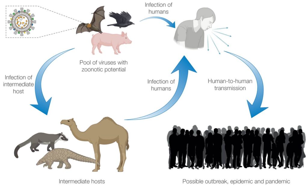
  • Primary Route of Transmission: Airborne transmission via nasopharyngeal respiratory droplet aerosols expelled during sneezing, coughing, or prolonged conversation by untreated, highly bacilliferous multibacillary (MB) patients. Millions of viable bacilli are shed daily in their nasal secretions.
  • Infectivity Requirements: Transmission is not casual. It requires prolonged, close, frequent contact (months to years) within the same household or immediate living environment.
  • Innate Human Immunity: Approximately 95% of all human beings possess complete natural, innate cell-mediated immunity against M. leprae and will never develop clinical disease even upon heavy exposure.
  • Infectivity Cessation: Within 72 hours to a few days of initiating multidrug therapy (MDT), the bactericidal potency of Rifampicin renders the patient non-infectious, permanently arresting forward transmission.
  • Non-Transmission Routes: There is no transplacental transmission (cannot be passed to an unborn fetus), no sexual transmission, and no transmission via casual social contacts (shaking hands, hugging, sharing utensils, sitting together).
  • Zoonotic Reservoirs: While humans are the primary reservoir, wild nine-banded armadillos in the southern United States and red squirrels in the United Kingdom harbor endemic M. leprae strains capable of zoonotic transmission to humans via direct handling.

Incubation Period

  • Due to the exceptionally slow bacterial division rate, the incubation period is prolonged.
  • Average Duration: Approximately 5 years.
  • Clinical Range: Ranges from 9 months to 20 years or more. Symptoms in Paucibacillary (PB) forms typically appear earlier (2–5 years), whereas Multibacillary (MB) forms average 8–12 years.

Risk Factors

  • Close Household Contact: Living with an untreated index patient with Multibacillary (MB) disease elevates disease risk up to 5- to 10-fold.
  • Geographic Clustering: Residing in hyper-endemic pockets or cross-border corridors (e.g., the West Nile region in Uganda).
  • Genetic Predisposition: Host polymorphisms in human leukocyte antigen (HLA-DR2 and HLA-DR3 associated with tuberculoid disease; HLA-DQ1 associated with lepromatous disease) and non-HLA genes (PARK2/PACRG, NOD2, LTA).
  • Malnutrition & Extreme Poverty: Food insecurity, overcrowded poor housing, and poor sanitary conditions that weaken baseline mucosal cell-mediated immunity.

3. Pathophysiology & Immunological Spectrum

Neural Tropism & Molecular Pathogenesis of Nerve Damage

Leprosy is fundamentally a neuro-dermatological disease. Mycobacterium leprae is the only human pathogen capable of directly invading the parenchyma of peripheral nerves:

  1. Attachment: The unique cell wall glycolipid PGL-1 and bacterial adhesins bind specifically to the G-domain of laminin-α2 situated within the basal lamina surrounding peripheral Schwann cells.
  2. Invasion & Demyelination: This interaction triggers receptor-mediated endocytosis via the α-dystroglycan complex. Once intracellular, the bacillus induces Schwann cell dedifferentiation, downregulating myelin gene expression and upregulating stem-cell-like progenitor traits, initiating spontaneous demyelination and axonal atrophy.
  3. Immunological Destruction: The subsequent destruction of the peripheral nerve is an immunologically mediated bystander injury. In patients with vigorous cell-mediated immunity (tuberculoid pole), CD4+ T-helper 1 (Th1) lymphocytes infiltrate the endoneurium, releasing interferon-gamma (IFN-γ) and TNF-α, forming compact epithelioid granulomas within the non-distensible perineurial sheath. This generates high intra-neural compartment pressure, localized ischemia, caseous necrosis, and permanent fibrotic nerve trunk strangulation.
  4. Lepromatous Infiltration: Conversely, in lepromatous leprosy, deficient cellular immunity permits unhindered intracellular bacterial replication within Schwann cells, perineurial macrophages, and vascular endothelial cells, causing painless, gradual, diffuse replacement of nerve fibers by foamy lepra cells and widespread ascending sensorimotor axonal loss.

The Ridley-Jopling Immunological Spectrum

The clinical, histological, and bacteriological manifestations of leprosy represent a continuous biological spectrum dictated entirely by the potency of the host’s Cell-Mediated Immunity (CMI) against M. leprae:

ClassificationCell-Mediated ImmunityBacterial Index (BI)Skin Lesion FeaturesHistopathological Hallmarks
Tuberculoid (TT)High / Robust (Th1 dominant: IFN-γ, IL-2)0 (Negative)Single or very few (≤3) large, well-demarcated hypopigmented anesthetic plaques with raised, erythematous, dry, hairless, scaly edges. Marked nerve thickening.Epithelioid cell granulomas with Langerhans giant cells surrounding dermal nerve twigs; dense cuffing by CD4+ lymphocytes. No demonstrable AFB.
Borderline Tuberculoid (BT)Moderate / Variable0 to 1+Few lesions (typically 3–10) resembling TT but smaller, less sharply defined margins; multiple satellite lesions; asymmetric nerve enlargement.Well-formed epithelioid granulomas, few giant cells, moderate lymphocytic infiltration, rare AFB in Schwann cells.
Mid-Borderline (BB)Unstable / Equivocal2+ to 3+Moderate number of lesions; characteristic "punched-out" or "inverted saucer" plaques (sloping outer margin with a sharply punched-out, anesthetic center).Diffuse epithelioid granulomas without giant cells, variable lymphocytes, moderate AFB present.
Borderline Lepromatous (BL)Low / Deficient3+ to 5+Numerous bilateral, asymmetric macules, papules, plaques, and small nodules; sensation mildly reduced or normal in early stages.Predominantly foamy histiocytes, few lymphocytes, abundant AFB singly and in globi, perineurial onion-skin lamellation.
Lepromatous (LL)Absent / Anergy (Th2 dominant: IL-4, IL-10)5+ to 6+ (Extreme)Innumerable, small, symmetric, shiny, ill-defined erythematous macules, papules, diffuse infiltration, plaques, and nodules. Leonine facies, loss of eyebrows, saddle-nose.Extensive sheets of foamy Virchow histiocytes packed with thousands of AFB in globi. Uninfiltrated Grenz zone directly below the epidermis. No granulomas.
Indeterminate Leprosy (I):
The earliest, uncommitted clinical stage of leprosy. Characterized by a single, faint, hypopigmented or erythematous macule with vague borders and only slightly diminished sensation. Histologically shows non-specific perivascular or periadnexal lymphocytic infiltration with rare bacilli located within tiny dermal neurovascular bundles. Depending on the individual's evolving cell-mediated immunity, indeterminate leprosy will either resolve spontaneously or evolve toward one of the Ridley-Jopling poles (TT or LL).

4. Clinical Presentation & Diagnostic Cardinal Signs

The 3 Cardinal Signs of Leprosy

In endemic countries, clinical case confirmation requires the definitive presence of at least ONE of the following three Cardinal Signs:

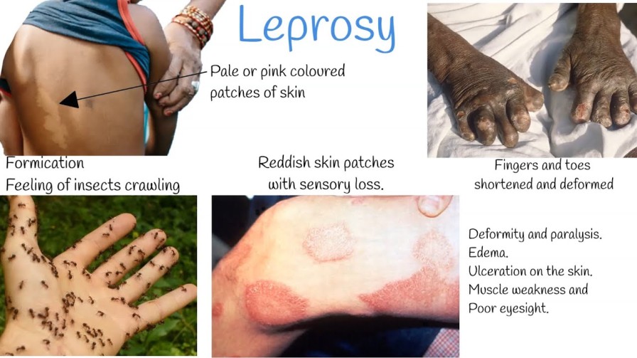
  1. Definite Loss of Sensation in a Hypopigmented or Reddish Skin Patch: Tested carefully using a light wisp of cotton wool, a feather, or standardized Semmes-Weinstein monofilaments (0.07g to 2.0g) applied to the patch while the patient's eyes are closed, comparing responses with adjacent normal skin. Thermal and pinprick sensation are lost prior to light touch.
  2. Thickened, Enlarged Peripheral Nerve Trunk(s): Palpable, tender, or firm peripheral nerves, accompanied by objective sensory impairment in its cutaneous distribution and/or motor weakness of the muscle groups it innervates.
  3. Microscopic Demonstration of Acid-Fast Bacilli (AFB): Visualized in a stained slit-skin smear or full-thickness skin/nerve biopsy specimen.

Nerve Palpation & Neurological Anatomy

Clinicians must routinely palpate specific superficial nerve trunks bilaterally for tenderness, enlargement, irregular beading, and firmness:

  • Ulnar Nerve: Palpated in the groove between the olecranon process and medial epicondyle of the humerus.
    • Sensory Deficit: Numbness over the hypothenar eminence, medial palm, little finger, and ulnar half of the ring finger.
    • Motor Deficit: Weakness of intrinsic hand muscles, interossei wasting, lumbrical paralysis, producing Claw Hand (main-en-griffe) deformity (hyperextension of metacarpophalangeal joints and flexion of interphalangeal joints, most marked in digits 4 and 5).
  • Median Nerve: Palpated as it passes deep to the flexor retinaculum at the wrist.
    • Sensory Deficit: Numbness over the thenar eminence, palmar aspects of thumb, index, middle finger, and radial half of the ring finger.
    • Motor Deficit: Thenar atrophy and paralysis of the abductor pollicis brevis, resulting in loss of thumb opposition (Ape Hand deformity) and total clawing when combined with ulnar paralysis.
  • Radial Cutaneous Nerve: Palpated across the anatomical snuffbox over the styloid process of the radius. Motor branch (posterior interosseous) involvement leads to Wrist Drop.
  • Common Peroneal (Lateral Popliteal) Nerve: Palpated rolling over the neck of the fibula on the lateral aspect of the knee.
    • Motor Deficit: Paralysis of the ankle dorsiflexors and peronei, resulting in Foot Drop with an unnatural high-stepping gait, leading to severe dragging of toes and extensive neuropathic trophic ulcerations.
  • Posterior Tibial Nerve: Palpated behind and below the medial malleolus of the ankle.
    • Sensory Deficit: Complete anesthesia of the entire sole of the foot.
    • Motor Deficit: Paralysis of intrinsic foot muscles leading to Clawed Toes and high peak planter pressures during ambulation, predisposing to chronic, painless neuropathic plantar ulcers.
  • Facial Nerve (Cranial Nerve VII): Specifically its zygomatic and temporal branches running over the zygomatic arch.
    • Motor Deficit: Paralysis of the orbicularis oculi muscle causing Lagophthalmos (inability to close the eyelid completely), absence of the corneal blink reflex, continuous exposure keratitis, corneal ulceration, secondary bacterial infection, and irreversible blindness.
  • Great Auricular Nerve: Palpated crossing diagonally across the superficial surface of the sternocleidomastoid muscle in the lateral neck; frequently visibly enlarged and cord-like in lepromatous and borderline cases.

Advanced Physical Signs in Multibacillary Leprosy

  • Leonine Facies (Facies Leonina): Extensive nodular thickening, deep furrowing of the forehead, glabellar ridges, and earlobes, imparting a lion-like facial appearance.
  • Madarosis: Bilateral, symmetrical, permanent loss of eyebrows (beginning characteristically at the outer lateral thirds) and eyelashes.
  • Saddle-Nose Deformity: Extensive chronic granulomatous infiltration and ischemic necrosis of the cartilaginous nasal septum, leading to septal perforation, structural collapse, and sunken bridge of the nose.
  • Gynecomastia & Testicular Atrophy: Direct bacillary infiltration of the seminiferous tubules and interstitial Leydig cells, causing hypergonadotropic hypogonadism, impotence, and sterility.

5. Diagnostic Investigations & Laboratory Workup

Slit-Skin Smear (SSS) Examination

A simple, highly specific semi-invasive bedside test performed to detect and quantify viable and non-viable acid-fast bacilli:

  • Standard Sampling Sites (Minimum 4 to 6 Sites):
    • Both earlobes (lower fleshy margin).
    • At least two active, erythematous, indurated skin lesion edges.
    • Dorsal aspect of fingers or normal-appearing skin over the forehead or chin.
  • Procedure: Pinch the skin firmly between thumb and forefinger to render the tissue ischemic (preventing bleeding, which dilutes the smear with red blood cells). Make a superficial scalpel slit (5 mm long, 2 mm deep) into the dermis. Turn the scalpel blade 90 degrees and scrape the side and bottom of the slit firmly to obtain clear dermal tissue pulp and cellular fluid. Smear onto a clean glass slide, heat fix, and stain with the modified Ziehl-Neelsen method.
  • Microscopic Enumeration:
    • Bacterial Index (BI - Ridley Scale): Reflects total bacterial load (live and dead):
      • 0: No bacilli observed in 100 oil immersion fields.
      • 1+: 1 to 10 bacilli per 100 fields.
      • 2+: 1 to 10 bacilli per 10 fields.
      • 3+: 1 to 10 bacilli per single field.
      • 4+: 10 to 100 bacilli per single field.
      • 5+: 100 to 1,000 bacilli per single field (clumps/globi).
      • 6+: >1,000 bacilli per field (massive globi throughout).
    • Morphological Index (MI): The percentage of solid-staining, fully intact, viable bacilli out of 200 counted bacilli. A falling MI is the earliest, most sensitive indicator of bacteriological response to effective MDT.

Skin & Nerve Biopsy (Histopathology)

Full-thickness punch or incisional biopsy taken from the advancing, elevated border of an active skin lesion including subcutaneous fat, or a superficial sensory nerve twig (e.g., radial cutaneous or sural nerve). Stained with Hematoxylin & Eosin (H&E) and the Fite-Faraco acid-fast stain. Provides absolute confirmation by demonstrating neural architecture, perineurial inflammation, granuloma morphology, and intra-neural bacilli.

Polymerase Chain Reaction (PCR) & Molecular Assays

Utilizes targeted nucleic acid amplification targeting the repetitive element (RLEP), groEL, or the 16S rRNA genes of M. leprae. Highly sensitive (>90%) and specific, capable of detecting as few as 1 to 10 bacterial genomes in paucibacillary lesions, indeterminate cases, blood, or fine-needle aspirates where smears are completely negative.

Differential Diagnosis

  • Hypopigmented Macules: Tinea versicolor (pityriasis versicolor—shows superficial scaling, KOH-positive fungal hyphae, completely normal sensation); Vitiligo (total chalky depigmentation, no sensory loss); Pityriasis alba (common in atopic children, cheeks, normal sensation); Post-inflammatory hypopigmentation.
  • Infiltrated Plaques & Nodules: Cutaneous Leishmaniasis; Sarcoidosis (non-caseating naked granulomas, no sensory deficit); Cutaneous Tuberculosis (Lupus vulgaris); Secondary and Tertiary Syphilis; Neurofibromatosis type 1; Kaposi Sarcoma; Granuloma annulare.
  • Peripheral Neuropathies: Diabetic peripheral sensory-motor polyneuropathy (bilateral, symmetrical, glove-and-stocking without thickened nerve trunks); Alcoholic neuropathy; Nutritional neuropathies (Vitamin B12 deficiency); Hereditary motor-sensory neuropathy (Charcot-Marie-Tooth disease).

6. WHO Classification & Medical Management (MDT Protocols)

WHO Operational Classification for Treatment

To eliminate reliance on laboratory microscopy at primary health centers, the WHO established a pragmatic operational classification based entirely on clinical lesion counting:

  • Paucibacillary (PB) Leprosy: 1 to 5 skin lesions with definite sensory loss, affecting no more than one nerve trunk. Slit-skin smears (if performed) are negative. (Corresponds to TT and BT on the Ridley-Jopling spectrum).
  • Multibacillary (MB) Leprosy: 6 or more skin lesions, OR involvement of more than one peripheral nerve trunk (regardless of lesion count), OR any positive slit-skin smear finding. (Corresponds to BB, BL, and LL on the Ridley-Jopling spectrum).

I. Multidrug Therapy (MDT) Regimens

MDT blister packs are supplied by the WHO free of charge globally. Treatment combines three core antimycobacterial agents:

Crucial Paradigm Shift — WHO 2018 Guidelines:

Historically, PB leprosy was treated with only 2 drugs (Rifampicin and Dapsone for 6 months), while MB leprosy received 3 drugs (adding Clofazimine for 12 months). The WHO Guidelines for the Diagnosis, Treatment, and Prevention of Leprosy 2018 strongly recommended a Uniform 3-Drug Regimen (Rifampicin, Dapsone, and Clofazimine) for BOTH PB and MB leprosy (6 months for PB, 12 months for MB). This prevents relapse, simplifies drug procurement, and ensures that misclassified MB cases are not undertreated.

*Note on Operational Practice: While Uganda and many national programmes have phased in 3-drug therapy, traditional 2-drug blister packs remain clinically relevant in many field settings. Both regimens are detailed below.

A. Adult Standard MDT Dosing Protocols

DrugDosage & FrequencyMode of AdministrationDuration
Rifampicin600 mg once monthlyDirectly Observed Therapy (DOT) by health workerPB: 6 months
MB: 12 months
Clofazimine300 mg once monthly (supervised DOT)
PLUS 50 mg daily
DOT (monthly dose)
Self-administered (daily dose)
PB: 6 months (WHO 2018)
MB: 12 months
Dapsone100 mg dailySelf-administered dailyPB: 6 months
MB: 12 months

B. Pediatric Standard MDT Dosing Protocols

MedicationChildren Aged 10 to 14 YearsChildren <10 Years (or <40 kg Body Weight)
Rifampicin450 mg once monthly (supervised DOT)10 mg/kg body weight once monthly (DOT)
Clofazimine150 mg once monthly (DOT) + 50 mg every other day6 mg/kg once monthly (DOT) + 1 mg/kg every other day
Dapsone50 mg daily2 mg/kg body weight daily

C. Treatment Duration & Completion Criteria

  • PB Leprosy: 6 monthly blister packs to be completed within a maximum timeframe of 9 months.
  • MB Leprosy: 12 monthly blister packs to be completed within a maximum timeframe of 18 months.
Critical Pharmacological Interaction — Dapsone and Cotrimoxazole (Septrin):
In HIV-positive patients already receiving daily Cotrimoxazole (TMP-SMX) preventive prophylaxis, DAPSONE MUST NEVER BE CO-ADMINISTERED. Both agents are folate-synthesis inhibitors (sulfonamides/sulfones) and share identical pathways of hematological toxicity. Concurrent use precipitates catastrophic, life-threatening bone marrow aplasia, severe agranulocytosis, and severe hemolytic crisis. In such patients, Dapsone must be replaced with Clofazimine or second-line regimens under specialist guidance.

Mechanisms of Action, Pharmacokinetics & Toxicities of MDT Drugs

  • Rifampicin (Rifadin):
    • Mechanism: Binds to the β-subunit of bacterial DNA-dependent RNA polymerase, blocking transcription and RNA synthesis. It is the single most powerful, rapidly bactericidal drug in the regimen, killing 99.9% of viable M. leprae within a single 600 mg dose.
    • Adverse Effects: Harmless orange-red discoloration of urine, sweat, saliva, and tears (essential patient counseling point!); transient hepatotoxicity; cutaneous flushing; rare flu-like syndrome with intermittent high-dose therapy.
  • Dapsone (4,4’-diaminodiphenylsulfone):
    • Mechanism: Competitive antagonist of para-aminobenzoic acid (PABA), inhibiting bacterial dihydropteroate synthase and halting folic acid synthesis. Slow, bacteriostatic agent.
    • Adverse Effects: Dose-dependent mild hemolysis and methemoglobinemia; severe, life-threatening acute hemolytic crisis in patients with Glucose-6-Phosphate Dehydrogenase (G6PD) deficiency; agranulocytosis; peripheral neuropathy; drug-induced lupus; and the severe idiosyncratic "Dapsone Hypersensitivity Syndrome" (exfoliative dermatitis, high fever, lymphadenopathy, hepatosplenomegaly, mononucleosis, and hepatitis, occurring 2–6 weeks after starting therapy; requires immediate, permanent discontinuation).
  • Clofazimine (Lamprene):
    • Mechanism: Lipophilic rhimophenazine dye that binds selectively to bacterial DNA guanine bases, inhibiting replication and generating toxic intracellular reactive oxygen species. Possesses potent anti-inflammatory properties by suppressing lysosomal enzymes and neutrophil chemotaxis, significantly suppressing Type 2 Lepra Reactions (ENL).
    • Adverse Effects: Causes reversible brownish-black or purplish hyperpigmentation of the skin, macules, and conjunctivae (occurs in >90% of patients and persists for months after treatment completion—a major driver of psychosocial distress); severe skin xerosis (ichthyosis); gastrointestinal deposition in mesenteric lymph nodes and bowel wall causing intermittent abdominal cramps, diarrhea, and rare paralytic ileus.
  • Second-Line & Alternative Agents: Indicated in documented rifampicin resistance, severe Dapsone hypersensitivity, or patient refusal of Clofazimine pigmentation:
    • Fluoroquinolones (Ofloxacin 400 mg daily or Moxifloxacin 400 mg daily): Inhibit bacterial DNA gyrase (topoisomerase II). Highly bactericidal.
    • Minocycline (100 mg daily): Semisynthetic tetracycline; lipophilic, crosses host cell membranes and inhibits bacterial protein synthesis at the 30S ribosomal subunit.
    • Clarithromycin (500 mg daily): Macrolide; binds the 50S ribosomal subunit, suppressing peptide translocation. Highly active against M. leprae.

7. Lepra Reactions: Pathophysiology & Emergency Management

Lepra reactions are acute, immunologically mediated systemic inflammatory episodes that occur before, during, or years after successful completion of MDT. They do NOT indicate treatment failure or bacterial relapse, but represent a sudden immunological response to dead bacterial antigens. If not treated promptly, reactions cause irreversible, permanent nerve trunk destruction within days.

I. Type 1 Lepra Reaction (Reversal Reaction)

  • Immunological Basis: Delayed-type hypersensitivity (Type IV immune reaction) driven by an abrupt, spontaneous upregulation of Cell-Mediated Immunity (CMI) against M. leprae antigens residing within skin lesions and Schwann cells. Seen almost exclusively in the borderline spectrum (BT, BB, BL).
  • Clinical Features:
    • Existing skin patches become acutely swollen, erythematous, shiny, hot, and highly tender. New satellite lesions may erupt. Desquamation and skin ulceration can follow.
    • Acute Neuritis (Medical Emergency): Peripheral nerves become enlarged, intensely painful, and exquisitely tender to light palpation. Rapid loss of motor and sensory function (e.g., sudden onset of foot drop, complete facial palsy, or rapid loss of hand sensation).
    • Systemic fever and constitutional symptoms are usually mild or absent.
  • Management Protocol:
    • Continue MDT without interruption.
    • Systemic Corticosteroids: High-dose Oral Prednisolone is the definitive first-line standard. Initiate at 40 mg daily (or 1 mg/kg/day) taken in the morning. Maintain for 2 weeks, then gradually taper down by 5 mg every 2 weeks over a 12- to 24-week period depending on clinical recovery.
    • Limb Splinting: Immobilize and rest the acutely inflamed limb in functional positions using plaster slabs or splints to relieve mechanical tension and prevent contractures.
    • Surgical Nerve Decompression: Indicated for unrelenting, excruciating neural pain or progressive motor/sensory paralysis failing to respond to 72 hours of maximal steroid therapy. Involves longitudinal neurolysis and incision of the epineurium.

II. Type 2 Lepra Reaction (Erythema Nodosum Leprosum - ENL)

  • Immunological Basis: Systemic immune-complex-mediated vasculitis (Type III hypersensitivity). High bacterial antigen loads combine with circulating polyclonal antibodies, forming antigen-antibody complexes that deposit within post-capillary venule walls, activating the classical complement cascade and recruiting neutrophils. Seen exclusively in multibacillary patients (BL and LL).
  • Clinical Features:
    • Cutaneous Eruptions: Sudden eruption of crops of bright red, warm, intensely tender, evanescent subcutaneous nodules and plaques situated predominantly on the extensor surfaces of limbs, thighs, and face. Nodules subside in 3 to 7 days, leaving faint brown macules, while new crops continue to erupt. In severe cases, nodules undergo central necrosis and ulcerate (ENL necroticans).
    • Severe Systemic Manifestations: High swinging fever (39°C–40°C), profound prostration, generalized arthralgia, synovitis, bone pain (dermo-periostitis), lymphadenopathy, and hepatosplenomegaly.
    • Visceral & Organ Complications: Acute bilateral iridocyclitis/uveitis (risk of secondary glaucoma and blindness), acute orchitis/epididymitis (extreme testicular agony, testicular necrosis, secondary sterility), glomerulonephritis, and severe acute polyneuritis.
  • Management Protocol:
    • Continue MDT without interruption.
    • Systemic Corticosteroids: Oral Prednisolone (40–60 mg daily), tapering slowly over several months. Recurrences are exceptionally common when tapering below 15–20 mg.
    • Thalidomide: The single most potent, rapidly acting therapeutic agent for severe ENL. Inhibits TNF-α synthesis, alters integrin expression, and suppresses neutrophil extravasation. Dose: 100 to 400 mg orally at bedtime, tapering gradually to 50–100 mg maintenance.

      ABSOLUTE CONTRAINDICATION: Thalidomide is devastatingly teratogenic, causing severe phocomelia (seal-like limb deformities) and fetal agenesis. It is strictly prohibited in women of childbearing potential unless absolute contraception (dual barrier methods) is documented under rigid regulatory frameworks.

    • High-Dose Clofazimine: Increased to 300 mg daily (divided into 100 mg three times daily) for 1 month, then 200 mg daily for 3 to 6 months, and finally 100 mg daily. Highly effective for controlling steroid-dependent chronic ENL and weaning patients off prolonged steroid regimens.
    • Analgesics / NSAIDs: Paracetamol, Indomethacin, or high-dose Ibuprofen for mild ENL manifestations.
    • Ophthalmic Care: Topical 1% Atropine drops (to dilate the pupil and prevent posterior synechiae formation) plus topical 0.1% Dexamethasone eye drops for acute leprotic anterior uveitis.

8. Comprehensive Nursing Care Plan (ADPIE Framework)

Nursing DiagnosisAssessment FindingsDesired OutcomesNursing InterventionsScientific RationaleEvaluation
Impaired Skin Integrity
related to sensory loss, bacterial infiltration, and pressure necrosis.
- Painless ulcers on soles/palms
- Anesthetic hypopigmented plaques
- Fissured, dry skin
- Loss of protective pain sensation
Patient will demonstrate intact skin integrity, wound healing of ulcers, and absence of secondary osteomyelitis. 1. Teach daily foot/hand visual inspection.
2. Implement the "Soak, Scrape, Oil" protocol daily: soak feet for 20 mins, gently scrape hyperkeratotic calluses with a pumice stone, coat immediately with petroleum jelly or mineral oil.
3. Perform sterile wound dressing for open ulcers.
4. Ensure total off-loading of plantar ulcers with plaster boots or custom orthotics.
1. Loss of pain sensation prevents awareness of microtrauma; visual inspection compensates for absent nociception.
2. Denervated skin loses autonomic sweat function (anhidrosis); hydration and lubrication prevent cracking and secondary infection.
3. Off-loading eliminates shearing forces, allowing epithelial migration.
Goal Met: Trophic plantar ulcers granulated completely; skin margins soft and supple; no new skin breaks.
Risk for Peripheral Neurovascular Dysfunction / Injury
related to nerve inflammation (lepra reactions) and compression.
- Acute nerve tenderness
- Developing claw hand or foot drop
- Inability to hold objects securely
- Tingling or sudden numbness
Patient will maintain baseline motor/sensory nerve function without progressive paralysis or irreversible deformity. 1. Perform voluntary muscle testing (VMT) and sensory testing with monofilaments at every visit.
2. Immediately administer prescribed systemic Prednisolone upon detection of neuritis.
3. Apply resting splints to affected extremities.
4. Instruct patient to use padded handles, insulated cooking utensils, and wear thick leather gloves.
1. Early detection of nerve function impairment within 6 months enables steroid reversal.
2. Glucocorticoids reduce endoneurial edema and halt ischemic axonal death.
3. Insulation prevents catastrophic thermal burns from hot pans and fire embers.
Goal Met: Nerve tenderness eliminated; ulnar/peroneal motor power preserved; no accidental burn injuries sustained.
Disrupted Body Image
related to visible facial infiltrations, madarosis, skin discoloration, and physical deformities.
- Clofazimine reddish-brown skin pigmentation
- Leonine facies / saddle nose
- Depressed affect, social withdrawal
- Avoidance of eye contact
Patient will verbalize realistic acceptance of physical alterations and actively participate in social and self-care activities. 1. Pre-counsel patient extensively before initiating Clofazimine that skin darkening is temporary and will fade post-treatment.
2. Encourage verbalization of grief and feelings regarding facial/limb changes.
3. Refer for reconstructive surgery (temporalis transfer for lagophthalmos, rhinoplasty, claw hand tendon transfers).
4. Connect with peer survivor networks.
1. Foreknowledge prevents non-compliance fueled by cosmetic alarm.
2. Therapeutic listening validates self-worth and decreases depressive symptomatology.
3. Corrective reconstructive surgery restores physical function and normalizes anatomical appearance.
Goal Met: Patient verbalizes that drug-induced hyperpigmentation is a sign of effective cure; joins local self-help cooperative.
Inadequate health Knowledge
regarding transmission, treatment duration, and lepra reactions.
- Inquiring whether disease is a curse
- Fear of infecting family members
- Stopping MDT when lesions flatten
- Confusing lepra reaction with cure failure
Patient and family will explain the biological cause, mode of transmission, and necessity of finishing the 6- or 12-month MDT course. 1. Educate patient that leprosy is a bacterial disease, not a curse, witchcraft, or inherited genetic trait.
2. Reassure family that patient is non-infectious within days of beginning MDT.
3. Provide color-coded calendar reminders for monthly clinic blister-pack collections.
4. Teach warning signs of lepra reactions (red, painful swelling of skin or nerves) requiring immediate hospital visit.
1. Dispels destructive cultural stigma and eliminates domestic isolation.
2. Directly observed therapy (DOT) and structured health literacy prevent premature treatment default.
3. Early presentation for reactions prevents irreversible paralysis.
Goal Met: Caregiver accurately details 3 cardinal signs and commits to uninterrupted 12-month clinic visits.

9. Complications & Disability Prevention

WHO Disability Grading System

To standardize clinical evaluation and programmatic monitoring globally, the WHO developed a simplified three-tier grading system assessing the hands, feet, and eyes:

GradeHands and Feet CriteriaEyes Criteria
Grade 0No anesthesia (loss of sensation); no visible deformity or damage.No eye problems due to leprosy; no loss of vision (vision 6/60 or better; can count fingers at 6 meters).
Grade 1Loss of sensation (anesthesia) present, but NO visible deformity or damage.Eye problems present due to leprosy (e.g., lagophthalmos, iridocyclitis), but vision is not severely impaired (can count fingers at 6 meters).
Grade 2Visible deformity or damage PRESENT: Ulcers, claw hand, foot drop, wrist drop, shortened/reabsorbed digits, severe bone absorption.Severe visual impairment (vision worse than 6/60; inability to count fingers at 6 meters); severe lagophthalmos, corneal opacity, or complete blindness.

Mechanisms of Bone Reabsorption & "Auto-Amputation"

The myth that digits "spontaneously rot and drop off" is completely false. The true sequence of progressive phalangeal shortening involves:

  1. Severe, unperceived repetitive mechanical trauma or thermal injury to totally anesthetic digits.
  2. Breakdown of epidermal barrier leading to indolent, painless soft-tissue ulcerations.
  3. Secondary invasive polymicrobial bacterial infection extending through fascial planes into the periosteum and bone cortex (pyogenic osteomyelitis).
  4. Development of avascular bone necrosis, microfractures, chronic osteolytic remodeling, and gradual aseptic osteoclast-mediated bone reabsorption by the host tissue, leading to stubbed, shortened, retracted digits.

Ophthalmological Complications & Prevention of Blindness

  • Lagophthalmos Care: Paralysis of the seventh cranial nerve prevents protective blinking. Patients must perform active forced eyelid closure exercises, wear wide-brimmed hats and wrap-around sunglasses outdoors to block dust and insects, apply sterile artificial tear drops hourly during the day, and apply thick antibiotic ointment (Tetracycline) at night, taping the eyelids shut with hypoallergenic paper tape.
  • Corneal Anesthesia: Sensory denervation of the ophthalmic branch of the trigeminal nerve (Cranial Nerve V1) results in total loss of corneal sensation; small abrasions and foreign bodies produce no pain, predisposing to undetected ulceration and hypopyon.

Microcellular Rubber (MCR) Footwear

Standard footwear exerts high localized mechanical shear forces over the metatarsal heads and heel, rapidly inducing blister formation and chronic neuropathic ulcers in anesthetic feet. MCR footwear utilizes specialized microcellular rubber insoles (15° to 20° Shore hardness) that conform dynamically to the plantar arch, distributing weight evenly over the entire sole surface, combined with rigid outer soles and firm heel counters to eliminate shearing.

10. Prevention, Chemoprophylaxis & The WHO 2021–2030 Strategy

I. Contact Tracing & Post-Exposure Prophylaxis (SDR-PEP)

Single-Dose Rifampicin Post-Exposure Prophylaxis (SDR-PEP) is the global standard for interrupting the transmission chain of leprosy among close contacts.

Eligibility Criteria:

  • All household, neighborhood, and social contacts who have had close association with a newly diagnosed PB or MB index patient within the past 1–2 years.
  • Contact must be at least 2 years of age.
  • Must undergo thorough clinical screening to rule out active leprosy disease and active tuberculosis prior to administration (giving monotherapy Rifampicin to a patient with undiagnosed active TB precipitates rifampicin-resistant TB!).
  • No history of liver disease or jaundice; not pregnant; no known rifampicin allergy; has not received rifampicin for any indication within the preceding 2 years.

SDR-PEP Weight- & Age-Based Dosing Protocol

Age / Weight CategorySingle Oral Dose of Rifampicin
Adults (≥15 years, ≥35 kg)600 mg single dose
Children aged 10 to 14 years (or 20–34 kg)450 mg single dose
Children aged 6 to 9 years (or 15–19 kg)300 mg single dose
Children aged 2 to 5 years (or <15 kg)10 to 15 mg/kg body weight single dose

II. Immunoprophylaxis (BCG Vaccination)

Administration of the Bacillus Calmette-Guérin (BCG) vaccine at birth (part of national infant immunization schedules) confers significant cross-reactive cell-mediated protective immunity against M. leprae, reducing the overall incidence of clinical leprosy by approximately 50% to 60% and providing even higher protective efficacy against severe multibacillary and lepromatous forms.

III. WHO Global Leprosy Strategy 2021–2030: "Towards Zero Leprosy"

The global roadmap is anchored on four fundamental goals: Zero Infection, Zero Disease, Zero Disability, and Zero Stigma and Discrimination:

  • Pillar 1: Implement integrated, country-owned zero-leprosy roadmaps in all endemic nations.
  • Pillar 2: Scale up leprosy prevention alongside integrated active case detection (shifting from passive waiting to proactive active case searches in communities and schools).
  • Pillar 3: Manage leprosy and its complications early, preventing new disabilities through expanded access to reconstructive surgery, assistive devices, and psychological support.
  • Pillar 4: Combat discrimination and promote inclusion, ensuring human rights protection and repealing outdated, discriminatory legislation against persons affected by leprosy.

11. Uganda National TB and Leprosy Strategic Plan (NSP 2025/26–2029/30)

Epidemiological Profile of Leprosy in Uganda

  • Current National Burden: In 2024, Uganda reported 275 new leprosy cases, corresponding to an incidence of 6 per 1,000,000 population.
  • The Geographic Epicenter (West Nile): Leprosy transmission is heavily geographically clustered. The West Nile sub-region registers the highest disease rates in the nation, ranging between 56 and 280 per 1,000,000 population. Between 2020 and 2024, 49% of all districts in Uganda (72 out of 146) recorded at least one new case, proving that low-level active transmission persists widely.
  • High Proportion of Child Cases: A critical 17% of all new cases occur in children (<15 years). This is an alarming epidemiological indicator demonstrating intense, recent, ongoing community transmission.
  • Overwhelming Multibacillary Ratio: Approximately 86% of newly notified cases are Multibacillary (MB), carrying severe risks of extensive onward droplet spread and high bacterial loads.
  • Alarmingly High Grade 2 Disability (G2D) at Diagnosis: In 2024, 24% of new leprosy patients presented with established Grade 2 visible disabilities at the very time of initial clinical diagnosis, an increase from previous years and dramatically higher than the national elimination target of <5%, underscoring severe, chronic diagnostic delays at primary healthcare levels.
  • Treatment Completion Rate: Treatment completion stood at 78% in 2022, improving to 91% in 2024 through intensified tracking, but remaining vulnerable to drug supply imbalances.

Core Strategic Targets of the Uganda Leprosy Response (NSP 2025/26–2029/30)

Strategic Objective / IndicatorBaseline (2024/25)Target by 2029/30
SO5: Proportion of child leprosy notifications14% – 17%< 3%
SO5.1: Districts reporting zero new autochthonous leprosy cases116 districts (79%)132 districts (90%)
SO5.2: Absolute annual new and relapse cases detected nationally273 – 275 casesReduce to 30 cases
SO5.3.1: Household contact tracing coverage67%≥ 90%
SO5.3.2: Proportion of index contacts screened for signs/symptoms74%90%
SO5.3.3: Eligible contacts receiving Single-Dose Rifampicin (SDR-PEP)55%≥ 90%
SO5.3.4: New leprosy cases presenting with Grade 2 Disability (G2D)18% – 24%< 5%
SO5.4: Treatment completion rate (TCR) for leprosy91%≥ 95%
SO5.5: Patients in need provided with custom protective MCR footwear72%≥ 90%

Strategic Interventions for Leprosy under the Uganda NSP

  • Child Case Audits: Conducting rapid epidemiological audits of every newly identified child leprosy case within 2 weeks of diagnosis, mapping exposure networks across household, school, and neighborhood contacts.
  • CAST+ Integration: Embedding targeted leprosy screening modules into high-impact Community Awareness, Screening, and Testing (CAST+) outreach campaigns across high-burden West Nile districts and border areas.
  • Designation of Specialized Skin Clinics: Establishing dedicated skin clinics at all Regional Referral Hospitals (RRHs) and Health Centre IVs (HC IVs) equipped with standardized SOPs for cardinal sign examination, slit-skin smears, reaction triage, and referral.
  • End-to-End Contact Tracing with SDR-PEP: Enforcing universal, immediate line-listing of all household contacts, offering same-day clinical examination and SDR-PEP delivery within 7 days.
  • Decentralized MCR Footwear Production: Fixing last-mile procurement bottlenecks for Microcellular Rubber (MCR) sandals, standardizing foot-mapping at baseline, and scheduling mandatory replacement every 12 to 18 months.
  • Digital Surveillance Integration: Fully migrating leprosy case notifications and contact tracking into the electronic Case-Based Surveillance System (eCBSS) and integrating routine reporting into DHIS2 (HMIS 106a).
  • Supply Chain Security via TWOS: Incorporating MDT and SDR-PEP rifampicin supplies into the web-based ordering system (TWOS) and National Medical Stores (NMS) distribution schedules to eradicate facility-level stock-outs.

12. Myths, Misconceptions & Stigma Reduction

Myth 1: "Leprosy is a hereditary punishment, witchcraft, or divine curse."

Clinical Fact: Leprosy is purely a biological infectious disease caused by a bacterium (M. leprae). It has zero connection to genetics, ancestral spirits, or curses. It is completely treatable and curable with modern antibiotics.

Myth 2: "Leprosy causes body parts and fingers to spontaneously rot and drop off."

Clinical Fact: The bacterium destroys peripheral sensory nerves, eliminating protective pain sensation. Patients unknowingly suffer severe burns, cuts, and pressure injuries. Unperceived secondary bacterial infections and bone infections (osteomyelitis) lead to aseptic bone reabsorption, making digits appear shortened and retracted. With daily self-care and protective footwear, deformities are 100% preventable.

Myth 3: "Leprosy is highly contagious and warrants immediate physical isolation."

Clinical Fact: Over 95% of all humans have complete natural immunity to leprosy. Furthermore, within days of starting MDT, patients can no longer transmit the infection. There is zero medical justification for isolating patients in segregated settlements or barring them from employment, school, or community life.

References

  • Ministry of Health Uganda. National Tuberculosis and Leprosy Strategy 2025/26–2029/30. Kampala: National Tuberculosis and Leprosy Control Programme (NTLP). November 2025.
  • Ministry of Health Uganda. Uganda Clinical Guidelines (UCG) 2023: National Guidelines for Common Medical Conditions. Section 2.1.4: Leprosy.
  • World Health Organization. WHO Guidelines for the Diagnosis, Treatment and Prevention of Leprosy. Geneva: World Health Organization; 2018.
  • World Health Organization. Towards Zero Leprosy: Global Leprosy (Hansen’s Disease) Strategy 2021–2030. New Delhi: WHO Regional Office for South-East Asia; 2021.
  • Doctors Revision Uganda. Clinical Guide: Leprosy (Hansen's Disease) Diagnosis, Referral, Management and NTLP Protocols. 2026.
  • National Tuberculosis and Leprosy Programme (NTLP). Manual for Health Workers in Uganda. Ministry of Health, Kampala.

Quick Quiz

Leprosy Quiz

Medical Nursing

Privacy: Your details are used only for quiz tracking and certificates.

Leprosy Read More »

Tuberculosis

Tuberculosis

Comprehensive Clinical Guide to Tuberculosis: Pulmonary & Extra-Pulmonary Disease Management

1. Definition, Etiology & Microbiological Properties

Definition of Tuberculosis

Tuberculosis (TB) is a chronic, subacute, or acute communicable granulomatous infectious disease caused by acid-fast bacilli belonging to the Mycobacterium tuberculosis complex. Although the disease primarily targets the respiratory system (Pulmonary Tuberculosis - PTB), hematogenous and lymphatic dissemination can infect virtually any vascularized anatomical organ or tissue structure in the human body (Extra-Pulmonary Tuberculosis - EPTB).

Microbiological Properties of Mycobacterium tuberculosis

  • Taxonomy: Member of the Mycobacteriaceae family within the Mycobacterium tuberculosis complex (which includes M. tuberculosis, M. bovis, M. africanum, M. microti, and M. canettii). M. tuberculosis (MTB) is responsible for the vast majority of human disease.
  • Cellular Morphology & Physiology: MTB is a slender, slightly curved or straight, non-motile, non-spore-forming, obligate aerobic rod-shaped bacillus measuring 0.5 μm in width and 2 to 4 μm in length. As an obligate aerobe, it thrives in tissues with high oxygen tension, explaining its pronounced predilection for the apical and posterior segments of the upper pulmonary lobes.
  • The Unique Mycobacterial Cell Envelope: The cell wall architecture is exceptionally thick, hydrophobic, waxy, and rich in complex lipids (accounting for up to 60% of the total dry wall weight). Key structural components include:
    • Mycolic Acids: Long-chain β-hydroxy fatty acids that form a dense, lipid-shell barrier rendering the organism impervious to Gram staining, drying, osmotic lysis, chemical disinfectants, acids, alkalis, and many conventional antibacterial agents.
    • Arabinogalactan & Peptidoglycan: Provide covalent structural rigidity and anchor the outer mycolate layer to the inner plasma membrane.
    • Lipoarabinomannan (LAM): A surface lipoglycan functionally analogous to endotoxin; it inhibits macrophage activation, scavenges cytotoxic oxygen free radicals, suppresses T-cell proliferation, and blocks host phagosome-lysosome fusion.
    • Cord Factor (Trehalose 6,6'-dimycolate): Causes virulent bacilli to align in parallel serpentine microscopic cords; it induces chronic granuloma formation, inhibits leukocyte migration, and disrupts host mitochondrial respiration.
  • Acid-Fastness: Once stained with heated phenolic carbolfuchsin (Ziehl-Neelsen or Kinyoun method), the dye forms a stable complex with mycolic acids that resists destaining with strong mineral acids and ethanol (3% hydrochloric acid in 95% ethanol). Hence, they are defined as Acid-Alcohol Fast Bacilli (AAFB). Under fluorescent microscopy, they retain fluorochrome dyes (Auramine-O and Rhodamine).
  • Slow Growth Rate: MTB possesses an extraordinarily prolonged generation time of 15 to 20 hours. Consequently, visible colonies typically require 2 to 6 weeks of incubation on solid egg-based media (Lowenstein-Jensen) and 1 to 3 weeks in automated liquid culture systems (Bactec MGIT).

Transmission Dynamics

  • Airborne Droplet Nuclei: Transmission occurs via inhalation of microscopic airborne droplet nuclei (measuring 1 to 5 μm in diameter) generated when an individual with active, untreated cavitary pulmonary or laryngeal TB coughs, sneezes, shouts, or sings. A single forceful cough produces up to 3,000 droplet nuclei, each containing 1 to 3 viable bacilli.
  • Environmental Persistence: Due to their tiny mass, droplet nuclei remain buoyantly suspended in ambient air currents for hours in enclosed, poorly ventilated spaces.
  • Ingestion Route: Ingestion of unpasteurized cow's milk contaminated with Mycobacterium bovis penetrates intestinal Peyer's patches, producing primary gastrointestinal and mesenteric lymph node tuberculosis (zoonotic TB).
  • Non-Transmissibility: TB is not transmitted through fomites, clothing, bedding, sharing eating utensils, or casual physical touch. Extrapulmonary TB (without concomitant pulmonary involvement) is non-infectious to others.

2. Comprehensive Pathophysiology & Natural History

I. Primary Infection & Granuloma Formation

  1. Inhalation & Alveolar Implantation: Droplet nuclei bypass the mucociliary escalator of the upper airways and settle in the peripheral subpleural alveoli, typically in the mid-to-lower pulmonary zones.
  2. Phagocytosis & Intracellular Parasitism: Resting alveolar macrophages phagocytose the bacilli via complement (CR3) and mannose receptors. MTB evades destruction by secreting protein kinase G and the cell wall lipid sulfatide, which prevent the fusion of phagosomes with lysosomes and inhibit phagosomal acidification (maintaining pH > 6.4). The bacilli replicate unhindered within the macrophage cytosol.
  3. Lysis & Early Hematogenous Seeding: Macrophages rupture, releasing bacilli that recruit fresh blood monocytes. Bacilli drain via pulmonary lymphatics to the bronchopulmonary and hilar lymph nodes. A low-grade, clinically silent bacteremia disseminates bacilli throughout the vascular tree, seeding the apices of the lungs, renal cortices, vertebral bodies, meninges, and epiphyses of long bones (forming dormant metastatic foci).
  4. Cell-Mediated Immunity (CMI) & Delayed Hypersensitivity: Between 2 to 8 weeks post-infection, dendritic cells transport mycobacterial antigens to regional lymph nodes, priming CD4+ T-helper 1 (Th1) cells. Activated Th1 cells secrete Interferon-gamma (IFN-γ) and Interleukin-2 (IL-2). IFN-γ potently activates naive macrophages, upregulating inducible nitric oxide synthase (iNOS), generating reactive nitrogen intermediates , hydrogen peroxide, and lysosomal enzymes capable of destroying intracellular bacilli.
  5. The Tuberculous Granuloma: Activated macrophages transform into large epithelioid histiocytes with abundant pink cytoplasm and interdigitating cell membranes. These fuse together under the influence of IFN-γ to form multinucleated Langhans giant cells (nuclei arranged in a peripheral horseshoe pattern). The lesion is enclosed by a dense cuff of CD4+ and CD8+ T-lymphocytes, B-cells, and an outer rim of fibroblasts depositing collagen.
  6. Ghon Focus, Ghon Complex, and Ranke Complex:
    • Ghon Focus: The localized primary parenchymal subpleural granuloma in the mid/lower lung field.
    • Ghon (Primary) Complex: The anatomical combination of the Ghon focus, draining inflamed lymphatics (lymphangitis), and enlarged caseous regional hilar/mediastinal lymph nodes.
    • Ranke Complex: The late, burnt-out, fibrocalcified radiological remnant of the Ghon complex.

II. Latent TB Infection (LTBI) vs. Post-Primary (Reactivation) TB

  • Latent TB Infection (LTBI): In approximately 90% of immunocompetent individuals, the cell-mediated immune response arrests bacillary proliferation. Granulomas undergo peripheral fibrosis, hyalinization, and dystrophic calcification. Viable bacilli enter a dormant, non-replicating metabolic state (persisters) inside caseous cores for the lifetime of the host. Individuals with LTBI have no clinical signs or symptoms, cannot transmit the organism, have normal chest radiographs, but test positive on Tuberculin Skin Tests (TST) or Interferon-Gamma Release Assays (IGRA).
  • Post-Primary (Secondary / Reactivation) Tuberculosis: Occurs when host cell-mediated immunity declines (due to HIV infection, malnutrition, aging, diabetes mellitus, end-stage renal disease, anti-TNF-α biologics, or systemic corticosteroids), allowing dormant bacilli within old apical seeding sites (Simon foci) to resume active multiplication. Less commonly, it occurs via exogenous reinfection.
  • Caseation, Liquefaction, and Cavitation:
    • The sensitized host mounts a massive delayed-type hypersensitivity reaction, releasing high levels of Tumor Necrosis Factor-alpha (TNF-α), leading to avascular coagulative tissue necrosis characterized by soft, friable, debris resembling clotted cheese (caseous necrosis).
    • Hydrolytic enzymes released by dying macrophages digest protein matrices, transforming solid caseum into liquid material (liquefaction).
    • The liquefied necrotic mass erodes through the wall of an adjacent bronchial branch and discharges into the bronchial tree, leaving behind an empty, air-filled, rigid cavity (cavitation).
    • Bacteriological Explosion: Cavity walls provide an open, oxygen-saturated, hyper-vascular environment that fuels uncontrolled extracellular bacterial replication, generating up to $$10^7$$ to $$10^9$$ bacilli per cavity. Coughing expels enormous loads of infectious droplet nuclei into the air.
  • Rasmussen Aneurysm: Progressive granulomatous necrotizing inflammation of a cavity wall erodes and thins the adventitia and media of an adjacent branch of the pulmonary artery. The weakened vascular wall dilates into a pseudoaneurysm. Rupture of a Rasmussen aneurysm produces sudden, cataclysmic, fatal arterial massive hemoptysis.

3. Clinical Presentation: Pulmonary Tuberculosis (PTB)

Hallmark Systemic & Respiratory Manifestations

PTB has an insidious onset, frequently progressing over weeks to months prior to clinical presentation:

  • Persistent Chronic Cough: Typically productive of mucoid or mucopurulent sputum lasting ≥ 2 weeks (any duration in HIV-infected patients). Cough may initially be dry and hacking.
  • Hemoptysis: Ranging from blood-streaked sputum to life-threatening frank hemorrhage (due to cavitary erosion or Rasmussen aneurysm rupture).
  • Low-Grade Afternoon / Evening Fever: Diurnal temperature elevation characteristically spiking between 37.8°C and 39.0°C in the late afternoon or evening, subsiding by morning.
  • Drenching Night Sweats: Severe nocturnal diaphoresis requiring the patient to change clothes and bedsheets, occurring as the evening fever lysis.
  • Significant Unintentional Weight Loss ("Consumption"): Progressive wasting of somatic fat and skeletal muscle mass driven by high levels of circulating TNF-α (cachectin) and catabolic cytokines.
  • Anorexia & Profound Asthenia: Loss of appetite and debilitating physical lethargy.
  • Pleuritic Chest Pain: Sharp, stabbing, localized pain exacerbated by deep inspiration and coughing, indicating subpleural inflammation or parietal pleural extension.
  • Dyspnea: Exertional breathlessness developing in extensive parenchymal destruction, spontaneous secondary pneumothorax, large tuberculous pleural effusion, or end-stage fibrocavitary disease.

Physical Examination Findings

  • General Inspection: Cachexia, temporal wasting, marked pallor of the conjunctivae, digital clubbing (in chronic advanced disease).
  • Chest Palpation & Percussion: Dullness to percussion over areas of dense lobar consolidation, fibro-atelectasis, or pleural effusion. Hyperresonance if a tension pneumothorax complicates cavitary rupture. Asymmetrical chest expansion.
  • Auscultation: Bronchial breath sounds and aegophony over consolidations; coarse post-tussive apical crackles (amphoric breathing over large empty cavities); absent or diminished breath sounds at the base in pleural effusion.

4. Extra-Pulmonary Tuberculosis (EPTB): Specialized Manifestations

EPTB accounts for 15% to 25% of all TB cases in immunocompetent hosts, and upwards of 50% in individuals co-infected with HIV. It arises from early hematogenous or lymphatic seeding during primary infection or late reactivation.

I. Tuberculous Lymphadenitis (Scrofula)

The single most frequent clinical manifestation of EPTB globally, accounting for 35% of extrapulmonary cases.

  • Anatomical Distribution: Primarily targets the anterior and posterior cervical lymph nodes along the sternocleidomastoid muscle, followed by supraclavicular, submandibular, and axillary chains. Usually unilateral.
  • Natural Progression (The 5 Stages of Jones & Campbell):
    • Stage 1: Firm, discrete, movable, painless, non-tender enlarged nodes without peri-adenitis.
    • Stage 2: Large rubbery nodes becoming fixed to each other and surrounding subcutaneous tissues due to peri-adenitis (matted lymph nodes).
    • Stage 3: Central caseous liquefaction and necrosis producing a soft, fluctuant mass ("cold abscess").
    • Stage 4: The cold abscess erodes through deep cervical fascia; the skin becomes violaceous, thin, and adheres to the mass (collar-stud abscess).
    • Stage 5: Spontaneous rupture through the skin surface forming an indolent, chronic tuberculous sinus tract that discharges pale, thick caseous material and heals with hypertrophic, puckered scarring.
  • Diagnosis: Fine-Needle Aspiration Biopsy (FNAB) evaluated with GeneXpert MTB/RIF, AFB staining, and cytology (epithelioid histiocytes, Langhans giant cells, and background caseous necrosis).

II. Pleural Tuberculosis (Tuberculous Pleurisy & Empyema)

  • Pathogenesis: Rupture of an immediate subpleural caseous focus into the pleural space introduces mycobacterial proteins, triggering an acute delayed hypersensitivity immune reaction with intense capillary permeability.
  • Clinical Features: Acute or subacute onset of dry cough, high fever, and severe unilateral pleuritic chest pain. As the pleural fluid accumulates, pleuritic pain resolves, replaced by progressive dyspnea.
  • Diagnostic Pleural Fluid Tap (Thoracentesis):
    • Macroscopic Appearance: Serous, straw-colored, clear or turbid fluid (pus indicates tuberculous empyema).
    • Exudative Criteria (Light’s Criteria): Pleural fluid to serum total protein ratio > 0.5; pleural fluid to serum LDH ratio > 0.6.
    • Cytology: Predominantly marked lymphocytic pleocytosis (often >80% small, mature lymphocytes); absence of mesothelial cells (<5%).
    • Biochemical Markers: Adenosine Deaminase (ADA) > 40 U/L and elevated Interferon-gamma levels are highly sensitive and specific for TB pleurisy.
    • Bacteriology: Direct smear AFB positivity is low (<10%), but GeneXpert and pleural biopsy with an Abram's needle yield granulomas in >80% of cases.

III. Tuberculosis of the Spine (Pott's Disease / Tuberculous Spondylodiscitis)

Musculoskeletal TB accounts for approximately 10% of EPTB cases, and the vertebral spine is involved in 50% of these cases. It is the most clinically destructive form of skeletal tuberculosis.

Pathogenesis & Anatomical Evolution:

  1. Vascular Seeding: Bacilli seed the vertebral column via the arterial arcades of the anterior spinal artery or retrograde flow through the valveless Batson’s paravertebral venous plexus from the pelvis or abdomen.
  2. Site Predilection: The anterior inferior and superior margins of the vertebral bodies adjacent to the intervertebral disc are most vascularized; hence, the paradiscal region of the lower thoracic (T8–T12) and upper lumbar (L1–L3) spine is the most frequent primary site.
  3. Disc Infiltration & Destruction: Unlike pyogenic bacterial spondylodiscitis (where proteolytic enzymes rapidly liquefy the intervertebral disc early), MTB lacks collagenolytic enzymes. In Pott's disease, the disc initially survives but is secondarily starved of nutrients as the subchondral bone plates are destroyed by caseous osteomyelitis. Infection spreads beneath the anterior longitudinal ligament to involve adjacent contiguous vertebral bodies.
  4. Vertebral Collapse & Kyphosis: Avascular necrosis destroys the anterior portion of the vertebral bodies while the strong posterior vertebral elements (pedicles, laminae, spinous processes) are relatively spared. Axial gravity causes wedging and structural collapse of the anterior column, producing a sharp, acute backward posterior angulation known as an angular Gibbus deformity (kyphosis).
  5. Cold Abscess Formation: Liquefied caseous necrotic pus tracks along tissue planes and fascial sheaths without classical heat or erythema:
    • Cervical Spine: Forms a retropharyngeal abscess (causing dysphagia, stridor, and respiratory distress) or tracks along the scalene fascia into the posterior triangle of the neck.
    • Thoracic Spine: Expands fusiformly along the anterior longitudinal ligament, producing a characteristic bilateral "bird's nest" or paravertebral spindle shadow on plain AP radiographs.
    • Lumbar Spine: Enters the psoas muscle sheath, forming a massive Psoas Abscess that tracks downward beneath the inguinal ligament to present as a painless, fluctuant swelling in the femoral triangle, groin, or gluteal region.

Clinical Presentation of Pott's Disease:

  • Localized, dull, deep-seated aching back pain and stiffness; characteristically aggravated by movement and weight-bearing, relieved by recumbency.
  • "Night Cries" in children: Muscle spasms relax during deep sleep, permitting diseased bone ends to rub against each other, awakening the child screaming with pain.
  • Palpable spinous process tenderness and visible angular kyphotic step-off (gibbus).
  • Pott's Paraplegia (Neurological Deficits): Motor weakness, spasticity, hyperreflexia, positive Babinski reflex, ankle clonus, sensory level deficits, and bladder/bowel incontinence caused by spinal cord compression from:
    • Early-onset paraplegia (Active Disease): Extradural mechanical compression by inflammatory cold abscess, granulation tissue, caseous debris, or sequestrated necrotic disc fragments. (High reversibility with chemotherapy).
    • Late-onset paraplegia (Healed Disease): Permanent mechanical cord transection over a sharp, calcified internal bony ridge at the apex of severe kyphotic deformity or pachymeningitis. (Poor neurological prognosis).

Kumar & Tuli Clinical-Radiological Staging of Pott's Disease:

StageDesignationPathological & Radiological Features
Stage IPre-destructiveMild paradiscal osteopenia; disc space narrowing on MRI; no bone collapse; minimal back spasm.
Stage IIEarly destructiveParadiscal erosion; anterior wedging of 1–2 vertebral bodies; early paraspinal cold abscess visible on CT/MRI; mild localized spinal tenderness.
Stage IIIMild KyphosisDefinite vertebral body collapse (>50%); distinct gibbus deformity; moderate paravertebral cold abscess; early nerve root compression.
Stage IVSevere Deformity / ParaplegiaMultiple vertebral body destruction; severe angular kyphosis (>60 degrees); mechanical cord compression producing Pott's paraplegia/tetraplegia.

Management of Pott's Disease:

  • Medical Chemotherapy: Extended-duration antitubercular therapy is the primary treatment: 2 months 2RHZE followed by 7 to 10 months of 4RH (Total duration: 9 to 12 months).
  • Conservative Immobilization: Strict bed rest during the acute painful phase on a firm mattress, followed by rigid external spinal orthosis (Taylor brace, molded thoraco-lumbar-sacral orthosis [TLSO], or Minerva cast).
  • Absolute Indications for Surgical Decompression & Spinal Fusion:
    • Development or progression of neurological deficits (Pott's paraplegia) despite active chemotherapy.
    • Severe mechanical spinal instability or pan-vertebral destruction.
    • Severe kyphotic deformity (>40–60 degrees) or progression of deformity in growing children.
    • Large, tense, non-resolving cold abscess producing secondary mechanical obstruction or sepsis.
    • Diagnostic uncertainty failing needle biopsy.
    • Surgical Procedure: Anterior radical transpleural or retroperitoneal debridement (Hong Kong operation), canal decompression, strut bone grafting, and posterior instrumentation/stabilization.

IV. Tuberculous Meningitis (TBM)

TBM is the most devastating, neurologically catastrophic, and lethal complication of tuberculosis. Mortality exceeds 30%, and over 50% of survivors suffer permanent neurological sequelae (hemiplegia, cognitive deficit, blindness, or hydrocephalus).

Pathophysiology: The Rich Focus

  1. During primary bacteremia, tiny subependymal or subpial caseous granulomas seed the cerebral cortex and meninges (Rich foci).
  2. Years or months later, a Rich focus undergoes caseous necrosis and ruptures directly into the subarachnoid space.
  3. A massive cell-mediated delayed hypersensitivity reaction is unleashed in the basal subarachnoid cisterns, generating a thick, gelatinous, greenish-gray basal exudate.
  4. Basilar Entrapment: The exudate coats the circle of Willis and entraps cranial nerves emerging from the brainstem, specifically Cranial Nerves III (Oculomotor), VI (Abducens), and VII (Facial).
  5. Vasculitis & Stroke: Intense inflammation of the adventitia and media of the middle and anterior cerebral arteries induces obliterative endarteritis, thrombosis, and cerebral infarction (most commonly in the basal ganglia, internal capsule, and thalamus).
  6. Hydrocephalus: The gelatinous exudate obliterates the foramen of Luschka and Magendie and coats the arachnoid villi, producing either non-communicating or communicating hydrocephalus.

British Medical Research Council (BMRC) Clinical Staging of TBM:

BMRC StageClinical Sensorium & Glasgow Coma Scale (GCS)Neurological Findings & Prognosis
Stage I (Early)Fully conscious, alert; GCS 15. Non-specific symptoms (malaise, low fever, irritability, apathy, headache).No neurological deficits; no cranial nerve palsies; meningismus may be absent or minimal. Excellent prognosis if treated immediately.
Stage II (Intermediate)Altered sensorium; lethargic, drowsy, confused; GCS 11 to 14. Obvious meningismus (neck stiffness, positive Kernig/Brudzinski).Definite neurological deficits: Cranial nerve palsies (CN III, VI, VII), ptosis, diplopia, anisocoria, or mild hemiparesis. Moderate prognosis; high risk of residual deficits.
Stage III (Advanced)Comatose, stuporous, unarousable; GCS ≤ 10. Severe delirium or vegetative state.Severe neurological deficits: Dense hemiplegia, paraplegia, decerebrate or decorticate posturing, intractable seizures, severe hydrocephalus. Extremely grave prognosis (>50% mortality).

Cerebrospinal Fluid (CSF) Analysis Profile in TBM:

ParameterTypical TBM Diagnostic FindingPhysiological / Clinical Interpretation
Opening PressureElevated (>200 to 250 mm $$H_2O$$)Due to basal cistern block, hydrocephalus, and cerebral edema.
Macroscopic AppearanceClear, opalescent, or slightly xanthochromicForms a delicate, characteristic "spiderweb" or "cobweb" coagulum (pellicle) upon standing undisturbed for 12–24 hours due to high fibrinogen content.
Cell Count & DifferentialPleocytosis of 100 to 500 cells/μL (rarely >1,000)Overwhelmingly lymphocytic predominance (>75–80%). (Note: May show transient neutrophil predominance in very early disease or acute HIV).
Protein ConcentrationMarkedly elevated (100 to 500 mg/dL; can exceed 1,000 mg/dL)Reflects massive blood-brain barrier breakdown and basal meningeal exudate. Extreme levels (>1,000 mg/dL) indicate complete spinal subarachnoid block (Froin's syndrome).
CSF-to-Serum Glucose RatioMarkedly decreased (<0.3 to 0.5; often absolute CSF glucose <2.2 mmol/L)Reflects impaired active glucose transport across the inflamed choroid plexus and bacterial utilization.
Molecular / MicroscopyGeneXpert MTB/RIF Ultra positive; AFB smear positive in <30%High-volume CSF centrifugation (≥5–10 mL) improves diagnostic yield.

Medical & Surgical Management of TBM:

  • Antitubercular Chemotherapy: Extended 12-month regimen: 2 months 2RHZE followed by 10 months of 4RH. High-dose Isoniazid and Pyrazinamide possess outstanding CSF penetration; Rifampicin achieves 10–20% CSF penetration through inflamed meninges.
  • Mandatory Corticosteroid Therapy: Adjuvant systemic steroids are mandatory for ALL patients with TBM regardless of clinical stage to reduce basal exudates, intracranial pressure, vasculitis-induced strokes, and mortality:
    • Intravenous Dexamethasone: 0.4 mg/kg/day for week 1, 0.3 mg/kg/day for week 2, 0.2 mg/kg/day for week 3, and 0.1 mg/kg/day for week 4; followed by transition to oral tapering over an additional 4 weeks (total duration: 6 to 8 weeks).
    • Alternative (Prednisolone): 60 mg/day (or 2 mg/kg/day in children) orally for 2 to 4 weeks, tapered gradually over the subsequent 4 to 6 weeks.
  • Neurosurgical Interventions: Ventriculoperitoneal (VP) shunting, external ventricular drainage (EVD), or endoscopic third ventriculostomy (ETV) for acute obstructive hydrocephalus.

V. Other Severe Extrapulmonary Manifestations

  • Tuberculous Pericarditis: Granulomatous inflammation of the pericardium presenting with chest pain, venous congestion, and friction rub. Rapid accumulation of fluid causes cardiac tamponade. Chronic healing leads to dense fibrocalcific constrictive pericarditis requiring surgical pericardiectomy. Systemic corticosteroids decrease the risk of constrictive pericarditis.
  • Abdominal / Gastrointestinal TB: Primarily involves the ileocecal junction (high lymphoid density in Peyer’s patches) presenting with right lower quadrant pain, mass, and intestinal obstruction. Tuberculous peritonitis presents with fever, weight loss, and painless, "doughy" abdomen with high-protein ascites (serum-ascites albumin gradient [SAAG] < 1.1 g/dL).
  • Genitourinary TB: Renal seeding leads to chronic pyuria with repeatedly negative routine bacterial cultures ("sterile pyuria"), microscopic hematuria, hydronephrosis, and autonephrectomy (putty kidney). In females, it attacks the fallopian tubes, leading to bilateral salpingitis, pelvic adhesions, and intractable primary infertility. In males, it causes chronic epididymo-orchitis and beaded vas deferens.
  • Miliary Tuberculosis: Disseminated hematogenous tuberculosis characterized by thousands of tiny (1 to 2 mm), yellowish-white, firm granulomas resembling millet seeds seeded throughout the lungs, liver, spleen, bone marrow, and kidneys. Clinically presents with high septic swinging fevers, wasting, hepatosplenomegaly, and choroidal tubercles on fundoscopy. Chest radiograph demonstrates a diffuse, uniform, symmetrical reticulonodular "miliary" infiltrate throughout both lung fields.

5. Diagnostic Investigations & Laboratory Workup

I. Molecular World Health Organization-Recommended Rapid Diagnostics (mWRDs)

Rapid molecular assays are the mandatory first-line initial diagnostic test for all individuals with presumptive TB.

GeneXpert MTB/RIF & Xpert Ultra:

  • Technology: Fully automated, closed, hemi-nested real-time Polymerase Chain Reaction (PCR) cartridge platform.
  • Mechanism: Multi-color molecular beacons target the 81-base pair core region of the rpoB gene of MTB.
    • Detects M. tuberculosis complex DNA.
    • Simultaneously detects mutations within the rpoB hotspot region conferring Rifampicin resistance (which serves as a reliable surrogate marker for Multidrug-Resistant TB [MDR-TB] in >95% of cases).
  • Analytical Sensitivity: The upgraded Xpert MTB/RIF Ultra cartridge incorporates two different multi-copy insertion elements (IS6110 and IS1081), dramatically dropping the limit of detection to 16 colony-forming units (CFU)/mL (compared to 131 CFU/mL for the classic cartridge).
  • Turnaround Time: Direct result available within 90 to 120 minutes.
  • Validated Specimen Types: Sputum, gastric aspirate, induced sputum, bronchoalveolar lavage (BAL), cerebrospinal fluid (CSF), lymph node aspirates/biopsy tissue, synovial fluid, pleural fluid, and stool samples (particularly validated in pediatric cohorts unable to expectorate).

TrueNat MTB & TrueNat MTB Plus:

  • Chip-based micro-PCR platform designed for point-of-care operation in rural peripheral primary healthcare facilities. Battery-operated, temperature-resilient, and reports results within 60 minutes.

II. Conventional Microbiological & Biomarker Tests

  • Sputum Smear Acid-Fast Microscopy: Light microscopy using Ziehl-Neelsen staining or LED fluorescence microscopy using Auramine-O. Highly specific, but requires a high bacterial threshold (5,000 to 10,000 bacilli/mL of sputum), rendering it frequently false-negative in paucibacillary pediatric TB, extra-pulmonary disease, and advanced HIV immunosuppression. Primarily reserved for monitoring therapeutic bacteriological response at months 2, 5, and 6.
  • Mycobacterial Culture (Gold Reference Standard):
    • Solid Media: Lowenstein-Jensen (LJ) egg-based medium (incubated at 37°C for up to 8 weeks).
    • Liquid Media: Mycobacterial Growth Indicator Tube (MGIT) utilizing automated fluorometric oxygen sensors (results within 1 to 3 weeks).
    • Essential for phenotypic Drug Susceptibility Testing (DST) and genomic surveillance.
  • Lateral Flow Urine Lipoarabinomannan (LF-LAM): Rapid, point-of-care lateral flow dipstick test that detects LAM antigen shed in the urine of TB patients. Indicated specifically for:
    • HIV-positive inpatients and outpatients with advanced HIV disease (CD4 count ≤ 200 cells/μL, or WHO clinical stages 3 or 4).
    • Any severely ill or hemodynamically unstable HIV-positive patient regardless of CD4 count.

III. Radiological Imaging

  • Chest Radiography (CXR):
    • Post-Primary TB: Focal, patchy, or confluent heterogenous infiltrates in the apical and posterior segments of the upper lobes or superior segments of the lower lobes; dynamic thin- or thick-walled cavitations; volume loss and fibrosis.
    • Primary TB: Unilateral hilar or mediastinal lymphadenopathy; middle/lower lobe consolidation; pleural effusion.
    • Miliary TB: Classical "millet-seed" pattern: bilateral, diffuse, uniform 1–2 mm micro-nodular opacities evenly distributed throughout both lungs.
  • Computer-Aided Detection (CAD / Artificial Intelligence): Digital CXR systems utilizing AI algorithms that automatically generate an abnormality score (0–100) for pulmonary TB, enabling massive triage screening in resource-limited or high-volume congregate environments (prisons, mining camps, urban slums).

6. Medical Pharmacology & Standard Anti-TB Regimens

I. Core First-Line Anti-TB Drugs: Mechanisms & Toxicities

1. Isoniazid (INH / H):

  • Mechanism of Action: A prodrug activated by the mycobacterial catalase-peroxidase enzyme KatG. The activated isonicotinic acyl radical couples with NAD+ to inhibit the enzyme InhA (enoyl-acyl carrier protein reductase), completely blocking the biosynthesis of mycolic acids essential for the mycobacterial cell wall. Highly bactericidal against actively replicating extracellular bacilli.
  • Dosage: 5 mg/kg body weight daily (typical adult dose: 300 mg once daily).
  • Pharmacokinetics & Metabolism: Metabolized in the liver by N-acetyltransferase 2 (NAT2). The human population is divided into "slow acetylators" (higher drug levels, prone to peripheral neuropathy and hepatotoxicity) and "fast acetylators" (rapid clearance).
  • Adverse Effects:
    • Peripheral Neuropathy: INH competitively inhibits pyridoxal phosphokinase and increases the urinary excretion of Pyridoxine (Vitamin B6), causing glove-and-stocking paresthesias. Prevention: Co-administer Pyridoxine 25 to 50 mg daily to all high-risk patients (pregnant women, malnourished individuals, alcoholics, diabetics, and PLHIV).
    • Hepatotoxicity: Asymptomatic transaminase elevation in up to 20% of patients; severe, fatal toxic hepatitis occurs via accumulation of toxic hydrazine metabolites. Discontinue if transaminases rise >3 times upper limit of normal (ULN) with symptoms, or >5 times ULN without symptoms.
    • Drug-Induced Lupus Erythematosus & Central Neurotoxicity: Seizures, optic neuritis, psychosis.

2. Rifampicin (RIF / R):

  • Mechanism of Action: Binds specifically to the β-subunit of the bacterial DNA-dependent RNA polymerase (encoded by the rpoB gene), physically blocking the elongation of nascent messenger RNA chains. Unmatched, potent bactericidal and sterilizing activity against both actively dividing and slow-metabolizing "persister" bacilli inside necrotic caseum.
  • Dosage: 10 mg/kg body weight daily (typical adult dose: 600 mg once daily; taken on an empty stomach 1 hour before or 2 hours after meals).
  • Adverse Effects & Pharmacological Interactions:
    • Benign Secretory Discoloration: Imparts a bright orange-red or reddish-brown discoloration to urine, tears, saliva, sweat, and contact lenses. (Universal patient counseling is essential).
    • Potent Cytochrome P450 (CYP3A4) Induction: Rifampicin is one of the most powerful known inducers of the hepatic microsomal CYP450 enzyme system. It massively accelerates the clearance and drops the plasma levels of: protease inhibitors, NNRTIs, oral contraceptives, warfarin, digoxin, sulfonylureas, methadone, and corticosteroids.
    • Hepatotoxicity: Cholestatic jaundice, elevated conjugated bilirubin, and transaminasemia.

3. Pyrazinamide (PZA / Z):

  • Mechanism of Action: A synthetic prodrug converted by mycobacterial pyrazinamidase (encoded by pncA) into active pyrazinoic acid. Active exclusively in acidic microenvironments (pH ≤ 5.5), such as the hyper-acidic phagolysosomes of macrophages and the borders of caseous necrotic lesions. Disrupts the bacterial membrane potential and collapses intracellular ATP synthesis. Possesses unique sterilizing activity against semi-dormant intracellular bacilli.
  • Dosage: 25 mg/kg body weight daily (typical adult dose: 1,500 mg to 2,000 mg once daily).
  • Adverse Effects:
    • Hyperuricemia & Arthralgia: Metabolites inhibit the renal excretion of uric acid, producing asymptomatic hyperuricemia and non-gouty polyarthralgia in >50% of patients. Frank acute gouty arthritis may occur.
    • Dose-Dependent Hepatotoxicity: Pyrazinamide is the most hepatotoxic of all standard first-line antitubercular medications.

4. Ethambutol (EMB / E):

  • Mechanism of Action: Inhibits the enzyme arabinosyl transferase III (encoded by embB), blocking the polymerization of arabinose into arabinogalactan, halting cell wall biosynthesis. Unlike HRZ, Ethambutol is primarily bacteriostatic. Its primary role in first-line therapy is to prevent the emergence of drug resistance mutations to companion drugs.
  • Dosage: 15 to 20 mg/kg body weight daily (typical adult dose: 800 mg to 1,200 mg once daily). Excreted predominantly by the kidneys; dose adjustment required in renal insufficiency.
  • Adverse Effects:
    • Retrobulbar / Optic Neuritis: Dose- and duration-dependent toxicity. Manifests as decreased visual acuity, central scotomas, and loss of red-green color discrimination (dyschromatopsia). Mandatory baseline and periodic visual acuity and Ishihara color testing. Strictly avoid or use with caution in young children unable to report visual changes reliably.

II. Standard Treatment Regimens for Drug-Susceptible TB (DS-TB)

A. Adult Standard Regimen (2RHZE / 4RH)

Administered using Fixed-Dose Combination (FDC) tablets (RHZE: 150/75/400/275 mg; RH: 150/75 mg) to ensure adherence and prevent monotherapy:

Patient Weight BandIntensive Phase: Daily 4-FDC (RHZE) (2 Months)Continuation Phase: Daily 2-FDC (RH) (4 Months)
30 to 39 kg2 tablets once daily2 tablets once daily
40 to 54 kg3 tablets once daily3 tablets once daily
55 to 70 kg4 tablets once daily4 tablets once daily
> 70 kg5 tablets once daily5 tablets once daily

B. Pediatric Treatment Regimens

  • Non-Severe Pediatric TB: World Health Organization guidelines recommend a 4-month regimen (2RHZ / 2RH) for children aged 3 months to 16 years with non-severe, smear-negative pulmonary TB or peripheral lymphadenitis.
  • Severe Pediatric Pulmonary TB or Suspected Resistance: Standard 6-month regimen: 2 months of daily RHZE followed by 4 months of daily RH (2RHZE / 4RH) using child-friendly dispersible FDC tablets.
  • Pediatric TBM and Spinal TB: 12 months total therapy: 2RHZE / 10RH.

III. Drug-Resistant Tuberculosis (DR-TB) Protocols

  • Definitions:
    • Rifampicin-Resistant TB (RR-TB): Resistance to rifampicin detected with or without resistance to other drugs.
    • Multidrug-Resistant TB (MDR-TB): Resistance to at least both Isoniazid and Rifampicin simultaneously.
    • Pre-Extensively Drug-Resistant TB (Pre-XDR-TB): MDR/RR-TB with additional resistance to any fluoroquinolone (Levofloxacin or Moxifloxacin).
    • Extensively Drug-Resistant TB (XDR-TB): MDR/RR-TB resistant to any fluoroquinolone AND at least one additional WHO Group A drug (Bedaquiline or Linezolid).
  • The Modern Standard: Shorter All-Oral 6-Month BPaLM / BPaL Regimen:
    • Completely supersedes older 9- to 24-month toxic injectable regimens containing kanamycin/amikacin.
    • Composition: Bedaquiline (B), Pretomanid (Pa), Linezolid (L), and Moxifloxacin (M) administered for 6 months (BPaL is used if fluoroquinolone resistance is documented).
    • Bedaquiline (Group A): Diarylquinoline; inhibits mycobacterial ATP synthase. Causes QTc interval prolongation (requires serial baseline and routine ECG monitoring).
    • Pretomanid: Nitroimidazooxazine; inhibits mycolic acid biosynthesis and induces respiratory poisoning via nitric oxide release under anaerobic conditions.
    • Linezolid (Group A): Oxazolidinone; inhibits protein synthesis at the 50S ribosomal subunit. Requires intensive monitoring for myelosuppression (anemia, thrombocytopenia, leukopenia) and mitochondrial toxicities (peripheral neuropathy, irreversible optic neuritis, and lactic acidosis).

7. Tuberculosis Preventive Treatment (TPT)

TPT is the systematic administration of antitubercular medicines to individuals with documented latent infection or documented high risk of exposure to prevent progression to active clinical disease.

Target Populations for Mandatory TPT

  1. People Living with HIV (PLHIV): All adults, adolescents, and children living with HIV, irrespective of CD4 count, ART status, or pregnancy, once active TB is ruled out.
  2. Household Contacts of Pulmonary TB:
    • All children aged < 5 years in household contact with a bacteriologically confirmed pulmonary TB index case (highest risk of fatal miliary TB or TBM).
    • All children ≥ 5 years, adolescents, and adult household contacts.
  3. Clinical Risk Groups: Patients initiating anti-TNF-α biologic therapies, systemic immunosuppressive therapy/long-term steroids, hemodialysis patients, and silicosis sufferers.

Ruling Out Active Tuberculosis (The Clinical Algorithm)

DANGER: Never initiate TPT without rigorously ruling out active TB. Providing monotherapy or dual therapy to an individual with active TB rapidly induces drug resistance!

  • Screen for the presence of the 4 Cardinal Symptoms: (1) Current cough of any duration, (2) Fever, (3) Unexplained weight loss, and (4) Night sweats.
  • If ANY symptom is present, the patient is presumed to have TB: perform immediate sputum GeneXpert Ultra and digital chest radiography. If negative, reconsider TPT.
  • If ALL four symptoms are absent, active TB is excluded with >95% negative predictive value: initiate TPT immediately.

Modern TPT Regimens

Regimen CodeDrug Composition & Dosing ScheduleTotal DurationTarget Population / Advantages
3HPRifapentine + Isoniazid taken together once weekly for 12 weeks.
Adult Dosing: Rifapentine 900 mg + Isoniazid 900 mg once weekly (+ Pyridoxine).
12 weeks (3 Months)Preferred standard for adults, adolescents, and children ≥ 2 years. High completion rates (>90%) due to convenient once-weekly dosing.
1HPRifapentine + Isoniazid taken together once daily for 28 consecutive days.
Adult Dosing: Rifapentine 600 mg + Isoniazid 300 mg daily.
1 Month (28 Days)Ultra-short duration; maximized treatment completion in mobile populations and adults/adolescents ≥ 13 years.
3HRRifampicin + Isoniazid taken together once daily.
Administered via dispersible pediatric FDC tablets.
3 MonthsPreferred first-line regimen for infants and children aged < 2 years. Outstanding tolerability.
6HIsoniazid monotherapy taken once daily (Adult dose: 300 mg daily + Pyridoxine 25 mg daily).6 MonthsTraditional standard; utilized where rifamycins are strictly contraindicated or unavailable.

8. Tuberculosis & HIV Co-Infection Dynamics

The Deadly Bidirectional Synergy

  • Impact of HIV on TB: HIV depletes and disorganizes CD4+ T-lymphocytes, destroying granuloma structural integrity. Latent TB reactivation risk surges from 10% per lifetime in HIV-negative individuals to 10% per year in untreated PLHIV. PTB manifests atypically (frequently smear-negative, lower-lobe infiltrates without cavities, high rates of miliary and fatal extra-pulmonary disease).
  • Impact of TB on HIV: Mycobacterial cell wall components stimulate CD4+ T-cell chemokine receptors (CCR5 and CXCR4), triggering nuclear factor kappa B (NF-κB) transcription, massively multiplying HIV viral load, and accelerating progression to Stage 4 AIDS and death.

Antiretroviral Therapy (ART) Initiation Timing

  1. Initiate Anti-TB Therapy First: Always stabilize the patient on antitubercular therapy first.
  2. Timing of ART:
    • Standard PTB / Non-CNS EPTB: Initiate ART within 2 weeks of starting TB treatment, regardless of CD4 count. (Early ART markedly drops mortality).
    • CRUCIAL EXCEPTION — Tuberculous Meningitis: In TBM, immediate early ART is dangerous. Rapid immune reconstitution within the closed intracranial vault produces catastrophic intracranial hypertension, severe brainstem herniation, and death. Delay ART initiation for 4 to 8 weeks after starting TB chemotherapy and high-dose corticosteroids.

Immune Reconstitution Inflammatory Syndrome (IRIS)

  • Mechanism: Rapid restoration of pathogen-specific CD4+ T-cells following ART initiation triggers a massive, dysregulated hyper-inflammatory cytokine storm directed against viable or dead mycobacterial antigens.
  • Forms:
    • Paradoxical TB-IRIS: A patient with known TB on active therapy who was clinically improving suddenly develops high spiking fevers, expanding lymph nodes, enlarging pleural effusions, or worsening pulmonary infiltrates shortly after commencing ART.
    • Unmasking TB-IRIS: An occult, subclinical, undiagnosed TB infection presents precipitously as an acute, severe, hyper-inflammatory illness immediately following ART start.
  • Management: DO NOT STOP ART; DO NOT STOP TB MEDICATION. Continue both therapies. Administer oral Prednisone (1.0 to 1.5 mg/kg/day) tapered over 4 weeks to suppress inflammatory damage. Aspirate tense, fluctuant cold abscesses.

9. Comprehensive Nursing Management (ADPIE Framework)

Nursing DiagnosisAssessment Cues / FindingsGoals & OutcomesNursing InterventionsScientific RationaleEvaluation
Ineffective Airway Clearance
related to copious tracheobronchial secretions and hemoptysis.
- Productive cough
- Tenacious mucopurulent sputum
- Coarse crackles/rhonchi
- Dyspnea, $$SpO_2 < 92\%$$
Patient will maintain patent airways with clear lung sounds and effortless expectoration within 48 hours. 1. Assist patient into semi-Fowler's or high-Fowler's position.
2. Teach effective huff coughing and deep breathing exercises.
3. Encourage oral hydration (2.5 to 3 L/day unless contraindicated).
4. Administer humidified oxygen if $$SpO_2 < 92\%$$.
1. Gravity promotes maximal diaphragmatic excursion and lung expansion.
2. Mobilizes secretions from peripheral bronchioles to central airways.
3. Adequate hydration thins bronchial mucus viscosity, preventing mucus plugging.
Goal Met: Secretions expectorated easily; clear lung sounds on auscultation; $$SpO_2$$ stable at 96% on room air.
Inadequate protein energy intake
related to anorexia, TNF-α hyper-catabolism, and malaise.
- Weight loss >10% baseline
- BMI < 18.5 kg/m²
- Severe muscle wasting
- Refusal of meals
Patient will demonstrate progressive weight gain of 0.5 kg/week and normal serum albumin. 1. Provide small, frequent, calorie- and protein-dense meals (6 times daily).
2. Perform daily dietary intake audits and weekly weight measurements.
3. Time anti-TB medications appropriately to minimize mealtime nausea.
4. Provide nutritional supplements (ready-to-use therapeutic food [RUTF]).
1. Prevents gastric over-distension and overcomes profound disease-related anorexia.
2. Monitors therapeutic response and detects nutritional failure.
3. Protein restores depleted muscle tissue and normalizes cellular immunity.
Goal Met: Weight increased by 1.8 kg over 3 weeks; albumin normalized to 3.8 g/dL; eats 100% of meals.
Risk for Impaired Gas Exchange / Injury (Spinal Cord Compression)
related to vertebral collapse in Pott's disease.
- Localized spinal tenderness
- Angular gibbus deformity
- Progressive leg weakness
- Hyperreflexia / positive Babinski
Patient will maintain intact lower-limb neurological motor/sensory function and spinal alignment. 1. Enforce strict bed rest on a firm fracture mattress during acute destruction.
2. Log-roll the patient every 2 hours when turning.
3. Apply and maintain rigid spinal orthosis (TLSO/Taylor brace) when out of bed.
4. Perform 4-hourly neurological motor/sensory assessments of lower extremities.
1. Eliminates gravitational axial loading across necrotic vertebral bodies.
2. Log-rolling prevents twisting shear forces that sever the compressed spinal cord.
3. Detects early acute cord compression requiring emergency decompressive laminectomy.
Goal Met: Lower extremity motor power maintained at 5/5; sensation intact; spinal stability maintained.
Inadequate health knowledge
related to complex multi-month drug regimen and stigma.
- Verbalizing fear of toxicity
- Intention to stop drugs when fever subsides
- Alarm over orange urine
- Missing clinic appointments
Patient will verbalize the necessity of completing the full 6-month course and identify 3 potential side effects. 1. Explain that orange-red urine/sweat is a normal, harmless effect of Rifampicin.
2. Emphasize that feeling clinically better does NOT mean bacilli are eradicated.
3. Educate on warning signs of toxic hepatitis (jaundice, nausea, abdominal pain).
4. Enroll patient into a community Directly Observed Therapy (DOTS) framework.
1. Dispels cosmetic panic that leads to premature self-discontinuation.
2. Premature discontinuation leaves viable persisters, selecting for lethal MDR-TB.
3. Early recognition of hepatotoxicity prevents acute liver failure.
Goal Met: Patient accurately explains the 6-month regimen, attends all DOT appointments, and reports zero missed doses.

10. Complications of Tuberculosis

Respiratory & Thoracic Sequelae

  • Massive Hemoptysis: Defined as expectoration of >200 to 600 mL of blood in 24 hours. Represents acute asphyxiation emergency from cavitary necrosis or Rasmussen aneurysm rupture.
  • Spontaneous Secondary Pneumothorax: Rupture of an apical tuberculous cavity directly through the visceral pleura into the pleural space. Rapidly progresses to a life-threatening tension pneumothorax or bronchopleural fistula.
  • Tuberculous Empyema & Fibrothorax: Chronic purulent effusion leads to extensive pleural calcification and dense collagen deposition ("pleural peel"), entrapping the lung, collapsing hemithorax compliance, and requiring surgical decortication.
  • Post-TB Lung Disease (PTLD): Widespread residual cicatricial scarring, architectural distortion, traction bronchiectasis, and destructive emphysema resulting in permanent chronic obstructive pulmonary disease (COPD) and chronic respiratory insufficiency.
  • Aspergilloma ("Fungus Ball"): Saprophytic colonization of a pre-existing, healed, open tuberculous cavity by Aspergillus fumigatus. Manifests with recurrent, refractory hemoptysis. Chest X-ray displays a mobile, round intracavitary mass topped by a crescent of air (Monod’s sign).
  • Cor Pulmonale: Extensive destruction of the pulmonary capillary bed by fibro-cavitary disease leads to severe chronic pulmonary hypertension, right ventricular hypertrophy, and right-sided congestive heart failure (jugular venous distension, hepatomegaly, ascites, peripheral edema).

Systemic & Neurological Sequelae

  • Systemic AA Amyloidosis: Chronic, longstanding, uncontrolled inflammatory stimulation drives the hepatic synthesis of serum amyloid A (SAA) protein, which deposits as insoluble β-pleated fibrils in the renal glomeruli (nephrotic syndrome), spleen, liver, and myocardium.
  • Obstructive Hydrocephalus & Ischemic Stroke: Permanent complications of TBM producing lifelong cognitive deficits, cranial nerve palsies, or spastic paraplegia.
  • Addisonian Crisis: Granulomatous destruction of >90% of the adrenal cortex produces primary adrenal insufficiency (Addison's disease) characterized by hyperpigmentation, severe hypotension, hyponatremia, hyperkalemia, and cardiovascular collapse during physiological stress.

11. Infection Prevention & Control (IPC) and Patient Education

I. The Three-Tiered Hierarchy of TB Infection Control

1. Administrative (Managerial) Controls (First & Most Critical Line of Defense):

  • Triage: Immediately identify, separate, and fast-track all patients presenting with chronic cough at health facility entry points.
  • Cough Etiquette: Issue surgical masks or paper tissues to coughing patients immediately upon triage arrival. Instruct them to cover mouth and nose during coughing/sneezing.
  • Rapid Diagnosis & Treatment: Shorten the time from sputum collection to GeneXpert results, immediately initiating effective chemotherapy. (An effective regimen renders the patient largely non-infectious within 2 weeks).

2. Environmental (Engineering) Controls:

  • Maximize Natural Ventilation: Cross-ventilation in consultation rooms, waiting areas, and wards by keeping opposing windows and doors open continuously. Maintain a minimum ventilation rate of 60 to 120 liters/second/patient (or >12 air changes per hour [ACH]).
  • Mechanical Negative Pressure Ventilation: Specialized airborne infection isolation rooms (AIIR) maintaining negative air pressure relative to corridors (-2.5 Pa) with exhaust discharged outdoors away from air intakes.
  • Upper-Room Ultraviolet Germicidal Irradiation (UVGI): Specialized shielded UV-C fixtures emitting light at 254 nm in upper room air spaces to destroy airborne bacilli in stagnant or cold climates.

3. Personal Respiratory Protection:

  • Healthcare workers caring for patients with infectious pulmonary TB or undergoing aerosol-generating procedures (sputum induction, bronchoscopy, intubation) must wear properly fit-tested particulate respirators (N95, FFP2, or equivalent).
  • Important Distinction: Ordinary surgical masks protect others from the wearer's droplets, but do NOT filter microscopic airborne droplet nuclei. Respirators protect the healthcare worker from inhaling infectious aerosols.

II. Essential Patient & Family Education Points

  • Adherence Counseling: Take all tablets once daily on an empty stomach. Emphasize that taking pills intermittently or stopping early causes the bacteria to mutate into incurable Drug-Resistant TB (DR-TB).
  • Color Changes: Reassure the patient that tears, sweat, saliva, and urine will turn reddish-orange from Rifampicin; this is completely harmless.
  • Hepatotoxicity Warning Signs: Instruct the patient to stop medication and report immediately if they develop: yellow eyes (jaundice), dark tea-colored urine, persistent vomiting, or right upper quadrant abdominal pain.
  • Home Hygiene: Sleep alone in a well-ventilated room with open windows during the first 2 weeks of therapy. Avoid crowded public transport, enclosed spaces, and contact with infants or unvaccinated children.
  • Household Contact Screening: Bring all household members, particularly children under 5 years and PLHIV, to the clinic for screening and initiation of Tuberculosis Preventive Treatment (TPT).

References

  • Doctors Revision Uganda. Clinical Guide: Tuberculosis (TB) Disease Management. Available at: https://doctorsrevisionuganda.com/clinical-guide/tuberculosis-tb-disease-management/ [Accessed 2026].
  • Doctors Revision Uganda. Clinical Guide: Tuberculosis of the Spine (Pott's Disease). Available at: https://doctorsrevisionuganda.com/clinical-guide/tuberculosis-of-the-spine-potts-disease/ [Accessed 2026].
  • Doctors Revision Uganda. Clinical Guide: Tuberculosis Preventive Treatment (TPT). Available at: https://doctorsrevisionuganda.com/clinical-guide/tuberculosis-preventive-treatment-tpt/ [Accessed 2026].
  • Doctors Revision Uganda. Clinical Guide: Tuberculous Meningitis (TB Meningitis / TBM). Available at: https://doctorsrevisionuganda.com/clinical-guide/tuberculous-meningitis-tb-meningitis/ and https://doctorsrevisionuganda.com/clinical-guide/tuberculous-meningitis-tbm/ [Accessed 2026].
  • World Health Organization. WHO Consolidated Guidelines on Tuberculosis: Module 4: Treatment - Drug-Susceptible Tuberculosis Treatment. Geneva: World Health Organization.
  • World Health Organization. WHO Consolidated Guidelines on Tuberculosis: Module 5: Management of Tuberculosis in Children and Adolescents. Geneva: World Health Organization.
  • Ministry of Health Uganda. National Tuberculosis and Leprosy Control Programme (NTLP) Manual for Health Workers. Kampala: Ministry of Health Uganda.

Quick Quiz

Tuberculosis Quiz

Medical Nursing

Privacy: Your details are used only for quiz tracking and certificates.

Tuberculosis Read More »

Measles

Measles

Measles

Measles, also known as Morbilli, is a highly contagious acute infection of the respiratory system caused by the morbillivirus. 

It is characterized by a widespread skin rash, fever, and inflammation of the mucous membranes.

The transmission of measles occurs through respiration, mainly by coming into contact with fluids from the nose and mouth of an infected person. Due to its high contagion, it can easily spread among individuals.

The incubation period for measles typically lasts from 9 to 12 days.

 Risk Factors

Several risk factors increase the likelihood of contracting measles, including:

  1. Immunodeficiency in children.
  2. Traveling to regions where measles is common or having contact with individuals who have visited these areas.
  3. Malnutrition, which can weaken the immune system.
  4. Pregnancy, as it may increase susceptibility to the virus.
  5. Vitamin A deficiency, which can compromise the body’s ability to fight infections.

Signs and symptoms of measles

Signs and symptoms of measles/Stages

Measles, an acute and highly communicable infection caused by the morbillivirus, presents a clinical picture that can be divided into three distinct stages: prodromal, eruptive, and convalescent. Suspecting measles becomes crucial when patients exhibit the classic triad of the three ”Cs”: cough, conjunctivitis, and coryza.

Stage 1: Prodromal Stage

  • The incubation period lasts approximately 10-14 days.
  • Patients may not show any signs or symptoms during this stage.
  • Abrupt onset of mild to moderate symptoms, characterized by:
    • Fever
    • Headache
    • General malaise
    • Loss of appetite (anorexia)
    • Enlarged neck lymph nodes
    • Abdominal pain
    • Diarrhea
    • Vomiting

Stage 2: Eruptive Stage

  • Abrupt onset with severe symptoms, including:
    • Very high fever
    • Cough
    • Photophobia (sensitivity to light)
    • Red eyes and conjunctivitis
    • Hoarseness of the voice
    • Distinctive Koplik spots on the mucous membrane of the mouth, next to the molar teeth. These spots may disappear once the rash appears.
  • Temperature rises on the first day (37.8-39.4 degrees Celsius), may slightly fall on the third day, then rise again on the fourth day with the onset of the rash.
  • The rash appears around the fourth day and starts on the forehead, behind the ears, neck, and then spreads over the face and entire body. The rash is a red maculo-papular eruption, giving the face a bloated, swollen appearance.

Stage 3: Convalescent Stage

  • Improvement and disappearance of signs and symptoms begin.
  • Key features include:
    • Desquamation of the skin (shedding of the rash)
    • A decline in body temperature
    • Resolution of hoarseness of the voice
    • Weight gain as the patient’s condition improves.

Nursing Care/Management for a Patient within 72 Hours of Measles:

Aims of Care/Management:

  1. To reduce body temperature.
  2. To correct dehydration.
  3. To prevent further complications.

Admission:

  1. Admit the child to a well-ventilated room in an isolation unit in the children’s ward.
  2. Record the patient’s particulars, including name, age, next of kin, and full address on the admission forms.
  3. Reassure the mother/caregiver about the child’s condition.

Observations:

  1. Monitor vital signs (Temperature, pulse, respiration, blood pressure, and weight) and record them in an observation chart for baseline monitoring.
  2. Conduct a comprehensive head-to-toe assessment to identify any abnormalities such as jaundice, edema, dehydration, cyanosis, anemia, and lymphadenopathy. Document findings in the patient file.
  3. Inform the doctor about the patient’s condition and prepare for any required investigations and medical treatments.
  4. Carry out procedures, such as tepid sponging, based on the patient’s findings (e.g., in case of high fever).
  5. Advertisements

Investigations:
  1. Conduct necessary investigations to rule out other diseases, such as:
    • Blood slide for malaria parasites
    • Full blood count (FBC) to rule out other infections
    • Urinalysis
    • Salivary measles-specific IgA testing (rarely done).
Medical Treatments:
  1. There is no specific treatment for measles; it is managed symptomatically.
  2. Prescribe the following drugs based on symptoms:
    • Antibiotics to treat underlying infections (e.g., Cephalexin or Amoxyl syrup).
    • Intravenous Ceftriaxone for severe cases.
    • Analgesics to reduce pain and fever (e.g., Syrup Cetamol).
    • Antihistamines to reduce itching (e.g., Calamine lotion).
    • Vitamins A capsules for children below 1 year to prevent eye complications.
    • Grovit drops or syrup multivitamin to improve appetite.

Fluids and Diet:

  1. Provide plenty of oral fluids to replace lost fluids due to vomiting and diarrhea.
  2. Offer easily digestible foods rich in vitamins and proteins for quick recovery.
  3. Encourage the child to take frequent small meals.
  4. Use a nasogastric tube for feeding if the child cannot eat or drink.
  5. Administer intravenous fluids in cases of severe dehydration.

Skin Care:

  1. Pad the fingers to prevent excessive scratching of the skin.
  2. Apply prescribed calamine lotion to relieve itching.

Mouth and Eye Care:

  1. Emphasize oral hygiene with frequent mouth care using warm saline.
  2. Keep the nostrils clean and maintain cleanliness around the nasogastric tube.
  3. Apply gentian violet 1% for mouth ulcers.
  4. Use glycerin borax to lubricate the lips and prevent cracking.
  5. Clean the eyes with warm saline and avoid rubbing them.
  6. Apply TEO ointment if necessary.
  7. If one eye is affected, encourage the child to lie on the affected side to prevent infecting the other eye.
  8. Avoid direct sunlight on the eyes.

Hygiene and Bed Rest:

  1. Give the patient a daily bath and change bedding frequently.
  2. Use appropriate precautions for discharging ears and administer antibiotics as needed.
  3. Disinfect used soiled linen and utensils.
  4. Properly dispose of used swabs, discharges, or secretions.

Visitor and Ward Management:

  1. Restrict visitors and maintain visiting hours.
  2. Keep radio and TV volumes low to allow for patient rest.
  3. Encourage dim lighting due to photophobia.
  4. Encourage adequate sleep by switching off lights and minimizing noise.

Observations:

  1. Continue monitoring the patient’s general condition and vital signs regularly.
  2. Take note of any deviations from the normal and act accordingly.
  3. Perform tepid sponging, give cold drinks, and apply cold compress on the forehead if the temperature is very high.

Bowel and Bladder Care:

  1. Observe and treat diarrhea or constipation as needed.
  2. Monitor and address any issues with the child’s urine output.

Exercises and Health Education:

  1. Encourage the patient to do active and passive exercises, including deep breathing exercises.
  2. Stimulate the child’s mind with play objects like toys.
  3. Educate the mother/caregiver about the mode of spread, signs, symptoms, and prevention of measles.

Complications of Measles:

  • Pneumonia
  • Acute Laryngo-Tracheo-Bronchitis (LTB)
  • Otitis media leading to deafness
  • Conjunctivitis
  • Encephalitis
  • Acute gastroenteritis
  • Malnutrition (PEM) – Kwashiorkor and marasmus
  • Subacute sclerosing panencephalitis.
  • Advertisements

Test Questions

MCQ: Which virus causes measles?
a) Influenza virus
b) Morbillivirus
c) Respiratory syncytial virus
d) Rotavirus
Answer: b) Morbillivirus
Explanation: Measles is caused by the morbillivirus, a member of the Paramyxoviridae family.

MCQ: During which stage of measles does the characteristic red maculo-papular rash appear?
a) Incubation stage
b) Prodromal stage
c) Catarrhal stage
d) Convalescence stage
Answer: c) Catarrhal stage
Explanation: The characteristic red maculo-papular rash appears during the catarrhal or eruptive stage of measles.

MCQ: What is the primary aim of nursing care in managing measles?
a) To reduce the risk of bacterial infection
b) To relieve itching and rash discomfort
c) To prevent complications and dehydration
d) To administer specific antiviral medication
Answer: c) To prevent complications and dehydration
Explanation: The primary aim of nursing care in managing measles is to prevent complications and dehydration, as there is no specific antiviral medication for measles.

MCQ: Which symptom is part of the classic triad used for suspecting measles?
a) Fever
b) Cough
c) Diarrhea
d) Jaundice
Answer: b) Cough
Explanation: The classic triad for suspecting measles includes cough, conjunctivitis, and coryza (common cold).

MCQ: What is the incubation period for measles?
a) 2-5 days
b) 7-10 days
c) 10-14 days
d) 21-28 days
Answer: c) 10-14 days
Explanation: The incubation period for measles typically lasts from 10 to 14 days.

MCQ: Which vitamin is administered to prevent eye complications related to measles in children below one year?
a) Vitamin B
b) Vitamin C
c) Vitamin D
d) Vitamin A
Answer: d) Vitamin A
Explanation: Vitamin A capsules are administered to children below one year to prevent eye complications associated with measles.

MCQ: Which stage of measles is characterized by an abrupt onset of severe symptoms, including very high fever and photophobia?
a) Incubation stage
b) Prodromal stage
c) Catarrhal stage
d) Convalescence stage
Answer: c) Catarrhal stage
Explanation: The catarrhal or eruptive stage of measles is characterized by an abrupt onset of severe symptoms, including very high fever and photophobia.

MCQ: What is the primary mode of measles transmission?
a) Contact with contaminated food
b) Direct skin-to-skin contact with an infected person
c) Airborne droplets from an infected person’s respiratory secretions
d) Ingestion of contaminated water
Answer: c) Airborne droplets from an infected person’s respiratory secretions
Explanation: Measles is primarily transmitted through airborne droplets when an infected person coughs or sneezes.

MCQ: Which of the following is NOT a risk factor for measles?
a) Immunodeficiency in children
b) Travel to areas where measles is endemic
c) Malnutrition
d) Taking vitamin supplements
Answer: d) Taking vitamin supplements
Explanation: Immunodeficiency, travel to endemic areas, and malnutrition are risk factors for measles, but taking vitamin supplements is not directly associated with measles risk.

MCQ: Which stage of measles marks the beginning of improvement, characterized by skin desquamation and a decline in body temperature?
a) Incubation stage
b) Prodromal stage
c) Catarrhal stage
d) Convalescence stage
Answer: d) Convalescence stage
Explanation: The convalescence or recovery stage of measles marks the beginning of improvement, characterized by skin desquamation, a decline in body temperature, and the resolution of symptoms.

Measles Read More »

Malaria

Malaria

Comprehensive Clinical Guide to Malaria: Diagnosis, Management, Treatment Failure Protocols, and Prevention

1. Introduction, Definitions, and Epidemiology

What is Malaria?

Malaria is an acute febrile, potentially life-threatening infectious disease caused by protozoan parasites of the genus Plasmodium and transmitted to humans through the nocturnal bite of an infected female Anopheles mosquito.

The disease manifests characteristically with paroxysms of severe shaking chills, high-spiking fevers, generalized body aches, and profuse drenching sweats, corresponding directly to the synchronous rupture of infected red blood cells.

Etiological Agents (The Plasmodium Species)

There are five identified species of Plasmodium known to cause disease in human beings:

  • Plasmodium falciparum: Responsible for practically all severe, complicated, and fatal malaria worldwide. It is the overwhelmingly dominant parasite in Uganda and sub-Saharan Africa. It has a high propensity for high parasite densities and vascular sequestration.
  • Plasmodium vivax: Capable of causing classic, debilitating relapsing malaria due to dormant liver stages (hypnozoites). Prevalent in Asia and the Americas, though less common in West/Central Africa due to the protective Duffy antigen-negative phenotype.
  • Plasmodium ovale: Also forms dormant hypnozoites in hepatocytes, leading to late relapses months to years later. Endemic predominantly in tropical Africa.
  • Plasmodium malariae: Causes quartan malaria (fever spikes every 72 hours). It can persist asymptomatically in the human bloodstream at low levels for decades and is associated with chronic immune-complex nephrotic syndrome.
  • Plasmodium knowlesi: A zoonotic parasite naturally infecting long-tailed and pig-tailed macaques in Southeast Asia; can cause rapid, life-threatening hyperparasitaemia in humans.

The Vectors

In Uganda, malaria is transmitted primarily by two highly efficient mosquito vectors:

  • Anopheles gambiae complex (the most efficient vector globally)
  • Anopheles funestus

Both species breed primarily in clean, stagnant fresh water collections. They are strictly nocturnal, anthropophilic (prefer feeding on human blood), and endophagic/endophilic (they prefer to enter homes, bite indoors at night, and rest on internal walls after feeding).

Epidemiological Burden

  • Malaria remains the single leading cause of illness and death in Uganda.
  • It accounts for approximately 25% to 40% of all outpatient health facility attendances, 20% of inpatient hospital admissions, and 9% to 14% of all inpatient hospital deaths.
  • The disease extracts a devastating physical and economic toll, causing chronic severe anaemia, low birth weight, impaired child physical and cognitive development, and catastrophic household expenditures.
  • The populations at highest risk of severe disease, complications, and death are:
    • Children under 5 years of age (due to lack of acquired semi-immunity).
    • Pregnant women (due to immunological changes and parasite sequestration in the placenta).
    • Non-immune individuals (such as international travelers and residents of non-endemic highland areas).
    • Individuals living with HIV/AIDS or severe malnutrition.

2. Transmission Dynamics & Biological Life Cycles

Determinants of Malaria Transmission

For sustainable malaria transmission to occur within an ecosystem, four interrelated biological and environmental criteria must be met:

  1. Human Reservoir: The presence of human hosts carrying viable sexual forms of the parasite (gametocytes) in their peripheral blood. Notably, asymptomatic individuals often serve as silent reservoirs.
  2. Breeding Sites: Presence of stagnant, unpolluted water bodies (pits, road puddles, agricultural ditches, brick-making excavations, ponds, marshes, animal footprints).
  3. Climatic Conditions: Warm, humid conditions with relative humidity greater than 60%. Parasite sporogony within the mosquito requires a mean daily ambient temperature consistently above 18°C for P. falciparum.
  4. Vector Density & Human Contact: Sufficient densities of long-lived female Anopheles mosquitoes biting susceptible individuals.
Debunking Common Cultural Myths:
In many rural communities, there is a widespread belief that eating unwashed mangoes, consuming freshly harvested maize or millet, walking in heavy rain, or drinking dirty water directly causes malaria. Health workers must clearly explain that these beliefs arise because the rainy harvest seasons coincide directly with the explosive multiplication of mosquito breeding sites, resulting in a spike in infectious mosquito bites, not nutritional or waterborne transmission.

I. Life Cycle of the Anopheles Mosquito

The female mosquito undergoes complete metamorphosis (Egg ➔ Larva ➔ Pupa ➔ Adult) alongside a continuous gonadotrophic cycle:

  • Night 1 (Feeding): The female mosquito seeks a blood meal to obtain protein necessary for egg development. If unsuccessful, she rests and tries again the following night.
  • Day 2 (Resting & Digestion): She rests indoors in dark, cool, humid corners. Digestion of the blood meal occurs, and her ovaries develop eggs.
  • Night 2 (Half-Gravid): Continues to rest as egg maturation proceeds; may relocate to other resting surfaces.
  • Day 3 (Fully Gravid): Egg maturation is fully complete.
  • Day 3 at Dusk (Oviposition / Egg Laying): The gravid female flies to a suitable water body and deposits 50 to 150 eggs on the water surface. She then immediately seeks another blood meal, repeating the cycle.
  • Larval Stage (2–3 Days): Eggs hatch into aquatic larvae that breathe via surface spiracles and filter-feed on microscopic algae. Over 3 days, they undergo 3 successive moults.
  • Pupal Stage (2–3 Days): After the 3rd moult, larvae become non-feeding, comma-shaped pupae that breathe through two cephalothoracic air trumpets. Internal transformation into the adult form takes place.
  • Emergence: The pupal skin splits along the dorsal surface, and the soft adult climbs out, rests on the surface to dry and harden its cuticle, and flies away.
  • Mating: Males form swarms at twilight. Mating takes place in flight, and spermatozoa are deposited into the female's spermatheca to fertilize eggs throughout her lifespan.

II. Life Cycle of the Malaria Parasite

The life cycle requires two hosts: a definitive host (the female Anopheles mosquito, where sexual reproduction occurs) and an intermediate host (humans, where asexual reproduction occurs).

A. The Human Phase (Asexual Cycle / Schizogony)

  1. Inoculation: During a blood meal, an infected mosquito injects saliva containing motile, thread-like sporozoites into the human dermis and capillary bed.
  2. Exo-Erythrocytic (Hepatic) Stage: Within 30 to 45 minutes, sporozoites migrate through the bloodstream and invade hepatocytes (liver parenchymal cells).
    • Inside liver cells, each sporozoite multiplies asexually by schizogony over 7 to 14 days, transforming into a massive liver schizont containing up to 30,000 to 40,000 daughter merozoites. This phase is clinically completely silent (asymptomatic).
    • Hypnozoite Formation: In P. vivax and P. ovale infections, a proportion of sporozoites do not divide immediately but enter a dormant "sleeping" state called hypnozoites. These remain latent in liver tissue for weeks, months, or years, periodically reactivating to cause clinical relapses.
  3. Erythrocytic Stage (Asexual Cycle): The liver schizont ruptures, discharging waves of merozoites into the bloodstream. These merozoites rapidly invade red blood cells (erythrocytes).
    • Inside the red cell, the parasite consumes host hemoglobin and develops into a ring-form, which matures into an active, amoeboid trophozoite.
    • The trophozoite undergoes nuclear division to form an erythrocytic schizont containing 8 to 24 merozoites.
    • The erythrocyte eventually bursts, releasing new merozoites, metabolic toxins (hemozoin), and pyrogenic cellular debris into systemic circulation. The released merozoites immediately invade fresh erythrocytes, repeating the cycle every 48 hours (in P. falciparum, P. vivax, P. ovale) or 72 hours (in P. malariae).
  4. Gametocytogenesis (Sexual Differentiation): After several erythrocytic cycles, a small fraction of intra-erythrocytic merozoites differentiate into male and female sexual forms known as microgametocytes and macrogametocytes, respectively. These forms do not cause symptoms and circulate freely, waiting to be ingested by a biting mosquito.

B. The Mosquito Phase (Sexual Cycle / Sporogony)

  1. Ingestion: When a female Anopheles feeds on an infected human, she ingests erythrocytes containing mature gametocytes.
  2. Gametogenesis: Within the mosquito midgut, erythrocytes lyse. The male microgametocyte undergoes exflagellation, producing eight whiplike, highly motile microgametes.
  3. Fertilization: A microgamete fertilizes a female macrogamete, resulting in a diploid zygote.
  4. Ookinete Formation: Within 18 to 24 hours, the zygote elongates into a motile, worm-like ookinete. The ookinete penetrates the peritrophic membrane and epithelial lining of the midgut wall.
  5. Oocyst Development: Underneath the gut's outer basal lamina, the ookinete encysts into an oocyst. Inside this expanding cyst, extensive asexual division (sporogony) takes place, generating thousands of active, spindle-shaped sporozoites.
  6. Migration to Salivary Glands: The mature oocyst bursts, discharging sporozoites into the mosquito's hemolymph (body cavity). The sporozoites migrate forward and lodge within the acinar cells of the mosquito's salivary glands, ready to be inoculated into another human during the next blood meal.

3. Pathophysiology of Malaria

I. Fever Paroxysms and Cytokine Storm

The clinical manifestations of malaria correlate directly with the synchronized rupture of mature erythrocytic schizonts. The rupture dumps large amounts of foreign materials into the blood: free merozoites, red cell membrane remnants, and hemozoin (malaria pigment formed by the parasite digesting hemoglobin). These foreign products stimulate circulating monocytes and macrophages to release potent endogenous pyrogens, primarily Tumor Necrosis Factor-alpha (TNF-α), Interleukin-1 (IL-1), and Interleukin-6 (IL-6). These cytokines reset the hypothalamic thermoregulatory center, producing acute, incapacitating shivering, followed by intense high fever, tachycardia, headache, and finally diaphoresis.

II. Cytoadherence, Rosetting, and Microvascular Sequestration

The hallmark that makes P. falciparum uniquely lethal is its capacity to alter the biomechanical properties of infected erythrocytes:

  • PfEMP-1 Expression: The growing parasite expresses adhesive protein complexes on the erythrocyte surface, visualized under electron microscopy as electron-dense "knobs." The primary ligand is Plasmodium falciparum erythrocyte membrane protein 1 (PfEMP-1).
  • Cytoadherence: PfEMP-1 binds with high affinity to endothelial receptors lining post-capillary venules (such as CD36, ICAM-1, VCAM-1, and Chondroitin Sulfate A in the placenta).
  • Rosetting: Infected red cells simultaneously adhere to uninfected red cells, forming clustered "rosettes," and clump to blood platelets (auto-agglutination).
  • Consequences: The rigid, adherent red blood cells completely plug the microvasculature of vital organs. This provides an evolutionary advantage to the parasite by preventing infected cells from circulating through the spleen where they would be destroyed. However, for the host, microvascular clogging results in tissue hypoperfusion, severe localized ischemia, endothelial leakage, tissue hypoxia, and organ failure.

Mechanisms of Specific Severe Complications:

  • Cerebral Malaria: Microvascular sequestration within deep cerebral capillaries leads to blood-brain barrier disruption, localized cerebral edema, petechial hemorrhages, intracranial hypertension, and coma.
  • Severe Metabolic Acidosis: Caused by systemic tissue hypoperfusion from microvascular obstruction combined with parasite-driven anaerobic glycolysis and severe anemia, resulting in high levels of lactic acid. Clinically, this presents as deep, rapid, laboured breathing ("respiratory distress" or Kussmaul-like breathing).
  • Severe Anaemia: Rapid, massive destruction of both parasitized and non-parasitized red blood cells via complement-mediated hemolysis, hypersplenic clearance, and profound bone marrow suppression (dyserythropoiesis).
  • Acute Renal Failure: Occurs due to acute tubular necrosis triggered by renal cortical ischemia, hemoglobin cast deposition, and systemic shock. When massive intravascular hemolysis produces heavy hemoglobinuria that stains urine dark brown or black, it is termed Blackwater Fever.
  • Hypoglycemia: Caused by heavy parasite consumption of host glucose, liver dysfunction (impaired gluconeogenesis and glycogenolysis), and drug-induced hyperinsulinemia (specifically from Quinine therapy stimulating pancreatic beta-cell insulin secretion).
  • Non-Cardiogenic Pulmonary Edema (ARDS): Triggered by severe pulmonary microvascular endothelial damage, increased capillary permeability, and cytokine-induced fluid extravasation into alveoli, independent of cardiac volume overload.

4. Clinical Presentation & Diagnostic Assessment

Incubation Period

The time between the infectious bite and the onset of clinical symptoms typically spans 9 to 14 days for P. falciparum, 12 to 17 days for P. vivax and P. ovale, and 18 to 40 days for P. malariae. However, prior chemoprophylaxis, self-medication, or partial host immunity can delay onset for weeks or months.

I. Uncomplicated Malaria

Defined as clinical malaria manifesting with fever without any evidence of vital organ dysfunction or danger signs of severe disease.

The Classical Three-Stage Paroxysm:

  1. The Cold Stage (15 to 60 Minutes): Sudden onset of intense, uncontrollable shivering, teeth chattering, peripheral vasoconstriction, pale cold skin, and cyanotic nail beds. The patient wraps themselves in blankets despite an already rapidly rising core body temperature.
  2. The Hot Stage (2 to 6 Hours): Peripheral vasodilation takes over. The skin becomes flushed, burning hot, and dry. Temperature frequently peaks between 39°C and 41°C. Accompanied by severe throbbing frontal headache, nausea, vomiting, intense thirst, tachypnea, and delirium (especially in young children).
  3. The Sweating Stage (2 to 4 Hours): Profuse, drenching diaphoresis breaks out. The core temperature drops precipitously to normal or subnormal levels. The patient experiences immediate relief from headache and pain, followed by exhaustion, profound weakness, and deep sleep.
Important Clinical Caveat on Fever Patterns:
In malaria-endemic areas like Uganda, the classical periodic paroxysms are frequently absent, especially in P. falciparum infections where fever is often continuous, irregular, or erratic. Furthermore, partially immune individuals or patients who have taken incomplete antimalarial doses may show an altered, blunted presentation. The body temperature may be entirely normal during a daytime clinic visit, only to spike severely hours later. Always rely on a documented history of fever within the past 24–48 hours.

Presenting Symptoms by Age Group

Children Under 5 Years:
  • High fever (or history of fever)
  • Anorexia (poor feeding / refusal to suckle)
  • Vomiting and diarrhea
  • Marked irritability, restlessness, or lethargy
  • Generalized weakness and malaise
  • Cough and mild tachypnea
Older Children and Adults:
  • Intermittent or continuous fever/chills
  • Severe generalized headache
  • Diffused arthralgia (joint pains) and myalgia (muscle aches)
  • Nausea, vomiting, and abdominal cramps
  • Severe loss of appetite and physical fatigue
  • Postural dizziness and mild jaundice

Common Physical Examination Signs (Uncomplicated Malaria)

  • Elevated body temperature (axillary temperature > 37.5°C).
  • Mild pallor of the conjunctivae, palms, and buccal mucosa.
  • Mild dehydration (dry tongue, cracked lips, reduced skin turgor).
  • Splenomegaly: In acute attacks, the spleen may be soft, palpable, and mildly tender. (Requires no independent medical treatment).
  • Hepatomegaly: Soft, mildly tender liver margin.

II. Danger Signs of Severe Illness (Red Flags)

Any patient presenting with fever and ONE or more of the following danger signs must be managed immediately as a life-threatening medical emergency for Severe Malaria:

  • Convulsions/Fits: Generalized seizures occurring within the last 2 days or observed currently.
  • Inability to Drink or Breastfeed: Complete oral intolerance, unable to swallow fluids.
  • Persistent Vomiting: Vomiting all food, drinks, and oral medicines immediately.
  • Altered Level of Consciousness: Extreme lethargy, drowsiness, confusion, disorientation, somnolence, delirium, or unarousable coma.
  • Prostration: Extreme generalized weakness where an adult cannot sit or stand without assistance, or a child cannot sit up or hold their head up.
  • Severe Respiratory Distress: Deep, rapid, acidotic breathing (Kussmaul breathing), nasal flaring, intercostal/subcostal chest indrawing, or grunting.
  • Severe Anemia: Profound, chalky pallor of the palms, tongue, nailbeds, and palpebral conjunctivae.
  • Severe Dehydration: Sunken eyes, very dry mouth, skin pinch retracting very slowly (>2 seconds), and lethargy.
  • Shock (Algid Malaria): Cold, clammy extremities, weak/thready rapid pulse, prolonged capillary refill time (>2 seconds), and systolic hypotension.
  • Jaundice: Frank yellowing of the sclerae and skin combined with signs of vital organ dysfunction.
  • Hemoglobinuria: Passage of dark red, brown, or black urine ("coca-cola urine").
  • Oliguria or Anuria: Marked reduction in urine volume (<0.5 mL/kg/hour in adults).
  • Spontaneous Bleeding: Spontaneous gingival bleeding, epistaxis, or petechiae (indicative of Disseminated Intravascular Coagulation - DIC).

Integrated Management of Childhood Illnesses (IMCI) Respiratory Thresholds

When assessing children with fever, respiratory rate must be counted for a full 60 seconds when the child is calm to identify pneumonia or acidotic respiratory distress:

Age GroupFast Breathing Threshold (Respiratory Distress)
Infants aged 2 months up to 12 months50 breaths per minute or more
Children aged 12 months up to 5 years40 breaths per minute or more

Differential Diagnosis of Febrile Illness

Do not assume all fevers are malaria. In a patient with fever, always carefully examine for other causes:

  • Respiratory Tract Infections: Pneumonia (cough, fast breathing, chest indrawing), Acute Bronchitis.
  • Urinary Tract Infection (UTI): Dysuria, urgency, suprapubic or flank tenderness.
  • Central Nervous System Infections: Bacterial or viral meningitis (neck stiffness, positive Kernig's/Brudzinski's signs, bulging fontanelle in infants).
  • Acute Otitis Media: Earache, pulling at ears, red bulging tympanic membrane, or discharge.
  • Acute Tonsillitis / Pharyngitis: Enlarged, fiery red tonsils with purulent exudate, cervical lymphadenopathy.
  • Deep Abscesses / Skin Sepsis: Pyomyositis, severe cellulitis, infected wounds.
  • Viral Exanthems: Measles (Koplik spots, conjunctivitis, descending maculopapular rash), Chickenpox.
  • Enteric/Typhoid Fever: Step-ladder fever, relative bradycardia, abdominal pain.

5. Diagnostic Investigations & Parasite Enumeration

I. Microscopic Blood Examination (The Gold Standard)

A properly stained and expertly examined Giemsa blood smear remains the clinical reference gold standard for malaria diagnosis.

Thick vs. Thin Blood Smears:

  • Thick Blood Film: Consists of several layers of lysed erythrocytes. Highly concentrated, making it 20 to 40 times more sensitive than a thin film. Used primarily for detecting the presence of parasites and for quantifying parasite density.
  • Thin Blood Film: Consists of a single layer of intact red blood cells. It preserves red cell morphology, making it essential for accurate species identification and assessing red blood cell morphology.

Parasite Enumeration Methods (Quantification)

Quantifying the parasite burden is vital for assessing clinical severity, identifying hyperparasitaemia (>4% or >100,000/μL), and tracking therapeutic response to treatment.

1. Counting Against White Blood Cells (WBCs) on Thick Film:

This is the standard laboratory method. Parasites and white blood cells are counted in the same microscopic fields using two mechanical tally counters.

  • Count parasites alongside leukocytes until 200 leukocytes have been enumerated.
  • If 10 or more parasites are identified after counting 200 leukocytes, stop and record the result.
  • If 9 or fewer parasites are observed after counting 200 leukocytes, continue counting until 500 leukocytes are reached.
  • Convert the count to parasites per microlitre (μL) of blood assuming a standard average human leukocyte count of 8,000 WBCs/μL using the formula:

Calculation Multipliers: If 200 WBCs are counted, multiply the number of parasites by 40. If 500 WBCs are counted, multiply the number of parasites by 16.

2. Method of Counting on Thin Blood Films (Percentage Parasitaemia):

Used primarily in high-density infections (>100,000 parasites/μL). Count the number of parasitized erythrocytes against a total of 500 or 1,000 red blood cells under oil immersion:

3. The Semi-Quantitative "Plus System":

When resources or high patient loads preclude exact numerical enumeration, the simplified Plus System is utilized:

ScoreMicroscopic Field DensityEstimated Parasites per mm³ (μL)
+1 to 10 parasites per 100 thick film fields4 to 40 / mm³
++11 to 100 parasites per 100 thick film fields40 to 400 / mm³
+++1 to 10 parasites per single thick film field400 to 4,000 / mm³
++++More than 10 parasites per single thick film field4,000 to 40,000+ / mm³

II. Malaria Rapid Diagnostic Tests (mRDTs)

Lateral flow immuno-chromatographic antigen-detection tests that utilize capture antibodies directed against specific malaria antigens:

  • Histidine-Rich Protein 2 (HRP-2): Specific to Plasmodium falciparum. Highly sensitive, but HRP-2 can persist in the human bloodstream for up to 2 to 4 weeks after complete clinical and parasitological cure. Thus, an RDT can remain false-positive after successful treatment.
  • Plasmodium Lactate Dehydrogenase (pLDH) or Aldolase: Found across all human species (pan-specific). Disappears rapidly from the bloodstream following parasite clearance, making it useful for tracking treatment success.

Critical Limitations of RDTs:

  • Cannot quantify parasite density (cannot differentiate low parasitaemia from hyperparasitaemia).
  • Subject to false-negative results if there is gene deletion (e.g., pfhrp2/pfhrp3 gene deletions) or the "prozone effect" (hyperparasitaemia blocking antigen-antibody binding).
  • High environmental temperatures (>30°C) and humidity rapidly degrade RDT nitrocellulose strips.

III. Essential Baseline Laboratory Tests

  • Complete Blood Count (CBC) & Hemoglobin: Severe normocytic anemia, marked thrombocytopenia, and leukopenia or normal WBC count with a toxic left shift.
  • Random Blood Glucose: Essential to immediately rule out hypoglycemia (<2.2 mmol/L or <40 mg/dL).
  • Renal Function Tests (Urea, Creatinine, Electrolytes): To identify acute kidney injury and metabolic hyperkalemia.
  • Liver Function Tests & Serum Bilirubin: Elevated conjugated and unconjugated bilirubin, transaminasemia.
  • Venous Blood Gas / Serum Lactate: To measure metabolic lactic acidosis (base deficit > -8 mEq/L or blood lactate > 5 mmol/L).
  • Urinalysis: Inspecting for microscopic or gross hemoglobinuria and proteinuria.

6. Medical Management of Uncomplicated Malaria

General Principles of Therapy

  • All cases of suspected malaria must have parasitological confirmation (microscopy or RDT) prior to treatment, except when testing is immediately unavailable and clinical risk of delay is high.
  • Artemisinin-based Combination Therapies (ACTs) are the mandatory first-line standard. Monotherapy with artemisinin derivatives is strictly prohibited to prevent the development and spread of parasite drug resistance.
  • The full 3-day course must always be completed even if symptoms resolve after the first dose.

I. First-Line Treatment: Artemether / Lumefantrine (Coartem®)

Available as co-formulated tablets containing 20 mg Artemether and 120 mg Lumefantrine.

  • Regimen: A total 6-dose regimen administered over 3 days (given at 0 hours, 8 hours, 24 hours, 36 hours, 48 hours, and 60 hours).
  • Programmatic Simplification: Dose 1 immediately at diagnosis; Dose 2 given between 8 to 12 hours later on Day 1. On Days 2 and 3, doses are given twice daily (morning and evening, 12 hours apart).
  • Crucial Administration Advice: Artemether and lumefantrine are highly lipophilic. Absorption on an empty stomach is very poor. The medicine MUST be taken with fatty food, whole milk, or a meal to maximize systemic absorption.
  • Vomiting Protocol: If the patient vomits within 30 minutes of taking the dose, the entire full dose must be re-administered immediately. If vomiting occurs between 30 to 60 minutes, a half-dose is re-administered.
  • Contraindications: Children weighing < 5 kg, infants under 4 months, and pregnant women in their first trimester.
Body WeightApproximate AgeDose per Timing (Tablets)Total Tablets (3-Day Course)Packaging Color Code
5 kg to 14 kg4 months up to 3 years1 tablet twice daily6 tabletsYellow
15 kg to 24 kg3 years up to 7 years2 tablets twice daily12 tabletsBlue
25 kg to 34 kg7 years up to 12 years3 tablets twice daily18 tabletsBrown
> 35 kg12 years and above / Adults4 tablets twice daily24 tabletsGreen

II. Alternative First-Line Treatment: Artesunate + Amodiaquine (AS+AQ)

Used when Artemether/Lumefantrine is unavailable or contraindicated. Co-packaged or co-formulated as Artesunate (50 mg) and Amodiaquine base (153 mg) tablets. Administered once daily for 3 consecutive days.

Age GroupArtesunate (50 mg) DoseAmodiaquine (153 mg) DoseDuration
5 to 11 months25 mg (½ tablet)76 mg (½ tablet)Once daily for 3 days
1 to 6 years50 mg (1 tablet)153 mg (1 tablet)Once daily for 3 days
7 to 13 years100 mg (2 tablets)306 mg (2 tablets)Once daily for 3 days
> 13 years / Adults200 mg (4 tablets)612 mg (4 tablets)Once daily for 3 days

III. Second-Line Treatment for Uncomplicated Malaria

Indicated when first-line ACTs fail or are strictly contraindicated.

  • Preferred Modern Standard: Dihydroartemisinin + Piperaquine (DP) administered once daily for 3 days.
  • Oral Quinine Sulfate: Administered at a dose of 10 mg salt/kg body weight (maximum single dose 600 mg) every 8 hours for 7 full days. Strict adherence is vital to prevent late recrudescence.
Age GroupBody WeightQuinine Sulfate (300 mg Salt) DoseAdministration Frequency
3 months to 1 year5 kg to 10 kg75 mg (¼ tablet)Every 8 hours for 7 days
1 year to 5 years10 kg to 18 kg150 mg (½ tablet)Every 8 hours for 7 days
5 years to 7 years18 kg to 24 kg225 mg (¾ tablet)Every 8 hours for 7 days
7 years to 10 years24 kg to 30 kg300 mg (1 tablet)Every 8 hours for 7 days
10 years to 13 years30 kg to 40 kg375 mg (1¼ tablets)Every 8 hours for 7 days
13 years to 15 years40 kg to 50 kg450 mg (1½ tablets)Every 8 hours for 7 days
15 years and olderOver 50 kg600 mg (2 tablets)Every 8 hours for 7 days

IV. Supportive and Symptomatic Therapy

  • Antipyretic Management: Give Paracetamol if axillary temperature is ≥ 38.5°C to reduce metabolic demand and prevent febrile seizures. Dose: 10 to 15 mg/kg body weight every 8 hours (maximum 1,000 mg per dose; maximum 4,000 mg per day in adults).
    • Children under 8 years: Only Paracetamol is permitted. Aspirin (acetylsalicylic acid) is strictly contraindicated due to the risk of Reye's syndrome.
Age GroupParacetamol (500 mg Tablet) DoseMaximum Tablets per 24 Hours
2 months to 3 years125 mg (¼ tablet)¾ tablet
3 years to 7 years250 mg (½ tablet)1½ tablets
7 years to 10 years500 mg (1 tablet)3 tablets
10 years to 15 years750 mg (1½ tablets)4½ tablets
15 years and older1,000 mg (2 tablets)6 tablets
  • Physical Cooling: Remove excess heavy blankets and tight clothing. Perform tepid sponging with lukewarm water (never cold or ice water, which causes shivering and raises core body temperature).
  • Fluid & Nutritional Support: Encourage small, frequent sips of clean water, oral rehydration salts (ORS), and light carbohydrate-rich juices. Breastfed infants must continue on-demand breastfeeding.

7. Malaria in Pregnancy

Pregnant women have decreased immunity and are prone to severe anemia, miscarriages, intrauterine fetal death, stillbirths, and delivering low birth-weight infants due to massive parasite sequestration in placental vascular beds.

Treatment of Clinical Malaria in Pregnancy

  • First Trimester: Oral Quinine sulfate (10 mg/kg 8-hourly for 7 days) has historically been the primary recommendation. *Recent WHO guideline updates now support Artemether/Lumefantrine as safe during the first trimester, but clinicians should always adhere to prevailing National Ministry of Health protocols.
  • Second and Third Trimesters: Artemether/Lumefantrine (Coartem®) or Artesunate + Amodiaquine are safe, effective, and recommended.
  • Severe Malaria in Pregnancy: Intravenous Artesunate is the drug of choice across ALL trimesters. Saving the mother's life takes absolute precedence. Quinine is the alternative if Artesunate is unavailable, with continuous blood glucose monitoring due to extreme risk of hyperinsulinemic hypoglycemia.

Intermittent Preventive Treatment in Pregnancy (IPTp)

  • All pregnant women in areas of moderate-to-high malaria transmission should receive Sulfadoxine/Pyrimethamine (SP) at every scheduled antenatal care (ANC) visit.
  • Dosing Schedule: Each dose consists of 3 tablets of SP (500/25 mg) taken under Directly Observed Therapy (DOT).
  • Initiation begins as early as possible in the second trimester (at quickening, around 16 weeks gestation).
  • Doses must be spaced at least one month (4 weeks) apart, aiming for a minimum of 3 doses (IPTp3) up to the time of delivery.
  • Contraindications: Do not give SP to women with known allergy to sulfonamides, or those already taking daily Cotrimoxazole (Septrin) prophylaxis for HIV.

8. Severe Malaria Management & Pre-Referral Care

Definitive First-Line Therapy: Intravenous Artesunate is the global gold standard for severe malaria in all adults, children, and pregnant women.

Intravenous Artesunate Dosing & Reconstitution

  • Dose:
    • Children weighing < 20 kg: 3.0 mg/kg per dose.
    • Children ≥ 20 kg and Adults: 2.4 mg/kg per dose.
  • Dosing Schedule: Administered IV at 0 hours, 12 hours, and 24 hours, then once daily until the patient can tolerate oral medication.
  • Reconstitution Protocol: Reconstitute artesunate powder using the supplied sodium bicarbonate solvent to make a clear solution, followed immediately by dilution in 5 mL of Normal Saline or 5% Dextrose. Administer slowly over 1 to 2 minutes.
  • Switching to Oral Therapy: Once the patient has received a minimum of 24 hours of parenteral artesunate, can sit up, and can swallow reliably, complete treatment with a full 3-day course of an oral ACT (Artemether/Lumefantrine or Dihydroartemisinin-Piperaquine).

Pre-Referral Emergency Intervention (Primary Health Facilities)

If a lower-level health facility cannot manage severe malaria, the patient must be stabilized and transferred immediately. Prior to referral, administer pre-referral treatment:

Intramuscular Quinine Dilution Protocol (Box 5):

  • Standard Quinine ampoule: 2 mL ampoule containing 600 mg of Quinine dihydrochloride (300 mg/mL).
  • DANGER: Never inject undiluted Quinine IM, as it causes massive sterile muscle necrosis, abscess formation, and nerve paralysis. Never inject Quinine into the buttock due to high risk of sciatic nerve injury.
  • Dilution Method: Draw 2 mL of quinine (600 mg) and add 4 mL of sterile water for injection to produce 6 mL of diluted solution.
  • The resulting concentration is 100 mg of Quinine per 1 mL.
  • Dose Calculation: Dose required = 10 mg/kg = 0.1 mL of diluted solution per kg body weight.
  • Splitting the Volume: If the total volume exceeds 3 mL, split the dose equally and inject into the anterior aspects of both right and left thighs.

Emergency Stabilization Measures

  • Convulsion Control: Administer Diazepam 0.5 mg/kg rectally (using a small syringe without needle) or 0.2 mg/kg IV slowly. Repeat once after 10 minutes if seizures persist.
  • Hypoglycemia Management: Immediately check blood glucose. If <3.0 mmol/L (or if testing is unavailable in a convulsing/unconscious patient), administer 2 to 5 mL/kg of IV 10% Dextrose over 5 minutes. If IV access fails, give sugar solution via nasogastric tube (NGT) or sublingually.
  • Airway & Breathing: Place patient in the recovery position (lateral tilt), clear secretions via gentle suctioning, and provide supplemental oxygen if available.
  • Documentation: Complete a detailed referral note detailing vital signs, neurological status, time of symptom onset, and the exact names, dosages, and administration times of all pre-referral medications.

9. Malaria Treatment Failure Management Algorithm

Treatment failure must be distinguished from reinfection, non-compliance, medication vomiting, and misdiagnosis.

Defining Treatment Failure:

  • Uncomplicated Malaria: Failure of clinical improvement or recurrence of parasitaemia within 28 days for Artemether/Lumefantrine (AL), or within 42 days for Dihydroartemisinin/Piperaquine (DP) and Artesunate-Pyronaridine (ASPY).
  • Severe Malaria: Lack of distinct clinical improvement within 24 hours of initiating IV Artesunate.
StepClinical Scenario / FindingRequired Action & Management Protocol
Step 1Suspected failure (Clinical lack of improvement) Immediately perform a Microscopic Blood Smear (B/S).
Laboratory must report both species identification and parasite density.
Step 2ABlood Smear is NEGATIVE NOT treatment failure.
Re-evaluate the patient thoroughly and manage for other alternative causes of febrile illness (sepsis, pneumonia, UTI, meningitis). Discharge on Dihydroartemisinin-Piperaquine (DP) if clearing malaria.
Step 2BBlood Smear is POSITIVE Collect a Dried Blood Spot (DBS) sample on filter paper for molecular resistance surveillance (PCR testing). Compare current parasite count to baseline.
Branch 1B/S Parasitaemia ≥ Baseline Count
(True Treatment Failure)
- Change regimen immediately to Intravenous Quinine.
- Repeat Blood Smear after 24 hours.
- If improved: Transition and discharge on oral DP.
- If no improvement: Immediately refer to a higher-level facility; schedule clinical review at 2 weeks.
Branch 2B/S Parasitaemia < Baseline Count
(Slow Parasite Clearance)
- Assess patient thoroughly and manage other underlying comorbidities.
- Continue IV Artesunate. Schedule clinical review date in 2 weeks.
- Sub-pathway A: If parasitaemia is decreasing but no clinical improvement, investigate and manage other co-existing medical conditions.
- Sub-pathway B: If clinical condition is improving, complete therapy and consider discharge on DP.
Follow-UpDay 14 Post-Discharge Review If the patient presents with a positive blood smear at the Day 14 follow-up visit, collect a follow-up Dried Blood Spot (DBS) sample for genotyping.

10. Pharmacology of Antimalarials & Pharmacovigilance

I. Pharmacological Profiles

1. Artemether / Lumefantrine (Coartem®)

  • Artemether: A semi-synthetic sesquiterpene lactone with an endoperoxide bridge derived from the plant Artemisia annua. In the presence of intraparasitic heme-iron, the endoperoxide bond breaks down, generating highly reactive free radicals that damage and lyse the parasite's food vacuole membrane and proteins. Rapidly acting blood schizonticide and gametocytocide with a short terminal elimination half-life (~2 hours).
  • Lumefantrine: A synthetic aryl-amino alcohol structurally related to quinine and halofantrine. Inhibits the polymerization of toxic heme into inert hemozoin within the parasite food vacuole. Long elimination half-life (3 to 6 days), providing synergistic clearance of residual parasites and preventing reinfection.
  • Adverse Effects: Headache, dizziness, palpitations, abdominal pain, nausea, vomiting, pruritus, arthralgia, myalgia, cough, and insomnia.
  • Stability: Shelf life is 2 years. Highly hygroscopic; rapidly absorbs atmospheric moisture and degrades at temperatures > 30°C. Must remain sealed in protective blister packs until consumption.

2. Artesunate + Amodiaquine

  • Artesunate: Water-soluble hemisuccinate artemisinin derivative. Hydrolyzed rapidly into active dihydroartemisinin (DHA). Lyses ring-stage trophozoites.
  • Amodiaquine: A 4-aminoquinoline related to chloroquine. Concentrates heavily in acidic parasite food vacuoles, preventing the crystallization of toxic hematin. Metabolized in the liver to desethylamodiaquine (elimination half-life of 1 to 10 days).
  • Toxicity Warning: Amodiaquine is associated with severe toxicities: Agranulocytosis (approximately 1 in 2,000 users when taken repeatedly), severe hepatotoxicity, and involuntary extrapyramidal movements. Avoid concurrent use in hepatic failure.

3. Quinine Dihydrochloride / Sulfate

  • A natural cinchona alkaloid. Rapid blood schizonticide acting on food vacuole hemoglobin digestion.
  • Cinchonism (Dose-Dependent Toxicity): Tinnitus (ringing in the ears), temporary high-frequency hearing loss, dizziness, visual disturbances, headache, nausea, and abdominal cramps. Usually resolves upon drug discontinuation.
  • Cardiac & Endocrine Toxicity: Prolongs the QT interval (risk of ventricular arrhythmias, torsades de pointes) and potently stimulates pancreatic beta cells, causing profound hypoglycemia.

II. National Pharmacovigilance Program (Uganda NDA)

Pharmacovigilance is the science and activities relating to the detection, assessment, understanding, and prevention of adverse effects or any other drug-related problem.

Why Report Adverse Drug Reactions (ADRs)?

  • Pre-marketing animal studies and human clinical trials cannot detect rare, delayed, or idiosyncratic reactions (such as amodiaquine-induced agranulocytosis).
  • Critical for detecting counterfeit, degraded, or substandard antimalarial medicines in the supply chain.
  • Evaluates medicine interactions in complex, real-world vulnerable populations (pregnant women, infants <5 kg, malnutrition, advanced HIV).

What Must Be Reported?

  • All suspected adverse reactions to newly introduced medicines (such as new ACT formulations).
  • All serious, unexpected, or fatal reactions.
  • All irreversible toxicities (retinopathy, permanent hearing loss, bone marrow failure).
  • Suspected drug-drug, drug-herb, or drug-food interactions.
  • Suspected pharmaceutical product quality defects (e.g., discoloration, abnormal crumbling, failure to dissolve).
  • Accidental medication errors or overdoses.

Reporting Procedure to the National Drug Authority (NDA):

  1. Verify the temporal sequence: Confirm the adverse event began after the medicine was consumed, not before.
  2. Assess and stabilize the patient. Discontinue the offending drug immediately and switch to a safe alternative class (e.g., switch from ACT to Quinine).
  3. Fill out the standardized Adverse Drug Reaction (ADR) Reporting Form (capturing Patient Details, Suspected Drug details with Batch Number, Reaction Details, Concomitant Medications, and Reporter Information).
  4. Submit the completed form to the District Health Officer (DHO) / District Director of Health Services (DDHS), or forward directly within 24 to 48 hours to the National Pharmacovigilance Centre at the National Drug Authority (NDA):
    Plot 46/48 Lumumba Avenue, P.O. Box 23096 Kampala. Website: www.nda.or.ug. Email: ndaug@nda.or.ug.

11. Nursing Management using the Nursing Process (ADPIE)

Nursing DiagnosisAssessment FindingsGoals & OutcomesNursing InterventionsScientific RationaleEvaluation
Hyperthermia
related to pyrogen-induced hypothalamic resetting.
- Body temp > 38.5°C
- Shivering/chills
- Flushed, warm skin
- Diaphoresis, tachycardia
Patient's temperature will maintain between 36.5°C and 37.5°C within 2 hours of intervention. 1. Administer prescribed antipyretic (Paracetamol 10–15 mg/kg).
2. Perform tepid sponging with lukewarm water.
3. Remove heavy clothing/blankets; improve room ventilation.
4. Monitor vitals every 1–2 hours.
1. Paracetamol inhibits prostaglandin synthesis in the hypothalamus.
2. Tepid water increases heat loss via evaporation without causing peripheral vasoconstriction.
3. Minimizes convective heat trapping.
Goal Met: Temperature reduced to 37.2°C; shivering resolved; patient rests comfortably.
Inadequate Fluid Volume balance
related to diaphoresis, vomiting, and high fever.
- Dry mucous membranes
- Sunken eyes
- Poor skin turgor
- Concentrated dark urine
- Weak rapid pulse
Patient will maintain adequate hydration as evidenced by moist mucosa, normal turgor, and urine output > 0.5 mL/kg/hr. 1. Encourage frequent oral sips of ORS or water.
2. Initiate IV crystalloids (Ringers Lactate/Normal Saline) as prescribed if vomiting.
3. Maintain strict Intake & Output fluid balance chart.
4. Check daily body weight.
1. Replaces fluid and essential electrolytes lost through sweating.
2. Restores intravascular volume and organ perfusion.
3. Detects early oliguria and prevents volume overload.
Goal Met: Mucous membranes moist; skin turgor elastic; urine output 1.2 mL/kg/hr.
Acute Pain (Headache, Myalgia)
related to circulating inflammatory cytokines.
- Grimacing
- Verbal report of throbbing headache
- Backache, muscle aches
- Pain rating > 6/10
Patient will report pain reduction to ≤ 3/10 within 1 hour of analgesic administration. 1. Assess pain location and intensity using numeric scale.
2. Administer prescribed analgesics.
3. Provide a quiet, dimly lit, restful environment.
4. Apply a cool, damp compress to forehead.
1. Establishes baseline and monitors therapy.
2. Reduces systemic inflammatory pain.
3. Reduces sensory stimulation that aggravates headaches.
Goal Met: Patient reports pain score reduced from 8/10 to 2/10; resting quietly.
Risk for Injury
related to cerebral hypoxia, fever convulsions, or hypoglycemia.
- High fever
- History of twitching
- Drowsiness/confusion
- Restlessness
Patient will remain free from physical injury, aspiration, or unmanaged seizure episodes. 1. Keep padded bed rails raised.
2. Have suction apparatus and oxygen ready at bedside.
3. Monitor blood glucose 4-hourly.
4. Position patient in recovery position if sensorium is clouded.
5. Administer Diazepam if seizures occur.
1. Prevents traumatic falls during convulsions.
2. Secures airway clearance in case of vomiting.
3. Quinine and malaria parasites consume glucose, precipitating sudden neuro-hypoglycemia.
Goal Met: No seizures occurred; blood glucose remained stable (5.4 mmol/L); patient safe.
Inadequate health Knowledge
regarding disease transmission, dosing, and prevention.
- Inquiring about cause of fever
- History of defaulting antimalarials
- Not using mosquito nets
Patient/caregiver will verbalize the cause of malaria, correct dosing schedule, and 3 key prevention measures. 1. Explain malaria transmission by mosquitoes in local language.
2. Demonstrate how to take ACT with milk/fatty food.
3. Emphasize taking the full 3-day course.
4. Educate on sleeping under an ITN every night.
1. Dispels harmful cultural myths.
2. Fats enhance lumefantrine bioavailability.
3. Prevents recrudescence and drug resistance.
4. Provides a physical and chemical mosquito barrier.
Goal Met: Caregiver repeated correct 3-day dosing schedule and demonstrated ITN use.

12. Prevention, Vector Control, and Health Education

Integrated Vector Management (IVM)

  1. Insecticide-Treated Mosquito Nets (ITNs / LLINs): The primary intervention. Long-Lasting Insecticidal Nets provide both a physical barrier preventing mosquito-human contact and a chemical barrier that repels and rapidly kills mosquitoes upon landing. Ensure daily use, hanging nets properly over sleeping areas, and prompt repair of tears.
  2. Indoor Residual Spraying (IRS): Spraying long-acting residual insecticides (organophosphates, carbamates, or neonicotinoids) on the interior walls and ceilings of houses where Anopheles mosquitoes rest after feeding. Highly effective at interrupting transmission in high-burden settings.
  3. Larval Source Management (Larviciding): Destroying mosquito larvae in stagnant water bodies before they emerge as flying adults:
    • Chemical larvicides: Temephos, Pirimiphos-methyl.
    • Biological control: Application of bacterial toxins such as Bacillus thuringiensis israelensis (Bti) which selectively destroys mosquito larval digestive tracts, or introducing larvivorous fish (Gambusia affinis) into artificial ponds.
  4. Environmental Management: Eliminating temporary breeding sites by draining stagnant water pools, filling potholes, disposing of broken containers, tin cans, coconut shells, and uncollected plastic bags, and clearing vegetation around homesteads.
  5. House Screening: Installing fine mesh on windows, doors, and eaves to prevent mosquito entry.

Chemoprophylaxis for Special Risk Groups

  • Sickle Cell Anemia Patients: Lifelong daily/weekly chemoprophylaxis (often with Proguanil or Mefloquine) reduces hemolytic crises and high mortality.
  • Non-Immune Travelers: Prophylaxis with Atovaquone-Proguanil, Doxycycline, or Mefloquine started prior to entering an endemic zone and continued post-departure.

Patient and Caregiver Health Education Counseling Points

  • Complete the full 3-day course: Symptoms often disappear within 24–48 hours, but parasites remain; stopping early leads to recurrence and drug resistance.
  • Always take Coartem with food: Fatty food or milk ensures the medicine works effectively.
  • Repeat vomiting doses: If medicine is vomited within 30 minutes, immediately take another full dose.
  • Watch for danger signs: Bring the child back to the clinic immediately if they stop drinking, vomit everything, become unusually sleepy, or develop convulsions.
  • Hang and sleep under LLINs every night: Every member of the household, especially pregnant women and young children, must sleep under an insecticide-treated net all year round.

References

  • Ministry of Health Uganda. Guidelines for the Management of Uncomplicated Malaria (3rd Edition). Kampala: National Malaria Control Programme.
  • Ministry of Health Uganda. Malaria Treatment Failure Management Algorithm. National Malaria Control Division, supported by Jhpiego & CHAI.
  • World Health Organization. WHO Guidelines for Malaria. Geneva: World Health Organization. Current digital edition.
  • National Drug Authority (NDA) Uganda. Pharmacovigilance Guidelines and Adverse Drug Reaction Reporting System. Kampala, Uganda.

Quick Quiz

Malaria Quiz

Medical Nursing

Privacy: Your details are used only for quiz tracking and certificates.

Malaria Read More »

Typhoid Fever (Enteric Fever)

Typhoid Fever (Enteric Fever)

Typhoid Fever (Enteric Fever)

Typhoid fever is an acute, life-threatening systemic bacterial infection characterized by prolonged fever and is primarily spread through the fecal-oral route via contaminated food and water. The term "Enteric Fever" collectively refers to both typhoid and paratyphoid fevers.

1. Causes and Transmission

Causative Organisms

  • Primary Cause: Salmonella enterica serotype Typhi (commonly referred to as Salmonella typhi).
  • Secondary Causes (Paratyphoid): Salmonella paratyphi A, B, and C (which usually cause a milder illness with a similar clinical picture).

Modes of Transmission (The Fecal-Oral Route)

Typhoid fever is contracted by the ingestion of the bacteria in contaminated food or water. The spread is classically summarized by the "5 Fs": Feces, Fingers, Food, Fomites, and Flies.

  • Contaminated Water/Food: Patients with acute illness shed the bacteria in their stool and urine, containing a high concentration of the pathogen. If sewage contaminates the surrounding water supply, or if infected food handlers do not wash their hands, the bacteria spread rapidly.
  • Environmental Survival: The bacteria are highly resilient and can survive for weeks in water, ice, dust, or dried sewage.
  • The Chronic Carrier State: About 3-5% of patients become chronic carriers of the bacteria after the acute illness resolves.
    • Some patients suffer a very mild illness that goes completely unrecognized, yet they become long-term carriers.
    • The bacteria persistently multiply in the gallbladder, bile ducts, or liver and pass continuously or intermittently into the bowel and out in the stool.
    • These chronic carriers may have absolutely no symptoms but can be the source of new outbreaks of typhoid fever for many years.

Clinical Example: The Story of "Typhoid Mary"

The most famous example of an asymptomatic carrier was Mary Mallon (Typhoid Mary), a cook in the early 1900s in New York. Because she felt perfectly healthy, she refused to believe she was spreading disease. However, her poor hand hygiene while preparing food led to dozens of infections and multiple deaths. This highlights why identifying and treating asymptomatic carriers (especially food handlers) is critical in public health.

2. Detailed Pathophysiology

Understanding how Salmonella typhi behaves inside the body is crucial to understanding the timeline of symptoms and the appearance of complications. It is a highly specialized intracellular pathogen.

  1. Ingestion and Gastric Survival: The bacteria are ingested via contaminated food/water. They must survive the highly acidic environment of the stomach. (Patients on antacids or with low stomach acid are at higher risk of infection with a lower bacterial dose).
  2. Intestinal Invasion (Peyer's Patches): The bacteria reach the distal end of the small intestine (the terminal ileum). Here, they attach to and invade specialized immune tissues called Peyer's patches (specifically via M cells).
  3. Intracellular Survival: Once they cross the intestinal lining, they are engulfed by macrophages (immune cells). However, instead of being destroyed, S. typhi survives and multiplies inside these macrophages.
  4. Primary Bacteremia (Incubation Period): The infected macrophages travel through the lymphatic system to the mesenteric lymph nodes, and then enter the bloodstream. This brief, asymptomatic spread is the primary bacteremia.
  5. Reticuloendothelial System (RES) Multiplication: The bacteria are carried to organs rich in macrophages: the liver, spleen, bone marrow, and gallbladder. Here, they multiply massively over 1 to 3 weeks (the incubation period).
  6. Secondary Bacteremia (Onset of Symptoms): Once critical mass is reached, large numbers of bacteria burst from these organs back into the bloodstream. This triggers the onset of clinical symptoms (fever, headache, malaise) characterizing the First Week of illness.
  7. Re-invasion of the Bowel: From the infected gallbladder, bacteria are dumped into the bile and back into the intestine in massive numbers. This causes severe secondary inflammation of the Peyer's patches, leading to necrosis (tissue death), sloughing, and ulceration by the Third Week.

3. Signs and Symptoms (The Classic 4-Week Course)

Classically, the course of untreated typhoid fever is divided into four distinct stages, each lasting approximately one week. Modern early antibiotic treatment often halts this progression, but understanding the natural course is essential for recognizing delayed presentations.

First Week: The Stage of Invasion

The secondary bacteremia begins. The Peyer's patches of the distal ileum are invaded by the bacillus and become inflamed.

  • Step-Ladder Fever: A gradual, daily rise in temperature, dropping slightly in the morning but peaking higher each successive evening (resembling a step-ladder).
  • Relative Bradycardia (Faget's Sign): Normally, heart rate rises with a high fever. In typhoid, the pulse is unexpectedly slow relative to the high temperature.
  • Constitutional Symptoms: Severe persistent frontal headache, general malaise, generalized body aching, anorexia, nausea, vomiting, restlessness, and a dry cough.
  • Intestinal Upset: Both diarrhea or constipation can occur early on.
  • Epistaxis: Nosebleeds are observed in about a quarter of cases.
  • Lab Findings: Depression of bone marrow causes leukopenia (low WBCs), eosinopenia, and relative lymphocytosis. The classic Widal test is negative, but Blood Culture is positive.

Second Week: The Stage of Fastigium (Peak Illness)

The patient becomes severely ill and toxic as the Peyer's patches form a slough (a layer of necrotic, dead tissue).

  • High Fever: Temperature plateaus, remaining constantly high, often reaching around 40°C (104°F), maintaining the relative bradycardia.
  • Neurological: Delirium is common, characterized by a state of calmness, apathy, or at times, severe agitation.
  • Rose Spots: Faint, pink, blanching macules (spots) appear on the lower chest and abdomen in about one-third of patients (more visible on lighter skin). They fade after 2-4 days.
  • Abdominal Signs: The abdomen becomes distended and painful, especially in the right lower quadrant (over the terminal ileum).
  • Pea-Soup Diarrhea: Diarrhea may occur, with stool appearing green and having a characteristic foul smell resembling pea soup. (However, severe constipation can also remain frequent).
  • Hepatosplenomegaly: The spleen and liver enlarge and become tender to palpation.
  • Severe Toxemia Signs: The tongue is coated with a brownish/white fur. The patient develops sordes (dark, foul crusts on the teeth and lips indicating severe dehydration and toxemia).
  • Lab Findings: Widal reaction shows strong positivity. Blood cultures may still be positive, and stool cultures become positive.

Third Week: The Stage of Complications

This is the most dangerous phase. The slough over the Peyer's patches separates, leaving deep, raw ulcers in the intestinal wall.

  • Extreme Toxicity: The patient becomes extremely ill, emaciated, and toxic. Temperature remains very high and intermittent; pulse becomes rapid and feeble.
  • The "Typhoid State": The patient lapses into a profound state of apathy, muttering delirium, confusion, and fluctuating levels of consciousness (described historically as coma vigil).
  • Neuromuscular Signs:
    • Carphologia: Aimless picking at the bedclothes or imaginary objects (interpreted previously as "carforragic picking").
    • Subsultus Tendinum: Involuntary twitching of the limbs and tendons (partially due to electrolyte loss, e.g., calcium in diarrheal states).
  • Dehydration & CCF: Severe dehydration from profuse diarrhea leads to dry, cracked lips. Toxemia weakens the myocardium, leading to signs of Congestive Cardiac Failure (CCF).
  • Gastrointestinal Emergencies:
    • Hemorrhage: Ulcers erode into blood vessels, causing sudden, massive GI bleeding (melena or hematochezia).
    • Perforation: Ulcers perforate completely through the ileum wall, spilling intestinal contents into the peritoneal cavity, leading to acute, life-threatening Peritonitis.

Fourth Week: The Stage of Convalescence

If the patient survives the third week, healing begins.

  • The deep intestinal ulcers begin to heal slowly through granulation tissue formation.
  • The fever begins to gradually decline (lysis), and other toxemia symptoms slowly reduce as the temperature normalizes.
  • Recovery is protracted and extremely slow. Relapses are common (occurring in 5-10% of cases as bacteria emerge from intracellular hiding).
  • If left completely untreated, typhoid fever proves fatal in up to 25% of all cases.

4. Investigations and Diagnosis

Accurate diagnosis relies on isolating the organism or detecting antibodies. The timing of the tests is crucial based on the disease week.

Mnemonic for Testing Timeline: B-A-S-U
Week 1: Blood Culture
Week 2: Agglutination (Widal Test)
Week 3: Stool Culture
Week 4: Urine Culture
InvestigationDetails and Rationale
Blood CultureA blood sample is cultured to isolate S. typhi. This is the gold standard in the early stages, most effective in the first week of illness (positive in 80% of cases), before antibodies form.
Stool CultureInvolves incubating a stool sample to isolate the bacteria. Highly effective in the 2nd and 3rd weeks when bacteria are shed into the gut via bile. Also essential for identifying chronic carriers after recovery.
Widal Test (Serology)A classic serological test detecting anti-O (somatic) and anti-H (flagellar) antibodies against S. typhi. Becomes positive in the 2nd week. Caveat: Must be interpreted cautiously; false positives occur due to previous vaccination, past infection, or cross-reactivity with malaria. A rising titer over two samples is more diagnostic than a single high titer.
Bone Marrow Culture(Advanced/Optional but highly sensitive) The most sensitive diagnostic test overall (positive in >90% of cases), even if the patient has already started antibiotics, because bacteria hide within bone marrow macrophages.
PCR (Polymerase Chain Reaction)A highly sensitive and specific molecular test detecting Salmonella DNA/RNA directly from blood or stool. Provides rapid results for early detection and timely treatment.
Complete Blood Count (CBC)May show leucopenia (low WBCs), eosinopenia (absence of eosinophils), and relative lymphocytosis (high lymphocyte percentage) due to bone marrow suppression.
Liver Function Tests (LFTs)Hepatic involvement is common, revealing elevated transaminases (ALT/AST) and mild jaundice.
Urinalysis / Urine CultureChecks for white blood cells/kidney involvement. Urine cultures become positive late in the disease (Week 3/4).

5. Complications of Typhoid Fever

Complications generally manifest in the 3rd week of untreated illness and can affect nearly every organ system due to the systemic nature of the bacteremia and toxemia.

I. Gastrointestinal Complications (Most Common & Lethal)

  • Hemorrhage: Erosion of blood vessels by deep Peyer's patch ulcers causes GI bleeding (melena), leading to sudden hypovolemic shock and severe anemia.
  • Intestinal Perforation: The ulcer eats completely through the ileum wall, causing intestinal contents to leak into the abdominal cavity.
  • Peritonitis: Resulting from perforation; inflammation of the peritoneum causing a rigid, board-like abdomen, severe pain, rebound tenderness, and rapid progression to septic shock. Requires emergency surgery.

II. Gallbladder Complications

  • Cholecystitis: The bacteria massively colonize the gallbladder, causing acute inflammation, right upper quadrant pain, fever, and tenderness. This is the primary site leading to the chronic carrier state.

III. Respiratory Complications

  • Pneumonia: Secondary bacterial pneumonia or direct typhoid pneumonia causes severe coughing, chest pain, and respiratory distress.

IV. Cardiovascular Complications

  • Toxic Myocarditis / Heart Failure: The profound toxemia puts severe strain on the heart muscle, leading to weak pulse, arrhythmias, and congestive heart failure (fluid accumulation in the lungs and periphery).

V. Neurological & Psychiatric Complications

  • Typhoid Encephalopathy/Encephalitis: Severe brain inflammation leading to the "typhoid state," confusion, altered mental state, and muttering delirium.
  • Meningitis: Infection spreads to the meninges (membranes covering the brain/spinal cord).
  • Mental Confusion: Delirium and paranoia induced by high fever and neurotoxins.

VI. Musculoskeletal Complications

  • Osteomyelitis: Bacteria spread to the bone/bone marrow. Clinical note: Patients with Sickle Cell Disease are particularly prone to Salmonella osteomyelitis.

6. Medical and Nursing Management

Management requires a multidisciplinary approach focusing on eradicating the bacteria, preventing complications, and supporting the patient's fluid and nutritional needs.

Nursing and Supportive Care

  • Hospital Admission: Essential for severe cases to provide continuous monitoring for GI bleeding or perforation (checking for sudden drops in blood pressure, rising pulse, or rigid abdomen).
  • Isolation / Barrier Nursing: Institute strict contact precautions. Proper disposal of all feces and urine is critical to prevent nosocomial spread.
  • Hygiene & Infection Control: Meticulous handwashing by staff and patient. Limit close contact. Ensure sterile handling of food and water.
  • Fluid and Electrolyte Management:
    • Monitor strict Intake and Output (I&O).
    • Administer IV fluids (Normal Saline, Dextrose 5%) for rehydration due to fever and diarrhea. Use Oral Rehydration Salts (ORS) for mild cases.
  • Nutrition: Metabolism is highly elevated, but the bowel is deeply ulcerated. Provide a bland, low-fiber, soft, and easily digestible diet (high calorie, high protein) to avoid irritating the ulcers. Withhold oral feeding entirely (NPO) if perforation or severe hemorrhage is suspected.
  • Symptom Control:
    • Antipyretics: Administer Paracetamol (PCM) and use tepid sponging to safely manage high fevers. Avoid NSAIDs/Aspirin as they increase the risk of GI bleeding.
    • Delirium: Keep the environment quiet and safe. Administer mild sedatives like Phenobarbital (30-60 mg) if delirium or severe agitation occurs.

Pharmacological Therapy (Antibiotics)

Antibiotic therapy is the cornerstone of curing the infection and preventing mortality. Note: Multi-Drug Resistant (MDR) typhoid is common globally, so therapy is often guided by local sensitivity patterns.

Drug Class / NameDosage / DurationNotes / Indications
Fluoroquinolones
(e.g., Ciprofloxacin)
500-750 mg twice daily for 10-14 days.Historically the drug of choice. Excellent tissue penetration (including gallbladder). Rising resistance is a major concern.
Macrolides
(e.g., Azithromycin)
10 mg/kg daily (or 500mg daily) for 7 days.Very effective for uncomplicated typhoid and areas with high fluoroquinolone resistance.
Cephalosporins
(e.g., Ceftriaxone)
1-2g IV daily for 10-14 days.The empirical drug of choice for severe/complicated cases or in regions with MDR strains.
Cotrimoxazole (Bactrim)960 mg twice daily for 10-14 days (or weight-based for children).Older, cheaper alternative, but high resistance rates globally limit its use.

Management of Long-Term Carriers

  • After clinical signs have completely passed, serial stool tests (usually 3 negative cultures taken weeks apart) must be conducted to ensure clearance.
  • Patients identified as chronic carriers (bacteria residing in the gallbladder) require a prolonged 28-day course of high-dose antibiotics (often Ciprofloxacin or Amoxicillin) to eradicate the bacteria.
  • In rare, refractory cases with co-existing gallstones, a cholecystectomy (surgical removal of the gallbladder) may be required to cure the carrier state.

7. Prevention and Public Health Measures

Because Typhoid is heavily linked to poor sanitation, public health infrastructure and personal hygiene are the ultimate preventive tools.

Sanitation and Hygiene

  • Safe Water Supply: Ensure drinking water is free from pathogens. Water should be boiled, chlorinated, or adequately filtered before consumption. Treat and store water properly in clean, covered containers.
  • Waste Disposal: Maintain clean premises, construct proper latrines away from water sources, and ensure hygienic disposal of sewage and rubbish.
  • Personal Hygiene: Wash hands thoroughly with soap and water after using the toilet, changing diapers, and before eating or handling food. Maintain short, clean fingernails.
  • Food Safety ("Boil it, cook it, peel it, or forget it"):
    • Avoid high-risk, raw, or semi-cooked foods (especially raw shellfish, unpasteurized milk, and street-vendor food).
    • Food handlers must wear clean aprons and caps.
    • Scrub and rinse all raw fruits and vegetables in safe water; peeling is preferred.
    • Cook food thoroughly and serve/consume while steaming hot.
    • Refrigerate perishables and leftovers promptly; reheat thoroughly before eating.
  • Carrier Exclusion: Legally exclude known infected individuals and identified asymptomatic carriers from handling public food supplies, working in restaurants, or providing direct care to children/elderly until they are medically cleared with negative stool cultures.

Immunization (Vaccines)

Vaccination is highly recommended for travelers heading to endemic areas, clinical microbiology lab workers, and close contacts of known carriers.

  • Oral Live-Attenuated Vaccine (Ty21a): Given as 4 capsules taken every other day. Provides protection for about 5 years. (Requires refrigeration and cannot be given with antibiotics).
  • Injectable Vi Capsular Polysaccharide Vaccine: A single IM injection given at least 2 weeks before travel. Provides protection for about 2 years.
  • Typhoid Conjugate Vaccine (TCV): A newer, highly effective injectable vaccine that can be given to children as young as 6 months and provides longer-lasting immunity.

Quick Quiz

Typhoid Quiz

Medical Nursing

Privacy: Your details are used only for quiz tracking and certificates.

Typhoid Fever (Enteric Fever) Read More »

Dysentery

Dysentery

Clinical Guide to Gastrointestinal Infections: Bacillary Dysentery (Shigellosis) & Amoebic Dysentery (Amoebiasis)

Overview: Bacillary vs. Amoebic Dysentery

Dysentery is defined clinically as an acute inflammatory disorder of the gastrointestinal tract characterized by frequent, small-volume stools containing visible blood, mucus, and inflammatory exudates, accompanied by severe abdominal cramps, fever, and tenesmus (a painful, straining sensation of urgent, incomplete rectal evacuation).

Clinical / Diagnostic FeatureBacillary Dysentery (Shigellosis)Amoebic Dysentery (Amoebiasis)
Primary Etiological AgentBacterial: Genus Shigella (S. flexneri, S. dysenteriae, S. sonnei, S. boydii).Protozoan parasite: Entamoeba histolytica (invasive trophozoites).
Onset & Incubation PeriodAcute, explosive onset; incubation typically 1 to 3 days (range: 12 hours–7 days).Gradual, insidious onset over weeks; incubation typically 2 to 4 weeks (range: days to months/years).
General Patient AppearanceToxic, febrile, acutely ill; high-grade pyrexia common; severe prostration.Non-toxic, afebrile or low-grade fever; chronic wasting or mild malaise.
Stool Gross CharacteristicsSmall volume, frequent (20–40/day), odorless or faint odor, bright red blood mixed with thick, purulent mucus ("bloody red currant jelly" or pure pus and blood).Larger volume, less frequent (4–10/day), foul-smelling (rotting meat odor), dark altered blood mixed with mucus, floating on liquid fecal matter.
Tenesmus & Abdominal PainSevere, continuous, excruciating tenesmus; generalized or left lower quadrant griping colicky pain.Mild to moderate tenesmus (worse if rectal ulceration); localized cecal, right iliac fossa, or lower abdominal tenderness.
Microscopic Examination of StoolDense sheets of degenerate polymorphonuclear leukocytes (pus cells >50/HPF), abundant erythrocytes, absent amoebae, sterile background.Scattered red blood cells in clumps, few leukocytes/macrophages, Charcot-Leyden crystals, and motile E. histolytica trophozoites with ingested RBCs (erythrophagocytosis).
Primary Pathological LesionSuperficial, diffuse, intense mucosal erythema, micro-abscesses, and shallow irregular ulcerations confined to the mucosa/lamina propria.Discrete, deep, sub-mucosal flask-shaped ulcers with overhanging mucosal margins, separated by normal mucosal architecture.
Systemic & Extra-Intestinal SpreadBacteremia is rare (except in severe malnutrition/HIV); systemic disease mediated via absorbed exotoxins (Shiga toxin: Hemolytic Uremic Syndrome).Common vascular invasion via mesenteric venules into the portal system, causing Amoebic Liver Abscess (ALA), pulmonary or cerebral abscesses.
Primary Definitive TreatmentAntibacterial therapy: Oral Ciprofloxacin, Azithromycin, or IV/IM Ceftriaxone + oral rehydration + Zinc.Tissue amoebicide (Metronidazole or Tinidazole) followed by a luminal amoebicide (Diloxanide furoate or Paromomycin).

PART I: Bacillary Dysentery (Shigellosis)

1. Definition, Bacteriology & Risk Factors

Bacillary dysentery (Shigellosis) is an acute, highly infectious bacterial enteritis characterized by invasion, necrosis, and inflammatory destruction of the colonic and terminal ileal epithelium, leading to severe abdominal cramps, high fever, tenesmus, and frequent loose stools laden with blood and mucus.

Etiological Agents

Shigellosis is caused by bacteria of the genus Shigella, which belong to the family Enterobacteriaceae. They are Gram-negative, facultatively anaerobic, non-motile, non-spore-forming, non-lactose-fermenting bacilli, classified serologically into four distinct species:

  • Shigella dysenteriae (Serogroup A): The most virulent species. Produces the potent Shiga toxin (Stx). Responsible for devastating, epidemic outbreaks of severe dysentery, life-threatening complications, and high mortality.
  • Shigella flexneri (Serogroup B): The most common cause of endemic bacillary dysentery in low- and middle-income countries, including Uganda and sub-Saharan Africa. Associated with chronic, persistent infection and secondary nutritional stunting in children.
  • Shigella boydii (Serogroup C): Less common globally, predominantly restricted to the Indian subcontinent.
  • Shigella sonnei (Serogroup D): The predominant species in high-income and industrialized countries. Produces a milder illness, often presenting as watery diarrhea with low rates of frank mucosal ulceration.

Transmission Dynamics & Inoculum Size

  • Exceptionally Low Infectious Inoculum: Unlike Vibrio cholerae or Salmonella which require the ingestion of hundreds of thousands to millions of organisms, ingestion of as few as 10 to 100 Shigella bacilli is sufficient to cause clinical disease. This high infectivity is due to the organism's extreme resistance to normal gastric acidity.
  • Transmission Pathways: Primarily transmitted via the fecal-oral route:
    • Direct Person-to-Person Contact: Fecal contamination on hands, clothing, bedding, and toys, driving rapid household and institutional transmission (daycare centers, schools, refugee settlements, barracks, and prisons).
    • Indirect Ingestion: Consuming fecally contaminated water supplies, unpasteurized milk, raw vegetables, or street foods washed in untreated water.
    • Mechanical Vectors: Common houseflies (Musca domestica) feed on human feces in open pit latrines and mechanically deposit bacilli directly onto exposed food.

High-Risk Populations

  • Children aged 6 months to 5 years (due to lack of acquired mucosal immunity, frequent hand-to-mouth behaviors, and weaning from protective maternal IgA).
  • Severely malnourished children (kwashiorkor and marasmus dramatically amplify case-fatality rates).
  • Individuals living with advanced HIV disease (prone to severe bacteremia and protracted mucosal destruction).
  • Displaced persons residing in overcrowded refugee camps, informal urban settlements, or flood-prone regions with collapsed water and sanitation infrastructure.

2. Molecular Pathophysiology of Shigella

The pathogenesis of Shigella involves a multi-step cascade of cellular invasion, intracellular movement, macrophage apoptosis, and intense inflammatory tissue destruction:

  1. Gastric Passage & Entry: Ingested bacilli survive the gastric acid barrier and transit through the small intestine into the distal colon and rectum. Because they lack flagella and cannot penetrate the thick apical brush border of mature enterocytes directly, they exploit specialized Microfold (M) cells located in the follicle-associated epithelium overlying lymphoid follicles (Peyer's patches).
  2. Macrophage Transcytosis & Apoptosis: M cells endocytose the bacilli and deliver them to underlying sub-epithelial macrophages. Shigella uses a Type III Secretion System (T3SS) to inject invasion plasmid antigens (IpaB, IpaC, IpaD) into the macrophage cytoplasm, lysing the phagosome. Inside the macrophage, the bacterium activates caspase-1, inducing rapid macrophage pyroptosis (pro-inflammatory programmed cell death) and releasing high concentrations of IL-1β and IL-18.
  3. Basolateral Invasion of Enterocytes: The released bacteria invade the basolateral surface of adjacent colonic enterocytes via T3SS-induced membrane ruffling and macropinocytosis. Once intracellular, the bacterium escapes into the host cell cytoplasm.
  4. Actin Polymerization & Intercellular Spread (IcsA / VirG): Shigella utilizes an outer membrane protein called IcsA (VirG), which recruits the host cell's neural Wiskott-Aldrich syndrome protein (N-WASP) and the Arp2/3 complex to polymerize host actin monomers into an explosive "actin tail" at one bacterial pole. This actin comet tail propels the bacterium through the cytoplasm and pushes against the host cell membrane, forming finger-like protrusions that are engulfed by adjacent enterocytes. This allows the bacterium to spread laterally from cell to cell without entering the extracellular space, effectively evading circulating humoral antibodies and complement.
  5. Mucosal Necrosis & Intense Infiltration: Intracellular bacterial replication and the release of inflammatory cytokines (IL-8) recruit massive numbers of polymorphonuclear neutrophils (PMNs) from the submucosal capillaries. Neutrophils migrate through tight junctions into the epithelial layer, destroying the intestinal barrier. The infected mucosa undergoes patchy ischemic necrosis, forming micro-abscesses, shedding dead epithelial cells, and creating superficial, bleeding ulcerations. The combination of extravasated blood, inflammatory leukocytes, necrotic debris, and mucus forms the characteristic dysenteric stool.
  6. Shiga Toxin (Stx) Cytotoxicity: Produced predominantly by S. dysenteriae type 1. Stx is an AB5-subunit holotoxin. The B-subunits bind to the globotriaosylceramide (Gb3) glycolipid receptor on host endothelial cells; the catalytic A-subunit is endocytosed and cleaves an adenine residue from the 28S ribosomal RNA of the 60S ribosomal subunit, completely arresting eukaryotic protein synthesis and causing endothelial cell death. Absorbed Stx enters the systemic bloodstream, damaging renal glomerular microvasculature and triggering Hemolytic Uremic Syndrome (HUS).

3. Clinical Presentation of Shigellosis

Classical Clinical Phases

Shigellosis characteristically evolves through two distinct phases:

  • Initial Early Phase (Small Bowel Secretory Diarrhea): Begins abruptly within 12 to 48 hours of ingestion. Manifests with high-grade fever (>38.5°C to 40°C), constitutional symptoms (severe frontal headache, anorexia, lethargy, generalized myalgia), nausea, vomiting, and voluminous, watery diarrhea without blood. This initial watery phase is mediated by enterotoxins (ShET-1 and ShET-2) stimulating intestinal fluid secretion in the jejunum.
  • Second Dysenteric Phase (Colonic Invasion): Occurs 24 to 72 hours later as bacteria invade the colon. The watery diarrhea subsides and is replaced by frequent (20 to 40 times daily), small-volume stools composed entirely of blood, thick mucus, and pus. Characterized by severe lower quadrant colicky abdominal pain and continuous, painful tenesmus. Young children may display continuous straining, rectal prolapse, and painful abdominal distension.

Severe & Systemic Manifestations

  • Dehydration: Manifests as sunken eyes, dry buccal mucosa, loss of skin turgor, tachycardia, hypotension, and oliguria.
  • Neurological Manifestations (Ekiri Syndrome): Febrile convulsions, encephalopathy, confusion, lethargy, meningismus, and obtundation, occurring frequently in young children with S. flexneri or S. dysenteriae infection, even prior to the onset of frank dysentery.
  • Hypoglycemia & Severe Hyponatremia: Caused by metabolic exhaustion, impaired gluconeogenesis, and inappropriate antidiuretic hormone secretion (SIADH).

4. Diagnostic Investigations for Shigellosis

  1. Stool Microscopy (Immediate Wet Mount):
    • Direct microscopic examination of fresh, warm, unstained stool or saline/methylene blue preparations.
    • Hallmark: Presence of dense sheets of polymorphonuclear leukocytes (>50 pus cells per high-power field), abundant erythrocytes, and cellular debris. The background is noticeably clear of motile protozoa.
  2. Stool Culture & Sensitivity (The Gold Standard):
    • Collect fresh stool containing blood and mucus, or a rectal swab, and plate immediately onto selective media (MacConkey Agar, Xylose Lysine Deoxycholate [XLD] Agar, or Salmonella-Shigella [SS] Agar).
    • Colony Morphology: Non-lactose-fermenting, smooth, colorless, translucent colonies on MacConkey agar; pink/red translucent colonies without black centers (H2S negative) on XLD agar.
    • Biochemical Confirmation: Oxidase-negative, non-motile, glucose-fermenting without gas production (except certain S. flexneri biotypes), urea-negative, indole-variable.
    • Antimicrobial Susceptibility Testing: Vital due to widespread plasmid-mediated multi-drug resistance (MDR) against older antimicrobials.
  3. Complete Blood Count (CBC): Marked leukocytosis (15,000 to 30,000 WBCs/μL) with a pronounced left shift (abundant band forms) or a severe leukemoid reaction (WBC counts exceeding 50,000/μL in children with S. dysenteriae type 1).
  4. Serum Electrolytes & Renal Function: Urea, creatinine, sodium, potassium, and chloride monitoring to detect prerenal azotemia, acute tubular necrosis, or hemolytic uremic syndrome.

5. Medical Management of Shigellosis

General Principles of Case Management

  • Antimicrobial Therapy is Mandatory: While mild disease can be self-limiting, antimicrobial treatment is indicated for all patients with bloody diarrhea to shorten illness duration, eradicate fecal excretion of bacilli, and halt transmission.
  • Absolute Contraindication of Antimotility Agents: Loperamide, Diphenoxylate-atropine, and opiates are strictly contraindicated. Suppressing intestinal peristalsis impedes the clearance of invasive bacteria and toxic luminal contents, significantly increasing the risk of toxic megacolon, bowel perforation, septic shock, and death.

First-Line & Second-Line Antimicrobial Regimens

Correction of Outdated Information:

Older clinical protocols widely recommended Nalidixic acid, Ampicillin, Chloramphenicol, and Cotrimoxazole (Septrin). These agents are no longer recommended by the WHO and Uganda Ministry of Health due to widespread plasmid-mediated resistance (>80%). Fluoroquinolones, modern macrolides, and third-generation cephalosporins are the current standard of care.

Drug & ClassDosage & RouteDurationMechanism of Action & Clinical Notes
Ciprofloxacin
(Fluoroquinolone)
Adults: 500 mg orally every 12 hours.
Children: 15 mg/kg body weight orally every 12 hours.
3 consecutive daysInhibits bacterial DNA gyrase (topoisomerase II) and topoisomerase IV, halting DNA replication and transcription. Rapidly bactericidal. First-line oral drug of choice.
Azithromycin
(Macrolide)
Adults: 500 mg orally on Day 1, then 250 mg daily on Days 2–5.
Children: 10–12 mg/kg body weight orally once daily.
3 to 5 daysBinds reversibly to the 50S ribosomal subunit, inhibiting transpeptidation and protein synthesis. Excellent alternative for fluoroquinolone-resistant strains and young infants.
Ceftriaxone
(3rd Generation Cephalosporin)
Adults: 1 to 2 g IV/IM once daily.
Children: 50 to 100 mg/kg IV/IM once daily (max 2 g/day).
3 to 5 daysInhibits bacterial cell wall synthesis by binding to penicillin-binding proteins (PBPs), causing cell lysis. Drug of choice for hospitalized patients unable to retain oral medicines, severe toxic shigellosis, or bacteremic infants.

Fluid Resuscitation & Supportive Therapy

  • Oral Rehydration Therapy (ORT): Use WHO low-osmolarity Oral Rehydration Salts (ORS) solution to replace lost water and electrolytes. Encourage frequent, small sips.
  • Intravenous Resuscitation: For patients with severe dehydration, hypovolemic shock, or intractable vomiting, rapidly infuse Ringer’s Lactate or Normal Saline (100 mL/kg divided per WHO Plan C guidelines).
  • Zinc Supplementation: Reduces the duration, severity, and recurrence of diarrhea in children:
    • Infants <6 months: 10 mg elemental zinc orally once daily for 14 days.
    • Children ≥6 months to 5 years: 20 mg elemental zinc orally once daily for 14 days.
  • Nutritional Management: Continue feeding without interruption. Do not withhold food. Provide frequent, small meals rich in potassium, digestible proteins, and energy (porridges, mashed bananas, boiled rice, soups, lean meats). For breastfed infants, maintain frequent on-demand breastfeeding. The historical "starve the bowel" or rigid "BRAT diet only" concept has been superseded by continuous, active, calorie-dense feeding to prevent acute nutritional deterioration.
  • Antiemetics: If persistent vomiting prevents oral rehydration, administer Ondansetron (0.15 mg/kg oral/IV). Avoid high-dose Metoclopramide in young children due to acute extrapyramidal dystonic reactions.

6. Nursing Care Plan for Bacillary Dysentery (ADPIE)

Nursing DiagnosisAssessment CuesNursing InterventionsScientific RationaleEvaluation Criteria
Inadequate Fluid Volume balance related to frequent, bloody diarrheal stools, vomiting, and high fever.Sunken fontanelle/eyes, dry mucous membranes, poor skin turgor (>2 sec), tachycardia, oliguria, hypotension. 1. Assess degree of dehydration using WHO diarrhea assessment scale.
2. Administer prescribed IV fluids or low-osmolarity ORS solution.
3. Maintain strict Intake and Output (I&O) chart; weigh all diapers.
4. Check vital signs and capillary refill every 2 to 4 hours.
1. Identifies fluid deficit and determines fluid volume requirements.
2. Restores intravascular volume, preserving organ perfusion.
3. Quantifies fluid losses and ensures accurate replacement.
4. Early detection of hypovolemic shock.
Patient maintains elastic skin turgor, moist oral mucosa, vital signs within normal parameters, and urine output ≥ 1 mL/kg/hr.
Acute Pain related to localized colonic mucosal inflammation, mucosal ulcerations, and hyperperistalsis.Severe abdominal cramping, facial grimacing, writhing, guarding of lower abdomen, continuous painful tenesmus. 1. Assess pain severity using age-appropriate visual analog/FLACC scale.
2. Apply warm compresses to the lower abdomen.
3. Encourage frequent, comfortable bed rest in lateral flexed-knee position.
4. Administer prescribed Paracetamol (10–15 mg/kg); avoid NSAIDs and antispasmodics/loperamide.
1. Establishes baseline and tracks therapeutic response.
2. Local warmth promotes smooth muscle relaxation and alleviates abdominal wall spasms.
3. Flexion relieves intra-abdominal muscle tension.
4. NSAIDs aggravate mucosal bleeding; antimotility agents provoke toxic megacolon.
Patient reports pain score ≤ 3/10; abdomen soft and non-distended; tenesmus frequency reduced.
Impaired Skin Integrity (Perianal) related to irritation from frequent, acidic, bloody diarrheal stools.Perianal erythema, excoriation, skin maceration, open fissuring, crying during defecation or perineal cleaning. 1. Inspect perianal area after each diarrheal episode.
2. Cleanse perineal area gently using warm water and mild non-perfumed soap; pat dry with soft cloth (do not rub).
3. Apply thick layer of zinc oxide paste or petroleum barrier ointment.
4. Expose perineum to air when resting in bed if possible.
1. Identifies early breakdown before deep ulceration develops.
2. Removes enzymatic fecal irritants without causing frictional micro-abrasions.
3. Forms an impermeable lipid barrier, shielding denuded dermis from caustic stool.
4. Promotes drying and accelerated re-epithelialization.
Perianal skin intact, pink, free of secondary fungal/bacterial excoriation and ulcerations.
Risk for Infection related to excretion of highly infectious pathogens in fecal matter.Bloody diarrheal stools, low infectious inoculum requirement, household contacts present. 1. Implement strict Contact Isolation Precautions in medical ward.
2. Enforce strict 5-moment hand hygiene with soap and running water before and after entering room.
3. Disinfect bed rails, commodes, and bedpans with 0.5% sodium hypochlorite (chlorine) solution.
4. Safely dispose of excreta in functional pit latrines or deep sewer lines; incinerate soiled nappies.
1. Contains the pathogen to the patient's immediate microenvironment.
2. Mechanical handwashing removes resistant bacilli; alcohol rubs alone are less effective against spore/heavy bio-burden.
3. Hypochlorite destroys bacterial cell membranes.
4. Interrupts fecal-oral transmission to healthcare workers and visitors.
Zero nosocomial transmission to healthcare staff, other ward patients, or accompanying family caregivers.

7. Complications of Bacillary Dysentery

  • Hemolytic Uremic Syndrome (HUS): The classic triad of Microangiopathic Hemolytic Anemia, Thrombocytopenia, and Acute Renal Failure. Typically appears 7 to 10 days after dysentery onset due to systemic absorption of Shiga toxin. Microvascular endothelial injury in renal glomeruli triggers platelet microthrombi, consuming platelets, shearing erythrocytes (producing fragmented schistocytes on peripheral blood smear), and causing severe oliguric renal failure.
  • Toxic Megacolon: Severe transmural inflammation and bacterial invasion paralyze the colonic muscularis propria, causing acute, massive non-obstructive dilatation of the transverse colon (>6 cm diameter on abdominal radiograph). Marked by high fever, prostration, absent bowel sounds, severe rebound tenderness, and high risk of bowel perforation and peritonitis.
  • Intestinal Perforation & Peritonitis: Deep mucosal ulcerations eroding through the muscularis and serosa of the colon, spilling fecal matter into the peritoneal cavity.
  • Rectal Prolapse: Full-thickness or mucosal evagination of the rectum through the anal sphincter, provoked by relentless tenesmus, straining, and pelvic muscle fatigue in young, malnourished children.
  • Post-Infectious Reactive Arthritis (Reiter’s Syndrome): Triad of arthritis, urethritis, and conjunctivitis occurring 1 to 4 weeks after infection with Shigella flexneri, occurring predominantly in HLA-B27 positive individuals.
  • Severe Metabolic Disturbances: Hyponatremia (SIADH), severe hypokalemia, and refractory hypoglycemia.

PART II: Amoebic Dysentery (Amoebiasis) & Amoebic Liver Abscess

1. Definition, Parasitology & Risk Factors

Amoebiasis is an invasive parasitic infection caused by the pseudopod-forming protozoan Entamoeba histolytica, presenting either as intestinal disease (ranging from asymptomatic colonization to acute amoebic dysentery, fulminant necrotizing colitis, and amoeboma) or extra-intestinal disease, most notably Amoebic Liver Abscess (ALA).

Microbiological Properties: The Two Morphological Stages

Entamoeba histolytica exists in two distinct biological forms during its life cycle:

  • The Cyst Stage (Infective & Resistant Form):
    • Spherical, non-motile, measuring 10 to 15 μm in diameter.
    • Enclosed by a rigid, refractile, chitinous wall that provides protection against environmental desiccation, osmotic lysis, and normal municipal water chlorination concentrations. Can survive for weeks in moist soil or cold water.
    • Morphology: Contains 1 to 4 characteristic round nuclei with a tiny, centrally located karyosome (endosome) and fine, evenly distributed peripheral chromatin, accompanied by blunt-ended, sausage-shaped chromatoid bodies in immature cysts.
    • Function: The mature quadrinucleate cyst is the sole infective stage transmitted via fecal contamination. Cysts do not cause tissue invasion or disease symptoms.
  • The Trophozoite Stage (Invasive, Amoeboid & Pathogenic Form):
    • Pleomorphic, actively motile organism measuring 20 to 40 μm in diameter.
    • Moves directionally using rapidly projecting, finger-like, clear ectoplasmic pseudopodia.
    • Morphology: Contains a single nucleus with a delicate central karyosome. The endoplasm is finely granular and, during invasive disease, contains ingested host red blood cells (erythrophagocytosis).
    • Function: The vegetative, tissue-destroying form that colonizes the colon, invades tissue, and causes clinical ulceration. Trophozoites are labile; they die rapidly in the external environment and are destroyed by gastric acid if ingested, meaning they cannot transmit the infection.

The Non-Pathogenic Commensal Look-Alike: Entamoeba dispar

Under light microscopy, Entamoeba histolytica is morphologically identical to the non-pathogenic, non-invasive commensal amoeba Entamoeba dispar. The vast majority (90%) of asymptomatic individuals passing amoebic cysts in their stool are colonized by E. dispar, which never invades tissue, requires no medical treatment, and causes no pathology. The only definitive light-microscopic feature distinguishing pathogenic E. histolytica from E. dispar is the demonstration of hematophagous trophozoites containing ingested host erythrocytes (erythrophagocytosis).

Transmission Pathways & Risk Factors

  • Primary Transmission: Ingestion of mature quadrinucleate cysts from fecally contaminated drinking water, uncooked vegetables fertilized with human nightsoil, unwashed hands of food handlers, or unpeeled fruits.
  • Direct Sexual Transmission: Oral-anal sexual contact (fecal-oral contact), common among men who have sex with men (MSM).
  • High-Risk Populations:
    • Travelers or immigrants returning from highly endemic tropical countries.
    • Institutionalized individuals with intellectual disabilities.
    • Immunocompromised patients, corticosteroid users, pregnant women, and malnourished individuals (who face dramatically increased risk of fulminant necrotizing colitis and fatal liver abscess rupture).

2. Pathophysiology: Invasive Colitis & Liver Abscess

I. Intestinal Colonization & Flask-Shaped Ulcer Formation

  1. Excystation: Ingested quadrinucleate cysts pass through the stomach unharmed. In the alkaline environment of the terminal ileum, excystation occurs: the cyst wall dissolves, releasing a tetranucleate metacyst that divides into 8 motile amoebic trophozoites that colonize the cecum and ascending colon.
  2. Adherence (Gal/GalNAc Lectin): Trophozoites attach to colonic mucins and the apical membrane of enterocytes via a surface transmembrane heterodimer: the Galactose/N-acetylgalactosamine (Gal/GalNAc)-specific lectin.
  3. Contact-Dependent Lysis & Amoebapores: Upon binding, trophozoites insert small, amphipathic peptide channels termed amoebapores into the host cell membrane, creating ion-permeable pores that cause rapid cell lysis. They concurrently release potent cysteine proteinases (CPs) that degrade the mucosal protective mucus layer, cleave host extracellular matrix proteins (collagen, fibronectin), and inactivate host secretory IgA and complement components (C3a, C5a).
  4. Flask-Shaped Ulcer Development: Trophozoites breach the single-cell epithelial layer and enter the lamina propria. The muscularis mucosae forms a temporary barrier. Unable to penetrate downward easily, the parasites spread laterally within the loose submucosal connective tissue, digesting tissue planes. This creates the classic flask-shaped (collar-button) ulcer: a narrow, pinhead-sized mucosal neck opening into a widely excavated, necrotic base situated within the submucosa. Intervening mucosal regions between ulcers remain entirely normal and uninflamed.

II. Vascular Dissemination & Amoebic Liver Abscess (ALA)

  1. Hematogenous Seeding: As submucosal ulcers erode deeper, trophozoites erode through the walls of the tributary venules of the superior mesenteric vein. Entering the mesenteric venous circulation, they travel via the portal vein directly into the liver parenchyma.
  2. Hepatic Sinusoid Colonization: The amoebae lodge in hepatic sinusoids, most commonly within the posterosuperior aspect of the right hepatic lobe (80% of cases), reflecting the preferential laminar stream of blood from the superior mesenteric vein into the right portal branch.
  3. Liquefactive Necrosis ("Anchovy Paste"): Trophozoites release cysteine proteinases that destroy surrounding hepatocytes and degrade cellular architecture. Recruited host neutrophils are lysed by the amoebae, spilling toxic lysosomal enzymes that worsen tissue destruction. This results in progressive, aseptic liquefactive necrosis of hepatic parenchyma.
  4. Abscess Morphology: The center of the abscess contains thick, odorless, reddish-brown, sterile, liquefied necrotic debris classically described as "anchovy paste" or "chocolate pudding" fluid. This fluid consists of dead hepatocytes, fragmented cellular debris, and altered blood, and is completely free of bacterial infection and leukocytes. Live, invading trophozoites are NOT found floating in the necrotic pus center, but reside exclusively at the viable, advancing periphery within the fibrous wall of the abscess.

3. Clinical Presentation of Amoebiasis

A. Intestinal Amoebiasis (Amoebic Colitis / Dysentery)

  • Asymptomatic Cyst Passage: Seen in >90% of individuals infected with E. histolytica; they shed infective cysts in stool for months without developing tissue invasion or clinical symptoms.
  • Acute / Subacute Amoebic Dysentery:
    • Gradual onset over 1 to 3 weeks of worsening diarrhea, lower abdominal colicky pain, flatulence, and fatigue.
    • Stools progress from semi-formed feces to 4 to 10 loose, voluminous bowel movements daily, containing dark altered blood and copious mucus thoroughly blended with fecal matter, imparting an offensive, foul odor.
    • Tenesmus is mild to moderate (severe only when ulcers involve the rectosigmoid junction).
    • High fever is uncommon (present in <40% of uncomplicated colitis cases, distinguishing it from the high-spiking pyrexia of shigellosis).
  • Fulminant / Necrotizing Amoebic Colitis: Rare (<0.5%), catastrophic complication occurring primarily in pregnant women, malnourished children, or patients inadvertently treated with corticosteroids. Manifests with high fever, severe continuous abdominal pain, profuse bloody diarrhea, rebound tenderness, toxic megacolon, diffuse intestinal gangrene, and high mortality (>40%).
  • Amoeboma: A chronic, inflammatory, granulomatous mass lesion formed in response to persistent amoebic ulceration, occurring most commonly in the cecum or ascending colon. Simulates an obstructing adenocarcinoma on colonoscopy and CT imaging.

B. Extra-Intestinal Amoebiasis: Amoebic Liver Abscess (ALA)

The most common extra-intestinal manifestation, occurring in approximately 1% to 4% of symptomatic patients. Males are affected 7 to 10 times more frequently than females (especially aged 20–50 years), hypothesized to be due to testosterone-driven immune differences or hepatic iron storage differences.

  • The Classical Diagnostic Triad:
    1. High, Spiking Fever: Intermittent or remittent pyrexia (38.5°C to 40°C), accompanied by prominent nocturnal rigors and profuse diaphoresis.
    2. Right Upper Quadrant (RUQ) Abdominal Pain: Constant, dull, aching, or throbbing pain localized over the hepatic area, often radiating superiorly to the right shoulder or right scapular region (phrenic nerve irritation). Pain is aggravated by coughing, deep inspiration, or turning onto the right side.
    3. Tender Hepatomegaly: Palpable, smoothly enlarged liver with exquisite point tenderness elicited by light percussion over the lower right intercostal spaces (between the 7th and 10th ribs).
  • Systemic Signs: Significant anorexia, rapid weight loss, physical exhaustion, mild jaundice (present in <10% unless multiple/large abscesses obstruct the biliary tree), right-sided pleural effusion, and basilar lung atelectasis.
  • Clinical Pearl: More than 70% of patients with amoebic liver abscess have NO concurrent active diarrhea or intestinal dysentery at the time of presentation; their primary intestinal infection occurred months or years prior.

4. Diagnostic Investigations for Amoebiasis

  1. Stool Microscopy:
    • Examine fresh, warm stool (within 30 minutes of passage) in saline and Lugol's iodine preparations.
    • Finding in Dysentery: Identification of actively motile E. histolytica trophozoites with clear, finger-like pseudopodia containing ingested red blood cells (erythrophagocytosis). Ingested RBCs definitively establish invasive pathogenic E. histolytica, excluding E. dispar.
    • Finding in Chronic/Asymptomatic States: Spherical cysts with 1 to 4 nuclei and blunt chromatoid bars.
    • Cellular Background: Few pus cells (leukocytes are lysed by amoebic enzymes), clumped RBCs, and Charcot-Leyden crystals (diamond-shaped eosinophil breakdown products).
  2. Stool Antigen Detection & Molecular Assays (ELISA & PCR):
    • Fecal Antigen ELISA: Commercial monoclonal antibody assays detecting the Gal/GalNAc lectin of E. histolytica in fresh stool. Sensitivity >95%; highly specific, differentiating E. histolytica from E. dispar.
    • Polymerase Chain Reaction (PCR): Amplifies 16S-like rRNA genes; considered the international diagnostic reference standard.
  3. Serological Antibody Testing (Essential for Amoebic Liver Abscess):
    • Indirect Hemagglutination (IHA) or Enzyme Immunoassay (EIA) for circulating anti-amoebic antibodies.
    • Positive in >95% of patients with Amoebic Liver Abscess and >85% of patients with invasive colitis. Serology remains positive for years after cure; a negative serology virtually excludes the diagnosis of amoebic liver abscess.
  4. Abdominal Ultrasonography (The First-Line Imaging Modality for ALA):
    • Demonstrates a round or oval, well-defined, hypoechoic, non-calcified cystic cavity with low-level internal echoes and continuous internal acoustic enhancement, located in 80% of cases in the right hepatic lobe subcapsular zone.
  5. Diagnostic / Therapeutic Needle Aspiration:
    • Indicated for large abscesses at risk of imminent rupture, abscesses in the left hepatic lobe (risk of pericardial rupture), or to rule out a pyogenic bacterial abscess.
    • Fluid Characteristics: Thick, reddish-brown, odorless "anchovy-paste" material. Microscopic examination of aspirate reveals amorphous proteinaceous necrotic debris without polymorphonuclear pus cells. Trophozoites are rarely identified in the fluid itself (they reside in the abscess wall). Gram stain and bacterial cultures are sterile.

5. Medical Management of Amoebiasis

The Obligatory Two-Step Eradication Protocol

CRITICAL CLINICAL MANDATE: Medical cure of invasive amoebiasis REQUIRES sequential therapy using two distinct classes of amoebicides:

  1. Step 1: A Systemic Tissue Amoebicide (Metronidazole or Tinidazole) to eradicate invasive trophozoites residing within the intestinal mucosa and liver parenchyma.
  2. Step 2: A Luminal Amoebicide (Diloxanide furoate, Paromomycin, or Iodoquinol) to eradicate residual cysts and trophozoites residing in the colonic lumen. Tissue amoebicides achieve sub-therapeutic concentrations in the bowel lumen; failure to administer a subsequent luminal amoebicide results in a relapse rate of up to 40% to 50% and continued transmission of infective cysts.
Drug Category & Generic NameDosage & Route (Adults & Children)Treatment DurationMechanism of Action, Side Effects & Clinical Notes
Metronidazole (Flagyl)
(Systemic Tissue Amoebicide: Nitroimidazole)
Adults: 750 mg orally every 8 hours (or 500 mg IV every 8 hours).
Children: 35 to 50 mg/kg/day orally divided into 3 equal doses (every 8 hours).
7 to 10 consecutive daysReduced by parasite ferredoxin into toxic nitro free radicals that cause DNA helical strand breakage and death. Side effects: Metallic taste, nausea, transient leukopenia, peripheral neuropathy. Strict disulfiram-like ethanol reaction (abstain from alcohol during and for 48 hours post-treatment).
Tinidazole
(Systemic Tissue Amoebicide: 2nd Gen Nitroimidazole)
Adults: 2 g orally once daily.
Children: 50 mg/kg body weight orally once daily (max 2 g/day).
3 to 5 consecutive daysSimilar mechanism to metronidazole but features a longer half-life (12–14 hrs), allowing convenient once-daily dosing with improved gastrointestinal tolerability. Drug of choice for outpatient therapy.
Diloxanide Furoate
(Luminal Amoebicide: Dichloroacetamide)
Adults: 500 mg orally every 8 hours.
Children: 20 mg/kg/day orally divided into 3 doses.
10 consecutive days
(Initiated immediately after tissue amoebicide)
Hydrolyzed in the gut into diloxanide, which directly destroys luminal trophozoites before cyst formation. Side effects: Severe flatulence, abdominal bloating, nausea. First-line luminal agent in Uganda.
Paromomycin
(Luminal Amoebicide: Non-absorbable Aminoglycoside)
Adults & Children: 25 to 35 mg/kg/day orally divided into 3 equal doses with meals.7 consecutive daysBinds irreversibly to the 30S ribosomal subunit of amoebic trophozoites, halting protein translation. Poorly absorbed from the GI tract, maintaining high luminal concentrations. Safe in pregnancy.

Management of Amoebic Liver Abscess (ALA)

  • Medical Cure: Over 95% of uncomplicated amoebic liver abscesses respond to medical management alone with Metronidazole (750 mg 8-hourly for 10 days) or Tinidazole (2 g daily for 5 days), accompanied by marked clinical improvement (fever lysis and pain resolution) within 72 to 96 hours.
  • Indications for Percutaneous Ultrasound-Guided Needle Aspiration:
    • High risk of imminent rupture: abscess diameter > 5 cm or thin liver tissue mantle (<10 mm) over the abscess.
    • Left hepatic lobe abscess (high risk of fatal intrapericardial rupture causing cardiac tamponade).
    • Complete lack of clinical defervescence or worsening pain after 72 hours of systemic metronidazole therapy.
    • Diagnostic uncertainty: inability to rule out a bacterial pyogenic liver abscess.

6. Nursing Care Plan for Amoebiasis & Amoebic Liver Abscess (ADPIE)

Nursing DiagnosisAssessment CuesNursing InterventionsScientific RationaleEvaluation Criteria
Acute Pain related to colonic mucosal ulcerations, hepatic capsule distension, and diaphragmatic irritation.Right upper quadrant pain radiating to right shoulder, hepatomegaly tender to light percussion, facial guarding, abdominal cramps. 1. Assess pain location, character, and radiation using numeric pain scale (0–10).
2. Position patient in low Fowler's position supporting the right flank with pillows.
3. Administer prescribed systemic amoebicides (Metronidazole) on time.
4. Administer prescribed non-hepatotoxic analgesics.
5. Avoid palpating or compressing the right upper quadrant.
1. Monitors therapeutic response and detects pain changes indicative of rupture.
2. Relieves tension on the hepatic capsule and abdominal wall musculature.
3. Eradicates invading trophozoites, halting hepatic tissue necrosis.
4. Unnecessary physical palpation risks bursting thin-walled liver abscesses.
Patient reports pain reduction to ≤ 2/10; RUQ and right shoulder pain resolved; able to sleep comfortably.
Hyperthermia related to circulating endogenous pyrogens and pyogenic necrosis within hepatic parenchyma.Core body temperature 39.0°C–40.2°C, profuse diaphoresis, rigors, tachycardia (>110 bpm), flushed warm skin. 1. Monitor core temperature every 2 hours until afebrile for 24 hours.
2. Administer prescribed Paracetamol; avoid NSAIDs.
3. Perform tepid sponge bathing with lukewarm water.
4. Maintain clean, dry clothing and bed linens; replace damp sheets promptly.
1. Detects temperature spikes and monitors defervescence within 72 hrs of amoebicide therapy.
2. Resets the hypothalamic thermostat; NSAIDs are avoided to minimize gastric mucosal injury.
3. Promotes conductive and evaporative heat loss without triggering peripheral shivering.
4. Promotes comfort and prevents chills during diaphoresis.
Core body temperature maintains between 36.5°C and 37.4°C; tachycardia resolves; patient comfortable.
Inadequate protein energy intake related to anorexia, metallic taste from metronidazole, and chronic diarrhea.Weight loss, muscle wasting, anorexia, nausea, reports of foul "metallic taste" in mouth. 1. Record baseline weight and weigh patient every other day.
2. Offer frequent, small, energy-dense meals; coordinate feeding during peak wakefulness.
3. Provide citrus candies, mints, or oral rinses before meals.
4. Administer Metronidazole with meals or milk.
5. Record calorie and protein intake daily.
1. Monitors progression of cachexia and nutritional recovery.
2. Small meals minimize nausea and gastric distension.
3. Citrus flavors help mask the persistent metallic dysgeusia induced by metronidazole.
4. Food decreases gastrointestinal irritation.
Patient consumes >75% of offered meals; stabilizes weight; nausea and dysgeusia controlled.
Inadequate health Knowledge regarding the two-step treatment regimen, cyst carriage, and alcohol interactions.Patient questioning why a second drug is required after symptoms resolve; expressing desire to consume alcoholic beverages. 1. Educate patient on the mandatory two-step protocol: explain that Metronidazole treats symptoms in the tissue, while the second drug clears intestinal cysts to prevent relapse.
2. Explain the absolute contraindication of alcohol during Metronidazole/Tinidazole treatment (severe nausea, vomiting, palpitations, flushing).
3. Instruct on boiling all drinking water and washing raw vegetables.
1. Ensures compliance with luminal therapy (Diloxanide), preventing relapse and cyst transmission.
2. Prevents the disulfiram-like reaction caused by acetaldehyde accumulation.
3. Interrupts the fecal-oral cycle, preventing reinfection.
Patient completes both courses of medications without interruption; avoids alcohol; describes safe water practices.

7. Complications of Amoebiasis

  • Rupture of Amoebic Liver Abscess: The most catastrophic complication:
    • Intraperitoneal Rupture: Spillage of necrotic amoebic debris into the peritoneal cavity, precipitating acute generalized peritonitis, shock, and high mortality.
    • Intrathoracic Rupture: Erosion through the diaphragm into the right pleural space (amoebic empyema) or bronchial tree, causing patients to cough up large volumes of characteristic "anchovy-paste" sputum (hepatobronchial fistula).
    • Intrapericardial Rupture: Left lobe abscesses eroding directly through the left diaphragm into the pericardial sac, triggering acute cardiac tamponade, pulsus paradoxus, hemodynamic collapse, and immediate death if not decompressed.
  • Fulminant Necrotizing Colitis with Toxic Megacolon: Massive transmural colonic gangrene, multiple perforations, and peritonitis.
  • Amoeboma (Amoebic Granuloma): A large, firm, vascular, fibrous inflammatory pseudotumor of the bowel wall that can cause complete mechanical large bowel obstruction or intussusception.
  • Secondary Bacterial Infection of Abscess: Superinfection of the sterile liver abscess with enteric bacteria (E. coli, Klebsiella), transforming it into a mixed pyogenic abscess requiring broad-spectrum intravenous antibiotics.

Combined Public Health Prevention & Sanitation (WASH Framework)

Water, Sanitation & Hygiene (WASH) Interventions

  1. Safe Water Treatment:
    • Boiling Water: The most reliable method for destroying both Shigella bacteria and Entamoeba cysts. Water must be brought to a rolling boil for a full 1 to 3 minutes.
    • The Chlorination Limitation: Standard municipal water chlorination concentrations easily destroy vegetative Shigella bacteria, but normal chlorine levels do NOT kill thick-walled Entamoeba histolytica cysts. Water purification in amoebic endemic areas requires filtration (pore size ≤ 1 micron) followed by boiling or specialized ultraviolet (UV) treatment.
  2. Sanitary Excreta Disposal: Construction and consistent utilization of ventilated improved pit (VIP) latrines and pour-flush toilets. Complete cessation of open defecation, especially around agricultural fields, rivers, and water collection points. Prohibit the use of untreated human nightsoil as agricultural fertilizer.
  3. Strict Hand Hygiene: Mandatory handwashing with clean running water and soap at critical times: before preparing food, before eating or feeding children, after using the toilet, and after cleaning a child's perineum.
  4. Safe Food Handling & Food Handler Screening:
    • Food must be thoroughly cooked to an internal temperature >70°C and eaten hot.
    • Raw vegetables and salad greens grown in contaminated soil must be thoroughly washed in clean, boiled water or peeled completely; unpeeled raw vegetables should be avoided in endemic zones.
    • Exclude all food handlers with active diarrhea or documented asymptomatic cyst passage until treatment completion and negative repeat stool examinations.
  5. Vector Control: Control housefly populations using insecticide sprays, flyscreens on kitchen doors and windows, and covering all food items and latrine drop-holes.

References

  • World Health Organization. Guidelines for the Control of Shigellosis, including Epidemics Due to Shigella dysenteriae 1. Geneva: WHO Press.
  • World Health Organization. Amoebiasis: WHO/PAHO/UNESCO Technical Report. Bulletin of the World Health Organization.
  • Ministry of Health Uganda. Uganda Clinical Guidelines (UCG) 2023: National Guidelines for Common Medical Conditions. Section: Gastrointestinal Tract Infections (Dysentery & Invasive Colitis). Kampala, Uganda.
  • Doctors Revision Uganda. Clinical Guide: Bacillary Dysentery (Shigellosis) Pathology, Diagnosis and Management.
  • Doctors Revision Uganda. Clinical Guide: Amoebiasis (Amoebic Colitis and Amoebic Liver Abscess) Protocol.
  • Kotloff KL, et al. Shigellosis. The Lancet, 391(10122), 801-812.
  • Haque R, et al. Amebiasis. New England Journal of Medicine, 348(16), 1565-1573.

Dysentery Read More »

Cholera

Cholera

Cholera: Comprehensive Clinical Overview, Pathophysiology, and Management

1. Introduction to Cholera

Cholera is an acute, severe epidemic infection of the small intestine primarily caused by the bacterium Vibrio cholerae.

The infection is classically characterized by the sudden onset of profuse, painless watery stools, uncontrollable vomiting, rapid and severe dehydration, and cardiovascular collapse. Without prompt and appropriate medical intervention, cholera can be rapidly fatal, sometimes killing a previously healthy individual within hours due to massive fluid loss.

2. Cause and Microbiological Characteristics

Cholera is specifically caused by the bacterium Vibrio cholerae. Most massive global outbreaks are driven by two specific serogroups: O1 and O139.

Key Bacterial Characteristics:

  • Gram Stain Negative: The bacterium appears pink/red under a Gram stain and has a distinctive curved, comma-like shape.
  • Flagellum: It possesses a single, highly active polar flagellum, a long projecting, whip-like tail part that acts like a propeller, enabling it to move rapidly through the mucus of the gastrointestinal tract.
  • Pili: The bacteria are covered in pili (specifically, toxin-coregulated pili or TCP), which are hair-like structures that it uses to tightly attach to the epithelial tissue of the intestinal wall, preventing it from being flushed out by diarrhea.

3. Transmission and Susceptibility

Mode of Transmission

The primary mode of transmission for cholera is through the fecal-oral route, specifically via the fecal contamination of food and water. This often results from poor sanitation practices and inadequate sewage treatment.

  • Contaminated Waterways: When individuals infected with cholera have untreated diarrheal discharge (which contains billions of bacteria), the bacteria can enter local waterways, rivers, or drinking water supplies, heavily contaminating them.
  • Contaminated Food: Consuming food that has been washed or prepared in contaminated water can also lead to transmission. Example: Raw or undercooked seafood (especially shellfish/oysters) harvested from contaminated coastal waters, or fruits and vegetables irrigated with raw sewage.
  • Person-to-Person: It is important to note that cholera is rarely spread directly from person to person through casual contact. Because a massive dose of bacteria is required to cause illness, simply touching an infected person is unlikely to transmit the disease if proper hand hygiene is maintained.

Factors Influencing Susceptibility

Not everyone exposed to V. cholerae becomes severely ill. Several factors influence the susceptibility to cholera:

Susceptibility FactorClinical Explanation
Ingestion of Bacteria (Infectious Dose)In a normal, healthy adult with intact stomach acid, approximately 100 million to 1 billion bacteria must typically be ingested to cause cholera. This highlights the importance of a significant bacterial load for infection to occur. Note: If consumed with food, the food buffers stomach acid, dropping the required infectious dose significantly to around 1 million bacteria.
AgeChildren, particularly those between the ages of two and four, are highly susceptible to cholera infection. This could be attributed to their underdeveloped immune systems, lack of previous exposure, and an increased likelihood of exposure due to their behavior, play habits, and developing hygiene practices.
Lowered Immunity and MalnutritionIndividuals with weakened immune systems, such as those with HIV/AIDS or severely malnourished children, are at a much higher risk of experiencing severe, life-threatening cases if they become infected. Their compromised immune function makes it more difficult for their bodies to fight off the infection effectively.
Gastric Acidity (Achlorhydria)Stomach acid is the first line of defense against cholera. People with low stomach acid (e.g., due to antacid use, proton pump inhibitors, or conditions like pernicious anemia) are highly susceptible to infection even at very low bacterial doses.

4. Pathophysiology of Cholera

Cholera is a purely gastrointestinal illness. The bacteria do not invade the bloodstream or deep tissues; instead, they produce a potent toxin that causes the body to lose massive amounts of water and electrolytes, leading to severe, life-threatening secretory diarrhea.

How the Bacteria Enter the Body and Survive:

Upon consumption, most Vibrio cholerae bacteria do not survive the highly acidic environment of the human stomach; they are rapidly killed. However, if a massive dose is ingested, or if the acid is buffered by food, a small number of bacteria manage to survive.

As they exit the stomach and reach the alkaline environment of the small intestine, they use their flagella to navigate through the thick mucus lining. In order to reach the intestinal walls, they utilize their pili to attach securely to the microvilli of the intestinal tissue, where they establish themselves, colonize, and multiply rapidly without damaging the physical structure of the cells.

The Toxin and Cellular Mechanism:

Once attached, Vibrio cholerae bacteria produce an enterotoxin known as Cholera Toxin (CTX). This toxin is responsible for causing the most severe symptoms of cholera.

  1. The cholera enterotoxin binds to specific receptors (GM1 gangliosides) on the surface of the cells in the small intestine.
  2. Once inside the cell, the toxin permanently activates an enzyme called adenylate cyclase.
  3. This causes a massive overproduction of cyclic AMP (cAMP) inside the intestinal cells.
  4. High cAMP levels force the cells to extract water and electrolytes (especially chloride and sodium ions) from the body's tissues and upper gastrointestinal tract, pumping them rapidly into the intestinal lumen.
  5. This results in the excretion of massive amounts of diarrheal fluid (up to 1 liter per hour in adults).

5. Signs and Symptoms (The Three Stages)

The incubation period for cholera is remarkably short, typically 2-3 days, but it can range from 2 hours to 5 days. The first signs and symptoms are an abrupt onset of painless, watery diarrhea and vomiting. The diarrhea can be so profuse that it quickly leads to profound dehydration and shock.

In a typical case of severe cholera (Cholera Gravis), the disease progresses through three distinct clinical stages:

Stage I: Stage of Evacuation

  • Profuse watery stools: Stools are expelled by the patient effortlessly and painlessly. Over time, fecal matter is cleared, and the stool becomes a nearly clear, grayish fluid with mucous flakes, giving it the classic and characteristic "rice-water" appearance. It possesses a slightly fishy, non-fecal odor.
  • Vomiting: Uncontrollable vomiting occurs, initially expelling recently eaten food and later becoming restricted to the same clear, rice-water-like fluid. Nausea is often absent.
  • Severe cramps: Agonizing cramps develop in the abdomen and limbs (especially the calves) primarily due to massive salt loss (hypokalemia and hyponatremia).

Stage II: Stage of Collapse (Algid Stage)

This is the most dangerous stage, resulting directly from extreme hypovolemia (low blood volume).

  • Dehydration and collapse occur rapidly during this stage. The patient appears lethargic, apathetic, or unconscious.
  • Skin changes: The body becomes cold and clammy. The skin appears dry, wrinkled (classic "washerwoman's hands"), and severely inelastic (skin pinch goes back very slowly). Eyes are deeply sunken.
  • Cardiovascular collapse: Blood pressure drops precipitously, sometimes becoming completely unrecordable. The radial pulse becomes rapid, thready, and weak, eventually disappearing.
  • Anuria: Urine production completely stops (anuria) because the kidneys are deprived of blood flow. The patient is in imminent risk of hypovolemic shock and death.

Stage III: Stage of Recovery

  • This stage marks the recovery phase, which can happen spontaneously if the patient survives the collapse, but typically occurs rapidly with aggressive fluid replacement treatment.
  • Diarrhea progressively decreases, allowing the patient to tolerate oral fluids and food.
  • Blood pressure normalizes, urine output returns, and the general condition of the patient rapidly improves over a few days.

6. Diagnosis of Cholera

Rapid diagnosis is crucial in a cholera outbreak to initiate life-saving treatment and public health interventions.

Clinical and Epidemiological Diagnosis:

  • History: The patient may have a history of recent travel to an area where cholera is endemic or epidemic, or they may have been in direct contact with someone who has cholera.
  • Symptoms: The classic triad: abrupt onset of painless watery diarrhea, vomiting, and rapid progression to dehydration and shock.
  • Physical examination: The doctor/nurse will examine the patient for classic signs of severe dehydration: dry skin with poor turgor, sunken eyes, dry mucous membranes, absent tears, tachycardia, hypotension, and decreased or absent urination.

Laboratory Tests:

Diagnostic TestDescription and Use
Stool Culture (Gold Standard)This test is used to grow the bacteria in the laboratory. Fresh stool or a rectal swab is transported in Cary-Blair medium and cultured on specific media (like TCBS agar). It allows for confirmation of the exact serotype and antibiotic susceptibility testing.
Polymerase Chain Reaction (PCR)This test is used to rapidly detect the specific genetic material (DNA) of the Vibrio cholerae bacteria. It is highly sensitive and specific.
Rapid Diagnostic Test (RDT)This is a rapid, point-of-care dipstick test used to detect the cholera antigen directly from stool samples within minutes. It is heavily used in field settings and outbreak situations for quick screening, though positive results are usually confirmed by culture.

7. Prevention of Cholera

Cholera is a serious disease that can be rapidly fatal, but it is highly preventable. The absolute best way to prevent cholera is to implement and follow proper Water, Sanitation, and Hygiene (WASH) practices.

Key Preventive Measures:

  • Hand hygiene: Always wash hands thoroughly with water and soap for at least 20 seconds before preparing, serving, or consuming food. Additionally, it is critically important to wash hands with soap and water after using a latrine or cleaning a child's bottom.
  • Safe drinking water: Boil all drinking water vigorously, or treat it chemically with chlorine (e.g., Aquatabs). Store the treated water in a clean, covered container with a narrow neck or a spigot to prevent recontamination by dirty hands or cups.
  • Food safety: Consume food when it is still hot. Follow the rule: "Boil it, cook it, peel it, or leave it." If consuming raw foods such as fruits and vegetables, ensure they are properly washed in safe water, and when possible, peeled by yourself before eating. Avoid raw seafood.
  • Food storage: Cover all foods securely to prevent contamination by dust, house flies, and cockroaches, which can carry the bacteria on their feet.
  • Disinfection: Kill the germs by sprinkling strong germ-killing solutions, such as JIK (0.2% to 2% chlorine solution), heavily on stool or vomitus, as well as on any other materials, linens, or floors used by the person suffering from cholera.
  • Reporting and burial practices: In the unfortunate event of a cholera-related death, report it immediately to local health authorities. Burial should take place promptly under supervised, safe conditions (Safe and Dignified Burials). It is crucial to avoid washing the body manually and to avoid serving food during funeral gatherings to prevent mass outbreaks.
  • Surveillance and reporting: Active community surveillance and prompt reporting of suspected cases allow authorities to map hotspots and achieve rapid containment of cholera epidemics.
  • Water and sanitation improvement: Enhance municipal water and sanitation infrastructure to reduce the transmission of infection, such as by improving access to clean piped water sources, eliminating open defecation, and implementing proper human waste management systems.
  • Outbreak investigations: Conduct thorough epidemiological investigations of diarrheal outbreaks (testing water sources, contact tracing) to identify the index source of contamination and implement appropriate control measures.
  • Cholera vaccination: Consider immunization with Oral Cholera Vaccines (OCV), such as Shanchol, Euvichol, or Dukoral, in areas prone to outbreaks, refugee camps, or for individuals at high risk of exposure. OCV is a complementary measure, not a replacement for WASH.
  • Treatment of malnutrition: Address underlying malnutrition in the community, as individuals with weakened immune systems are far more susceptible to severe cholera. Providing adequate nutrition can help improve their overall physiological resilience.

8. Management and Treatment of Cholera

The core principle of cholera treatment is aggressive, immediate fluid and electrolyte replacement. If rehydration is prompt and adequate, the mortality rate drops from 50% to less than 1%.

1. Facility Setup and Patient Admission

The patient can be admitted to dedicated Cholera Treatment Centers (CTCs) or temporary units in hospitals, schools, or churches. Cholera beds (cots made of canvas or plastic with a central hole) are used. This allows continuous watery stools to pass directly into a calibrated bucket containing a chlorine disinfectant (like JIK) underneath the bed, allowing staff to easily measure fluid loss and maintain sanitation.

2. Rehydration Therapy (The Cornerstone of Treatment)

  • Oral Rehydration Solution (ORS): ORS is the primary treatment for mild to moderate cholera. It is a precise mixture of water, glucose, and salts recommended for rehydrating patients and replenishing electrolytes lost through diarrhea. The glucose is crucial as it helps the intestines actively absorb the sodium and water.
  • Intravenous (IV) Fluids: In cases of severe dehydration (shock, unrecordable pulse, unconsciousness), intravenous Ringer's lactate (or normal saline if Ringer's is unavailable), along with ORS via a nasogastric tube, must be administered rapidly.
    • During the first 24 hours of treatment, a severely dehydrated patient may require 200ml/kg or more of IV fluid.
    • The patient should be reassessed every one to two hours, and strict hydration monitoring should be continued. If there is no improvement in hydration, the intravenous drip rate must be increased.
    • If hydration improves, the pulse returns, and the patient is fully conscious and able to drink, switching to oral ORS solution is highly recommended to prevent IV complications and manage potassium levels.
  • Nasogastric tube: In young children, or patients too fatigued to drink but not requiring IV access, a nasogastric tube can be used to administer ORS fluids steadily, ensuring adequate hydration.
  • Fluid balance chart: A strict fluid balance chart (measuring input via oral/IV and output via stool/vomit/urine) MUST be instituted to monitor the patient's hydration status closely and guide ongoing fluid prescriptions.

3. Pharmacological Management

  • Antibiotics: While hydration saves the life, antibiotics reduce the volume of diarrhea, shorten the duration of illness, and decrease the period of bacterial shedding.
    • Adults: Doxycycline 300mg as a single dose, or Ciprofloxacin, or Tetracycline. Note: Doxycycline/Tetracycline are contraindicated in pregnancy.
    • Pregnant women: Erythromycin or Azithromycin (or Septrin/Cotrimoxazole depending on local guidelines).
    • Children: Azithromycin, Cotrimoxazole, or Erythromycin may be considered based on specific circumstances and local resistance patterns.
  • Hypoglycemia management: Children are particularly prone to dropping their blood sugar during cholera. If hypoglycemia is suspected or present (lethargy, seizures), intravenous dextrose (e.g., D50W or D10W for kids) should be administered immediately to correct low blood sugar levels.
  • Zinc supplementation: Zinc supplementation (10-20mg daily for 10-14 days) is highly effective in treating and preventing diarrhea, especially among children under 5. It reduces the severity and duration of the episode and aids in intestinal recovery.

4. Infection Prevention and Control (IPC)

  • Isolation: Patients should be strictly isolated in the CTC to prevent the spread of infection, as their stools and vomit are highly infectious. Restrict visitors to one dedicated caregiver.
  • Waste Disposal: Proper disposal of stools and vomit should be carried out safely, preferably emptied into a designated, deep pit latrine after being treated with strong chlorine.
  • Equipment and instrument disinfection: All hospital equipment, floors, and cholera beds should be cleaned routinely with a strong disinfectant such as 0.2% JIK (chlorine solution). Medical instruments must be sterilized to prevent the transmission of the infection.

9. Complications of Cholera

Complications arise almost exclusively due to the profound loss of fluid and electrolytes, or failure to recognize concurrent metabolic issues.

ComplicationClinical Mechanism and Presentation
Hypovolemic ShockThe most immediate cause of death. Massive loss of circulating blood volume leads to plummeting blood pressure, tissue hypoxia, and organ failure.
Electrolyte ImbalanceMassive loss of potassium (hypokalemia) leads to severe muscle cramps, weakness, paralytic ileus, and fatal cardiac arrhythmias. Loss of bicarbonate leads to severe metabolic acidosis (presenting as deep, rapid Kussmaul breathing).
Acute Renal FailureDue to prolonged hypotension and severe dehydration, the kidneys are starved of blood flow, leading to acute tubular necrosis. The patient stops making urine (anuria) and toxins build up in the blood.
Hypoglycemia (Convulsions/Fits)Especially dangerous in malnourished children. Depleted glycogen stores combined with an inability to eat leads to critically low blood sugar, which triggers seizures, lethargy, and coma.
ComaResults from a combination of poor brain perfusion (shock), severe hypoglycemia, and extreme metabolic acidosis/electrolyte derangement.
Pulmonary EdemaA complication of treatment (iatrogenic). If IV fluids are given too rapidly without monitoring, especially in the elderly or those with heart disease, fluid can back up into the lungs causing breathing difficulty and crackles.

References

  • World Health Organization (WHO). Cholera Fact Sheet and Global Task Force on Cholera Control (GTFCC) Guidelines.
  • Centers for Disease Control and Prevention (CDC). Cholera - Vibrio cholerae infection clinical guidance.
  • Uganda Ministry of Health. Prevention, Control and Elimination of Cholera: Operational Guidelines (2025).
  • Note: Standard treatment protocols (e.g., ORS composition, IV fluid rates) are based on current WHO/UNICEF recommendations for the management of acute diarrhea and cholera.

Quick Quiz

Cholera Quiz

Medical Nursing

Privacy: Your details are used only for quiz tracking and certificates.

Cholera Read More »

Gastroenteritis (GE)

Gastroenteritis (GE)

Gastroenteritis (GE)

Gastroenteritis is a medical condition characterized by inflammation of the gastrointestinal tract that involves both the stomach (”gastro”-) and the small intestine (”entero”-), resulting in some  combination of diarrhea, vomiting, and abdominal pain and cramping.  

The severity of infectious gastroenteritis depends on the immune system’s ability to resist the  infection.  

Electrolytes (mainly sodium and potassium) may be lost as the infected individual vomits and  experiences diarrhea.  

different-types-of-gastroenteritis

Causes of Gastroenteritis

  1. Viruses 
  •  Rotavirus, norovirus, adenovirus, and astrovirus are known to cause viral gastroenteritis. 
  •  Rotavirus is the most common cause of gastroenteritis in children. 
  •  Norovirus  is the leading cause of gastroenteritis among adults, causing greater than 90%  of outbreaks.
  1. Bacteria 
  • In the developed world Campylobacter jejuni is the primary cause of bacterial GE. 
  • Escherichia coli 
  •  Salmonella, Shigella, and Campylobacter species. 
  • Salmonella is contracted by ingesting the bacteria in contaminated food or water and by  handling poultry. 
  • Campylobacter occurs by the consumption of raw or undercooked poultry meat and other  foods. It is also associated with unpasteurized milk or contaminated water.  
  • Clostridium difficile is an important cause of diarrhea that occurs more often in the elderly. It is a common cause of diarrhea in those who are hospitalized and is frequently  associated with antibiotic use. 
  •  Staphylococcus aureus infectious diarrhea may also occur in those who have used  antibiotics. 
  1. Parasites 

A number of protozoans can cause gastroenteritis – most commonly: – Giardia lamblia 

  • – Entamoeba histolytica 
  • – Cryptosporidium 
  1. Non-infectious causes 
  • – Medications like NSAIDs 
  • – Certain foods such as lactose (in those who are intolerant). 
  • – Crohn’s disease.
  • Advertisements

transmission of gastroenteritis.

Transmission:

The transmission of germs occurs through the feces or vomit of individuals infected with the illness. Gastroenteritis can be spread through the following means:

  • Consuming untreated or unboiled water from rivers, streams, lakes, ponds, or unprotected springs.
  • Eating cold food that has been exposed to dust, flies, or cockroaches.
  • Neglecting to wash hands with soap and water after using a latrine.
  • Eating unwashed fruits and vegetables.
  • Serving food and drinks in dirty containers.
  • Storing drinking water in unclean containers.
  • Improper disposal of feces.
  • Presence of open rubbish in areas that attract flies and cockroaches.

Signs and Symptoms:

The primary symptom is diarrhea, often accompanied by vomiting. Infected individuals may notice the presence of blood or mucus in their stools. Crampy abdominal pain is a common occurrence, which may temporarily ease after passing stool. There may be a low-grade fever, headache, and body aches. Symptoms of dehydration may include:

  • Muscular cramps, sunken eyes, decreased urine output, dry mouth and tongue, weakness, and irritability. In severe cases, adults may experience symptoms such as fatigue, dizziness or lightheadedness, headache, weakness, confusion, rapid heart rate, coma, and significantly reduced urine production.

Diagnosis:

  • Gastroenteritis is diagnosed clinically, based on a person’s signs and symptoms.
  • Stool cultures should be performed especially in those with blood in the stool.

Management:

  • Gastroenteritis is usually an acute and self-limiting disease that does not require medication.
  • The preferred treatment in those with mild to moderate dehydration is oral rehydration therapy (ORT).
  • Intravenous delivery may be required if there is a decreased level of consciousness or if dehydration is severe.
  • Plain water may be used if more specific and effective ORT preparations are unavailable or not palatable.
  • A nasogastric tube can be used in young children to administer fluids if necessary.
  • Institute a fluid balance chart.
  • Metoclopramide may be helpful in some children, and butylscopolamine is useful in treating abdominal pain.
  • Fermented milk products (such as yogurt) are similarly beneficial.
  • Zinc supplementation is effective in both treating and preventing diarrhea among children.
  • Antibiotics are not usually used for gastroenteritis, although they are sometimes recommended if symptoms are particularly severe or if a susceptible bacterial cause is isolated or suspected.
    • If antibiotics are to be employed, a macrolide (such as azithromycin) is preferred. Metronidazole or Tinidazole is used if the cause is protozoa.
    • Advertisements

  • Isolation of the patient to prevent cross-infection.
  • Proper disinfection and disposal of stool and vomit.

Prevention and Control of gastroenteritis.

Prevention and Control:

  • Always wash hands with water and soap before preparing, serving, or eating food.
  • Always wash hands with soap and water after using a latrine.
  • Boil all drinking water or treat it with chlorine. Store it in a clean container.
  • Consume food while it is still hot.
  • Ensure that raw foods such as fruits and vegetables are properly washed and, whenever possible, peeled before eating.
  • Cover all foods to prevent contamination by dust, house flies, and cockroaches.
  • In the event of a person’s death due to diarrhea, report it immediately to the health authorities.
  • Kill germs by using germ-killing solutions like JIK (bleach) on stool or vomit and on all other materials used by the person suffering from diarrhea.
  • Improve water and sanitation to reduce the transmission of infection.
  • Conduct investigations of diarrheal outbreaks.
  • Treat other infections such as typhoid, dysentery, etc.
  • Address and treat malnutrition.
  • Consider immunization with Rota vaccine, which provides protection against rotavirus, a common cause of gastroenteritis.

Gastroenteritis (GE) Read More »

Introduction to communicable diseases

Introduction to communicable diseases

Introduction to communicable diseases

Communicable diseases, also known as infectious diseases or transmissible diseases are diseases  that spreads from one person or animal to another or from a surface to a person. 

Communicable diseases occur at all age groups outmost serious in childhood due to intensive exposure and poorly developed immunity. These diseases are to a great extent preventable

In countries where the disease have been largely prevented, other conditions like accidents, and degenerative and malignant diseases that occur at an old age have become the commonest, the process known as epidemiological transmission

Tropical countries, Uganda, inclusive have continued to struggle with poverty related diseases that occur at an old age which include: diarrhea, parasite infestations, respiratory infections, immunizable childhood infections, eye infections and malnutrition. These countries are at the same time facing steady increase of diabetes, CVA, rheumatic conditions and cancer

Communicable’ diseases are divided into 

  • Contact contagious diseases
  • STDs and HIV/AIDs
  • Vector borne diseases
  • Diseases related to contaminated water and food
  • Airborne diseases
  • Blood borne diseases
  • Diseases from the animals and their products
  • Helminthic diseases

Some Communicable diseases and there causative agents.

Causative OrganismDisease/Infection
Rabies virusRabies
Influenza A virusAvian influenza (Bird flu)
Vibrio choleraeCholera
Plasmodium speciesMalaria
Severe acute respiratory syndrome coronavirus (SARS-CoV)Severe acute respiratory syndrome (SARS)
Trypanosoma speciesTrypanosomiasis
Tsetse flySleeping sickness (African trypanosomiasis)
Wuchereria bancroftiElephantiasis
Dracunculus medinensisGuinea worm disease
RotavirusDiarrhea (caused by rotavirus)
Mumps virusMumps
Human immunodeficiency virus (HIV)HIV/AIDS
Varicella-zoster virusChickenpox
Measles virusMeasles
Yellow fever virusYellow fever
ArbovirusesArboviral diseases
African swine fever virusAfrican swine fever
Brucella speciesBrucellosis
Salmonella enterica serovar TyphiTyphoid fever
Schistosoma speciesSchistosomiasis
PoliovirusPoliomyelitis
Shigella speciesDysentery
Bacillus anthracisAnthrax

Why are communicable diseases important in Africa?  

  1. Many of them are very common 
  2. Some of them are very serious and cause death and disability 
  3. Some of them cause widespread outbreaks of the disease- epidemics 4. Many of them are preventable by fairly simple means 
  4. Many are particularly serious and more common in infants and children. 

Organisms and agents of disease 

The living organisms that cause communicable diseases are of different sizes and sorts. The largest, like tape or filarial worms are visible to the eyes. They are made of many cells and  are called metazoa.  

Complicated but single celled organisms like malaria parasites and amoeba are called protozoa.  They are smaller and can only be seen when magnified with a microscope. Smaller still are bacteria which are simple, single cell, best seen under a microscope after they  have been stained with dyes. 

Advertisements

Rickettsiae and chlamydiae are smaller and can only multiply within cells. Smallest of all are the viruses. These cannot be seen with an ordinary microscope.

Epidemiological Triad

Patterns of communicable diseases 

Different diseases are common in different places and different times. To understand why this  happens we need to consider the living organisms of disease- the agent; the people they infect the host and the surrounding in which they live- the environment. 

The agents need a suitable environment in which to grow and multiply and must be able to  spread and infect other hosts. If they do not succeed in doing this, they die out.

There is therefore a balance between the agent, the host and the environment which can change  and be made to change in different ways.

epidemiological triad

Hosts (people) are affected by environment, for example, they may live in a hot climate in which  there many mosquitoes. But people can also change this environment by draining swamps,  changing the vegetation and adding competing hosts such as animals. 

Similarly, the environment can affect the agent, for example, the altitude and the temperature  for malaria. 

Terminology 

Infectious disease 

An infectious disease is an illness due to a specific infectious agent or its toxic products that arise  through a transmission of that agent or its products from an infected person, animal or reservoir  to a susceptible host, either directly through an intermediate plant or animal host, vector or inanimate environment. 

Infection 

Infection is the entry and development of an infectious agent in the body accompanied by an  immune response. 

Disease 

Manifestation of infection through symptoms and signs 

Exposed 

Someone who has met with an infectious agent in a way that is known to cause disease 

Colonization 

Colonization is the presence of a replicating microorganism without clinical or subclinical  infection or disease. No immune response. 

Carrier 

Carrier is a person that harbours a specific infectious agent in the absence of clinical disease and  serves as a potential source of infection.

Reservoir 

The reservoir of infection is the animal or place in which a particular organism usually lives and  multiplies. Most of the important communicable diseases humans are the main reservoirs. 

Route of transmission 

The route of transmission is the way in which an organism leaves the infected host or source and  travels to a new susceptible person. 

Source  

The source of infection is the animal or place from which the particular organism spreads to its  new host. 

Incubation period 

The incubation period is the time between infection and the appearance of signs and symptoms  of illness. 

Epidemiology 

Epidemiology is the study of the distribution and patterns of health events, health characteristics  and their causes or influences in a well defined population. Or 

It is a branch of medicine that deals with the study of the causes, distribution and control of  diseases in the population. 

Endemic 

It means the disease is present in the community at all times but in a relatively low frequency Something that is endemic is typically restricted or peculiar to a locality or region. For example  malaria is endemic in some areas of Africa. 

Advertisements

Epidemic 

An epidemic is a sudden severe outbreak of an infectious disease that spreads rapidly within a  region or group, affecting a large proportion of people. 

Pandemic 

A pandemic occurs when an epidemic becomes widespread and affects a whole region, a  continent or the entire world. 

Clinical disease 

A clinical disease is a disease which has physical manifestations (clinical signs and symptoms). 

Susceptible host 

A susceptible is someone that is exposed to an infectious disease. 

Vector 

A vector is an animal, usually an insect that transmits parasitic microorganisms from person to  person or from infected animals to human beings. 

Transmission cycle 

The transmission cycle describes how organisms grow, multiply and spread. In some cases humans may be the only host, in which case the infection spreads directly from  person to person, e.g. measles. In other cases, humans are the final hosts from whom the  organism has no chance to pass further, e.g. tetanus. 

COMMUNICABLE

There are three parts of a transmission cycle for an agent or organism: 

Source —-> Transmission  —-> Susceptible Host 

Source 

The source of an infection can be an infected person or animal, or soil. People and animals may  have clinical disease, subclinical disease or be carriers. 

Transmission 

The main routes of transmission are: 

  • ❖ Direct contact (skin, mucous membrane, sexual intercourse) 
  • ❖ Vector transmission 
  • ❖ Fecal contamination of soil, food and water which are ingested. 
  • ❖ Contact with animals or their products (e.g. biting) 
  • ❖ Airborne transmission (inhalation) 
  • ❖ Transplacental (mother to child) transmission 
  • ❖ Blood contact (injections, surgery, blood transfusion) 

Susceptible Host 

A susceptible host is one with low resistance to a particular infection. Low resistance may be due  to: 

  •  Not having met the organism before and therefore not having any immunity to it. For  example, at the age of 6-12 months, a child loses the passive immunity against measles  which was acquired from the mother during pregnancy. When in contact with another child who has measles, the child will develop the disease because of no immunity against  measles 
  •  Having another serious illness like AIDS at the same time. Such people have a higher risk  of developing tuberculosis. 
  •  Malnutrition which can make the infection worse. 

Principles of communicable disease control and prevention 

The aim of control is to tip the balance against the agent. This may be done by: 

  1. Attacking the source 
  2.  Interrupting route of transmission 
  3.  Protecting the host 

Attacking the Source 

Interrupting Transmission 

Protecting the Host

Treatment 

Environmental sanitation 

Immunization

Isolation 

Personal hygiene 

Chemoprophylaxis

Reservoir control 

Vector control 

Personal protection

Notification 

Disinfection and sterilization 

Better nutrition 

Introduction to communicable diseases Read More »

Psychosocial support to terminally ill patients

Psychosocial support to terminally ill patients

Psychosocial Support to Terminally Ill Patients

INTRODUCTION TO TERMINAL ILLNESS

What is Terminal Illness?

Terminal illness refers to a condition that cannot be cured and is expected to result in the patient's death within a certain timeframe. The patient has reached a stage where curative treatment is no longer possible or appropriate, and the focus shifts to comfort, quality of life, and dignity.

This devastating diagnosis affects not only the physical health of the individual but also has profound emotional, psychological, social, and spiritual implications. The knowledge that one's life will soon come to an end can trigger intense feelings of fear, sadness, anxiety, anger, and despair — both for the patient and for their loved ones.

💡 Psychological Mechanism: Kübler-Ross Stages of Grief

When a patient receives a terminal diagnosis, they (and their family) typically undergo the classic five stages of grief (DABDA): Denial ("The lab results must be wrong"), Anger ("Why me? The doctors failed me!"), Bargaining ("God, if you heal me, I will never sin again"), Depression (Profound sadness and withdrawal), and Acceptance (Finding peace with the inevitable). Remember, these stages are not linear; patients bounce back and forth between them daily.

Common Terminal Illnesses

Nurses in Uganda must be familiar with the terminal illnesses they are most likely to encounter:

Terminal IllnessDescription & Pathophysiological Expansion
CancerA group of diseases involving abnormal cell growth with the potential to invade or spread to other parts of the body. Common cancers in Uganda include cervical cancer, breast cancer, Kaposi's sarcoma, prostate cancer, and lymphoma.

Expansion: Metastasis occurs when malignant cells degrade the basement membrane, enter the lymphatic or vascular system, and seed in distant organs (like liver, lungs, brain), ultimately causing organ failure.
DementiaA general term for loss of memory, language, problem-solving, and other thinking abilities severe enough to interfere with daily life. Includes Alzheimer's disease.

Expansion: Physiologically caused by the buildup of neurotoxic proteins (Amyloid-beta plaques and Tau tangles) which physically destroy neurons in the hippocampus and cerebral cortex, ultimately leading to fatal loss of autonomic functions (like swallowing).
Heart DiseaseA group of conditions affecting the heart, caused by high blood pressure, high cholesterol, smoking, obesity, and infections like rheumatic heart disease.

Expansion: End-stage Congestive Heart Failure (CHF) means the heart pump is so weak (low ejection fraction) that fluid backs up massively into the lungs (pulmonary edema) and body, leading to chronic, terminal suffocation.
Lung DiseaseAny condition affecting the lungs, caused by smoking, air pollution, infections (TB, pneumonia), or occupational hazards. Includes COPD and lung cancer.
Neurological DiseasesDiseases affecting the brain, spinal cord, or nerves. Caused by genetics, infections, toxins, or trauma.
End-Stage Renal Disease (ESRD)Kidneys can no longer function properly. Caused by diabetes, high blood pressure, infections, or glomerulonephritis. Requires dialysis or leads to death.

Expansion: Without kidneys to filter blood, toxic urea and potassium build up (uremia/hyperkalemia), leading to terminal cardiac arrhythmias, severe pericarditis, and uremic encephalopathy (coma).
HIV/AIDSA chronic, life-threatening condition caused by the human immunodeficiency virus (HIV). HIV attacks the immune system, making it difficult to fight off infections. In advanced stages (Stage IV), it becomes terminal.

Expansion: The virus selectively destroys CD4+ T-helper cells. When the CD4 count drops below 200 cells/mm³, the body is entirely defenseless against fatal opportunistic infections (like Cryptococcal meningitis or Pneumocystis pneumonia).
AmyloidosisA group of diseases where abnormal amyloid proteins build up in organs and tissues, damaging them.
Lou Gehrig's Disease (ALS)Also known as amyotrophic lateral sclerosis. A progressive neurodegenerative disease affecting nerve cells in the brain and spinal cord. Causes muscle weakness, wasting, paralysis, and death.

Expansion: Death usually results from the paralysis of the diaphragm muscle, causing respiratory failure.
Parkinson's DiseaseA chronic, progressive neurological disorder affecting movement. Caused by loss of dopamine-producing cells in the brain (specifically in the substantia nigra).

In the Uganda, the most common terminal illnesses nurses will encounter are:

  • HIV/AIDS (still a major cause of death despite ART availability)
  • Cancer (often presenting late due to limited screening and diagnostic services)
  • Tuberculosis (especially drug-resistant TB and TB in HIV-positive patients)
  • Heart failure and chronic kidney disease

UNDERSTANDING PSYCHOSOCIAL SUPPORT

What is Psychosocial Support?

Psychosocial support is the care provided to address the psychological, emotional, social, and spiritual needs of patients and their families facing terminal illness. It recognizes that illness affects the whole person — not just the body.

The term combines two words:

  • Psycho = mind, emotions, thoughts, mental health
  • Social = relationships, family, community, culture, economic situation

Why is Psychosocial Support Essential in Palliative Care?

Terminal illness creates suffering in many dimensions. This aligns with the "Total Pain" concept coined by Dame Cicely Saunders (founder of the modern hospice movement), which dictates that pain is not just physical tissue damage, but an amalgamation of all human suffering.

DimensionHow Illness Affects It
PhysicalPain, weakness, nausea, breathlessness, loss of function
Emotional/PsychologicalFear, sadness, anxiety, depression, anger, hopelessness
SocialIsolation, loss of role, financial ruin, family conflict, stigma
SpiritualLoss of meaning, questioning God, fear of death, guilt, unfinished business

Without psychosocial support, a patient may have their physical pain controlled but still suffer terribly from loneliness, fear, or spiritual despair. Holistic palliative care requires addressing all dimensions.

KEY COMPONENTS OF PSYCHOSOCIAL SUPPORT

Psychosocial support is not one single action. It is a comprehensive approach with five key components.

COMPONENT A: EMOTIONAL SUPPORT — Nurturing Mental Well-Being

Emotional support plays a pivotal role in promoting the mental well-being of terminally ill individuals.

What Emotional Support Involves:

  • Actively listening to their concerns without interrupting or judging.
  • Validating their emotions — letting them know that what they feel is real and understandable.
  • Offering empathy and compassion — putting yourself in their place and showing you care.
  • Providing a safe space where patients can freely express their fears, hopes, and anxieties.

Why It Matters:

  • Terminal illness brings intense emotions: fear of death, grief for lost health, anger at fate, guilt about burdening family.
  • When patients can express these emotions, they feel lighter, understood, and less alone.
  • Suppressed emotions lead to depression, anxiety, and even physical worsening (stress hormones like cortisol increase physical pain perception).
  • Emotional support helps build emotional resilience — the ability to face challenges without breaking down.

Nursing Actions for Emotional Support:

ActionHow to Do It
Sit with the patientDo not rush. Give them your full attention. (Hovering at the door implies you want to leave).
Use therapeutic touchHold their hand, pat their shoulder (if culturally appropriate). Human touch releases oxytocin, which naturally reduces pain and anxiety.
Listen without fixingDo not rush to give solutions. Sometimes listening IS the solution.
Validate feelings"It makes sense that you are scared. Anyone in your situation would be."
Allow cryingTears are healing. Do not say "Don't cry." Pass tissues and sit quietly.
Be present in silenceSilence is not awkward. It is comforting.
Use the patient's name"Mama Grace, I am here with you." This maintains their identity beyond just "the cancer patient."
Remember detailsAsk about their children, their farm, their church. Show you remember.

COMPONENT B: COUNSELING AND THERAPY — Addressing Psychological Distress

Psychological distress commonly accompanies terminal illness. It ranges from mild sadness to severe depression, anxiety, and existential crises.

Types of Psychological Distress in Terminal Illness:

TypeDescriptionSigns
DepressionPersistent sadness, loss of interest, hopelessness, guilt.Crying, withdrawal, refusing to eat, saying "I want to die"
AnxietyExcessive worry, restlessness, fear of the future.Fast breathing (hyperventilation), trembling, insomnia, asking the same questions repeatedly
Existential crisisQuestioning the meaning of life, fear of non-existence."Why me?" "What is the point?" "Will I just disappear?"
Adjustment disorderDifficulty coping with the diagnosis.Mood swings, anger, denial, social withdrawal
DeliriumConfusion, disorientation, agitation (especially near death).
Mechanism: Often caused by organ failure (uremia/hepatic encephalopathy) or opioid toxicity affecting the brain cortex.
Not knowing where they are, seeing things (hallucinations), restlessness

Counseling Approaches:

ApproachHow It WorksNursing Application
Cognitive-Behavioral Therapy (CBT)Helps patients challenge and reframe negative thoughts (Cognitive distortions)."You said you are worthless because you can't work. But your family loves you. Your life has value in who you are, not just what you do."
Supportive counselingProvides a safe space to talk, vent, and process emotions.Regular sessions with the nurse, counselor, or chaplain.
Grief counselingHelps patients and families process anticipated grief."Let's talk about what you will miss and what you want to leave behind."
Life review therapyHelps patients look back on their lives and find meaning."Tell me about your childhood. What are you most proud of?"

Pharmacological Support for Psychological Distress (High Yield):

ConditionMedications UsedNotes & Pharmacological Mechanisms
DepressionSSRIs (Sertraline, Paroxetine, Citalopram), Tricyclics (Amitriptyline), Mirtazapine, MethylphenidateStart promptly. SSRIs block serotonin reuptake in the brain. Methylphenidate (Ritalin) is a central nervous system stimulant; it is highly effective in palliative care because it rapidly treats the extreme fatigue, apathy, and opioid-induced sedation in cancer/HIV patients, lifting their mood much faster than SSRIs (which take 2-4 weeks to work).
AnxietyBenzodiazepines (Diazepam, Lorazepam), SSRIsEffectiveness in palliative care is mixed. Use cautiously. Mechanism: Benzos enhance GABA, the primary inhibitory neurotransmitter, causing sedation. Beware of respiratory depression when combined with opioids!
DeliriumHaloperidol, antipsychoticsCommon near death. Reassure family that confusion is often part of the dying process. Mechanism: Haloperidol blocks D2 dopamine receptors in the brain, effectively stopping hallucinations and agitation without causing severe respiratory depression.

Important: Antidepressant therapy is generally well-tolerated. Expert consensus recommends starting treatment promptly for depression in terminal illness. Do not wait until the patient is severely depressed.

COMPONENT C: SOCIAL SUPPORT — Fostering Connections and Combating Isolation

Social support plays a critical role in the well-being of terminally ill individuals. Illness often leads to isolation — physical, emotional, and social.

Why Social Support Matters:

  • Patients may be abandoned by friends who fear illness or death.
  • Stigma (especially with HIV/AIDS and cancer) drives people away. In many Ugandan communities, cancer is falsely believed to be contagious or a curse.
  • The patient loses their social role — they can no longer work, farm, parent, or lead. This destroys their self-identity.
  • Loneliness increases suffering and can worsen physical symptoms.
  • Family conflict may arise over care, money, or inheritance.

Nursing Actions for Social Support:

ActionHow to Do It
Encourage family visitsTalk with family about the importance of presence, not just providing things.
Link with support groupsConnect patients with groups for cancer survivors, people living with HIV, or bereaved families.
Facilitate communicationHelp the patient use a phone to call distant relatives.
Address stigmaEducate family and community that the illness is not contagious or shameful.
Preserve social rolesIf the patient was a teacher, let them "teach" the nurse something. If a farmer, let them advise on crops. (This restores dignity).
Create community connectionsInvolve church members, neighbors, village health teams.
Address family conflictMediate gently. Help families talk openly about care and worries.

The Role of Support Groups in Uganda:

  • TASO (The AIDS Support Organization) — provides peer support for HIV patients.
  • Cancer support groups — offer emotional support and practical help.
  • Church groups — often provide food, prayers, and visitation.
  • Women's groups and clan networks — can rally around a sick member.

❓ Clinical Scenario: Social Role Reversal

Case: A 55-year-old Ugandan man with end-stage prostate cancer is profoundly depressed. He was the sole provider for his large family, but is now bedridden. His sons are now making all the decisions and paying the bills. He tells you, "I am useless. I am just a burden eating their money." How do you apply Component C (Social Support) here?

Answer: Acknowledge his loss of his "provider" role. Facilitate a family meeting where you encourage the sons to still consult their father for advice, wisdom, and blessings. By letting him retain his role as the "head of the family" in a consultative manner, you preserve his social dignity even when he cannot provide financially.

COMPONENT D: SPIRITUAL CARE — Enhancing Existential Well-Being

Spiritual care is not the same as religious care, though religion is often part of it. Spiritual care addresses the patient's search for meaning, purpose, peace, and connection in the face of death.

Why Spiritual Care is Vital in Uganda:

  • Uganda is a deeply religious country. Most people are Christian or Muslim, and many also hold traditional spiritual beliefs.
  • Terminal illness often triggers spiritual crisis: "Why has God allowed this?" "Am I being punished?" "What happens after death?"
  • Addressing spiritual needs can bring profound peace even when physical cure is impossible.

Dimensions of Spiritual Care:

DimensionWhat the Patient NeedsNursing Action
Meaning"Does my life still have purpose?"Help the patient identify their legacy: children, values, contributions.
Hope"Is there anything to hope for?"Reframe hope: "You can hope for a peaceful death, for reconciliation, for no pain." (Hope shifts from cure to comfort).
Forgiveness"Can I be forgiven? Can I forgive?"Facilitate conversations with estranged family members. Support confession or reconciliation rituals.
Transcendence"Is there something greater than me?"Support prayer, meditation, connection with nature, or religious community.
Ritual"Are there rituals I need to complete?"Ask about last rites, baptism, traditional ceremonies, or cultural practices.
Beliefs about death"What do I believe happens after death?"Listen respectfully. Do not impose your own beliefs.

Spiritual Assessment (FICA Tool):

This is a universally recognized tool for taking a spiritual history.

LetterQuestionPurpose
F — Faith"Do you have faith or spiritual beliefs that help you cope?"Understand the patient's spiritual foundation.
I — Importance"How important are these beliefs in your daily life?"Know how much spirituality matters.
C — Community"Are you part of a spiritual or religious community?"Identify sources of support.
A — Address"How would you like me to address these issues in your care?"Respect the patient's wishes.

Nursing Actions for Spiritual Care:

  • Ask about spiritual needs gently and respectfully.
  • Arrange visits from pastors, priests, imams, or traditional elders if requested.
  • Pray with the patient if they ask and if you are comfortable.
  • Respect traditional beliefs — do not dismiss them as "superstition."
  • Support life review and legacy work (writing letters, recording messages).
  • Be present during spiritual distress — you do not need to have answers, just compassion.

COMPONENT E: SUPPORTING FAMILIES AND CAREGIVERS

Psychosocial support must extend beyond the patient to include families and caregivers. They are the unsung heroes of palliative care, but they also suffer.

Why Caregivers Need Support (The Pathophysiology of Burnout):

Chronic stress in caregivers leads to continuous activation of the Hypothalamic-Pituitary-Adrenal (HPA) axis, flooding their bodies with cortisol. This suppresses their immune system, making them highly susceptible to illnesses while caring for their loved one.

ChallengeHow It Affects Caregivers
ExhaustionPhysical tiredness from lifting, bathing, feeding, and sleepless nights.
Sleep deprivationMany caregivers sleep on the floor next to the patient and wake repeatedly.
Physical demandsBack pain from lifting, hand pain from washing, infections from wound care.
Nutritional neglectCaregivers eat poorly because they are too busy or too sad to cook.
Financial strainPaying for medicines, transport, and food while losing income.
Emotional burdenWatching a loved one suffer, grieving while still providing care.
Social isolationFriends avoid them. They cannot leave the house.
Guilt and conflictFamily arguments about care decisions, money, and inheritance.

Nursing Actions for Caregiver Support:

ActionHow to Do It
Assess caregiver well-beingAsk: "How are YOU coping? When did you last sleep? Eat?" (Shift focus to them for a moment).
Teach caregiving skillsShow them how to turn the patient, give medicines, do mouth care. (Reduces feelings of helplessness).
Provide respiteArrange for someone else to sit with the patient so the caregiver can rest.
Offer counselingCaregivers also need someone to talk to about their fears and grief.
Link with resourcesFood programs, financial support, community volunteers.
Support groups for caregiversConnect them with other caregivers who understand.
Bereavement supportAfter death, follow up with the family. Grief does not end at the funeral.

SIGNS AND SYMPTOMS FACED BY PATIENTS WITH TERMINAL ILLNESSES

Patient-Facing Signs and Symptoms

DomainSigns and SymptomsNursing Implications & Clinical Mechanisms
PainSevere, uncontrolled, or worsening painAggressive pain management using WHO ladder.
Expansion: Distinguish between nociceptive pain (tissue damage, responds well to NSAIDs/opioids) and neuropathic pain (nerve damage, described as burning/shooting, requires adjuvants like Amitriptyline or Gabapentin).
SleepInsomnia, reversed day-night cycle, terminal restlessnessTreat pain, anxiety, and other symptoms. Create calm environment.
Expansion: Terminal restlessness (agitated delirium) is often due to the buildup of toxins as the kidneys and liver fail, altering brain chemistry.
NutritionLoss of appetite, difficulty swallowing, weight loss, cachexiaDo not force feed. Offer small, soft, favorite foods. Explain that decreased appetite is normal near death.
Expansion: Cachexia is not just starvation; it is a metabolic syndrome driven by tumor necrosis factor (TNF-alpha) and cytokines that actively break down skeletal muscle. Artificial nutrition (IV fluids/feeding tubes) at this stage often causes fluid overload and worsens suffering (edema, secretions).
Medication side effectsConstipation, nausea, drowsiness, confusionPrevent constipation. Give antiemetics. Monitor and adjust doses.
Expansion: Opioids universally cause constipation by binding to mu-receptors in the gut, slowing peristalsis. "The hand that writes the opioid prescription must write the laxative prescription."
Activities of Daily Living (ADLs)Loss of mobility, inability to bathe or toilet independentlyAssist with dignity. Prevent pressure sores. Use commodes, bedpans, catheterization if needed.
ResponsivenessDecreased consciousness, confusion, comaReassure family this is often part of dying. Provide mouth care and positioning.
EmotionsAnger, embarrassment, sadness, withdrawalDo not take anger personally. Validate feelings. Provide privacy for embarrassing symptoms.

Caregiver-Facing Signs and Symptoms

DomainSigns and SymptomsNursing Implications
ExhaustionPhysical and mental fatigueEncourage rest. Arrange respite.
Sleep deprivationUnable to sleep due to patient's needs or worryTeach family members to share night duties.
Physical demandsBack pain, infections, injuries from caregivingTeach safe lifting. Provide gloves for infection control.
Nutritional neglectSkipping meals, weight lossRemind caregivers to eat. Link with food support.

MANAGEMENT OF TERMINAL ILLNESS

Symptom-Based Management

Terminal illness is managed according to symptoms, not by trying to cure the disease. The goal is comfort and quality of life.

SymptomManagement Approach & Pharmacology
PainWHO analgesic ladder. Morphine for severe pain. Adjuvants for nerve pain.
BreathlessnessMorphine (reduces distress), oxygen if available, positioning, fan.
Expansion: Morphine helps dyspnea by decreasing the central respiratory drive and blunting the brain's perception of "air hunger". A fan blowing cool air across the trigeminal nerve on the face also neurologically reduces the sensation of breathlessness.
Nausea/vomitingAntiemetics (metoclopramide, haloperidol). Treat underlying cause.
ConstipationLaxatives with ALL opioids. Increase fluids and fiber if possible.
AnxietyCounseling, benzodiazepines, reassurance, presence.
DepressionAntidepressants, counseling, activity, social connection.
InsomniaTreat pain and anxiety. Create calm bedtime routine.
DeliriumHaloperidol, reorientation, calm environment, reassure family.
Excess secretionsHyoscine butylbromide, positioning on side.
Expansion: Hyoscine (Scopolamine) is an anticholinergic. It blocks muscarinic receptors, drying up saliva and respiratory secretions to prevent the "death rattle".
Skin breakdownRegular turning, pressure-relieving mattress, clean dry skin.

Cognitive Management

Managing the cognitive and informational needs of patients and families:

ActionWhat to Do
Assess understanding of prognosis"What have the doctors told you about your illness?"
Address uncertaintiesProvide clear, honest information in small amounts.
Explain the nature and trajectory of illness"The cancer is advanced. It will not get better, but we can keep you comfortable."
Discuss meaning and impact"How has this illness changed your life? What matters most to you now?"
Explain symptoms and emergency managementTeach family what to expect and when to call for help.
Address financial and legal concernsWills, inheritance, guardianship for children, funeral planning.
Discuss end-of-life decisionsWhere does the patient want to die? What treatments do they want or refuse?
Guide through the process of death and dyingExplain the signs of approaching death so family is prepared.

Environmental Management

Creating a supportive environment for both patient and caregivers:

FactorWhat to Ensure
Continuity of careThe same nurse or team should visit when possible. Builds trust.
Structured care processClear plans, schedules, and written instructions.
Supplies and accommodationsMedicines, wound dressings, gloves, soap, clean water, comfortable bedding.
Community resourcesInformation about shopping help, cleaning assistance, transport.
Sensory stimuliSoft lighting, gentle music, pleasant smells, comfortable temperature.
Comfortable environmentClean, quiet, private space. Fresh air. Mosquito net.
Home vs. hospitalMost Ugandan patients prefer to die at home. Support home care with regular visits and phone support.

WHY TERMINALLY ILL PATIENTS DIE WITH UNCONTROLLED PAIN

Understanding the barriers to good pain control helps nurses advocate for better care. Here are 12 key reasons why patients die with uncontrolled pain:

  • Inadequate Pain Assessment: Failure to accurately assess the intensity and characteristics of the patient's pain leads to ineffective treatment. Nurses may not ask about pain regularly. They may rely on vital signs instead of the patient's report. Solution: Use PQRST and pain scales at every contact.
  • Underestimation of Pain Severity: Healthcare professionals may think the patient is "exaggerating" or "getting used to it." Chronic pain patients often do not look like they are in pain (no crying, no sweating). Mechanism: The autonomic nervous system adapts to chronic pain, meaning tachycardia and hypertension disappear over time, even though the pain is still severe. Solution: Believe the patient. Use pain scales. Do not judge by appearance.
  • Fear of Opioid Addiction: Misconceptions about morphine lead to under-prescribing. Families refuse morphine because they fear the patient will become a "drug addict." Solution: Educate that psychological addiction is extremely rare in appropriate medical use for terminal pain. Differentiate addiction from tolerance (needing a higher dose for the same effect) and physical dependence (withdrawal symptoms if stopped abruptly).
  • Inadequate Knowledge of Pain Management: Lack of training in pain management techniques among health workers. Many nurses and doctors in Uganda have never been taught the WHO analgesic ladder (Step 1: Non-opioids, Step 2: Weak opioids, Step 3: Strong opioids). Solution: Continuous professional education. Every nurse must know the ladder.
  • Suboptimal Medication Administration: Incorrect techniques, inadequate dosing intervals, or failure to provide breakthrough doses. Nurses may give lower doses than prescribed out of fear. Solution: Follow prescriptions exactly. Give medicines on time (by the clock). Do not skip doses. Provide rescue doses for breakthrough pain.
  • Reluctance to Escalate Pain Medication: Healthcare providers hesitate to increase doses or switch to stronger opioids. Fear of side effects or respiratory depression. Solution: Titrate morphine gradually. Side effects are manageable. Respiratory depression is remarkably rare with oral morphine when titrated properly.
  • Lack of Access to Pain Specialists: Limited availability of palliative care teams, especially in rural Uganda. Patients in remote areas may never see a pain specialist. Solution: Train all nurses in basic palliative care. Use telemedicine where possible. Advocate for decentralized services.
  • Physical Tolerance and Inadequate Opioid Titration: Some patients develop tolerance to opioids over time. Failure to increase the dose appropriately leaves the patient in pain. Solution: Titrate morphine by 30-50% increments. Crucial Note: There is no "maximum ceiling dose" for pure opioid agonists like morphine. You titrate until the pain is controlled or unmanageable side effects occur.
  • Psychological Factors: Emotional distress, anxiety, and depression can amplify pain. A patient who is terrified or hopeless feels more pain even with the same injury. Solution: Treat depression and anxiety. Provide counseling and emotional support.
  • Inadequate Support for Non-Pharmacological Interventions: Limited access to physical therapy, relaxation techniques, massage, or complementary therapies. These methods can reduce pain and medication needs. Solution: Teach families simple techniques. Use community resources. Advocate for integration of complementary therapies.
  • Co-Existing Medical Conditions: Conditions like kidney or liver impairment affect how pain medicines are processed. Doses may need adjustment that is not made. Solution: Assess organ function. Adjust doses for elderly and frail patients. Monitor for toxicity.
  • Communication Barriers: Ineffective communication between patients, caregivers, and healthcare providers. Language barriers, low health literacy, cultural differences, or family secrets (conspiracy of silence). Solution: Use interpreters. Speak simply. Involve family. Create safe spaces for honest talk.

PSYCHOSOCIAL SUPPORT IN THE UGANDAN CONTEXT

Cultural Considerations

Cultural FactorHow It Affects Psychosocial SupportNursing Response
Extended family decision-makingThe patient may not be told their diagnosis. The family decides. (Known as the Conspiracy of Silence).Respect family dynamics while advocating for the patient's right to know (balancing autonomy vs. beneficence). Negotiate with the family.
Stigma around HIV/AIDS and cancerPatient may be isolated, rejected, or hide their illness.Educate community. Protect confidentiality. Reduce blame.
Belief in witchcraft or cursesPatient may believe illness is punishment.Do not dismiss. Explore beliefs gently. Offer spiritual support alongside medical care.
Gender rolesWomen are often primary caregivers and may also be the patient.Support female caregivers. Advocate for their rest and health.
Economic povertyFamilies cannot afford medicines, food, or transport.Link with NGOs, church support, government programs. Simplify care to reduce costs.
Preference for home deathMost Ugandans want to die at home surrounded by family.Support home-based palliative care. Train family members. Provide phone support.
Religious faithStrong belief in God's will and prayer.Support prayer and religious practices. Do not blame God for illness. Help patient find meaning in suffering.

The Role of the Nurse in Psychosocial Support

As a nurse in Uganda, you are often the most important source of psychosocial support. You may be the only health worker the patient sees regularly.

  • Listener: Hear the patient's fears, hopes, and stories without judgment.
  • Counselor: Provide basic counseling. Refer to specialists when needed.
  • Educator: Teach the patient and family about the illness, symptoms, and care.
  • Advocate: Speak up for the patient's needs, rights, and preferences.
  • Coordinator: Link the patient with social workers, spiritual leaders, community resources.
  • Comforter: Provide physical and emotional comfort through presence, touch, and care.
  • Family supporter: Support caregivers, teach skills, prevent burnout.
  • Bereavement supporter: Follow up with families after death. Recognize complicated grief.

END-OF-LIFE PSYCHOSOCIAL CARE

Preparing for Death

TaskHow to Support
Life reviewHelp the patient remember and celebrate their life. Record stories.
Legacy workLetters to family, memory books, gifts for children, planting a tree.
ReconciliationFacilitate forgiveness conversations with estranged relatives.
Practical planningWills, guardianship for children, funeral wishes, debt settlement.
Saying goodbyeCreate opportunities for the patient and family to express love and gratitude.
Spiritual preparationLast rites, prayer, communion, traditional rituals.

Signs of Approaching Death

Teach families what to expect so they are not frightened:

SignExplanation for Family & Clinical Mechanism
Decreased responsiveness"She is sleeping more. This is normal. She can still hear you. Keep talking to her." (Hearing is widely considered the last sense to be lost).
Changes in breathing"The breathing may become irregular or noisy. This is called Cheyne-Stokes breathing. It is not distressing to the patient."
Mechanism: Medullary respiratory centers lose sensitivity to CO2, leading to periods of deep, rapid breathing followed by apnea (no breathing).
Cool, mottled skin"Blood is moving to the vital organs. Her hands and feet may feel cold."
Mechanism: Peripheral perfusion shuts down to shunt blood to the brain and heart.
Decreased urine output"Her body is slowing down. This is expected."
Mechanism: Falling blood pressure drops the GFR (glomerular filtration rate) in the kidneys.
Loss of appetite"She does not need food anymore. Do not force her. Offer sips of water or ice chips."

Supporting the Family at Death

  • Be present: Sit with the family. Do not leave them alone immediately.
  • Allow expressions of grief: Crying, wailing, silence — all are normal in Ugandan culture.
  • Provide privacy: Give the family time alone with the body if they wish.
  • Offer practical help: Help with washing the body, contacting the funeral home, or arranging transport.
  • Respect cultural rituals: Ask about traditional practices for handling the dead body.
  • Follow up: Call or visit the family in the days and weeks after death. Bereavement support prevents complicated grief.

NURSE SELF-CARE

Providing psychosocial support is emotionally demanding. Nurses are at risk of:

ConditionSignsPrevention
Compassion fatigueEmotional exhaustion, feeling numb, dreading work. (The cost of caring).Set boundaries. Debrief with colleagues. Take breaks.
BurnoutCynicism, feeling ineffective, detachment. (Often related to systemic/workplace stress).Seek supervision. Rotate duties. Find meaning in your work.
Secondary traumaNightmares, anxiety, reliving patient's suffering.Talk to a counselor. Practice self-care. Pray or meditate.
Moral distressKnowing what the patient needs but being unable to provide it (e.g., due to lack of morphine in the hospital).Advocate for resources. Document needs. Seek support from management.

Remember: You cannot pour from an empty cup. To care for others, you must care for yourself.

MNEMONICS AND MEMORY AIDS

🧠 The Five Components of Psychosocial Support

"Every Child Seems Special"

  • Emotional support
  • Counseling and therapy
  • Social support
  • Spiritual care
  • (And) Supporting families and caregivers

🧠 The 12 Reasons for Uncontrolled Pain

"A Big Ugly Cat Really Scares Tiny People In Cold Countries"

  • Assessment inadequate
  • Belief (underestimation)
  • Ugly fear of addiction
  • Cknowledge inadequate (Lack of knowledge)
  • Radministration suboptimal
  • Sescalation reluctance
  • Tspecialists lacking
  • Ptolerance not titrated
  • Ipsychological factors
  • Complementary therapies lacking
  • Co-existing conditions
  • Communication barriers

🧠 FICA for Spiritual Assessment

"Faith Is Crucial Always"

  • Faith
  • Importance
  • Community
  • Address in care

🧠 Caregiver Support Needs

"REST"

  • Respite (break from caregiving)
  • Education (skills and knowledge)
  • Support (emotional and practical)
  • Time for self-care

📝 EXAM TIPS & CHECKLIST

  • Define terminal illness and list at least 8 common terminal illnesses.
  • Name and explain the five components of psychosocial support (Hint: Every Child Seems Special).
  • Describe emotional support and give 5 nursing actions.
  • Explain why spiritual care is important in the Ugandan context.
  • Use the FICA tool to assess spiritual needs.
  • List 5 signs and symptoms faced by patients and 4 faced by caregivers.
  • Explain cognitive management — what information do patients and families need?
  • Describe environmental management for home-based palliative care.
  • List all 12 reasons why patients die with uncontrolled pain (Hint: A Big Ugly Cat...).
  • Discuss cultural considerations in psychosocial support specific to Uganda.
  • Explain the nurse's role in supporting families and caregivers.
  • Describe end-of-life psychosocial care — life review, legacy work, saying goodbye, bereavement support.

REFERENCES

  • Kübler-Ross, E. (1969). On Death and Dying. Macmillan.
  • Uganda Ministry of Health. National Palliative Care Guidelines.
  • World Health Organization (WHO). Guidelines on Palliative Care and Pain Management.
  • Saunders, C. (1964). The Symptomatic Treatment of Incurable Malignant Disease. (Concept of Total Pain).

Quick Quiz

Psychosocial Support Quiz

Palliative Care

Privacy: Your details are used only for quiz tracking and certificates.

UNMEB related question.(feb 2022)

33 (b) Outline 12 reasons why terminally ill patients die with uncontrolled pain

  1. Inadequate pain assessment: Failure to accurately assess the intensity and characteristics of the patient's pain can lead to ineffective pain management and uncontrolled pain.

  2. Underestimation of pain severity: Healthcare professionals may underestimate the severity of pain experienced by terminally ill patients, leading to insufficient treatment and uncontrolled pain.

  3. Fear of opioid addiction: Misconceptions and fears surrounding opioid addiction may result in healthcare providers prescribing lower doses of pain medication than necessary, resulting in inadequate pain relief.

  4. Inadequate knowledge of pain management: Lack of knowledge or training in pain management techniques can contribute to ineffective pain control and uncontrolled pain.

  5. Suboptimal medication administration: Incorrect administration techniques, inadequate dosing intervals, or failure to provide breakthrough pain medication as needed can result in uncontrolled pain.

  6. Reluctance to escalate pain medication: Healthcare providers may be hesitant to increase pain medication doses or switch to stronger opioids, leading to uncontrolled pain due to fear of side effects or concerns about respiratory depression.

  7. Lack of access to pain specialists: Limited availability of pain specialists or palliative care teams can result in inadequate pain management, especially in resource-limited settings.

  8. Physical tolerance and opioid titration: Some patients may develop tolerance to opioid medications over time, requiring dose adjustments or switching to alternative medications. Failure to titrate opioids appropriately can lead to uncontrolled pain.

  9. Psychological factors: Emotional distress, anxiety, or depression can exacerbate the experience of pain and make it more challenging to achieve adequate pain control.

  10. Inadequate support for non-pharmacological interventions: Non-pharmacological approaches, such as physical therapy, relaxation techniques, or complementary therapies, can complement pain management. However, limited access or lack of support for these interventions can contribute to uncontrolled pain.

  11. Co-existing medical conditions: The presence of comorbidities, such as renal or hepatic impairment, can affect the choice and dosing of pain medications, potentially leading to inadequate pain control.

  12. Communication barriers: Ineffective communication between patients, caregivers, and healthcare providers can impede the understanding of pain symptoms and hinder appropriate pain management, resulting in uncontrolled pain.

Psychosocial support to terminally ill patients Read More »

Want notes in PDF? Join our classes!!

Send us a message on WhatsApp
0726113908

Scroll to Top
Enable Notifications OK No thanks