Nurses Revision

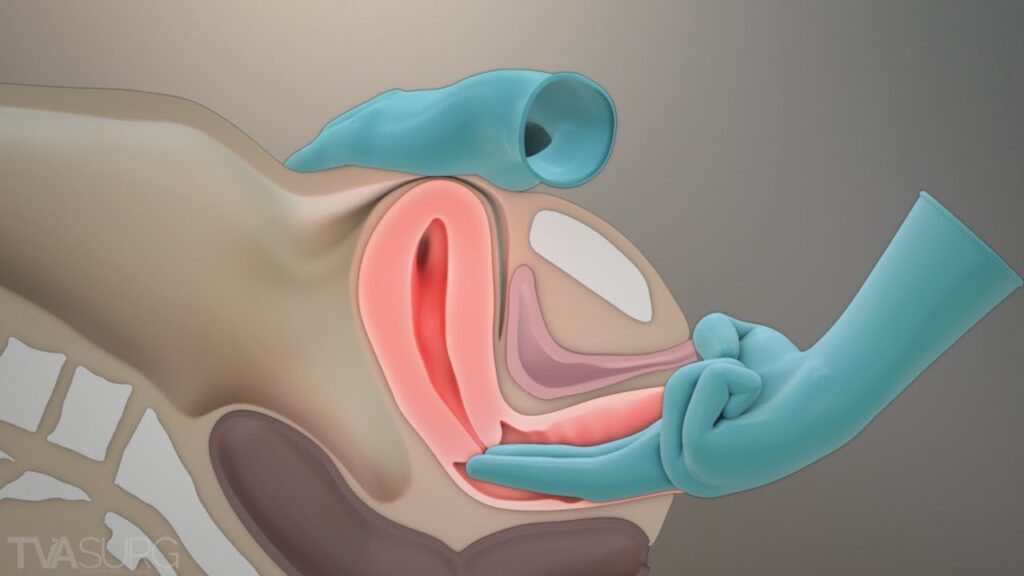
Uncategorized

uterus, fallopian tube, ovaries midwives revision

Uterus, Fallopian tubes and Ovaries

THE NON-PREGNANT UTERUSĀ 

The uterus is a hollow muscular organ of the female reproductive system where offspring gestate.
A pear-shaped organ, the uterus lies posterior-superior to the bladder and anterior to the rectum in the female pelvis. It consists of the fundus (top), body (middle), and cervix (lower). The uterus is composed of the endometrium (inner mucosal lining), myometrium (smooth muscular middle layer), and perimetrium.

Situation: The uterus lies in the true pelvis in an anteverted (leans forward) and ante-flexed (bends over its body) position. The body of the uterus lies above the urinary bladder.

Shape – it resembles that of an avocado or it is pear shaped.

Size – 7.5cm long, 5cm wide and 2.5cm thick.

Weight – 60 grams.

Gross-structure:Ā 

The uterus consists of two main parts; the body or corpus and the neck or cervix.

\"uterus,

1. THE BODY OR CORPUS
Forms the upper 2/3 of the uterus and it is the whole part above the cervix.

  • The Fundus: This is the portion of the body which lies between and above the cornua.
    NOTE: This is the part a midwife palpates during abdominal examination to measure the height of the uterus/fundus during pregnancy and fundal height during puerperium.
  • The cornua: These are the lateral angles of the uterine body where the fallopian tubes are attached. In normal labour, the uterine contractions start at the cornua and spread downwards the uterus.
  • The cavity: This is the potential space between the posterior and anterior walls. It is triangular in shape and it is occupied by the products of conception during pregnancy, mainly the fetus and the placenta.
  • The isthmus: Is the narrowest part of the body of the uterus immediately above the internal os. During pregnancy, this develops into the lower uterine segment.

2. THE NECK OF THE UTERUS
This forms the lower 1/3 of the uterus and enters the vagina at right angles.


\"Microscopic

Microscopic structure of the uterus
There are three layers of tissue from within.

1. The endometrium: It is the mucus membrane which lines the cavity of the uterus. It is made up of three layers; The compact layer, the Functional (spongy) layer and the Basal layer. The appearance of this lining varies with each day of the menstruation cycle. During menstruation, it is shed up to the basal layer.

2. The myometrium: It is the thickest muscle layer and has three layers.

  • Outer layer or longitudinal fibres — These pass over the fundus from front to back, starting and finishing at the level of the internal os with a few fibres into the cervix. In labour as the muscles contract and relax, they help to shorten the upper uterine segment and help to pull up and shorten the cervix.
  • The middle layer — Oblique fibres
    These are the interlacing fibres. They are known as living ligatures. They close the bleeding blood vessels after the separation of the placenta and so control bleeding.
  • The circular muscle fibres — Found in the cervix and cornua. Help in dilatation of the cervix. They relax during labour as they are more in the cervix.

3. The perimetrium – This is the outer covering of the peritoneum which covers the uterus except at the side where it forms the broad ligaments and at the level of the isthmus.

Relations to the uterus

  • Superiorly: The intestines and the abdominal muscles.
  • Anteriorly: The bladder, Uterovesical pouch, anterior vaginal fornix.
  • Posteriorly: The rectal uterine pouch, rectum, utero-sacral ligaments.
  • Laterally: The broad ligaments, ureters, Fallopian tubes, ovaries and round ligaments.
  • Inferiorly: The vagina.


\"Blood-Supply-to-Female-Reproductive-Tract

  • Blood supply: Uterine arteries: These arise from the internal iliac arteries and ovarian arteries.
  • Venous return: Uterine veins.
  • Lymphatic drainage: Into the internal iliac and sacral glands.
  • Nerve supply: By the sympathetic and the parasympathetic nerves which are branches of the lee Franken Hauser plexus.

Supports

  • The round ligaments: Maintain the uterus in its position of anteverted and anteflexed. They extend from the cornua at each side, pass downwards and insert into the tissue of the labia majora.
  • The broad ligaments: These are not true ligaments but folds of peritoneum extending laterally between the uterus and the side walls of the pelvis.
  • Cardinal ligament or Transverse cervical ligaments: They run out from the side walls of the cervix to the side walls of the pelvis.
  • The uterosacral ligament: These pass from the cervix to the sacrum.
  • Pub cervical ligament: These pass from the cervix under the bladder to the pubic bone.
  • The ovarian ligament: These begin at the cornua and attach to the ovaries.


\"ligament

Functions of the Uterus:

  1. Responsible for menstruation as the endometrium sheds during each monthly period.
  2. The endometrial cavity accommodates the fetus during pregnancy.
  3. Uterine muscles facilitate contractions during labor, enabling the expulsion of the infant through the birth canal.
  4. To accommodate and nourish the fertilized ovum for the gestation period.
  5. To involute following childbirth.
  6. It is a site for intra-Uterine Device insertion.

Clinical note

The uterus undergoes physiological changes in structure when one is pregnant due to the action of progesterone and oestrogen hormone.

Guiding questions

  1. Describe the non-pregnant uterus with a well-labeled diagram.
  2. List three layers of the uterus.
  3. Describe the myometrium of the uterus.
  4. Outline three uterine supports.
  5. Outline four functions of the uterus.

Clinical procedure: Antenatal Examination

An antenatal mother is waiting to be examined. Using the Leopold method, palpate her abdomen.

TASK: Abdominal examination

Step

Action:

Rationale:

1.

Welcome and explain the procedure to the mother, wash hands.

To allay anxiety and promote cooperation.

2.

Request mother to empty bladder.

For comfort.

3.

Put the mother in a recumbent position and expose the xiphisternum and symphysis pubis.Ā 

To aid relaxation of the abdominal muscles.

4.

While standing at the foot of the bed, inspect and observe the abdomen for:
– Striae gravidarum, linear nigra,

Ā Fetal movements,
Size and shape
– Linea alba,
– Skin rashes, skin hyper-pigmented spots and scars

To rule out pregnancy.

5.

Stand on the right hand side of the mother,
– Ask the woman if she has any pain palpation in the abdomen.Ā 

– Do light palpation of her left and right abdomen, and ensure to exclude enlargement of the spleen and liver.

To detect anatomical conditions of the organs.

6.

Fundal grip. Palpate the upper abdomen with both hands to feel the gravid uterus.

To detect the size and position of the uterus.

7.

Umbilical grip. Place the hand to apply deep pressure with the palm on the lateral sides to feel the uterus.
Take note of the irregular nodules which indicate fetal limbs.

To identify the location and position of the fetal back.

8.

Pawlik\’s grip – Turn and face the mother\’s legs.
– Use the fingers and thumbs to feel the moving fetal limbs in the lower abdomen.

To determine the presentation and the amniotic fluid volume.

9.

Pelvic grip. Move the finger towards the pelvis to determine where the brow is located.

To confirm presentation and the lie.

10.

Auscultation Place Pinard\’s stethoscope over the maternal abdomen where the fetal back was felt. Move the stethoscope until maximum intensity is felt. Place the right hand on the maternal radial pulse and compare it with the fetal heart rate. Count the fetal heart beats per minute.

To determine fetal viability.

11.

Tell the mother findings

Ā 


\"Fallopian

Fallopian Tubes

Fallopian tubes are two muscular tubes leading from the ovaries to the uterus.Ā 

They consist of the infundibulum (with fimbriae near the ovary), ampullary region, isthmus (narrowest part linking to the uterus), and interstitial part traversing the uterine musculature. They are also called oviduct or fallopian tubes named after Fallopius, an ancient Greek anatomist.

Situation

  1. They are situated in the true pelvis on either side of the uterus.
  2. Each tube extends from the cornua of the uterus and travels towards the side walls of the pelvis, then turns downwards and backwards before reaching it.
  3. The tubes lie in the broad ligaments.

Shape

  • They are tubes. The lumen of each communicates with the cavity of the uterus superiorly and the peritoneal cavity inferiorly.

Size

  1. The length of each tube is approximately 10 cm.
  2. The lumen is about 3mm.
  3. The thickness is that of an ordinary pencil.

Gross Structure/Surface Anatomy

Each tube is divided into four parts namely;

1. The interstitial portion/intramural.
a. This lies within the walls of the uterus.
b. It is 1.25cm thick.
c. Its lumen is about 1mm in diameter.

2. The isthmus.
Is the narrow part immediately adjoining the uterus. It is 2.5cm long.

\"\"

3. The ampulla.
Is a widened area of the tube where fertilization is thought to occur. It is 5cm long.

Ā 

4. The infundibulum.
a. It is the funnel-shaped extremity.
b. It is the terminal portion of the tubes, which turns backwards and downwards.
c. It extends in finger-like processes which surround the orifice of the tube.
d. It measures 1.25cm long.


\"Microscopic

Microscopic Structure

The fallopian tube has 4 coats (from within outwards):

  1. A lining of ciliated columnar epithelium.
    a. This forms the lining of the tube and aids the passage of the ovum to the uterus.
    b. This epithelium is arranged in folds known as placae which slows down the journey of the fertilized ovum and so making it ready for embedding when it reaches the uterus.
  2. A layer of connective tissue:
    Lies beneath the epithelium.

  3. Muscle coat/Muscularis.
    This is a thin muscular coat arranged in two layers:
    a. Inner layer of circular fibres, which are numerous, near the infundibulum.
    b. Outer layer of longitudinal fibres.

  4. Peritoneum: A covering of the peritoneum of the broad ligament.
    hangs over the tubes but absent on their inferior surface.

Blood Supply

  • The blood comes from the uterine and ovarian arteries.
  • The venous return is by corresponding veins.

Lymphatic Drainage: The lymphatic drainage is into the lumbar glands.

Nerve Supply: From the ovarian plexus.

Supports: The Infundibulo-pelvic ligaments. These are formed from folds of the broad ligament and run from the infundibulum of the tube to the side walls of the pelvis.

FUNCTIONS

  • The tube forms a canal through which the ovum and sperm pass.
  • Provide a site for fertilization (ampulla) and guide the zygote to the uterus for implantation.
  • Commencements of early development of the fertilized ovum take place in the tube.
  • Female sterilization is hence a Family Planning site.
  • Facilitate sperm movement using tubal cilia and transport the ovum from the ovaries to the uterus.
  • Supply nutrients to the fertilized ovum during its journey to the uterus.

Relations

  • Anteriorly: Intestines and the peritoneal cavity.
  • Posteriorly: The peritoneal cavity and the intestines.
  • Superiorly: Peritoneal cavity.
  • Inferiorly: The broad ligaments and the ….
  • Laterally: Infundibulo-pelvic ligaments and the side walls of the pelvis.
  • Medial: The uterus.

Clinical note

Conditions like ectopic pregnancy and Salpingitis are associated with the fallopian tubes.

Important

  1. Cleanliness of the vulva is very significant.
  2. Early detection of abnormal vaginal discharge like the pus discharge of Gonorrhea is important so that treatment is given on time.

Revision Questions

  1. List four parts of the uterine tubes.
  2. Outline two functions of the ampulla.
  3. Describe the uterine tubes with the aid of a diagram.
  4. Explain three functions of the uterine tubes.


\"\"

Ovaries:Ā 

The ovaries are two small glandular structures. They are the female sex endocrine glands in which ova are produced.

Two glands on each side of the uterus, ovaries are attached to the uterus by the ovarian ligament and the pelvic wall by the suspensory ligament. Covered by the mesovarium (part of the broad ligament), the ovary\’s size varies with age and menstrual cycle stage.

Situation

a. They lie within the peritoneal cavity in a small depression of the posterior wall of the broad ligaments.

b. They are situated at the fimbriated end of the uterine tubes, about the level of the pelvic brim.

c. Each is attached to the upper part of the uterus by the ligament of the ovary and the tissue called Mesovarium, a band of the broad ligament.

Shape: They are small, with a corrugated surface, like an organ, dull white in colour.

Size: The size of ovaries varies in age and in different individuals. They are about 2.5cm-3.5 cm long, 2cm wide and 1cm thick.

Development: The ovaries develop in the germinal ridges of the posterior wall and during fetal life, they descend into the pelvic cavity in the same manner as the testes.

Gross Structure/Surface Anatomy

The structure of the ovaries varies with the age of the woman.

  • From birth to puberty: The organs are smooth, dull white and solid in consistency.
  • Menstrual phase: Between puberty and menopause, the organs are larger and irregular on the surface, more like a walnut than an almond.
  • Post-menopausal phase: The ovaries become smaller and shrunken and are covered with scar tissue where month after month, the Graffian follicles have ruptured.

\"\"
\"ovary

Ā 

Microscopic Structure

The ovaries have two layers of tissue or zones:

The Medulla.

  • This is the central portion, consisting chiefly of fibrous tissue, blood vessels, lymphatics and nerves.
  • Has the hilum which is the central point of entry for blood vessels, lymphatics and nerves.
  • Contains no follicular structures but in pregnancy, the corpus luteum may spread towards the medulla.

The cortex.

  • This is the functional part of the ovary, because it\’s where Graffian follicles grow.
  • It surrounds the medulla.
  • It consists mostly of stroma in which the Graffian follicles are embedded.
  • Before puberty, the ovaries are inactive, but the stroma already contains immature or primitive follicles known as primordial follicles.
  • In the cortex of each ovary of every female child, 100,000 to 200,000 primordial follicles can be found, although not all of them reach maturity.
  • As the child grows, the primitive structures become mature and are known as Graafian follicles.
  • With the onset of puberty, one Graffian follicle grows more rapidly each month than the others.
  • And after its full development, it bursts, releasing a mature ovum. This process is called ovulation.
  • The empty Graffian follicle starts to undergo certain changes and develops into a corpus luteum (a yellow body).
  • If fertilisation does not take place, the corpus luteum lasts for two weeks, then fibrosis occurs, and so it turns into corpus albicans (white body) then into the final stage of corpus fibrosum.
  • If the ovum becomes fertilized, the corpus luteum does not die but increases in size. It produces progesterone and a little amount of oestrogen under the influence of the Luteinizing hormone from the anterior pituitary gland. This maintains pregnancy until the placenta has developed sufficiently to fulfil its own function at 12 weeks of gestation.

Tunica albuginea – This is a dense fibrous coat which surrounds the cortex.

Germinal epithelium encloses the ovary.

  • Blood Supply: Ovarian arteries.
  • Venous drainage: Is into the ovarian veins.
  • Lymphatic drainage: Into the lumbar glands.
  • Nerve supply: Ovarian plexus.

Supports

  • The fossa where it lies.
  • The ovarian ligament.
  • The broad ligament that extends between the uterine tube and the ovary.
  • Ovarian fimbria and infundibulo-pelvic ligament.

Ovarian Functions:

  • Produce ova and female sex hormones—predominantly estrogen and progesterone.
  • Oestrogen promotes the development of secondary sex characteristics, growth, and maturity of reproductive organs.
  • Progesterone prepares the endometrium for pregnancy, aids in placental development, breast enlargement during pregnancy, and inhibits ovum production during gestation.
  • Together, estrogen and progesterone regulate menstrual cycle changes in the endometrium.

Clinical note:

The ovaries may become infected with microorganisms (Oophritis), a fertilized ovum may embed in the ovary (Ovarian pregnancy) and tumors and cysts can affect them as well.

Revision questions

  1. Describe the development of the Graffian follicle.
  2. Describe the cortex of the ovary.
  3. State two functions of the medulla.
  4. State two hormones produced by the ovary.
  5. With the aid of a diagram, describe the mature ovary.
  6. Define ovulation.
  7. Give three reasons why ovulation takes place.
  8. List four ovarian supports.
  9. State two functions of the ovary.

Uterus, Fallopian tubes and Ovaries Read More Ā»

PELVIC ASSESSMENT MIDWIVES REVISION

PELVIC ASSESSMENT


Pelvic Assessment During Labor

A mother who is pregnant for the second time (Gravida 2, Para 1+0) reports with labor pains. Your task is to perform an internal pelvic assessment and create a plan for the mode of delivery.

Pelvic Assessment

Pelvic assessment is a process to determine whether a mother’s pelvis is wide enough for a baby to pass through safely during delivery.

Methods of Pelvic Assessment

  1. External Pelvic Assessment: This is a non-invasive assessment that can be performed by a midwife or healthcare provider. It involves observing the woman\’s physical characteristics and taking a detailed history.
  2. Internal Pelvic Assessment: This is a more invasive assessment that requires a vaginal examination. It is typically performed by a doctor, especially during labor.

External Pelvic Assessment

The woman is observed/as she moves towards the HCW/ midwife, note the stature, gait and shape of the abdomen. Any mother with a pendulous abdomen is suspected of having a contracted pelvis.

1. Observation:
Observe the woman as she approaches. Pay attention to her;

  • Stature: Observe the woman\’s height and build. A woman with a shorter stature might have a smaller pelvis.
  • Gait: Observe how the woman walks. A waddling gait can indicate a wider pelvis.
  • Abdomen: Observe the shape of the abdomen. A pendulous abdomen (protruding belly) can suggest a contracted pelvis

2. History Taking:

  • Social History:
      • Age:

  • Under 18 years may indicate an immature pelvis with smaller diameters.
  • Over 30 years may suggest that the pelvic joints are less flexible due to ossification, making labor more difficult.
      • Tribe: Some tribes are known to have smaller or larger pelvises, which can influence delivery outcomes.

  • Medical History:
    Ask if the mother has had diseases like poliomyelitis or rickets, which can affect the pelvis’s shape and size.
  • Surgical History:
    Inquire about any accidents or surgeries involving the spine, pelvis, or lower limbs, as these may lead to a contracted pelvis.Ā 
  • Past Obstetrical History:
  1. This is especially important for mothers who have been pregnant before (multigravida).
  2. Ask about previous deliveries: Were they normal or assisted?
  3. Ask about the condition of babies at birth: This can help rule out obstructed labor.
  4. Ask about the baby\’s health: This can help rule out mental retardation, which could be a result of abnormal labor.
  5. Ask about the baby\’s birth weight: This gives an idea of the size of baby that can pass through the pelvis without complications.

3. General Examination:

  • Shoe Size: A woman wearing a smaller shoe size (size 4 or less) might have a smaller pelvis.
  • Size of Hands and Feet: Smaller hands and feet can indicate a smaller pelvis.
  • Height: A woman shorter than 152 cm might have a smaller pelvis that may not allow an average-sized baby to pass through.


\"Internal

Internal Pelvic Assessment

Internal pelvic assessment is usually done around 36 weeks of pregnancy for first-time mothers (primigravida) or by a midwife during labor. This assessment helps determine if the pelvis can accommodate the baby during delivery.

Scenario

A mother who is pregnant for the third time (Gravida 3, Para 1) arrives with labor pains. Your task is to perform a pelvic assessment to evaluate pelvic capacity.

Objectives

  1. Prepare the necessary equipment for an internal pelvic assessment.
  2. Conduct the internal pelvic assessment for the mother in labor.

Requirements

  • A pack containing

  • Two receivers

  • A gallipot of sterile swabs

  • Clean pad, Antiseptic lotion, Sterile gloves, Sterile bowl for lotion, Clean gloves, Lubricant, Mackintosh and draw sheet.Ā 

  • At the bedside

  • Screen

  • Hand washing equipmentĀ 

  • Bedpan

NOTE: Measure the length of your fingers from the curve of the thumb to the middle finger, to measure the diagonal conjugate.Ā 

Procedure

Step

Action

Rationale

1

Explain the procedure to the mother using soft skills.

To ensure the mother understands and feels comfortable.

2

Ask the mother to empty her bladder and provide privacy by screening the bed.

To allow accurate assessment and maintain the mother’s privacy.

3

Put on clean gloves.

To maintain hygiene.

4

Assist the mother into the dorsal position.

To allow proper access for the examination.

5

Place the mackintosh and draw sheet under her buttocks.

To provide a clean field.

6

Drape the mother.

To create a sterile area for the procedure.

7

Remove gloves, wash hands, and put on sterile gloves.

To prevent infection.

8

Observe the vulva.

To rule out any abnormalities.

9

Swab the vulva.

To prevent infection.

10

Lubricate the index and middle fingers of your dominant hand and insert them into the vagina, reaching under the symphysis pubis to feel for the sacral promontory.

This must not be prominent /tipped as this will reduce the Anteroposterior diameter of

the pelvic brim.Ā 

To measure the diagonal conjugate.

11

Examine the sacral hollow, ensuring it is well curved, to allow proper rotation off the fetal head.

To check if internal rotation of the fetal head is possible.

12

Feel the left and right greater sciatic notches. They should be wide and round.

To assess the transverse diameter of the pelvic outlet.

13

Feel for the ischial spines; they should be blunt and round, not sharp, not to reduce the diameters of the outlet. If prominent, it can cause obstructed labour.Ā 

Prominent spines can obstruct labor.

14

Examine the subpubic arch; it should accommodate two fingers with some space left.

If the space is less, it may reduce the pelvic outlet diameter.

15

Place four knuckles between the ischial tuberosities.

To measure the intertuberous diameter.

16

Clean the vulva, make the mother comfortable, and provide feedback.

To ensure the mother knows her status.

17

Clear the surroundings and record findings.

For follow-up and documentation.

Note:
During labour, while performing pelvic assessment, also assess the station of the fetus. Stations indicate how far the fetus has descended into the pelvis and can be felt during a vaginal examination, especially at stations -3, -2, and -1.

Station Table:

Station

Measurement from the Ischial Spine

Part of the True Pelvis

-3

-5 cm

Pelvic inlet or brim

-2

-3.3 cm

-1

-1.6 cm

0

0

Ischial spine

+1

+1.6 cm

Pelvic outlet

+2

+3.3 cm

+3

+5 cm

  • The table represents fetal station measurements during labor, which describe the position of the fetus\’s presenting part (usually the head) in relation to the maternal ischial spines.

  • The ischial spines are bony protrusions in the pelvis and serve as a key landmark in determining the station.


\"\"

Stations and their Significance:

Station 0: When the fetal head is at the level of the ischial spines, it is said to be at \”0 station.\” This is considered the midpoint, meaning the fetal head has engaged in the pelvis but hasn\’t descended past the spines.

Negative Stations (-3 to -1): When the fetal head is above the ischial spines, it is in a negative station. The numbers reflect the distance in centimetres above the spines. For example:

  • -3 Station: The head is 5 cm above the ischial spines, closer to the pelvic inlet.
  • -2 Station: The head is 3.3 cm above the ischial spines, indicating descent but not yet engaged.

Positive Stations (+1 to +3): When the fetal head is below the ischial spines, it is in a positive station. The numbers reflect the distance in centimetres below the spines:

  • +1 Station: The head is 1.6 cm below the ischial spines.
  • +3 Station: The head is 5 cm below the ischial spines, nearing the pelvic outlet, indicating significant descent and progress toward delivery.


PELVIC ASSESSMENT CHECKLIST

\"\"

PELVIC ASSESSMENT Read More Ā»

female pelvis midwives revision

Female Pelvis


Introduction To Obstetric Anatomy

This field focuses on the anatomical structures involved in pregnancy, labor, and the postpartum period. Key areas of study include the pelvis, pelvic floor, female reproductive system, female breast, male reproductive system, embryology, fetal skull, and the female urinary system.

Definition of Terms

  • Anatomy: The study of the structures of the body.
  • Physiology: The study of how the body functions.
  • Obstetrics: A branch of medicine that focuses on pregnancy, childbirth, and the postpartum period (puerperium).

The Female Pelvis

The pelvis, or pelvic girdle, is a bony structure that forms a canal through which a fetus passes during birth.

Location: The pelvis is positioned between the movable vertebral column, which it supports, and the lower limbs, upon which it rests. It connects to the fifth lumbar vertebra above and the head of the femur (thigh bone) in the acetabulum (hip socket) below.

Shape: The pelvis resembles a bony basin.

Size: It is the largest bony structure in the body, with size varying based on individual age and body size.

Structure: The pelvis consists of the following components:

  1. Bones
  2. Joints
  3. Ligaments

\"bones

Bones of the Pelvis

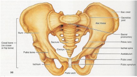
1. The Innominate Bones:

These are two large bones on either side of the sacrum, where the femur bones connect. Each innominate bone is made up of three parts that meet at a cup-shaped depression known as the acetabulum:

\"\"
\"Innominate

Ilium:
The largest and flared-out part of the innominate bone. It articulates with the alae (wings) of the sacrum and forms the upper two-fifths of the acetabulum.

  • Iliac Crest: The upper border of the ilium.
  • Anterior Superior Iliac Spine: The point where the iliac crest ends at the front.
  • Anterior Inferior Iliac Spine: Located about 2.5 cm below the anterior superior iliac spine.
  • Posterior Superior Iliac Spine: The point where the iliac crest ends at the back.
  • Posterior Inferior Iliac Spine: Located about 2.5 cm below the posterior superior iliac spine. This marks the upper border of the greater sciatic notch, where the sciatic nerves pass.

Ischium:
The lowest part of the innominate bone, forming the lower two-fifths of the acetabulum.Ā 

  • Ischial Tuberosity: The body of the ischium, where the body rests.
  • Ischial Spine: Located about 2.5 cm above the ischial tuberosity. It divides the lesser and greater sciatic notches.


\"\"

Pubis:
The smallest part, forming the lowest fifth of the acetabulum. It includes the superior ramus, body, and inferior ramus. The two pubic bones join at the symphysis pubis.

  • Superior Ramus: The upper part of the pubis.
  • Body: The main part of the pubis.
  • Inferior Ramus: The lower part of the pubis.
  • Symphysis Pubis: The right and left pubic bones fuse together with a pad of cartilage at the symphysis pubis.
  • Obturator Foramen: The space surrounded by the inferior and superior pubic rami.

Ā 2. The Sacrum:

A wedge-shaped structure made up of five fused sacral vertebrae, with foramina (holes) through which blood vessels, nerves, and lymphatics pass. The sacrum is smooth inside to protect organs and rough outside for muscle attachment.
Parts of the Sacrum:

  • Sacral Promontory: The upper border of the first sacral vertebra, projecting forward over the hollow of the sacrum.
  • Hollow of Sacrum: The smooth, concave anterior surface.
  • Alae of Sacrum: Wing-like extensions on each side of the first sacral vertebra.
  • Sacral Canal: Runs through the center of the bone and opens at the level of the fifth sacral vertebra. It provides a passage for the spinal cord and spinal nerves. At the level of the second and third sacral vertebrae, the nerves spread out to form the Cauda equina (horse\’s tail).

\"sacrum_coccyx_

3. The Coccyx:

A vestigial tailbone, made up of four fused coccygeal vertebrae. It is triangular in shape and articulates with the sacrum at the sacro-coccygeal joint.


\"The-pelvis-and-its-joints

Pelvic Joints

1. Sacroiliac Joints:
The strongest joints in the body, located between the first two sacral vertebrae and the ilium. These joints allow limited movement and are supported by ligaments.

  • Location: Between the first two bodies of the sacral vertebrae and the upper surface of the ilium.
  • Function: Strongest joints in the body, allowing limited movement. They are surrounded and supported by ligaments.

2. Symphysis Pubis:
A pad of cartilage between the two pubic bones, forming a cartilaginous joint that unites the pubic rami.

  • Definition: A pad of cartilage lying between the two bodies of the pubic bone.
  • Function: A cartilaginous joint uniting the two rami of the pubic bone.
  • Size: Approximately 4 cm in length, with supporting ligaments around it. Clinical Note:
    During pregnancy, the hormone progesterone relaxes and softens the smooth muscles of these joints, causing backache and pain at the symphysis.

3. Sacrococcygeal Joint:
This joint allows the coccyx to bend backward during labor, providing more room for the fetal head to pass through the birth canal.

  • Location: Where the base of the coccyx articulates with the tip of the sacrum.
  • Function: Allows a bend backwards during labor, providing more room for the fetal head as it passes through the birth canal.


\"Pelvic

Pelvic Ligaments

These strong fibrous bands strengthen the pelvic joints and support the pelvic organs. They include:

  1. Sacro-Iliac Ligaments: Strengthen the sacroiliac joint.
  2. Sacro-Tuberous Ligaments: Stretch from the lower sacrum to the ischial tuberosities.
  3. Sacro-Spinous Ligaments: Extend from the lower sacrum to the ischial spines, forming the posterior wall of the pelvic outlet.
  4. Inter-Pubic Ligament: Strengthens the pubic bones.
  5. Inguinal Ligaments: Extend between the anterior superior iliac spine and the pubic body.
  6. Lacuna Ligaments: Lie beneath the inguinal ligaments.
  7. Sacro-Coccygeal Ligament: Strengthens the sacro-coccygeal joint.
  8. Obturator Ligaments: Cover the obturator foramen, allowing the passage of blood vessels, nerves, and lymphatics.


\"Greater-and-Lesser-Pelvis-Divided-by-the-Pelvic-Brim

Divisions of the Pelvis

The pelvis is divided into:

1. The Lesser or True Pelvis (Pelvis Minor): The lower part of the pelvis, crucial in childbirth.

  • Location: The bottom part of the false pelvis.
  • Importance: It is for childbirth as it forms the birth canal.
  • Components:
  1. Brim (Inlet): The ring-shaped bone separating the false pelvis from the true pelvis.
  2. Cavity: The space between the brim and the outlet.
  3. Outlet: The lowest part of the true pelvis.

2. The Greater or False Pelvis (Pelvis Major): The upper part, less important in midwifery.

  • Location: The part above the pelvic brim.
  • Importance: Less significant in midwifery.

The True Pelvis

Brim/Inlet: A ring-shaped bone separating the false pelvis from the true pelvis.

  • Importance: The site where the engagement of the fetal head takes place.
  • Shape: Round, except where the sacral promontory projects into it.
  • Circumference: Approximately 36 cm, allowing a fetal head with a circumference of 35 cm to pass.

Landmarks:

  1. Promontory of the sacrum
  2. Alae of the sacrum
  3. Sacroiliac joint
  4. Ilio-pectineal eminence
  5. Superior pubic ramus
  6. Upper inner border of the body of the pubic bone
  7. Upper border of the symphysis pubis
  8. Ilio-pectineal line


\"pelvic

Important Landmarks:
  1. Sacral Promontory: If prominent, it can reduce the antero-posterior diameter, obstructing labor.
  2. Ilio-pectineal Eminence: Important for determining the fetal head\’s position.Ā  Important because it\’s the point where the denominator (the presenting part of the fetus) faces during labor.
  3. Symphysis Pubis: If narrow, it reduces the antero-posterior diameter.
  4. Engagement: The oblique diameter is crucial for the engagement of the fetal head.

Diameters of the Pelvic Brim/Inlet:(11-12-13)

  1. Antero-Posterior Diameter: Measures 11 cm from the sacral promontory to the upper inner border of the symphysis pubis. Three conjugates are involved: anatomical (12 cm), obstetrical, and diagonal (12-13 cm, subtracting 1-2 cm for tissues).
  2. Oblique Diameters: Right and left, measuring 12 cm from the sacro-iliac joint to the ilio-pectineal eminence.
  3. Transverse Diameter: The largest diameter on the brim, measuring 13 cm from the ilio-pectineal line.

\"pelvic-dimensions

The Pelvic Outlet

  • Definition: The lowest part of the true pelvis.
  • Significance: Forms the narrow pelvic strait through which the fetus must pass.
    The pelvic outlet is the narrowest part of the pelvis, through which the fetus must pass.
  • Shape: Diamond-shaped.
  • Circumference: 36 cm.

\"Borders-of-the-Pelvic-Outlet

Measurements of the outlet:
  1. Antero-posterior Diameter: Measured from the lower border of the symphysis pubis to the lower border of the sacrum (13 cm).
  2. Oblique Diameter: Difficult to measure accurately due to the stretching of the sacro-tuberous ligaments by the fetal head. It\’s accepted to lie parallel to the oblique diameter of the brim and cavity and should be at least 12 cm.
  3. Transverse Diameter: Measured between the ischial spines (11 cm).
Important Landmarks of the Pelvic Outlet:
  • Coccyx: Important because it tilts (extends) backwards during labor to give more room for the passing fetus.
  • Sub-pubic Arch: Should accommodate at least two fingers and leave space for the passage of the baby.
  • Ischial Spines: Should be round. If prominent, they reduce the transverse diameter and obstruct labor.
  • Sacro-spinous Ligament: Should be soft and stretch outwards for the baby to pass.

Functions of the Pelvis:

  1. Birth Canal: Provides the passage through which the fetus must pass to be born.
  2. Protection: Contains and protects internal reproductive organs such as the bladder, uterus, and vagina.
  3. Weight Transmission: Transmits the weight of the trunk to the legs, acting as a bridge between the femurs.
  4. Movement: The primary function of the pelvic girdle is to allow movement of the body.
  5. Sitting and Kneeling: Permits a person to sit or kneel.
  6. Nerve Transmission: The sacrum transmits the cauda equina (the continuation of the nerve roots in the lumbar and sacral region) to and conveys nerves to various parts of the pelvis.


\"types

Types of Pelvis

There are four main types of pelvis, each with distinct characteristics:

Gynaecoid Pelvis (Normal Female Pelvis)

The normal female pelvis for childbirth. Found in women of average weight and height with a shoe size 4 or larger.

Characteristics:

  • Brim: Round, except where the sacral promontory protrudes slightly inward.
  • Cavity: Generous fore pelvis (the part in front of the transverse diameter), shallow cavity, broad, and well-curved sacrum.
  • Outlet: Blunt ischial spines, well-rounded sciatic notches, and a subpubic angle of 90 degrees.

Effects on Labour:

  • The rounded shape is favourable for childbirth. The fetus presents with its head in the occipito anterior position, which aligns with the rounded part of the pelvis, facilitating a smoother labor.
  • Favorable for the fetus at the start of labor because the pelvis is well-rounded anteriorly, allowing the fetus to present with the most rounded part of its head (occipital anterior).

Android Pelvis (Male-like Pelvis)

Resembles a male pelvis. Found in short and heavily built women.

Characteristics

  • Brim: Heart or triangular-shaped.
  • Cavity: Narrow fore pelvis, deep cavity, and straight sacrum.
  • Outlet: Prominent ischial spines, narrow sciatic notches, and a subpubic angle less than 90 degrees.

Effects on Labour:

  • Brim: Favours a posterior position of the occiput (fetus presents with the occiput lying posteriorly).
  • Outlet: May become obstructed at the outlet due to prominent ischial spines reducing the transverse diameter (Deep transverse arrest). Emergency Caesarean section is often necessary.

Platypelloid Pelvis (Flat Pelvis)

A flat pelvis characterized by a kidney/bean-shaped brim and a short anteroposterior diameter.

Characteristics

  • Brim: Kidney or bean-shaped with a short anteroposterior diameter.
  • Cavity: Wide transverse diameter, flat sacrum, and shallow cavity.
  • Outlet: Blunt ischial spines, wide sciatic notches, and a subpubic angle greater than 90 degrees.

Effects on Labour:

The fetus\’s head usually engages in the transverse diameter. However, due to the narrow anteroposterior diameter, the head may require tilting (asynclitism) to pass through, sometimes leading to face presentation or requiring a Caesarean section if the head remains high.

  • Engagement: The head must engage with the sagittal suture in the transverse diameter.
  • Descent: Descent through the cavity is usually without difficulty.
  • Asynclitism: Lateral tilting of the head is necessary to allow the bi-parietal diameter to pass the narrowest anteroposterior diameter of the brim.
  • Presentation: Can result in face presentation.
  • Contracted Brim: If the brim is severely contracted, the fetal head remains floating high above it, requiring a Cesarean section.

Anthropoid Pelvis

Found in tall women with narrow shoulders.

Characteristics

  • Brim: Long oval shape, with a longer anteroposterior diameter than the transverse diameter.
  • Cavity: Long, deep sacrum with side walls that diverge.
  • Outlet: Less prominent ischial spines, very wide sciatic notch, and a subpubic angle greater than 90 degrees.

Effects on Labour:
Labour is usually not problematic, but the fetus often remains in a posterior position, leading to delivery with the face towards the pubis instead of the perineum.

  • Labor: Usually does not present any difficulties.
  • Position: Direct occipito posterior position is often a feature, and the position adopted for engagement may persist up to delivery.
  • Delivery: The fetus passes through the pelvis remaining in the same position and so delivers face to pubis instead of face to perineum.

Summary of Pelvic Types

Features

Gynaecoid

Android

Anthropoid

Platypelloid

Brim

Rounded

Heart-shaped

Long oval

Kidney-shaped

Fore pelvis

Generous

Narrow

Narrowed

Wide

Side walls

Straight

Convergent

Divergent

Divergent

Ischial spines

Blunt

Prominent

Blunt

Blunt

Sciatic notch

Rounded

Narrow

Wide

Wide

Subpubic angle

90°

<90°

>90°

>90°

Incidence

50%

20%

25%

5%


Other Pelvic Variations

These variations can result from developmental anomalies, dietary deficiencies, injuries, or diseases. They often lead to a contracted pelvis, where one or more diameters are reduced, complicating the normal labor process.

1. Developmental Anomalies.

  • Robert\’s Pelvis: The sacrum\’s wings are underdeveloped or absent, causing contraction in all diameters, requiring a Caesarean section.
  • Naegele’s Pelvis: The sacrum has only one wing due to congenital abnormalities or disease, a true Naegele\’s pelvis may occur in a woman who has walked with a limp for many years, also requiring a Caesarean section.
  • Assimilation Pelvis: Can be high (six vertebrae in the sacrum) or low (four vertebrae in the sacrum). Normal is 5.
  1. High Assimilation Pelvis: The sacrum consists of six vertebrae.
  2. Low Assimilation Pelvis: The sacrum consists of four vertebrae.
  • Justominor Pelvis: A smaller version of the gynaecoid pelvis, common in petite women, with proportional reductions in all measurements. A Gynaecoid type of pelvis where all measurements are reduced but in correct proportions. Common in petite women of small stature. Effects on Labor:
  1. Small Baby: Can be delivered vaginally with little or no problems.
  2. Large Baby: May require Cesarean section.

2. Dietary Deficiencies.

  • Rachitic Pelvis: Deformity due to rickets, which affects the person in early childhood due to lack of vitamin D and calcium.
  • Osteomalacic Pelvis: An extreme deformity due to osteomalacia from dietary deficiencies, caused by a deficient diet and lack of vitamin D, leading to softening of the bones and a Y-shaped pelvic brim.

3. Injuries and Diseases.

  • Asymmetrical Pelvis: May result from congenital hip dislocation or polio, causing distortion on one side.

Main Differences Between Male and Female Pelvis

Feature

Male

Female

General structure

Thick and heavy

Thin and light

Muscle attachments

Well marked

Poorly marked

False pelvis

Deep

Shallow

True pelvis

Narrow and deep

Wide and shallow

Superior pelvic aperture

Heart-shaped

Oval or rounded

Inferior pelvic aperture

Comparatively small

Comparatively large

Subpubic angle

Narrow

Wide

Obturator foramen

Round

Oval

Acetabulum

Large

Small

Revision Questions

  • Describe the four bones of the gynaecoid pelvis.
  • Describe the innominate bones.
  • List four diameters of the pelvic brim.
  • Outline three important landmarks of the pelvic brim.
  • Describe the three parts of the true pelvis in structure.
  • List four joints of the pelvis.
  • Outline six ligaments of the pelvis.
  • Describe the main four types of pelvis.
  • A prime gravida at 36 weeks of gestation comes for antenatal care. How would you assess her pelvis?
  • Explain five causes of a contracted pelvis.

Female Pelvis Read More Ā»

METRORRHAGIA/INTERMENSTRUAL BLEEDING

METRORRHAGIA/INTERMENSTRUAL BLEEDING

METRORRHAGIA/INTERMENSTRUAL BLEEDING

Metrorrhagia, now commonly called Intermenstrual bleeding is vaginal bleeding that occurs at irregular intervals not associated with the menstrual cycle.

Metrorrhagia is a medical term used to describe irregular or abnormal uterine bleeding that occurs between menstrual periods.Ā 

It is characterized by light spotting that occurs outside of one’s period patterns and does not need sanitary protection.

This is a symptom of some underlying pathology which may be organic or functional.

Causes of Metrorrhagia

  • Fibroid Uterus: Presence of uterine fibroids, noncancerous growths that can cause irregular and abnormal bleeding.
  • Adenomyosis: A disorder involving the glands that secrete cervical mucus and fluids, contributing to abnormal uterine bleeding.
  • Pelvic Endometriosis: Presence of endometrial tissue outside the uterine lining, leading to premenstrual pain and dysmenorrhea.
  • Chronic Tubo-Ovarian Mass: Persistent mass involving the fallopian tubes and ovaries, contributing to irregular uterine bleeding.
  • Retroverted Uterus (Due to Congestion): Uterus tilted backward, causing congestion and contributing to metrorrhagia.
  • Uterine Polyp: Presence of a polyp with a rich blood supply, making it prone to easy bleeding.
  • Cervical Erosions: Presence of wounds on the cervix with increased blood supply, leading to bleeding.
  • Cancer of the Cervix or Endometrial Cancer: Malignant growths in the cervix or endometrium causing abnormal bleeding.
  • Chronic Threatened Abortion or Incomplete Abortion: Prolonged or incomplete abortion affecting uterine function and causing irregular bleeding.
  • Retained Pieces of Placenta: Residual placental fragments interfering with uterine contraction, preventing proper closure of blood vessels after childbirth.
  • Mole Pregnancy: Abnormal uterine mass growing post-fertilization, characterized by an excess of blood capillaries leading to bleeding.
  • Ovulation Bleeding: Bleeding associated with the process of ovulation.
  • Short Cycles like Polymenorrhea: Menstrual cycles shorter than the average duration, contributing to irregular and frequent uterine bleeding.
  • Infections: Having lower abdominal or pelvic infections such as vaginitis can lead to metrorrhagia.
Signs and symptoms of Metrorrhagia

Signs and symptoms of Metrorrhagia

  1. Bleeding Between Menstrual Periods: Presence of abnormal bleeding episodes occurring between regular menstrual cycles.
  2. Irregular Menstrual Cycles: Variations in the normal pattern of menstrual cycles, including changes in cycle length or timing.
  3. Heavier or Lighter Bleeding Than Usual During Menstrual Periods: Experiencing unusually heavy or light menstrual flow compared to the individual’s typical pattern.
  4. Prolonged Bleeding That Lasts Longer Than Normal: Extended duration of menstrual bleeding beyond the usual timeframe.
  5. Pelvic Pain or Discomfort: Presence of pain or discomfort in the pelvic region, often associated with abnormal bleeding.
  6. Fatigue or Tiredness Due to Blood Loss: Feeling tired or fatigued as a result of significant blood loss during irregular bleeding episodes.
  7. Anaemia Symptoms: Manifestations of anaemia, including: Shortness of Breath, Dizziness, Weakness.

Investigations of Metrorrhagia

Assessment:

  • Thorough medical history and physical examination.
  • Detailed historyĀ  about menstrual cycles, associated symptoms, and relevant medical conditions.
  • Pelvic examination to assess the condition of reproductive organs.

Diagnostic Tests:

  • Hormone Level Assessment: Blood tests to evaluate hormone levels, including oestrogen, progesterone, and thyroid hormones. This helps identify hormonal imbalances that may contribute to metrorrhagia.
  • Transvaginal Ultrasound: Imaging test for visualizing the uterus and ovaries, detecting structural abnormalities or conditions such as fibroids.
  • Endometrial Biopsy: Collection of a sample from the uterine lining for microscopic evaluation. This procedure helps identify abnormalities, including signs of cancer.
  • Hysteroscopy: Procedure involving the insertion of a thin, lighted tube into the uterus to visualize the uterine cavity. It aids in detecting abnormalities or issues affecting the uterine lining.
  • Digital and Speculum Examination: Examination techniques to visualize the cervix for any signs of abnormality.
  • Pelvic Scan: Imaging scan focused on visualizing pelvic organs, assisting in ruling out any abnormalities.
  • Urine Test: Urine samples to check for pregnancy, infection, or STDs.
  • Pap Smear: To rule out cancer.
METRORRHAGIA rx
Management of Metrorrhagia
The best management to investigate and treat the cause.

Aims of Management:

  • Alleviate Symptoms.
  • Determine and address the root causes of metrorrhagia.
  • Prevent Complications

Medical and Nursing Management of Metrorrhagia:

  1. Assessment: Medical History and Physical Examination: Gather detailed information on menstrual cycles, symptoms, and relevant medical conditions. Conduct a pelvic examination to assess reproductive organs.
  2. Hormonal therapy: Depending on the underlying cause, hormonal medications, such as birth control pills or progestin therapy, may be prescribed to regulate the menstrual cycle and reduce abnormal bleeding.
  3. Nonsteroidal anti-inflammatory drugs (NSAIDs): These medications can help manage pain and reduce bleeding during episodes of metrorrhagia.
  4. Treatment of underlying conditions: If metrorrhagia is caused by conditions such as fibroids, polyps, or infections, appropriate treatment strategies will be implemented to address the specific cause.
  5. Surgical interventions: In some cases, surgical procedures may be necessary to remove uterine abnormalities or address the underlying cause of metrorrhagia.
  6. Supportive care: Nursing management focuses on providing emotional support, educating patients about menstrual hygiene and symptom management, and promoting overall well-being.
  7. Rest: Rest is advised during the bleeding phase. Assurance and sympathetic handling are helpful particularly in adolescents. Anaemia should be corrected energetically by diet, hematinics, and even by blood transfusion.
  8. Monitoring and follow-up: Monitor patients’ response to treatment, assess the effectiveness of interventions, and ensure appropriate follow-up care.

METRORRHAGIA/INTERMENSTRUAL BLEEDING Read More Ā»

Tetanus

Tetanus

Tetanus

Tetanus is an acute infectious disease of the central nervous system caused by clostridium tetani and is characterized by spasms of the skeletal muscles frequently attacking the muscles of the jaw.Ā 

Tetanus is commonly known as ā€˜Lock-jaw.’

Cause of Tetanus

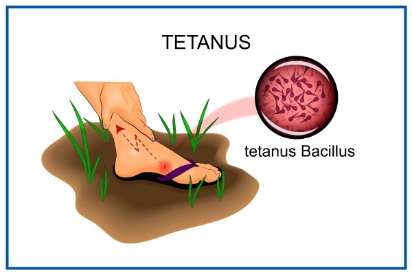
Tetanus is caused by the exotoxins of clostridium tetani.

  • The organism can live for a long time in any condition, especially dirty environments. So it can be found in dust, soil, or grass.
  • The clostridia can be normal organisms in the alimentary canal of animals but when passed out and gain entry to the human body, they become harmful. The organism can be found in cows, horse, sheep, or goat.

Incidence of tetanus:

  • In babies born at home before arrival at the hospital.
  • In homes where domestic animals are kept.

\"route

Pathophysiology of Tetanus

The pathophysiology of tetanus involves the invasion of the body by bacilli or spores, typically through deep puncture wounds or cuts. These bacilli find a suitable environment to multiply in anaerobic conditions. It is crucial to note that all unclean wounds pose a significant risk. Once the clostridium tetani organisms enter the wound, they unleash two forms of exotoxins into the surrounding tissues: tetanospasmin and tetanolysin.

Tetanospasmin, a potent toxin, plays a critical role in producing the disease\’s clinical manifestations. It primarily affects the central nervous system (CNS). The toxins specifically target the motor nerve cells of the spinal cord and the brain. As a result, spasms develop in the muscles that are supplied by the corresponding nerves.

Route of entry

The route of entry for the clostridium tetani organisms includes various pathways, all of which can lead to infection and subsequent tetanus:

  1. Infected ulcerated wound.
  2. Postoperative wounds.
  3. Umbilical stumps (in newborns).
  4. Gun-shot wounds.
  5. Septic abortion.
  6. Jiggers or foreign bodies.
  7. Burns and scalds.

Signs and Symptoms

Tetanus manifests with a set of distinctive signs and symptoms, indicating the severity of the infection:

  1. Stiffness of the muscles, particularly noticeable in the jaw.
  2. Spasms affecting the muscles of the face, especially the cheek and jaw, leading to difficulty in opening the mouth, a condition referred to as trismus.
  3. The angles of the mouth are pulled outwards, causing a forced smile known as risus sardonicus or the \”Devil\’s grin.\”
  4. The head is thrown back, and the back becomes arched due to the rigid muscles in the neck, a condition called opisthotonus.
  5. Signs of inflammation may be evident, such as swelling of the umbilical cord (if present in newborns), a wet cord, offensive smell, or pus discharge from the wound site.
  6. Patients may experience an elevated temperature and rapid pulse.
  7. Weight loss may occur due to difficulties in eating, leading to starvation.
  8. Spasms of the sphincters can result in retention of urine or stool, and in severe cases, sphincter rupture may occur.
  9. Swallowing becomes challenging as the muscles of the mouth and esophagus are affected by spasms.
  10. Spasms affecting the respiratory muscles may lead to prolonged periods without oxygen (anoxia), which can be life-threatening and result in death.
  11. Patients may have a wound or a history of a wound, which could be the point of entry for the tetanus-causing bacteria.

Alblett Classification of Tetanus

There are several grading systems; the scale proposed by Ablett5
is the most widely used . This categorizes patients
into four grades depending upon the intensity of spasms, and
respiratory and autonomic involvement.

\"\"

Management of Tetanus

There\’s no cure for tetanus. A tetanus infection requires emergency and long-term supportive care while the disease runs its course.

Aims of Management

  • To control spasms.
  • To eliminate the causative organism and its toxins.
  • To prevent complications, and ensure adequate nutrition for the patient.Ā 

Specific treatment measures include:

  1. Penicillin: Administering penicillin is a crucial step in destroying the tetanus-causing organism.

  2. Anti-tetanus serum: The administration of anti-tetanus serum helps neutralize the spreading toxins and halt their further detrimental effects.

  3. Sedation and muscle relaxants: Medications like diazepam and chlorpromazine are given to provide sedation and muscle relaxation, effectively alleviating spasms and minimizing discomfort.

  4. Wound management: If there is a wound or focus of infection where the tetanus bacteria may have entered, the dead tissue is excised, and the area is irrigated with hydrogen peroxide. Leaving the wound open without suturing promotes oxygen exposure, hindering the growth of tetanus bacilli, which thrive in anaerobic conditions.

Control of spasms involves the following measures:

  • Absolute rest and isolation: The patient should be kept in a quiet room with dim lighting to minimize triggers for spasms.
  • Prevention of external stimuli: Measures such as fitting the door with suitable closing materials or springs prevent slamming noises that could stimulate the patient.
  • Warming hands before touching: Nurses should warm their hands before touching the patient to avoid any stimulation that might trigger spasms.
  • Medication administration: Sedatives and muscle relaxants, such as chlorpromazine (Largactil), and Diazepam, are given regularly through a nasogastric tube to maintain a controlled state and alleviate spasms.
  • o Example of 6 hourly regimenĀ 

    DrugĀ 

    6-9Ā 

    am

    9-12Ā Ā 

    pm

    12-3Ā Ā 

    pm

    3-6Ā Ā 

    pmĀ 

    6-9Ā 

    pm

    9-12Ā 

    am

    12-3Ā 

    am

    3-6Ā 

    am

    6-9Ā 

    am

    LargactilĀ 

    āœ”Ā 

    ⫼⫼⫼⫼ 

    āœ”Ā 

    ⫼⫼⫼⫼ 

    āœ”Ā 

    ⫼⫼⫼⫼ 

    āœ”Ā 

    ⫼⫼⫼⫼ 

    āœ”

    DiazepamĀ 

    ⫼⫼⫼⫼ 

    āœ”Ā 

    ⫼⫼⫼⫼ 

    āœ”Ā 

    ⫼⫼⫼⫼ 

    āœ”Ā 

    ⫼⫼⫼⫼ 

    āœ”Ā 

    ⫼⫼⫼⫼

General Management:

  1. Close observation and airway management: Monitor the patient closely, ensuring a clear airway and using a mucous extractor if necessary.

  2. Vital signs monitoring: Regularly check temperature, pulse, and respirations, noting the severity of the condition. Record the strength, frequency, duration, and body part involved in spasms using a spasm chart.

  3. Nutrition: Maintain adequate nutrition through nasogastric tube feeding to avoid stimulating spasms with injections. Prevent aspiration of fluids into the airway.

  4. Catheterization: Catheterize the patient to maintain proper bladder function.

  5. Fluid balance chart: Monitor and maintain fluid balance, initially using intravenous fluids if necessary and later transitioning to nasogastric tube feeding.

  6. Hygiene: Ensure daily cleaning of the cord with normal saline and perform oral care carefully. Turn the patient every two hours to prevent pressure sores.

  7. Vaccination: Prevent future tetanus cases by ensuring all individuals receive a full course of DPT (diphtheria, pertussis, and tetanus) vaccination.

  8. Use sterile equipment: Prevent cross-infection by using sterile equipment during medical procedures.

  9. Bowel and bladder care: Monitor and assist the patient in passing stool and urine.

  10. Medication: Administer prescribed drugs as instructed.

  11. Physiotherapy: Implement physiotherapy sessions for deep breathing exercises and active limb movements.

Prevention:

  1. Public health education: Raise awareness about the dangers of using unsterilized equipment during childbirth and applying native medicine or other substances to the umbilical cord.

  2. Immunization: Ensure that all women of childbearing age are vaccinated against tetanus.

  3. Safe childbirth practices: Promote safe and clean practices during childbirth to prevent infections.

  4. Discourage harmful practices: Discourage practices like applying cow dung to a child\’s umbilical cord.

  5. Wound care education: Educate people on cleaning wounds thoroughly with water and soap to prevent infections. Encourage covering wounds with sterile dressings and seeking early medical attention for cut wounds.

  6. Protective gear: Encourage the use of gum boots when digging to prevent soil-related infections.

Complications of Tetanus

  1. Fracture of bones or the spine: The intense and frequent muscle spasms can cause fractures in bones or the spine, especially if the spasms are severe and uncontrolled.

  2. Pneumonia: Heavy sedation, a common treatment for managing tetanus spasms, may lead to shallow breathing or difficulty in clearing the airway, increasing the risk of pneumonia, a potentially severe respiratory infection.

  3. Brain damage: The potent toxins produced by the tetanus-causing bacteria can affect the central nervous system, leading to brain damage in severe cases.

  4. Growth retardation: In children affected by tetanus, the disease can interfere with proper nutrition and growth, potentially causing growth retardation.

  5. Exhaustion: The continuous and strenuous muscle spasms can lead to extreme exhaustion, further weakening the patient\’s overall condition.

  6. Respiratory failure: In severe cases, the spasms can affect the respiratory muscles, resulting in respiratory failure, where the patient is unable to breathe adequately on their own.

  7. Retention of urine: Spasms in the pelvic region can cause the sphincters to contract, leading to difficulty in passing urine and possible urine retention.

  8. Death due to airway obstruction: In the most severe cases, the intense spasms, particularly those affecting the muscles of the jaw and neck, can obstruct the airway, leading to suffocation and potential death.

Test Questions

Which bacterium causes tetanus?
a) Streptococcus pyogenes
b) Staphylococcus aureus
c) Clostridium tetani
d) Escherichia coli
Answer: c) Clostridium tetani

Explanation: Clostridium tetani is the bacterium responsible for causing tetanus.

What is the common term used to describe tetanus due to spasms in the jaw muscles?
a) Trismus
b) Opisthotonus
c) Risus sardonicus
d) Tetanospasmin
Answer: a) Trismus

Explanation: Trismus is the medical term for difficulty in opening the mouth due to jaw muscle spasms, which is commonly known as \”Lock-jaw.\”

What is the primary goal of managing tetanus?
a) Preventing complications
b) Destroying the toxin
c) Controlling fever
d) Alleviating pain
Answer: a) Preventing complications

Explanation: The main aim of managing tetanus is to prevent complications associated with the disease and ensure the best possible outcome for the patient.

What is the specific treatment given to destroy the tetanus-causing organism?
a) Penicillin
b) Paracetamol
c) Aspirin
d) Ibuprofen
Answer: a) Penicillin

Explanation: Penicillin is administered to destroy the Clostridium tetani bacterium responsible for causing tetanus.

Which complication of tetanus can lead to respiratory failure?
a) Pneumonia
b) Fracture of bones
c) Brain damage
d) Respiratory muscle spasms
Answer: d) Respiratory muscle spasms

Explanation: Tetanus-induced spasms affecting the respiratory muscles can lead to respiratory failure, where the patient is unable to breathe adequately on their own.

Why is the use of a mucous extractor important in tetanus management?
a) To prevent dehydration
b) To clear the airway
c) To alleviate muscle spasms
d) To prevent fever
Answer: b) To clear the airway

Explanation: A mucous extractor is used to clear the airway and prevent obstruction caused by excessive secretions, which is crucial in tetanus management.

What is the recommended method for feeding tetanus patients to avoid spasms triggered by injections?
a) Intravenous feeding
b) Nasogastric tube feeding
c) Oral feeding
d) Intramuscular injections
Answer: b) Nasogastric tube feeding

Explanation: Nasogastric tube feeding is used to provide nutrition and administer drugs to tetanus patients while avoiding the stimulation of spasms caused by intramuscular injections.

Which immunization should be given to prevent future tetanus cases?
a) Hepatitis B
b) Measles, Mumps, and Rubella (MMR)
c) Diphtheria, Pertussis, and Tetanus (DPT)
d) Polio
Answer: c) Diphtheria, Pertussis, and Tetanus (DPT)

Explanation: The DPT vaccine provides immunity against diphtheria, pertussis (whooping cough), and tetanus, preventing future tetanus cases.

What measure can be taken to prevent tetanus in newborns with umbilical stumps?
a) Cleaning the stump with cow dung
b) Applying native medicine on the stump
c) Keeping the stump dry and clean
d) Ignoring the stump until it falls off naturally
Answer: c) Keeping the stump dry and clean

Explanation: Maintaining cleanliness and dryness of the umbilical stump can prevent infection and the risk of tetanus in newborns.

What is the purpose of physiotherapy in tetanus management?
a) To provide pain relief
b) To promote muscle strength
c) To control spasms
d) To increase body temperature
Answer: b) To promote muscle strength

Explanation: Physiotherapy aims to promote muscle strength and mobility, which can be helpful in the recovery process of tetanus patients.

Tetanus Read More Ā»

Leprosy

Leprosy

Leprosy

Leprosy, also known as Hansen\’s disease, is a chronic infection caused by the bacteria Mycobacterium leprae and Mycobacterium lepromatosis.Ā 

It primarily affects the skin and peripheral nerves.

Cause of Leprosy

Ā Leprosy is caused by Mycobacterium leprae and Mycobacterium lepromatosis. M. lepromatosis is a relatively newly identified mycobacterium isolated from a fatal case of diffuse lepromatous leprosy in 2008.

Transmission of Leprosy

  • Ā Nasal route via secretionsĀ 
  • Ā Transplacental and breast feedingĀ 
  • Ā Genetic predisposition

\"Types

Types of Leprosy:

  1. Lepromatous leprosy (90%): This is the most common type of leprosy, accounting for about 90% of cases. It is characterized by widespread skin lesions and a weak cellular immune response. The bacteria multiply profusely in the body, leading to severe skin and nerve damage.

  2. Tuberculoid leprosy: In this type, the immune response is stronger, and the skin lesions are few and well-defined. The affected areas may have a loss of sensation, but nerve damage is less severe compared to lepromatous leprosy.

  3. Borderline leprosy: Borderline leprosy lies in between lepromatous and tuberculoid leprosy in terms of immune response and clinical features. It displays mixed characteristics, with moderate skin lesions and nerve involvement.

  4. Undeterminate (Dismorphoid or Undetermined) leprosy: This type is diagnosed when the symptoms and immune response are not well-defined, making it difficult to classify precisely. It often occurs early in the disease\’s progression and may eventually develop into one of the other types.

Differences between Tuberculoid, Lepromatous and Borderline leprosyĀ 

Cutaneous lesionĀ 

TuberculoidĀ 

LepromatousĀ 

BorderlineĀ 

Characteristic number of lesionsĀ 

FewĀ 

ManyĀ 

ManyĀ 

Size of lesionĀ 

LargeĀ 

SmallĀ 

Both – large & small

Symmetry of lesionsĀ 

AsymmetricalĀ 

SymmetricalĀ 

SymmetricalĀ 

Surface of lesionsĀ 

Rough and scalyĀ 

SmoothĀ 

Rough & scaly

EdgesĀ 

SharpĀ 

VagueĀ 

SharpĀ 

Incubation Period:Ā 

The incubation period of leprosy refers to the time between when a person is exposed to the bacteria Mycobacterium leprae and the onset of symptoms. In leprosy, this period usually lasts from 2 to 5 years. However, in certain cases, especially in lepromatous leprosy, the incubation period may extend to a longer duration, lasting 8 to 12 years before signs of the disease become apparent.

Signs and Symptoms:

Ā Leprosy presents with a variety of signs and symptoms, which may vary depending on the type and stage of the disease. Some common manifestations include:

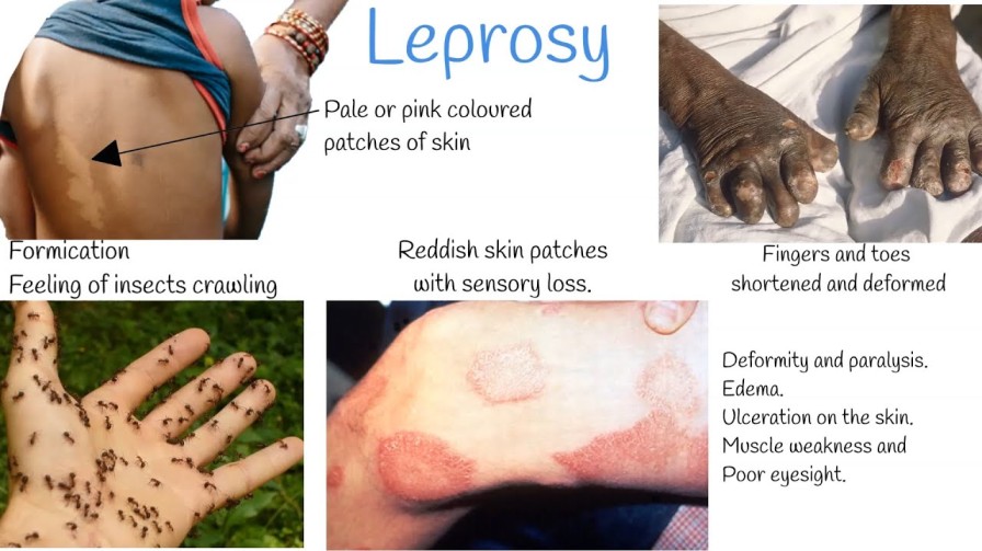
  • Anaesthetic skin lesions: These are patches of skin that lose their ability to feel sensation. Individuals may not be able to detect pain, heat, or touch in these areas, making them prone to injuries.

  • Thickened peripheral nerves: Leprosy can affect the peripheral nerves, leading to their thickening and enlargement, often seen as lumps under the skin.

  • Nasal stuffiness: In some cases, leprosy can cause inflammation and swelling in the nasal passages, leading to nasal stuffiness and congestion.

  • Saddled nose (Saddle nose): This refers to the collapse of the nasal bridge due to the destruction of the nasal septum, which can occur in advanced cases of leprosy.

  • Loss of eyebrows and lashes: Leprosy can cause the loss of eyebrows and eyelashes, leading to changes in facial appearance.

  • Erythema nodosum: This is a condition characterized by painful, red nodules that can occur on the skin or under the skin\’s surface.

  • Inflammatory eye changes: Leprosy may affect the eyes, leading to various eye problems, including inflammation and potential vision impairment.

Investigations

To diagnose leprosy and confirm the presence of Mycobacterium leprae, healthcare professionals may conduct several investigations, such as:

  • Histamine test: This test helps assess the level of nerve damage by evaluating the body\’s response to histamine injection.

  • Lepromine test: Lepromine is a substance derived from the leprosy bacteria. The test measures the immune response to lepromine to determine the type of leprosy and the individual\’s immune status.

  • Polymerase Chain Reaction (PCR): PCR is a molecular technique used to detect the genetic material of the bacteria in skin samples, aiding in early and accurate diagnosis.

  • Skin snip for Mycobacterium leprae (modified ZN): A small sample of skin is taken and stained using the modified Ziehl-Neelsen method to visualize the presence of Mycobacterium leprae under a microscope.

Treatment for Leprosy:

  1. Tuberculoid leprosy:

    • Dapsone + Rifampicin
  2. Lepromatous leprosy:

    • Dapsone + Rifampicin + Clofazimine
  3. Borderline leprosy:

    • Dapsone
Non-leprosy drugs used in the management of leprosy:
  • Steroids
  • Vitamin B complex

Drugs for leprosy are commonly administered in fixed drug combinations known as MDT (Multi-Drug Therapy). MDT has been a highly effective approach in treating leprosy and preventing the development of drug resistance.

Complications of leprosy include:

  • Madorosis (loss of eyebrows and eyelashes)
  • Nasal bridge collapse
  • Ocular complications: corneal ulcer, blindness
  • Leonine faces (thickened, lion-like appearance of facial skin)
  • Loss of sensation to heat, pain, and light touch
  • Multiple ulcerations due to nerve damage and loss of sensation
  • Nerve enlargement
  • Orchitis (inflammation of the testicles)
  • Disuse of some parts of the body
  • Contractures and shortening of phalanges, especially the 4th and 5th fingers and toes
  • Elongated soft ear lobes
  • Sterility in men secondary to orchitis
  • Hammer toes (abnormal bending of the toes)
  • Reactional states secondary to successful drug therapy (erythema nodosum leprosum).

Test Questions

Question: Leprosy primarily affects which body parts?
a) Liver and kidneys
b) Skin and peripheral nerves
c) Lungs and heart
d) Brain and spinal cord
Answer: b) Skin and peripheral nerves
Explanation: Leprosy is a chronic infection that primarily affects the skin and peripheral nerves.

Question: What is the incubation period for lepromatous leprosy?
a) 1-2 years
b) 3-5 years
c) 6-8 years
d) 8-12 years
Answer: d) 8-12 years
Explanation: Lepromatous leprosy has a longer incubation period of 8-12 years, compared to other types of leprosy.

Question: Which type of leprosy has well-defined, few skin lesions and less severe nerve damage?
a) Tuberculoid leprosy
b) Lepromatous leprosy
c) Borderline leprosy
d) Indeterminate leprosy
Answer: a) Tuberculoid leprosy
Explanation: Tuberculoid leprosy is characterized by well-defined, few skin lesions and less severe nerve damage.

Question: Which drug combination is used to treat lepromatous leprosy?
a) Dapsone
b) Dapsone + Rifampicin
c) Dapsone + Rifampicin + Clofazimine
d) Rifampicin
Answer: c) Dapsone + Rifampicin + Clofazimine
Explanation: Lepromatous leprosy is treated with a combination of Dapsone, Rifampicin, and Clofazimine.

Question: What are the complications of leprosy that may lead to blindness?
a) Loss of eyebrows and lashes
b) Nasal bridge collapse
c) Corneal ulcer
d) Erythema nodosum leprosum
Answer: c) Corneal ulcer
Explanation: Leprosy can cause corneal ulcers, which may lead to blindness if left untreated.

Question: Which investigation helps diagnose leprosy by detecting Mycobacterium leprae in skin samples?
a) Lepromine test
b) Histamine test
c) PCR
d) Skin snip for Mycobacterium leprae (modified ZN)
Answer: d) Skin snip for Mycobacterium leprae (modified ZN)
Explanation: Skin snip test with modified Ziehl-Neelsen staining is used to visualize Mycobacterium leprae under a microscope.

Question: What is the most common type of leprosy?
a) Tuberculoid leprosy
b) Lepromatous leprosy
c) Borderline leprosy
d) Indeterminate leprosy
Answer: b) Lepromatous leprosy
Explanation: Lepromatous leprosy is the most common type, accounting for about 90% of leprosy cases.

Question: Which type of leprosy has a weak cellular immune response and widespread skin lesions?
a) Tuberculoid leprosy
b) Lepromatous leprosy
c) Borderline leprosy
d) Indeterminate leprosy
Answer: b) Lepromatous leprosy
Explanation: Lepromatous leprosy is characterized by a weak cellular immune response and widespread skin lesions.

Question: What is the term used to describe the thickened, lion-like appearance of facial skin in leprosy?
a) Madorosis
b) Leonine faces
c) Erythema nodosum
d) Nasal bridge collapse
Answer: b) Leonine faces
Explanation: Leonine faces refer to the thickened, lion-like appearance of facial skin seen in some cases of leprosy.

Question: Which non-leprosy drug is commonly used in the management of leprosy?
a) Antibiotics
b) Steroids
c) Antifungals
d) Antivirals
Answer: b) Steroids
Explanation: Steroids are used in the management of leprosy to control inflammation and reduce immune reactions in some cases.

Ā 

Leprosy Read More Ā»

Tuberculosis

Tuberculosis

Tuberculosis

Tuberculosis, commonly known as TB (short for tubercle bacillus), is a widespread and often deadly infectious disease caused by various strains of mycobacteria.

While primarily affecting the lungs, it can also impact other parts of the body. Around one-third of the world\’s population (1 in 3 or 3 out of 10 people) is affected by this condition. Individuals with HIV/AIDS have a higher risk of contracting tuberculosis.

Aetiology:

The disease is caused by mycobacterium tuberculosis, which is a small, aerobic, non-motile bacillus.

Mode of Spread

TB spreads through the air when individuals with an active infection cough, sneeze, or transmit respiratory fluids. Additionally, it can spread through the blood (haematogenous spread).

\"Types

Types of Tuberculosis

  • Pulmonary tuberculosis.
  • Extra-pulmonary tuberculosis.
  • Primary and Secondary tuberculosis.

Clinical Features:

Pulmonary TB:

  • Fever and chills
  • Night sweats
  • Loss of appetite
  • Weight loss
  • Easy fatigability
  • Persistent cough lasting more than 3 weeks, with or without haemoptysis (coughing up blood)
  • Significant finger clubbing (abnormal swelling of the fingertips)
  • Chest pain
  • Productive cough or non-productive cough in smear-negative TB
  • Lymphadenopathy (swollen lymph nodes)

Extrapulmonary TB:

In approximately 15-20% of active TB cases, the infection spreads beyond the lungs, resulting in various forms of extrapulmonary tuberculosis. This type is more common in individuals with weakened immune systems and young children. In people with HIV, extrapulmonary TB occurs in more than 50% of cases.

Notable sites of extrapulmonary infection include:

  • The pleura, leading to tuberculous pleurisy.
  • The central nervous system, causing tuberculous meningitis.
  • The lymphatic system, resulting in TB lymph nodes.
  • The genitourinary system, causing urogenital tuberculosis.
  • The bones and joints, leading to Pott\’s disease of the spine. When it affects the bones, it is known as \”osseous tuberculosis,\” a form of osteomyelitis.
  • Sometimes, a tubercular abscess may burst through the skin, resulting in a tuberculous ulcer. Ulcers originating from infected lymph nodes nearby are typically painless.
  • A potentially severe and widespread form of TB is \”disseminated\” TB, commonly known as miliary tuberculosis. Miliary TB accounts for about 10% of extrapulmonary cases.

Risk factors

Several factors increase the susceptibility of individuals to TB infections:

  • HIV infection is a significant global risk factor, contributing to 13% of all TB cases.
  • Tuberculosis is closely associated with overcrowding and malnutrition, making it a prevalent disease in impoverished communities.
  • Inhabitants and employees of places where vulnerable individuals gather, such as prisons and homeless shelters, face higher risks.
  • Medically underserved and resource-poor communities, as well as high-risk ethnic minorities, are more susceptible.
  • Children in close contact with high-risk patients are at increased risk.
  • Health care providers serving TB patients are also at higher risk.
  • Chronic lung disease is another significant risk factor.
  • Smokers have nearly double the risk of TB compared to nonsmokers.
  • Other conditions like alcoholism and diabetes mellitus can also elevate the risk of developing tuberculosis.

Epidemiology:

  • Approximately one-third of the world\’s population is infected with tuberculosis.
  • TB causes 25% of preventable adult deaths, and three-fourths of these affected individuals are in their productive age.
  • South Eastern Asia has the highest number of TB cases.
  • Africa has the world\’s highest incidence rate, with an annual incidence rate of 345 cases per 100,000 people.
  • The infection rate is higher in men than in women.
Primary Tuberculosis:
  • It occurs in individuals who have never been exposed to tubercle bacilli before.
  • Tubercle bacilli are inhaled and reach the lungs, where they multiply and can spread to the hilar lymph nodes through the lymphatic system and blood.
  • Approximately six weeks after the primary infection, the body\’s immune response kicks in, preventing further multiplication of the tubercle bacilli.
  • Some bacilli may die, and the remaining ones are walled off by immune cells called epithelioid cells, forming a ghon focus. This ghon focus can persist for years in primary tuberculosis.
  • The ghon focus and hilar lymphadenopathy together form a primary complex in primary tuberculosis.
  • Only about 10% of those with primary infection progress to develop tuberculosis disease.
\"ghon
Secondary Tuberculosis:
  • This type of tuberculosis can result from either the reactivation of tubercle bacilli acquired during primary infection or reinfection by tubercle bacilli in a person previously exposed to the organisms.

Pathogenesis:

  • TB infection starts when mycobacteria reach the pulmonary alveoli and invade and replicate there.
  • In addition to the lungs, tuberculosis can spread through the bloodstream, leading to infection in distant sites like peripheral lymph nodes, kidneys, brain, and bones.
  • The infection triggers an inflammatory response, resulting in the formation of granulomas. Granulomas are collections of activated macrophages, T lymphocytes, B lymphocytes, and fibroblasts.
  • The tubercle bacilli are surrounded by lymphocytes, forming a peripheral rim and creating a Ghon focus. (see image below)
  • Inside the granulomas, the bacteria can become dormant, causing latent infection. Caseation, a type of abnormal cell death, occurs in the center of the tubercles.
  • In severe cases, where TB bacteria enter the bloodstream from damaged tissue, multiple foci of infection can develop throughout the body, appearing as tiny white tubercles in the tissues. This condition is known as miliary tuberculosis and is more common in young children and individuals with HIV.
  • Tissue destruction and necrosis are balanced by healing and fibrosis. Scarring and cavities filled with caseous necrotic material replace affected tissue.

Diagnosis (Investigations):

When tuberculosis is suspected, the following investigations can aid in confirming the diagnosis:

  1. Signs of lung disease or constitutional symptoms lasting longer than two weeks.
  2. Chest X-ray: Imaging the chest can reveal characteristic abnormalities, such as infiltrates, cavities, or nodules, which can indicate tuberculosis.
  3. Multiple sputum cultures for acid-fast bacilli (AFB): Sputum samples are collected at different times, typically spot samples and early morning samples, to increase the chances of detecting the tuberculosis bacteria.
  4. Tuberculin skin tests: Also known as the Mantoux or Heaf test, it is commonly used to assess TB infection in children and identify individuals at risk of developing tuberculosis.
  5. Haematological tests:
    • Full blood cell count (FBC): This test helps evaluate any abnormalities in blood cell counts that may be indicative of an infection or inflammation.
    • Erythrocyte Sedimentation Rate (ESR): An elevated ESR can raise suspicion of tuberculosis, as it indicates inflammation in the body.
  6. Tissue biopsy: In cases where tuberculosis affects extrapulmonary sites or when other tests are inconclusive, a biopsy of the affected tissue may be performed to examine the presence of tubercle bacilli.

Relationship between HIV and TB:

Effects of HIV on TB:
  1. Development of active TB: Individuals infected with HIV have a higher risk of developing active tuberculosis once exposed to the TB bacteria.
  2. High risk of re-infection: HIV-positive individuals are more susceptible to being infected with a second strain of TB after already having the infection.
  3. Increased incidence of TB: The overall incidence of tuberculosis increases due to the higher prevalence of HIV, which weakens the immune system and makes individuals more susceptible to TB.
  4. Changes in TB presentation: TB in HIV-positive individuals may present with clinical and bacteriological changes, such as a non-productive cough, absence of hemoptysis (coughing up blood), and a miliary pattern on imaging instead of cavitations.
  5. Quicker development of TB complications: HIV accelerates the progression of TB and its associated complications.
Effects of TB on HIV:
  1. Increased HIV replication: TB infection can enhance the replication of HIV, leading to a higher viral load and faster progression to AIDS.
  2. Common opportunistic infection: TB is one of the most common opportunistic infections in individuals living with HIV and is a leading cause of death in this population.
  3. Interference with ARV treatment: Some anti-TB medications, such as Rifampicin, can interfere with certain antiretroviral drugs (ARVs), like Nevirapine and protease inhibitors, necessitating adjustments in treatment.

Consequences of dual infection with HIV and TB:

  • Increased morbidity and mortality.
  • Higher recurrence rate of TB after completing treatment.
  • Drug resistance leading to multidrug-resistant TB (MDR-TB) and extensively drug-resistant TB (XDR-TB).
  • Higher rates of treatment non-adherence due to overlapping medication regimens.
  • Increased risk of drug toxicity from the combined treatment.

Management of HIV and TB co-infection:

  • Prioritize TB treatment before starting ARVs.
  • Start ARVs if CD4 count is below 350 cells/mm³, either after finishing TB treatment or during the intensive phase, depending on the clinical situation.
  • Consider drug interactions between TB and HIV regimens when selecting medications.
  • Use directly observed therapy (DOTs) for TB treatment and closely monitor patients for toxicity and adherence.
  • Administer prophylaxis for opportunistic infections as indicated.

Complications of TB:

  • Pleural effusion: Accumulation of fluid in the pleural space of the lungs.
  • Pericardial effusion: Accumulation of fluid around the heart.
  • Empyema: Pus-filled cavity in the pleural space.
  • Pneumothorax: Presence of air or gas in the pleural cavity, causing lung collapse.
  • Lung fibrosis: Scarring of lung tissue, leading to impaired lung function.
  • Lung collapse: Collapse of a lung or part of a lung due to blockage or compression.
  • Extra-pulmonary TB: TB affecting organs other than the lungs, such as TB meningitis.

\"\"

Treatment of TB:

Aims of TB Treatment:

  • To cure the patient of tuberculosis.
  • To prevent complications and death from TB.
  • To reduce the transmission of TB to others.

Case Definitions:

  1. New Case: A person who has never received TB treatment or has taken TB treatment for four weeks or less.
  2. Relapse: A patient who was previously diagnosed with TB, completed the course of anti-TB drugs, was declared cured, but has now become smear positive again.
  3. Failure: A person who continues to be smear positive at five months, despite adequately taking anti-TB drugs, or who was smear negative and becomes smear positive at two months.
  4. Defaulter: A patient who starts taking anti-TB drugs for more than four weeks but interrupts treatment (stops taking the drugs) for four weeks or more.
Drugs Used in TB Treatment:
  1. Rifampicin (R)
  2. Isoniazid (H or INH)
  3. Ethambutol (E)
  4. Pyrazinamide (Z)
  5. Streptomycin (S)
Standard TB Treatment Regimen:Ā 

The World Health Organization (WHO) recommends a standard six-month treatment regimen for drug-sensitive TB cases, which typically includes the following four drugs for the first two months:

  1. Rifampicin (R)
  2. Isoniazid (H)
  3. Pyrazinamide (Z)
  4. Ethambutol (E)

This initial phase is followed by a continuation phase for the next four months, during which the drugs used may vary depending on the patient\’s response to treatment and drug sensitivity testing.

It\’s important to note that TB treatment should be administered under direct observation (DOTs) whenever possible to ensure proper adherence and to prevent the development of drug-resistant TB strains. Check DOT below in details.

Treatment regimenĀ 

Ā Short course TB treatment regimen

Patient category (type of TB)Ā 

Initial phaseĀ 

Continuation phase

1. New smear positiveĀ 

2. New smear negativeĀ 

3. Severe extra-pulmonary

2EHRZĀ 

6EH

4. Previously treated smearĀ  POSITIVE:Ā 

– RelapseĀ 

– Failure to respondĀ 

– Return after interruption

2SEHRZ/1EHRZĀ 

5EHR

5. Any form of TB in children

6. Adult non-severe extra pulmonary

2HRZĀ 

4HR

Non-anti-TB Drugs Used in TB:
  1. Pyridoxine (Vitamin B₆): Administered to prevent or treat peripheral neuropathy, a side effect of Isoniazid (H) used in TB treatment. Pyridoxine supplementation helps prevent nerve damage caused by Isoniazid.

  2. Steroids: Used as adjunct therapy in specific forms of TB to reduce inflammation and improve outcomes. Steroids are commonly used in the treatment of:

    • TB Meningitis
    • TB Pericarditis
    • TB of Adrenals
DOTS (Directly Observed Therapy Short course):

How it works:

  • DOTS is a community-based TB care approach adopted by countries to improve TB treatment outcomes.
  • Trained workers or treatment supporters ensure that patients take their daily treatment doses and record the administration on the TB card.
  • DOTS is the standard of care for all TB cases and suspects.
  • It helps decrease relapse, defaulter rates, and the development of acquired drug resistance.
  • When combined with other measures, DOTS promotes treatment adherence.
  • After diagnosing TB and initiating treatment, the diagnostic center records the information in the health unit\’s TB register.
  • The sub-county health worker transfers this information to the sub-county health worker register and identifies a treatment supporter in the patient\’s village.
  • The treatment supporter is trained to observe the patient taking their treatment, record it on the TB card, keep the drugs, and remind the patient of follow-up assessments at the health unit at 2 months, 5 months, and 8 months.
  • The sub-county health worker collects medication from the health unit and delivers it to the treatment supporter.

Prevention of TB:

  • Early detection and proper management of TB cases.
  • Early case findings to identify and treat TB cases promptly.
  • Health education to raise awareness about TB transmission and prevention.
  • Training of all health workers to recognize early signs of TB.
  • Vaccination of children with the Bacille Calmette-GuĆ©rin (BCG) vaccine to protect against severe forms of TB in childhood.
  • Prophylaxis with Isoniazid for individuals at high risk of developing TB, such as those with latent TB infection or individuals with HIV.
  • Prevention and management of medical conditions like HIV, which increase the risk of TB.
  • Implementation of the DOTS program to improve treatment adherence and outcomes.

\"midwives

Test Questions

Question: Which bacterium is responsible for causing tuberculosis?

a) Streptococcus pneumoniae
b) Mycobacterium tuberculosis
c) Escherichia coli
d) Staphylococcus aureus

Answer: b) Mycobacterium tuberculosis
Explanation: Mycobacterium tuberculosis is the specific bacterium that causes tuberculosis.

Question: What is the most common site of TB infection in the human body?
a) Liver
b) Lungs
c) Heart
d) Kidneys

Answer: b) Lungs
Explanation: Pulmonary tuberculosis is the most common form of TB, affecting the lungs.

Question: In HIV-positive individuals, the risk of developing active tuberculosis:
a) Decreases
b) Stays the same
c) Increases
d) Remains unaffected

Answer: c) Increases
Explanation: HIV weakens the immune system, making individuals more susceptible to developing active tuberculosis once infected with Mycobacterium tuberculosis.

Question: Which of the following is NOT a clinical feature of pulmonary tuberculosis?
a) Fever and chills
b) Night sweats
c) Loss of appetite
d) Severe abdominal pain

Answer: d) Severe abdominal pain
Explanation: Severe abdominal pain is not a typical clinical feature of pulmonary tuberculosis.

Question: What is the standard duration of treatment for drug-sensitive tuberculosis?
a) 3 months
b) 6 months
c) 9 months
d) 12 months

Answer: b) 6 months
Explanation: The standard treatment regimen for drug-sensitive TB lasts for 6 months.

Question: In which form of TB, patients may present with a non-productive cough and a miliary pattern on imaging?
a) Drug-resistant TB
b) Extrapulmonary TB
c) Latent TB
d) Multidrug-resistant TB

Answer: b) Extrapulmonary TB
Explanation: Extrapulmonary TB can present with atypical symptoms like a non-productive cough and a miliary pattern on imaging.

Question: What is the main purpose of the DOTS program in TB management?
a) To prevent TB transmission
b) To promote TB vaccination
c) To monitor drug resistance
d) To improve treatment adherence

Answer: d) To improve treatment adherence
Explanation: The main aim of the DOTS program is to ensure that patients adhere to their TB treatment, which leads to better outcomes and reduced relapse rates.

Question: Which non-anti-TB drug is used to prevent or treat peripheral neuropathy, a side effect of Isoniazid?
a) Vitamin C
b) Vitamin D
c) Pyridoxine (Vitamin B₆)
d) Folic acid

Answer: c) Pyridoxine (Vitamin B₆)
Explanation: Pyridoxine is used to prevent or treat peripheral neuropathy caused by Isoniazid.

Question: TB Meningitis is best managed with the addition of which adjunct therapy?
a) Antibiotics
b) Antifungals
c) Antivirals
d) Steroids

Answer: d) Steroids
Explanation: TB Meningitis is often treated with the addition of steroids to reduce inflammation and improve outcomes.

Question: What is the primary aim of TB prevention?
a) Eradicate Mycobacterium tuberculosis from the environment
b) Reduce the incidence of drug-resistant TB
c) Prevent the transmission of TB from person to person
d) Increase vaccination coverage in high-risk populations

Answer: c) Prevent the transmission of TB from person to person
Explanation: The primary aim of TB prevention is to break the chain of transmission by preventing the spread of Mycobacterium tuberculosis from infected individuals to others.

Tuberculosis Read More Ā»

Measles

Measles

Measles

Measles, also known as Morbilli, is a highly contagious acute infection of the respiratory system caused by the morbillivirus.Ā 

It is characterized by a widespread skin rash, fever, and inflammation of the mucous membranes.

The transmission of measles occurs through respiration, mainly by coming into contact with fluids from the nose and mouth of an infected person. Due to its high contagion, it can easily spread among individuals.

The incubation period for measles typically lasts from 9 to 12 days.

Ā Risk Factors

Several risk factors increase the likelihood of contracting measles, including:

  1. Immunodeficiency in children.
  2. Traveling to regions where measles is common or having contact with individuals who have visited these areas.
  3. Malnutrition, which can weaken the immune system.
  4. Pregnancy, as it may increase susceptibility to the virus.
  5. Vitamin A deficiency, which can compromise the body\’s ability to fight infections.

\"Signs

Signs and symptoms of measles/Stages

Measles, an acute and highly communicable infection caused by the morbillivirus, presents a clinical picture that can be divided into three distinct stages: prodromal, eruptive, and convalescent. Suspecting measles becomes crucial when patients exhibit the classic triad of the three \”Cs\”: cough, conjunctivitis, and coryza.

Stage 1: Prodromal Stage

  • The incubation period lasts approximately 10-14 days.
  • Patients may not show any signs or symptoms during this stage.
  • Abrupt onset of mild to moderate symptoms, characterized by:
    • Fever
    • Headache
    • General malaise
    • Loss of appetite (anorexia)
    • Enlarged neck lymph nodes
    • Abdominal pain
    • Diarrhea
    • Vomiting

Stage 2: Eruptive Stage

  • Abrupt onset with severe symptoms, including:
    • Very high fever
    • Cough
    • Photophobia (sensitivity to light)
    • Red eyes and conjunctivitis
    • Hoarseness of the voice
    • Distinctive Koplik spots on the mucous membrane of the mouth, next to the molar teeth. These spots may disappear once the rash appears.
  • Temperature rises on the first day (37.8-39.4 degrees Celsius), may slightly fall on the third day, then rise again on the fourth day with the onset of the rash.
  • The rash appears around the fourth day and starts on the forehead, behind the ears, neck, and then spreads over the face and entire body. The rash is a red maculo-papular eruption, giving the face a bloated, swollen appearance.

Stage 3: Convalescent Stage

  • Improvement and disappearance of signs and symptoms begin.
  • Key features include:
    • Desquamation of the skin (shedding of the rash)
    • A decline in body temperature
    • Resolution of hoarseness of the voice
    • Weight gain as the patient\’s condition improves.

Nursing Care/Management for a Patient within 72 Hours of Measles:

Aims of Care/Management:

  1. To reduce body temperature.
  2. To correct dehydration.
  3. To prevent further complications.

Admission:

  1. Admit the child to a well-ventilated room in an isolation unit in the children\’s ward.
  2. Record the patient\’s particulars, including name, age, next of kin, and full address on the admission forms.
  3. Reassure the mother/caregiver about the child\’s condition.

Observations:

  1. Monitor vital signs (Temperature, pulse, respiration, blood pressure, and weight) and record them in an observation chart for baseline monitoring.
  2. Conduct a comprehensive head-to-toe assessment to identify any abnormalities such as jaundice, edema, dehydration, cyanosis, anemia, and lymphadenopathy. Document findings in the patient file.
  3. Inform the doctor about the patient\’s condition and prepare for any required investigations and medical treatments.
  4. Carry out procedures, such as tepid sponging, based on the patient\’s findings (e.g., in case of high fever).
Investigations:
  1. Conduct necessary investigations to rule out other diseases, such as:
    • Blood slide for malaria parasites
    • Full blood count (FBC) to rule out other infections
    • Urinalysis
    • Salivary measles-specific IgA testing (rarely done).
Medical Treatments:
  1. There is no specific treatment for measles; it is managed symptomatically.
  2. Prescribe the following drugs based on symptoms:
    • Antibiotics to treat underlying infections (e.g., Cephalexin or Amoxyl syrup).
    • Intravenous Ceftriaxone for severe cases.
    • Analgesics to reduce pain and fever (e.g., Syrup Cetamol).
    • Antihistamines to reduce itching (e.g., Calamine lotion).
    • Vitamins A capsules for children below 1 year to prevent eye complications.
    • Grovit drops or syrup multivitamin to improve appetite.

Fluids and Diet:

  1. Provide plenty of oral fluids to replace lost fluids due to vomiting and diarrhea.
  2. Offer easily digestible foods rich in vitamins and proteins for quick recovery.
  3. Encourage the child to take frequent small meals.
  4. Use a nasogastric tube for feeding if the child cannot eat or drink.
  5. Administer intravenous fluids in cases of severe dehydration.

Skin Care:

  1. Pad the fingers to prevent excessive scratching of the skin.
  2. Apply prescribed calamine lotion to relieve itching.

Mouth and Eye Care:

  1. Emphasize oral hygiene with frequent mouth care using warm saline.
  2. Keep the nostrils clean and maintain cleanliness around the nasogastric tube.
  3. Apply gentian violet 1% for mouth ulcers.
  4. Use glycerin borax to lubricate the lips and prevent cracking.
  5. Clean the eyes with warm saline and avoid rubbing them.
  6. Apply TEO ointment if necessary.
  7. If one eye is affected, encourage the child to lie on the affected side to prevent infecting the other eye.
  8. Avoid direct sunlight on the eyes.

Hygiene and Bed Rest:

  1. Give the patient a daily bath and change bedding frequently.
  2. Use appropriate precautions for discharging ears and administer antibiotics as needed.
  3. Disinfect used soiled linen and utensils.
  4. Properly dispose of used swabs, discharges, or secretions.

Visitor and Ward Management:

  1. Restrict visitors and maintain visiting hours.
  2. Keep radio and TV volumes low to allow for patient rest.
  3. Encourage dim lighting due to photophobia.
  4. Encourage adequate sleep by switching off lights and minimizing noise.

Observations:

  1. Continue monitoring the patient\’s general condition and vital signs regularly.
  2. Take note of any deviations from the normal and act accordingly.
  3. Perform tepid sponging, give cold drinks, and apply cold compress on the forehead if the temperature is very high.

Bowel and Bladder Care:

  1. Observe and treat diarrhea or constipation as needed.
  2. Monitor and address any issues with the child\’s urine output.

Exercises and Health Education:

  1. Encourage the patient to do active and passive exercises, including deep breathing exercises.
  2. Stimulate the child\’s mind with play objects like toys.
  3. Educate the mother/caregiver about the mode of spread, signs, symptoms, and prevention of measles.

Complications of Measles:

  • Pneumonia
  • Acute Laryngo-Tracheo-Bronchitis (LTB)
  • Otitis media leading to deafness
  • Conjunctivitis
  • Encephalitis
  • Acute gastroenteritis
  • Malnutrition (PEM) – Kwashiorkor and marasmus
  • Subacute sclerosing panencephalitis.

Test Questions

MCQ: Which virus causes measles?
a) Influenza virus
b) Morbillivirus
c) Respiratory syncytial virus
d) Rotavirus
Answer: b) Morbillivirus
Explanation: Measles is caused by the morbillivirus, a member of the Paramyxoviridae family.

MCQ: During which stage of measles does the characteristic red maculo-papular rash appear?
a) Incubation stage
b) Prodromal stage
c) Catarrhal stage
d) Convalescence stage
Answer: c) Catarrhal stage
Explanation: The characteristic red maculo-papular rash appears during the catarrhal or eruptive stage of measles.

MCQ: What is the primary aim of nursing care in managing measles?
a) To reduce the risk of bacterial infection
b) To relieve itching and rash discomfort
c) To prevent complications and dehydration
d) To administer specific antiviral medication
Answer: c) To prevent complications and dehydration
Explanation: The primary aim of nursing care in managing measles is to prevent complications and dehydration, as there is no specific antiviral medication for measles.

MCQ: Which symptom is part of the classic triad used for suspecting measles?
a) Fever
b) Cough
c) Diarrhea
d) Jaundice
Answer: b) Cough
Explanation: The classic triad for suspecting measles includes cough, conjunctivitis, and coryza (common cold).

MCQ: What is the incubation period for measles?
a) 2-5 days
b) 7-10 days
c) 10-14 days
d) 21-28 days
Answer: c) 10-14 days
Explanation: The incubation period for measles typically lasts from 10 to 14 days.

MCQ: Which vitamin is administered to prevent eye complications related to measles in children below one year?
a) Vitamin B
b) Vitamin C
c) Vitamin D
d) Vitamin A
Answer: d) Vitamin A
Explanation: Vitamin A capsules are administered to children below one year to prevent eye complications associated with measles.

MCQ: Which stage of measles is characterized by an abrupt onset of severe symptoms, including very high fever and photophobia?
a) Incubation stage
b) Prodromal stage
c) Catarrhal stage
d) Convalescence stage
Answer: c) Catarrhal stage
Explanation: The catarrhal or eruptive stage of measles is characterized by an abrupt onset of severe symptoms, including very high fever and photophobia.

MCQ: What is the primary mode of measles transmission?
a) Contact with contaminated food
b) Direct skin-to-skin contact with an infected person
c) Airborne droplets from an infected person\’s respiratory secretions
d) Ingestion of contaminated water
Answer: c) Airborne droplets from an infected person\’s respiratory secretions
Explanation: Measles is primarily transmitted through airborne droplets when an infected person coughs or sneezes.

MCQ: Which of the following is NOT a risk factor for measles?
a) Immunodeficiency in children
b) Travel to areas where measles is endemic
c) Malnutrition
d) Taking vitamin supplements
Answer: d) Taking vitamin supplements
Explanation: Immunodeficiency, travel to endemic areas, and malnutrition are risk factors for measles, but taking vitamin supplements is not directly associated with measles risk.

MCQ: Which stage of measles marks the beginning of improvement, characterized by skin desquamation and a decline in body temperature?
a) Incubation stage
b) Prodromal stage
c) Catarrhal stage
d) Convalescence stage
Answer: d) Convalescence stage
Explanation: The convalescence or recovery stage of measles marks the beginning of improvement, characterized by skin desquamation, a decline in body temperature, and the resolution of symptoms.

Measles Read More Ā»

Want notes in PDF? Join our classes!!

Send us a message on WhatsApp
0726113908

Scroll to Top
Enable Notifications OK No thanks