nursesrevision@gmail.com

Hypoxic Ischemic Encephalopathy

Hypoxic Ischemic Encephalopathy

Hypoxic Ischemic Encephalopathy

Hypoxic Ischemic Encephalopathy (HIE) is a type of newborn brain damage caused by oxygen deprivation and limited blood flow.

Hypoxic Ischemic Encephalopathy is when the brain does not get enough oxygen, permanent brain damage can result. Hypoxic Ischemic Encephalopathy is a type of birth injury; this is a broad term used to refer to any harm that a baby experiences at or near the time of birth. Other terms used for HIE include birth asphyxiaperinatal asphyxia, and neonatal encephalopathy.

Hypoxic Ischemic Encephalopathy falls under the Broad term “Encephalopathy

Encephalopathy

Encephalopathy is a general term used to describe damage or disease affecting brain function. The causes are varied and can be related to infection, liver conditions, drug toxins, and more.

Some of the major types include the following:

  • Chronic Traumatic Encephalopathy: This condition occurs due to direct injury to the brain, leading to nerve damage. This type is commonly found in athletes.
  • Hepatic Encephalopathy: Often caused by liver cirrhosis, when the liver is not able to filter and function properly, toxins accumulate in the blood and brain.
  • Glycine Encephalopathy: This type of encephalopathy is genetic with symptoms appearing after birth.
  • Toxic Metabolic Encephalopathy: This type of encephalopathy results from toxins, infections, or organ failure. When chemical composition in the body becomes imbalanced, it can impact the brain’s normal function. 
  • Hypoxic-Ischemic Encephalopathy: When the brain does not get enough oxygen, permanent brain damage can result.
  • Hypertensive Encephalopathy: Prolonged hypertension that is not treated can cause the brain to swell resulting in neurological damage.
  • Uremic Encephalopathy: If the kidneys are not functioning properly, they cannot filter harmful substances. The buildup of uremic toxins can cause confusion and other symptoms.

Etiology of Hypoxic Ischemic Encephalopathy

Pathologically, any factors which interfere with the circulation between maternal and fetal blood exchange in form of  maternal factors, delivery factors and fetal factors.

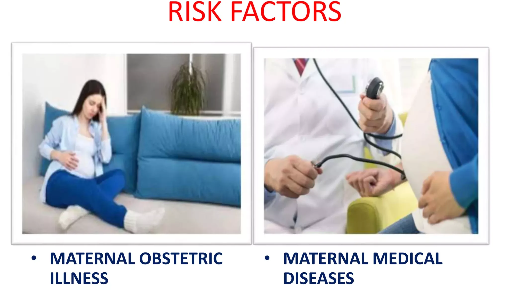
Maternal factor: 
  • hypoxia,
  • anemia,
  • diabetes,
  • hypertension,
  • smoking,
  • nephritis,
  • heart disease,
  • too old or too young,etc 
Delivery condition: 
  • Abruption of placenta,
  • placenta previa,
  • prolapsed cord,
  • premature rupture of membranes,etc 
Fetal factor
  • Multiple birth,
  • congenital or malformed fetus,etc 

High Risk Factors

  • Mismanagement of a high-risk pregnancy: Women with conditions such as preeclampsia and gestational diabetes require more extensive monitoring and treatment.
  • Umbilical cord complications: The umbilical cord is like a lifeline between mother and baby, supplying oxygen and nutrients and removing fetal waste. Anything that compresses the cord or reduces its function puts the baby at risk of HIE.
  • Placental or uterine complications: The placenta and uterus also play very important roles in providing oxygenated blood to the baby. Examples of placental and uterine issues that may cause HIE include:
    • Placental abruption: when the placenta separates from the uterus before the baby is born
    • Placenta previa: when the placenta attaches too close to the cervix; this can cause dangerous bleeding and oxygen deprivation during delivery
    • Placental insufficiency: when the placenta is unable to deliver enough blood to the baby
    • Uterine rupture: when the uterus tears, partially or completely
  • Infections: Infections in the mother can spread to the baby during labor and delivery, especially if the medical team do not take adequate precautions (such as doing indicated infection screening and prescribing antibiotics when needed).
  • Improper fetal heart monitoring: If a baby shows signs of fetal distress on the fetal heart monitor, doctors and nurses can often intervene so that their oxygen supply is restored. If necessary, this may involve an emergency C-section. However, if monitoring is sporadic or does not occur, important signs of danger may be missed.
  • Failure to prevent a premature birth: Premature babies are at higher risk for HIE and other birth injuries because their lungs are so underdeveloped. Therefore, it is very important that doctors do what they can to prevent premature birth, such as performing a cervical cerclage (a stitch placed in the cervix to stop it from opening too early) or providing progesterone treatment.
  • Allowing prolonged labor to continue: Labor is stressful for babies because uterine contractions compress the placenta and umbilical cord that supply their oxygen. If something is preventing labor from progressing, and physicians do not offer intervention (such as an emergency C-section), this is negligence. Prolonged labor is more likely to occur when a baby is larger than normal, or the mother’s pelvis is smaller than normal.
  • Medication problems: Sometimes physicians prescribe medications such as Pitocin and Cytotec in order to induce or enhance labor. Unfortunately, these medications can also cause uterine contractions to become so strong and frequent that the baby is dangerously deprived of oxygen.
  • Mismanagement of a neonatal condition: Hypoxic-ischemic injury can be caused by complications during the neonatal period, i.e. a baby’s first month of life. Problems such as respiratory distress, jaundice, and neonatal hypoglycemia can all contribute to an HIE diagnosis, especially if mismanaged.

Clinical Features of Hypoxic Ischemic Encephalopathy

  • Breathing problems
  • Feeding problems
  • Missing reflexes (for example, the baby does not respond to loud noises)
  • Seizures
  • Low Apgar scores
  • Low or high muscle tone
  • Altered level of consciousness (e.g. not alert)

Classification of Hypoxic Ischemic Encephalopathy 

Clinical Classification

  • Mild(stage I): hyperalert, irritable, normal muscular tone & reflex, no seizure, normal EEG 
  • Moderate(stage II): lethargy, hypotonia, weak sucking & Moro response, often seizure, EEG+
  • Severe(stage III): coma, absent muscular tone & reflex, persistent seizure, EEG++

Levene Classification

FeatureMildModerateSevere
CONSCIOUSNESSIrritableLethargyComatose
TONEHypotoniaMarkedSevere
SEIZURENoYesProlonged
SUCKING/ RESPIRATIONPoor SuckUnable to SuckUnable to maintain Spontaneous respiration

Sarnat Staging Classification 

(Commonly used)

Sarnat staging is used alongside electroencephalogram findings to provide information about the prognosis for the infant.

 Grade I MildGrade II ModerateGrade III Severe
AlertnessHyperalertLethargyComa
Muscle toneNormal or increasedHypotonicFlaccid
SeizuresNoneFrequentUncommon
PupilsDilated, reactiveSmall, reactiveVariable, fixed
RespirationRegularPeriodicApnoea
Duration< 24 Hours2 – 14 DaysWeeks

EEG brain scans may detect signs of autism in 2-year-olds - CBS Newselectroencephalogram


Management of Hypoxic Ischemic Encephalopathy

This is a pediatrics emergency.

  • HIE is managed using a treatment called therapeutic hypothermia, where the baby’s brain or body is cooled down below normal temperatures to slow damage.
  • This allows the baby’s brain to recover and reduces the level of disability they may have as they grow. According to current guidelines, the treatment must be given within six hours of birth, not exceeding  24 hours.
  • The treatment of encephalopathy varies, depending on the underlying cause of the condition.

Generalized treatment

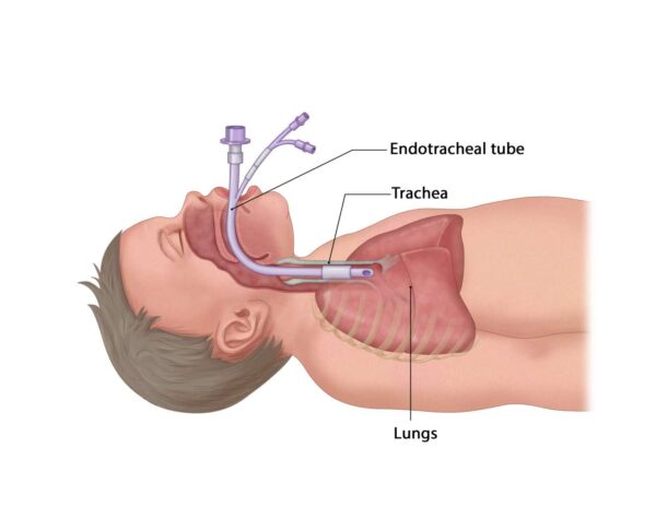
  • Ventilation: CPAP(continuous positive airway pressure) , CMV(continuous mandatory ventilation), HFOV (High-frequency oscillatory ventilation )
  • Perfusion/Circulation: Dopamine/Dobutamine
  •  Energy: normal glucose level maintained (50-110mg/dl): Hypo and hyperglycemia avoided
  • Fluid: 60-80ml/kg/d restriction if SIADH(Syndrome of inappropriate antidiuretic hormone secretion)
  • Electrolytes- Sodium and Calcium should be monitored.
  • Avoid Polycythemia: If Hct>65-70, partial exchange transfusion is done to bring Het level to 55.
  • Control of seizures: HIE seizures are difficult to control ;
  • Phenobarbital loading dose 15-20mg/kg, iv maintenance dose 3-5mg/kg, iv
  •  Phenytoin  loading dose 15-20mg/kg, iv maintenance dose 5mg/kg, iv Midazolam: 0.1-0.3mg/kg, iv Leveracetam, Topiramate
  • EEG 
Special Investigations 
  • Continous AEEG- Amplified EEG used for cerebral function monitoring. Detects voltage pattern- burst, low voltage, isoelectric Detects electrical seizure activity
  •  CT scan: only indicated in emergency 
Prognosis 

Depend on the severity of brain damage & medical treatment, usually

  • Mild or moderate cases could be cured completely, but severe cases represent poor prognosis with high mortality or cerebral complications such as mental retardation & cerebral palsy.
  • Overall mortality 20%
  • Overall incidence of sequele 30%
  • Mild: 100% good prognosis
  • Moderate: 80% normal
  • Severe : 50% death, 50% sequele 

Presence of seizure increases chance of Cerebral palsy by 50-70 times

Prevention 
  • Better Obstetric care
  • Skilled resuscitation teams and neonatal facilities.

Nursing Diagnosis

  1. Acute Confusion related to Hypoxia ,Disturbance in cerebral metabolism ,Accumulation of toxins in the brain, Structural changes in the brain as evidenced by Cognitive dysfunction , Altered psychomotor performance , Tremors, Fluctuation in the level of consciousness, Agitation, Misperception, Neurobehavioral manifestations, Difficulty initiating purposeful behavior.
  2.  Impaired Memory related to Neurological disturbances related to encephalopathy, Inadequate intellectual stimulation, Changes in brain structure and processes, Irreversible brain damage, Depressive symptoms as evidenced by Reports experiences of forgetfulness, Consistently forgets to schedule or keep appointments , Difficulty recalling events, Difficulty recalling familiar names, objects, and words, Inability to learn or retain new skills or information, Inability to perform a previously learned skill.
  3. Disturbed Thought Processes related to Insufficient oxygen supply to the brain secondary to encephalopathy, Head trauma related to encephalopathy, Infections as evidenced by Incorrect perception of stimuli, Difficulty performing activities of daily living , Difficulty communicating verbally, Impaired interpretation of events, Impaired judgment, Impaired decision making, Inadequate emotional responses, Disorientation

Hypoxic Ischemic Encephalopathy Read More »

conjunctivitis

Conjunctivitis

Conjunctivitis

Conjunctivitis is an inflammation of the lining of the eye and eyelid caused by bacteria, viruses, chemicals or allergies, fungal, parasites.

  • It is characterized by dilatation of the conjunctival vessels, resulting in hyperemia and edema of the conjunctiva, typically with associated discharge.
  • It’s the most common cause of red eye usually painless and characterized by pussy or watery discharge.

Types of Conjunctivitis

  1. Bacterial conjunctivitis :- caused by bacterium e.g. staphylococcus or streptococcus
  2. Acute viral conjunctivitis:- e.g. herpes simplex
  3. Allergic conjunctivitis:- caused by allergy e.g. smoke, cosmetic and medicines
Bacterial Conjunctivitis

Predisposing factors for bacterial conjunctivitis

  • Flies
  • Poor hygienic conditions
  • Hot dry climate
  • Poor sanitation
  • Dirty habits

Causes of Bacterial Conjunctivitis

  • Staphylococcus aureus is the most common cause of bacterial conjunctivitis
  • Pneumococcus.
  • Streptococcus pyogenes (haemolyticus) is virulent and usually produces pseudomembranous conjunctivitis.
  • Pseudomonas pyocyanea is a virulent organism, which readily invades the cornea.
  • Neisseria gonorrhoeae typically produces acute purulent conjunctivitis in adults and ophthalmic neonatorum in newborn
  • Neisseria meningitidis may produce muco-purulent conjunctivitis.
  • Corynebacterium diphtheriae causes acute membranous conjunctivitis

Mode of Infection

Conjunctiva may get infected from three sources:-

1.Exogenous infections may spread:

  • Directly through close contact
  • Vector transmission e.g., flies
  • Material transfer e.g. infected fingers of health workers, common towels,handkerchiefs & tonometers
  1. Local spread may occur from neighbouring structures such as infected lacrimal sac, lids, and nasopharynx.
  2. Endogenous infections may occur very rarely through blood, e.g., gonococcal and meningococcal infections.
Pathology
  • Pathological changes of bacterial conjunctivitis consist of:
  1. Vascular response. It is characterized by congestion and increased permeability of the conjunctival vessels associated with proliferation of capillaries.
  2. Cellular response. It is in the form of exudation of polymorphonuclear cells and other inflammatory cells into the substantia propria of conjunctiva as well as in the conjunctival sac.
  3. Conjunctival tissue response. Conjunctiva becomes edematous. The superficial epithelial cells degenerate, become loose and even desquamate. There occurs proliferation of basal layers of conjunctival epithelium and increase in the number of mucin-secreting goblet cells.
  4. Conjunctival discharge. It consists of tears, mucus, inflammatory cells, desquamated epithelial cells, fibrin and bacteria. If the inflammation is very severe, diapedesis of red blood cells may occur and discharge may become blood stained.
Acute Bacterial Conjunctivitis
  • Acute bacterial conjunctivitis is characterized by marked conjunctival hyperemia and mucopurulent discharge from the eye.
  • It is the most common type of bacterial conjunctivitis.
  • Common causative bacteria are: Staphylococcus aureus, Pneumococcus and Streptococcus.

Infectious period

The time during which an infected person can infect others thus while the eye discharge is present.

Clinical Presentation

  • Typically there is conjunctival infection, especially in the fornices where the blood supply is rich.
  • Discharge is variable, but typically is present in the mornings, and on waking the eye is difficult to open because the eyelids are stuck together.
  • The eyelids may be red and inflamed.
  • The condition may be unilateral or bilateral.
  • The vision is always unaffected and there is usually no pain.
  • The patient may complain of a gritty or foreign body sensation, some discomfort and very occasionally very mild photophobia.
  • Slight blurring of vision due to mucous flakes in front of cornea.
  • Flakes of mucopus seen in the fornices, canthi and lid margins is a critical sign

Clinical course and diagnosis

  • Mucopurulent conjunctivitis is usually bilateral, although one eye may become affected 1–2 days before the other.
  • The disease usually reaches its height in three to four days. If untreated, in mild cases the infection may be overcome and the condition is cured in 10–15 days

Diagnosis

  • It is by clinical and/or microscopic examination and culture of discharge from the eye.
  • It is usually not possible to tell whether the conjunctivitis is caused by bacteria or viruses without laboratory tests.

Management of bacterial Conjunctivitis

  • Clean the eyes and apply topical antibiotics
  • Bacterial conjunctivitis usually resolves without treatment
  • Topical antibiotics may be needed only if no improvement is observed after 3 days
  • Dark goggles should be used to prevent photophobia.
  • No bandage should be applied in patients with mucopurulent conjunctivitis. Exposure to air keeps the temperature of conjunctival cul-de-sac low which inhibits the bacterial growth
  • No steroids should be applied, otherwise infection will flare up and bacterial corneal ulcer may develop.
  • Topical Antibiotics:
  • Therefore, treatment may be started with chloramphenicol (1%), or gentamicin (0.3%), or tobramycin 0.3% or framycetin 0.3% eye drops 3–4 hourly in day and ointment used at night will not only provide antibiotic cover but also help to reduce the early morning stickiness.
  • Quinolone antibiotic drops such as ciprofloxacin (0.3%), ofloxacin (0.3%), gatifloxacin (0.3%) or moxifloxacin (0.5%) may be used as alternative in severe cases
Chronic Bacterial Conjunctivitis
  • Chronic bacterial conjunctivitis also known as ‘Chronic catarrhal conjunctivitis’ or ‘simple chronic conjunctivitis’ is characterized by mild catarrhal inflammation of the conjunctiva.
Viral Conjunctivitis

Viral causes include:

  • Adenovirus
  • Herpes simplex virus
  • Poxvirus
  • Mycovirus and Paramyxovirus

Signs of Viral conjunctivitis

  • Red/pink eye.
  • Chemosis, if severe.
  • Follicles may be present on the palpebral conjunctiva.
  • Bleeding from conjunctival vessels in severe adenoviral conjunctivitis.

Management/Treatment

  1. Supportive treatment for amelioration of symptoms is the only treatment required and includes:
  • Apply cold compresses
  • If photophobia is present, advise patient to wear dark glasses
  • Artificial lubricant can be prescribed for patient comfort.
  1. Topical antibiotics help to prevent superadded bacterial infections.
  2. Topical antiviral drugs are not beneficial in adenoviral conjunctivitis.
  3. Topical steroids should not be used during active inflammation as these may enhance viral replication and extend the period of infectivity.
  • Weak steroids such as fluorometholone or loteprednol (0.5%) are indicated in patients with subepithelial infiltrates, and in those with membrane formation.
  1. Full explanation of the condition to increase patient awareness and reduce discomfort.


Preventive measures include:

  • Frequent hand washing
  • Relative isolation of infected individual
  • Avoiding eye rubbing and common use of towel or handkerchief sharing
  • Disinfection of ophthalmic instruments and clinical surfaces after examination of a patient is essential.
Allergic Conjunctivitis
  • It is the inflammation of conjunctiva due to allergic or hypersensitivity reactions which may be immediate (humoral) or delayed (cellular).

Types

  1. Simple allergic conjunctivitis
  • Seasonal allergic conjunctivitis (SAC)
  • Perennial allergic conjunctivitis (PAC)
  1. Vernal keratoconjunctivitis (VKC)
  2. Atopic keratoconjunctivitis (AKC)

Simple Allergic Conjunctivitis

  • It is a mild, nonspecific allergic conjunctivitis characterized by itching, hyperaemia and mild papillary response.

Etiology

  • Simple allergic conjunctivitis, is a type-I immediate hypersensitivity reaction mediated by IgE.

Types

  1. Seasonal allergic conjunctivitis (SAC). SAC is a response to seasonal allergens such as tree and grass pollens. It is of very common occurrence and may be associated with allergic rhinitis.
  2. Perennial allergic conjunctivitis (PAC) is a response to perennial allergens such as house dust, animal dander and mite. The onset is subacute, the condition is chronic in nature and occurring all through the year.

Clinical Features

Symptoms

  • Intense itching and burning sensation in the eyes associated with watery mucus and mild photophobia.

Signs

  • Hyperaemia and chemosis which give a swollen juicy appearance to the conjunctiva.
  • Oedema of lids is often present.

Diagnosis

Diagnosis is made from:

  • Typical symptoms and signs,
  • Normal conjunctival flora, and
  • Presence of abundant eosinophils in the discharge.

Treatment

  1. Elimination of allergens if possible.
  2. Topical vasoconstrictors like naphazoline, antizoline and tetrahydrozoline provide immediate decongestion.
  3. Artificial tears like carboxymethyl cellulose provide soothing effect. As they sometimes relieve discomfort in mild cases
  4. Mast cell stabilizers such as sodium cromoglycate and nedocromil sodium are very effective in preventing recurrences in atopic cases.
  5. Steroids, if condition is severe
  6. Cool water poured over the face with the head inclined downward constricts capillaries.

Other forms of Conjunctivitis

  Fungal conjunctivitis

  it is caused by Candida albicans. Babies can be affected during birth through an infected birth canal.

  • Fine white plaques are apparent on the conjunctiva.
  • The treatment is with nystatin drops and ointment.
  Parasitic conjunctivitis
  • In hot climates, parasites causing onchocerciasis (river blindness) and schistosomiasis (bilharzia) can induce conjunctivitis.

Prevention

  • Exclude people with conjunctivitis from public places until discharge from the eyes has ceased.
  • Good personal hygiene must be followed. Careful hand washing, using soap and warm water.
  • Do not share towels and wash clothes

Conjunctivitis Read More »

eye anatomy and physiology

Eye Anatomy and Physiology

Eye Anatomy.

 Eye  is the organ for sight. The globe-shaped eyeball occupies the anterior part of the orbit/eye socket. The eyeball is embedded in the orbital cavity.  

The eye contains the receptors for vision and a refracting system that focuses light rays on the receptors in the retina.

Diagram Showing the structure of the Eye.


The Structure of the Eye

  • The eye is spherical in shape and the diameter of an adult eye is approximately 2.5cm.   
  • Internally, the eye is divided into 2 chambers.  
  • The lens, suspensory ligaments and ciliary body separate the 2 chambers; 

Anterior and posterior chamber 

Anterior chamber. It is filled with a clear watery fluid called aqueous humour.  
  • This chamber is in front of the lens.  
  • It is further divided into 2 cavities ie anterior and posterior cavities. 

Posterior chamber. It is filled with a jelly like substance called vitreous humour (vitreous body).  This chamber is behind the lens. 

There are three main layers of tissue in the walls of the eye: 

  • The outer fibrous layer consisting of sclera and cornea 
  • The middle vascular layer or uveal tract consisting of the choroid, ciliary body and iris 
  • The inner nervous tissue layer consisting of the retina. 

The outer fibrous layer

  • This consists of sclera and cornea.  

The sclera or white of the eye forms the outermost layer of the posterior and lateral aspects of the eyeball.  

  • It is continuous anteriorly with a clear transparent epithelial membrane, the cornea.  

The cornea is transparent due to its vascularity and the regular arrangement of its fibres.  

  • Its surface is lined by the conjunctiva.  
  • It is well supplied with nerve endings from the trigeminal nerve. 
  • It consists of a firm fibrous membrane that maintains the shape of the eye.  
  • This membrane gives attachment to the extrinsic muscles of the eye. 
  • Light rays pass through the cornea to reach the retina.  
  • The cornea is convex anteriorly. 
  • It is involved in refracting (bending) light rays to focus them on the retina. 

The middle vascular layer

  • The middle vascular layer is also known as the uveal tract
  • This layer consists of the choroid, ciliary body and iris. 

The choroid lines sclera in the posterior compartment of the eye.  

  • The choroid is rich in blood supply and is chocolate brown in colour.  

The ciliary body is an anterior continuation of the choroid which is inserted into suspensory ligaments.  

  • These ligaments extend to the lens and hold it in position.   
  • The ciliary body is supplied by the 3rd cranial nerve (Oculomotor).  
  • The ciliary body also consists of;  
  • Ciliary muscles. Contraction and relaxation of these smooth muscles determine the size and thickness of the lens. 
  • Secretory epithelial cells (Ciliary glands). These secrete aqueous humour which nourishes structures in the anterior chamber. 

The iris is the visible coloured ring at the front of the eye. 

  • The iris extends anteriorly from the ciliary body lying behind the cornea and in front of the lens.  
  • It divides the anterior chamber of the eye into anterior and posterior cavities.  
  • It contains both circular and radiating muscle fibres which control the size of the pupil.  
  • The colour of iris is genetically determined and depends on the number of pigment cells present. 
  • NB: The Oculomotor nerve supplies the muscles of the iris and ciliary body (intrinsic eye muscles).  

The inner nervous tissue layer

  • The inner layer of the eye ball is the retina.  
  • It is the light sensitive (photosensitive) part of the eye.  
  • It contains several millions of sensory photo receptor cells. 
  • These cells are responsible for converting light into nerve impulses.  

The retina consists of two layers;  

  • The pigmented outer layer which lines choroid. 
  • The inner most neural layer which is in contact with the vitreous humour.  
  • The light sensitive layer consists of sensory receptor cells ie rods and cones. 
  • These contain photosensitive pigments that convert light rays into nerve impulses. 
  • Rod cells pre-dominate in the periphery and function best in dim light.  These cells are much more numerous. 
  • Cone cells pre-dominate near the centre of the retina. These are adapted for bright light and colour vision. 
  • Near the posterior of the retina is a part called macula lutea or yellow spot.  
  • The greatest concentration of cone cells is at a small area in the yellow spot called the fovea centralis.  
  • It is the most vital part of retina for high definition or vision.  
  • The optic disc or blind spot is a small area where the optic nerve leaves the eye.  
  • The blind spot does not have light sensitive cells. 
eye anatomy
Parts of the Eye and functions.

Eyebrows-protect eyeball from sweat, dust and other foreign bodies.

Eyelids –movable folds acting as curtains, preventing injuries. Meet at palpebral fissure(both eyelids meet). It contains sebaceous glands, sweat glands and accessory lacrimal glands all aligned with conjuctival material.

Conjunctiva-clear, delicate mucous membrane. it lines the eyelids and is highly vascularised. It protects the eye against infections. It also acts as a physical barrier, and produces mucin (goblet cells)which lubricates the eye ball.

Sclera-is a fibrous tissue of the eye(white),is tough and contains collagen fibres, and covers 5/6 of the eyeball. It protects inner structures maintains the shape of the eyeball. It also acts as a passage of blood vessels and nerves

Cornea-covers 1/6 of the eyeball. Its clear, transparent and has 5 layers.

Its functions include;

  • protection of the eye as it’s very
  • Refractive media of the and
  • Prevents aqueous from coming out of the eye Anterior chamber

Is just behind the cornea and its functions include;

  • Refractive media
  • Maintains shape and structure of the eyeball
  • Bathes/nourishes the

Production and flow of aqueous humor.

Aqueous Humor is secreted by the epithelial cells of the ciliary body. it passes through suspensory ligaments into the posterior chamber, then flow through the pupil into the anterior chamber. From anterior chamber it drains through trabecular meshwork into the canal of schlemm (scleral venous sinus) then goes to the general circulation

Iris-is the thin visible, contractile, and coloured part of the eye, with a central aperture known as the pupil. It divides the anterior segment of the eye into anterior and posterior chambers. It controls amount of light entering the eye and plays a role in accommodation.

Ciliary body-Is continuous with choroid(middle layer of eyeball). It suspends the lens which is important during accommodation, and produces aqueous.

Choroid-Is the soft brown part behind the eye. Is most vascularized, and nourishes the retina.

Lens-is the transparent, highly elastic biconvex body, that lies immediately behind the pupil/front of the vitreous body. Its thickness is controlled by ciliary muscle through the suspensory ligaments.

Its functions include:

  • Refractive media
  • Absorbs ultra violet rays

Retina-Innermost layer of eyeball where images are formed. It has macula, optic disk, rods and cones. It consists of 2 layers.

  • Epithelial
  • Nervous layer
  • It absorbs
  • Stores and releases vitamin

Vitreous body-is transparent, jelly like media.

  • It maintains the shape of eyeball and acts as refractive
blood and nerve supply of the eye

Blood and Nerve supply to the eye

  • The blood supply of the eye is from the ciliary and central retinal arteries. 
  • These are branches of ophthalmic artery which is also a branch of the internal carotid artery.  
  • Venous drainage is by the central retinal vein.  
  • These vessels run alongside the optic nerve. 
  • Nerve supply is by the optic nerve which is the 2nd cranial nerve.  
  • The retinal nerve fibres originate in the retina. 
  • These fibres converge to form the optic nerve at the optic disc. 

Physiology of Sight 

  • Light rays from objects are bent (refracted) as they pass through varying densities of the clear media of the eye to focus onto the retina.  
  • In the eye, the biconvex shape of the lens refracts and focuses light rays on the retina.  
  • Before reaching the retina, the light rays pass through the cornea. 

Physiology of vision - Online Biology Notes

  • The cornea also plays a role in the refractive power of the eyes. 
  • The lens is elastic thus has ability to change shape. Change in shape varies the amount of refraction for clarity of focus.  This is known as accommodation.  
  • Accommodation is necessary in order for objects at different distances to be visualized with equal clarity. 
  • The normal eye in its relaxed state brings rays of light from distant objects into sharp focus.  
  • However for clear focusing on near objects, an autonomic reflex comes into play.  
  • The reflex involves accommodation, miosis and convergence as follows; 

Accommodation. This refers to the increase in the refractive power of the lens in order to focus light rays from near objects on the retina.  The ciliary muscle contracts and changes shape of the lens to bulge increasing its convexity and refractive power. 

Miosis. This is also known as constriction of pupils. 

  • It accompanies accommodation.  
  • It ensures that light rays are concentrated to pass through the centre of the lens and focus on the retina. 

Convergence (movement of the eyeballs). This refers to bilateral movement of the eyes at the same time in order to focus on a nearby object eg focusing the tip of one’s nose.   

  • The light sensitive layer in the retina containing sensory photo receptor cells (rods and cones) convert light rays into nerve impulses.  
  • These are transmitted through the visual pathways to the visual area in occipital lobe of cerebrum. 
  • Here, they are interpreted as sensation of light form. 
  • They are processed into images of objects which are given meaning by other cerebral areas. 
  • This process involves interaction with information stored as memory in the association areas of the brain. 

NB: The images refracted on the retina are upside down. 

  • The brain adapts to this early in life so that objects are perceived as upside/upright. 

Anatomy and Physiology of Human Eye - GeeksforGeeks

Accessory Organs of the Eye

  • The eye is a delicate organ on the body and it is protected by several structures.  

These include; 

(1).  The eye brows:

  • These are numerous hairs that project from the skin at the supra orbital margins of the frontal bone. 
  • These protect the eye from sweat, dust and other foreign bodies. 

(2). Eyelids and eyelashes: 

  • These are two movable folds of tissue above and below the front of each eye.  
  • There are sebaceous glands, some open into the hair follicles of the eye lids. 

The eyelids contain two muscles. 

  • These include;  
  •  Levator palpebrae superioris which raises the upper eyelid
  •  Orbicularis oculi which closes the eyelids. 
  • The hair on the eye lid is called eye lashes. 
  • The eyelids have a lining (mucous membrane) of the conjunctiva. 
  • This lining is a fine transparent membrane that is on the inner surface of the eyelid.  
  • This layer also covers the eyeball.  Where it lines the eyelids, there is a highly vascularized columnar epithelium  The corneal conjunctiva has avascular stratified epithelium.  
  • This means that the conjunctiva has epithelium without blood vessels at the cornea.  
  • The medial and lateral angles where the eyelids come together are called medial and lateral canthus respectively. 
  • At the edges of the eyelids, are eyelid margins that have numerous sebaceous glands.  
  • These are modified and secrete an oily material (meibum) spread over the conjunctiva by blinking.  
  • The material delays evaporation of the tears. 
    • Protect the eye from injury  
    • Blinking at about 3 to 7 seconds interval spreads tears and oily secretions over the cornea.  This prevents drying of the eyeball. 
      • Function of the eyelids and eyelashes 

(3). Lacrimal apparatus: 

  • The lacrimal apparatus consists of the structures that secrete tears and drain them from the front of the eyeball.  
  • These include;  
  • 1 lacrimal gland and its ducts 
  • 2 lacrimal canaliculi ie superior and inferior to the caruncle of the eye. 
  • 1 lacrimal sac 
  • 1 nasolacrimal duct 
  • Each eye has a lacrimal gland behind the supra orbital margin.  
  • Lacrimal glands are exocrine glands.  
  • They secrete tears which are composed of water, mineral salts, antibodies and bactericidal enzymes. 
  • The tears leave the lacrimal glands by several small ducts. 
  • They then pass over to the front of the eye under the eyelids towards the medial canthus where they drain into two lacrimal canaliculi.  
  • The opening of canaliculi on each side is called punctum.  
  • The canaliculi lie above one another separated by a red body called caruncle. 
  • The tears then drain into the lacrimal sac which is the upper expanded part of the nasolacrimal duct
  • When foreign bodies or other irritants enter the eye, secretion of tears is greatly increased and the conjunctival blood vessels dilate.  
  • Secretion of tears is also increased in emotional states like crying and laughing. 
  • Excess tears are drained from the eye via the lacrimal apparatus into the lacrimal sac and then into the nasolacrimal duct. 
  • Functions of the lacrimal apparatus.
  • It has a fluid which is filled into the conjunctival sac. 
  • This fluid consists of tears and oily (meibum) secretions of meibomian/tarsal glands. 
  • The fluid is spread over the cornea by blinking. 
  • This mixture washes away irritants eg dust. 
  • It provides oxygen and nutrients to the avascular corneal conjunctiva and drains off wastes. 
  • Bactericidal enzyme lysozyme protects the eye by preventing microbial infection. 
  • The oiliness nature of the fluid delays its evaporation and prevents drying/friction of the conjunctiva. 
  • The fluid also prevents the eyelids from sticking together while sleeping. 
  • Main function of tears / tear fluid
  • To lubricate the eye to facilitate oxygen and carbon dioxide exchange. 
  • To produce an optically smooth cornea surface. 
  • To cleanse the eye with a bactericidal enzyme lysozyme.
  • To prevent the conjunctiva from drying. 


(4). Extrinsic muscles :

  • These are also called extrinsic muscles.  
  • They are 6 in number and include the following; 
  • Medial rectus which rotates the eyeball inwards. 
  • Lateral rectus which rotates the eyeball outwards 
  • Superior rectus which rotates the eyeball upwards 
  • Inferior rectus which rotates the eyeball downwards 

Function of the muscles 

  • They protect the eye through the flexible movement of the types of muscles.  
  • These movements help us to see in all directions of the eyeball movement.  
  • Hence they also play a protective function ie protecting the eye and the whole body. 

Eye Anatomy and Physiology Read More »

Intracranial Hemorrhage

Intracranial Hemorrhage

INTRACRANIAL HEMORRHAGE

An intracranial hemorrhage is a type of bleeding that occurs inside the skull (cranium).

Bleeding around or within the brain itself is known as a cerebral hemorrhage (or intracerebral hemorrhage).

Bleeding caused by a blood vessel in the brain that has leaked or ruptured is called a hemorrhagic stroke.

All bleeding within the skull is referred to as intracranial hemorrhage.

Causes of Intracranial Hemorrhage.

  1. Head Trauma: Injury to the head from falls, car accidents, sports incidents, or seizures.
  2. Hypertension: High blood pressure leading to damage in blood vessel walls, causing leakage or rupture.
  3. Blood Clot: Blockage of a brain artery by a clot formed in the brain or traveling from another body part.
  4. Cerebral Aneurysm: Rupture of a weak spot in a blood vessel wall, forming a balloon-like bulge that bursts.
  5. Malformed Arteries or Veins: Leaking of improperly formed arteries or veins.
  6. Bleeding Tumors: Hemorrhage from tumors causing bleeding.
  7. Pregnancy-Related Conditions: Conditions linked to pregnancy or childbirth, including eclampsia, difficult delivery, and assisted delivery.
  8. Coagulopathy or Anticoagulation Medicine: Blood clotting disorders, use of anticoagulants like warfarin or heparin, or bleeding disorders like hemophilia and thrombocytopenia.
  9. Child Abuse Syndrome: Shaken baby syndrome as a result of child abuse.
  10. Postsurgical Causes: Hemorrhage occurring after surgeries like craniotomy or shunting.

Pathophysiology:

The brain relies on a network of blood vessels to supply oxygen and nutrients. Intracranial hemorrhage disrupts this supply, preventing oxygen from reaching brain tissue. The pooled blood from the hemorrhage increases pressure on the brain, further limiting oxygen delivery.

During a hemorrhage or stroke, if oxygen deprivation persists for more than three or four minutes, brain cells begin to die. This results in damage to affected nerve cells and the related functions they control. The interruption of blood flow around or inside the brain is a critical factor leading to cellular damage and dysfunction.

Types of Intracranial Hemorrhage

  • Epidural hematoma
  • Subdural hematoma
  • Subarachnoid hemorrhage
  • Intra cerebral hemorrhage
Types of intracranial hemorrhage

Epidural Hematoma (Subgalea hemorrhage.

Subgaleal hemorrhage occurs when emissary veins between the skull and intracranial venous sinuses tear, leading to blood collection between the dura/apo-neurosis and periosteum of the skull.

High-pressure bleeding is a prominent feature. An epidural hematoma, may briefly lead to lose consciousness and then consciousness is regained latter.

Epidural hematoma is accumulation of blood between the Dura and the skull following fracture of the skull

  • Most commonly from rupture of middle meningeal artery.
  • The hematoma expands rapidly since accumulating blood is arterial in origin and causes compression of the Dura and flattening of underlying gyri
  •  The patient develops progressive loss of consciousness if hematoma is not drained early.

Signs and symptoms

  • Swelling of the ears
  • Increasing head circumference as bleeding expands into this space. (hydrocephalus)
  • Hypovolemic shock,
  • Tachycardia,
  • Hypotension

Diagnosis

  • Subgaleal hemorrhage may present as a large, boggy fluid collection palpable on the head’s surface. Characteristic of a subgaleal hemorrhage is that it is not restricted by suture lines and may shift with movement. This is in contrast to the more common cephalohematoma, a superficial collection of blood restricted to the space between the periosteum and skull, which is contained along suture lines.
  • Neonates with subgaleal hemorrhage are at high risk for rapid decompensation; the subgaleal space can expand to collect a newborn’s entire intravascular blood volume if bleeding continues unrecognized.

Subdural hematoma (SDH)

Subdural hematoma (SDH)

A subdural hemorrhage occurs when bridging veins carrying blood through the dura mater to the arachnoid mater of the meninges are torn.

 This causes bleeding, with blood collecting below the dura and brain.

Presence of blood on the surface of the brain beneath its outer covering.

SDH is a collection of blood below the inner layer of the dura mater but external to the arachnoid membrane.

  • Subdural hematoma is accumulation of blood between the Dura and subarachnoid.
  • Develops most often from rupture of veins which cross the surface convexities of the cerebral hemispheres.

Subdural hematoma may be acute or chronic.

  • Acute subdural hematoma; develops following trauma and consists of clotted blood, often in the front parietal region. There is no significant compression of gyri. Since the accumulated blood is of venous origin, symptoms appear slowly and may become chronic with passage of time if not fatal.
  • Chronic subdural hematoma; occurs often with brain atrophy. Chronic subdural hematoma is composed of liquid blood. Separating the hematoma from underlying brain is a membrane composed of granulation tissue.

Diagnosis

  • Because subdural bleeders are located within the skull, there is often no physical sign on the scalp that reflects injury. Instead, the presence of hemorrhage may initially be unrecognized. For most neonates, subdural hemorrhage remains asymptomatic and resolves without consequence.
  • Clinical problems can arise in case of large volume hemorrhage or if bleeding slowly continues over hours or even days, as in cases of bleeding disorders.
  • Symptomatic neonates often present 24–48 hours after birth with nonspecific signs such as apnea, respiratory distress, altered neurologic state, or seizures.

Subarachnoid hemorrhage

Subarachnoid hemorrhage occurs when the veins of the subarachnoid villi are torn, leading to a collection of blood in the subarachnoid space

There’s bleeding between the brain and the thin tissues that cover the brain. These tissues are called meninges.

A sudden, sharp headache usually comes before a subarachnoid hemorrhage. Typical symptoms also include loss of consciousness and vomiting.

  • Hemorrhage into the subarachnoid space is most common, caused by;
  •  rupture of an aneurysm,  and rarely, rupture of a vascular malformation.
  • Of the three types of aneurysms affecting the larger intracranial arteries—berry, mycotic and fusiform, berry aneurysms are most important and most common.
  • Berry aneurysms are saccular in appearance with rounded or lobulated bulge arising at the bifurcation of intracranial arteries and varying in size from 2 mm to 2 cm or more.
  • They account for 95% of aneurysms which are liable to rupture.
  •  Berry aneurysms are rare in childhood but increase in frequency in young adults and middle life.
  • They are, therefore, not congenital anomalies but develop over the years from developmental defect of the media of the arterial wall at the bifurcation of arteries forming thin-walled saccular bulges.
  •  Although most berry aneurysms are sporadic in occurrence, there is an increased incidence of their presence in association with congenital polycystic kidney disease and coarctation of the aorta.
  • In more than 85% cases of subarachnoid hemorrhage, the cause is massive and sudden bleeding from a berry aneurysm on or near the circle of Willis.

The four most common sites are;

  1. In relation to anterior communicating artery.
  2. At the origin of the posterior communicating artery from the stem of the internal carotid artery.
  3. At the first major bifurcation of the middle cerebral artery.
  4. At the bifurcation of the internal carotid into the middle and anterior cerebral arteries

Intracerebral hemorrhage

An intracerebral brain hemorrhage (ICH) is bleeding in the brain caused by the rupture of a damaged blood vessel in the head.

As the amount of blood increases, the build-up of pressure can lead to brain damage, unconsciousness or even death.

Intra cerebral hemorrhage is when there’s bleeding inside the brain.

This is bleeding into the brain’s ventricular system, where the cerebrospinal fluid is produced and circulates through towards the subarachnoid space. It can result from physical trauma or from hemorrhaging in stroke ( HTN). This is the most common type of ICH that occurs with a stroke. It’s not usually the result of injury.

  • Spontaneous intracerebral hemorrhage occurs mostly in patients of hypertension. Children with systemic diseases that manifest with HTN are at risk because they have micro aneurysms in very small cerebral arteries in the brain tissue.
  • Rupture of one of the numerous micro aneurysms is believed to be the cause of intracerebral hemorrhage
  • Not common to have recurrent intracerebral hemorrhages like is the case of subarachnoid hemorrhages
  •  The common sites of hypertensive intracerebral hemorrhage are the region of the basal ganglia (particularly the putamen and the internal capsule), pons and the cerebellar cortex

Diagnosis

  • There are very few clinical symptoms of IcH. When present, signs may include an acute drop in hematocrit, new-onset hypotension, and lethargy.
  • However, these symptoms are often present in extremely low birth weight and prematures

Signs and Symptoms

A prominent warning sign is the sudden onset of neurological deficit. This is a problem with the brain’s functioning. The symptoms progress over minutes to hours and they include:

  • Headache accompanied by neck stiffness
  • Drowsiness
  • Difficulty speaking/crying
  • Nausea
  • Vomiting
  • Decreased consciousness
  • Seizure
  • Coma
  • Weakness in one part of the body
  • Elevated blood pressure
  • Cognitive dysfunction or memory loss
  • Sudden tingling, weakness, numbness, or paralysis of the face, arm or leg, particularly on one side of the body
  • Loss of balance or coordination in older children
  • Babies less than 12 months old may develop a swollen fontanel, or soft spot

Diagnosis

  • History taking
  • Computed temography (CT- scan) of head
  • MRI of head
  • CBC
  • Coagulation profile e.g. INR, PT
  • Physical examination e.g. glasgow coma scale (GCS):

    • Eye Opening
    • Verbal response
    • Best motor response

GLASGOW COMA SCALE

Management

  • Admission in icu or surgical ward
  • Resuscitation (ABC); All patients with GCS < 8 should be intubated for airway protection
  • Surgical management

ICH is a medical emergency. Survival depends on getting treatment right away. It may be necessary to operate to relieve the pressure on the skull (craniotomy)

  • Craniotomy; to evacuate blood
  • Endovascular treatment; to occlude parent artery
  • Aneurysm coiling; obstruct aneurysm site with coil

MEDICAL MANAGEMENT

  1. Steroids to Reduce Swelling: Steroids help reduce inflammation and swelling in the brain. Minimizing swelling is important to prevent further damage to delicate brain tissue.
  2. Anticoagulants: Reduces clotting to prevent the formation of blood clots. Clots can exacerbate the existing hemorrhage and lead to complications like stroke.
  3. Anti-Seizure Medications: Controls and prevents seizures. Seizures can further damage the brain and hinder the recovery process.
  4. Medications to Counteract Anticoagulants: Reverses the effects of any blood thinners previously taken. Prevents excessive bleeding and facilitates clotting.
  5. Blood Pressure Management: Maintain mean arterial pressure below 130 mm Hg. Helps control bleeding, but excessive hypotension should be avoided to ensure adequate blood flow to the brain.
  6. Avoiding Hyperthermia: Prevents elevated body temperature. Elevated temperature can worsen brain damage; controlling it is essential for recovery.
  7. Correction of Coagulopathy: Using interventions like fresh frozen plasma, vitamin K, or platelet transfusions. Correcting coagulation issues ensures proper blood clotting and reduces the risk of complications.
  8. Anticonvulsant Initiation: Controls seizures. Seizures can cause additional harm to the brain and hinder recovery.
  9. Transfer to Operating Room or ICU: Facilitates specialized care and monitoring. Swift transfer ensures prompt and appropriate management of the patient’s condition.
  10. Consideration of Nonsurgical Management: For patients with minimal neurological deficits. Nonsurgical approaches may be appropriate in less severe cases, avoiding unnecessary interventions.
  11. Dietary Measures: Initiating enteral feedings, possibly via nasogastric tube or percutaneous device. Ensures proper nutrition and supports the patient’s recovery.
  12. Activity Management: Bed rest initially, followed by a progressive increase in activity. Balancing rest and activity promotes recovery without causing undue stress on the healing brain.

Nursing Concerns Intracranial Hemorrhage:

  1. Risk for Increased Intracranial Pressure: Bleeding within the brain can lead to increased intracranial pressure, which can damage brain tissue.
  2. Risk for Neurological Deficits: The hemorrhage can cause permanent neurological damage, such as paralysis, speech impairment, or cognitive decline.
  3. Risk for Seizures: The hemorrhage can trigger seizures.
  4. Risk for Complications of Immobility: The patient may be bedridden, increasing the risk of complications such as pneumonia, deep vein thrombosis, and pressure ulcers.
  5. Risk for Anxiety and Fear: The patient and family may experience anxiety and fear about the diagnosis and prognosis.
  6. Risk for Family Dysfunction: The patient’s illness can put a strain on family relationships.
  7. Risk for Post-Traumatic Stress Disorder: The patient may experience PTSD after a traumatic brain injury.

Complications to Monitor:

  • Seizures: Can occur and require prompt management.
  • Paralysis: Possible impairment of motor functions.
  • Memory Loss: Cognitive deficits may arise.
  • Stroke: Hemorrhage can lead to a secondary stroke.
  • Permanent Brain Damage: A risk, especially if complications are not managed effectively.
  • Cerebral Coning: Herniation of brain tissue, a serious complication.
  • Depression: Emotional and psychological impact.
  • Bedsore: Potential complication due to immobility, requiring preventive measures.

Intracranial Hemorrhage Read More »

Congenital toxoplasmosis

Congenital Toxoplasmosis

Congenital Toxoplasmosis

Congenital toxoplasmosis is a disease that occurs in fetuses or new-borns infected with Toxoplasma gondii, a protozoan parasite, which is transmitted from mother to fetus.

  • It can cause miscarriage or stillbirth.
  • It can also cause serious and progressive visual, hearing, motor, cognitive, and other problems in a child.
  • In healthy people it causes asymptomatic infection however in immunocomprised people and pregnant mothers it may cause serious infection
  • At birth there is no obvious damage but develops later in early childhood or adult hood.
  • The severity of the disease depends on the gestational age at transmission
  •  

Causes of  Toxoplasmosis

Toxoplasmosis is caused by Toxoplasma gondii.

It which burrows in wild and domestic cats through which their infectious forms, oocysts, get excreted in their feces.

Mode of Transmission

There are different ways for a person to contract toxoplasmosis:

Vertical transmission.

  • Congenital transmission. A patient with toxoplasmosis can infect the unborn child. The patient may not present symptoms, but the unborn baby can have serious complications which can affect the nervous system and eyesight

Horizontal Transmission

  1. Foodborne. Humans can contract toxoplasmosis by eating undercooked meat containing infective tissue forms of the parasite T. gondii. It can also be transferred to food and therefore to humans through contaminated utensils and cutting boards. Also, drinking unpasteurized goat’s milk can cause toxoplasmosis infection.
  2. Zoonotic transmission. Zoonotic transmission refers to animal to human transfer of the infection. Cats play a major role in this type of transmission. Cats serve as hosts to T. gondii. They shed their oocysts through their feces, and these oocysts are microscopic and can be transferred to humans through accidental ingestion by not washing hands after cleaning the cat’s litter box, drinking water infected with oocysts, or not using gloves when gardening.
  3. Rare means of transmission. In very rare occasions, toxoplasmosis can be transmitted through organ donation and transplant, as well as in blood transfusion.
Congenital toxoplasmosis lifecycle

LIFE CYCLE OF TOXOPLASMA GONDII

  • It is an ubiquitous obligate intracellular protozoa that infects animals and humans
  • It has intestinal and extra intestinal cycles in cats and only extra intestinal cycles in hosts.
  • It exists in 3 infective forms; Bradyzoites, Tachyzoites and Sporozoites

Bradyzoites: – slowly multiplying contained in tissue cysts usually localized to skeletal and cardiac muscles, Eyes and the brain. These live in their host cells for months to years.

  • Once ingested, gastric enzymes degrade the cyst wall liberating viable Bradyzoites

Tachyzoites: – rapidly dividing organisms found in the tissue in acute phases of the infection. They localize in neural and muscle tissues and develop into tissue cysts. They are responsible for tissue destruction. Multiplication continues till either the cyst formation or host cell destruction occurs. After cell death, the free Tachyzoites invade other cells and resume rapid multiplication

Sporozoites: – result from the parasite’s sexual cycle which takes place in the cat’s intestines. When eliminated by the cat these cysts first undergo sporulation to become infectious (2-3 days) therefore the risk of spread is minimized if cat litter is cleaned daily.

Pathogenesis for vertical transmission

  • Acute infection with Tachyzoites in blood during pregnancy increase the risk of transplacental infections
  • The Tachyzoites colonize in the placenta and cross the barrier to reach the fetus.
  • The frequency of transmission of the Tachyzoites to the fetus is related to the gestational age where the transmission rate is low in the first trimester and highest in the third trimester however the disease is more severe if the infection is acquired in early pregnancy.

Clinical Features

  • Premature birth — as many as half of infants with congenital toxoplasmosis are born prematurely
  • Abnormally low birth weight
  • Eye damage (Blurred vision, Photophobia, Epiphora)
  • Intrauterine growth restriction
  • Low-grade fever
  • Vomiting
  • Jaundice, yellowing of the skin and whites of the eyes
  • Diarrhea
  • Anemia
  • Hearing loss
  • Motor and developmental delays
  • Hydrocephalus
  • Difficulty feeding
  • Swollen lymph nodes (Lympadenopathy; painless, firm and confined to one chain)
  • Enlarged liver and spleen
  • Macrocephaly, an abnormally large head
  • Microcephaly, an abnormally small head
  • Rash (usually Maculopapular that spares hands and soles)
  • Bulging fontanelle
  • Abnormal muscle tone
  • Seizures
  • Motor and developmental delays
  • Hydrocephalus, a buildup of fluid in the skull
  • Intracranial calcifications, evidence of areas of damage to the brain caused by the parasites

Diagnosis of Toxoplasmosis

  • History taking
  • Physical examination

Investigations

  • Serologic testing – a blood test to measure the level of immunoglobulin G (IgG) can tell if a person has been infected. Immunoglobulin M (IgM) may also be tested if the time of infection needs to be determined. This is mostly appropriate to pregnant women as the time of infection is necessary to give the clinician a better understanding of the possible effects of toxoplasmosis to the unborn baby.
  • Culture – tissue sample like cerebrospinal fluid may be used to observe the parasite through culture. However, this method is not commonly done as the sample is not easily obtained.
  • Amniotic fluid testing – to check for the presence of the parasite’s DNA. This is particularly helpful in pregnant women with toxoplasmosis.
  • Brain biopsy – if the individual is not responding to treatment, a brain biopsy is performed to check for toxoplasmosis cysts.
  • LFT’s will be elevated(ALT, AST)
  • CBC: will show Eosinophilia.
  • RFT’s: elevated blood urea, creatinine.
  • Electrolytes: elaveted pottasium, calcium and sodium.

Imaging 

  • ultrasound can be performed in pregnant women. It will not definitely diagnose toxoplasmosis, but it will give clinicians a visual of the baby’s brain for the presence of hydrocephalus. If the fetus is between 20 – 24 weeks of gestation, scan will show hepatosplenomegaly, intrahepatic calcification.
  • Magnetic Resonance Imaging (MRI) may also be performed to get images of the brain if nervous system involvement is suspected.
  • CT Scan will show intracranial calcifications, ventriculomegaly, hydrocephalus.

Treatment and Management 

  • Usually managed as outpatient cases in patients who are immune competent or those without vial organ damage
  • Limitation of activity in patients with toxoplasmosis depends on the severity of the disease and organ system involved
  • Patient education on prevention methods, effects of T. gondii on the fetus to pregnant mothers.
  • Follow-up every 2 weeks till the patient is stable then monthly during the therapy. Perform CBC weekly for the first month then every 2 weeks perform LFTs and RFTs monthly.
  • Administer drugs such as 
  • Pyrimethamine. This drug is typically used for malaria. It is a folic acid antagonist, and it prevents the body from absorbing folate.
  • Sulfadiazine. It is commonly prescribed together with pyrimethamine to treat toxoplasmosis.
  • Sulfadiazine is active against Tachyzoites however adjust doses it in renal insufficiency because it is only excreted by the kidneys, also avoids in G6PD because it causes haemolysis. It can be substituted with Clindamycin
  • Sulfadiazine
  • Dose: 1-1.5g QID for 3-4 weeks or 100mg/kg/day in 2DD
  • Pyrimethamine when given in high doses may cause haemolytic anaemia therefore monitor closely
  • Pyrimethamine
  • Dose: 50-75mg OD PO for 2-3weeks then 25-37.5mg OD PO for 4-5 weeks
  • Corticosteroids; esp. with elevated CSF protein and vision threatening Chorioretinitis administer Predinsolone 1mg/kg/day till they resolve
  • Trimethoprim Sulphamethoxazole: 40mg/kg/day in 2DD
  • Dapsone(in combination with Pyrimethamine): 100mg/kg/day PO for 1-3weeks
  • Clindamycin (in combination with Pyrimethamine for sulfadiazine sensitive patients):10-12mg/kg BD PO for 4 weeks
  • Folic acid: to prevent hematological effects associated with bone marrow suppression and also reduce effects of Pyrimethamine
  • Dose: 10mg 3times per week
Prevention
    • This is particularly important to pregnant mothers and immunocompromised
    • Avoid consuming raw or half cooked meat, unpasteurized milk or uncooked eggs
    • Wash hands after touching raw meat, gardening or having contact with soil
    • Avoid contact with cat feces
    • Disinfect litter for 5 minutes with boiling water
    • Cook food thoroughly
    • Wash and peel all fruits and vegetables
    • Wash hands frequently and any cutting boards used to prepare meat, fruits or vegetables
    • Wear gloves when gardening or avoid gardening altogether to avoid contact with soil that may contain cat waste
    • Avoid cleaning the litter box
Complications
  • Intrauterine growth restriction
  • Chorioretinitis (Blurred vision, Photophobia, Epiphora)
  • Cerebral calcifications
  • T. Encephalitis
  • Mental retardation
  • Seizures

Nursing Diagnosis for Toxoplasmosis

  1. Hyperthermia related to parasitic infection secondary to toxoplasmosis, as evidenced by temperature of 38.5 degrees Celsius, rapid and shallow breathing, flushed skin, profuse sweating, and weak pulse.
  2.  Deficient Knowledge related to new diagnosis of toxoplasmosis as evidenced by patient’s verbalization of “I want to know more about my condition, cause and treatment”

Congenital Toxoplasmosis Read More »

Mental Health Assessment

Assessment of the Mentally Ill

Mental Health Assessment

The psychiatric interview is the most important tool in psychiatry. It is the primary tool used to understand a patient’s problems, elicit signs and symptoms, uncover etiologies, and identify complications. This process is essential to making an accurate diagnosis, initiating treatment, and predicting outcomes.

A mental health assessment is a comprehensive evaluation of a person’s emotional, cognitive, and behavioral functioning. It’s a process used to diagnose mental health conditions, understand a person’s strengths and challenges, and develop a treatment plan.

Overview of the Assessment Process

The mental health assessment involves several key steps:

  1. History Taking: Gathering information from the patient and, when possible, collateral sources (family, friends, or other close contacts).
  2. Psychiatric Interview and Assessment: A comprehensive exploration of the patient’s mental state using structured interviews and observations.
  3. Physical Examination: Evaluating physical health, which may influence or mimic psychiatric conditions.
  4. Investigations: Requesting relevant investigations including biological tests (blood, urine, X-rays), psychological testing, social evaluations (home visits, environmental assessments), and any other assessments deemed necessary.

Conditions for an Effective Consultation

For the consultation to yield high-quality information, several environmental and practical factors must be met:

Factor

Details/Considerations

Adequate Time

Ensure that sufficient time is allocated so that the patient does not feel rushed.

Privacy

Conduct the interview in a private setting to encourage openness and honesty.

Tidy Environment

A neat and organized consultation room can positively influence the patient’s mood and level of comfort.

Minimized Interference

Avoid interruptions (e.g., answering phone calls) to maintain the focus of the consultation.

Professional Appearance

The appearance and grooming (e.g., well-kept nails, eyebrows, lips, and hair) of the health worker can affect the patient’s willingness to share personal details.

Establishing a Therapeutic Relationship

The quality of information gathered in a psychiatric interview greatly depends on the level of trust and confidentiality the patient perceives. A strong rapport encourages the patient to share personal and diagnostically important details. The following elements are essential to establishing an effective therapeutic relationship:

  1. Respect: Treat the patient with respect regardless of appearance or socioeconomic status. This respect is often immediately sensed by the patient.
  2. Compassion: Display genuine concern and empathy for the patient’s suffering and distress.
  3. Genuineness and Non-Judgment: Approach the patient with a sincere, non-judgmental attitude. This helps build trust, making it easier for patients to open up about sensitive issues.
  4. Cultural Sensitivity: Be aware of and respect cultural differences. For example, when taking a sexual history or discussing personal matters, consider cultural norms (such as attire or communication styles).
  5. Flexibility with Accompaniment: If a patient prefers to have a relative or friend present, allow this unless confidentiality is required for certain parts of the discussion.

Essential Must-Do’s for the Interview

  • Explain the Purpose: Clearly inform the patient about the reasons for the interview.
  • Reassurance: Provide reassurance regarding the need and benefits of the interview.

General Principles of the Psychiatric Interview

A successful interview involves active participation from both the clinician and the patient. Key principles include:

  • Active Observation: Notice behavioral cues such as gait, physical appearance, and facial expressions.
  • Two-Way Assessment: Recognize that the patient is also evaluating you. Show genuine attention, listen carefully, and engage with empathy.
  • Acceptance: Understand that every behavior has meaning. Avoid making premature assumptions and strive to fully comprehend the patient’s perspective.
  • Avoiding Arguments: Maintain assertiveness without engaging in confrontations. Focus on understanding rather than debating.
  • Emphasis on Feelings: Encourage the patient to express their emotions (for example, allow space for tears and exploration of emotionally charged topics).
  • Interpersonal Focus: Nurture a sense of connection and trust during the interaction.
  • Tolerance of Silence: Recognize that pauses can be valuable, allowing the patient time to reflect and respond.

Psychiatric History Components

A comprehensive psychiatric history is gathered from the patient and, when possible, from family members or close contacts. It includes the following sections:

1. Identifying Data

Name

Patient’s full name

Age

Chronological age

Tribe/Ethnicity

Cultural or ethnic background

Occupation

Employment status and type of work

Religion

Religious affiliation

Next of Kin

Primary contact or emergency contact

Marital Status

Current relationship status

Education

Highest level of education achieved

2. Referral System

Source of Referral

Who referred the patient (e.g., health worker, family member, police)

Reason for Referral

The main concerns or symptoms prompting the referral

Chief Complaints

Primary issues as reported by the patient, along with the duration of symptoms

3. History of Present Illness

Exploration of Problems

Detailed discussion of the current issues and emotional state.

Diagnostic Focus

Information should guide differential diagnoses, identify stressors, and note any complications.

4. Past Psychiatric and Medical History

Previous Illnesses

Past physical and emotional health issues

Investigations and Results

Relevant tests (including HIV tests) and their outcomes

Previous Diagnoses

Prior psychiatric diagnoses

Treatment History

Treatments received and their outcomes

5. Family History(Information to Gather)

Family Members

Note each member’s relationship with the patient

Current Health Conditions

Health status of family members

Dependency Issues

Whether any relative is dependent on the patient and how that affects the patient emotionally

Presence of Mental Illness

Any history of mental illness among nuclear or extended family

6. Personal and Developmental History

Early Development

Details about pregnancy, birth, and early childhood (up to 6 years, particularly important in children).

Childhood to Adolescence

School performance, peer group activities, and early social experiences.

Adolescence to Young Adulthood (up to 19 years)

Sexual history, personal interests, and identity formation.

7. Occupational and Marital History

Occupational History

Details

Nature of Work

Type of job and job description

Job Satisfaction and Issues

Level of satisfaction and any workplace challenges

Marital History

Details

Age at Marriage

The age when the patient got married

Spouse’s Occupation

Occupation and background of the spouse

Family Health

Health status of the spouse and children

Marital Relationship

Quality and dynamics of the marital relationship

8. Forensic History

  • Legal Encounters: Document any previous problems with the law or involvement in legal matters.

Mental Status Examination (MSE)

The Mental Status Examination (MSE) is the psychiatric equivalent of a physical examination in medical assessments. It provides a structured way to evaluate a patient’s mental health by systematically observing and documenting their psychological and cognitive functioning.

MSE observations begin the moment the clinician meets the patient and continue throughout the interaction until the patient leaves.

The MSE is a systematic appraisal of the patient’s appearance, behavior, mental functioning, and overall demeanor.

It is divided into several components:

The main elements of the MSE can be remembered with the mnemonic ASEPTIC:

  • A: Appearance and Behavior
  • S: Speech
  • E: Emotion (Mood and Affect)
  • P: Perception
  • T: Thought Content and Process
  • I: Insight and Judgment
  • C: Cognition
1. Appearance and Behavior

Observation

Examples/Observations

Sample Questions/Comments

Apparent Age

Compare stated age vs. observed appearance (Does the patient look younger or older than stated?)

“Can you confirm your age?” (This also helps compare self-report with observation.)

Dress

Clothing style and condition (casual, formal, disheveled, poorly maintained)

“How do you decide what to wear each day?” (Or simply note your observations.)

Grooming & Hygiene

Overall grooming, cleanliness, and personal care (well-groomed vs. disheveled; good vs. poor hygiene)

“Have you been taking care of yourself recently?” (Observation is usually key.)

Gait

The way a person walks (brisk, slow, intoxicated, ataxic, rigid, shuffling, staggering, uncoordinated)

“I’ve noticed a certain way you move—have you felt any changes in your energy or balance?”

Psychomotor Activity

Overall motor activity (normal, reduced, or excessive movements)

“Do you feel more or less energetic in your movements than usual?”

Abnormal Movements

Involuntary movements (grimaces, tics, tardive dyskinesias, foot tapping, ritualistic behaviors)

“Have you experienced any involuntary movements or twitches?”

Eye Contact

Level and quality of eye contact (good or poor)

“Do you feel comfortable maintaining eye contact during conversations?”

Attitude

Interpersonal stance (cooperative, belligerent, oppositional, submissive, etc.)

“How are you feeling about discussing your current situation today?”

2. Speech

Observation

Examples/Observations

Sample Questions/Comments

Speech Rate

Speed of speaking (rapid, pressured, or slowed)

“Do you feel you speak more quickly or more slowly than you normally do?”

Speech Rhythm

Flow of speech (hesitant, rambling, halting, stuttering, jerky, with long pauses)

“Do you ever feel that your thoughts are hard to get out in order?”

Tone of Voice

Quality of tone (appropriate or inappropriate for the context)

(Often observed; you may comment, “Your tone seems different today.”)

Volume

Loudness of speech (loud, soft, whispered, yelling, inaudible)

“Have you noticed any changes in how loudly or softly you speak?”

Clarity & Quantity

Articulation, pronunciation, and amount of speech (clear, accented, slurred; responds only when asked, overly repetitive, verbose)

“Do you think people understand you easily when you speak?”

3. Emotion (Mood and Affect)

Observation

Examples/Observations

Sample Questions/Comments

Mood

The patient’s subjective report of their emotional state (e.g., “good,” “depressed,” “anxious”)

“How have you been feeling emotionally lately?”

Affect

The observable expression of emotion (e.g., appears down, euphoric, blunted) and whether it matches the reported mood (congruent vs. incongruent)

“Does the way you feel inside match how you’re expressing yourself now?”

Range & Stability

Range: Broad versus restricted emotional expression; Stability: Fixed versus labile (rapid changes)

“Have you noticed any sudden changes in your mood during the day?”

4. Perception

Observation

Examples/Observations

Sample Questions/Comments

Hallucinations

Sensory experiences without external stimuli (auditory – hearing voices; visual – seeing things; olfactory – unusual smells)

“Have you experienced any sensations, like hearing voices or seeing things that others do not?”

Illusions

Misinterpretations of real sensory stimuli (e.g., mistaking a shadow for a person)

“Do you sometimes perceive things differently from others around you?”

Depersonalization/Derealization

Feelings of unreality regarding self (depersonalization) or surroundings (derealization)

“Do you ever feel as if you’re not real, or that the world around you isn’t real?”

5. Thought Content and Process

A. Thought Process

Observation

Examples/Observations

Sample Questions/Comments

Coherence & Organization

How well thoughts are connected (logical, coherent, relevant) versus disorganized (circumstantial, tangential, flight of ideas, loosening of associations)

“Do you find it easy to organize your thoughts when you speak?”

Specific Abnormalities

Instances of thought blocking (sudden stops), word salad (incoherent jumble), echolalia (repeating others’ words), or neologisms (making up new words)

“Have you noticed moments where your thoughts seem to just stop or jumble together?”

B. Thought Content

Observation

Examples/Observations

Sample Questions/Comments

Delusions

Fixed false beliefs (paranoid delusions: e.g., “people are watching you”; delusions of grandeur: e.g., “I have special powers”)

“Have you had any strong or unusual beliefs recently—such as feeling that people are out to get you or that you possess extraordinary abilities?”

Suicidal Ideation

Thoughts about life not being worth living or ending one’s life

“When things get overwhelming, have you ever felt that life isn’t worth living? Can you tell me more about those thoughts?”

Homicidal Ideation

Thoughts about hurting others

“Have you ever had thoughts about hurting someone else?”

6. Insight and Judgment

Observation

Examples/Observations

Sample Questions/Comments

Insight

Awareness of one’s own mental health (good insight: recognizes illness and need for treatment; partial: acknowledges a problem but is reluctant; poor: denies issues)

“What do you think is contributing to your current difficulties?”

Judgment

The ability to make sound decisions (good, fair, or impaired based on the patient’s reasoning and decision-making skills)

“Can you walk me through how you make decisions when faced with a difficult situation?”

7. Cognition

Observation

Examples/Observations

Sample Questions/Comments

Level of Consciousness

Overall alertness (alert, confused, lethargic, stuporous)

(Generally observed, but you might ask, “How aware do you feel right now?” if needed.)

Orientation

Awareness of person, place, and time (e.g., “What is your name? Where are you right now? What is the date today?”)

“Can you tell me your full name, your current location, and today’s date?”

Attention/Concentration

Ability to focus (good vs. poor concentration)

“Do you feel that you have any difficulty staying focused on tasks?”

Memory

Short-term memory (recalling recent events) and long-term memory (recalling distant events)

“What did you have for breakfast this morning?” (for short-term) and “Can you describe an important memory from your past?”

Intellectual Functioning

Overall cognitive abilities as inferred from speech and comprehension (below average, average, or above average)

“How do you solve everyday problems? Could you explain your thought process when faced with a challenge?”

Developing the Nursing Care Plan

Based on the findings from the interview, history, MSE, and physical examination, a nursing care plan is developed. This plan should include:

  1. Assessment: Group findings into objective (observable) and subjective (reported) data.
  2. Nursing Diagnosis: Identify the patient’s needs and formulate clear nursing diagnoses.
  3. Goal Setting:  Establish realistic, measurable goals for the patient’s treatment and recovery.
  4. Planning and Implementation: Identify the methods, resources, and interventions required. Implement the care plan with a focus on holistic recovery.
  5. Evaluation: Continuously assess and adjust the care plan based on the patient’s progress and feedback.

Assessment of the Mentally Ill Read More »

mental health

Mental Health

MENTAL HEALTH

Mental health is a state of balance between the individual and the surrounding world. 

Mental health is a  state of harmony between oneself and others. 

Mental health is a co-existence between the realities of the self and that of other people and that of the environment.

HEALTH; it is a state of well being of an individual, socially, physically, mentally, not merely the absence of a disease or infirmity. (WHO)

PSYCHIATRY; is a branch of medicine which deals with assessment, diagnosis and treatment of mental disorders. 

Concepts of Mental Health

There are many concepts of mental health and each person or society sees mental health in different perspective.

  1. Medical Concept: According to Medical concept one is considered to be mentally healthy if he or she is described to be  free from pain, gross pathology and disability.


  1. Cultural Concept: of mental health described it as the capacity to be competent in performance of social roles within a wide range of behaviours.


  1. Statistical Concept: of mental health is described as the behaviours distributed within a normal curve with deviant behaviours occurring at both extremities. Statistics indicate that:


  1. Legal Concept: of insanity is described as the inability to distinguish right from wrong and to conformbehaviour to law. Codes to define mental health is been developed by the states.


  1. Process Concept: of mental health is the ability to effectively integrate biological, psychological and social system as life events are met at progressive stages of growth and development.

Stress and Mental Health

Stress: Is a stimulus or demand that generates disruption in homeostasis or produces a reaction.

Stress: Is a state of disequilibrium that occurs when there is a disharmony between demands occurring within an individual’s internal and external environment and his or her ability to cope with those demands.

Stressor: a demand from within an individual’s internal and external environment that elicits a physiological and or psychological response.

Stressor:  is a source of stress.

Stress can produce adaptive and maladaptive responses.

Responses to Stress

  1. Neurobiological responses: Stimulation of the autonomic nervous system prepares the person for “fight and flight” Physical manifestations of stress include: increased heart beat, increased respiration rate, increased visual acuity.
  2. Behavioural responses: Behavioural responses are determined by client’s coping mechanism/skills. These responses include: Anger, Uncooperativeness, Perceptual disturbances, Sensory disturbances

Determinants of response to stress

Responses are influenced by internal and external resources.

  1. Internal resources include:
  • Personality traits: determine ones appraisal of events, tolerance, to stress,self esteem and ability to form meaningful relationships. Coping patterns
  • Biological response to stress are determined by ones cognitive processes, genetic predisposition, developmental stage and biochemical processes that influence the appraisal of the event. Ones stress may be uneventful to another. When an event is appraised as threatening the usual response are anxiety, fear, worry, agitation/restlessness or denial. Neuro-endocrine is mobilized to maintain biological stability (homeostasis)


  1. External resources: close relationship with others that foster support, protection and self reliance during stressful periods. The quality of the relationships influences susceptibility to maladaptive responses and buffers people against distress.

Other factors include ; number of stressors and severity of the stressor.

  1. Causes:
    • Bewitching
    • Spirits from ancestors
    • Failure to perform cultural rituals.
    • Lack of respect from the elders
    • Over reading books
  2. Treatment:
  • Prayer
  • Performing cultural rituals
  • Visiting traditional healers
  • Left to roam about
  • Chased away from homes
  • Home and property are destroyed

Other beliefs include:

  • A normal person will never be abnormal.
  • Mentally sick should be treated in asylums
  • Mental illness is incurable
  • There is no treatment for mental illness
  • Mental patient admitted in mental hospital is dangerous.
  • Mental illness is not related to physical illness.
  • Mental illness is something to be ashamed of.

 How does the belief affect health care delivery

  1. Delayed treatment
  2. Refusal to accept modern medicine
  3. The disorder becomes chronic
  4. Frequent relapses

Characteristics of a mentally healthy person 

  • He has the ability to make adjustments 
  • He has a sense of personal worth ,feels worthwhile and important 
  • He solves his problems largely by his own effort and makes his own decisions 
  • He has a sense of personal security and feels secure in a group ,shows understanding of other people’s problems and motives 
  • He has a sense of responsibility 
  • He can give and accept love 
  • He lives in a world of reality rather than fantasy 
  • He shows emotional maturity in his behavior and develops a capacity to tolerate frustration and disappointment in his daily life
  • He has developed a philosophy of life that gives meaning and purpose to his daily activities
  • He has a variety of interests and generally lives in a well balanced life of work, rest and recreation.
  • Adequate contact with reality 
  • Control of thoughts and imaginations
  • Efficiency in work and play
  • Social acceptance
  • Positive self concept 
  • A healthy emotional life 

MENTAL ILLNESS 

Mental illness is the maladjustment in living.

The inability to cope with stress and environment.

It produces a disharmony in the person’s ability to meet human needs comfortably or effectively and function with culture 

Mentally ill person loses his ability to respond according to the expectations he has for himself and the demands that society has for him

In general an individual may be considered to be mentally ill if 

  • The personal behavior is causing distress to self and others 
  • The person’s behavior is causing disturbance in his day-to-day activities, job and interpersonal relationships

Common signs and symptoms of mental illness  

  • Disturbances in motor behavior; motor retardation, stupor, stereotype, negativism, ambitendency, waxy flexibility, echopraxia, restlessness, agitation and excitement 
  • Disorders of thought ,language and communication; pressure of speech ,poverty of speech  ,flight of ideas ,circumstantially ,loosening of association ,tangentially ,incoherence ,perseveration ,neologism ,clang associations ,thought block ,thought insertion ,thought broadcasting echo-Lilia ,delusions ,obsessions and phobias 
  • Disorders of perception: illusions, Hallucinations: depersonalization, derealization.
  • Disorders of emotions: blunt affect, labile affect, elated mood, euphoria, ecstasy, dysphonic mood, depression, anhedonia.
  • Disturbances of consciousness; clouding of consciousness, delirium and coma.
  • Disturbances in attention; distractibility, selective inattention 
  • Disturbance in orientation; disorientation of time, place or person.
  • Disturbance of memory; amnesia, confabulation 
  • Impairment judgment 
  • Disturbance in biological function e.g. Persistence deviations in temperature, pulse and respiration, nausea, vomiting, headache, loss of appetite or increased appetite, loss of weight, pain, fatigue, weight gain, insomnia or hypersonic and sexual dysfunction.

 PROBLEMS ASSOCIATED WITH MENTAL DISODERS 

  • Self –care limitations or impaired functioning related to mental illness.
  • Significant deficits in biological ,emotional and cognitive functioning 
  • Disability ,life-process changes
  • Emotional problems such as anxiety ,anger, sadness, loneliness and grief 
  • Physical symptoms that occur along with altered psychological functioning 
  • Alteration in thinking ,perceiving ,communicating and decision making 
  • Difficulties in relating with others
  • Behavior may be dangerous to self or others
  • Adverse effects on the well-being of the  individual ,family and community 
  • Financial ,marital ,family ,academic and occupational problems 

ETIOLOGY OF MENTAL ILLNESS

Many factors are responsible for the causation of mental illness. These factors may predispose an individual to mental illness, precipitate or perpetuate the mental illness

Predisposing factors

These factors determine an individual’s susceptibility mental illness. They interact with precipitating factors resulting in mental illness 

These are 

  • Genetic make up 
  • Physical damage to the central nervous system 
  • Adverse psychological influence 
Precipitating factors

These are factors that occur shortly before the onset of a disorder and appear to have induced it 

These are 

  • Physical stress
  • Psychosocial stress.
Perpetuating factors

 These factors are responsible for aggravating or prolonging the disease already existing in an individual. psychological stress is an example 

Thus etiological factors of mental illness can be 

  • Biological factors 
  • Psychological factors 
  • Social factors 
Biological factors 

Heredity .what one inherits is not the illness or its symptom, but a predisposing to the illness which is determined by genes that we inherit directly. Studies have shown three –fours of mental defectives and one third of psychotic individuals owe their condition mainly to unfavorable heredity.

Biochemical factors; biochemical abnormalities in the brain are considered to be the cause of some psychological disorders. Disturbances in neuro-transmitters in the brain is found to play an important role in etiology of certain psychiatric disorders 

Brain damage : Any damage to the structure and functioning of the brain may be due to one of the following causes.

  • Infection e.g. neuro syphilis, encephalitis ,HIV infection 
  • Injury ;loss of brain tissue due to head injury 
  • Intoxication; damage to the brain tissue due to toxins such as alcohol ,barbiturate ,lead etc
  • Vascular ;poor blood supply ,bleeding ,(intra-cranial hemorrhage)
  • Alteration in brain function; changes in blood chemistry that interfere with the brain functioning such as disturbance in blood glucose levels, hypoxia, anoxia and fluid and electrolyte imbalance.
  • Tumors; brain tumors
  • Vitamin deficiency and malnutrition ,in particular deficiency of vitamin B complex 
  • Degenerative diseases; dementia
  • Endocrine disturbances ;hypothyroidism ,thyrotoxicosis etc
  • Physical defects and physical illness; acute physical illness as well as chronic illnesses with all their handicapping conditions may result in loss of mental capacities.
Physiological changes 

It has been observed that mental disorders are more likely to occur at certain critical periods of life namely-puberty, menstruation, pregnancy, delivery, puerperium and climacteric.. These periods are marked not only by also by psychological issues that diminish the adaptive capacity of the individual. Thus the individual becomes more susceptible to mental illness during this period 

Psychological factors 

  • It’s observed that some specific personality types are more prone to develop certain psychological disorders. For example those who are unsocial and reserved (schizoid) are vulnerable to schizophrenia when they face adverse situations and psychosocial stresses.
  • Psychological factors like ,strained interpersonal relationships at home, place of work ,school or college, bereavement ,loss of prestige, loss of job etc
  • childhood insecurities due to parents with pathological personalities ,faulty attitude of parents (over-strictness, over-leniency), abnormal parent child relationship (over-protection, rejection, unhealthy comparisons) deprivation of child’s essential psychological and social needs etc 
  • Social and recreational deprivations resulting in boredom, isolation and alienationation.
  • Marriage problems like ,forced bachelorhood ,disharmony due to physical ,emotional, social, educational or financial incapability , childlessness or having too many children etc 
  • Sexual difficulties arising out improper sex education, unhealthy attitudes towards sexual functions, guilt feelings about masturbation, pre and extra –marital sexual relations, worries about sexual perversions.
  • Stress, frustration, climatic conditions and seasonal variations, seasonal variations and seasonal differences are sometimes noted in the occurrence of mental diseases.
Social factors 
  • Poverty, unemployment, injustice, insecurity, migration, urbanization.
  • Gambling, alcoholism, prostitution ,broken home ,divorce ,very big family ,religion .traditions political up heals and other social crises .


CLASSIFICATION OF MENTAL ILLENESS

It’s important to classify mental illness because it serves as a guide to Diagnosis and prognosis (outcome)

In psychiatry classification is based on clinical description of disease.

General classification

Neurosis

It  means a group of mental disorder which have a combination of symptoms in which there’s is no evidence of organic brain disorder .People who suffer from those conditions don’t lose touch with the external reality ,the behavior  may be affected but remains within socially acceptable limits.

In neuroses, there are no hallucinations and delusions.

Patients may have insight and seek help.

Examples of neurosis are:

Anxiety

Disorders occur in various combination of psychological and physical or symptons.  Anxiety is vague feeling, worry and tension characterized by excessive fear and apprehension.

Obsessive

Compulsive disorder [OCD] recurred, persistent thought, impulses or images that the pt regards as upgrade, while recognizing them as physical and dissociative problems

Phobic   – fear

Panic –     Extreme of fear.

PTSD [post – Traumatic stress Disorders]

This is a group of mental symptoms that usually follow a traumatizing experience like war, floods, and epidemics like Ebola, rape, defilement, and accident.

The condition is characterized by severe anxiety persistent disturbing and reoccurring thought or night mares of the experience.

Conversion and dissociation disorder

(Hysteria) present as physical problem present as psychological.

Psychosis: 

This is a severe form of mental disorder that is characterized by loss of touch with reality.

A person who has lost touch with reality has abnormal thoughts or beliefs (delusions) and abnormal sensory experience (hallucinations)

She or he may also have disorganized speech and behavior.

Psychoses are divided into function and organic psychosis.


ORGANIC PYSCHOSIS.

Results from identifiable cause e.g malaria, HIV/AIDS, gonorrhea, syphilis, head injury.

Organic mental disorder can be acute or chronic.

Acute organic disorder (delirium)

In this condition there’s fluctuation level of consciousness or clouding of consciousness, hallucinations and loss of memory.

Chronic organic disorder (dementia)

There is no impairment of consciousness but there’s a gross impairment of memory which is due to drainage.

FUNCTIONAL PSYCHOSIS.

Don’t result from early identifiable cause.

No structure damage in brain cell e.g. schizophrenia which is one of the worst form of chronic illness characterized by loss of touch with reality, social withdraw, disturbed thinking, altered perception and behavior.

Affective disorder.

It’s characterized by mood changes i.e. mania and depression.

Depression

Is one of the most mental disorders in the community characterized by persistent low mood, reduced activity and persistent physical complaints.

Mania

Is one of the major mental disorders characterized by excessive happiness increased activity and pressure of speech.

Note: The classification of mental disorders into psychosis and neurosis of an old way of classifying mental disorders though still being used by many clinicians.

Difference between neurosis and psychosis

Neurosis

Psychosis.

A minor form of M.I

Severe form of M.I

no loss of contact with reality

Loss of contact with reality

No abnormal thoughts and beliefs

Abnormal thoughts and beliefs

No abnormal sensory experience and illusion

Abnormal sensory experience and illusion

Have sight

No sight

Doesn’t require hospitalization

Hospitalization is mandatory

Continue to function socially at work

Does not act normally in society and can easily hurt himself or others

The patient frequently talks about his symptoms (has sight)

Patient denies that there’s nothing wrong with him/her.

NEWLY ADOPTED TWO MAIN CLASSIFICATIONS

  1. Diagnostic and Statistical Manual for Classification of Mental disorders (DSM) – American.
  2. International Classification of Disorders (ICD) – WHO.

ICD-10

  • More general categories.
  • Generally single axis.

But uses broad aetiology. Uses term neurotic.

DSM-IV-TR

Larger no. of discrete categories. Uses a multi-axial system. Uses term psychotic.

The inclusion of the axes reflect the assumption that most disorders are caused by the interaction of:

  • Biological
  • Sociological
  • Psychological factors.
  • The patient is assessed more broadly giving a more global in depth picture.

Conditions include

  1. Disorders usually first diagnosed in infancy, childhood or adolescence
  2. Delirium, Dementia & amnestic, & other cognitive disorders
  3. Mental disorders due to a general medical condition
  4. Substance related disorders
  5. Schizophrenia & other psychotic disorders
  6. Mood disorders
  7. Anxiety disorders 
  8. . Somatoform disorders
  9. 10.Factitious disorders
  10. Dissociative disorders
  11. Sexual & Gender identity disorders
  12. Eating disorders
  13. Sleep disorders
  14. Impulse control disorders not elsewhere classified
  15. Adjustment disorders
  16. Personality disorders
  17. Other conditions that may be a focus of clinical attention 

THE FIVE AXES OF THE DSM-IV-TR.

  1. Axis I Clinical syndromes. (All mental disorders & criteria for rating them except personality disorders/mental retardation, also abuse/neglect) 
  2. Axis II Personality disorders, Mental retardation. (Life long deeply ingrained, inflexible & maladaptive) 
  3. Axis III General medical condition. (Any medical condition that could affect the patients mental state.) 
  4. Axis IV Psychosocial & environmental problems. (Stressful events that have occurred within theprevious year) 
  5. Axis V global assessment functioning. (How well the patient performed during the previous year) 

GENERAL SYMPTOMATOLOGY OF MENTAL DISORDERS

Symptoms of mental disorders are exaggerated of normal patterns of behavior in everyday life. These exaggerations occur in mood, beliefs, perception, awareness and memory.

Most people who suffer from mental disorders may present with unexplained persistent headaches, vague, but general health, change in pattern of general gainful economic activity.

Signs and symptoms of mental disorders

 The signs and symptoms of mental disorders are going to be described according to the area of the brain they affect and or the behavior cause and these are;

Appearance 

One can identify mental disorders from a person’s appearance. A person with mental disorders may have poor grooming and hygiene .these will include dirty clothing, hair, and nails.

Behavior 

This refers to how a person reacts to present situation e.g. mentally ill person may be withdrawn, hostile, uncommunicative, guarded etc 

Disorders of movement 

These symptoms include the way the patients move their limbs and body .symptoms include:

  1. Slow in movement and speech (psychomotor retardation)
  2. One cannot sustain purposeful movement (restlessness)
  3. Imitating other people’s behavior 
  4. Pacing up and down in one spot 
  5. Involuntary movement of the muscles like uncontrolled shaking (tremors and ties )
  6. Bizarre posturing (involuntarily taking on abnormal posture for a long time), also called mannerisms, for example a person prolonged facial expressions, standing in one position for a long time.

Speech

Is the way we put together statements, when we are talking, their meaning and appropriateness, tone and rate. Symptoms of mental disorders in relation to speech are:-

  1. The speed –speaking too fast (extremely rapid )or too slow (can be slurred ,not clear )
  2. Volume of speech, the volume may be low or whispered or inappropriately loud and difficult to understand as in mania.
  3. Absent speech or muteness as may occur in depression 
  4. The appropriateness of speech –where it may be relevant to a particular situation.
  5. Echolalia –echoing or repeating everything that the health provider or another people around the patient say.
  6. Slow with speech (taking too long to answer) as in depression.
  7. Pressured and forceful speech (talking too much or too fast without giving the health work the chance to ask more )
  8. Word salad –saying words that do not connect to make an intelligible sentence.
  9. Neologism –the patient makes up words of which the meaning is only understood to him /her.

Mood and effect 

Mood is the state of one’s sustained feelings or emotions which often influence individuals’ behavior and their perception of the world as described as sadness or happiness.

A person’s emotions or feelings need to be appropriate to the situation that they are in .in mental disorders the mood may be elated ‘extreme happiness’ or depression ‘extreme sadness’

Affect refers to the health provider’s assessment of the appropriateness of emotions of the health provider. This maybe normal, elated, depressed, labile ‘alternating between extremes’, inappropriate, blurred or flat ‘total or nearly absent emotional expression’

Perception 

Perception is the process through which we become aware of our environment through the five senses of touch, taste, hearing, smell and sight. Some mental disorders affect the way can occur in any of these five senses.

Perceptions include; Illusions and hallucinations

An illusion refers to the misinterpretation of a sensory stimulus e.g. mistaking a rope for a snake in broad day light. Illusions occur in normal people and should be associated with other symptoms to detect mental disorder.

Hallucinations; refers to a perception without sensory stimulus .symptoms may present in all sensory modalities as follows 

  1. Auditory hallucinations –hearing voices or sounds which other people cannot hear. this is the most common type of hallucination 
  2. Visual hallucinations; seeing things which other people cannot see.
  3. Tactile hallucination-feeling something on the skin without existing stimulus, such as feeling insects crawling on the body.
  4. Olfactory hallucinations; smelling things which other people cannot smell
  5. Gustatory; hallucinations-a sense of taste which other people cannot taste. 

Thinking 

This is the ability to process information in one’s mind .the processing of information includes stream, content and form.

Symptoms associated with thinking include;

  • Stream of thoughts
  • Form of thoughts
  • Content of thought 

Stream of thought refers to the mount and speed of things one reports that they are thinking about .the symptoms are ;

  • Pressure of thought-thoughts are rapid ,abundant and varied .the patient will feel over whelmed by these thoughts 
  • Flight of ideas; too many varied ideas that don’t connect.
  • Poverty of thoughts :the patient will report feelings un able to sustain thinking i.e. very few thoughts
  • Thought block-this is when the mind is suddenly empty and the individual loses truck of his / her own thoughts. The patient may report that the thoughts are being stolen from him 

Form of thought; 

this refers to the logical order of the flow of ideas or how ideas are connected and related to each other .the symptoms of thought are 

  • Perseveration. Persistent repetition of the some words or ideas irrespective of the nature of question or conversation  
  • An abstract thought is the ability to interpret complex information according to expected ability.

Content of thought. This refers to what the patient is thinking about.

The disorders of thought content include delusions, phobias and obsessions

 Delusions; these are personal beliefs that cannot be changed by rational arguments or evidence and they are not shared by people with same social, culture or religious background and experiences. 

Types of delusions include 

  • Grandiose delusions; the patient believes s/he is somebody great /important ,knowledgeable or powerful contrary to the social cultural ,religious background and experiences 
  • Delusion of guilty and worthlessness; the patient believes s/he is not worth to live even though there’s nothing to justify this belief.
  • Delusions of jealousy –the patient believes that spouse/partner is being unfaithful even when there is no evidence to suggest so.
  • Delusion of persecution: the patient believes they’re being deliberately wronged, conspired or harmed by another person or agency even when there’s no evidence to suggest so.
  • Religious delusions; the individual believes he or she has a special link with God that is out keeping with people of the same religious belief.
  • Delusions of control, influence or phenomenon , these are three types ;belief that the person performs activities as a result of an extreme force .

This includes 

  • Thought insertion; the patient will report that his ideas are not his own and have been inserted into his mind by another person or force.
  • Thought withdrawal; the patient states that his ideas, thoughts are being taken away by another person for use.
  • Thought broadcasting; this is where the patient feels that his ideas are being broadcasted live by other people on radio, television or newspaper.
  • Phobia; these are excess fears e.g. fear of a cat.
  • Obsessions –excessive ,preoccupation with an idea e.g. excessive orderliness, cleanliness etc 

AWARENESS AND MEMORY 

This includes the level of consciousness, orientation, attention, concentration, memory, intellect and abstract thoughts.

  • Level of consciousness: consciousness refers to the state of alertness of a person. Disturbance of consciousness usually fluctuate from mild (lethargy or drowsiness) to severe impairment of consciousness (coma).

Progress symptoms of consciousness include:

  • Clouding of consciousness or lack of clear mindedness in perception and attitude.
  • Delirium or being bewildered, confused, restless and disoriented.
  • State of sustained motionlessness despite being aware of what’s going on around them.
  • Coma or unconsciousness and the patient cannot be aroused  
  • Orientation: this refers to a state in which an individual is aware of his current place in time .the person can tell what the day it is, where he is and correctly identifies the people around him.
  • Attention and concentration: attention refers to the ability of an individual to focus his mind on a task at hand, while concentration refers to the ability to sustain this focus. Concentration can be assessed by asking the individual to name the month of the year. The individual is expected to give correct answers in a maximum of two and half to three minutes .a person with poor attention and concentration may fail to learn new information and will therefore have poor registration and short term memory.


Depersonalization and derealization

  • Depersonalization: this is when the patient says his body has changed, looks different or looks unreal. Depersonalization is a change of awareness of the self, and is sometimes described as being unable to feel emotion 
  • Derealsation; is a sense of being detached from ones environment. This is when the patient states that everything in his surrounding looks changed, strange and feels distanced from the world .this may occur in anxiety, stress, fatigue, affective disorder and hyperventilation.

Memory

 Refers to the a ability to recall present and past events and general knowledge .Symptoms manifest in the form of forgetfulness and inability to remember important things .symptoms related to memory can be immediate ,short and long term.

Intellect; refers to the ability to receive, process, interpret and use information and other forms of experience for survival and adaptation in life. It is also the ability to learn and retain new information .for example, ask the patient what they would do if they found a child playing with a a razor blade.

Insight: refers to the individual’s awareness of his or her situation and illness. There are varying degrees of insight .lack of insight generally means that it will be difficult to encourage the individual to accept treatment  

Others Symptoms of mental disorder may also present with problems in relationships, appetite and sleep disturbances

Relationship; is the way we interact with others.

Symptoms related with relationships include 

  • Social withdrawal: Not wanting or desiring to participate in social activities.
  • Isolation keeping to one’s self ,even when in a social environment 
  • Poor interpersonal relations: Gets into fights or quarrels very easily with other people.

Appetite and weight :

Appetite and weight disorders tend to go together .one who has an increased appetite will gain weight whereas on with a decreased appetite will lose weight .one may refuse one may refuse to eat, hind the food and is excessively worried about their weight and body image.

Sleep disorders 

Patients with mental disorder may present with sleep problems .The examples of these include;

  • Altered pattern of sleep i.e. awake all night, dozing all day.
  • Failure to fall asleep in the early hours of the night 
  • Failure to sleep in late in late hours i.e. from 3 am to dawn .i.e. early morning awakening that occurs in depression.
  • Interrupted sleep associated with horrifying dreams or with florid dreams.
  • Quality of sleep; some people may sleep the whole night through but wake up not feeling refreshed

Mental Health Read More »

midwifery pregnancy

Normal Midwifery Questions and answers

Normal Midwifery

Abdominal examination; do systematically from the head to toes to find out the lie, presentation, position, level of descent, nature of contractions and fetal heart moulding if any.

    1. Hygiene given; bath and a clean gown provided.
    2. Records: All the information about the mother is charted on the record sheet.
    3. Position: Mother is allowed to adopt any comfortable position especially sitting up position.
    4. Ambulation: In early 1st SOL mother is allowed to move about where the midwife can observe her.
    5. Nutrition: Mother is allowed to eat alight diet like porridge, but later in established 1st SOL she is given hot sweetened tea.
    6. Care of the bladder: Mother is encouraged to empty the bladder after every 2hours and urine should be tested for any abnormality.
    7. Rest and sleep: assist the mother by dimming off lights and control noise and visitors.
    8. Prevention of infections: Ensure aseptic techniques and proper hygiene of the ward to prevent infections.
    9. Bowel actions: Mother is encouraged to empty the bowel, move frequently and clean the bed pan if offered.
  1.  

Write short notes on the following

    1. Causes of pain in labour.
    2. Factors that affect pain perception during
    3. Observation done during fourth stage of Labour indicating importance of each.
    4. List indications of ultra sound scan during

SOLUTIONS

LABOUR

Is the physiological process by which the fetus, placenta and membranes are expelled through the birth canal by the help of the mother‘s effort after 28 weeks of gestation and there are four stages of labour that is to say; first, second, third and fourth stage and there are two types of labour and that is normal and abnormal labour.

PAIN

Is a sensory /emotional experience of what the patient feels and there are two types of pain; somatic and visceral pain.

CAUSES OF PAIN

There are two major causes of pain;

  • Hormonal factors
  • Mechanical factors

Hormonal factors

These include;

  • Oxytocin stimulation. This increases the strength, intensity, duration and frequency of the contractions leading to pain.
  • Progesterone withdrawal. The reduced levels of progesterone lead to an increase in estrogen levels which stimulates the muscles of the deciduae muscles of the uterus to produce prostaglandin which stimulates the smooth muscles of the uterus to contract.

Mechanical factors

These include;

  • Strength and frequency of Braxton hick‘s contractions occurring in late pregnancy which leads to over stretching of the uterus which irritates the uterine muscles to contract leading to pain.
  • Pressure of the presenting part on the sacra-nerves and lumbar nerves which has pain receptor.
  • Pressure of the presenting part on the cervix. The presenting part exerts pressure on the cervix muscles hence leading to pain.
  • Displacement of the pelvic floor muscles. The advancing presenting part distends the vagina and displaces the pelvic floor muscles which are over stretched and the nerves are compressed leading to pain.

PART (B)

PERCEPTION.

Is the process of becoming aware of the environment through the five senses.

Factors that affect pain perception during labour

These factors are emotional experience involving physical and psychological mechanism and can be contributed by the mother, fetus, health workers and structural environment.

Mother

  • Maternal medical; conditions like pre-eclampsia and eclampsia, cardiac conditions which can affect pain perception.
  • Compromised immunity due to chronic conditions like HIV and cancer which can affect pain perception.
  • Size of the pelvis. Any pelvic deviation from normal leading to cephalous pelvic disproportion which can affect pain perception.
  • Age. Young prime gravidae below 18years their pelvic bones are not fully developed and the mother above 35years their pelvic bones are contracted and this hinders normal progress of labour and affects pain.
  • Parity. Prime gravidae‘s their muscles are still intact and sensitive to pain which leads to strong contractions. The multipara mothers their muscles are laced which leads to uterine itial.
  • Past obstetrical history like caesarian section which increases the risks of uterine rapture and affects normal labour
  • Social economic factors for example lack of support which can affect pain perception.
  • Cultural factors like use of native drugs can affect pain perception.
  • Past experience can also affect pain perception
  • Level of education, occupation, religion can also affect pain perception.

Fetus

  • Fetal abnormalities like hydrocephalous, macrosomic babies can lead to cephalopelvic disproportion hence affecting pain.
  • Lie, position and presenting pain can affect pain perception during labour
  • Size of the fetus that is to say big babies which cannot pass through the pelvis hence affecting pain perception
  • Gestation age. In past maturity the sutures and the fontanels are closing and molding can hot take place hence affecting pain perception.

Health workers

  1. Poor screening of mothers during antenatal Poor management during labour
  2. Poor attitude towards the mother

Structural environment

  • Hospital setting where there is no privacy for mothers, all stages of labour in one room which can affect pain perception.
  • Lack of recreation in labour rooms like newspapers, television to occupy the mothers.

PART (C)

Forth stage of labour

Is the 1-2 hours following third stage of labour. Observations done during the fourth stage of labour

To the mother

  • Per vagina

Perineum to inspect for perineal tears, episiotomy and other injuries Bleeding to rule out anemia and post partner hemorrhage

  • Per abdominal

Fundal height estimation to rule out the retained products of conception State of the uterus whether it has contracted

Bladder encourages the mother to pass urine to prevent PPH

  • Vital observations like blood pressure, temperature, pulse, and respiration for consciousness of the mother to rule out pre –eclampsia and eclampsia, dehydration and
  • Breast examination – to rule out abnormalities of the nipple which can hinder breast feeding like inverted nipples.
  • Observe the bowel action if the bowel movements are present and able to pass out stool
  • Observe the legs for varicose veins

To the baby

  • Assessment of the baby after five minutes for example APGAR scoring the baby General examination of the baby to rule out abnormalities
  • Observation of the cord for bleeding and well ligatured
  • Bowel for passage of meconium to rule out anal impaction
  • Observe if the baby is breast feeding for the presence of the sucking reflex.

PART (D)

Ultra- sound scan

Is a tool used in pregnancy and obstetrics generally for diagnosis. The system uses waves of sound that create images by bounding off and td safe to the developing fetus.

Methods

  • Trans abdominal
  • Trans vaginal

INDICATIONS

  • To determine the gestation age
  • To detect the sex of the baby
  • To detect the fetal abnormalities
  • To know the site of the placenta
  • To determine the maturity where the dates are not accurate
  • To rule out intra- uterine fetal death
  • To rule out intra- fetal growth retardation
  • To confirm pregnancy
  • To asses amniotic fluid amount an order to rule out polyhydrominous and oligohydrominous
  • To determine the causes of bleeding in pregnancy
  • For detection of multiple pregnancies
  • To determine the size of the baby
  • For diagnostic purposes
  • Improves the woman‘s pregnancy experience

For pelvic assessment.

      • Vital observations. Vital observations like blood pressure is taken to rule out conditions like pre-eclampsia that will necessitate referral.

      • Physical examination. This is done to exclude conditions like anaemia, jundice, dehydration, oedema, malnutrition.
      • External pelvic assessment. This is done especially from 36 weeks of gestation those mothers who have not delivered vaginally and prime gravidas by considering gait, stature, height and weight.

      • Blood tests. Blood test like complete blood count(CBC),blood grouping and rhesus factor, venereal disease research laboratory test(VDRL),Random blood sugar (RBS),Routine counseling and testing(RCT) to rule out conditions like anaemia, rhesus incompatibility, syphilis, diabetes, HIV &Aids respectively that will necessitate referral.
      • Urine test. Urinalysis is done at every visit to exclude the presence of glucose, protein and ketones in the urine which indicates conditions like diabetes, hypertensive disorders, urinary tract infection and unmet fetal demand respectively which necessitate referral.

        • Breast examination. It is done to exclude conditions like breast abscess ,breast cancer and any abnormality of the breast that will necessitate referral.

      Abdominal examination

      On inspection. Observe the size and shape of the abdomen and previous scars that indicates condition like polyhydrominous and oligohydrominous and fresh previous scars that will necessitate referral.

      On palpation. It is done to ascertain any abnormality like; Height of fundus not corresponding with gestational weeks, oblique and transverse lie, breech presentation at term especially prime gravidas and absence of fetal movement that will necessitate referral.

      Auscultation. This is to exclude abnormal fetal heart tone, regularity, rhythm and absence of fetal heart that will necessitate referral.

        1. Define a partograph.
        2. What information is recorded on the partograph?
        3. List the observations/ nursing care that can be done for a woman in first stage of labour but cannot be plotted on the partograph.

        1.  

        1.  

        SOLUTIONS

        A partograph is a tool used to monitor the progress of labour, condition of the fetus and the mother.

        OR

        Is the chart used to record all information and observations of a woman in labour/its a partogram before plotting and becomes a partograph after plotting.

         A PARTOGRAPH IS STARTED

            • When a woman is in active phase of labour that is 4cm or more of cervical opening.

            • When the pregnancy of at least 30 completed weeks.

            • When the presenting part is cephalic or breech.

            • When there is no complication that needs immediate action.

          THE INFORMATION RECORDED ON A PARTOGRAPH.

          The following information is recorded on a partograph;

            • Mothers demographic data.
            • Fetal conditions
            • Labour progress.
            • Maternal condition.
            • Outcome of labour.

            MOTHERS DEMOGRAPHIC DATA

            This is the first information entered on a partograph, this is essential as it helps a midwife to easily identify the mother she is following to know the weeks of Amenorrhoea and any risk factor etc. It includes name of the health facility, in patient‘s number, name, and data of admission on the partograph, time of admission of the partograph, LNMP, EDD, WOA risk factors and time of rapture of membranes.

            FETAL CONDITION.

            This part of the graph is used to monitor and assess fetal condition.

            It consists of the following; fetal heart, membranes, liquor, molding and caporal.

            • Fetal heart; Listening to and recording feta heart is safe and reliable away of knowing that the fetus is well. It‘s recorded after every 30 minutes when a mother is active phase of labour .The normal range is between 120b/m to 160b/m.

            • Membranes; Liquor can assist in assessing the fetal condition.

              • If membranes are intact record 1 on the partograph.

              • If ruptured record R.

            • Liquor; The following are recorded about the color of liquor when membranes have ruptured;

              • If membranes rapture and liquor is clear: C
              • If membranes rupture and liquor is blood stained: B
              • If membranes rupture and liquor is Meconium stained: M
              • If membranes rupture and; liquor is absent: A
              • If membranes rupture and liquor is brown: B

              • Moulding; This indicates how well the cervix will accommodate the fetal head.

                  • Bones separatable, sutures can be felt easily. O
                  • Bones are flit fast touching each other. +
                  • Bones are overlapping but can be easily separated with pressure from your fingers ++
                  • Bones are overlapping but can not be separated easily with pressure from your fingers +++
                  • In ++ and +++ refer a mother if in a maternity centre or lower health facility in a hospital inform a Doctor.

                •  

                •  

                •  

                The labour progress.

                Cervical dilation;

                First stage of labour is divided into two; latent phase and active phase;-

                    1. Latent phase; This is a slow period of cervical dilation 0.3cm with gradual shortening of the cervix. It should not exceed 8hours.

                    1. Active phase; This is the faster period of cervical dilation from 4-10cm.

                  The cervix dilates at a rate of at least 1cm/hr.

                  Cervical dilation is plotted with letter X and vaginal examination is done every 4 hours from 4cm to 6cm.Then from 7cm after 3 hours, 8cm after 2hours, 9cm hourly.

                  If the progress of labour is satisfactory plotting of cervical dilation will remain on the left of the alertline. It should not cross the right of the action line. If cervical dilation is going towards or is on action line the mother is referred from the health centre, in the hospital and Dr is informed.

                  Desent of the head;

                  For labour to progress well, dilation of the cervix should be accompanied by desent of the head. For desent to occur at an expected rate the head should be well flexed. This is assessed abdominally.

                  Desent is plotted with O on the partograph.

                  Uterine contractions;

                  Good uterine contractions are necessary for progress of labour; normally contractions become more frequent and last longer as labour progresses and contraction are observed after every 30mins for frequency, strength and duration.

                  Maternal conditions;

                  All observations for a mother‘s condition are recorded at the bottom of the partograph e.g. temperature 4hours, pulse ½ hourly. B.P 2hourly, urine output 2hourly.

                  Drug, rehydration fluids, Oxytocin, if labour is augmented and analysis of proteins, sugar and acetones are cleared.

                  Out comes of labour;

                  This information is entered in a partograph after the mother has delivered e.g. Date of delivery, type of delivery, duration of 1st SOL and 2nd SOL, post delivery vitals, time of delivery of the placenta and membranes Oxytocin given, amount of blood loss, state of the

                  perineum whether intact has sustained a tear or episiotomy was made immediate post, partum care and treatment.

                  Only the baby;

                  Involves weight, sex, time of delivery, APGAR score, treatment given at birth, any physical abnormality and immediate care.

                  Observation / Nursing care;

                  Observation while the mother is coming; the gait whether normal stature, short indicates risk mothers.

                  General examination from head to toe to examine Anaemia, jaundice and oedema.

                  Abdominal examination; do systematically from the head to toes to find out the lie, presentation, position, level of descent, nature of contractions and fetal heart moulding if any.

                    1. Hygiene given; bath and a clean gown provided.
                    2. Records: All the information about the mother is charted on the record sheet.
                    3. Position: Mother is allowed to adopt any comfortable position especially sitting up position.
                    4. Ambulation: In early 1st SOL mother is allowed to move about where the midwife can observe her.
                    5. Nutrition: Mother is allowed to eat alight diet like porridge, but later in established 1st SOL she is given hot sweetened tea.
                    6. Care of the bladder: Mother is encouraged to empty the bladder after every 2hours and urine should be tested for any abnormality.
                    7. Rest and sleep: assist the mother by dimming off lights and control noise and visitors.
                    8. Prevention of infections: Ensure aseptic techniques and proper hygiene of the ward to prevent infections.
                    9. Bowel actions: Mother is encouraged to empty the bowel, move frequently and clean the bed pan if offered.
                  1.  

                  Write short notes on the following

                    1. Causes of pain in labour.
                    2. Factors that affect pain perception during
                    3. Observation done during fourth stage of Labour indicating importance of each.
                    4. List indications of ultra sound scan during

                  SOLUTIONS

                  LABOUR

                  Is the physiological process by which the fetus, placenta and membranes are expelled through the birth canal by the help of the mother‘s effort after 28 weeks of gestation and there are four stages of labour that is to say; first, second, third and fourth stage and there are two types of labour and that is normal and abnormal labour.

                  PAIN

                  Is a sensory /emotional experience of what the patient feels and there are two types of pain; somatic and visceral pain.

                  CAUSES OF PAIN

                  There are two major causes of pain;

                  • Hormonal factors
                  • Mechanical factors

                  Hormonal factors

                  These include;

                  • Oxytocin stimulation. This increases the strength, intensity, duration and frequency of the contractions leading to pain.
                  • Progesterone withdrawal. The reduced levels of progesterone lead to an increase in estrogen levels which stimulates the muscles of the deciduae muscles of the uterus to produce prostaglandin which stimulates the smooth muscles of the uterus to contract.

                  Mechanical factors

                  These include;

                  • Strength and frequency of Braxton hick‘s contractions occurring in late pregnancy which leads to over stretching of the uterus which irritates the uterine muscles to contract leading to pain.
                  • Pressure of the presenting part on the sacra-nerves and lumbar nerves which has pain receptor.
                  • Pressure of the presenting part on the cervix. The presenting part exerts pressure on the cervix muscles hence leading to pain.
                  • Displacement of the pelvic floor muscles. The advancing presenting part distends the vagina and displaces the pelvic floor muscles which are over stretched and the nerves are compressed leading to pain.

                  PART (B)

                  PERCEPTION.

                  Is the process of becoming aware of the environment through the five senses.

                  Factors that affect pain perception during labour

                  These factors are emotional experience involving physical and psychological mechanism and can be contributed by the mother, fetus, health workers and structural environment.

                  Mother

                  • Maternal medical; conditions like pre-eclampsia and eclampsia, cardiac conditions which can affect pain perception.
                  • Compromised immunity due to chronic conditions like HIV and cancer which can affect pain perception.
                  • Size of the pelvis. Any pelvic deviation from normal leading to cephalous pelvic disproportion which can affect pain perception.
                  • Age. Young prime gravidae below 18years their pelvic bones are not fully developed and the mother above 35years their pelvic bones are contracted and this hinders normal progress of labour and affects pain.
                  • Parity. Prime gravidae‘s their muscles are still intact and sensitive to pain which leads to strong contractions. The multipara mothers their muscles are laced which leads to uterine itial.
                  • Past obstetrical history like caesarian section which increases the risks of uterine rapture and affects normal labour
                  • Social economic factors for example lack of support which can affect pain perception.
                  • Cultural factors like use of native drugs can affect pain perception.
                  • Past experience can also affect pain perception
                  • Level of education, occupation, religion can also affect pain perception.

                  Fetus

                  • Fetal abnormalities like hydrocephalous, macrosomic babies can lead to cephalopelvic disproportion hence affecting pain.
                  • Lie, position and presenting pain can affect pain perception during labour
                  • Size of the fetus that is to say big babies which cannot pass through the pelvis hence affecting pain perception
                  • Gestation age. In past maturity the sutures and the fontanels are closing and molding can hot take place hence affecting pain perception.

                  Health workers

                  1. Poor screening of mothers during antenatal Poor management during labour
                  2. Poor attitude towards the mother

                  Structural environment

                  • Hospital setting where there is no privacy for mothers, all stages of labour in one room which can affect pain perception.
                  • Lack of recreation in labour rooms like newspapers, television to occupy the mothers.

                  PART (C)

                  Forth stage of labour

                  Is the 1-2 hours following third stage of labour. Observations done during the fourth stage of labour

                  To the mother

                  • Per vagina

                  Perineum to inspect for perineal tears, episiotomy and other injuries Bleeding to rule out anemia and post partner hemorrhage

                  • Per abdominal

                  Fundal height estimation to rule out the retained products of conception State of the uterus whether it has contracted

                  Bladder encourages the mother to pass urine to prevent PPH

                  • Vital observations like blood pressure, temperature, pulse, and respiration for consciousness of the mother to rule out pre –eclampsia and eclampsia, dehydration and
                  • Breast examination – to rule out abnormalities of the nipple which can hinder breast feeding like inverted nipples.
                  • Observe the bowel action if the bowel movements are present and able to pass out stool
                  • Observe the legs for varicose veins

                  To the baby

                  • Assessment of the baby after five minutes for example APGAR scoring the baby General examination of the baby to rule out abnormalities
                  • Observation of the cord for bleeding and well ligatured
                  • Bowel for passage of meconium to rule out anal impaction
                  • Observe if the baby is breast feeding for the presence of the sucking reflex.

                  PART (D)

                  Ultra- sound scan

                  Is a tool used in pregnancy and obstetrics generally for diagnosis. The system uses waves of sound that create images by bounding off and td safe to the developing fetus.

                  Methods

                  • Trans abdominal
                  • Trans vaginal

                  INDICATIONS

                  • To determine the gestation age
                  • To detect the sex of the baby
                  • To detect the fetal abnormalities
                  • To know the site of the placenta
                  • To determine the maturity where the dates are not accurate
                  • To rule out intra- uterine fetal death
                  • To rule out intra- fetal growth retardation
                  • To confirm pregnancy
                  • To asses amniotic fluid amount an order to rule out polyhydrominous and oligohydrominous
                  • To determine the causes of bleeding in pregnancy
                  • For detection of multiple pregnancies
                  • To determine the size of the baby
                  • For diagnostic purposes
                  • Improves the woman‘s pregnancy experience

                  For pelvic assessment.

                    • Plasma volume increase by 30% this results into hydraemia.
                    • Increased blood flow to the uterus to aid placental circulation by 10-15% or about 750mls per minute, kidneys for excretion of extra waste product of metabolism.

                    Identification of abnormalities that necessitate referral.

                      • History taking. A comprehensive history is taken on medical, surgical, past and present obstetrical, social and family history that may complicate or be complicated by pregnancy, labour and pueperium like diabetes, hypertension, epilepsy, sickle cell disease and accident involving the spine, pelvis and the lower limbs that will necessitate referral.

                      • Vital observations. Vital observations like blood pressure is taken to rule out conditions like pre-eclampsia that will necessitate referral.

                      • Physical examination. This is done to exclude conditions like anaemia, jundice, dehydration, oedema, malnutrition.
                      • External pelvic assessment. This is done especially from 36 weeks of gestation those mothers who have not delivered vaginally and prime gravidas by considering gait, stature, height and weight.

                      • Blood tests. Blood test like complete blood count(CBC),blood grouping and rhesus factor, venereal disease research laboratory test(VDRL),Random blood sugar (RBS),Routine counseling and testing(RCT) to rule out conditions like anaemia, rhesus incompatibility, syphilis, diabetes, HIV &Aids respectively that will necessitate referral.
                      • Urine test. Urinalysis is done at every visit to exclude the presence of glucose, protein and ketones in the urine which indicates conditions like diabetes, hypertensive disorders, urinary tract infection and unmet fetal demand respectively which necessitate referral.

                      • Breast examination. It is done to exclude conditions like breast abscess ,breast cancer and any abnormality of the breast that will necessitate referral.

                    Abdominal examination

                    On inspection. Observe the size and shape of the abdomen and previous scars that indicates condition like polyhydrominous and oligohydrominous and fresh previous scars that will necessitate referral.

                    On palpation. It is done to ascertain any abnormality like; Height of fundus not corresponding with gestational weeks, oblique and transverse lie, breech presentation at term especially prime gravidas and absence of fetal movement that will necessitate referral.

                    Auscultation. This is to exclude abnormal fetal heart tone, regularity, rhythm and absence of fetal heart that will necessitate referral.

                    1. Define a partograph.
                    2. What information is recorded on the partograph?
                    3. List the observations/ nursing care that can be done for a woman in first stage of labour but cannot be plotted on the partograph.

                    1.  

                    1.  

                    SOLUTIONS

                    A partograph is a tool used to monitor the progress of labour, condition of the fetus and the mother.

                    OR

                    Is the chart used to record all information and observations of a woman in labour/its a partogram before plotting and becomes a partograph after plotting.

                     A PARTOGRAPH IS STARTED

                      • When a woman is in active phase of labour that is 4cm or more of cervical opening.

                      • When the pregnancy of at least 30 completed weeks.

                      • When the presenting part is cephalic or breech.

                      • When there is no complication that needs immediate action.

                    THE INFORMATION RECORDED ON A PARTOGRAPH.

                    The following information is recorded on a partograph;

                    • Mothers demographic data.
                    • Fetal conditions
                    • Labour progress.
                    • Maternal condition.
                    • Outcome of labour.

                    MOTHERS DEMOGRAPHIC DATA

                    This is the first information entered on a partograph, this is essential as it helps a midwife to easily identify the mother she is following to know the weeks of Amenorrhoea and any risk factor etc. It includes name of the health facility, in patient‘s number, name, and data of admission on the partograph, time of admission of the partograph, LNMP, EDD, WOA risk factors and time of rapture of membranes.

                    FETAL CONDITION.

                    This part of the graph is used to monitor and assess fetal condition.

                    It consists of the following; fetal heart, membranes, liquor, molding and caporal.

                    • Fetal heart; Listening to and recording feta heart is safe and reliable away of knowing that the fetus is well. It‘s recorded after every 30 minutes when a mother is active phase of labour .The normal range is between 120b/m to 160b/m.

                    • Membranes; Liquor can assist in assessing the fetal condition.

                      • If membranes are intact record 1 on the partograph.

                      • If ruptured record R.

                    • Liquor; The following are recorded about the color of liquor when membranes have ruptured;

                    • If membranes rapture and liquor is clear: C
                    • If membranes rupture and liquor is blood stained: B
                    • If membranes rupture and liquor is Meconium stained: M
                    • If membranes rupture and; liquor is absent: A
                    • If membranes rupture and liquor is brown: B

                    • Moulding; This indicates how well the cervix will accommodate the fetal head.

                      • Bones separatable, sutures can be felt easily. O
                      • Bones are flit fast touching each other. +
                      • Bones are overlapping but can be easily separated with pressure from your fingers ++
                      • Bones are overlapping but can not be separated easily with pressure from your fingers +++
                      • In ++ and +++ refer a mother if in a maternity centre or lower health facility in a hospital inform a Doctor.

                    •  

                    •  

                    •  

                    The labour progress.

                    Cervical dilation;

                    First stage of labour is divided into two; latent phase and active phase;-

                      1. Latent phase; This is a slow period of cervical dilation 0.3cm with gradual shortening of the cervix. It should not exceed 8hours.

                      1. Active phase; This is the faster period of cervical dilation from 4-10cm.

                    The cervix dilates at a rate of at least 1cm/hr.

                    Cervical dilation is plotted with letter X and vaginal examination is done every 4 hours from 4cm to 6cm.Then from 7cm after 3 hours, 8cm after 2hours, 9cm hourly.

                    If the progress of labour is satisfactory plotting of cervical dilation will remain on the left of the alertline. It should not cross the right of the action line. If cervical dilation is going towards or is on action line the mother is referred from the health centre, in the hospital and Dr is informed.

                    Desent of the head;

                    For labour to progress well, dilation of the cervix should be accompanied by desent of the head. For desent to occur at an expected rate the head should be well flexed. This is assessed abdominally.

                    Desent is plotted with O on the partograph.

                    Uterine contractions;

                    Good uterine contractions are necessary for progress of labour; normally contractions become more frequent and last longer as labour progresses and contraction are observed after every 30mins for frequency, strength and duration.

                    Maternal conditions;

                    All observations for a mother‘s condition are recorded at the bottom of the partograph e.g. temperature 4hours, pulse ½ hourly. B.P 2hourly, urine output 2hourly.

                    Drug, rehydration fluids, Oxytocin, if labour is augmented and analysis of proteins, sugar and acetones are cleared.

                    Out comes of labour;

                    This information is entered in a partograph after the mother has delivered e.g. Date of delivery, type of delivery, duration of 1st SOL and 2nd SOL, post delivery vitals, time of delivery of the placenta and membranes Oxytocin given, amount of blood loss, state of the

                    perineum whether intact has sustained a tear or episiotomy was made immediate post, partum care and treatment.

                    Only the baby;

                    Involves weight, sex, time of delivery, APGAR score, treatment given at birth, any physical abnormality and immediate care.

                    Observation / Nursing care;

                    Observation while the mother is coming; the gait whether normal stature, short indicates risk mothers.

                    General examination from head to toe to examine Anaemia, jaundice and oedema.

                    Abdominal examination; do systematically from the head to toes to find out the lie, presentation, position, level of descent, nature of contractions and fetal heart moulding if any.

                      1. Hygiene given; bath and a clean gown provided.
                      2. Records: All the information about the mother is charted on the record sheet.
                      3. Position: Mother is allowed to adopt any comfortable position especially sitting up position.
                      4. Ambulation: In early 1st SOL mother is allowed to move about where the midwife can observe her.
                      5. Nutrition: Mother is allowed to eat alight diet like porridge, but later in established 1st SOL she is given hot sweetened tea.
                      6. Care of the bladder: Mother is encouraged to empty the bladder after every 2hours and urine should be tested for any abnormality.
                      7. Rest and sleep: assist the mother by dimming off lights and control noise and visitors.
                      8. Prevention of infections: Ensure aseptic techniques and proper hygiene of the ward to prevent infections.
                      9. Bowel actions: Mother is encouraged to empty the bowel, move frequently and clean the bed pan if offered.
                    1.  

                    Write short notes on the following

                      1. Causes of pain in labour.
                      2. Factors that affect pain perception during
                      3. Observation done during fourth stage of Labour indicating importance of each.
                      4. List indications of ultra sound scan during

                    SOLUTIONS

                    LABOUR

                    Is the physiological process by which the fetus, placenta and membranes are expelled through the birth canal by the help of the mother‘s effort after 28 weeks of gestation and there are four stages of labour that is to say; first, second, third and fourth stage and there are two types of labour and that is normal and abnormal labour.

                    PAIN

                    Is a sensory /emotional experience of what the patient feels and there are two types of pain; somatic and visceral pain.

                    CAUSES OF PAIN

                    There are two major causes of pain;

                    • Hormonal factors
                    • Mechanical factors

                    Hormonal factors

                    These include;

                    • Oxytocin stimulation. This increases the strength, intensity, duration and frequency of the contractions leading to pain.
                    • Progesterone withdrawal. The reduced levels of progesterone lead to an increase in estrogen levels which stimulates the muscles of the deciduae muscles of the uterus to produce prostaglandin which stimulates the smooth muscles of the uterus to contract.

                    Mechanical factors

                    These include;

                    • Strength and frequency of Braxton hick‘s contractions occurring in late pregnancy which leads to over stretching of the uterus which irritates the uterine muscles to contract leading to pain.
                    • Pressure of the presenting part on the sacra-nerves and lumbar nerves which has pain receptor.
                    • Pressure of the presenting part on the cervix. The presenting part exerts pressure on the cervix muscles hence leading to pain.
                    • Displacement of the pelvic floor muscles. The advancing presenting part distends the vagina and displaces the pelvic floor muscles which are over stretched and the nerves are compressed leading to pain.

                    PART (B)

                    PERCEPTION.

                    Is the process of becoming aware of the environment through the five senses.

                    Factors that affect pain perception during labour

                    These factors are emotional experience involving physical and psychological mechanism and can be contributed by the mother, fetus, health workers and structural environment.

                    Mother

                    • Maternal medical; conditions like pre-eclampsia and eclampsia, cardiac conditions which can affect pain perception.
                    • Compromised immunity due to chronic conditions like HIV and cancer which can affect pain perception.
                    • Size of the pelvis. Any pelvic deviation from normal leading to cephalous pelvic disproportion which can affect pain perception.
                    • Age. Young prime gravidae below 18years their pelvic bones are not fully developed and the mother above 35years their pelvic bones are contracted and this hinders normal progress of labour and affects pain.
                    • Parity. Prime gravidae‘s their muscles are still intact and sensitive to pain which leads to strong contractions. The multipara mothers their muscles are laced which leads to uterine itial.
                    • Past obstetrical history like caesarian section which increases the risks of uterine rapture and affects normal labour
                    • Social economic factors for example lack of support which can affect pain perception.
                    • Cultural factors like use of native drugs can affect pain perception.
                    • Past experience can also affect pain perception
                    • Level of education, occupation, religion can also affect pain perception.

                    Fetus

                    • Fetal abnormalities like hydrocephalous, macrosomic babies can lead to cephalopelvic disproportion hence affecting pain.
                    • Lie, position and presenting pain can affect pain perception during labour
                    • Size of the fetus that is to say big babies which cannot pass through the pelvis hence affecting pain perception
                    • Gestation age. In past maturity the sutures and the fontanels are closing and molding can hot take place hence affecting pain perception.

                    Health workers

                    1. Poor screening of mothers during antenatal Poor management during labour
                    2. Poor attitude towards the mother

                    Structural environment

                    • Hospital setting where there is no privacy for mothers, all stages of labour in one room which can affect pain perception.
                    • Lack of recreation in labour rooms like newspapers, television to occupy the mothers.

                    PART (C)

                    Forth stage of labour

                    Is the 1-2 hours following third stage of labour. Observations done during the fourth stage of labour

                    To the mother

                    • Per vagina

                    Perineum to inspect for perineal tears, episiotomy and other injuries Bleeding to rule out anemia and post partner hemorrhage

                    • Per abdominal

                    Fundal height estimation to rule out the retained products of conception State of the uterus whether it has contracted

                    Bladder encourages the mother to pass urine to prevent PPH

                    • Vital observations like blood pressure, temperature, pulse, and respiration for consciousness of the mother to rule out pre –eclampsia and eclampsia, dehydration and
                    • Breast examination – to rule out abnormalities of the nipple which can hinder breast feeding like inverted nipples.
                    • Observe the bowel action if the bowel movements are present and able to pass out stool
                    • Observe the legs for varicose veins

                    To the baby

                    • Assessment of the baby after five minutes for example APGAR scoring the baby General examination of the baby to rule out abnormalities
                    • Observation of the cord for bleeding and well ligatured
                    • Bowel for passage of meconium to rule out anal impaction
                    • Observe if the baby is breast feeding for the presence of the sucking reflex.

                    PART (D)

                    Ultra- sound scan

                    Is a tool used in pregnancy and obstetrics generally for diagnosis. The system uses waves of sound that create images by bounding off and td safe to the developing fetus.

                    Methods

                    • Trans abdominal
                    • Trans vaginal

                    INDICATIONS

                    • To determine the gestation age
                    • To detect the sex of the baby
                    • To detect the fetal abnormalities
                    • To know the site of the placenta
                    • To determine the maturity where the dates are not accurate
                    • To rule out intra- uterine fetal death
                    • To rule out intra- fetal growth retardation
                    • To confirm pregnancy
                    • To asses amniotic fluid amount an order to rule out polyhydrominous and oligohydrominous
                    • To determine the causes of bleeding in pregnancy
                    • For detection of multiple pregnancies
                    • To determine the size of the baby
                    • For diagnostic purposes
                    • Improves the woman‘s pregnancy experience

                    For pelvic assessment.

                    Identification of abnormalities that necessitate referral.

                    Abdominal examination

                    On inspection. Observe the size and shape of the abdomen and previous scars that indicates condition like polyhydrominous and oligohydrominous and fresh previous scars that will necessitate referral.

                    On palpation. It is done to ascertain any abnormality like; Height of fundus not corresponding with gestational weeks, oblique and transverse lie, breech presentation at term especially prime gravidas and absence of fetal movement that will necessitate referral.

                    Auscultation. This is to exclude abnormal fetal heart tone, regularity, rhythm and absence of fetal heart that will necessitate referral.

                    1. Define a partograph.
                    2. What information is recorded on the partograph?
                    3. List the observations/ nursing care that can be done for a woman in first stage of labour but cannot be plotted on the partograph.

                    1.  

                    1.  

                    SOLUTIONS

                    A partograph is a tool used to monitor the progress of labour, condition of the fetus and the mother.

                    OR

                    Is the chart used to record all information and observations of a woman in labour/its a partogram before plotting and becomes a partograph after plotting.

                     A PARTOGRAPH IS STARTED

                      • When a woman is in active phase of labour that is 4cm or more of cervical opening.

                      • When the pregnancy of at least 30 completed weeks.

                      • When the presenting part is cephalic or breech.

                      • When there is no complication that needs immediate action.

                    THE INFORMATION RECORDED ON A PARTOGRAPH.

                    The following information is recorded on a partograph;

                    • Mothers demographic data.
                    • Fetal conditions
                    • Labour progress.
                    • Maternal condition.
                    • Outcome of labour.

                    MOTHERS DEMOGRAPHIC DATA

                    This is the first information entered on a partograph, this is essential as it helps a midwife to easily identify the mother she is following to know the weeks of Amenorrhoea and any risk factor etc. It includes name of the health facility, in patient‘s number, name, and data of admission on the partograph, time of admission of the partograph, LNMP, EDD, WOA risk factors and time of rapture of membranes.

                    FETAL CONDITION.

                    This part of the graph is used to monitor and assess fetal condition.

                    It consists of the following; fetal heart, membranes, liquor, molding and caporal.

                    • Fetal heart; Listening to and recording feta heart is safe and reliable away of knowing that the fetus is well. It‘s recorded after every 30 minutes when a mother is active phase of labour .The normal range is between 120b/m to 160b/m.

                    • Membranes; Liquor can assist in assessing the fetal condition.

                      • If membranes are intact record 1 on the partograph.

                      • If ruptured record R.

                    • Liquor; The following are recorded about the color of liquor when membranes have ruptured;

                    • If membranes rapture and liquor is clear: C
                    • If membranes rupture and liquor is blood stained: B
                    • If membranes rupture and liquor is Meconium stained: M
                    • If membranes rupture and; liquor is absent: A
                    • If membranes rupture and liquor is brown: B

                    • Moulding; This indicates how well the cervix will accommodate the fetal head.

                      • Bones separatable, sutures can be felt easily. O
                      • Bones are flit fast touching each other. +
                      • Bones are overlapping but can be easily separated with pressure from your fingers ++
                      • Bones are overlapping but can not be separated easily with pressure from your fingers +++
                      • In ++ and +++ refer a mother if in a maternity centre or lower health facility in a hospital inform a Doctor.

                    •  

                    •  

                    •  

                    The labour progress.

                    Cervical dilation;

                    First stage of labour is divided into two; latent phase and active phase;-

                      1. Latent phase; This is a slow period of cervical dilation 0.3cm with gradual shortening of the cervix. It should not exceed 8hours.

                      1. Active phase; This is the faster period of cervical dilation from 4-10cm.

                    The cervix dilates at a rate of at least 1cm/hr.

                    Cervical dilation is plotted with letter X and vaginal examination is done every 4 hours from 4cm to 6cm.Then from 7cm after 3 hours, 8cm after 2hours, 9cm hourly.

                    If the progress of labour is satisfactory plotting of cervical dilation will remain on the left of the alertline. It should not cross the right of the action line. If cervical dilation is going towards or is on action line the mother is referred from the health centre, in the hospital and Dr is informed.

                    Desent of the head;

                    For labour to progress well, dilation of the cervix should be accompanied by desent of the head. For desent to occur at an expected rate the head should be well flexed. This is assessed abdominally.

                    Desent is plotted with O on the partograph.

                    Uterine contractions;

                    Good uterine contractions are necessary for progress of labour; normally contractions become more frequent and last longer as labour progresses and contraction are observed after every 30mins for frequency, strength and duration.

                    Maternal conditions;

                    All observations for a mother‘s condition are recorded at the bottom of the partograph e.g. temperature 4hours, pulse ½ hourly. B.P 2hourly, urine output 2hourly.

                    Drug, rehydration fluids, Oxytocin, if labour is augmented and analysis of proteins, sugar and acetones are cleared.

                    Out comes of labour;

                    This information is entered in a partograph after the mother has delivered e.g. Date of delivery, type of delivery, duration of 1st SOL and 2nd SOL, post delivery vitals, time of delivery of the placenta and membranes Oxytocin given, amount of blood loss, state of the

                    perineum whether intact has sustained a tear or episiotomy was made immediate post, partum care and treatment.

                    Only the baby;

                    Involves weight, sex, time of delivery, APGAR score, treatment given at birth, any physical abnormality and immediate care.

                    Observation / Nursing care;

                    Observation while the mother is coming; the gait whether normal stature, short indicates risk mothers.

                    General examination from head to toe to examine Anaemia, jaundice and oedema.

                    Abdominal examination; do systematically from the head to toes to find out the lie, presentation, position, level of descent, nature of contractions and fetal heart moulding if any.

                      1. Hygiene given; bath and a clean gown provided.
                      2. Records: All the information about the mother is charted on the record sheet.
                      3. Position: Mother is allowed to adopt any comfortable position especially sitting up position.
                      4. Ambulation: In early 1st SOL mother is allowed to move about where the midwife can observe her.
                      5. Nutrition: Mother is allowed to eat alight diet like porridge, but later in established 1st SOL she is given hot sweetened tea.
                      6. Care of the bladder: Mother is encouraged to empty the bladder after every 2hours and urine should be tested for any abnormality.
                      7. Rest and sleep: assist the mother by dimming off lights and control noise and visitors.
                      8. Prevention of infections: Ensure aseptic techniques and proper hygiene of the ward to prevent infections.
                      9. Bowel actions: Mother is encouraged to empty the bowel, move frequently and clean the bed pan if offered.
                    1.  

                    Write short notes on the following

                      1. Causes of pain in labour.
                      2. Factors that affect pain perception during
                      3. Observation done during fourth stage of Labour indicating importance of each.
                      4. List indications of ultra sound scan during

                    SOLUTIONS

                    LABOUR

                    Is the physiological process by which the fetus, placenta and membranes are expelled through the birth canal by the help of the mother‘s effort after 28 weeks of gestation and there are four stages of labour that is to say; first, second, third and fourth stage and there are two types of labour and that is normal and abnormal labour.

                    PAIN

                    Is a sensory /emotional experience of what the patient feels and there are two types of pain; somatic and visceral pain.

                    CAUSES OF PAIN

                    There are two major causes of pain;

                    • Hormonal factors
                    • Mechanical factors

                    Hormonal factors

                    These include;

                    • Oxytocin stimulation. This increases the strength, intensity, duration and frequency of the contractions leading to pain.
                    • Progesterone withdrawal. The reduced levels of progesterone lead to an increase in estrogen levels which stimulates the muscles of the deciduae muscles of the uterus to produce prostaglandin which stimulates the smooth muscles of the uterus to contract.

                    Mechanical factors

                    These include;

                    • Strength and frequency of Braxton hick‘s contractions occurring in late pregnancy which leads to over stretching of the uterus which irritates the uterine muscles to contract leading to pain.
                    • Pressure of the presenting part on the sacra-nerves and lumbar nerves which has pain receptor.
                    • Pressure of the presenting part on the cervix. The presenting part exerts pressure on the cervix muscles hence leading to pain.
                    • Displacement of the pelvic floor muscles. The advancing presenting part distends the vagina and displaces the pelvic floor muscles which are over stretched and the nerves are compressed leading to pain.

                    PART (B)

                    PERCEPTION.

                    Is the process of becoming aware of the environment through the five senses.

                    Factors that affect pain perception during labour

                    These factors are emotional experience involving physical and psychological mechanism and can be contributed by the mother, fetus, health workers and structural environment.

                    Mother

                    • Maternal medical; conditions like pre-eclampsia and eclampsia, cardiac conditions which can affect pain perception.
                    • Compromised immunity due to chronic conditions like HIV and cancer which can affect pain perception.
                    • Size of the pelvis. Any pelvic deviation from normal leading to cephalous pelvic disproportion which can affect pain perception.
                    • Age. Young prime gravidae below 18years their pelvic bones are not fully developed and the mother above 35years their pelvic bones are contracted and this hinders normal progress of labour and affects pain.
                    • Parity. Prime gravidae‘s their muscles are still intact and sensitive to pain which leads to strong contractions. The multipara mothers their muscles are laced which leads to uterine itial.
                    • Past obstetrical history like caesarian section which increases the risks of uterine rapture and affects normal labour
                    • Social economic factors for example lack of support which can affect pain perception.
                    • Cultural factors like use of native drugs can affect pain perception.
                    • Past experience can also affect pain perception
                    • Level of education, occupation, religion can also affect pain perception.

                    Fetus

                    • Fetal abnormalities like hydrocephalous, macrosomic babies can lead to cephalopelvic disproportion hence affecting pain.
                    • Lie, position and presenting pain can affect pain perception during labour
                    • Size of the fetus that is to say big babies which cannot pass through the pelvis hence affecting pain perception
                    • Gestation age. In past maturity the sutures and the fontanels are closing and molding can hot take place hence affecting pain perception.

                    Health workers

                    1. Poor screening of mothers during antenatal Poor management during labour
                    2. Poor attitude towards the mother

                    Structural environment

                    • Hospital setting where there is no privacy for mothers, all stages of labour in one room which can affect pain perception.
                    • Lack of recreation in labour rooms like newspapers, television to occupy the mothers.

                    PART (C)

                    Forth stage of labour

                    Is the 1-2 hours following third stage of labour. Observations done during the fourth stage of labour

                    To the mother

                    • Per vagina

                    Perineum to inspect for perineal tears, episiotomy and other injuries Bleeding to rule out anemia and post partner hemorrhage

                    • Per abdominal

                    Fundal height estimation to rule out the retained products of conception State of the uterus whether it has contracted

                    Bladder encourages the mother to pass urine to prevent PPH

                    • Vital observations like blood pressure, temperature, pulse, and respiration for consciousness of the mother to rule out pre –eclampsia and eclampsia, dehydration and
                    • Breast examination – to rule out abnormalities of the nipple which can hinder breast feeding like inverted nipples.
                    • Observe the bowel action if the bowel movements are present and able to pass out stool
                    • Observe the legs for varicose veins

                    To the baby

                    • Assessment of the baby after five minutes for example APGAR scoring the baby General examination of the baby to rule out abnormalities
                    • Observation of the cord for bleeding and well ligatured
                    • Bowel for passage of meconium to rule out anal impaction
                    • Observe if the baby is breast feeding for the presence of the sucking reflex.

                    PART (D)

                    Ultra- sound scan

                    Is a tool used in pregnancy and obstetrics generally for diagnosis. The system uses waves of sound that create images by bounding off and td safe to the developing fetus.

                    Methods

                    • Trans abdominal
                    • Trans vaginal

                    INDICATIONS

                    • To determine the gestation age
                    • To detect the sex of the baby
                    • To detect the fetal abnormalities
                    • To know the site of the placenta
                    • To determine the maturity where the dates are not accurate
                    • To rule out intra- uterine fetal death
                    • To rule out intra- fetal growth retardation
                    • To confirm pregnancy
                    • To asses amniotic fluid amount an order to rule out polyhydrominous and oligohydrominous
                    • To determine the causes of bleeding in pregnancy
                    • For detection of multiple pregnancies
                    • To determine the size of the baby
                    • For diagnostic purposes
                    • Improves the woman‘s pregnancy experience

                    For pelvic assessment.

                      1.  

                      Normal pregnancy

                      Is the growth and development of the fetus into the uterine cavity without any complication.

                      Pregnancy: Refers to growth and development of the fetus into the body.

                      Circulatory system: Is the transport system that deals with movement of nutrients, hormones, gases and waste products of metabolism into and out of the body cells and tissues.

                      Referral: This is the process of transferring or forwarding clients or patients from a lower health unit to a higher unit for further management.

                      Characteristics of normal pregnancy

                      Changes that takes place in the circulatory system during pregnancy.

                      Heart

                      Blood vessels

                      Blood

                      Identification of abnormalities that necessitate referral.

                      Abdominal examination

                      On inspection. Observe the size and shape of the abdomen and previous scars that indicates condition like polyhydrominous and oligohydrominous and fresh previous scars that will necessitate referral.

                      On palpation. It is done to ascertain any abnormality like; Height of fundus not corresponding with gestational weeks, oblique and transverse lie, breech presentation at term especially prime gravidas and absence of fetal movement that will necessitate referral.

                      Auscultation. This is to exclude abnormal fetal heart tone, regularity, rhythm and absence of fetal heart that will necessitate referral.

                      1. Define a partograph.
                      2. What information is recorded on the partograph?
                      3. List the observations/ nursing care that can be done for a woman in first stage of labour but cannot be plotted on the partograph.

                      1.  

                      1.  

                      SOLUTIONS

                      A partograph is a tool used to monitor the progress of labour, condition of the fetus and the mother.

                      OR

                      Is the chart used to record all information and observations of a woman in labour/its a partogram before plotting and becomes a partograph after plotting.

                       A PARTOGRAPH IS STARTED

                        • When a woman is in active phase of labour that is 4cm or more of cervical opening.

                        • When the pregnancy of at least 30 completed weeks.

                        • When the presenting part is cephalic or breech.

                        • When there is no complication that needs immediate action.

                      THE INFORMATION RECORDED ON A PARTOGRAPH.

                      The following information is recorded on a partograph;

                      • Mothers demographic data.
                      • Fetal conditions
                      • Labour progress.
                      • Maternal condition.
                      • Outcome of labour.

                      MOTHERS DEMOGRAPHIC DATA

                      This is the first information entered on a partograph, this is essential as it helps a midwife to easily identify the mother she is following to know the weeks of Amenorrhoea and any risk factor etc. It includes name of the health facility, in patient‘s number, name, and data of admission on the partograph, time of admission of the partograph, LNMP, EDD, WOA risk factors and time of rapture of membranes.

                      FETAL CONDITION.

                      This part of the graph is used to monitor and assess fetal condition.

                      It consists of the following; fetal heart, membranes, liquor, molding and caporal.

                      • Fetal heart; Listening to and recording feta heart is safe and reliable away of knowing that the fetus is well. It‘s recorded after every 30 minutes when a mother is active phase of labour .The normal range is between 120b/m to 160b/m.

                      • Membranes; Liquor can assist in assessing the fetal condition.

                        • If membranes are intact record 1 on the partograph.

                        • If ruptured record R.

                      • Liquor; The following are recorded about the color of liquor when membranes have ruptured;

                      • If membranes rapture and liquor is clear: C
                      • If membranes rupture and liquor is blood stained: B
                      • If membranes rupture and liquor is Meconium stained: M
                      • If membranes rupture and; liquor is absent: A
                      • If membranes rupture and liquor is brown: B

                      • Moulding; This indicates how well the cervix will accommodate the fetal head.

                        • Bones separatable, sutures can be felt easily. O
                        • Bones are flit fast touching each other. +
                        • Bones are overlapping but can be easily separated with pressure from your fingers ++
                        • Bones are overlapping but can not be separated easily with pressure from your fingers +++
                        • In ++ and +++ refer a mother if in a maternity centre or lower health facility in a hospital inform a Doctor.

                      •  

                      •  

                      •  

                      The labour progress.

                      Cervical dilation;

                      First stage of labour is divided into two; latent phase and active phase;-

                        1. Latent phase; This is a slow period of cervical dilation 0.3cm with gradual shortening of the cervix. It should not exceed 8hours.

                        1. Active phase; This is the faster period of cervical dilation from 4-10cm.

                      The cervix dilates at a rate of at least 1cm/hr.

                      Cervical dilation is plotted with letter X and vaginal examination is done every 4 hours from 4cm to 6cm.Then from 7cm after 3 hours, 8cm after 2hours, 9cm hourly.

                      If the progress of labour is satisfactory plotting of cervical dilation will remain on the left of the alertline. It should not cross the right of the action line. If cervical dilation is going towards or is on action line the mother is referred from the health centre, in the hospital and Dr is informed.

                      Desent of the head;

                      For labour to progress well, dilation of the cervix should be accompanied by desent of the head. For desent to occur at an expected rate the head should be well flexed. This is assessed abdominally.

                      Desent is plotted with O on the partograph.

                      Uterine contractions;

                      Good uterine contractions are necessary for progress of labour; normally contractions become more frequent and last longer as labour progresses and contraction are observed after every 30mins for frequency, strength and duration.

                      Maternal conditions;

                      All observations for a mother‘s condition are recorded at the bottom of the partograph e.g. temperature 4hours, pulse ½ hourly. B.P 2hourly, urine output 2hourly.

                      Drug, rehydration fluids, Oxytocin, if labour is augmented and analysis of proteins, sugar and acetones are cleared.

                      Out comes of labour;

                      This information is entered in a partograph after the mother has delivered e.g. Date of delivery, type of delivery, duration of 1st SOL and 2nd SOL, post delivery vitals, time of delivery of the placenta and membranes Oxytocin given, amount of blood loss, state of the

                      perineum whether intact has sustained a tear or episiotomy was made immediate post, partum care and treatment.

                      Only the baby;

                      Involves weight, sex, time of delivery, APGAR score, treatment given at birth, any physical abnormality and immediate care.

                      Observation / Nursing care;

                      Observation while the mother is coming; the gait whether normal stature, short indicates risk mothers.

                      General examination from head to toe to examine Anaemia, jaundice and oedema.

                      Abdominal examination; do systematically from the head to toes to find out the lie, presentation, position, level of descent, nature of contractions and fetal heart moulding if any.

                        1. Hygiene given; bath and a clean gown provided.
                        2. Records: All the information about the mother is charted on the record sheet.
                        3. Position: Mother is allowed to adopt any comfortable position especially sitting up position.
                        4. Ambulation: In early 1st SOL mother is allowed to move about where the midwife can observe her.
                        5. Nutrition: Mother is allowed to eat alight diet like porridge, but later in established 1st SOL she is given hot sweetened tea.
                        6. Care of the bladder: Mother is encouraged to empty the bladder after every 2hours and urine should be tested for any abnormality.
                        7. Rest and sleep: assist the mother by dimming off lights and control noise and visitors.
                        8. Prevention of infections: Ensure aseptic techniques and proper hygiene of the ward to prevent infections.
                        9. Bowel actions: Mother is encouraged to empty the bowel, move frequently and clean the bed pan if offered.
                      1.  

                      Write short notes on the following

                        1. Causes of pain in labour.
                        2. Factors that affect pain perception during
                        3. Observation done during fourth stage of Labour indicating importance of each.
                        4. List indications of ultra sound scan during

                      SOLUTIONS

                      LABOUR

                      Is the physiological process by which the fetus, placenta and membranes are expelled through the birth canal by the help of the mother‘s effort after 28 weeks of gestation and there are four stages of labour that is to say; first, second, third and fourth stage and there are two types of labour and that is normal and abnormal labour.

                      PAIN

                      Is a sensory /emotional experience of what the patient feels and there are two types of pain; somatic and visceral pain.

                      CAUSES OF PAIN

                      There are two major causes of pain;

                      • Hormonal factors
                      • Mechanical factors

                      Hormonal factors

                      These include;

                      • Oxytocin stimulation. This increases the strength, intensity, duration and frequency of the contractions leading to pain.
                      • Progesterone withdrawal. The reduced levels of progesterone lead to an increase in estrogen levels which stimulates the muscles of the deciduae muscles of the uterus to produce prostaglandin which stimulates the smooth muscles of the uterus to contract.

                      Mechanical factors

                      These include;

                      • Strength and frequency of Braxton hick‘s contractions occurring in late pregnancy which leads to over stretching of the uterus which irritates the uterine muscles to contract leading to pain.
                      • Pressure of the presenting part on the sacra-nerves and lumbar nerves which has pain receptor.
                      • Pressure of the presenting part on the cervix. The presenting part exerts pressure on the cervix muscles hence leading to pain.
                      • Displacement of the pelvic floor muscles. The advancing presenting part distends the vagina and displaces the pelvic floor muscles which are over stretched and the nerves are compressed leading to pain.

                      PART (B)

                      PERCEPTION.

                      Is the process of becoming aware of the environment through the five senses.

                      Factors that affect pain perception during labour

                      These factors are emotional experience involving physical and psychological mechanism and can be contributed by the mother, fetus, health workers and structural environment.

                      Mother

                      • Maternal medical; conditions like pre-eclampsia and eclampsia, cardiac conditions which can affect pain perception.
                      • Compromised immunity due to chronic conditions like HIV and cancer which can affect pain perception.
                      • Size of the pelvis. Any pelvic deviation from normal leading to cephalous pelvic disproportion which can affect pain perception.
                      • Age. Young prime gravidae below 18years their pelvic bones are not fully developed and the mother above 35years their pelvic bones are contracted and this hinders normal progress of labour and affects pain.
                      • Parity. Prime gravidae‘s their muscles are still intact and sensitive to pain which leads to strong contractions. The multipara mothers their muscles are laced which leads to uterine itial.
                      • Past obstetrical history like caesarian section which increases the risks of uterine rapture and affects normal labour
                      • Social economic factors for example lack of support which can affect pain perception.
                      • Cultural factors like use of native drugs can affect pain perception.
                      • Past experience can also affect pain perception
                      • Level of education, occupation, religion can also affect pain perception.

                      Fetus

                      • Fetal abnormalities like hydrocephalous, macrosomic babies can lead to cephalopelvic disproportion hence affecting pain.
                      • Lie, position and presenting pain can affect pain perception during labour
                      • Size of the fetus that is to say big babies which cannot pass through the pelvis hence affecting pain perception
                      • Gestation age. In past maturity the sutures and the fontanels are closing and molding can hot take place hence affecting pain perception.

                      Health workers

                      1. Poor screening of mothers during antenatal Poor management during labour
                      2. Poor attitude towards the mother

                      Structural environment

                      • Hospital setting where there is no privacy for mothers, all stages of labour in one room which can affect pain perception.
                      • Lack of recreation in labour rooms like newspapers, television to occupy the mothers.

                      PART (C)

                      Forth stage of labour

                      Is the 1-2 hours following third stage of labour. Observations done during the fourth stage of labour

                      To the mother

                      • Per vagina

                      Perineum to inspect for perineal tears, episiotomy and other injuries Bleeding to rule out anemia and post partner hemorrhage

                      • Per abdominal

                      Fundal height estimation to rule out the retained products of conception State of the uterus whether it has contracted

                      Bladder encourages the mother to pass urine to prevent PPH

                      • Vital observations like blood pressure, temperature, pulse, and respiration for consciousness of the mother to rule out pre –eclampsia and eclampsia, dehydration and
                      • Breast examination – to rule out abnormalities of the nipple which can hinder breast feeding like inverted nipples.
                      • Observe the bowel action if the bowel movements are present and able to pass out stool
                      • Observe the legs for varicose veins

                      To the baby

                      • Assessment of the baby after five minutes for example APGAR scoring the baby General examination of the baby to rule out abnormalities
                      • Observation of the cord for bleeding and well ligatured
                      • Bowel for passage of meconium to rule out anal impaction
                      • Observe if the baby is breast feeding for the presence of the sucking reflex.

                      PART (D)

                      Ultra- sound scan

                      Is a tool used in pregnancy and obstetrics generally for diagnosis. The system uses waves of sound that create images by bounding off and td safe to the developing fetus.

                      Methods

                      • Trans abdominal
                      • Trans vaginal

                      INDICATIONS

                      • To determine the gestation age
                      • To detect the sex of the baby
                      • To detect the fetal abnormalities
                      • To know the site of the placenta
                      • To determine the maturity where the dates are not accurate
                      • To rule out intra- uterine fetal death
                      • To rule out intra- fetal growth retardation
                      • To confirm pregnancy
                      • To asses amniotic fluid amount an order to rule out polyhydrominous and oligohydrominous
                      • To determine the causes of bleeding in pregnancy
                      • For detection of multiple pregnancies
                      • To determine the size of the baby
                      • For diagnostic purposes
                      • Improves the woman‘s pregnancy experience

                      For pelvic assessment.

                        1. What may make you refer this mother to hospital during first stage of labour? 
                        2. Outline the changes that take place in the uterus during the first stage of labour.
                        3. Explain how you would admit a mother who has reported in active phase of first stage.

                          SOLUTIONS

                          During the first stage of labour the following occurs:

                              1. Effacement or take up of the cervix: this is made possible by the work of muscle fibres surrounding the internal OS which are drawn upward by the retracted upper segment.
                              2. Pacemaker / Fundal dominance: each contraction begins from the pace maker situated at the cornua of the uterus. From the fundal region it spreads downwards being stronger and persisting longer in the upper region on reaching the lower region, the wave of contraction weakens and allows the cervix to dilate.
                              3. Dilatation of the cervix: this is the opening of the external OS to allow the passage of the fetal head; it occurs as a result of uterine action and the pressure from fore bag of waters and the well fitting presenting part
                              4. Contraction and Retraction: is the special ability of the uterine muscle where the contraction does not pass off completely and the muscle fibres retaining some of the contractions do not become completely relaxed instead they become gradually shorter and thicker.
                              5. Polarity: is the term used to define the coordination between two poles of uterus throughout labour
                              6. Development of upper and lower segment: the upper uterine segment is the thicker muscular contractile part. The lower part segment is the firm distensible are of 7.5cm – 10cm in length developed from isthmus to the uterus.
                              7. Development of retraction ring: is the ridge formed between the upper and the lower uterine segment. It‘s present in labour and normal as long as it‘s not marked enough to be visible above the symphysis pubis.   NB: It is called bandl‘s ring in obstructed labour
                              8. Show: is the blood stained mucoid discharge seen a few hours within or a few hours after when labour has started
                              9. General fluid pressure: while the membranes remain intact, the pressure of the uterine contraction is exerted on the fluid as the fluid is not compressible the pressure is equalized throughout the uterus.
                              10. Rupture of membranes: rupture of membranes may be early or later during the second stage of labour due to the malpresentation and pressure from the presenting part.
                              11. Fetal axis pressure: this is when the force of contractions from the uterus is transmitted via the long axis of the canal; this becomes more significant after the rupture of membranes and during second stage.
                            1.  
                            2. B. Explain how you would admit a mother who has reported in active phase of first stage

                            If she has not been attending ANC, a full antenatal history is taken, which involves the mother‘s name, address, tribe, religion, husband‘s name and address, her gravida and parity, obstetrical, surgical, social, and medical history, the time and date of admission are entered in the admission forms of the mother.

                            Then, history of labour under the following headings is recorded:

                            C. What will make you refer this mother to hospital during first stage of labour?


                            1. What are the characteristics of normal pregnancy?
                            2. Outline changes that take place in the circulatory system during pregnancy.
                            3. How can a midwife identify abnormalities that will necessitate referral during pregnancy?

                            1.  

                            SOLUTIONS

                            1.  

                            Normal pregnancy

                            Is the growth and development of the fetus into the uterine cavity without any complication.

                            Pregnancy: Refers to growth and development of the fetus into the body.

                            Circulatory system: Is the transport system that deals with movement of nutrients, hormones, gases and waste products of metabolism into and out of the body cells and tissues.

                            Referral: This is the process of transferring or forwarding clients or patients from a lower health unit to a higher unit for further management.

                            Characteristics of normal pregnancy

                            • It takes 40 weeks or 280 days.

                            • There is a single fetus growing in the uterine cavity.
                            • Amount of liquor amnii should be 500-1500ml.
                            • The lie is longitudinal.
                            • The fetus present by vertex.
                            • The height of fundus corresponds to the weeks of gestation.
                            • Maternal weight gain is by 12kg .
                            • Mother is healthy with no complications.

                            Changes that takes place in the circulatory system during pregnancy.

                            Heart

                              • Enlargement of the heart due to increased workload as a result of hypertrophy of the muscles especially the left ventricle .

                              • The growing uterus pushes the heart upwards and to the left.

                              • Cardiac output is increased by 40% due to increased blood volume and oxygen requirement.

                              • There is no physiological rise in blood pressure because of the relaxing effects of progesterone on the smooth muscles, but the diastolic pressure drops slightly due to peripheral vasodilation hence lower blood pressure during pregnancy

                              • Increased in pulse rate by 15 beats per minutes.

                            Blood vessels

                              • Relaxation of the plain muscles due to effects of progesterone hence vasodilation of the blood vessels.

                              • Poor venous return in late pregnancy results into varicose vein, hemorrhoids and Oedema.

                            Blood

                              • Increase in red cell volume by 20-30% due to increased oxygen requirements for the fetus and the mother.

                              • Slight increase in the level of white cell count to about 10-15,000mls but with low immunity due to the presents of human chorionic gonadotrophin(HCG).

                              • Platelet levels remains unchanged but clotting and fibrinolytic system undergo alteration to arrest bleeding during delivery.

                              • Plasma volume increase by 30% this results into hydraemia.
                              • Increased blood flow to the uterus to aid placental circulation by 10-15% or about 750mls per minute, kidneys for excretion of extra waste product of metabolism.

                            Identification of abnormalities that necessitate referral.

                              • History taking. A comprehensive history is taken on medical, surgical, past and present obstetrical, social and family history that may complicate or be complicated by pregnancy, labour and pueperium like diabetes, hypertension, epilepsy, sickle cell disease and accident involving the spine, pelvis and the lower limbs that will necessitate referral.

                              • Vital observations. Vital observations like blood pressure is taken to rule out conditions like pre-eclampsia that will necessitate referral.

                              • Physical examination. This is done to exclude conditions like anaemia, jundice, dehydration, oedema, malnutrition.
                              • External pelvic assessment. This is done especially from 36 weeks of gestation those mothers who have not delivered vaginally and prime gravidas by considering gait, stature, height and weight.

                              • Blood tests. Blood test like complete blood count(CBC),blood grouping and rhesus factor, venereal disease research laboratory test(VDRL),Random blood sugar (RBS),Routine counseling and testing(RCT) to rule out conditions like anaemia, rhesus incompatibility, syphilis, diabetes, HIV &Aids respectively that will necessitate referral.
                              • Urine test. Urinalysis is done at every visit to exclude the presence of glucose, protein and ketones in the urine which indicates conditions like diabetes, hypertensive disorders, urinary tract infection and unmet fetal demand respectively which necessitate referral.

                              • Breast examination. It is done to exclude conditions like breast abscess ,breast cancer and any abnormality of the breast that will necessitate referral.

                            Abdominal examination

                            On inspection. Observe the size and shape of the abdomen and previous scars that indicates condition like polyhydrominous and oligohydrominous and fresh previous scars that will necessitate referral.

                            On palpation. It is done to ascertain any abnormality like; Height of fundus not corresponding with gestational weeks, oblique and transverse lie, breech presentation at term especially prime gravidas and absence of fetal movement that will necessitate referral.

                            Auscultation. This is to exclude abnormal fetal heart tone, regularity, rhythm and absence of fetal heart that will necessitate referral.

                            1. Define a partograph.
                            2. What information is recorded on the partograph?
                            3. List the observations/ nursing care that can be done for a woman in first stage of labour but cannot be plotted on the partograph.

                            1.  

                            1.  

                            SOLUTIONS

                            A partograph is a tool used to monitor the progress of labour, condition of the fetus and the mother.

                            OR

                            Is the chart used to record all information and observations of a woman in labour/its a partogram before plotting and becomes a partograph after plotting.

                             A PARTOGRAPH IS STARTED

                              • When a woman is in active phase of labour that is 4cm or more of cervical opening.

                              • When the pregnancy of at least 30 completed weeks.

                              • When the presenting part is cephalic or breech.

                              • When there is no complication that needs immediate action.

                            THE INFORMATION RECORDED ON A PARTOGRAPH.

                            The following information is recorded on a partograph;

                            • Mothers demographic data.
                            • Fetal conditions
                            • Labour progress.
                            • Maternal condition.
                            • Outcome of labour.

                            MOTHERS DEMOGRAPHIC DATA

                            This is the first information entered on a partograph, this is essential as it helps a midwife to easily identify the mother she is following to know the weeks of Amenorrhoea and any risk factor etc. It includes name of the health facility, in patient‘s number, name, and data of admission on the partograph, time of admission of the partograph, LNMP, EDD, WOA risk factors and time of rapture of membranes.

                            FETAL CONDITION.

                            This part of the graph is used to monitor and assess fetal condition.

                            It consists of the following; fetal heart, membranes, liquor, molding and caporal.

                            • Fetal heart; Listening to and recording feta heart is safe and reliable away of knowing that the fetus is well. It‘s recorded after every 30 minutes when a mother is active phase of labour .The normal range is between 120b/m to 160b/m.

                            • Membranes; Liquor can assist in assessing the fetal condition.

                              • If membranes are intact record 1 on the partograph.

                              • If ruptured record R.

                            • Liquor; The following are recorded about the color of liquor when membranes have ruptured;

                            • If membranes rapture and liquor is clear: C
                            • If membranes rupture and liquor is blood stained: B
                            • If membranes rupture and liquor is Meconium stained: M
                            • If membranes rupture and; liquor is absent: A
                            • If membranes rupture and liquor is brown: B

                            • Moulding; This indicates how well the cervix will accommodate the fetal head.

                              • Bones separatable, sutures can be felt easily. O
                              • Bones are flit fast touching each other. +
                              • Bones are overlapping but can be easily separated with pressure from your fingers ++
                              • Bones are overlapping but can not be separated easily with pressure from your fingers +++
                              • In ++ and +++ refer a mother if in a maternity centre or lower health facility in a hospital inform a Doctor.

                            •  

                            •  

                            •  

                            The labour progress.

                            Cervical dilation;

                            First stage of labour is divided into two; latent phase and active phase;-

                              1. Latent phase; This is a slow period of cervical dilation 0.3cm with gradual shortening of the cervix. It should not exceed 8hours.

                              1. Active phase; This is the faster period of cervical dilation from 4-10cm.

                            The cervix dilates at a rate of at least 1cm/hr.

                            Cervical dilation is plotted with letter X and vaginal examination is done every 4 hours from 4cm to 6cm.Then from 7cm after 3 hours, 8cm after 2hours, 9cm hourly.

                            If the progress of labour is satisfactory plotting of cervical dilation will remain on the left of the alertline. It should not cross the right of the action line. If cervical dilation is going towards or is on action line the mother is referred from the health centre, in the hospital and Dr is informed.

                            Desent of the head;

                            For labour to progress well, dilation of the cervix should be accompanied by desent of the head. For desent to occur at an expected rate the head should be well flexed. This is assessed abdominally.

                            Desent is plotted with O on the partograph.

                            Uterine contractions;

                            Good uterine contractions are necessary for progress of labour; normally contractions become more frequent and last longer as labour progresses and contraction are observed after every 30mins for frequency, strength and duration.

                            Maternal conditions;

                            All observations for a mother‘s condition are recorded at the bottom of the partograph e.g. temperature 4hours, pulse ½ hourly. B.P 2hourly, urine output 2hourly.

                            Drug, rehydration fluids, Oxytocin, if labour is augmented and analysis of proteins, sugar and acetones are cleared.

                            Out comes of labour;

                            This information is entered in a partograph after the mother has delivered e.g. Date of delivery, type of delivery, duration of 1st SOL and 2nd SOL, post delivery vitals, time of delivery of the placenta and membranes Oxytocin given, amount of blood loss, state of the

                            perineum whether intact has sustained a tear or episiotomy was made immediate post, partum care and treatment.

                            Only the baby;

                            Involves weight, sex, time of delivery, APGAR score, treatment given at birth, any physical abnormality and immediate care.

                            Observation / Nursing care;

                            Observation while the mother is coming; the gait whether normal stature, short indicates risk mothers.

                            General examination from head to toe to examine Anaemia, jaundice and oedema.

                            Abdominal examination; do systematically from the head to toes to find out the lie, presentation, position, level of descent, nature of contractions and fetal heart moulding if any.

                              1. Hygiene given; bath and a clean gown provided.
                              2. Records: All the information about the mother is charted on the record sheet.
                              3. Position: Mother is allowed to adopt any comfortable position especially sitting up position.
                              4. Ambulation: In early 1st SOL mother is allowed to move about where the midwife can observe her.
                              5. Nutrition: Mother is allowed to eat alight diet like porridge, but later in established 1st SOL she is given hot sweetened tea.
                              6. Care of the bladder: Mother is encouraged to empty the bladder after every 2hours and urine should be tested for any abnormality.
                              7. Rest and sleep: assist the mother by dimming off lights and control noise and visitors.
                              8. Prevention of infections: Ensure aseptic techniques and proper hygiene of the ward to prevent infections.
                              9. Bowel actions: Mother is encouraged to empty the bowel, move frequently and clean the bed pan if offered.
                            1.  

                            Write short notes on the following

                              1. Causes of pain in labour.
                              2. Factors that affect pain perception during
                              3. Observation done during fourth stage of Labour indicating importance of each.
                              4. List indications of ultra sound scan during

                            SOLUTIONS

                            LABOUR

                            Is the physiological process by which the fetus, placenta and membranes are expelled through the birth canal by the help of the mother‘s effort after 28 weeks of gestation and there are four stages of labour that is to say; first, second, third and fourth stage and there are two types of labour and that is normal and abnormal labour.

                            PAIN

                            Is a sensory /emotional experience of what the patient feels and there are two types of pain; somatic and visceral pain.

                            CAUSES OF PAIN

                            There are two major causes of pain;

                            • Hormonal factors
                            • Mechanical factors

                            Hormonal factors

                            These include;

                            • Oxytocin stimulation. This increases the strength, intensity, duration and frequency of the contractions leading to pain.
                            • Progesterone withdrawal. The reduced levels of progesterone lead to an increase in estrogen levels which stimulates the muscles of the deciduae muscles of the uterus to produce prostaglandin which stimulates the smooth muscles of the uterus to contract.

                            Mechanical factors

                            These include;

                            • Strength and frequency of Braxton hick‘s contractions occurring in late pregnancy which leads to over stretching of the uterus which irritates the uterine muscles to contract leading to pain.
                            • Pressure of the presenting part on the sacra-nerves and lumbar nerves which has pain receptor.
                            • Pressure of the presenting part on the cervix. The presenting part exerts pressure on the cervix muscles hence leading to pain.
                            • Displacement of the pelvic floor muscles. The advancing presenting part distends the vagina and displaces the pelvic floor muscles which are over stretched and the nerves are compressed leading to pain.

                            PART (B)

                            PERCEPTION.

                            Is the process of becoming aware of the environment through the five senses.

                            Factors that affect pain perception during labour

                            These factors are emotional experience involving physical and psychological mechanism and can be contributed by the mother, fetus, health workers and structural environment.

                            Mother

                            • Maternal medical; conditions like pre-eclampsia and eclampsia, cardiac conditions which can affect pain perception.
                            • Compromised immunity due to chronic conditions like HIV and cancer which can affect pain perception.
                            • Size of the pelvis. Any pelvic deviation from normal leading to cephalous pelvic disproportion which can affect pain perception.
                            • Age. Young prime gravidae below 18years their pelvic bones are not fully developed and the mother above 35years their pelvic bones are contracted and this hinders normal progress of labour and affects pain.
                            • Parity. Prime gravidae‘s their muscles are still intact and sensitive to pain which leads to strong contractions. The multipara mothers their muscles are laced which leads to uterine itial.
                            • Past obstetrical history like caesarian section which increases the risks of uterine rapture and affects normal labour
                            • Social economic factors for example lack of support which can affect pain perception.
                            • Cultural factors like use of native drugs can affect pain perception.
                            • Past experience can also affect pain perception
                            • Level of education, occupation, religion can also affect pain perception.

                            Fetus

                            • Fetal abnormalities like hydrocephalous, macrosomic babies can lead to cephalopelvic disproportion hence affecting pain.
                            • Lie, position and presenting pain can affect pain perception during labour
                            • Size of the fetus that is to say big babies which cannot pass through the pelvis hence affecting pain perception
                            • Gestation age. In past maturity the sutures and the fontanels are closing and molding can hot take place hence affecting pain perception.

                            Health workers

                            1. Poor screening of mothers during antenatal Poor management during labour
                            2. Poor attitude towards the mother

                            Structural environment

                            • Hospital setting where there is no privacy for mothers, all stages of labour in one room which can affect pain perception.
                            • Lack of recreation in labour rooms like newspapers, television to occupy the mothers.

                            PART (C)

                            Forth stage of labour

                            Is the 1-2 hours following third stage of labour. Observations done during the fourth stage of labour

                            To the mother

                            • Per vagina

                            Perineum to inspect for perineal tears, episiotomy and other injuries Bleeding to rule out anemia and post partner hemorrhage

                            • Per abdominal

                            Fundal height estimation to rule out the retained products of conception State of the uterus whether it has contracted

                            Bladder encourages the mother to pass urine to prevent PPH

                            • Vital observations like blood pressure, temperature, pulse, and respiration for consciousness of the mother to rule out pre –eclampsia and eclampsia, dehydration and
                            • Breast examination – to rule out abnormalities of the nipple which can hinder breast feeding like inverted nipples.
                            • Observe the bowel action if the bowel movements are present and able to pass out stool
                            • Observe the legs for varicose veins

                            To the baby

                            • Assessment of the baby after five minutes for example APGAR scoring the baby General examination of the baby to rule out abnormalities
                            • Observation of the cord for bleeding and well ligatured
                            • Bowel for passage of meconium to rule out anal impaction
                            • Observe if the baby is breast feeding for the presence of the sucking reflex.

                            PART (D)

                            Ultra- sound scan

                            Is a tool used in pregnancy and obstetrics generally for diagnosis. The system uses waves of sound that create images by bounding off and td safe to the developing fetus.

                            Methods

                            • Trans abdominal
                            • Trans vaginal

                            INDICATIONS

                            • To determine the gestation age
                            • To detect the sex of the baby
                            • To detect the fetal abnormalities
                            • To know the site of the placenta
                            • To determine the maturity where the dates are not accurate
                            • To rule out intra- uterine fetal death
                            • To rule out intra- fetal growth retardation
                            • To confirm pregnancy
                            • To asses amniotic fluid amount an order to rule out polyhydrominous and oligohydrominous
                            • To determine the causes of bleeding in pregnancy
                            • For detection of multiple pregnancies
                            • To determine the size of the baby
                            • For diagnostic purposes
                            • Improves the woman‘s pregnancy experience

                            For pelvic assessment.

                            1. What may make you refer this mother to hospital during first stage of labour? 
                            2. Outline the changes that take place in the uterus during the first stage of labour.
                            3. Explain how you would admit a mother who has reported in active phase of first stage.

                            SOLUTIONS

                            During the first stage of labour the following occurs:

                              1. Effacement or take up of the cervix: this is made possible by the work of muscle fibres surrounding the internal OS which are drawn upward by the retracted upper segment.
                              2. Pacemaker / Fundal dominance: each contraction begins from the pace maker situated at the cornua of the uterus. From the fundal region it spreads downwards being stronger and persisting longer in the upper region on reaching the lower region, the wave of contraction weakens and allows the cervix to dilate.
                              3. Dilatation of the cervix: this is the opening of the external OS to allow the passage of the fetal head; it occurs as a result of uterine action and the pressure from fore bag of waters and the well fitting presenting part
                              4. Contraction and Retraction: is the special ability of the uterine muscle where the contraction does not pass off completely and the muscle fibres retaining some of the contractions do not become completely relaxed instead they become gradually shorter and thicker.
                              5. Polarity: is the term used to define the coordination between two poles of uterus throughout labour
                              6. Development of upper and lower segment: the upper uterine segment is the thicker muscular contractile part. The lower part segment is the firm distensible are of 7.5cm – 10cm in length developed from isthmus to the uterus.
                              7. Development of retraction ring: is the ridge formed between the upper and the lower uterine segment. It‘s present in labour and normal as long as it‘s not marked enough to be visible above the symphysis pubis.   NB: It is called bandl‘s ring in obstructed labour
                              8. Show: is the blood stained mucoid discharge seen a few hours within or a few hours after when labour has started
                              9. General fluid pressure: while the membranes remain intact, the pressure of the uterine contraction is exerted on the fluid as the fluid is not compressible the pressure is equalized throughout the uterus.
                              10. Rupture of membranes: rupture of membranes may be early or later during the second stage of labour due to the malpresentation and pressure from the presenting part.
                              11. Fetal axis pressure: this is when the force of contractions from the uterus is transmitted via the long axis of the canal; this becomes more significant after the rupture of membranes and during second stage.
                            1.  
                            2. B. Explain how you would admit a mother who has reported in active phase of first stage

                            If she has not been attending ANC, a full antenatal history is taken, which involves the mother‘s name, address, tribe, religion, husband‘s name and address, her gravida and parity, obstetrical, surgical, social, and medical history, the time and date of admission are entered in the admission forms of the mother.

                            Then, history of labour under the following headings is recorded:

                            C. What will make you refer this mother to hospital during first stage of labour?


                            1. What are the characteristics of normal pregnancy?
                            2. Outline changes that take place in the circulatory system during pregnancy.
                            3. How can a midwife identify abnormalities that will necessitate referral during pregnancy?

                            1.  

                            SOLUTIONS

                            1.  

                            Normal pregnancy

                            Is the growth and development of the fetus into the uterine cavity without any complication.

                            Pregnancy: Refers to growth and development of the fetus into the body.

                            Circulatory system: Is the transport system that deals with movement of nutrients, hormones, gases and waste products of metabolism into and out of the body cells and tissues.

                            Referral: This is the process of transferring or forwarding clients or patients from a lower health unit to a higher unit for further management.

                            Characteristics of normal pregnancy

                            • It takes 40 weeks or 280 days.

                            • There is a single fetus growing in the uterine cavity.
                            • Amount of liquor amnii should be 500-1500ml.
                            • The lie is longitudinal.
                            • The fetus present by vertex.
                            • The height of fundus corresponds to the weeks of gestation.
                            • Maternal weight gain is by 12kg .
                            • Mother is healthy with no complications.

                            Changes that takes place in the circulatory system during pregnancy.

                            Heart

                              • Enlargement of the heart due to increased workload as a result of hypertrophy of the muscles especially the left ventricle .

                              • The growing uterus pushes the heart upwards and to the left.

                              • Cardiac output is increased by 40% due to increased blood volume and oxygen requirement.

                              • There is no physiological rise in blood pressure because of the relaxing effects of progesterone on the smooth muscles, but the diastolic pressure drops slightly due to peripheral vasodilation hence lower blood pressure during pregnancy

                              • Increased in pulse rate by 15 beats per minutes.

                            Blood vessels

                              • Relaxation of the plain muscles due to effects of progesterone hence vasodilation of the blood vessels.

                              • Poor venous return in late pregnancy results into varicose vein, hemorrhoids and Oedema.

                            Blood

                              • Increase in red cell volume by 20-30% due to increased oxygen requirements for the fetus and the mother.

                              • Slight increase in the level of white cell count to about 10-15,000mls but with low immunity due to the presents of human chorionic gonadotrophin(HCG).

                              • Platelet levels remains unchanged but clotting and fibrinolytic system undergo alteration to arrest bleeding during delivery.

                              • Plasma volume increase by 30% this results into hydraemia.
                              • Increased blood flow to the uterus to aid placental circulation by 10-15% or about 750mls per minute, kidneys for excretion of extra waste product of metabolism.

                            Identification of abnormalities that necessitate referral.

                              • History taking. A comprehensive history is taken on medical, surgical, past and present obstetrical, social and family history that may complicate or be complicated by pregnancy, labour and pueperium like diabetes, hypertension, epilepsy, sickle cell disease and accident involving the spine, pelvis and the lower limbs that will necessitate referral.

                              • Vital observations. Vital observations like blood pressure is taken to rule out conditions like pre-eclampsia that will necessitate referral.

                              • Physical examination. This is done to exclude conditions like anaemia, jundice, dehydration, oedema, malnutrition.
                              • External pelvic assessment. This is done especially from 36 weeks of gestation those mothers who have not delivered vaginally and prime gravidas by considering gait, stature, height and weight.

                              • Blood tests. Blood test like complete blood count(CBC),blood grouping and rhesus factor, venereal disease research laboratory test(VDRL),Random blood sugar (RBS),Routine counseling and testing(RCT) to rule out conditions like anaemia, rhesus incompatibility, syphilis, diabetes, HIV &Aids respectively that will necessitate referral.
                              • Urine test. Urinalysis is done at every visit to exclude the presence of glucose, protein and ketones in the urine which indicates conditions like diabetes, hypertensive disorders, urinary tract infection and unmet fetal demand respectively which necessitate referral.

                              • Breast examination. It is done to exclude conditions like breast abscess ,breast cancer and any abnormality of the breast that will necessitate referral.

                            Abdominal examination

                            On inspection. Observe the size and shape of the abdomen and previous scars that indicates condition like polyhydrominous and oligohydrominous and fresh previous scars that will necessitate referral.

                            On palpation. It is done to ascertain any abnormality like; Height of fundus not corresponding with gestational weeks, oblique and transverse lie, breech presentation at term especially prime gravidas and absence of fetal movement that will necessitate referral.

                            Auscultation. This is to exclude abnormal fetal heart tone, regularity, rhythm and absence of fetal heart that will necessitate referral.

                            1. Define a partograph.
                            2. What information is recorded on the partograph?
                            3. List the observations/ nursing care that can be done for a woman in first stage of labour but cannot be plotted on the partograph.

                            1.  

                            1.  

                            SOLUTIONS

                            A partograph is a tool used to monitor the progress of labour, condition of the fetus and the mother.

                            OR

                            Is the chart used to record all information and observations of a woman in labour/its a partogram before plotting and becomes a partograph after plotting.

                             A PARTOGRAPH IS STARTED

                              • When a woman is in active phase of labour that is 4cm or more of cervical opening.

                              • When the pregnancy of at least 30 completed weeks.

                              • When the presenting part is cephalic or breech.

                              • When there is no complication that needs immediate action.

                            THE INFORMATION RECORDED ON A PARTOGRAPH.

                            The following information is recorded on a partograph;

                            • Mothers demographic data.
                            • Fetal conditions
                            • Labour progress.
                            • Maternal condition.
                            • Outcome of labour.

                            MOTHERS DEMOGRAPHIC DATA

                            This is the first information entered on a partograph, this is essential as it helps a midwife to easily identify the mother she is following to know the weeks of Amenorrhoea and any risk factor etc. It includes name of the health facility, in patient‘s number, name, and data of admission on the partograph, time of admission of the partograph, LNMP, EDD, WOA risk factors and time of rapture of membranes.

                            FETAL CONDITION.

                            This part of the graph is used to monitor and assess fetal condition.

                            It consists of the following; fetal heart, membranes, liquor, molding and caporal.

                            • Fetal heart; Listening to and recording feta heart is safe and reliable away of knowing that the fetus is well. It‘s recorded after every 30 minutes when a mother is active phase of labour .The normal range is between 120b/m to 160b/m.

                            • Membranes; Liquor can assist in assessing the fetal condition.

                              • If membranes are intact record 1 on the partograph.

                              • If ruptured record R.

                            • Liquor; The following are recorded about the color of liquor when membranes have ruptured;

                            • If membranes rapture and liquor is clear: C
                            • If membranes rupture and liquor is blood stained: B
                            • If membranes rupture and liquor is Meconium stained: M
                            • If membranes rupture and; liquor is absent: A
                            • If membranes rupture and liquor is brown: B

                            • Moulding; This indicates how well the cervix will accommodate the fetal head.

                              • Bones separatable, sutures can be felt easily. O
                              • Bones are flit fast touching each other. +
                              • Bones are overlapping but can be easily separated with pressure from your fingers ++
                              • Bones are overlapping but can not be separated easily with pressure from your fingers +++
                              • In ++ and +++ refer a mother if in a maternity centre or lower health facility in a hospital inform a Doctor.

                            •  

                            •  

                            •  

                            The labour progress.

                            Cervical dilation;

                            First stage of labour is divided into two; latent phase and active phase;-

                              1. Latent phase; This is a slow period of cervical dilation 0.3cm with gradual shortening of the cervix. It should not exceed 8hours.

                              1. Active phase; This is the faster period of cervical dilation from 4-10cm.

                            The cervix dilates at a rate of at least 1cm/hr.

                            Cervical dilation is plotted with letter X and vaginal examination is done every 4 hours from 4cm to 6cm.Then from 7cm after 3 hours, 8cm after 2hours, 9cm hourly.

                            If the progress of labour is satisfactory plotting of cervical dilation will remain on the left of the alertline. It should not cross the right of the action line. If cervical dilation is going towards or is on action line the mother is referred from the health centre, in the hospital and Dr is informed.

                            Desent of the head;

                            For labour to progress well, dilation of the cervix should be accompanied by desent of the head. For desent to occur at an expected rate the head should be well flexed. This is assessed abdominally.

                            Desent is plotted with O on the partograph.

                            Uterine contractions;

                            Good uterine contractions are necessary for progress of labour; normally contractions become more frequent and last longer as labour progresses and contraction are observed after every 30mins for frequency, strength and duration.

                            Maternal conditions;

                            All observations for a mother‘s condition are recorded at the bottom of the partograph e.g. temperature 4hours, pulse ½ hourly. B.P 2hourly, urine output 2hourly.

                            Drug, rehydration fluids, Oxytocin, if labour is augmented and analysis of proteins, sugar and acetones are cleared.

                            Out comes of labour;

                            This information is entered in a partograph after the mother has delivered e.g. Date of delivery, type of delivery, duration of 1st SOL and 2nd SOL, post delivery vitals, time of delivery of the placenta and membranes Oxytocin given, amount of blood loss, state of the

                            perineum whether intact has sustained a tear or episiotomy was made immediate post, partum care and treatment.

                            Only the baby;

                            Involves weight, sex, time of delivery, APGAR score, treatment given at birth, any physical abnormality and immediate care.

                            Observation / Nursing care;

                            Observation while the mother is coming; the gait whether normal stature, short indicates risk mothers.

                            General examination from head to toe to examine Anaemia, jaundice and oedema.

                            Abdominal examination; do systematically from the head to toes to find out the lie, presentation, position, level of descent, nature of contractions and fetal heart moulding if any.

                              1. Hygiene given; bath and a clean gown provided.
                              2. Records: All the information about the mother is charted on the record sheet.
                              3. Position: Mother is allowed to adopt any comfortable position especially sitting up position.
                              4. Ambulation: In early 1st SOL mother is allowed to move about where the midwife can observe her.
                              5. Nutrition: Mother is allowed to eat alight diet like porridge, but later in established 1st SOL she is given hot sweetened tea.
                              6. Care of the bladder: Mother is encouraged to empty the bladder after every 2hours and urine should be tested for any abnormality.
                              7. Rest and sleep: assist the mother by dimming off lights and control noise and visitors.
                              8. Prevention of infections: Ensure aseptic techniques and proper hygiene of the ward to prevent infections.
                              9. Bowel actions: Mother is encouraged to empty the bowel, move frequently and clean the bed pan if offered.
                            1.  

                            Write short notes on the following

                              1. Causes of pain in labour.
                              2. Factors that affect pain perception during
                              3. Observation done during fourth stage of Labour indicating importance of each.
                              4. List indications of ultra sound scan during

                            SOLUTIONS

                            LABOUR

                            Is the physiological process by which the fetus, placenta and membranes are expelled through the birth canal by the help of the mother‘s effort after 28 weeks of gestation and there are four stages of labour that is to say; first, second, third and fourth stage and there are two types of labour and that is normal and abnormal labour.

                            PAIN

                            Is a sensory /emotional experience of what the patient feels and there are two types of pain; somatic and visceral pain.

                            CAUSES OF PAIN

                            There are two major causes of pain;

                            • Hormonal factors
                            • Mechanical factors

                            Hormonal factors

                            These include;

                            • Oxytocin stimulation. This increases the strength, intensity, duration and frequency of the contractions leading to pain.
                            • Progesterone withdrawal. The reduced levels of progesterone lead to an increase in estrogen levels which stimulates the muscles of the deciduae muscles of the uterus to produce prostaglandin which stimulates the smooth muscles of the uterus to contract.

                            Mechanical factors

                            These include;

                            • Strength and frequency of Braxton hick‘s contractions occurring in late pregnancy which leads to over stretching of the uterus which irritates the uterine muscles to contract leading to pain.
                            • Pressure of the presenting part on the sacra-nerves and lumbar nerves which has pain receptor.
                            • Pressure of the presenting part on the cervix. The presenting part exerts pressure on the cervix muscles hence leading to pain.
                            • Displacement of the pelvic floor muscles. The advancing presenting part distends the vagina and displaces the pelvic floor muscles which are over stretched and the nerves are compressed leading to pain.

                            PART (B)

                            PERCEPTION.

                            Is the process of becoming aware of the environment through the five senses.

                            Factors that affect pain perception during labour

                            These factors are emotional experience involving physical and psychological mechanism and can be contributed by the mother, fetus, health workers and structural environment.

                            Mother

                            • Maternal medical; conditions like pre-eclampsia and eclampsia, cardiac conditions which can affect pain perception.
                            • Compromised immunity due to chronic conditions like HIV and cancer which can affect pain perception.
                            • Size of the pelvis. Any pelvic deviation from normal leading to cephalous pelvic disproportion which can affect pain perception.
                            • Age. Young prime gravidae below 18years their pelvic bones are not fully developed and the mother above 35years their pelvic bones are contracted and this hinders normal progress of labour and affects pain.
                            • Parity. Prime gravidae‘s their muscles are still intact and sensitive to pain which leads to strong contractions. The multipara mothers their muscles are laced which leads to uterine itial.
                            • Past obstetrical history like caesarian section which increases the risks of uterine rapture and affects normal labour
                            • Social economic factors for example lack of support which can affect pain perception.
                            • Cultural factors like use of native drugs can affect pain perception.
                            • Past experience can also affect pain perception
                            • Level of education, occupation, religion can also affect pain perception.

                            Fetus

                            • Fetal abnormalities like hydrocephalous, macrosomic babies can lead to cephalopelvic disproportion hence affecting pain.
                            • Lie, position and presenting pain can affect pain perception during labour
                            • Size of the fetus that is to say big babies which cannot pass through the pelvis hence affecting pain perception
                            • Gestation age. In past maturity the sutures and the fontanels are closing and molding can hot take place hence affecting pain perception.

                            Health workers

                            1. Poor screening of mothers during antenatal Poor management during labour
                            2. Poor attitude towards the mother

                            Structural environment

                            • Hospital setting where there is no privacy for mothers, all stages of labour in one room which can affect pain perception.
                            • Lack of recreation in labour rooms like newspapers, television to occupy the mothers.

                            PART (C)

                            Forth stage of labour

                            Is the 1-2 hours following third stage of labour. Observations done during the fourth stage of labour

                            To the mother

                            • Per vagina

                            Perineum to inspect for perineal tears, episiotomy and other injuries Bleeding to rule out anemia and post partner hemorrhage

                            • Per abdominal

                            Fundal height estimation to rule out the retained products of conception State of the uterus whether it has contracted

                            Bladder encourages the mother to pass urine to prevent PPH

                            • Vital observations like blood pressure, temperature, pulse, and respiration for consciousness of the mother to rule out pre –eclampsia and eclampsia, dehydration and
                            • Breast examination – to rule out abnormalities of the nipple which can hinder breast feeding like inverted nipples.
                            • Observe the bowel action if the bowel movements are present and able to pass out stool
                            • Observe the legs for varicose veins

                            To the baby

                            • Assessment of the baby after five minutes for example APGAR scoring the baby General examination of the baby to rule out abnormalities
                            • Observation of the cord for bleeding and well ligatured
                            • Bowel for passage of meconium to rule out anal impaction
                            • Observe if the baby is breast feeding for the presence of the sucking reflex.

                            PART (D)

                            Ultra- sound scan

                            Is a tool used in pregnancy and obstetrics generally for diagnosis. The system uses waves of sound that create images by bounding off and td safe to the developing fetus.

                            Methods

                            • Trans abdominal
                            • Trans vaginal

                            INDICATIONS

                            • To determine the gestation age
                            • To detect the sex of the baby
                            • To detect the fetal abnormalities
                            • To know the site of the placenta
                            • To determine the maturity where the dates are not accurate
                            • To rule out intra- uterine fetal death
                            • To rule out intra- fetal growth retardation
                            • To confirm pregnancy
                            • To asses amniotic fluid amount an order to rule out polyhydrominous and oligohydrominous
                            • To determine the causes of bleeding in pregnancy
                            • For detection of multiple pregnancies
                            • To determine the size of the baby
                            • For diagnostic purposes
                            • Improves the woman‘s pregnancy experience

                            For pelvic assessment.

                            Normal Midwifery Questions and answers Read More »

                             Blood and its composition

                            Module Unit CN-111: Anatomy and Physiology (I)

                            Contact Hours: 60

                            Module Unit Description: Introduces students to the anatomy and physiology of the human body, covering the structure and function of different body parts and systems, specifically skeletal, muscular, circulatory, and digestive systems.

                            Learning Outcomes for this Unit:

                            By the end of this unit, the student shall be able to:

                            • Identify various parts of the human body and their functions.
                            • Differentiate the normal structure and functioning of various systems from that of abnormal conditions of the skeletal, muscular, cardiovascular and digestive systems.

                            Topic: Structures and functions of various body systems - Blood

                            Blood

                            Blood is a vital connective tissue that circulates throughout the body, acting as a transport system and playing roles in defense and maintaining balance. About 7% of your body weight is blood.

                            Blood is a fluid connective tissue. It circulates continually around the body, allowing constant communication between tissues distant from each other.

                            It transports:

                            • oxygen from the lungs to the tissues, and carbon dioxide from the tissues to the lungs for excretion,
                            • nutrients from the alimentary tract to the tissues, and cell wastes to the excretory organs, principally the kidneys,
                            • hormones secreted by endocrine glands to their target glands and tissues,
                            • heat produced in active tissues to other less active tissues,
                            • protective substances, e.g. antibodies, to areas of infection
                            • clotting factors that coagulate blood, minimizing bleeding from ruptured blood vessels

                            Composition of Blood

                            Blood is composed of a clear, straw-coloured, watery fluid called plasma in which several different types of blood cell are suspended. Plasma normally constitutes 55% of the volume of blood. The remaining 45% is accounted for by the cellular fraction of blood. The two fractions of blood, blood cells and plasma, can be separated by centrifugation (spinning) or by gravity when blood is allowed to stand (See the picture below). Because the cells are heavier than plasma, they sink to the bottom of any sample.

                            Blood makes up about 7% of body weight (about 5.6 litres in a 70 kg man). This proportion is less in women and considerably greater in children, gradually decreasing until the adult level is reached.

                            Blood in the blood vessels is always in motion because of the pumping action of the heart. The continual flow maintains a fairly constant environment for body cells. Blood volume and the concentration of its many constituents are kept within narrow limits by homeostatic mechanisms.

                            Plasma

                            The constituents of plasma are water (90 to 92%) and dissolved and suspended substances, including:

                            • plasma proteins
                            • inorganic salts
                            • nutrients, principally from digested foods
                            • waste materials
                            • hormones
                            • gases.

                            Plasma proteins

                            Plasma proteins, which make up about 7% of plasma, are normally retained within the blood, because they are too big to escape through the capillary pores into the tissues. They are largely responsible for creating the osmotic pressure of blood, which keeps plasma fluid within the circulation. If plasma protein levels fall, because of either reduced production or loss from the blood vessels, osmotic pressure is also reduced, and fluid moves into the tissues (oedema) and body cavities.

                            Plasma viscosity (thickness) is due to plasma proteins, mainly albumin and fibrinogen. Plasma proteins, with the exception of immunoglobulins, are formed in the liver.

                            Albumins

                            These are the most abundant plasma proteins (about 60% of total) and their main function is to maintain normal plasma osmotic pressure. Albumins also act as carrier molecules for free fatty acids, some drugs and steroid hormones.

                            Globulins

                            Their main functions are:

                            • as antibodies (immunoglobulins), which are complex proteins produced by lymphocytes that play an important part in immunity. They bind to, and neutralize, foreign materials (antigens) such as microorganisms.
                            • transportation of some hormones and mineral salts, e.g. thyroglobulin carries the hormone thyroxine and transferrin carries the mineral iron.
                            • inhibition of some proteolytic enzymes, e.g. α2 macroglobulin inhibits trypsin activity.

                            Clotting factors

                            These are responsible for coagulation of blood. Serum is plasma from which clotting factors have been removed. The most abundant clotting factor is fibrinogen.

                            Electrolytes

                            These have a range of functions, including;

                            • muscle contraction (e.g. Ca2+),
                            • transmission of nerve impulses (e.g. Ca2+and Na+),
                            • maintenance of acid–base balance (e.g. phosphate, ).

                            The pH of blood is maintained between 7.35 and 7.45 (slightly alkaline) by an ongoing complicated series of chemical activities, involving buffering systems.

                            Nutrients

                            The products of digestion, e.g. glucose, amino acids, fatty acids and glycerol, are absorbed from the alimentary tract.

                            Together with mineral salts and vitamins they are used by body cells for

                            • energy,
                            • heat,
                            • repair and replacement, and for the
                            • synthesis of other blood components and body secretions.

                            Waste products

                            Urea, creatinine and uric acid are the waste products of protein metabolism. They are formed in the liver and carried in blood to the kidneys for excretion.

                            Hormones

                            These are chemical messengers synthesized by endocrine glands.

                            Hormones pass directly from the endocrine cells into the blood, which transports them to their target tissues and organs elsewhere in the body, where they influence cellular activity.

                            Gases

                            Oxygen, carbon dioxide and nitrogen are transported round the body dissolved in plasma. Oxygen and carbon dioxide are also transported in combination with haemoglobin in red blood cells.

                            Most oxygen is carried in combination with haemoglobin and most carbon dioxide as bicarbonate ions dissolved in plasma. Atmospheric nitrogen enters the body in the same way as other gases and is present in plasma but it has no physiological function.

                            Cellular Contents of Blood

                            There are three types of blood cell.

                            • erythrocytes (red blood cells)
                            • platelets (thrombocytes)
                            • leukocytes (white blood cells).

                            Blood cells are synthesized mainly in red bone marrow. Some lymphocytes, additionally, are produced in lymphoid tissue.

                            In the bone marrow, all blood cells originate from pluripotent stem cells (i.e. capable of developing into one of a number of cell types) and go through several developmental stages before entering the blood. Different types of blood cell follow separate lines of development. The process of blood cell formation is called haemopoiesis.

                            Stages in the development of blood cells diagramImage Placeholder - Stages in the development of blood cells diagram

                            Red Blood Cells

                            Red blood cells are biconcave discs; they have no nucleus, and their diameter is about 7.5 micrometres.

                            Their main function is in gas transport, mainly of oxygen, but they also carry some carbon dioxide. Their characteristic shape is suited to their purpose; the biconcavity increases their surface area for gas exchange, and the thinness of the central portion allows fast entry and exit of gases. The cells are flexible so they can squeeze through narrow capillaries, and contain no intracellular organelles, leaving more room for haemoglobin, the large pigmented protein responsible for gas transport.

                            Life span and function of erythrocytes

                            Erythrocytes or red blood cells are produced in red bone marrow, which is present in the ends of long bones and in flat and irregular bones. They pass through several stages of development before entering the blood.

                            Their life span in the circulation is about 120 days.

                            The process of development of red blood cells from stem cells takes about 7 days and is called erythropoiesis. The immature cells are released into the bloodstream as reticulocytes, and then mature into erythrocytes over a day or two within the circulation. During this time, they lose their nucleus and therefore become incapable of division.

                            Maturation of red blood cell diagram

                            Both vitamin B12 and folic acid are required for red blood cell synthesis. They are absorbed in the intestines, although vitamin B12 must be bound to intrinsic factor to allow absorption to take place. Both vitamins are present in dairy products, meat and green vegetables. The liver usually contains substantial stores of vitamin B12, several years’ worth, but signs of folic acid deficiency appear within a few months.

                            Haemoglobin

                            Haemoglobin is a large, complex protein containing a globular protein (globin) and a pigmented iron containing complex called haem.

                            Each haemoglobin molecule contains four globin chains and four haem units, each with one atom of iron. As each atom of iron can combine with an oxygen molecule, this means that a single haemoglobin molecule can carry up to four molecules of oxygen.

                            An average red blood cell carries about 280 million haemoglobin molecules, giving each cell a theoretical oxygen-carrying capacity of over a billion oxygen molecules.

                            Iron is carried in the bloodstream bound to its transport protein, transferrin, and stored in the liver. Normal red cell production requires a steady supply of iron. Iron absorption from the alimentary canal is very slow, even if the diet is rich in iron, meaning that iron deficiency can occur readily if losses exceed intake.

                            Blood haemoglobin molecule diagram

                            Control of Erythropoiesis

                            The rate of red blood cell production (erythropoiesis) is controlled by the body's demand for oxygen. The hormone erythropoietin, produced mainly by the kidneys, stimulates the bone marrow to produce more red blood cells when oxygen levels in the blood are low (hypoxia). This is a negative feedback mechanism that helps maintain homeostasis of oxygen carrying capacity in the blood.

                            Revision Questions for Blood and its Composition:

                            1. Describe the main functions of blood.

                            2. What are the two main components of blood by volume, and what percentage does each constitute?

                            3. List at least three types of substances transported by plasma.

                            4. Name the three main types of blood cells and briefly state the primary function of each.

                            5. Where are blood cells mainly synthesized?

                            6. What is the main function of erythrocytes? How is their shape suited to this function?

                            7. What is haemoglobin, and what is its key component for oxygen transport?

                            8. What is erythropoiesis, and what hormone controls this process?

                            References (from Curriculum for CN-1102):

                            Below are the core and other references listed in the curriculum for Module CN-1102. Refer to the original document for full details.

                            • Cohen, JB and Hull, L.K (2016) Memmlers – The Human body in Health and diseases 13th Edition, Wolters, Kluwer. (Core Reference)
                            • Cohen, J.B and Hull, L.K (2016) Memmler's Structure and Function of the Human Body. 11th Edition. Wolters Kluwer, China
                            • Kumar, M and Anand, M (2010) Human Anatomy and Physiology for Nursing and Allied Sciences. 2nd Edition. Jaypee Brothers Medical Publishers Ltd.
                            • Scott, N.W. (2011) Anatomy and Physiology made incredibly easy. 1st Edition. Wolwers Kluwers, Lippincotts Williams and Wilkins.
                            • Moore, L. K, Agur, M.R.A and Dailey, F.A. (2015) Essential Clinical Anatomy.15th Edition. Wolters Kluwer.
                            • Snell, S. R. (2012) Clinical Anatomy by Regions. 9th Edition. Wolters Kluwer, Lippincott Williams and Wilkins, China
                            • Wingerd, B, (2014) The Human Body-Concepts of Anatomy and Physiology. 3rd Edition Lippincott Williams and Wilkins and Wolters Kluwer.
                            • Rohen, Y.H-Orecoll. (2015) Anatomy.A Photographic Atlas 8th Edition. Lippincott Williams & Wilkins
                            • Waugh, A., & Grant, A. (2014). Ross and Wilson Anatomy & Physiology in Health and Illness (12th ed.). Churchill Livingstone Elsevier. (Added as per user's reference)

                             Blood and its composition Read More »

                            Hodgkin's Disease

                            Hodgkin’s Disease

                            Hodgkin’s disease

                            Hodgkin’s disease, also called Hodgkin’s lymphoma, is a type of lymphoma.

                            LYMPHOMAS

                            Lymphomas are malignant neoplasms (cancer) of lymphoid tissue.
                            Lymphoma is a form of cancer that affects the immune system – it is a cancer of immune cells called lymphocytes, a type of white blood cells.


                            There is abnormal proliferation of lymphatic cells leading to hepatomegaly, splenomegaly and lymphadenitis

                            Classification of lymphomas

                            1. Hodgkin’s lymphoma
                            2. Non-Hodgkin’s lymphoma

                            Hodgkin’s Lymphoma

                            • A malignant proliferation of the lymphoid cells that is characterized histologically by Reed “Sternberg cells.

                            Hodgkin's disease

                            • Hodgkin’s Lymphoma is a malignant disease in which the lymph glands are enlarged and there is an increase of lymphoid tissue in the liver spleen and bone marrow. This disease is fatal if not treated early It was described by a British physician called Thomas Hodgkin in 1832

                              The exact cause is unknown though it is associated with Epstein Birr Virus (EBV)

                            • Epstein Birr Virus exposure and infection has been linked to its etiology, especially in the under 15 and over 50 age ranges.
                            • It can occur in both sex and ages but more common in male and in the age of 20-35 & 50-70 years (bimodal age distribution)

                            • There is abnormal/neoplastic proliferation of an atypical form of lymphoid cell eponymously termed as reed-sternberg cell. It predominantly a B- cell disease

                            Clinical staging of Hodgkin’s lymphoma

                            Stage 1: involvement of a single lymph node or a single extra nodal site

                            Stage 11: involvement of two or more lymph node regions on the same site of the diaphragm or localized involvement of an extra nodal site and one or more lymph node regions of the same side of the diaphragm

                            Stage 111: involvement of lymph node region on both sides of the diaphragm

                            Stage 111 (1): lymphatic involvement of the upper abdomen in the spleen (ophiceliac and portal node)

                            Stage 111 (2): lymphatic involvement of the upper abdomen in the spleen (ophiceliac and portal node) and the lower abdominal nodes in the periaortic mesenteric and iliac region

                            Stage IV: diffuse or dissemination of the disease of one or more extra lymphatic organ or tissue with or without lymph node involvement

                            i.e H- Hepatic, L- lung, P – pleura, M- marrow, D-dermal, and O – osseous

                            Hodgkin's disease staging

                            WHO Classification

                            • Nodular sclerosing HL, the most common subtype with large tumor nodules
                            • Mixed cellularity subtype HL is also common subtype
                            • Lymphocyte rich is a rare subtype
                            • Lymphocyte depleted is a rare subtype

                            Clinical features of Hodgkin’s lymphoma

                            • Enlargement of one or group of nodes which may be discovered following investigation for non specific signs like weight loss (>60% in 6 months), fever and pruritus.

                            Hodgkin's disease growth

                            • Enlarged lymph nodes are replaced by firm rubbery (elastic character on palpation) pinkish white tissue
                            • Involvement of the spleen, liver and bone marrow if not treated
                            • Lymph node enlargement is insidious
                            • Involved nodes are painless and rubbery in consistency
                            • Fever weight loss, weakness, night sweats and anemia may be present in some cases
                            • Cough and dyspnoea due to enlarged hillar or thoracic lymph nodes
                            • Chills and tachycardia
                            • Hepatosplenomegaly
                            • Abdominal distention and ascites due to enlarged abdominal glands (retroperitoneal nodes)
                            • Patient complain of pain in the affected lymph node after taking alcohol (cause not known)
                            • Dysphagia if mediastinal nodes are involved
                            • Jaundice due to liver involvement
                            • Bone involvement leads to bone pain
                            • Paraplegia due spinal cord compression in extra Dural involvement
                            • Palpable abdominal masses

                            In advanced stages anemia occur due to decreased erythropoiesis and increase hemolysis

                            Hodgkin's disease

                            Differential diagnosis

                            • Non-Hodgkin’s lymphoma
                            • Lymphadenitis secondary to TB and cat scratch disease
                            • Pseudo lymphoma caused by phenytoin
                            • Lymphomatoid granulomatosis
                            • Sarcoidosis
                            • HIV disease
                            • SLE

                            Investigation done in Hodgkin’s lymphoma

                            • Lymph node biopsy
                            • Bone marrow examination
                            • Peripheral blood film reveals normochromic normocytic anemia, eosinophilia, neutrophilia and lymphopenia
                            • Raised ESR
                            • Chest x-ray reveals mediastinal mass
                            • CT-scan of the chest, abdomen and pelvis to define the extent of the disease
                            • Lymphangiography
                            • In TB lymph nodes are large tender, firm and may rapture to for sinuses

                            Management of Hodgkin’s lymphoma

                            • Radiation therapy for localized disease
                            • Short course combination therapy with less extensive radiation
                            • Radiation is combined with chemotherapy to treat disseminated disease
                            • Cytotoxic drugs are combined with steroids
                            • Two regimens are used i.e
                            • MOPP

                            Mustin/nitrogen                 mustard on day 1 and 8

                            Oncorin/ vincristine            day 1 and 8

                            Procarbazine            day 1 and 14

                            Predisone                 day I and 14

                            • ABVD

                            Adriamycin/ dexorubium            day 1 and 15

                            Bleomycin                          day 1 and 15

                            Vinblastin                           day 1 and 15

                            Decarbazine                         day 1 and 15

                            • Nursing care is based on pancytopenia (A condition in which there is a lower-than-normal number of red and white blood cells and platelets in the blood.) and other drug effects
                            • Psychological support
                            • Nutrition support
                            • Regular hygiene to prevent infections

                            NON-HODGKIN’S (NON-BURKITT’S) LYMPHOMA (NHL)

                            This is cancer that originates from the lymphatic system.

                            Cancer begins in the cell of the immune system. The disease can begin elsewhere in the lymphatic system but more common found in lymph nodes where by lymphocytes mainly B-cells become abnormal. T-cells can also be affected.

                            The abnormal cell makes copies which also divide continuously. These cells exceed normal life span and can’t offer defense. Extra cells often form a mass called tumor

                            There are more than 60 specific non Hodgkin’s lymphomas

                            Non-Hodgkin lymphomas - Knowledge @ AMBOSS

                            Staging of Non Hodgkin disease

                            The stage depends on how many areas are affected and imaging results

                            Stage I: The lymphoma cells are in one lymph node group such as in the neck or underarm. Or, if the abnormal cells are not in the lymph nodes, they are in only one part of a tissue or organ (such as the lung, but not the liver or bone marrow).

                            Stage II: The lymphoma cells are in at least two lymph node groups on the same side of either above or below the diaphragm or, the lymphoma cells are in one part of an organ and the lymph nodes near that organ (on the same side of the diaphragm). There may be lymphoma cells in other lymph node groups on the same side of the diaphragm.

                            Stage III: The lymphoma is in lymph nodes above and below the diaphragm. It also may be found in one part of a tissue or an organ near these lymph node groups.

                            Stage IV: Lymphoma cells are found in several parts of one or more organs or tissues (in addition to the lymph nodes). Or, it is in the liver, blood, or bone marrow.

                            stages of non Hodgkin's

                            Recurrent: The disease returns after treatment.

                            In addition to these stage numbers, the disease may also described as stage A or B:

                            A: You have not had weight loss, drenching night sweats, or fevers

                            B: You have had weight loss, drenching night sweats, or fevers.

                            Risk Factors for Non-Hodgkin’s lymphoma/ disease

                            Weakened immune system: The risk of developing lymphoma may be increased by weak immune system due to inherited condition or certain drugs used after an organ transplant Certain infections such as;

                            1. Human immunodeficiency virus (HIV): HIV is the virus that causes People who have HIV infection are at much greater risk of some types of non-Hodgkin lymphoma.
                            2. Epstein-Barr virus (EBV): EBV infection is linked to Burkitt
                            3. Helicobacter pylori: pylori are bacteria that can cause stomach ulcers increase a person’s risk of lymphoma in the stomach lining.
                            4. Human T-cell leukemia/lymphoma virus type 1 (HTLV-1): Infection with HTLV-1 increases a person’s risk of lymphoma and
                            5. Hepatitis C virus: Some studies have found an increased risk of lymphoma in people with hepatitis C More research is needed to understand the role of hepatitis C

                            Age: Although non-Hodgkin lymphoma can occur in young people, the chance of developing this disease goes up with age. Most people with non-Hodgkin lymphoma are older than 60.

                            Obesity is other possible risk factor for non-Hodgkin lymphoma.

                            Occupation: People who work with herbicides or certain other chemicals may be at increased risk of this disease.

                            Clinical features of Non-Hodgkin’s lymphoma


                            • Non-Hodgkin’s lymphoma (NHL) is rare before 40 years.
                            • Lymphadenopathy is common but extra nodal spread occurs early, so first presentation may be in the skin, gut, CNS, or lungs.
                            • Although often symptomless, systemic symptoms are as for HL.
                            • Marrow involvement may cause pancytopenia
                            • Infection is common.
                            •    Progressive non tender lymph node enlargement    
                            •    An unexplained weight loss
                            •    Night sweats     
                            •    Persistent fever   
                            •    Anemia
                            •    Persistent weakness and tiredness  
                            •   Hepatomegaly
                            •   Pain swelling and fullness in the abdomen   
                            •  Splenomegaly

                            Diagnosis and Investigation

                            Physical exam: checking for swollen lymph nodes in the neck, underarms, and groin. Also examine for swollen spleen or liver.

                            Blood tests: The lab does a complete blood count to check the number of white blood cells. The lab also checks for other cells and substances, such as lactate dehydrogenase (LDH). Lymphoma may cause a high level of LDH.

                            Chest x-rays: this checks for swollen lymph nodes or other signs of disease in the chest.

                            Biopsy: A biopsy is taken from lymph node to confirm lymphoma. Removing an entire lymph node is best. The pathologist uses a microscope to check the tissue for lymphoma cells.

                            Bone marrow biopsy: The doctor uses a thick needle to remove a small sample of bone and bone marrow the hipbone or breast bone. Local anesthesia can help control pain. A pathologist looks for lymphoma cells in the sample.

                            • LFTs and RFTs
                            • Serology tests for HIV

                            CT scan: An x-ray machine linked to a computer takes a series of detailed pictures of the head, neck, chest, abdomen, or pelvis. A patient is given an injection of contrast material. Also may be asked to drink another type of contrast material. The contrast material makes it easier for the doctor to see swollen lymph nodes and other abnormal areas on the x-ray.

                            MRI pictures of your spinal cord, bone marrow, or brain. MRI uses a powerful magnet linked to a computer. It makes detailed pictures of tissue on a computer screen or film.

                            Ultrasound: An ultrasound device sends out micro sound waves. A small hand-held device is held against the patient’s body. The waves bounce off nearby tissues, and a computer uses the echoes to create a picture. Tumors may produce echoes that are different from the echoes made by healthy tissues. The picture can show possible tumors.

                            Spinal tap: CSF is checked for lymphoma cells or other problems.

                            PET (positron emission tomography) scan: an injection of a small amount of radioactive sugar is given. A machine makes computerized pictures of the sugar being used by cells in the patient’s body. Lymphoma cells use sugar faster than normal cells and areas with lymphoma look brighter on the pictures. The stage is based on where lymphoma cells are found (in the lymph nodes or in other organs or tissues)

                            Management of Non Hodgkin’s disease

                            Specialists who treat non-Hodgkin lymphoma include hematologists, medical oncologists, radiation oncologists, oncology nurses and a registered dietitian. The choice of treatment depends mainly on the following:

                            1. The type of non-Hodgkin lymphoma
                            2. Stage of lymphoma
                            3. How quickly the cancer is growing (whether it is indolent or aggressive lymphoma)
                            4. Age of the patient
                            5. Other patient’s health problems

                            1.      Watchful waiting:

                            • If a patient has indolent non-Hodgkin lymphoma without symptoms, treatment for the cancer is not initiated immediately. The treatment team watches the patient’s health closely so that treatment can start when symptoms begin
                            • Indolent lymphoma with symptoms needs chemotherapy and biological Radiation therapy may be used for people with Stage I or Stage II lymphoma
                            • In aggressive lymphoma, the treatment is usually chemotherapy and biological therapy People with lymphoma that comes back after treatment may receive high doses of chemotherapy, radiation therapy, or both, followed by stem cell transplantation

                            Before treatment starts, health care team should explain possible side effects and ways of managing them to the patient

                            2. Chemotherapy uses drugs to kill cancer cells throughout the body; drug can be administered by oral route, intravenous or into spinal cord in phases depending on the cancer stage and nature of the drug. Drugs in initial stage cyclophosphamide and chlorambucil

                            In recurrence CDVP (cyclophosphamide, doxorubicin, vincristine and prednisone) or CVPP (cyclophoshamide, vinchristine, procarbozine and prednisone)

                            Side effects poor appetite, nausea and vomiting, diarrhea, trouble swallowing, or mouth and lip sores, hair loss, infections, bruise or bleeding easily, skin rashes or blisters, headaches, weakness and tiredness

                            3.      Biological therapy:

                            • People with certain types of non-Hodgkin lymphoma may have biological therapy. This type of treatment helps the immune system fight cancer.

                            Monoclonal antibodies i.e interferon, interlukin 2 and tumor necrosis factor (proteins made in the lab that can bind to cancer cell so that they can be destroyed). Patients receive this treatment through a vein at the doctor’s office, clinic, or hospital.

                            • Flu-like symptoms such as fever, chills, headache, weakness, and nausea may Most side effects are easy to treat. Rarely, a person may have more serious side effects, such as breathing problems, low blood pressure, or severe skin rashes.

                            4. Radiation therapy/ radiotherapy: uses high-energy rays to kill lymphoma cells. It can shrink tumors and help control Two types of radiation therapy are used for people with lymphoma:

                            • External radiation: A large machine aims the rays at the part of the body where lymphoma cells have collected. This is local therapy because it affects cells in the treated area only. Most people go to a hospital or clinic for treatment 5 days a week for several
                            • Systemic radiation: Some people with lymphoma receive an injection of radioactive material that travels throughout the body. The radioactive material is bound to monoclonal antibodies that seek out lymphoma The radiation destroys the lymphoma cells.
                            • External radiation to abdomen can cause nausea, vomiting, and diarrhea, on chest and neck there may be dry sore throat and difficult in swallowing, the skin may become red, dry, and People who get systemic radiation also may feel very tired, get infections and above signs worsen

                            5.      Stem cell transplantation:

                            If lymphoma returns after treatment, stem cell transplantation is considered. A transplant of blood-forming stem cells allows a patient to receive high doses of chemotherapy, radiation therapy, or both. The high doses destroy both lymphoma cells and healthy blood cells in the bone marrow. Transplant given through a flexible tube placed in a large vein in the neck or chest area after heavy chemotherapy. New blood cells develop from the transplanted stem cells. The stem cells may come from body of the patient (Autologous stem cell transplantation) or a donor who is a brother, sister or parent (Allogeneic stem cell transplantation) and Syngeneic stem cell transplantation for identical twins

                            Supportive care aims at controlling pain and other symptoms, to relieve the side effects of therapy and to help the patient cope with the diagnosis. It includes

                            6. Nutrition: give calories to maintain a good weight, protein to keep promote strength. Eating well may help the patient feel better and have more

                            7. Activity: Walking, swimming, and other activities can keep the patient strong and Exercise may reduce nausea and pain and make treatment easier to handle. It also can help relieve stress

                            8. Follow-Up Care: regular checkups after treatment for non-Hodgkin The health team watches patient’s recovery closely and check for recurrence of the lymphoma. Checkups monitors change in health and treatment needs of the patient. Checkups may include a physical exam, lab tests, chest x-rays, and other procedures.

                            9. Social support: this can be provided by Doctors, nurses, and other members of the health care team who answer many questions about patient’s treatment, working, or other procedures.

                            Social workers can suggest resources for financial aid, transportation, home care, or emotional support. Support groups like patients or family members meet with other patients or their families to share what they have learned about coping with the disease and the effects of treatment. Groups may offer support in person, over the telephone, or on the Internet. A patient may want to talk with a member of the health care team about finding a support group.

                            10.  Treat treatment side effects appropriately

                            1. Helicobacter pylori is treated with antibiotics
                            2. Surgical: this corrects stricture and obstruction
                            3. Encourage bladder training , habit retraining and intake of oral fluids

                            Hodgkin’s Disease Read More »

                            Want notes in PDF? Join our classes!!

                            Send us a message on WhatsApp
                            0726113908

                            Scroll to Top
                            Enable Notifications OK No thanks