Ethical Issues in Palliative Care
Introduction
Ethics is not an abstract subject for philosophers.
In palliative care, ethics is what you do at the bedside when a mother asks you to lie to her son about his diagnosis,
when a patient begs you to end his life,
or
when you must choose which of two patients gets the last dose of morphine. These moments arrive without warning, and they do not come with a textbook answer.
In the era of HIV/AIDS, ethical challenges have become even more prominent. Stigma forces secrecy. Scarce resources force rationing. Family dynamics force nurses into the middle of conflicts between what the patient wants and what the relatives demand. Understanding the principles of medical ethics gives you a framework to navigate these storms, but principles alone are not enough. You also need compassion, courage, and the wisdom to know when two good principles collide.
The foundation of modern medical ethics rests on four pillars first articulated clearly in the Hippocratic tradition and refined over centuries. These pillars are beneficence, non-maleficence, autonomy, and justice. They are not rules that tell you exactly what to do. They are lenses that help you see the moral dimensions of a situation more clearly.
The Four Pillars of Medical Ethics
Beneficence: Do Good
Beneficence means acting in the best interest of the patient. It is the positive duty to help, to heal, and to promote wellbeing. In palliative care, beneficence does not mean curing the disease. Often cure is impossible. Instead, it means relieving pain, easing breathlessness, preserving dignity, and creating moments of peace.
This principle demands that every intervention you offer must genuinely serve the patient's welfare. Giving chemotherapy to a dying patient who is too frail to tolerate it is not beneficent. It is harmful. Keeping a patient alive on a ventilator when they are suffering and have no hope of recovery is not beneficent. It prolongs agony. True beneficence in palliative care requires the courage to shift from curative thinking to comfort thinking, and to trust that comfort is a profound form of healing.
Non-Maleficence: Do No Harm
Non-maleficence is the duty to avoid causing harm. This principle is older than almost any other in medicine, rooted in the Hippocratic Oath itself. At first glance it seems simple: do not hurt people. But in palliative care, harm is not always obvious.
Morphine relieves pain, but too much can sedate a patient so deeply that they cannot say goodbye to their family. A urinary catheter prevents incontinence, but insertion can cause infection and discomfort. Telling a patient the full truth about their prognosis may cause psychological distress. Withholding the truth may destroy trust. Every intervention carries potential harms, and non-maleficence requires you to weigh these harms against benefits honestly.
Sometimes the greatest harm is not a physical injury but the loss of dignity, the violation of privacy, or the denial of a peaceful death. Non-maleficence means protecting the whole person, not just the body.
Autonomy: Respect the Person's Right to Choose
Autonomy means respecting the patient's right to make decisions about their own life and body. It recognises that the patient is not a passive recipient of care but an active participant. This principle insists on informed consent, honest information, and the right to refuse treatment.
Autonomy can be difficult in cultures where family decision-making is strong. In many Ugandan communities, the eldest son or the husband may expect to make medical decisions for a patient. Respecting autonomy does not mean dismissing family. It means ensuring that the patient's voice is heard first. If the patient is competent, their wishes prevail. If they wish to delegate decisions to a family member, that is their autonomous choice too. But it must be their choice, not an assumption.
Autonomy also protects privacy. A patient has the right to decide who knows their diagnosis, who visits them, and what photographs are taken. The days of medical paternalism, where the doctor or nurse decided what was best without asking, are gone. Patients have the right to know, to choose, and to refuse.
Justice: Be Fair
Justice in medical ethics means treating people fairly and distributing resources equitably. It asks: if there is not enough to go around, who should receive care? In Uganda, this is not a theoretical question. It is a daily reality. There may be only one oxygen cylinder, one dose of morphine, or one bed in the hospice. Justice demands that you allocate these resources without discrimination based on wealth, tribe, gender, religion, or social status.
Justice also means advocating for systems that reduce inequality. If ARVs are available in the city but not in the village, that is an injustice. If only wealthy patients can afford pain medication, that is an injustice. As a nurse, you may not control national policy, but you can practice fairness in your own ward, challenge bias when you see it, and speak up for the most vulnerable.
Working Through an Ethical Dilemma: The Case of James
Let us apply the four pillars to a real situation.
James is twelve years old, HIV positive, and has Kaposi's sarcoma. Over three months you have watched him lose weight and become increasingly breathless. His mother has told you that James must not be told his diagnosis, and that everything possible must be done for him. But James is tired and in pain. One day in the clinic, with his mother present, he turns to you and asks: "Am I ever going to get better?"
This moment freezes time. The mother is watching you. James is watching you. What do you say?
Step One: Identify the Ethical Tensions
Several principles are in conflict here. Beneficence suggests you should comfort James and give him hope. But truthfulness, which supports autonomy, suggests he has a right to know the truth. Non-maleficence warns you that lying may protect him from immediate distress but could harm him by denying him the chance to express his fears, say goodbye, or make sense of his life. Justice reminds you that children are often denied autonomy in medical settings, yet they still deserve age-appropriate honesty.
Step Two: Consider James's Capacity
At twelve, James may not be a legal adult, but he is old enough to sense that something is seriously wrong. Children often know more than adults think they do. They hear whispered conversations. They see the pity in faces. They feel the pain that does not go away. Pretending everything is fine when the child knows it is not creates a terrible loneliness. James may feel he cannot talk about his fears because the adults around him refuse to acknowledge them.
Step Three: Engage the Mother
The mother's desire to protect James comes from love, not cruelty. She is grieving too. She fears that telling James will destroy his spirit. Your job is not to override her but to guide her. You might say: "Mama, I know you want to protect James. That is natural. But children often sense more than we realise. When we do not answer their questions, they sometimes imagine things that are worse than the truth. If we allow James to ask questions and answer them gently, it may actually bring him peace. He may want to talk about things that matter to him. Would you be open to us doing that together?"
This approach respects the mother's love while inviting her into a partnership. It does not force disclosure against her will in that moment, but it plants a seed.
Step Four: Respond to James
If the mother agrees, or if James asks again when she is not there, you can answer with compassion and honesty without brutality. You do not need to say "You are dying." You can say: "James, your body is very sick. The medicines we are giving you are to help you feel more comfortable, but they are not going to make the sickness go away completely. I know that is hard to hear. I want you to know that I am here, and you can ask me anything. Nothing you feel is wrong."
This response tells the truth. It validates his reality. It opens the door for him to talk about his fears, his hopes, his unfinished business. It is an act of profound respect.
Confidentiality
Confidentiality is the foundation of trust. Without it, patients will not share the truths that nurses need to know. A patient who fears their HIV status will be revealed to their spouse, their employer, or their neighbour may hide symptoms, avoid testing, or abandon treatment. Confidentiality saves lives.
The Basics
Patient information belongs to the patient. It must be stored securely, discussed privately, and shared only with those directly involved in care. Patient records must be kept in a safe place accessible only to the care team. Ward rounds should not be conducted loudly in the middle of a shared ward. Screens are not soundproof, so lower your voice when discussing sensitive matters.
Exceptions to Confidentiality
Confidentiality is not absolute. There are times when breaking it is ethically justified, though these should be rare and carefully considered.
- Sharing within the care team: Information necessary for treatment may be shared with other health professionals involved in the patient's care. This is not gossip. It is coordinated care. Always ask yourself whether the person hearing the information needs it to help the patient.
- Notifiable diseases: By law, certain diseases must be reported to public health authorities. This is a legal duty that overrides individual confidentiality.
- Risk to others: If keeping a secret puts another person's life at risk, you may need to breach confidentiality. The classic example is an HIV-positive person who refuses to disclose their status to a sexual partner. In this situation, your first duty is to counsel the patient, help them develop disclosure skills, and support them through the process. Only if they absolutely refuse and the partner remains at significant risk might you consider disclosure, and even then you should seek senior guidance and document your reasoning carefully.
Confidentiality and HIV
HIV creates unique confidentiality challenges. A patient may tell you their status but ask you not to tell their spouse. Another family member may demand to know. The community may stigmatise the patient if word spreads. In this environment, your commitment to confidentiality must be fierce.
If a patient confides their HIV status to you, protect it. Do not discuss it where others can overhear. Do not share it with colleagues who do not need to know. Do not confirm or deny suspicions from family members without the patient's consent. If the patient is endangering a partner, work tirelessly to help them disclose voluntarily. Respect their autonomy while also fulfilling your duty to protect others from harm.
Truthfulness
Truth-telling is one of the most difficult ethical duties in palliative care. There is a difference between honesty and cruelty. Telling a patient bluntly "You have two weeks to live" may satisfy a technical duty to inform but violates the deeper duty to care. Truthfulness must be tempered with compassion, timing, and respect for the patient's readiness.
The Skill of Gentle Truth
Truthfulness is not a single event. It is a process. You begin by assessing what the patient already knows and what they want to know. Some patients ask directly. Others prefer not to know the details. Both preferences are valid. Autonomy includes the right to choose how much information to receive.
When a patient asks a direct question, evasion is a form of dishonesty. But you can answer in layers. You can say: "Your illness is serious. The treatment we are giving now is focused on keeping you comfortable and giving you the best quality of life. Would you like me to tell you more about what is happening in your body?" This gives the patient control. They can ask more, or they can stop. Either way, you have been honest without being brutal.
Cultural Considerations
In some cultures, it is traditional for the family, not the patient, to receive bad news. This creates tension with the principle of autonomy. The ethical approach is to ask the patient early, while they are still competent, whom they would like to be involved in decisions and how much they wish to know. Document this. If the patient has said "I want my husband to know everything and to decide for me," then involving the husband respects autonomy. If the patient has said "I want to know everything myself," then withholding information from them at the family's request is a violation.
Autonomy in Practice
Informed Consent
Informed consent means the patient understands the nature of their condition, the proposed treatment, the alternatives, the risks, and the benefits, and then agrees freely. In palliative care, informed consent applies to procedures, medications, and care plans. It also applies to the choice to stop treatment. A patient who refuses further chemotherapy is exercising autonomy. Your job is to ensure they understand the consequences of that choice and then to respect it.
Refusal of Treatment
A conscious, competent patient has the absolute right to refuse treatment, even if that refusal leads to death. This can be hard to accept. You may believe a treatment would help. The family may beg the patient to continue. But if the patient is mature and lucid, their refusal must be respected. Elderly patients in particular may refuse treatments because the side effects make them feel worse than the disease. They may consciously choose to accept deterioration as long as they remain comfortable. That is their right.
However, if you believe the patient is not competent to make that decision, because of delirium, dementia, severe depression, or coercion, then you have a duty to protect them. Seek senior review. Involve the multidisciplinary team. Do not simply override the patient's wishes, but do investigate whether their refusal is truly autonomous.
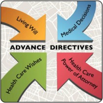
Advance Care Planning
Advance care planning allows patients to record their wishes before they lose the capacity to express them. This might include preferences about resuscitation, hospital admission, or place of death. In Uganda, formal advance directives are rare, but informal conversations are valuable. Ask patients early what they would want if they became too sick to speak. Talk to the family. Write it down. This is an act of respect that prevents terrible conflicts later.
Being the Patient's Advocate
An advocate speaks on behalf of another person, as that person perceives their own interests. In palliative care, patients are often vulnerable. They may be too weak to speak, too confused to understand, or too intimidated by family or medical authority to express their true wishes. The nurse is often the person who spends the most time at the bedside. You see things the doctor does not see. You hear things in the quiet hours.
Being an advocate means:
- Speaking up when a patient's pain is undertreated
- Ensuring the patient's cultural and spiritual needs are respected
- Protecting a patient from unwanted visitors
- Challenging a family member who is making decisions that clearly contradict the patient's expressed wishes
- Ensuring a dying patient is not subjected to painful or futile procedures simply because the family cannot let go
Advocacy requires courage. It may make you unpopular. But your loyalty is to the patient, not to the easiest path.
Ethical Issues at the End of Life
Euthanasia and Assisted Death
Euthanasia is derived from Greek words meaning "easy death" or "dying well." In modern usage, it generally refers to the intentional ending of a patient's life at their request, or helping them to end their own life. The terminology matters.
- Hastened death refers to any act that accelerates the dying process in response to suffering.
- Assisted death involves aiding a person who wants to die prematurely, either through counselling or by providing a lethal substance.
- Assisted suicide involves self-killing with the assistance of another person.
- Physician-assisted suicide (PAS) is assisted suicide carried out specifically by a physician or healthcare provider.
In Uganda, as in most of Africa, all forms of euthanasia and assisted suicide are illegal. As a nurse, you cannot and must not participate in ending a patient's life. But you must understand why patients ask for it, and you must know how to respond.
Why Patients Ask for Hastened Death
Patients do not ask to die because they want death. They ask because they see no other escape from suffering. Common reasons include:
- Feeling like a burden to their family
- Loss of control over their body and circumstances
- Lack of social support and loneliness
- Perceived loss of dignity, especially with incontinence or dependence
- Poor quality of life defined by unrelenting symptoms
- Lack of meaning or purpose
- Fear of a prolonged, undignified death
When a patient says "Please help me die," they are usually saying "I am suffering, and I do not believe anyone can help me." That is a call for better palliative care, not for death.
The Nurse's Response to a Request for Hastened Death
When a patient asks you to help them die, your response must be compassionate, thorough, and principled.
First, ensure you understand what they are asking. Do they want you to give them an overdose? Do they want you to stop their food and fluids? Or are they simply expressing despair? Clarify without judgment.
Second, acknowledge and validate their suffering. Do not brush off the request. Say: "I hear that you are in terrible distress. I cannot help you to die, but I can help you to live without so much suffering. Will you let me try?"
Third, assess comprehensively. Uncontrolled symptoms are often the real driver. Ask about pain, breathlessness, nausea, fatigue, constipation, insomnia, and itching. Review their medication. Are they receiving adequate analgesia? Are side effects making them miserable? Are they depressed? Depression can make any situation feel hopeless, and treating the depression may remove the desire for death.
Fourth, explore their past experiences with death. A patient who watched a parent die in agony may fear the same fate. Understanding this fear allows you to address it directly.
Fifth, identify a trusted team member. Find someone who can build a strong rapport with the patient, understand their cultural background, and facilitate communication. This might be a nurse, a counsellor, a chaplain, or a social worker.
Sixth, understand the nature of their suffering. Suffering is not just physical. It is existential, social, and spiritual. A patient may have no pain but feel their life has no meaning. Addressing meaning may require a chaplain, an elder, or simply a listener who helps them review their life and legacy.
Seventh, treat symptoms aggressively. Refer to palliative care specialists, anaesthetists for complex pain, psychiatrists for depression, and spiritual care providers for existential distress. When suffering is properly managed, requests for hastened death usually disappear.
The Ethical Controversy
The debate over assisted dying continues worldwide. Advocates argue that terminally ill patients have the right to die with dignity, and that denying this right forces people to suffer needlessly. Opponents argue that life is sacred, that the Hippocratic Oath forbids killing, and that legalising assisted suicide creates a slippery slope where vulnerable people feel pressured to die.
As a nurse in Uganda, the law is clear. But ethics is broader than law. Your ethical duty is to ensure that no patient requests death because their suffering has been ignored. Palliative care is the ethical answer to euthanasia. When you restore dignity, control, comfort, and meaning, you give patients a reason to live their remaining time fully.
Other Ethical Issues
High-Tech Medicine and Resuscitation
In resource-rich settings, patients can be kept alive on ventilators, feeding tubes, and dialysis machines. In Uganda, such technology is rarely available, but the ethical questions still arise. Should a patient be transferred to a hospital for ventilation? Should a dying patient be given antibiotics for a mild infection that will not change the outcome?
The principle of non-maleficence reminds us that interventions that prolong suffering without benefit are harmful. The principle of autonomy reminds us to ask the patient what they want. A do-not-resuscitate (DNR) order, discussed openly with the patient and family, can prevent a traumatic and futile attempt at resuscitation that breaks ribs and destroys dignity in the final moments of life.
Resource Allocation
When there is one oxygen cylinder and two patients need it, who gets it? There is no perfect answer. Justice demands that you decide based on medical need and likelihood of benefit, not on who can pay more or who is related to the hospital administrator. Document your reasoning. Consult colleagues. Be transparent. Even when resources are scarce, fairness must be visible.
Mnemonics and Exam Tips
Mnemonic for the Four Pillars: "BANJ"
| Letter |
Pillar |
Core Question |
| B |
Beneficence |
How does this help the patient? |
| A |
Autonomy |
What does the patient want? |
| N |
Non-maleficence |
Could this cause harm? |
| J |
Justice |
Is this fair to everyone? |
Mnemonic for Responding to Requests for Hastened Death: "UNDERSTAND"
| Letter |
Action |
| Understand |
Clarify exactly what the patient is asking |
| Name |
Acknowledge and validate their suffering |
| Depression |
Screen for depression as a driver |
| Explore |
Ask about past experiences with death |
| Rapport |
Identify a trusted team member |
| Suffering |
Assess physical, psychological, social, and spiritual dimensions |
| Treat |
Aggressively treat all symptoms and distress |
| Ask |
Involve palliative care specialists, psychiatrists, chaplains |
| No |
State clearly but compassionately that you cannot assist in ending life |
| Dignity |
Restore meaning, control, and quality of life |
Exam-Style Questions
Q1: A patient with terminal cancer refuses further chemotherapy. His family insists he must continue. What is your ethical duty?
Answer: If the patient is competent, his autonomy must be respected. His refusal of treatment is his right, even if the family disagrees. Your role is to ensure he understands the consequences, to support his decision, and to advocate for his wishes with the family. You may also explore whether the family understands the prognosis, as their insistence may come from grief and hope rather than malice.
Q2: A patient confides that he is HIV positive and asks you not to tell his wife. You know they are sexually active and she is at risk. What do you do?
Answer: Your first duty is to counsel the patient about the importance of disclosure, offer to support him through the conversation, and explore safer sex options. If he absolutely refuses to disclose and his wife remains at significant risk, this creates a conflict between confidentiality and protection of others. Seek senior guidance. In some jurisdictions, disclosure to a partner at risk may be legally justified, but it should never be done without careful ethical review and documentation.
Q3: James, the twelve-year-old boy with Kaposi's sarcoma, asks if he will get better. His mother has forbidden you from telling him the truth. How do you respond?
Answer: This is a conflict between the mother's protective authority and James's emerging autonomy. First, assess what James already knows. Children often sense the truth. Second, speak with the mother gently about the harm of secrecy, loneliness, and unspoken fear. Third, if appropriate and with the mother's eventual agreement, answer James with compassionate honesty appropriate to his age: that his body is very sick, that the focus is on comfort, and that he can ask anything. If the mother absolutely refuses and James is not in immediate danger, you may need to accept her authority while continuing to advocate for openness.
Q4: A dying patient asks you to give him an overdose of morphine to end his suffering. What is your response?
Answer: You must refuse clearly but compassionately. Explain that you cannot do this, but that you can do everything possible to relieve his suffering. Then conduct a comprehensive assessment of his physical, psychological, social, and spiritual distress. Treat his symptoms aggressively. Involve the palliative care team, a psychiatrist if depression is suspected, and spiritual support. When suffering is properly addressed, the request for death usually resolves.
Q5: What is the difference between euthanasia and palliative care?
Answer: Euthanasia intentionally ends life to relieve suffering. Palliative care relieves suffering so that life can continue with dignity and comfort until natural death occurs. Palliative care never intends to hasten death, though some treatments (like high-dose morphine) may have the incidental effect of shortening life. The intention is always comfort, not killing.
Summary: Key Nursing Points
- The four pillars of ethics are beneficence, non-maleficence, autonomy, and justice. They guide but do not dictate decisions.
- Beneficence in palliative care means promoting comfort and dignity, not futile cure.
- Non-maleficence means avoiding harm, including the harm of unnecessary procedures and dishonesty.
- Autonomy means respecting the patient's right to know, choose, and refuse. It includes the right to delegate decisions.
- Justice means fair resource allocation and challenging discrimination.
- Confidentiality is essential, especially with HIV, but may be breached in rare cases where another life is at serious risk.
- Truthfulness is a process, not a single announcement. Be honest without being brutal.
- Children deserve age-appropriate honesty. Secrecy creates loneliness and fear.
- Advocacy means speaking for the patient's interests, even when it is uncomfortable.
- Euthanasia and assisted suicide are illegal in Uganda. The ethical response to a request for hastened death is to assess and treat the underlying suffering comprehensively.
- Palliative care is the answer to euthanasia. When suffering is relieved and dignity restored, patients usually no longer want to die.
- There is no single right answer in every ethical dilemma. Use the pillars, consult colleagues, document your reasoning, and act with compassion.
References
- Beauchamp, T. L., & Childress, J. F. (2019). Principles of Biomedical Ethics (8th ed.). Oxford University Press.
- International Council of Nurses (ICN). (2021). The ICN Code of Ethics for Nurses. Geneva, Switzerland.
- African Palliative Care Association (APCA). (2010). Guidelines for Providing Palliative Care to People Living with HIV/AIDS. Kampala, Uganda.
- World Health Organization (WHO). (2020). Integrating Palliative Care and Symptom Relief into Primary Health Care. Geneva.