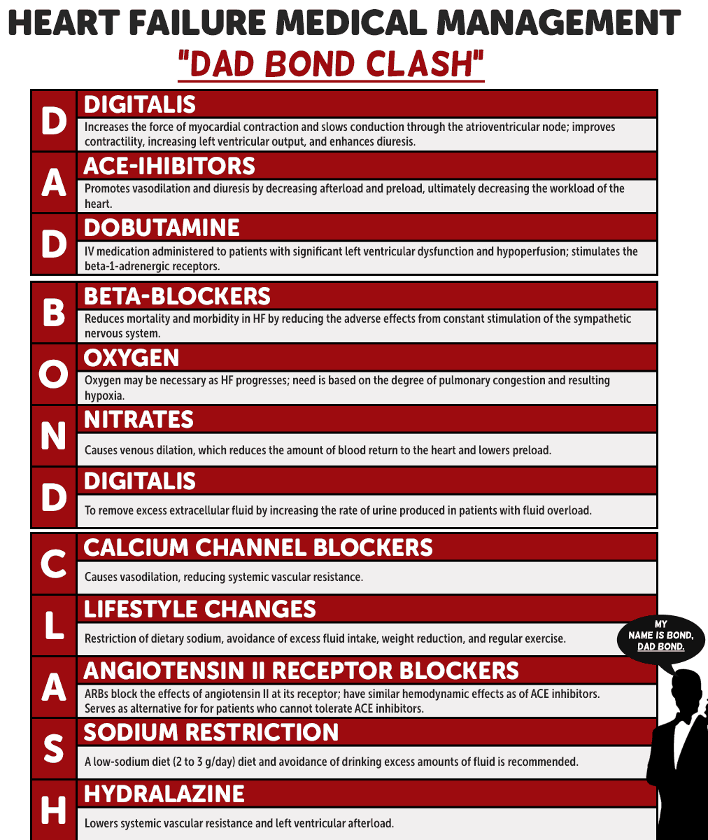
COAGULATION DISORDERS
COAGULATION DISORDERS
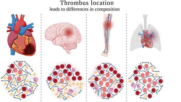
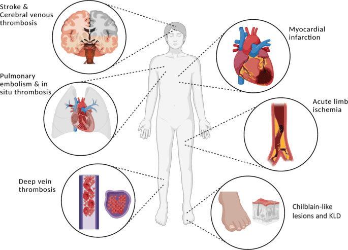
A coagulation disorder is a medical condition characterised by excessive bleeding occurring as a result of deficiency of any of the essential clotting factors. Coagulations disorders are conditions that affect the blood’s clotting activities. Hemophilia, Von Willebrand disease, clotting factor deficiencies, hypercoagulable states and deep venous thrombosis are all coagulations disorders. Hemophilia and Von Willebrand disease are among the best known.
Normal mechanism of blood clotting
- Damage or injury to the endothelium will initiate a cascade of events in an attempt to control bleeding.
- Disruption of the endothelium will first cause local vasoconstriction to occur, limiting blood flow to the area.
- Primary hemostasis initiates by platelets with the release of von Willebrand factor (vWF), a large plasma glycoprotein made and stored in endothelial cells and megakaryocytes.
- Platelets and vWF will combine to form a plug at the site of injury. Circulating vWF continues to bind with collagen and Factor VIII as well as other endothelial substances, allowing the platelet plug to adhere to the area of injury.
- Through activation of the clotting cascade and secondary hemostasis, this initial platelet plug will get reinforced to a sturdy fibrin clot.
- The clotting cascade operates through a dual process system in which the various clotting factors become activated with the result being the formation of a fibrin strand or clot at the site of tissue injury.
NB: A deficiency of any of the essential clotting factors will result in difficulty forming a fibrin clot, and excessive bleeding can occur.
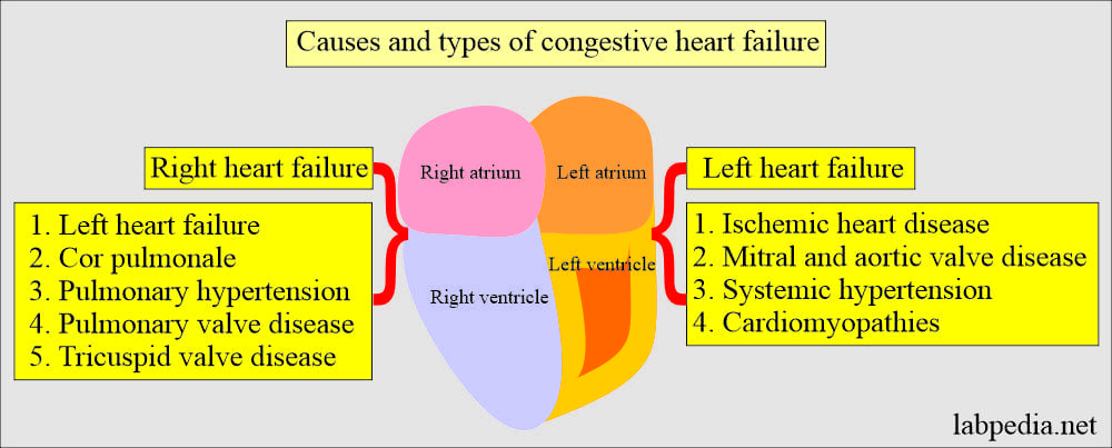
Types of coagulation disorders
Bleeding disorders fall into two main categories:
1. Inherited coagulation disorders: Hereditary bleeding disorders are due to the absence or deficiency of specific clotting proteins which act as pro-coagulants through precise interactions in the clotting cascade. The three most common are:- a. Hemophilia A (Factor VIII deficiency): Hemophilia A is an X-linked recessive genetic disorder affecting 1 in 5000 males making it the most common congenital coagulopathy.
- b. Hemophilia B (Factor IX deficiency): Hemophilia B is an X-linked genetic coagulopathy affecting 1 in 30000 male births.
- c. Von Willebrand disease: It is characterised by excessive bleeding as a result of deficiency of von-Willebrand factor hence causing failure of platelet plug formation.
- a. Liver disease
- b. Vitamin k deficiency
- c. Disseminated intravascular coagulation
Causes of Coagulation disorders
The major causes of acquired coagulation disorders are:
- Vitamin K deficiency
- Liver disease
- Disseminated intravascular coagulation (DIC)
- Development of circulating anticoagulants
- Severe liver disease (e.g. cirrhosis, fulminant hepatitis, acute fatty liver of pregnancy) may disturb hemostasis by impairing clotting factor synthesis. Because all coagulation factors are made in the liver (by hepatocytes and endothelial cells), both the prothrombin time (PT) and partial thromboplastin time (PTT) are prolonged in severe liver disorders. (PT results are typically reported as INR [international normalized ratio].)
The most common hereditary disorder of hemostasis is:
- a. Von Willebrand disease (VWD)
- b. The hemophilias
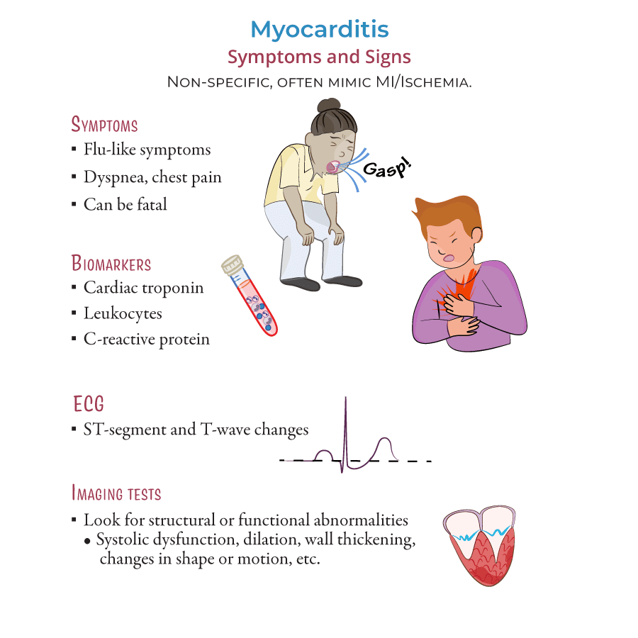
Clinical manifestations
Hemophilia:
- While mild hemophilia may only present after a traumatic injury or surgery.
- Those with a moderate to severe form of the disease may exhibit hallmark characteristics such as:
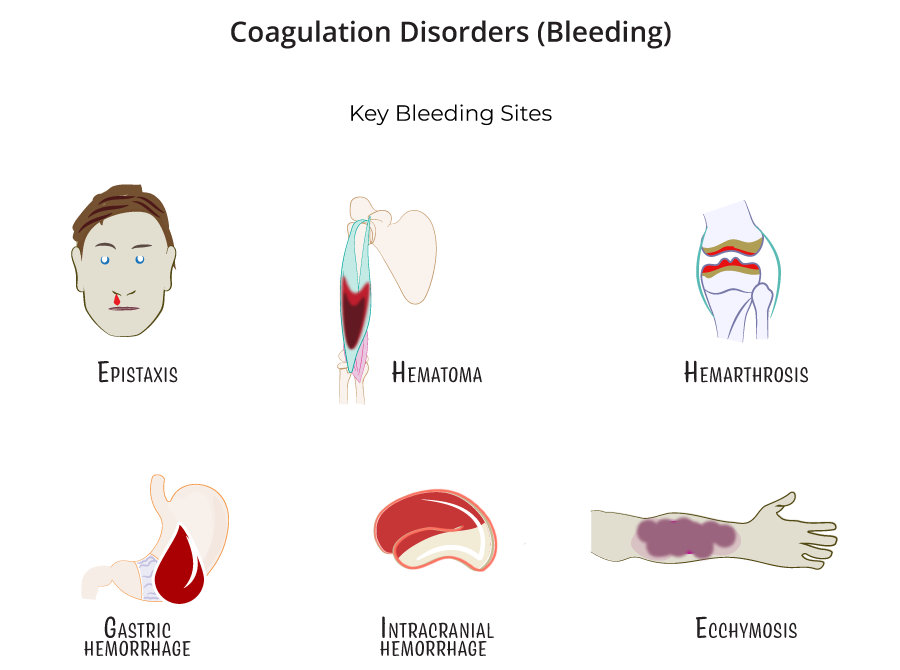
- a) Mucosal or gingival bleeding
- b) Easy bruising
- c) Hematoma formation
- d) Hemarthrosis: is bleeding into joints, particularly in the ankles.
- e) Bleeding into muscle tissue from minor traumas can result in anemia and
- f) Compression of vital structures and nerves leading to compartment syndrome.
- g) Intracranial bleeds
- Hemophilia can present in infancy with cephalohematoma formation after vaginal birth and with significant bleeding after circumcisions.
Von Willebrand Disease
- Von Willebrand disease can exhibit clinical signs and symptoms starting in childhood with a history of easy bruising and bleeding.
- While patients with a very mild version of the disease may not have clinical symptoms at all, patients with vWF that is qualitative or quantitatively low may present with a predisposition to mucosal bleeding and episodic epistaxis.
- Women with von Willebrand disease may have significant menorrhagia which is often a presenting sign of the illness, precipitating a workup and eventual diagnosis.
- These patients can also go unrecognized until undergoing major surgery or experiencing a traumatic injury.
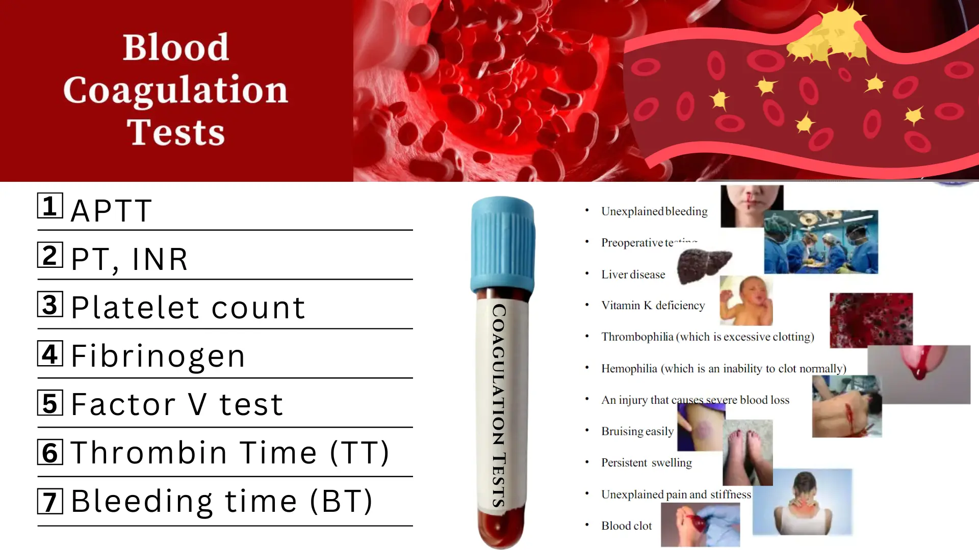
Test and Diagnosis for coagulation disorders
Hemophilia
- Chromogenic assay: This assay is considered by some to be more accurate, as it measures the level of plasma factor VIII activity but it is less widely available in clinical laboratories in the United States.
- Laboratory studies: Laboratory studies for suspected hemophilia include a complete blood cell count, coagulation studies, and a factor VIII (FVIII) assay.
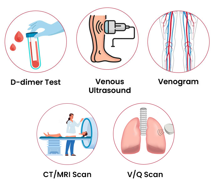
- CT scans: Head CT scans without contrast are used to assess for spontaneous or traumatic intracranial hemorrhage.
- MRI: Perform magnetic resonance imaging (MRI) on the head and spinal column for further assessment of spontaneous or traumatic hemorrhage; MRI is also useful in the evaluation of the cartilage, synovium, and joint space.
- Ultrasonography: Ultrasonography is useful in the evaluation of joints affected by acute or chronic effusions.
- Testing for inhibitors: Laboratory confirmation of a FVIII inhibitor is clinically important when a bleeding episode is not controlled despite infusion of adequate amounts of factor concentrate.
- Carrier testing: Screening for carrier status can be performed by measuring the ratio of FVIII coagulant activity to the concentration of von Willebrand factor (vWF) antigen; a ratio that is less than 0.7 suggests carrier status.
- Radiography: Radiography for joint assessment is of limited value in acute hemarthrosis; evidence of chronic degenerative joint disease may be visible on radiographs in patients who have been untreated or inadequately treated or in those with recurrent joint hemorrhages.
Management
Medical Management - Hemophilia
The treatment of hemophilia may involve prophylaxis, management of bleeding episodes, treatment of factor VIII (FVIII) inhibitors, and treatment and rehabilitation of hemophilia synovitis.
Pre-hospital care
- Rapid transport to definitive care is the mainstay of prehospital care; prehospital care providers should apply aggressive hemostatic techniques, assist patients capable of self-administered factor therapy, and gather focused historical data if the patient is unable to communicate.
- Emergency department care. Use aggressive hemostatic techniques; correct coagulopathy immediately; include a diagnostic workup for hemorrhage, but never delay indicated coagulation correction pending diagnostic testing; acute joint bleeding and expanding, large hematomas require adequate factor replacement for a prolonged period until the bleed begins to resolve, as evidenced by clinical and/or objective methods; life-threatening bleeding episodes are generally initially treated with FVIII levels of approximately 100%, until the clinical situation warrants a gradual reduction in dosage.
- Factor VIII and FIX concentrates. Various FVIII and FIX concentrates are available to treat hemophilia A and B; besides improved hemostasis, continuous infusion decreases the amount of factor used, which can result in significant savings; obtain factor level assays daily before each infusion to establish a stable pattern of replacement regarding the dose and frequency of administration.
- Desmopressin. Desmopressin vasopressin analog, or 1-deamino-8-D-arginine vasopressin (DDAVP), is considered the treatment of choice for mild and moderate hemophilia A; DDAVP stimulates a transient increase in plasma FVIII levels; DDAVP may result in sufficient hemostasis to stop a bleeding episode or to prepare patients for dental and minor surgical procedures.
- Management of bleeding. Immobilization of the affected limb and the application of ice packs are helpful in diminishing swelling and pain; early infusion upon the recognition of initial symptoms of a joint bleed may often eliminate the need for a second infusion by preventing the inflammatory reaction in the joint; prompt and adequate replacement therapy is the key to preventing long-term complications.
- Treatment of patients with inhibitors. Inhibitors are antibodies that neutralize factor VIII (FVIII) and can render replacement therapy ineffective; the treatment of patients with inhibitors of FVIII is difficult; assuming no anamnestic response, low-titer inhibitors (ie, concentrations below 5 Bethesda units [BU]) occasionally can be overcome with high doses of factor VIII; there is no established treatment for bleeding episodes in patients with high-titer inhibitors.
- Prophylactic factor infusions. The main goal of prophylactic treatment is to prevent bleeding symptoms and organ damage, in particular to joints; in December 2013, the US Food and Drug Administration (FDA) expanded the indication for anti-inhibitor coagulant complex (Feiba NF) to include routine prophylaxis in patients with hemophilia A or B who have developed inhibitors; approval was based on data from a pivotal phase III study in which a prophylactic regimen resulted in a 72% reduction in median annual bleed rate compared with on-demand treatment.
- Pain management. Hemophilic chronic arthropathy is associated with pain; narcotic agents have been used, but frequent use of these drugs may result in addiction; nonsteroidal anti-inflammatory drugs may be used instead because their effects on platelet function are reversible and because these drugs can be effective in managing acute and chronic arthritic pain; avoid aspirin because of its irreversible effect on platelet function.
- Activity. Generally, individuals with severe hemophilia should avoid high-impact contact sports and other activities with a significant risk of trauma; however, mounting evidence suggests that appropriate physical activity improves overall conditioning, reduces injury rate and severity, and improves psychosocial functioning.
- Gene therapy. With the cloning of FVIII and advances in molecular technologies, the possibility of a cure for hemophilia with gene therapy was conceived; ex vivo gene therapy, in which cells to be transplanted are genetically modified to secrete factor VIII and then are reimplanted into the recipient; in vivo gene therapy, in which a vector (typically a virus altered to include FVIII DNA) is directly injected into the patient; and nonautologous gene therapy, in which cells modified to secrete FVIII are packaged in immunoprotected devices and implanted into recipients.
- Radio-synovectomy. In patients who develop synovitis from joint bleeds, intra-articular injection of radioisotopes to ablate the synovium (radiosynovectomy) can be used to decrease bleeding, slow progression of cartilage and bone damage, and prevent arthropathy.
Pharmacologic Management - Hemophilia
Medications of choice for patients with hemophilia are:
- Factor VIII. Factor VIII (FVIII) is the treatment of choice for acute or potential hemorrhage in hemophilia A; recombinant FVIII concentrate is generally the preferred source of factor VIII; prophylactic administration of FVIII is often recommended for pediatric patients with severe disease.
- Anti-fibrinolytic agents. Antifibrinolytic agents, such as aminocaproic acid and tranexamic acid, are especially useful for oral mucosal bleeds but are contraindicated as initial therapies for hemophilia-related hematuria originating from the upper urinary tract because they can cause obstructive uropathy or anuria.
- Factor IX. Factor IX is the treatment of choice for acute hemorrhage or presumed acute hemorrhage in hemophilia B. Recombinant factor IX is the preferred source for replacement therapy.
- Coagulation factor VIIa. These agents can activate coagulation factor X to factor Xa as well as coagulation factor IX to IXa.
- Coagulation factors. FVIII concentrates replace deficient FVIII in patients with hemophilia A, with the goal of achieving a normal hematologic response to hemorrhage or preventing hemorrhage; recombinant products should be used initially and subsequently in all newly diagnosed cases of hemophilia that require factor replacement; agents that bypass FVIII activity in the clotting cascade (eg, activated FVII) are used in patients with FVIII inhibitors.
- Anti-hemophilic agents. These agents are used to control bleeding in hemophilia B or FIX deficiency and to prevent and/or control bleeding in patients with hemophilia A and inhibitors to FVIII.
- Monoclonal antibodies. Monoclonal antibodies are used to bind to one specific substance in the body (eg, molecules, antigens); this binding is very versatile and can mimic, block, or cause changes to enact precise mechanisms (eg, bridging molecules, replacing or activating enzymes or cofactors, immune system stimulation).
- Vasopressin-related. Desmopressin transiently increases the FVIII plasma level in patients with mild hemophilia A.
Management - Von Willebrand disease
Treatment depends on the type of VWD and should be decided by a hematologist. Options include the following:
- Hormonal treatments such as oral contraceptives and some intrauterine devices are highly effective in controlling menorrhagia. In fact, 88% of women with VWD report improvement in bleeding symptoms when treated only with oral contraceptives.
- Desmopressin (DDAVP) is effective in most patients with type 1 VWD and some patients with type 2. Recovery testing must be done to determine its effectiveness. During a recovery test, a blood sample is obtained before the medication is given and 30 to 60 minutes after administration. This test helps determine if the medication increases the patient’s factor levels enough to prevent or stop bleeding.
- Replacement factor made from plasma-derived concentrates can be used in any patient with VWD, but must be used in all patients with type 3 and in some patients with type 2. Replacement factor is also used when patients don’t respond to DDAVP.
- Anti-fibrinolytics such as aminocaproic acid and tranexamic acid are used in conjunction with factor or DDAVP to treat bleeding. Anti-fibrinolytics stabilize a clot by preventing it from breaking down too early, which would cause bleeding. Without anti-fibrinolytics, bleeding may occur several days or weeks after a procedure involving mucosal tissue. Antifibrinolytics are effective in treating mucosal bleeding such as with dental surgery, menstrual bleeding, nosebleeds, and gastrointestinal bleeding.
Nursing intervention/management
- Relieve pain. Immobilize joints and apply elastic bandages to the affected joint if indicated; elevate affected and apply a cold compress to active bleeding sites, but must be used cautiously in young children to prevent skin breakdown.
- Maintain optimal physical mobility. Provide gentle, passive ROM exercise when the child’s condition is stable; educate on preventive measures, such as the application of protective gear and the administration of factor products; and refer for physical therapy, occupational therapy, and orthopedic consultations, as required.
- Assist in the coping of the family. Encourage family members to verbalize problem areas and develop solutions on their own; encourage family members to express feelings, such as how they deal with the chronic needs of a family member and coping patterns that help or hinder adjustment to the problems.
- Prevent bleeding. Monitor hemoglobin and hematocrit levels; assess for inhibitor antibody to factor VIII; anticipate or instruct in the need for prophylactic treatment before high-risk situations, such as invasive diagnostic or surgical procedures, or dental work; and provide replacement therapy of deficient clotting factors.
- Prevent injury. Utilize appropriate toys (soft, not pointed or small sharp objects); for infants, may need to use padded bed rail sides on crib; avoid rectal temperatures; provide appropriate oral hygiene (use of a water irrigating device; use of a soft toothbrush or softening the toothbrush with warm water before brushing; use of sponge-tipped toothbrush); and avoid contact sports such as football, soccer, ice hockey, karate.
Complications
- Anemia
- Arthritis
COAGULATION DISORDERS Read More »