Nurses Revision

Pharmacology

nursing exam Nursing Management nursing exam

Nursing Exam Question Approach

Nursing Exam Question Approach

A comprehensive guide on how to interpret and answer UNMEB question types: EXPLAIN, OUTLINE, DESCRIBE, MENTION, IDENTIFY, STATE, LIST, WHAT, and GIVE.

This guide explores specific nursing interventions, considerations, concerns, and issues frequently tested in professional medical exams.
EX

The EXPLAIN Approach

In Simple Terms: "Explain" means to give details and reasons. You need to show *how* or *why* something happens, not just what it is.
1
Understand the question: Carefully read and identify the main concept. Pay attention to specific instructions.
2
Organize your response: Create a mental map. Start with a concise introduction, context, and clear thesis.
3
Provide thorough explanation: Elaborate using clear language. Use nursing terminology and case studies.

Simulated Examination Sheet

Qn: Explain the pathophysiology of diabetes mellitus and its effects on the body.

Diabetes mellitus is a chronic metabolic disorder characterized by high blood glucose levels due to impaired insulin secretion, insulin action, or both. The pathophysiology of diabetes involves multiple factors that contribute to the development and progression of the disease. Firstly, in type 1 diabetes, an autoimmune process leads to the destruction of insulin-producing beta cells in the pancreas. This results in a deficiency of insulin and requires external insulin administration. On the other hand, type 2 diabetes is primarily characterized by insulin resistance, where the body’s cells become less responsive to insulin. Insulin is a hormone produced by the beta cells of the pancreas, and its main function is to regulate glucose metabolism. In diabetes, the lack of insulin or the body’s inability to use it effectively leads to hyperglycemia. Persistently high blood glucose levels can have detrimental effects on various organs and systems in the body. The effects of diabetes on the body are many. It can lead to macrovascular complications, such as cardiovascular disease, stroke, and peripheral vascular disease. Also, microvascular complications may arise, affecting small blood vessels in the eyes, kidneys, and nerves. Diabetes can also increase the risk of infections, slow wound healing, and cause diabetic neuropathy and nephropathy
OU

The OUTLINE Approach

In Simple Terms: "Outline" means to create a structured summary. Use main headings and sub-points to show parts in an organized way.
1
Analyze the question: Identify main components that need to be outlined.
2
Organize your response: Identify main headings and arrange them in a coherent order.
3
Provide detailed information: Use concise and informative language under each heading. JUMP A LINE, UHPAB HAS VERY MANY BOOKLETS

Simulated Examination Sheet

Qn: Outline the steps involved in the nursing process.

1. Assessment: Gather relevant patient data, including physical, psychological, social, and cultural aspects. Perform a comprehensive health history and physical examination. Utilize assessment tools and techniques to collect objective and subjective data. Document and organize the collected data systematically.

2. Diagnosis: Analyze the assessment data to identify health problems, risks, or potential complications. Formulate nursing diagnoses based on the identified issues. Ensure that the diagnoses are accurate, concise, and specific.

Collaborate with other healthcare professionals when necessary. 3. Planning: Establish patient-centered goals and outcomes in collaboration with the patient.

Develop a nursing care plan that includes evidence-based interventions and strategies. Prioritize nursing actions based on the urgency and importance of each goal. Ensure that the care plan is feasible, realistic, and adaptable. 4. Implementation: Execute the planned nursing interventions effectively and efficiently.

Provide safe and compassionate care while considering the patient's preferences. Document the implementation process and any modifications made. Collaborate with the interdisciplinary healthcare team to deliver comprehensive care.

5. Evaluation: Assess the patient's response to the nursing interventions and the achievement of goals. Compare the actual outcomes with the expected outcomes. Modify the care plan if needed based on the evaluation findings. Document the evaluation results and communicate them to the healthcare team.
DE

The DESCRIBE Approach

In Simple Terms: "Describe" means to paint a picture with words. Give a detailed account of characteristics or features. Describe usually likes STEPS in order, even using IMAGES where applicable!
1
Understand the question: Identify the main topic that needs characterization.
2
Provide comprehensive description: Offer thorough details, features, or components.
3
Use terminology: Accurately describe concepts to demonstrate knowledge.

Simulated Examination Sheet

Qn: Describe the stages of wound healing.

1. Hemostasis: This initial stage begins immediately after the injury occurs. Blood vessels constrict to reduce blood flow and prevent excessive bleeding. Platelets aggregate to form a temporary clot. The clotting process releases various growth factors and cytokines, initiating the subsequent stages of healing.

2. Inflammatory phase: This phase typically lasts for 2-3 days. Inflammation occurs as a response to tissue injury. Vasodilation and increased vascular permeability allow immune cells to migrate to the wound site. Neutrophils arrive first to eliminate debris and prevent infection. Macrophages then remove dead tissue and release additional growth factors to stimulate healing.

3. Proliferative phase: This phase generally occurs between days 3 and 20. New blood vessels form to supply oxygen and nutrients to the wound. Fibroblasts produce collagen, which provides structural support for wound healing. Epithelial cells migrate from the wound edges to resurface the wound. Granulation tissue forms, consisting of new blood vessels, fibroblasts, and extracellular matrix.

4. Maturation phase: This final phase can last for several months to years. Collagen fibers reorganize and remodel, increasing the wound's tensile strength. Scar tissue forms, but it may not possess the same strength and flexibility as the original tissue. The scar gradually becomes more refined and fades over time.
ST

MENTION / IDENTIFY / STATE

In Simple Terms: These words mean "give a short, direct answer." Just name the facts without extra explanation.
1
Identify facts: Read and identify the specific information required.
2
Direct response: Offer a concise response. Avoid unnecessary elaboration.

Simulated Examination Sheet

Qn: State the types of delusions.

  • Grandiose delusions; the patient believes s/he is somebody great /important ,knowledgeable or powerful contrary to the social cultural ,religious background and experiences.

  • Delusion of guilty and worthlessness; the patient believes s/he is not worth to live even though there’s nothing to justify this belief.

  • Delusions of jealousy; the patient believes that spouse/partner is being unfaithful even when there is no evidence to suggest so.

  • Delusion of persecution: the patient believes they’re being deliberately wronged, conspired or harmed by another person or agency even when there’s no evidence to suggest so.

  • Religious delusions; the individual believes he or she has a special link with God that is out keeping with people of the same religious belief.

  • Delusions of control, influence or phenomenon; these are three types; belief that the person performs activities as a result of an extreme force.
  • LI

    The LIST Approach

    In Simple Terms: "List" means to present points one after another, usually with a short description for each.
    1
    Identify factors: Carefully identify the elements that need to be listed.
    2
    Organize: Present items in a logical order using bullet points.

    Simulated Examination Sheet

    Qn: List the risk factors for cardiovascular disease.

    - Hypertension: increases strain on heart.
    - Smoking: damages blood vessels.
    - Obesity: increases risk of diabetes.
    - Sedentary lifestyle: contributes to obesity.
    WH

    The WHAT Approach

    In Simple Terms: "What" asks for a definition. Give a clear, simple explanation of the term or concept.
    1
    Identify term: Pinpoint the specific procedure or concept to define.
    2
    Clear explanation: Offer a concise definition using simple language.

    Simulated Examination Sheet

    Qn: What is sepsis?

    Sepsis is a potentially life-threatening condition that occurs when the body’s response to an infection becomes unregulated, leading to widespread inflammation and organ dysfunction.

    Nursing Exam Question Approach Read More »

    MOOD STABILIZERS

    Mood Stabilizers

    Mood Stabilizers

    Mood stabilizers are psychotropic drugs which are used in controlling mood disorders.

    Indications of Mood Stabilizers

    The major indications are the following:

    • Bipolar affective disorder with frequent fluctuation of mood at short intervals.
    • Can also be used as maintenance drug in mood disorder.

    However, patients with acute-phases of mania or depression should be treated first with anti-psychotics or anti-depressants respectively.

    MODE OF ACTION

    Mood stabilizers are a class of psychiatric medications that are primarily used to treat bipolar disorder, but can also be used for other conditions that involve mood swings or instability. The exact mode of action of mood stabilizers is not fully understood, but there are several theories that attempt to explain how they work.

    One theory is that mood stabilizers work by regulating the levels of neurotransmitters in the brain, particularly serotonin, dopamine, and norepinephrine. These neurotransmitters are involved in the regulation of mood, and imbalances in their levels can lead to mood swings and instability. Mood stabilizers may help to normalize these imbalances, which can reduce symptoms of bipolar disorder and other mood disorders.

    Second theory is that mood stabilizers work by modulating the activity of certain ion channels in the brain, particularly those that regulate the flow of sodium and calcium ions. These ion channels are involved in the regulation of neuronal excitability, and abnormalities in their activity can lead to mood swings and other symptoms of bipolar disorder. Mood stabilizers may help to normalize the activity of these ion channels, which can reduce symptoms of bipolar disorder and other mood disorders.

    Third theory is that mood stabilizers work by affecting the structure and function of certain brain regions that are involved in the regulation of mood, such as the prefrontal cortex and the amygdala. These brain regions are thought to play a key role in the regulation of emotional processing and response, and abnormalities in their structure or function can lead to mood instability and other symptoms of bipolar disorder. Mood stabilizers may help to normalize the structure and function of these brain regions, which can reduce symptoms of bipolar disorder and other mood disorders.

    Pharmacodynamics of Mood Stabilizers

    1. Changes in heart rate and blood pressure: Some mood stabilizers, such as lithium, can affect the cardiovascular system and cause changes in heart rate and blood pressure. This is why people taking these medications may need to have regular check-ups to monitor their heart health.

    2. Changes in electrolyte balance: Some mood stabilizers, such as lithium, can affect the balance of electrolytes in the body, particularly sodium and potassium. This can lead to side effects such as nausea, vomiting, diarrhea, and muscle weakness. When cerebral sodium concentration is reduced, there is control of mania which reduces excitements.

    3. Changes in kidney function: Lithium is primarily excreted by the kidneys, and long-term use of this medication can sometimes lead to kidney damage or impairment. Regular kidney function tests may be required for people taking lithium.

    4. Changes in thyroid function: Some mood stabilizers, such as lithium and valproic acid, can affect thyroid function and lead to hypothyroidism (an underactive thyroid). This can cause symptoms such as fatigue, weight gain, and cold intolerance.

    5. Changes in liver function: Some mood stabilizers, such as valproic acid, can affect liver function and lead to elevated liver enzymes. Regular liver function tests may be required for people taking valproic acid.

    6. Changes in metabolism: Some mood stabilizers, such as valproic acid, can affect metabolism and lead to weight gain. 

    LITHIUM CARBONATE (ESKALITH)

    Lithium carbonate is a medication that is commonly used as a mood stabilizer to treat bipolar disorder. It is a naturally occurring element that is found in small amounts in the body, and it works by affecting the levels of certain neurotransmitters in the brain, particularly serotonin and norepinephrine.

    Indications of Lithium Carbonate
    • In bipolar affective where there are mood swings.
    • Prophylaxis therapy (preventive measure which prolongs the re-occurrence).
    • In aggressive disorders i.e. in personality disorders.
    • Schizoaffective disorder. 
    • Alcoholism.
     Contra indications of Lithium Carbonate.
    • Allergies: People who are allergic to lithium carbonate or any of its ingredients should not take this medication.

    • Kidney disease: Lithium is primarily excreted by the kidneys, and people with kidney disease may be at risk of toxicity if they take lithium. Lithium is contraindicated in people with severe kidney disease or end-stage renal disease.

    • Dehydration: Lithium can affect the body’s electrolyte balance, particularly sodium, and dehydration can increase the risk of toxicity. People who are dehydrated or at risk of dehydration should not take lithium.

    • Cardiovascular disease: Lithium can affect the cardiovascular system, and people with a history of heart disease or other cardiovascular conditions may be at increased risk of complications if they take lithium.

    • Pregnancy and breastfeeding: Lithium can cross the placenta and pass into breast milk, and it may be harmful to a developing fetus or nursing infant. Lithium is contraindicated during pregnancy and breastfeeding, unless the benefits outweigh the risks.

    • Low sodium levels: Lithium can cause or worsen hyponatremia (low sodium levels in the blood), which can be life-threatening. Lithium is contraindicated in people with severe or uncontrolled hyponatremia.

    • Seizures: Lithium can lower the seizure threshold, and it is contraindicated in people with a history of seizures or epilepsy.

    • Thyroid disease: Lithium can affect thyroid function and may exacerbate hypothyroidism (an underactive thyroid). Lithium is contraindicated in people with severe hypothyroidism.

    Dosage

    For adults:

    • The typical starting dose of lithium carbonate for the treatment of bipolar disorder is 300-600 mg per day, divided into two or three doses.
    • The dosage is then gradually increased, usually by 300-600 mg per week, until a therapeutic blood level is achieved.
    • Maintenance doses of lithium carbonate can range from 600-2400 mg per day, depending on the individual’s needs and the therapeutic blood level.

    For children:

    • Lithium carbonate is not typically prescribed for children under the age of 12, as there is limited research on its safety and efficacy in this population.
    • For adolescents aged 12-18, the starting dose of lithium carbonate is  300 mg per day, divided into two or three doses.
    • The dosage is then gradually increased as needed, usually by 150-300 mg per week, until a therapeutic blood level is achieved.

    Routine Investigations

    Note: When one is on the lithium carbonate, the following base line investigation should be done:

    1. Blood serum lithium level. The normal range 0.6-1.2 mEq/l (for prevention of relapse in BAD)
    2. Therapeutic levels 0.8-1.2mEq/l (for treatment of acute mania)
    3. Toxic lithium levels greater than 2.0 mEq/l 

    Other Investigations include;

    1. Blood tests: A complete blood count (CBC) and blood chemistry panel should be done to assess the person’s overall health and to check for any underlying medical conditions that could affect their response to lithium. The blood chemistry panel should include tests for electrolyte levels (including sodium and potassium), kidney function, liver function, and thyroid function.

    2. Urinalysis: A urinalysis should be done to assess kidney function and to check for any signs of kidney damage.

    3. ECG: An electrocardiogram (ECG) should be done to assess the person’s heart function and to check for any underlying cardiac conditions that could affect their response to lithium.

    4. Pregnancy test: Women of childbearing age should have a pregnancy test before starting lithium, as this medication can be harmful to a developing fetus.

    5. Medical history: A thorough medical history should be taken to assess the person’s overall health, including any past or current medical conditions, medications, or allergies.

    6. Psychiatric evaluation: A comprehensive psychiatric evaluation should be done to assess the person’s symptoms and to establish a diagnosis of bipolar disorder.

    7. Baseline mood assessment: A baseline assessment of the person’s mood and behavior should be done to establish a baseline for monitoring the effects of the medication.

    N.B: A patient on lithium carbonate should also be given thyroxin tablet (also known as levothyroxine), a medication that is used to treat an underactive thyroid gland (hypothyroidism). This is because lithium can affect the functioning of the thyroid gland

    Side effects: 

    Lithium carbonate is generally well-tolerated when used as a mood stabilizer for the treatment of bipolar disorder. However, like any medication, it can cause side effects, both in the short term and the long term.

    Short-term side effects of lithium can include:

    • Nausea, vomiting, and diarrhea
    • Dry mouth and thirst
    • Increased urination
    • Muscle weakness and tremors
    • Fatigue and drowsiness
    • Headaches
    • Increased appetite and weight gain
    • Mild cognitive impairment (difficulty with attention, memory, or problem-solving)
    • Skin rash or acne
    • Vertigo.
    • Dysarthria (impaired articulation of speech).
    • Cardiac arrhythmias.
    • In some patients they may have oedema.
    • Nystagmus

    Many of these side effects are usually mild and may resolve on their own as the body adjusts to the medication. Some people may be able to manage these side effects by adjusting the dosage or timing of their medication, or by taking it with food or milk.

    Long-term use of lithium can cause more serious side effects, including:

    • Kidney damage or kidney failure
    • Hypothyroidism (underactive thyroid gland)
    • Increased risk of diabetes mellitus
    • Cardiovascular disease (heart disease)
    • Neurological effects (such as hand tremors, slurred speech, and impaired coordination)
    • Teratogenicity (birth defects in fetuses exposed to lithium during pregnancy)
    • Hypothyroidism i.e. reduced thyroxin in the body as lithium carbonate interferes with metabolism
    • Nephrogenic diabetes insipidus   
    • Depletion of calcium in bones 
    • Memory impairment affects memory centers of the brain.

    NB: Big doses of neuroleptics e.g. haloperidol when used together with lithium for a long time may cause irreversible toxic encephalopathy.

    Management of lithium toxicity

    The following are some general principles for managing lithium toxicity:

    1. Stop taking lithium: The person should stop taking lithium immediately to prevent further toxicity.

    2. Fluid and electrolyte replacement: Intravenous fluids may be given to help flush out the excess lithium from the body and to restore electrolyte balance.

    3. Supportive care: The person may need to be hospitalized for close monitoring of their vital signs and mental status. In severe cases, mechanical ventilation and dialysis may be necessary.

    4. Pharmacological treatment: Depending on the severity of the symptoms, the person may be given medications to help control nausea, vomiting, seizures, or other symptoms.

    Continue;

    • Assess serum lithium levels, serum electrolytes, renal functions, ECG as soon as possible.
    • Maintenance of fluid and electrolyte balance 
    •  In a patient with serious manifestations of lithium toxicity, hemodialysis should be initiated.
    •  For significant short time ingestions, residual gastric content should be removed by induction of emesis, gastric lavage  and adsorption with activated charcoal
    • If possible instruct the patient to ingest fluids

    NURSE’S RESPONSIBILITIES FOR PATIENT RECEIVING LITHIUM

    1. Assessment: Nurses should perform a thorough assessment of the patient before starting lithium treatment, including a physical examination, medical history, and laboratory tests to establish baseline values. Nurses should also monitor the patient regularly for potential side effects or adverse reactions.

    2. Education: Nurses should educate the patient and their family members about the proper use of lithium, including the dosage, timing, and potential side effects. They should also instruct the patient to avoid dehydration and to maintain a consistent level of sodium intake.

    3. Medication administration: Nurses should administer lithium carbonate according to the prescribed dosage and timing. They should also monitor the patient’s compliance with the medication regimen and report any missed doses or concerns to the healthcare provider.

    4. Monitoring: Nurses should monitor the patient regularly for potential side effects or adverse reactions, such as tremors, confusion, or kidney dysfunction. They should also monitor the patient’s blood levels of lithium, electrolytes, and kidney function regularly and report any abnormalities to the healthcare provider.

    5. Collaboration: Nurses should collaborate with the healthcare provider and other members of the healthcare team to ensure the safe and effective use of lithium carbonate. They should also communicate any concerns or changes in the patient’s condition to the healthcare provider in a timely manner.

    6. The pre-lithium work up: A complete physical history, ECG, blood studies (FBS, creatinine, electrolytes) urine examination (routine and microscopic) must be carried out. It is important to assess renal function as renal side effects are common and the drug can be dangerous in an individual with compromised kidney function. Thyroid functions should also be assessed, as the drug is known to depress the thyroid gland.

    Precautions

    To achieve therapeutic effect and prevent lithium toxicity, the following precautions should be taken:

    • Lithium must be taken on regular basis, preferably at the same time daily. For example, a client taking lithium on TID schedule, who forgets a dose, should wait until the next scheduled time to take lithium and not take twice the amount at one time, because lithium toxicity can occur.
    • When lithium therapy is initiated, mild side effects such as fine tremors, increased thirst and urination, nausea, anorexia etc may develop. 
    • Serious side effects of lithium that necessitate its discontinuance include vomiting, extreme hand tremors, sedation, muscle weakness and vertigo. The psychiatrist should be notified immediately if any of these effects occur.
    • Since polyurea can lead to dehydration with risk of lithium intoxication, patients should advised to drink water to compensate for the fluid loss
    • Various situations can require an adjustment in the amount of lithium administered to a client, such as the addition of a new medicine to the client’s drug regimen, a new diet or an illness with fever or excessive sweating. People involved in heavy outdoor labor are prone to excessive sweating sodium loss through sweating. Must be advised to consume large quantities of water with water with salt, to prevent lithium toxicity due to decreased sodium levels.
    • Frequent serum lithium level evaluation is important. Blood for determination of lithium levels should be drawn in the morning approximately 12-14 hours after the last dose was taken.
    • The patient should be told about the importance of regular follow up. In every six months, blood sample should be taken for estimation of electrolytes, urea, creatinine, a full blood count, and thyroid function test. 

    SODIUM VALPROATE (EPILIM)

    Sodium valproate is a medication used to treat a variety of neurological and psychiatric conditions. It belongs to a class of drugs called anticonvulsants, which are typically used to treat epilepsy, but sodium valproate has also been found to be effective in treating bipolar disorder, migraine headaches, and certain types of seizures.

    Mode of action

    Sodium valproate works by increasing the levels of a neurotransmitter called gamma-aminobutyric acid (GABA) in the brain, which helps to calm overactive neurons and prevent seizures. It also has mood-stabilizing properties that make it effective in treating bipolar disorder.

    Dosage

    For Epilepsy:

    • Adults: The usual starting dose is 600-1000 mg per day, divided into two or three doses. The maintenance dose may be increased gradually up to 2500-3000 mg per day, as needed.
    • Children: The starting dose is usually 10-15 mg/kg/day, divided into two or three doses. The maintenance dose may be increased gradually up to 30-60 mg/kg/day, as needed.
    • Elderly: The starting dose may be lower, usually around 250-500 mg per day, divided into two or three doses. The maintenance dose may be increased gradually up to 2000 mg per day, as needed.

    For Bipolar Disorder:

    • Adults: The starting dose is usually 500-750 mg per day, divided into two or three doses. The maintenance dose may be increased gradually up to 2000-2500 mg per day, as needed.
    • Children: The starting dose is usually 10-15 mg/kg/day, divided into two or three doses. The maintenance dose may be increased gradually up to 60 mg/kg/day, as needed.
    • Elderly: The starting dose may be lower, usually around 250-500 mg per day, divided into two or three doses. The maintenance dose may be increased gradually up to 2000 mg per day, as needed.
    Indications of Sodium Valproate
    1. Epilepsy: Sodium valproate is used to prevent and control seizures in patients with epilepsy, including generalized and partial seizures, absence seizures, and myoclonic seizures.

    2. Bipolar disorder: Sodium valproate is used as a mood stabilizer in the treatment of bipolar disorder, which is characterized by episodes of mania and depression.

    3. Migraine prophylaxis: Sodium valproate is sometimes used to prevent migraines, particularly in patients who do not respond to other treatments or who have frequent, severe, or long-lasting migraines.

    4. Neuropathic pain: Sodium valproate may be used to treat certain types of neuropathic pain, such as trigeminal neuralgia and diabetic neuropathy.

    5. Agitation and aggression: Sodium valproate may be used to treat agitation and aggression in patients with dementia, autism, or other psychiatric conditions.

    6. Alcohol withdrawal: Sodium valproate may be used to treat alcohol withdrawal symptoms, such as seizures and delirium tremens.

    Side effects
    • Gastrointestinal effects: Nausea, vomiting, diarrhea, and abdominal pain are common side effects of sodium valproate.
    • Weight gain: Sodium valproate can cause weight gain and changes in appetite.
    • Sedation and drowsiness: Sodium valproate can cause sedation and drowsiness, which can affect the ability to operate machinery or drive.
    • Tremor: Sodium valproate can cause tremors, which are involuntary movements of the hands, arms, or other body parts.
    • Hair loss: Sodium valproate can cause hair loss, although this side effect is usually reversible.
    • Liver toxicity: Sodium valproate can cause liver toxicity in some patients, especially those who are taking other medications that affect the liver or who have pre-existing liver disease.
    • Blood disorders: Sodium valproate can affect blood cells, leading to anemia, low platelet counts, and increased risk of bleeding.
    • Pancreatitis: In rare cases, sodium valproate can cause inflammation of the pancreas, which can be a serious and potentially life-threatening condition.
    Contra indication
    • Hypersensitivity: Sodium valproate should not be used in patients who have had an allergic reaction to it in the past.
    • Liver disease: Sodium valproate can cause liver toxicity, so it should be used with caution or avoided altogether in patients with pre-existing liver disease or abnormal liver function tests.
    • Pancreatitis: Sodium valproate can cause inflammation of the pancreas, so it should not be used in patients with a history of pancreatitis.
    • Pregnancy: Sodium valproate can cause birth defects and other developmental problems in fetuses, so it should be avoided in pregnant women or used only if the benefits outweigh the risks.
    • Breastfeeding: Sodium valproate can pass into breast milk and harm a nursing baby, so it should not be used in breastfeeding women or used only if the benefits outweigh the risks.
    • Urea cycle disorders: Sodium valproate can cause hyperammonemia, which is an increase in ammonia levels in the blood, in patients with urea cycle disorders, a group of rare genetic conditions that affect the body’s ability to eliminate ammonia.

    Carbamazepine (tegretol)

    Carbamazepine is a medication used primarily to treat seizures and nerve pain, such as trigeminal neuralgia. It works by reducing the excessive electrical activity in the brain that can cause seizures and by reducing the sensitivity of nerve fibers, which can help to relieve pain. Carbamazepine belongs to a class of medications called anticonvulsants, which are also used to treat bipolar disorder and mood disorders.

    Indications:
    • In epilepsy especially in complex partial seizure (drug of choice)
    • Rapid cycling
    • Acute mania.
    • Trigeminal neuralgia i.e. inflammation of the trigeminal nerve.
    • Herpes zoster 
    • Schizoid affective disorder
    Dosage
    1. Children: The starting dose for children with epilepsy is usually 10-20 mg/kg/day, divided into two or three doses. The maximum dose should not exceed 1000 mg/day. For children with trigeminal neuralgia, the starting dose is usually 100 mg/day, divided into two doses, and can be increased gradually over time as needed.

    2. Adults: The starting dose for adults with epilepsy is usually 200-400 mg/day, divided into two or three doses. The maximum dose should not exceed 1200 mg/day. For adults with trigeminal neuralgia, the starting dose is usually 100-200 mg/day, divided into two doses, and can be increased gradually over time as needed.

    3. Elderly: The starting dose for elderly patients may be lower than that for younger adults, due to age-related changes in metabolism and potential for side effects. The dosage should be carefully monitored and adjusted as needed.

    Contra indications: 
    • Hypersensitivity or allergy to carbamazepine: Individuals who have had an allergic reaction to carbamazepine in the past should not take this medication.
    • Bone marrow suppression: Carbamazepine can cause bone marrow suppression, which can lead to a decrease in blood cell production. Therefore, it should not be used in individuals with bone marrow suppression or blood disorders.
    • History of agranulocytosis: Agranulocytosis is a rare but serious blood disorder that can cause a severe decrease in white blood cells. Individuals who have had this condition in the past should not take carbamazepine.
    • Use of MAO inhibitors: Carbamazepine should not be used in combination with monoamine oxidase (MAO) inhibitors, as this can cause a potentially life-threatening drug interaction.
    • Pregnancy: Carbamazepine can cause harm to a developing fetus, and therefore should be used with caution or avoided during pregnancy.
    • Breastfeeding: Carbamazepine can be present in breast milk and may cause harm to a nursing infant. Women who are breastfeeding should consult with their healthcare provider before taking this medication.
    Side effects:
    1. Dizziness or drowsiness
    2. Nausea or vomiting
    3. Headache
    4. Blurred vision or double vision
    5. Skin rash or itching
    6. Dry mouth
    7. Constipation or diarrhea
    8. Swelling or fluid retention
    9. Unsteadiness or loss of coordination
    10. Fatigue or weakness
    Adverse Effects

    More serious adverse effects may occur with carbamazepine, and may require medical attention. These can include:

      1. Severe skin reactions, such as Stevens-Johnson syndrome or toxic epidermal necrolysis
      2. Blood disorders, such as agranulocytosis or aplastic anemia
      3. Liver damage or hepatitis
      4. Allergic reactions, including anaphylaxis
      5. Increased risk of suicidal thoughts or behaviors, particularly in young adults
      6. Interactions with other medications, such as causing birth control pills to be less effective

    Mood Stabilizers Read More »

    Organophosphates poisoning

    Organophosphates poisoning

    Organophosphates Poisoning

    Organophosphates Poisoning is when a person develops an illness as a result of organophosphate exposure.

    Organophosphates are chemicals in insecticide used extensively in agriculture. When people, such as agricultural workers, are exposed to large quantities of organophosphates, these chemicals can be harmful.

    Organophosphates include: parathion, fenthion, malathion, diazinon, dursban, quinalphos and prothoate.

    Fast facts on organophosphate poisoning

    • Nearly 25 million cases of unintentional pesticide poisoning occur in the agricultural industry across the world each year.
    • Globally, it is reported that 3 million or more people are exposed to OPs every year, accounting for 300,000 mortalities.
    • In the United States, there are around 8000 exposures per year, with fewer deaths. Poisoning leads to significant morbidity and mortality each year in India. According to the National Crime Records Bureau of India, there were 27,657 deaths and suicides by poisoning in 2015.
    • Cases are most common in regions where workers do not use or do not have access to protective gear, such as suits or masks.
    • Symptoms and complications vary but can include death.

    Definitions

    1. Poison: A foreign chemical that is capable of producing a harmful effect on a biologic system (xenobiotic)
    2. Poisoning: The development of harmful effects on normal body functions following exposure to chemicals after it is swallowed, inhaled, injected or absorbed.

    Pathophysiology of Organophosphates Poisoning.

    Organophosphates exert their acute effects by causing overstimulation at cholinergic nerve terminals. Acetylcholine (Ach) is found in the central and peripheral nervous systems, neuromuscular junctions and red blood cells (RBCs). Normally, acetylcholinesterase (AChE) catalyzes the degradation of the neurotransmitter Ach into choline and acetic acid in the synapse. OP pesticides act by binding irreversibly to the AChE, thereby reducing the ability of the enzyme to break down the neurotransmitter. This produces an accumulation of Ach in the central and peripheral nervous systems, resulting in an acute cholinergic syndrome via continuous neurotransmission. The clinical onset of cholinergic overstimulation can vary from almost instantaneous to several hours after exposure. Although most patients rapidly become symptomatic, the onset and severity of symptoms depend on the specific compound, amount, route of exposure and rate of metabolic degradation.

    Routes of absorption during organophosphate poisoning

    1. Ingestion – Gastrointestinal (GI) tract (accidental, deliberate)
    2. Cutaneous – Skin
    3. Inhalation – Lungs.
    Organophosphates poisoning pathophysiology

    Signs and symptoms of organophosphates poisoning

    Organophosphate poisoning symptoms can range from mild to severe. In more severe cases, a person may die from the toxicity.

    The length and strength of the exposure will determine the nature of someone’s symptoms. Symptoms may start in as little as a few minutes or after several hours.

    Symptoms of mild exposure to organophosphates include:

    1. Blurry or impaired vision
    2. Watery eyes
    3. Narrowed pupils
    4. Stinging eyes
    5. Nausea
    6. Runny nose
    7. Muscle twitching
    8. Glassy eyes
    9. Extra saliva
    10. Headache
    11. Muscle fatigue or weakness
    12. Agitation

    Symptoms of moderate exposure to organophosphate include:

    1. Dizziness
    2. Very narrow pupils
    3. Fatigue
    4. Muscle tremors
    5. Muscle twitching
    6. Drooling
    7. Disorientation
    8. Wheezing or coughing
    9. Severe diarrhea
    10. Difficulty breathing
    11. Sneezing
    12. Uncontrolled urination or bowel movements
    13. Excessive phlegm
    14. Muscle weakness
    15. Severe vomiting

    Symptoms of emergency-level exposure to organophosphate include:

    1. Confusion
    2. Narrow pupils
    3. Convulsions
    4. Coma
    5. Agitation
    6. Excessive secretions, such as saliva, sweat, tears, and mucus
    7. Irregular or slow heartbeat
    8. Collapsing
    9. Breathing that is ineffective stops
    signs and symptoms of organophosphates poisoning

    Signs and Symptoms according to stimulation

    Muscarinic signs and symptoms

    “Musc leaks from everywhere”

    Remember this mnemonic SLUDGE, there is excessive secretions from everywhere in muscarinic overstimulation.

    • S – salivation
    • L- lacrimation
    • U- urination
    • D- defecation
    • G- GI cramps
    • E- emesis

    Nicotinic signs and symptoms

    “Nics give tension (hypertension), weakness and paralysis”

    Remember this mnemonic MT WTF my BP is high, and Paralysis is happening.

    • M-Mydriasis
    • T-Tachycardia
    • W-muscle weakness
    • T-muscle twitching
    • F-muscle fasciculation
    • BP is high- hypertension
    • Paralysis is happening – muscle paralysis

    Complications

    In addition to immediate signs and symptoms, organophosphate exposure can cause a number of long- term complications. Again, the severity of the complications depends on the extent and length of exposure.

    1. Paralysis
    2. Fertility issues
    3. Cancer
    4. Metabolic disorders, such as high blood sugar levels
    5. Inflammation of the pancreas
    6. Excess acid in the blood
    7. Brain and nerve problems

    Diagnosis of Organophosphate Poisoning

    1. History

    2. Physical examination

    3.  Vital signs

    •  Depressed respirations, bradycardia and hypotension are possible findings.

    4. Laboratory investigations:

    • Plasma pseudocholinesterase levels: Normal 3000–8000 U/L. Serum levels may be < 1000 U/L
    • RBC AChE level:
    • White blood cells (WBC) – Leucocytosis is seen
    • ABG values to rule out acidosis – Metabolic and/or respiratory acidosis
    • Potassium and magnesium levels are decreased.

    5. Imaging studies

    • Chest X-ray for
    • Electrocardiogram for ventricular

    Medical management

    Step 1: Identify the nature of poison i.e. OP, carbamate, chloride, pyrethroid.

    Step II: Decontamination

    1. Staff must have on protective equipment before commencing treatment including mask, gloves, gowns and eye protection. Staff involved in direct contact with patient’s bodily secretions should immediately and thoroughly wash the affected area with soap and water.
    2. Gastric lavage should be done only after stabilizing the forced emesis if patient is awake. Gastric lavage is given within 1 hour of ingestion of Organophosphates. Activated charcoal 0.5–1 g/kg can be given within 1 hour of ingestion, but studies have shown no benefit.

    Step III: Maintaining airway, breathing and circulation

    1. Airway: Maintain clear airway and ensure adequate oxygenation. Check gag reflex. If absent, intubate before stomach wash.
    2. Breathing: Administer oxygen 6 L/min by Intubation if breathing is inadequate, oximetry is <90%, or Glasgow coma scale (GCS) <8. Administer injection atropine 0.05 mg/kg (2 mg in adults) every 5 min to reduce bronchial and oral secretions until adequately atropinised.
    3. Circulation: Administer adequate intravenous (IV) fluids through a wide bore cannula to replace volume loss.

    Step IV: Cardiac monitoring

    1. Monitor for arrythmias.

    Step V: Specific therapy Antidotes

    • Atropine is given in intermittent boluses 2 mg every 5 min or as an infusion. The aim is to keep patient airway dry.
    • Atropinisation is to be initiated as soon as diagnosis is suspected.
    • Signs of atropinisation: Heart rate about 100/min, pupils mid position, bowel sounds just heard, clear lung sounds, dry skin
    • Protocol for atropinisation: Injection atropine 2 mg IV bolus is administered, and then the dose is doubled every 5 min till atropinisation is achieved.
    • Signs of atropine toxicity (anticholinergic toxidrome): Dry mucus membranes (dry as a bone), mental status changes (mad as a hatter), flushed skin (red as a beet), mydriasis (blind as a bat), fever (hot as hell), tachycardia, hypertension, decreased bowel sounds/GI motility and urinary retention.
    • Atropine toxicity is treated with injection haloperidol 5 mg intramuscular or IV and by reducing the dose of atropine.

    Antibiotics

    • Antibiotics are not usually indicated for OP.
    • Gastric lavage with an unprotected airway and/or a low GCS in the setting of poisoning are the risk factors for aspiration.
    • If aspiration pneumonia is suspected (fever, leucocytosis, pulmonary infiltrates with worsening oxygenation), antibiotics such as penicillin (ceftriaxone, amoxycillin or clauvulenic acid, piperacillin tazobacterium [Piptaz]) may be considered.

    Sedation

    • Agitation in the setting of OP poisoning may indicate over atropinisation, hypoxaemia, or distress due to pain/discomfort. Intubated patients need a combination of an analgesic and a sedative such as morphine + lorazepam as an infusion. Haloperidol may increase seizure threshold and is not recommended unless patients are unresponsive to other drugs.
    • Lasix is the drug of choice if pulmonary oedema persists even after full atropinisation

    Nurses roles during management of organophosphate poisoning

    1. Assessing the airway for bilateral equal air entry, respiratory rate and breath sounds
    2. Assessing for cough and gag reflex and for bronchospasms.
    3. Changed position every 2 hourly to mobilize secretions.
    4. Positioning him in semi-fowlers at 45° to promote lung expansion and to prevent aspiration.
    5. Maintaining adequate hydration by administering IV fluids.
    6. Providing humidification to airways to thin secretions.
    7. Checking for neck muscle weakness, use of accessory muscles for breathing.
    8. Assessing single breath count.
    9. Assisting for intubation.
    10. Checking the ventilator settings of the patient.
    11. Positioning patient in semi-fowler’s position to promote diaphragmatic descent and maximal inhalation.
    12. Performing suctioning whenever necessary.
    13. Assessing heart rate, rhythm for arrhythmias, BP, capillary refill time, skin turgor, vital signs every hour.
    14. Assessing peripheral sites for perfusion.
    15. Monitoring urine output every hour.
    16. Administered atropine infusion to maintain the heart rate above 90/min.
    17. Monitoring vital signs.
    18. Following strict aseptic technique while handling invasive lines and while performing suctioning.
    19. Providing oral care with chlorhexidine solution.
    20. Checking for the colour, consistency and volume of secretions.
    21. Monitored ABG values, WBC counts, culture and sensitivity results, chest X-ray.
    22. Administering injection Piptaz 4.5 g IV q 8 hourly as per the order.
    23. Evaluating his feelings and perception of the reasons for lack of power and sense of helplessness.
    24. Involving him in care.
    25. Identifying his usual belief/locus of control that influences his life.

    Opium/Opioid poisoning

    Voluntary or accidental overdose of opioid drugs

    Clinical features

    • Respiratory depression
    • Hypotension
    • Hypothermia
    • Pinpoint pupils
    • Decreased mental status or coma

    Management

    • Aim at restoring respiration not consciousness
    • Give antidote; naloxone 4-2mg IV or IM repeat dose every 2-3minutes if not improving up to max of 10mg
    • For children give 0.1mg/kg

    NB

    • Naloxone is contraindicated opioid-induced respiratory depression in chronic opioid use like in palliative care of cancer patients

    TOXICOLOGY/POISONING:

    Toxicology is a scientific study of adverse effects of chemicals/poisons and their effect on living system

    Poisoning refers to bodily entry of toxic substance in amounts that cause dysfunction of body system

    Antidote is a chemical substance that stops or counteracts effects of a poison

    It is caused by;

    • Micro organism e.g. in food poisoning
    • Inorganic sources e.g. lead, mercury, copper metal poisoning
    • Organic sources e.g. agriculture chemical, paraffin, petrol
    • Drug abuse e.g. alcohol or medicines in excess amounts

    GENERAL MANAGEMENT OF POISONING

    1. Refer patients to the admission
    2. In hospital, admit all patients with history or signs of poisoning even if they are well.
    3. Optimal management depends on the specific poison taken, presenting and suspected illness and time has elapsed between exposure and presentation.

    Management includes;

    1. Supportive care
    2. Decontamination and enhanced elimination techniques
    3. Antidote therapy

    Supportive therapy

    1. Airway and breathing support
    2. Position in semi prone to minimize risk of aspiration of vomitus
    3. Maintain airway patent and if necessary assist in ventilation
    4. Administer oxygen
    5. Blood pressure
    •  If hypotensive, raise foot of bed and start IV N/S
    • If hypertensive manage appropriately

          6. Temperature

    • If hypothermic, cover with heavy blanket
    • If hyperthermic, tepid sponging and give antipyretics

           7 .Convulsion

    • Give diazepam 10mg rectally of 5-10mg as slow IV in adults. Max dose is 30mg
    • In children 0.5mg/kg rectally or 0.2mg/kg as IV

          8. Counsel patient and families on poisoning

    Decontamination

    It refers to removal and elimination of poison.

    It has to be implemented after stabilization of vitals.

    It involves;

    1. Removal of the stomach, Do not induce vomiting
    2. Balance dangers of gastric emptying against the likely toxicity of swallowed medicine
    3. Insert NG tube perform gastric lavage. It is useful if done within 2 hours of ingestion of poison and is contraindicated in comatose patients, and in corrosive or petroleum products
    4. Prevention of absorption and enhance active elimination
    5. Administer activated charcoal to bind the poison in the stomach and reduce absorption. Give 50g (250mg) repeated every 4 hours if necessary. Grind the tablet into fine powered then mix with 100mls of  water. In children give 0.5-1g/kg.
    • This is effective within 2 hours of ingestion of poison and is contraindicated in intestinal obstruction, corrosive or petroleum products, toxins that are poorly absorbed by charcoal, depressed mental status in late presentation

    Benzodiazepine toxicity 

    • Benzodiazepines are used in anxiety and as a sedative drug its overdose can be international or accidental

    Clinical presentations

    • Confusion or drowsiness
    • Hypotension
    • Unresponsiveness or coma
    • Respiratory depression
    • Nystagmus
    • Hallucinations
    • Slurred speech
    • Body weakness/ hypotonia

    Management

    • Obtain a baseline prothrombin time and international normalized ratio (PT/INR) and make arrangements for a repeat measurement in 24-48 hours
    • Administer activated charcoal for recent (within the last 1-2 hours)
    • Gastric lavage is unnecessary if rapid administration of activated charcoal is feasible/ carried out
    • If the patient is elevated, the effects of wafarin can be reversed with vitamin K1 is appropriate; 10mg orally or by slow IV infusion or flesh frozen plasma (FFP)
    • For urgent reversal of the effects, prothrombin complex concentrate (PCC) also known as factor IX complex, has been approved in urgent reversal of acquired coagulation factor deficiency induced by wafarin

    Paracetamol toxicity

    • Accidental or international consumption of paracetamol
    • Toxic dose>150mg/kg or 7.5g

    Clinical features

    • First 24 hours, the individual may be asymptomatic or may present with nausea, vomiting, malaise and abdominal pain
    • In 24-72 hours, progressive signs of hepatoxicity appear such as right upper quadrant pain, enlarged tender liver and raised LFT
    • After 72 hours, its followed by either recovery after 5-7 days or progression to hepatic failure

    Management

    • If ingestion occurred less than two hours, perform gastric lavage to empty the stomach and remove any remaining medicine
    • Give repeated doses of activated charcoal like 25-50g every 4 hours
    • Give acetylcysteine IV preferably within 8 hours from ingestion. It work to reduce paracetamol toxicity by providing cysteine for glutat5haione synthesis which is an antioxidant. Glutathione reacts with the toxic metabolite so that it does not damage cells and can be safely excreted.

    It can be given as follows;

    • 150mg/kg (max 15g) in 200mls D5 in 60 minutes followed by
    • 50mg/kg (max5g) in 500ml D5 in 4 hours followed by
    • 100mg/kg (max 10g) in 100ml D5 in 16 hours

    Aspirin/acetylsalicylic poisoning

    • Overdose of ASA occurs when there is consumption of > 10g in adults and 3g in children

    Clinical features

    • Mild to moderate toxicity; hyperventilation, nausea, vomiting, vasodilation and tinnitus
    • Severe toxicity; hyperpyrexia, convulsions, altered mental status,
    • Acidosis

    Food poisoning 

    This is illness caused by consumption of food or water caused by pathogenic micro-organism. 

    It caused by infections with mainly salmonella typhi or toxins released by micro-organisms

    Clinical features

    • Nausea and vomiting
    • Intermittent abdominal pain
    • Diarrhoea
    • Fever

    Management

    1. Give ORS or IV fluids normal saline to rehydrate the patient
    2. Give paracetamol 1g 6 hourly incase of abdominal pain
    3. Establish the cause and treat accordingly
    4. In severe cases, give antibiotics like ciprofloxacin 500mg 12 hourly or metronidazole 400mg tds 

    Prevention

    • Heat cooked food thoroughly before eating and avoid leftovers
    • Ensure food and utensil hygiene
    • Ensure personal hygiene

    Carbon monoxide poisoning

    • Carbon monoxide is a colourless, odourless and non-irritating gas. The poisoning can result from inhalation of smoke, car exhaust or fumes or use charcoal stoves in unventilated rooms

    Clinical features

    • Headache, dizziness and confusion
    • Nausea and vomiting
    • Seizures, collapse and coma

    Management

    • Move person to fresh air
    • Clear airway
    • Give 100% oxygen via non-rebreather mask
    • Re-assess the ABGS
    • IV fluids incase of hypotension
    • Diazepam for seizures

    Methanol toxicity

    Methanol is used as an industrial solvent and is an ingredient of methylated spirits

    Methanol is a product of incomplete conversion of alcohol to ethanol especially in home- distilled crude alcohol. When taken, it’s transformed into toxic products in the body that cause acidosis

    Ingestion of a dose >1g/kg is lethal

    Clinical features

    • Initially presents with; headache, dizziness, nausea, vomiting and visual disturbances
    • Later CNS depression, respiratory failure and coma
    • Toxic metabolites may cause severe acidosis and retinal optical nerve damage

    Management

    • Gastric lavage if ingestion occurred within 1 hour to arrival
    • Charcoal is not effective
    • Give IV fluids to manage shock and hypovolemia
    • Administer deferoxamine as an antidote for iron toxicity. Give a continuous infusion of 5mg/kg/hr in N/S or D5. Continue until metabolic acidosis clears or symptoms improve. Do not use for more than 2 hours
    • Avoid the drug in cases of renal failure

    Paraffin and other petroleum products

    • Petroleum products include; paraffin, petrol, paint thinners and organic solvents

    Clinical features

    • Patient may smell paraffin or other petroleum products
    • Burning sensation in the mouth and throat
    • Patient looks pale, dyspnea and tachypnea
    • Vomiting, diarrhoea and bloody stool
    • Cough
    • Lethargy

    Management

    • Remove clothes and wash skin with soap and water if contaminated
    • Avoid gastric lavage or use of enemas.
    • Do not give charcoal
    • Treatment is supportive or symptomatic
    • Administer oxygen if hypoxic

    Organophosphates poisoning Read More »

    Narcotics

    Storage Of Narcotics

    Storage of Narcotics.

    Storage of narcotics:

    Being a drug that is associated with addiction and tolerance , it is prone abuse and the government has to prevent this by properly storing in such away that people have limited access to it.

    The following are the responsibilities in regard to storage of narcotics.

    Storage in pharmacy.

    • The drugs should be kept in a separate cupboard and the key handled by the pharmacist
    • A register book should be keep up to date indicating total quality of each drug, the date and where to sign.
    • During issuing of drugs, FEFO METHOD IS USED (First Expiry, First Out).
    • The register book should be kept for 2 years from the last entry.

    Storage on the ward:

    • All narcotics must be stored in a double locked compartment or automated dispensing cabinet except refrigerated narcotic infusion bag
    • The keys for locked compartment/cupboard must be carried by the nursing unit personnel especially ward I/C or stored in an approved lock box at all times and spare key with a pharmacist.
    • Areas with more than one narcotic key, must account for all keys at end of each shift and document this in NCD(Narcotic Drug) administration record books
    • Ampoules must be well labeled and separated
    • Keys lost or removed from the hospital premises require a lock key replacement by physical plant personnel.
    • There should be a register book for stock in and stock out
    • Empty ampoules must be kept for replacement.
    • Use the FEFO and keep the records for two years
    • All narcotics received and issued out on nursing units must be documented in the NCD administration record book or in an automated dispensing system record. Issues must include the patients name physician name and dose.
    • All wastage of NCD’S must be singled by a witness after observing the wastage into the sharps container on the units.
    • Counts must be performed once per shift by two nurses. An incident report  must be completed for  discrepancies not resolved prior to shift change
    • A count variance of less than 5% for oral narcotic solutions can be corrected without completion of an incident report. The patients services manager is responsible for ensuring discrepancies are  resolved and all required signature are obtained in the NCD administration book which must be returned to pharmacy within 2 weeks of completion.

    Key Considerations in Narcotic Storage

    The responsibility for the storage of narcotics typically falls on the Pharmacy staff, while on wards, storage falls on the nursing staff, who must ensure that the drugs are stored in a secure and controlled environment, and that their access is restricted only to authorized personnel. Nurses must be aware of the regulations and guidelines that govern the storage and handling of narcotics, and must follow strict protocols to ensure the safety and effectiveness of these drugs. By following these best practices, nurses can help prevent diversion, abuse, and misuse of narcotics, and ensure that they are used only for their intended therapeutic purposes.

    1. Secure storage: Narcotics must be stored in a secure and locked cabinet or safe, which is only accessible to authorized personnel. The storage area should be located in a secure and well-lit area, away from public access and preferably near the nursing station for easy monitoring, for ward storage.

    2. Proper labeling: All narcotics must be labeled with their generic name, strength, quantity, lot number, and expiration date. The labels must be legible and firmly affixed to the container, and any outdated or damaged labels should be replaced immediately.

    3. Accurate inventory: An accurate inventory of all narcotics must be maintained at all times, with regular checks and reconciliations between the actual stock and the recorded inventory. The nursing staff must also document any discrepancies, losses, or incidents related to the use or storage of narcotics.

    4. Temperature control: Some narcotics, such as fentanyl and hydromorphone, are particularly sensitive to temperature and humidity, and must be stored in a cool and dry environment to prevent degradation or loss of potency. The storage area must be monitored regularly for temperature and humidity levels, and any deviations from the recommended range must be promptly reported and addressed.

    5. Access control: Access to the narcotics storage area must be strictly controlled and limited to authorized personnel, who have been trained and approved to handle and administer narcotics. The nursing staff must follow strict protocols for accessing and dispensing narcotics, including checking the patient’s identity, verifying the prescription and dosage, and documenting the administration.

    6. Disposal: Narcotics that are expired, damaged, or no longer needed must be disposed of properly, in accordance with government regulations. The nursing staff must follow the prescribed procedures for disposing of narcotics.

    Expired, rejected or returned Class A drugs

    1. Unused drugs must be returned to the prescriber or dispenser.
    2. If expired or rejected for any reason return to pharmacy in charge who will contact the drug inspector.
    3. Expired drugs should be destroyed by the pharmacy in charge WITNESSED BY THE Drug inspector.
    4. Destruction follows the WHO guidelines.
    5. Details of quantity destroyed and reason must be written in the Class A register.

    Importation of Class A drugs

    1. Manufacture and wholesale of Class A drugs requires an annual import lincence.
    2. Currently NDA allows only National Medical store (Government) and Joint Medical Stores (NGO) to import narcotics.
    3. Private retail pharmacies and hospitals access through the above agencies.

    Prescription practices of narcotics:

      This is a process of sending a written document from prescriber to the dispenser ordering for narcotics.

    Ordering in the pharmacy to the  wards:

    •   In the pharmacy, the person responsible obtains the drugs from the registered body as far as ordering is concerned, the pharmacist keeps the records of all entries of drugs.
    •  Narcotics must be dispensed by a registered pharmacist or medical practitioners

    Ordering on the ward:

    • Being a group of drugs that can easily be abused, the prescription of narcotics has been limited to registered medical practitioners (doctors) who should prescribe it after evaluating  that other NSAIDS cannot relieve pain especially after surgery, cancer treatment e.t.c .
    • The doctor makes 2 copies, one is retained in stores/ pharmacy and the other in the patients file. It has to be written clearly with full names of the prescriber and signature, drug, the patients name, route, duration e.t.c.
    • The drug given must be indicated by empty ampoules  
    • If the in charge orders the drug, she or he must sign the orders properly
    • On collection, drugs must be checked
    • After checking, the nurse who receives the drugs signs them to confirm that he/she has received the drug.

    Prescription

    Only the following are allowed to prescribe Class A drugs;

    1. Registered medical doctor
    2. Registered dentist
    3. Registered veterinary Surgeon
    4. Specialized palliative care nurse or Clinical officer

    Prescription forms must have all the details because it is a legal document.

    Prescription is valid for 14 days. Supply must not exceed 1 month. It must be in duplicate.

    Prescription requirements

    The following must be included:

    1. Name, age, sex, address
    2. Total dose of drugs prescribed in words and figures
    3. Stipulated form of drug e.g. tablets, oral solution, injection.
    4. Specify strength where possible e.g. 5mg/5mls or 50mg/5mls oral morphine.

    Penalties

    Any person in the possession of classified drugs unlawfully is liable to:-

    1. A fine not exceeding Ug shs.2 million
    2. Imprisonment for a term not exceeding 2 years
    3. Both may be applied

    Note:

    1. NDA statute is under review
    2. Pharmacists’ council is established
    3. Guidelines for handling Class A drugs were established in 2001.

    Legal Implications of Narcotics as stipulated in the Narcotic Drugs and Psychotropic Substances(control) Act.

    The Narcotic Drugs and Psychotropic Substances (Control) Act, No. 3 of 2016 in Uganda has several legal implications for narcotics.

    • Firstly, it criminalizes the possession, sale, manufacture, and trafficking of narcotics, including cocaine, heroin, and marijuana. Those found guilty of these offenses can face severe penalties, including imprisonment and fines.
    • Secondly, the Act establishes the National Drug Authority, which is responsible for regulating the importation, exportation, and distribution of controlled substances in Uganda. The Authority has the power to issue licenses and permits for the manufacture, distribution, and sale of narcotics, and to conduct inspections to ensure compliance with the Act’s provisions.
    • Thirdly, the Act creates a legal framework for the treatment and rehabilitation of individuals with substance abuse problems. It establishes a National Drug Policy and a National Drug Abuse Prevention and Control Program, which are designed to prevent drug abuse and promote public awareness of the dangers of narcotics.
    • Those found guilty of these offenses can face severe penalties, including imprisonment and fines. For example, possession of narcotic drugs can result in up to 10 years’ imprisonment or a fine of up to 10 million Ugandan shillings (about 2,700 USD), or both. Trafficking, on the other hand, can result in life imprisonment or a fine of up to 20 billion Ugandan shillings (about 5.5 million USD), or both.

    Overall, the Narcotic Drugs and Psychotropic Substances (Control) Act, No. 3 of 2016 in Uganda aims to combat drug abuse and trafficking while also providing for the treatment and rehabilitation of individuals struggling with addiction. It is important for individuals in Uganda to understand the legal implications of narcotics and to comply with the provisions of this Act to avoid facing serious legal consequences.

    Administration of narcotics on the ward:

    • The drug to be administered should be prescribed by the doctor.
    • The drug must be administered by a qualified staff or a 3rd year student under a supervision of a qualified staff.
    • Both people must sign in the register after administration
    • The drug must be administered according to the 5R’S i.e. right patient, right drug, right dose, right route, right time.
    • Empty ampoules must be handed over to the in charge
    • In case of any remainder, it should be taken back to the pharmacy
    • The drug wasted must be recorded and signed for.

    Precautions on narcotics:

    • Dispensed by registered pharmacist or medical practitioner.
    • Medical practitioners should not get the drug for personal use
    • Keep the drug with an anti dote
    • Order must be from a doctor/ medical practitioner with a prescribed form
    • Transport should be legal (should be transported by legal means).
    • Comply with the rules from NDA.
    • Health inspector should be allowed to check on records and obtain sample
    • Not allowed to export or import. Trade by licensed pharmacist, drug shop.

    NARCOTIC DRUG ABUSE

    Narcotics are very good drugs used to mange pain however besides managing pain, it also causes euphoria, narcosis, tolerance and dependence which leads to abuse

            Drug abuse is the use of drugs to person gains with out physician prescription/ non-medical purpose.

    Narcotic abuse is therefore its use to seek feeling of well being other than pain killing.

             Drug dependence is a state resulting from the interactions of a person and a drug in which the person has a compulsion to continue taking the drug experience pleasurable psychological effects and some times to avoid discomfort due to withdraw.

            Drug tolerance is where by more of drug is needed to produce the same response. This usually happens with drug causing dependence.

    REASONS FOR NARCOTIC DRUG ABUSE AND DEPENDENCE:

    • Intermittent use of drugs for social or emotional reasons rather than medical reasons e.g. drinking alcohol to relieve stress or to forget problems (escapism)
    •  Continuous use of a drug for along time.
    • Curiosity and wanting to belong e.g. some one may be eager to know the taste of the drug and also wanting to be accepted in the groups of drunkards
    •  Genetics some are drunkards from generation to generation of grand parents.
    • Availability of drugs, Easy access to drugs perhaps can lead many into the vice.
    • Work pressure.
    • Weak laws
    • Irrational drug use
    • Poverty/stress
    • Recreational purpose
    • ADHD in children.
    • Pear pressure
    • Occupation.

    Effects of narcotics

    Addiction and dependence- is a complex set of behaviors typically associated with misuse of certain drugs, developing over time and with higher drug dosages. It is divide into physical and psychological.

    • Physical dependence: is when a person stops using narcotics and develops withdrawal symptoms.
    • Psychological dependence: using the drug for personal satisfaction even if the risks are known to the user.
    • Tolerance– decreased response to the drug where increased dosage leads to achieving the desired effect.

    The effects of narcotic abuse are;

    • Accidents.
    • Cognitive impairment.
    • Seizure/Coma
    • Opioid hyperalgesia
    • Infection at the injection site.
    • Transmission of infections like HIV, HEPB
    • Constipation
    • Pneumonia
    • Nausea and vomiting.

    SIGNS OF NARCOTIC DEPENDENCE

    • Ingestion of large amount /tolerance.
    • Craving.
    • Presence withdrawal symptoms
    • Shallow breath constipation
    • Nausea and vomiting.
    • Reduced recreation activities
    • Analgesia.
    • Sedation/euphoria.
    • Small pupils
    • Slurred speech.

    SIGNS OF WITHDRAWAL

    • Anxiety/immobility.
    • Tachypnoea.
    • Craving
    • Diarrhea
    • Abdomen cramp.
    • Yawning running nose.
    • Salvation.
    • Muscle ache.
    • Sweating.
    • Wide pupils.
    • Tremors.
    • Lack of appetite

    Intoxication

    • Mental status effects include euphoria, sedation, decreased anxiety, a sense of tranquility, and indifference to pain produced by mild-to-moderate intoxication. Severe intoxication can lead to delirium and coma.
    • Physiological effects:
      • Respiratory depression (may occur while the patient maintains consciousness)
      • Alterations in temperature regulations
      • Hypovolemia (true as well as relative), leading to hypotension
      • Miosis
      • Needle marks or soft tissue infection
      • Increase sphincter tone (can lead to urinary retention)

    TREATMENT OF NARCOTIC OVER DOSE:

    1.  The patient with narcotic over dose may be brought to emergency unit unconscious with other signs like constricted pupil
    • Collateral history and urine test may guide in making decision
    • Give naloxone 1.V which reverses the effects of narcotics in 1-5 minutes substituting the irrational drug with methadone.

    TREATMENT OF WITHDRAWAL SYMPTOMS:

    •  Clonidine relieves symptoms of withdrawal such as salvation, running nose, sweating, muscle ache.
    • Clonidine can be used together with naloxone which is along acting narcotic antagonist that produces rapid detoxification
    • Narcotic abuse group and counseling.

    PREVENTIVE MEASURES:

    • Health education of patients about narcotics.
    • Maintain lock and key for the drugs.
    • Allow the patient to express their feelings about the drug and advice accordingly.
    • Avoid long term therapy of narcotics.
    • Strict suppression of patients on narcotics.

    Nursing responsibility during administration of narcotics

             Narcotics are regulated by the federal law, the nurse must record the date, time, clients name, type and amount of the drug used and sign the entry in a narcotic inventory sheet, if the drug must be wasted after it is signed out, the sct must be witnessed and the narcotic sheet signed by the nurse and the witness. Computerized narcotic documentation method are also available.

    • Keep narcotic antagonists such as naloxone, readily available to treat respiratory depression
    • Assess allergies or adverse effects from narcotics previously experienced by the client.
    • Asses for any respiratory disease such as asthma that might increase the risk of respiratory depression
    • Asses the characteristics of pain and the effectiveness of drugs that have been previously used to treat pain
    • Take and record baseline vital parameters before administering the drug.
    •  Administer the drug following established guidelines.
    • Monitor vital signs and the L.O.C, pupilary response, nausea, bowel function, urinary function and effectiveness of pain management
    • Teach non-invasive methods of pain management for use in conjunction with narcotic analgesics, this is to avoid narcotic overuse

    Client and family teaching

    • The use of narcotic to treat severe pain is unlikely to cause addiction.
    • Do not drink alcohol.
    • Do not take over the counter medications unless approved by the health care provider.
    • Increase intake of fluids and fiber in the diet to prevent constipation.
    • The drugs often cause dizziness, drowsiness and impaired thinking. Use with caution when driving or making decisions.
    • Report decreasing effectiveness or the appearance of the side effects to the physician.

    Treatment is multistage process

    1. Assess the patient through the WHO criteria of CAGE (
    • a). Cut down
    • (b). Annoyed
    • (c). Guilty
    • (d). Eye opener
    1. Detoxification: patient should be motivated and helped to appreciate the disadvantage of alcohol use.
    • (a). Drugs include: Chlordiazepoxide 25mg three times a day or diazepam or haloperidol in large doses.
    • (b). Carbamazepine to guide against seizures or convulsions. 200-400mg b.d
    • (c). Vitamin B complex or multivitamins
    1. Motivational counseling
    • (a). Show the patient that he has a problem
    • (b). With the help of the person identify the cause of the problem and try to eliminate it if possible.
    • (c). Help the person to solve the problem.
    1. Prevent relapses
    • (a). Observe any change in behaviour
    • (b). Any sign of craving for the substance
    • (c). Ensure the client does not get access to the substance
    1. Rehabilitation:
    • (a). Treat any complications
    • (b). Provide proper nutrition especially protein foods for building damaged tissues
    1. Social reintegration:
    • (a). Encourage community or social support from the friends, families or communities as much as possible
    • (b). Encourage the client to join alcohol anonymous groups or any supportive groups.
    1. Group therapy and Counseling :
    • (a). Help client to manage difficult feelings and situations related to the use of substance.
    • (b). Encourage the client to be assertive.
    • (c). Identify relaxation techniques and use of leisure time
    • (d). Present materials associated with substance abuse and their effects in the body.
    1. Vocational rehabilitation:
    • Train the client in simple activities to keep busy and earn his or her
    1. Health education
    • (a). Create awareness about the dangers of alcohol use
    • (b). Encourage effective coping mechanism not through the use of alcohol
    • (c). Taking drugs as prescribed
    • (d). Share feelings and problems with people.

    Storage Of Narcotics Read More »

    Anxiolytic and Hypnotic Agents

    Anxiolytic and Hypnotic Agents

    Anxiolytic and Hypnotic Agents

    Anxiolytic agents are drugs used to depress the central nervous system (CNS) to prevents the signs and symptoms of anxiety.

    Hypnotic agents are drugs used to depress the CNS to causes sleep.

    Common Terms

    • Anxiety: unpleasant feeling of tension, fear, or nervousness in response to an environmental stimulus, whether real or
      imaginary.
    • Barbiturate: former mainstay drug used for the treatment of anxiety and for sedation and sleep induction; associated
      with potentially severe adverse effects and many drug–drug interactions, which makes it less desirable than some of the newer agents.
    • Benzodiazepine: drug that acts in the limbic system and the
      reticular activating system to make gamma-aminobutyric acid (GABA), an inhibitory neurotransmitter, more effective, causing interference with neuron firing; depresses CNS to
      block the signs and symptoms of anxiety, and may cause
      sedation and hypnosis in higher doses.
    • Hypnosis: extreme sedation resulting in CNS depression and sleep
    • Sedation: loss of awareness of and reaction to environmental
      stimuli.
    • Sedative: drug that depresses the CNS; produces a loss of
      awareness of and reaction to the environment .

    Drugs used as Anxiolytic and Hypnotic Agents

    BENZODIAZEPINES USED AS ANXIOLYTICSBARBITURATES USED AS ANXIOLYTIC-HYPNOTICS OTHER ANXIOLYTIC AND HYPNOTIC DRUGS
    alprazolam (Xanax)phenobarbital promethazine (Phenergan)
    diazepam (Valium)butabarbital zolpidem
    clonazepamamobarbitalbuspirone
    oxazepampentobarbitalmeprobamate

    BENZODIAZEPINES USED AS ANXIOLYTICS

    Benzodiazepines, the most frequently used anxiolytic drugs, prevent anxiety without causing much associated sedation. In addition, they are less likely to cause physical dependence than many of the older sedatives/hypnotics that are used to relieve anxiety.

    Dose
    BENZODIAZEPINES USED AS ANXIOLYTICS doses

    Indications of Benzodiazepines used as Anxiolytics

    The benzodiazepines are indicated for the treatment of the following conditions:

    1. anxiety disorders like, generalized anxiety disorder,  social anxiety disorder, panic disorder
    2. alcohol withdrawal
    3. hyperexcitability and agitation
    4. Obsessive-compulsive disorder (OCD)
    5. preoperative relief of anxiety and tension to aid in balanced anesthesia.

    Pharmacodynamics

    • These drugs act in the limbic system and the RAS to make gamma aminobutyric acid (GABA) more effective, causing interference with neuron firing.
    • GABA stabilizes the postsynaptic cell. This leads to an anxiolytic effect at doses lower than those required to induce sedation and hypnosis.
      Note. The exact mechanism of action is not clearly understood.

    Mechanism of Action

    • Anxiolytics enhance the effect of gamma amino butyric acid (GABA) and depress the CNS, which in turn depresses the limbic system that integrates other systems governing emotions. GABA causes relaxation of skeletal muscles, anticonvulsive effects, and calming of emotional response.
    • These drugs cause central nervous system (CNS) depression through potentiation of  GABA, a neurotransmitter that decreases neuronal excitability in the brain.

    Pharmacokinetics

    • The benzodiazepines are well absorbed from the gastrointestinal (GI) tract, with peak levels achieved in 30 minutes to 2 hours.
    • They are lipid soluble and well distributed throughout the body, crossing the placenta and entering breast milk.
    • The benzodiazepines are metabolized extensively in the liver. Patients with liver disease must receive a smaller dose and be monitored closely.
    • Excretion is primarily through the urine.

    Contraindications and Cautions

    • Allergy to any benzodiazepine.
    • Psychosis, which could be exacerbated by sedation.
    • Acute narrow-angle glaucoma, shock, coma, or acute alcoholic intoxication, all of which could be exacerbated by the depressant effects of these drugs.
    • Pregnancy: Contraindicated in pregnancy because a predictable syndrome of cleft lip or palate, inguinal hernia, cardiac defects, microcephaly, or pyloric stenosis occurs when they are taken in the first trimester. Neonatal withdrawal syndrome may also result.
    • Lactation: Breast-feeding is also a contraindication because of potential adverse effects on the neonate (e.g., sedation).
    • Use with caution in elderly or debilitated patients because of the possibility of unpredictable reactions and in cases of renal or hepatic dysfunction, which may alter the metabolism
      and excretion of these drugs, resulting in direct toxicity. Dose adjustments usually are needed for such patients

    Adverse Effects and Side Effects

    The adverse effects of benzodiazepines are associated with the impact of these drugs on the central and peripheral nervous systems.

    Nervous system effects include;

    • sedation
    • drowsiness
    • depression
    • lethargy
    • blurred vision
    • headaches
    • apathy
    • light-headedness
    • confusion
    • GI conditions such as dry mouth, constipation, nausea, vomiting, and elevated liver enzymes may result.
    • Cardiovascular problems may include hypotension, hypertension, arrhythmias, palpitations, and respiratory difficulties.
    • Hematological conditions such as blood dyscrasias and anemia are possible.
    • Genitourinary (GU) effects include urinary retention and
      hesitancy, loss of libido, and changes in sexual functioning.

    Note: Abrupt cessation of these drugs may lead to a withdrawal syndrome characterized by nausea, headache, vertigo, malaise, and nightmares.

    Drug Interactions

    • The risk of CNS depression increases if benzodiazepines are taken with alcohol or other CNS depressants, so such combinations should be avoided.
    • Effects of benzodiazepines increase if they are taken with cimetidine, oral contraceptives, or disulfiram. 
    • Impact of benzodiazepines may be decreased if they are given with theophyllines or ranitidine.

    Remember; Flumazenil is the antidote of benzodiazepine.

    Special Nursing Considerations when using Benzodiazepines as Anxiolytics.

    1. Do not administer intra-arterially because serious arteriospasm and gangrene could occur. Monitor injection sites carefully for local reactions to institute treatment as soon as possible.
    2.  Do not mix intravenous (IV) drugs in solution with any other drugs to avoid potential drug–drug interactions.
    3.  Give parenteral forms only if oral forms are not feasible or available, and switch to oral forms, which are safer and less
      likely to cause adverse effects, as soon as possible.
    4.  Give IV drugs slowly because these agents have been associated with hypotension, bradycardia, and cardiac arrest.
    5.  Arrange to reduce the dose of narcotic analgesics in patients receiving a benzodiazepine to decrease potentiated effects and sedation.
    6.  Maintain patients who receive parenteral benzodiazepines in bed for a period of at least 3 hours. Do not permit ambulatory patients to operate a motor vehicle after an injection to ensure patient safety.
    7.  Monitor hepatic and renal function, as well as CBC, during long-term therapy to detect dysfunction and to arrange to taper and discontinue the drug if dysfunction occurs.
    8.  Taper dose gradually after long-term therapy, especially in epileptic patients. Acute withdrawal could precipitate seizures
      in these patients. It may also cause withdrawal syndrome.
    9.  Provide comfort measures to help patients tolerate drug effects, such as having them void before dosing, instituting a
      bowel program as needed, giving food with the drug if GI upset is severe, providing environmental control (lighting, temperature, stimulation), taking safety precautions (use of side rails, assistance with ambulation), and aiding orientation.
    10.  Provide thorough patient teaching, including drug name, prescribed dose, measures for avoidance of adverse effects, and warning signs that may indicate possible problems. Instruct patients about the need for periodic monitoring and
      evaluation to enhance patient knowledge about drug therapy
      and to promote compliance.
    11.  Offer support and encouragement to help the patient cope with the diagnosis and the drug regimen.
    12.  If necessary, use flumazenil , the benzodiazepine
      antidote, for the treatment of overdose.
    Flumazenil 0.1mg/ml (Fresenius)

    BARBITURATES USED AS ANXIOLYTIC-HYPNOTICS

    The barbiturates were once the sedative/hypnotic drugs of choice.

    Not only is the likelihood of sedation and other adverse effects greater with these drugs than with newer sedative/hypnotic drugs, but the risk of addiction and dependence is also greater. For these reasons, newer anxiolytic drugs have replaced the barbiturates in most instances.

    Dose

    BARBITURATES USED AS ANXIOLYTIC-HYPNOTICS doses

    Indications

    • For the relief of the signs and symptoms of anxiety
    • For sedation, pre anesthesia,
    • Sleep disorders like insomnia
    • Treatment of seizures

    Pharmacodynamics

    • The barbiturates are general CNS depressants that inhibit
      neuronal impulse conduction in the ascending RAS, depress
      the cerebral cortex, alter cerebellar function, and depress motor output
    • Thus, they can cause sedation, hypnosis, anesthesia, and, in extreme cases, coma.

    Pharmacokinetics

    • The barbiturates are absorbed well, reaching peak levels in 20
      to 60 minutes.
    • They are metabolized in the liver.
    • Excreted in the urine.
    • The longer-acting barbiturates tend to be metabolized slower
      and excreted to a greater degree unchanged in the urine.

    Contraindications 

    • Allergy to any barbiturate 
    • Addiction. Previous history of addiction to sedative/hypnotic drugs because the barbiturates are more addicting than most other anxiolytics.
    • Porphyria, which may be exacerbated
    • Hepatic impairment or nephritis, which may alter the metabolism and excretion of these drugs
    • Respiratory distress or severe respiratory dysfunction, which could be exacerbated by the CNS depression caused by these drugs.
    • Pregnancy is a contraindication because of potential adverse
      effects on the fetus; congenital abnormalities have been
      reported with barbiturate use.

    Adverse Effects

    The adverse effects caused by barbiturates are more severe than those associated with other, newer hypnotics. For this reason, barbiturates are no longer considered the mainstay for the treatment of anxiety.

    • CNS effects may include drowsiness, somnolence, lethargy, ataxia, vertigo, a feeling of a “hangover,” thinking abnormalities, paradoxical excitement, anxiety, and hallucinations.
    • GI signs and symptoms such as nausea, vomiting, constipation, diarrhea, and epigastric pain may occur.
    • CVS effects may include bradycardia, hypotension (particularly with IV administration), and syncope.
    • Respiratory, Serious hypoventilation may occur, and respiratory
      depression and laryngospasm may also result, particularly
      with IV administration.
    • Hypersensitivity reactions, including rash, serum sickness, and Stevens–Johnson syndrome, which is sometimes fatal, may also occur.

    Drug Interactions

    • Increased CNS depression results if these agents are taken with other CNS depressants, including alcohol, antihistamines, and other tranquilizers. If other CNS depressants are used, dose adjustments are necessary.
    • There often is an altered response to phenytoin if it is combined with barbiturates.
    • If barbiturates are combined with monoamine oxidase (MAO) inhibitors, increased serum levels and effects occur.
    •  The following drugs may not be as effective as desired if taken with barbiturates: oral anticoagulants, digoxin, tricyclic antidepressants (TCAs), corticosteroids, oral contraceptives, estrogens, acetaminophen, metronidazole, phenmetrazine, carbamazepine, beta-blockers, griseofulvin, phenylbutazones,
      theophyllines, quinidine, and doxycycline, because of an enzyme induction effect of barbiturates in the liver.

    Special Nursing Considerations when using Barbiturates used as Anxiolytic-Hypnotic.

    1.  Do not administer these drugs intra-arterially because serious arteriospasm and gangrene could occur. Monitor injection sites carefully for local reactions.
    2.  Do not mix IV drugs in solution with any other drugs to avoid potential drug–drug interactions.
    3.  Give parenteral forms only if oral forms are not feasible or available, and switch to oral forms as soon as possible to avoid serious reactions or adverse effects.
    4. Give IV medications slowly because rapid administration may
      cause cardiac problems.
    5.  Provide standby life-support facilities in case of severe respiratory depression or hypersensitivity reactions.
    6.  Taper dose gradually after long-term therapy, especially in patients with epilepsy. Acute withdrawal may precipitate seizures or cause withdrawal syndrome in these patients.
    7. Provide comfort measures to help patients tolerate drug effects, including small, frequent meals; access to bathroom facilities; bowel program as needed; consuming food with the drug if
      GI upset is severe; and environmental control, safety precautions, orientation, and appropriate skin care as needed.
    8.  Provide thorough patient teaching, including drug name, prescribed dosage, measures for avoidance of adverse effects, and warning signs that may indicate possible problems.
    9. Instruct patients about the need for periodic monitoring and
      evaluation to enhance patient knowledge about drug therapy
      and to promote compliance.
    10.  Offer support and encouragement to help the patient cope with the diagnosis and the drug regimen.

    OTHER ANXIOLYTIC AND HYPNOTIC DRUGS

    Other drugs are used to treat anxiety or to produce hypnosis
    that do not fall into either the benzodiazepine or the barbiturate group.

    Antihistamines (promethazine [Phenergan], diphenhydramine [Benadryl]) can be very sedating in some people.
    They are used as preoperative medications and postoperatively to decrease the need for narcotics.

    Buspirone (BuSpar), a newer antianxiety agent, has no sedative, anticonvulsant, or muscle relaxant properties, and its mechanism of action is unknown. However, it reduces the signs and symptoms of anxiety without many of the CNS effects and severe adverse effects associated with other anxiolytic drugs. It is rapidly absorbed from the GI tract, metabolized in the liver, and excreted in urine.

    Zaleplon (Sonata) and zolpidem (Ambien), both of which cause sedation, are used for the short-term treatment of insomnia. They are thought to work by affecting serotonin levels in the sleep center near the RAS(The reticular activating system ). These drugs are metabolized in the liver and excreted in the urine.

    Other Indications and special consideration

    other anxiolytics

    Multiple Choice Questions.

    1. Drugs that are used to alter a patient’s response to the environment are called
    a. hypnotics.
    b. sedatives.
    c. antiepileptics.
    d. anxiolytics.

    The correct answer is d. anxiolytics. Anxiolytics are drugs that are used to reduce anxiety and alter a patient’s response to their environment. Hypnotics and sedatives are drugs that induce sleep or reduce agitation. Antiepileptics are drugs used to treat seizures.

    2. The benzodiazepines are the most frequently used anxiolytic drugs because
    a. they are anxiolytic at doses much lower than those needed for sedation or hypnosis.
    b. they can also be stimulating.
    c. they are more likely to cause physical dependence than older anxiolytic drugs.
    d. they do not affect any neurotransmitters.

    The correct answer is a. they are anxiolytic at doses much lower than those needed for sedation or hypnosis. Benzodiazepines are preferred as anxiolytic drugs because they are effective at much lower doses than those required for inducing sedation or hypnosis. They act by enhancing the effects of the neurotransmitter gamma-aminobutyric acid (GABA) in the brain, which results in a reduction of anxiety. While benzodiazepines can cause physical dependence with long-term use, they are not more likely to do so than older anxiolytic drugs. Some benzodiazepines can have stimulating effects, but this is not a reason why they are most frequently used as anxiolytic drugs.

    3. Barbiturates cause liver enzyme induction, which could lead to
    a. rapid metabolism and loss of effectiveness of other drugs metabolized by those enzymes.
    b. increased bile production.
    c. CNS depression.
    d. the need to periodically lower the barbiturate dose to
    avoid toxicity.

    The correct answer is a. rapid metabolism and loss of effectiveness of other drugs metabolized by those enzymes. Barbiturates are known to cause liver enzyme induction, which can accelerate the metabolism of other drugs that are metabolized by those same enzymes. This can result in a loss of effectiveness of these other drugs and can even lead to drug interactions that can be harmful or life-threatening. Increased bile production (option b) is not a common effect of barbiturates, while CNS depression (option c) is a well-known effect of these drugs. The need to periodically lower the barbiturate dose to avoid toxicity (option d) is also a common concern when using these drugs, but it is not directly related to their liver enzyme-inducing properties.

    4. A person who could benefit from an anxiolytic drug for short-term treatment of insomnia would not be prescribed
    a. zolpidem.
    b. chloral hydrate.
    c. buspirone.
    d. meprobamate.

    The correct answer is c. buspirone. Buspirone is not typically used to treat insomnia, as it has a slower onset of action and is not as effective at inducing sleep as other drugs that are specifically indicated for insomnia. Zolpidem (option a) is a commonly used sleep aid that can also have anxiolytic effects. Chloral hydrate (option b) and meprobamate (option d) are older drugs that are sometimes used for short-term treatment of insomnia and anxiety, but they are not as commonly used as some of the newer drugs in these classes.

    5. Anxiolytic drugs block the awareness of and reaction to the environment. This effect would not be beneficial
    a. to relieve extreme fear.
    b. to moderate anxiety related to unknown causes.
    c. in treating a patient who must drive a vehicle for a living.
    d. in treating a patient who is experiencing a stress
    reaction.

    The correct answer is c. in treating a patient who must drive a vehicle for a living. Anxiolytic drugs can produce a variety of effects on the patient’s awareness of and reaction to the environment, ranging from mild sedation to complete loss of consciousness. While these effects can be beneficial in some cases, such as in relieving extreme fear (option a) or moderating anxiety related to unknown causes (option b), they can be detrimental in situations where the patient’s ability to drive or operate machinery is critical. Therefore, treating a patient who must drive a vehicle for a living (option c) with an anxiolytic drug may not be appropriate. An anxiolytic drug may be beneficial in treating a patient who is experiencing a stress reaction (option d), but the decision to use such a drug would depend on the specific circumstances and the patient’s overall health status.

    6. Mr. Jones is the chief executive officer of a large company and has been experiencing acute anxiety attacks. His physical examination was normal, and he was diagnosed with anxiety. Considering his occupation and his need to be alert and present to large groups on a regular basis, the following anxiolytic would be a drug of choice for Mr. Jones:
    a. phenobarbital
    b. diazepam
    c. clorazepate
    d. buspirone

    The correct answer is d. buspirone. Given Mr. Jones’ occupation and need to be alert and present to large groups on a regular basis, an anxiolytic drug with minimal sedative effects would be the drug of choice. While all of the drugs listed can be used as anxiolytics, phenobarbital (option a) and diazepam (option b) are known to have sedative effects and can impair alertness and cognition, making them less than ideal choices for Mr. Jones. Clorazepate (option c) is less sedating than phenobarbital and diazepam, but it can still cause drowsiness and impair cognitive function. Buspirone (option d) is a non-benzodiazepine anxiolytic drug that does not have sedative effects and is well-suited for individuals who need to remain alert and attentive.

    7. The benzodiazepines react with
    a. GABA-receptor sites in the RAS to cause inhibition of neural arousal.
    b. norepinephrine-receptor sites in the sympathetic nervous system.
    c. acetylcholine-receptor sites in the parasympathetic nervous system.
    d. monoamine oxidase to increase norepinephrine breakdown.

    The correct answer is a. GABA-receptor sites in the RAS to cause inhibition of neural arousal. Benzodiazepines are a class of drugs that act as positive allosteric modulators of the GABA-A receptor, which is an inhibitory receptor in the central nervous system. When benzodiazepines bind to the GABA-A receptor, they enhance the effect of GABA and increase the inhibitory tone of the central nervous system, leading to sedative, anxiolytic, and anticonvulsant effects. The RAS (reticular activating system) is a group of nuclei in the brainstem that play a key role in regulating arousal and wakefulness, and the inhibition of neural arousal in this system is one of the mechanisms by which benzodiazepines produce their effects. Benzodiazepines do not react with norepinephrine-receptor sites in the sympathetic nervous system (option b), acetylcholine-receptor sites in the parasympathetic nervous system (option c), or monoamine oxidase (option d).

    8. A pediatric patient is prescribed phenobarbital preoperatively to relieve anxiety and produce sedation. After giving the injection, you should assess the patient for
    a. acute Stevens–Johnson syndrome.
    b. bone marrow depression.
    c. paradoxical excitement.
    d. withdrawal syndrome.

    The correct answer is c. paradoxical excitement. Phenobarbital is a barbiturate that can produce sedative effects by enhancing the activity of GABA, an inhibitory neurotransmitter in the central nervous system. However, in some patients, especially pediatric patients, barbiturates can produce paradoxical excitement instead of sedation, which is characterized by restlessness, agitation, and hyperactivity. Therefore, after giving phenobarbital to a pediatric patient preoperatively, it is important to assess the patient for paradoxical excitement, as this may require additional sedation or alternative anxiolytic medications to achieve the desired effect. Acute Stevens-Johnson syndrome (option a) and bone marrow depression (option b) are not expected adverse effects of phenobarbital at therapeutic doses, and withdrawal syndrome (option d) is a potential adverse effect of prolonged use of phenobarbital or other barbiturates, but it is not a concern in a single preoperative dose.

    Anxiolytic and Hypnotic Agents Read More »

    Antineoplastic Agents

    Antineoplastic Agents

    ANTINEOPLASTIC AGENTS

    Antineoplastic agents are a class of drugs designed to combat cancer by inhibiting the growth and proliferation of neoplastic cells (cancer cells). Also called Anticancer drugs.

    The action of antineoplastic agents can be broadly categorized into two main mechanisms: affecting cell survival and enhancing the immune system’s ability to fight abnormal cells.

    Common Terminology

    • Alopecia: Hair loss, a common side effect due to the drug’s action on rapidly dividing cells.
    • Angiogenesis: Formation of new blood vessels, which cancer cells induce to supply themselves with nutrients.
    • Carcinoma: A type of cancer that starts in epithelial cells.
    • Metastasis: The spread of cancer cells from the original site to other parts of the body.
    • Neoplasm: An abnormal growth of tissue, which can be benign or malignant (cancerous).
    • Sarcoma: A type of cancer that arises from connective tissues such as bone, muscle, and fat.
    • Anaplasia: loss of organization and structure; property of cancer cells.
    • Cancer: refers to a malignant neoplasm or new growth

    Cancer can be divided into;

    1. Solid tumors
    2. Hematological

    Solid Tumors; can further be differentiated into carcinomas, or tumors that originate in epithelial cells, and sarcomas, or tumors that originate in the mesenchyme and are made up of embryonic connective tissue cells.

    Haematological Malignancies; involve blood forming organs of the body, the bone marrow, the lymphatic system. These malignancies alter the body’s ability to produce and regulate the cells found in the blood.

    Classification of Antineoplastic Agents:

    1. Alkylating Agents: Interfere with DNA replication, most effective against slow-growing cancers.
    2. Antimetabolites: Resemble natural substances within the cell, disrupting DNA and RNA synthesis.
    3. Antineoplastic Antibiotics: Bind to DNA and prevent RNA synthesis, primarily affecting rapidly growing cells.
    4. Mitotic Inhibitors: Block cell division (mitosis), preventing the replication of cancer cells.
    5. Hormones and Hormone Modulators: Block or mimic hormones to inhibit the growth of hormone-sensitive tumors.
    6. Cancer Cell-Specific Agents: Target specific molecules involved in cancer cell growth and survival, minimizing damage to normal cells.
    7. Miscellaneous Antineoplastics: A diverse group with varying mechanisms of action, often used when other treatments are ineffective.
    1. Starts with G1 Phase: where the cell grows in size and releases enzymes for DNA replication. Cells may not progress hence remain at G0 phase.
    2. S Phase: Synthetic Phase: Where DNA replication occurs, cells make identical copies of their own chromosomes.
    3. G2 Phase: Check Point, When passed, they continue to grow and prepare themselves to divide.
    4. Mitosis Phase: where cell division occurs
    • Prophase: Chromosomes appear condensed and the nuclear envelope breaks down.
    • Metaphase: Where microtubules in the center align,
    • Anaphase: Chromosomes are separated
    • Telophase: 2 daughter cells formed
    • Cytokinesis: Either they become dormant(Gap 0) , or continue to G1 phase to create another cell division.
    MITOSIS

    Types of Antineoplastic Drugs

    Alkylating Agents/DNA Replication Inhibitors

    Alkylating agents work by adding an alkyl group to the DNA, thereby preventing the DNA strands from uncoiling and replicating. This is particularly effective in treating slow-growing cancers.

    Indications:

    • Lymphomas
    • Leukemias
    • Myelomas
    • Ovarian, testicular, and breast cancers
    • Pancreatic cancer
    • Pulmonary carcinoma (lung cancer)
    • Rheumatoid arthritis

    Contraindications:

    • Pregnancy and lactation (due to severe effects on the fetus and neonate)
    • Bone marrow suppression
    • Renal and hepatic dysfunction

    Adverse Effects:

    • Gastrointestinal (GI): Nausea, vomiting, diarrhea, mucous membrane deterioration
    • Genitourinary (GU): Renal toxicity, increased uric acid levels
    • Hematological: Bone marrow suppression, leading to anemia, thrombocytopenia, and leukopenia
    • Alopecia

    Examples of Alkylating Agents:

    Drug

    Indications

    Dosage

    Cyclophosphamide (Cytoxan, Neosar)

    Lymphomas, Leukemias, Myelomas, Breast cancer

    Induction: 40–50 mg/kg per day IV over 2–5 days; Maintenance: 1–5 mg/kg per day Orally/IV

    Busulfan (Busulfex, Myleran)

    Chronic myelogenous leukemia (CML), Lymphomas

    Induction: 4–8 mg/d Orally; Maintenance: 1–3 mg/d Orally

    Chlorambucil (Leukeran)

    Hodgkin’s disease, Non-Hodgkin’s lymphoma

    0.1–0.2 mg/kg per day Orally for 3–6 weeks; Maintenance: 0.03–0.1 mg/kg per day Orally

    Antimetabolites

    Antimetabolites mimic natural substances within the cell, interfering with DNA and RNA synthesis. These drugs are most effective against rapidly proliferating cells.

    Indications:

    • Leukemias
    • Gastrointestinal cancers
    • Breast, stomach, pancreas, and colon cancer

    Contraindications:

    • Pregnancy and lactation
    • Bone marrow suppression
    • Renal and hepatic dysfunction
    • GI ulceration

    Adverse Effects:

    • CNS: Headache, drowsiness, dizziness
    • Respiratory: Pulmonary toxicity, interstitial pneumonitis
    • Hematological: Bone marrow suppression
    • GI: Nausea, vomiting, diarrhea, hepatic toxicity
    • GU: Renal toxicity

    Examples of Antimetabolites:

    Drug

    Indications

    Dosage

    Methotrexate (Rheumatrex, Trexall)

    Leukemias, Rheumatoid arthritis

    15–30 mg Orally/IM depending on the disease being treated

    Fluorouracil (Adrucil, Carac)

    Breast, stomach, colon cancer

    12 mg/kg per day IV on days 1–4, then 6 mg/kg IV on days 6, 8, 10, and 12

    Antineoplastic Antibiotics

    These drugs bind to DNA and inhibit RNA synthesis, primarily targeting rapidly dividing cells.

    Indications:

    • Testicular cancer
    • Lymphomas
    • Squamous cell carcinoma
    • Choriocarcinoma

    Contraindications:

    • Pregnancy and lactation
    • Bone marrow suppression
    • Renal and hepatic dysfunction
    • Pre-existing pulmonary or cardiac conditions

    Adverse Effects:

    • CNS: Headache, drowsiness, dizziness
    • Respiratory: Pulmonary toxicity
    • Hematological: Bone marrow suppression
    • GI: Nausea, vomiting, hepatic toxicity
    • GU: Renal toxicity
    • Alopecia

    Examples of Antineoplastic Antibiotics:

    Drug

    Indications

    Dosage

    Bleomycin (Blenoxane)

    Testicular cancer, Lymphoma

    Test dose of 1-2 units given 2-4 hours before therapy; 0.25–0.5 units/kg IM, IV, or SC once/twice weekly

    Doxorubicin (Adriamycin, Doxil)

    Breast cancer, Kaposi’s sarcoma

    60–75 mg/m2 as a single IV dose; repeat every 21 days


    Mitotic Inhibitors/Vinca Alkaloids

    Mitotic inhibitors block cell division by inhibiting mitosis, specifically targeting the M phase of the cell cycle.

    Indications:

    • Leukemia
    • Lymphomas (e.g., Hodgkin’s lymphoma)
    • Kaposi’s sarcoma
    • Testicular and breast cancer

    Contraindications:

    • Pregnancy and lactation
    • Bone marrow suppression
    • Renal and hepatic dysfunction
    • GI ulceration

    Adverse Effects:

    • CNS: Headache, drowsiness, dizziness
    • Hematological: Bone marrow suppression
    • GI: Nausea, vomiting, mucous membrane deterioration
    • GU: Renal toxicity
    • Alopecia
    • Neuropathy, stomatitis, constipation

    Examples of Mitotic Inhibitors:

    Drug

    Indications

    Dosage

    Vincristine (Oncovin, Vincasar)

    Leukemia, Lymphoma

    Adult: 1.4 mg/m2 IV at weekly intervals

    Vinblastine (Velban)

    Hodgkin’s disease, Lymphoma

    Adult: 3.7 mg/m2 IV once weekly; Pediatric: 2.5 mg/m2 IV once weekly


    Hormones and Hormone Modulators

    Some cancers, particularly those involving the breast tissue, ovaries, uterus, prostate, and testes, are sensitive to estrogen stimulation. Estrogen-receptor sites on the tumor react with circulating estrogen, and this reaction stimulates the tumor cells to grow and divide

    Hormones and hormone modulators block or interfere with these receptor sites to prevent growth of the cancer and cause cell death.

    Some hormones are used to block the release of gonadotropic hormones in breast or prostate cancer if the tumors are responsive to gonadotropic hormones. Others may block androgen-receptor sites directly.

    Indications:

    • Breast cancer in postmenopausal women
    • Prostate cancer

    Contraindications and Cautions

    1. Known allergy to drug: Prevent hypersensitivity reactions
    2. Hypercalcemia: Contraindication to the use of toremifene because the drug can increase serum calcium
    3. Pregnancy and lactation: Severe effects on the fetus and neonate
    4. Bone marrow suppression: Index of re-dosing and dosing levels
    5. Renal and hepatic dysfunction: Interfere with drug metabolism and excretion
    6. Known GI ulceration or ulcerative diseases: Can be exacerbated by the effects of the drug.

    Adverse Effects:

    • Menopausal symptoms: Hot flashes, vaginal dryness, mood changes
    • Hematological: Bone marrow suppression
    • GI: Hepatic toxicity
    • GU: Renal toxicity
    • Hypercalcemia

    Examples of Hormones and Hormone Modulators:

    Drug

    Indications

    Dosage

    Tamoxifen (Nolvadex)

    Breast cancer

    20–40 mg Orally per day

    Anastrozole (Arimidex)

    Breast cancer in postmenopausal women

    1 mg Orally per day

    Cancer Cell-Specific Agents

    These drugs would not have the devastating effects on healthy cells in the body and would be more effective against particular cancer cells. Three groups of drugs are available for cancer cell–specific actions: protein tyrosine kinase inhibitors, an epidermal growth factor inhibitor, and a proteasome inhibitor.

    Therapeutic Action

    1. Protein tyrosine kinase inhibitors act on specific enzymes that are needed for protein building by specific tumor Blocking of these enzymes inhibits tumor cell growth and division. They do not
      affect healthy human cells, so the patient does not experience
      the numerous adverse effects associated with antineoplastic
      chemotherapy. The protein tyrosine kinase inhibitors that are available include everolimus (Afinitor), gefitinib (Iressa), imatinib
      (Gleevec), lapatinib (Tykerb), nilotinib (Tasigna), sorafenib
      (Nexavar), sunitinib (Sutent), and temsirolimus (Torisel). 
    2. Epidermal growth factor inhibitors are drugs that act on epidermal growth factor receptors which are found in both normal and cancerous cells but are more abundant on rapidly
      growing cells. Example is erlotinib (Tarceva),
    3. Proteasome inhibitors are drugs indicated for inhibition of proteasome in human cells, a large protein complex that works to maintain cell homeostasis and protein production.
      Without it, the cell loses homeostasis and dies. This drug was
      shown to delay growth in selected tumors. Example is bortezomib (Velcade)

    Indications

    Cancer cell-specific agents are indicated for the following medical conditions:

    1. Imatinib, the first drug approved protein tyrosine kinase inhibitor, is given orally and is approved to treat chronic myelocytic leukemia (CML). It selectively inhibits the Bcr-Abl tyrosine kinase created by the Philadelphia chromosome abnormality in
    2. Bortezomib is used for the treatment of multiple myeloma in patients whose disease had progressed after two standard

    Contraindications and Cautions

    1. Pregnancy: All drugs in this class is pregnancy category D.
    2. Women of childbearing age: Must be advised to use barrier contraceptives while taking these drugs.
    3. Lactation: Can enter breast milk and use is only justified if benefits outweigh the danger.
    4. Hepatic dysfunction: Increased risk of toxicity with imatinib.
    5. Nilotinib is contraindicated with patients who have
      or who are at risk for prolonged QT intervals (hypokalemia,
      hypomagnesia, or taking another drug that prolongs the QT
      interval) because it prolongs the QT interval, and sudden
      deaths could occur.
    6. Known allergy to the drug: Prevent hypersensitivity

    Adverse Effects

    Use of cancer cell-specific agents may result to these adverse effects:

    1. Imatinib: GI upset, muscle cramps, heart failure, fluid retention, skin Severe adverse effects of traditional antineoplastic therapy (severe bone marrow depression, alopecia, severe GI effects) do not occur.
    2. Gefitinib: potentially severe interstitial lung disease and various eye symptoms
    3. Pazopanib: some bone marrow depression, diarrhea, hypertension, and liver impairment, change in hair color
    4. Lapatinib: diarrhea, liver impairment, altered heart function
    5. Erlotinib and bortezomib: cardiovascular events, pulmonary toxicity
    6. Bortezomib: peripheral neuropathy, liver and kidney impairment

    Platinum analogues/ miscellaneous anti-neoplastics

    The mechanism of action of this unrelated group of drugs is not entirely clear.

    Examples of miscellaneous anti-neoplastics include

    1. Cisplatin: 20—70 mg/m2 IV
    2. Carboplatin: 360 mg/m2 IV
    3. Hydroxyurea: 20—80 mg/kg PO (it belongs to a class known as substituted ureas)

    Indications

    1. Testicular cancer
    2. Ovarian cancer
    3. Bladder cancer
    4. Sickle cell crisis prevention for Contra indications, side effects are the same.

    Nursing Considerations

    Here are important nursing considerations when administering antineoplastic agents:

    Nursing Assessment

    These are the important things the nurse should include in conducting assessment, history taking, and examination:

    1. Assess for the mentioned cautions and contraindications (e.g. drug allergies, hepatorenal impairment, bone marrow suppression, pregnancy and lactation, etc.) to prevent any complications.
    2. Perform a thorough physical assessment (other medications taken, orientation and reflexes, vital signs, bowel sounds, etc.) to establish baseline data before drug therapy begins, to determine effectiveness of therapy, and to evaluate for occurrence of any adverse effects associated with drug therapy.
    3. Monitor result of laboratory tests such as CBC with differential to identify possible bone marrow suppression and toxic drug effects and establish appropriate dosing for the drug; and liver and renal function tests to determine need for possible dose adjustment and identify toxic drug effects.

    Antineoplastic Agents Read More »

    Spinal Cord Compression

    Spinal Cord Compression

    Spinal Cord Compression (SCC)

    Spinal cord compression (SCC) results from processes that compress or displace arterial, venous, and cerebrospinal fluid spaces, as well as the cord itself.

    Spinal cord compression (SCC) refers to the mechanical or pathological compression of the spinal cord, resulting in the displacement or obstruction of arterial, venous, and cerebrospinal fluid spaces, as well as direct cord involvement

    This compression can arise due to intrinsic or extrinsic causes, leading to varying degrees of neurological dysfunction.

    Etiology of Spinal Cord Compression

    Etiology of Spinal Cord Compression

    SCC can result from multiple conditions, broadly categorized into traumatic, neoplastic, degenerative, inflammatory, infectious, vascular, or iatrogenic causes.

    1. Traumatic Causes

    Trauma is a leading cause of SCC, often resulting from accidents, falls, or sports injuries. The injury can lead to:

    • Vertebral fractures, commonly affecting the cervical spine.
    • Facet joint dislocation, which can lead to spinal instability and compression.
    • Complete transection of the spinal cord, resulting in irreversible neurological deficits.
    • Brown-Séquard syndrome, a condition caused by spinal hemisection, often due to penetrating injuries. This results in ipsilateral motor weakness and contralateral loss of pain and temperature sensation.

    2. Neoplastic Causes

    Tumors, whether benign or malignant, can lead to SCC. These include:

    • Primary spinal tumors (e.g., meningiomas, schwannomas, ependymomas).
    • Metastatic tumors, commonly from lung, breast, prostate, and renal cancers.
    • Hematologic malignancies such as lymphoma, multiple myeloma, and leukemia.
    • Paraneoplastic syndromes leading to acute myelopathy.
    • Meningeal carcinomatosis, in which cancer spreads to the meninges, causing extensive spinal cord involvement.

    3. Degenerative Causes

    Age-related degeneration of the spine can lead to compression via:

    • Intervertebral disc herniation, commonly at L4-L5 and L5-S1, potentially causing cauda equina syndrome.
    • Cervical disc herniation, which can result in myelopathy.
    • Cervical spondylotic myelopathy, a progressive condition due to osteophyte formation, disc herniation, and ligamentum flavum hypertrophy.

    4. Vascular Causes

    • Epidural or subdural hematomas, typically occurring after trauma, spinal procedures, or in patients on anticoagulation therapy.
    • Spinal cord infarction, which may occur due to atherosclerosis, embolism, or systemic hypotension.
    • Arteriovenous malformations (AVMs), which can rupture and cause compression.

    5. Inflammatory and Autoimmune Disorders

    • Rheumatoid arthritis (RA): Weakening of the ligamentous structures around the odontoid peg can result in atlantoaxial subluxation and high cervical cord compression.
    • Ankylosing spondylitis: Can cause severe kyphotic deformities leading to compression.
    • Multiple sclerosis (MS): Can lead to spinal cord demyelination and secondary compression.

    6. Infectious Causes

    • Bacterial infections, such as vertebral osteomyelitis and discitis, can result in spinal cord compression.
    • Tuberculosis (Pott’s disease), a chronic infection that can cause vertebral collapse and epidural abscess formation.
    • Fungal infections, such as aspergillosis or cryptococcosis, are more common in immunocompromised patients.

    7. Iatrogenic and Miscellaneous Causes

    • Complications from spinal surgery, including epidural fibrosis and post-operative hematomas.
    • Spinal manipulation: Though rare, chiropractic or osteopathic manipulation can lead to spinal injury.
    • Ossification of the posterior longitudinal ligament (OPLL): A condition seen in some Asian populations, leading to spinal canal narrowing.
    Clinical Presentation of Spinal Cord Compression

    Clinical Presentation of Spinal Cord Compression

    The symptoms of SCC vary depending on the location, severity, and rate of onset.

    Neurological Symptoms

    • Motor dysfunction: Progressive weakness, difficulty with fine motor tasks, clumsiness, and gait disturbances.
    • Sensory deficits: Loss of pain, temperature, proprioception, and vibration sensation, often in a dermatomal pattern.
    • Autonomic dysfunction: Loss of bladder, bowel, and sexual function.
    • Lhermitte’s sign: An electric shock-like sensation radiating down the spine and limbs upon neck flexion.

    Neurological Signs

    Upper motor neuron (UMN) signs (seen in spinal cord compression above the conus medullaris):

    • Hyperreflexia
    • Clonus
    • Spasticity
    • Positive Babinski sign (upgoing plantar reflex)

    Lower motor neuron (LMN) signs (seen in cauda equina syndrome or nerve root compression):

    • Muscle atrophy
    • Hyporeflexia
    • Flaccid paralysis

    Regional Effects of Compression

    • Cervical spine involvement: Can cause quadriplegia. Lesions at C3-C5 affect the phrenic nerve, leading to respiratory failure.
    • Thoracic spine involvement: Can cause paraplegia.
    • Lumbar spine involvement: Can affect the L4-S1 nerve roots, leading to radicular pain and cauda equina syndrome.

    Autonomic Dysfunction

    • Neurogenic shock: Loss of sympathetic tone leading to hypotension and bradycardia.
    • Paralytic ileus: Gastrointestinal stasis due to autonomic dysfunction.
    • Urinary retention: Loss of bladder control, leading to overflow incontinence.
    • Priapism: A sustained, painful erection due to autonomic dysfunction.
    • Loss of thermoregulation: Impaired ability to control body temperature below the lesion level.

    Diagnosis and Investigations

    A thorough diagnostic workup is necessary to determine the underlying cause of SCC.

    Laboratory Tests

    • Complete blood count (CBC): To assess for anemia, infection, or malignancy.
    • Inflammatory markers: ESR and CRP can be elevated in infections and inflammatory conditions.
    • Coagulation profile: Important if a hematoma is suspected.
    • Renal function and electrolytes: To assess for dehydration and metabolic abnormalities.

    Imaging

    • MRI of the entire spine (gold standard): Provides detailed visualization of the spinal cord, nerve roots, and soft tissues.
    • CT scan with myelography: Useful when MRI is contraindicated (e.g., pacemakers, certain implants).
    • X-rays: Can detect fractures, vertebral instability, and degenerative changes.

    Electrophysiological Studies

    • Somatosensory evoked potentials (SSEP): Can assess functional impairment of the spinal cord.
    • Electromyography (EMG) and nerve conduction studies (NCS): Useful in distinguishing SCC from peripheral neuropathy.

    Management of Spinal Cord Compression (SCC)

    Aims of Management

    Effective management of spinal cord compression (SCC) requires a multidisciplinary approach aimed at;

    • stabilizing the spine, 
    • preserving neurological function, 
    • alleviating pain, and addressing the underlying cause. 

    1. Immediate Management and Supportive Care

    Spinal Stability and Nursing Care

    • Keep the patient flat with the spine in a neutral alignment using logrolling techniques or specialized turning beds. This prevents further injury until spinal and neurological stability are confirmed.
    • Use rigid cervical collars or spinal orthoses for immobilization in cases of suspected instability.

    Corticosteroid Therapy

    • Dexamethasone is recommended to reduce spinal cord edema and inflammation.
    • A typical regimen includes a loading dose (e.g., 16 mg IV) followed by gradual tapering over days to weeks depending on the underlying condition.
    • Contraindications: Active infections, uncontrolled diabetes, gastrointestinal ulcers.

    Management of Hemodynamic Instability

    • Postural hypotension should be managed with gradual position changes, compression garments (e.g., abdominal binders, elastic stockings), and devices to enhance venous return.
    • Avoid overhydration, as fluid overload can exacerbate pulmonary complications.

    Bladder and Bowel Management

    • Urinary catheterization is often required for neurogenic bladder dysfunction to prevent urinary retention and infections.
    • Bowel management includes laxatives and scheduled bowel programs to prevent constipation or incontinence.

    Respiratory Support

    • Patients with high cervical cord injuries (above C3-C5) may require mechanical ventilation due to diaphragm paralysis.
    • Breathing exercises, assisted coughing, suctioning, and chest physiotherapy help prevent aspiration pneumonia and secretion retention.

    Psychosocial and Emotional Support

    • Patients may experience anxiety, depression, or distress due to functional limitations.
    • Counseling, psychiatric support, and spiritual care should be integrated into the treatment plan.

    2. Pain Management

    Pain control is essential for improving the patient’s quality of life and may involve a combination of pharmacologic and non-pharmacologic approaches.

    Pharmacologic Pain Management

    • First-line therapy: NSAIDs, acetaminophen.
    • Moderate to severe pain: Opioids (e.g., morphine, oxycodone, fentanyl patches).
    • Neuropathic pain: Gabapentin, pregabalin, or tricyclic antidepressants (e.g., amitriptyline).
    • Bisphosphonates (e.g., zoledronic acid, pamidronate) for pain relief in cases of vertebral involvement from myeloma or metastatic breast/prostate cancer.
    • Corticosteroids also have analgesic effects, particularly in malignancy-related SCC.

    Advanced Pain Control Strategies

    For intractable pain, specialized pain procedures may be required:

    • Epidural analgesia or spinal nerve blocks.
    • Palliative radiotherapy for pain relief in metastatic SCC.
    • Vertebroplasty or kyphoplasty for vertebral compression fractures causing severe pain.

    3. Definitive Treatment

    Timing of Intervention

    • Early intervention is crucial—treatment should ideally begin before the patient loses ambulation or experiences severe neurological deterioration.
    • In malignant SCC, interventions should commence within 24 hours of diagnosis.

    Surgical Intervention

    Surgery is often indicated for mechanical instability, progressive neurological deficits, or refractory pain. Common procedures include:

    • Laminectomy (posterior decompression ± internal fixation).
    • Anterior cervical discectomy and fusion (ACDF) for cervical compression.
    • Vertebral corpectomy with spinal reconstruction in cases of extensive vertebral destruction.
    • Spinal stabilization using rods, screws, or cages to restore structural integrity.

    Radiotherapy

    • Indicated in metastatic SCC or cases where surgery is contraindicated.
    • External beam radiotherapy (EBRT) is the most common modality.
    • Stereotactic body radiotherapy (SBRT) delivers precise high-dose radiation for certain tumors.

    Chemotherapy and Targeted Therapy

    • Used in cases of hematologic malignancies (e.g., lymphoma, multiple myeloma).
    • Hormonal therapy for SCC due to hormone-sensitive cancers (e.g., prostate, breast cancer).

    4. Discharge Planning and Rehabilitation

    Recovery from SCC often requires long-term multidisciplinary rehabilitation to improve function and quality of life.

    Comprehensive Discharge Planning

    • Assess home safety and support systems.
    • Train caregivers and family members in patient mobility, catheter care, and wound prevention.
    • Coordinate with community-based rehabilitation services.
    • Ensure follow-up appointments with neurologists, physiatrists, and oncologists (if applicable).

    Physical Rehabilitation

    • Early mobilization and physiotherapy to prevent muscle atrophy and improve strength.
    • Assistive devices (wheelchairs, walkers, braces) as needed.
    • Occupational therapy to enhance daily functioning.

    Psychological and Social Support

    • Coping mechanisms for disability adaptation.
    • Peer support groups for spinal cord injury (SCI) patients.

    Cancer Screening and SCC Detection

    Patients with known malignancies should undergo routine screening for SCC to ensure early detection.

    Red Flags for Spinal Metastases in Cancer Patients

    • Persistent thoracic or cervical spine pain.
    • Progressive, unrelenting lumbar spinal pain.
    • Spinal pain exacerbated by movement, coughing, or straining.
    • Nocturnal spinal pain that disrupts sleep.
    • Localized spinal tenderness.

    Symptoms Suggestive of Metastatic SCC

    • Radicular pain.
    • Limb weakness or gait disturbances.
    • Sensory loss or paresthesia.
    • Bladder or bowel dysfunction.
    • Neurological signs of cord or cauda equina compression.

    Imaging Guidelines

    MRI of the whole spine is the gold standard for diagnosis.

    • If spinal metastases are suspected: MRI within one week.
    • If SCC is suspected: MRI within 24 hours.
    • Urgent MRI (out of hours) for patients requiring emergency intervention.

    Complications of SCC

    • Permanent paraplegia or quadriplegia.
    • Autonomic dysfunction (hypotension, neurogenic bladder).
    • Chronic neuropathic pain.
    • Pressure ulcers from prolonged immobility → Requires frequent repositioning.
    • Osteoporosis and fractures due to prolonged immobilization.
    • Aspiration pneumonia, atelectasis, ventilation-perfusion mismatch.
    • Acute respiratory distress syndrome (ARDS).
    • Depression and anxiety due to loss of independence.
    • Reduced participation in daily activities and social isolation.

    Prognosis of SCC

    • Spinal cord regeneration is limited, so prognosis depends largely on the severity of the initial injury and timeliness of treatment.
    • Ambulatory status at the time of diagnosis is a key predictor of recovery—patients who are ambulatory at diagnosis have a significantly better prognosis.
    • Preventing complications (e.g., infections, pressure sores) is crucial for long-term outcomes.
    • Underlying etiology (e.g., trauma vs. malignancy) determines overall survival.

    In cases of malignant SCC, prognosis depends on:

    • Primary tumor type and response to treatment.
    • Presence of metastases elsewhere.
    • Effectiveness of pain and symptom management.

    Nursing care

    • Nurse the patient flat with the spine in neutral alignment (eg, using logrolling or turning beds) until spinal stability and neurological stability are ensured.
    • Give a course of dexamethasone unless contra-indicated until a definitive treatment plan is made.
    • Manage postural hypotension with positioning and devices to improve venous return; avoid overhydration.
    • Insert a catheter to manage bladder dysfunction.
    • Use breathing exercises, assisted coughing, and suctioning to clear airway secretions.
    • Offer and provide psychological and spiritual support as needed (including after discharge).
    • Analgesia, palliative radiotherapy, spinal orthoses, vertebroplasty or kyphoplasty, or spinal stabilization surgery may be required for pain control.
    • Bisphosphonates should be offered to all patients with vertebral involvement from myeloma and breast cancer and to patients with prostate cancer in whom conventional analgesia is inadequate.
    • Specialized pain control procedures may be needed for intractable pain (eg, epidural analgesia).
    • If definitive treatment of the cord compression is appropriate, it should be started before patients lose the ability to walk or before other neurological deterioration occurs, and ideally within 24 hours.
    • Definitive treatment may be using surgery (eg, laminectomy, posterior decompression ± internal fixation) or using radiotherapy.
    • Discharge should be fully planned and community-based rehabilitation and support should be available when the patient returns home. This includes support and any necessary training of carers and families.

    Spinal Cord Compression Read More »

    anticonvulsants

    Anticonvulsants

    Anticonvulsants

    Anticonvulsants / antiepileptic drugs are a type of drugs that are used to prevent or treat seizures or convulsions by controlling abnormal electrical activity in the brain.

    Common Terms

    • Absence seizure: type of generalized seizure that is characterized by sudden, temporary loss of consciousness, sometimes with staring or blinking for 3 to 5 seconds; formerly known as a petit mal seizure
    • Antiepileptic: drug used to treat the abnormal and excessive energy bursts in the brain that are characteristic of epilepsy.
    • Convulsion: tonic–clonic muscular reaction to excessive electrical energy arising from nerve cells in the brain.
    • Epilepsy: collection of various syndromes, all of which are characterized by seizures.
    • Generalized seizure: seizure that begins in one area of the brain and rapidly spreads throughout both hemispheres
    • Partial seizures: also called focal seizures; seizures involving one area of the brain that do not spread throughout the entire body.
    • Seizure: sudden discharge of excessive electrical energy from nerve cells in the brain
    • Status epilepticus: state in which seizures rapidly recur; most severe form of generalized seizure
    • Tonic–clonic seizure: type of generalized seizure that is characterized by serious clonic–tonic muscular reactions and loss of consciousness, with exhaustion and little memory of the event on awakening; formerly known as a grand mal seizure

    A seizure is a sudden burst of uncontrolled electrical activity in the brain that occurs when neurons become excessively active.

    Seizures can be generally classified into two major groups depending on where they begin in the brain;

    • Focal seizures affect initially only a portion of the brain typically one hemisphere and may occur with or without impairment of awareness.
    •  Generalized seizures affect both sides of the brain at the same time and almost always cause loss of consciousness.

     Seizures can be viewed as the result of an imbalance between inhibitory and excitatory processes in the brain that produces either too little inhibition or too much excitation. 

    Inhibition and Excitation neurotransmitters.

    • Excitatory. Excitatory neurotransmitters “excite” the neuron and cause it to “fire off the message,” meaning, the message continues to be passed along to the next cell. Examples of excitatory neurotransmitters include glutamate, epinephrine and norepinephrine.
    • Inhibitory. Inhibitory neurotransmitters block or prevent the chemical message from being passed along any farther. Gamma-aminobutyric acid (GABA), glycine and serotonin are examples of inhibitory neurotransmitters.
    • Modulatory. Modulatory neurotransmitters influence the effects of other chemical messengers. They “tweak” or adjust how cells communicate at the synapse. They also affect a larger number of neurons at the same time.
    anticonvulsant neurotransmitter

    DRUGS FOR TREATING GENERALIZED SEIZURES

    Hydantoins
    • Ethotoin
    • Fosphenytoin
    • Phenytoin
    Barbiturates and Barbiturate-Like Drugs
    • Mephobarbital
    • Phenobarbital
    • Primidone
    Benzodiazepines
    • Clonazepam
    • Diazepam
    Succinimides
    • Ethosuximide
    • Methsuximide
    Oxazolidinediones
    • Trimethdiaone
    • Paramethadione
    Sulfonamides
    • Acetazolamide
    • Zonisamide
    Valproates / Valproic Acid Derivatives.
    • Valproic acid
    • Sodium Valproate
    • Divalproex sodium

    DRUGS FOR TREATING PARTIAL SEIZURES

    Carboxamides
    • Carbamazepine
    • Oxcarbazepine
    Gaba analogs
    • Pregabalin
    • Gabapentin
    Triazines
    • Lamotrigine
    Fructose derivatives
    • Topiramate

    DRUGS FOR TREATING GENERALIZED SEIZURES

    Drugs typically used to treat generalized seizures stabilize the nerve membranes by blocking channels in the cell membrane or altering receptor sites.

    Because they work generally on the central nervous system (CNS), sedation and other CNS effects often result. Various drugs are used to treat generalized seizures, including hydantoins, barbiturates, barbiturate-like drugs, benzodiazepines, and succinimides. These drugs affect the entire brain and reduce the chance of sudden electrical
    outburst.

    Hydantoins

    Hydantoins include ethotoin (Peganone), phenytoin (Dilantin). Because hydantoins are generally less sedating than many other antiepileptics, they may be the drugs of choice for patients who are not willing to
    tolerate sedation and drowsiness. They do have significant adverse effects; thus, less toxic drugs, such as benzodiazepines, have replaced them in many situations. 

    Indications of Hydantoins
    • Treatment of tonic–clonic and psychomotor seizures.
    • Short-term control of status epilepticus, prevention of seizures after neurosurgery.

    Dose

    Phenytoin

    • Adult: 100 mg Orally t.d.s., up to 300–400 mg/d; 10–15 mg/kg IV
    • Children: 5–8 mg/kg per day Orally; 5–10 mg/kg IV in divided doses
    Contraindications of Hydantoins
    • Presence of allergy to any of these drugs to avoid hypersensitivity reactions.
    • Are associated with specific birth defects and should not be used in pregnancy or lactation unless the risk of seizures outweighs the potential risk to the fetus.
    • Women of childbearing age should be urged to use barrier contraceptives while taking these drugs.
    Adverse effects
    • Nystagmus
    • ataxia
    • slurred speech
    • depression
    • confusion
    • drowsiness
    • lethargy
    • fatigue
    • constipation
    • dry mouth
    • anorexia
    • cardiac arrhythmias and changes in blood pressure
    • urinary retention
    • loss of libido.

    Barbiturates and Barbiturate-Like Drugs

    The barbiturates and barbiturate-type drugs inhibit impulse conduction in the ascending reticular activating system (RAS), depress the cerebral cortex, alter cerebellar function, and depress motor nerve output. They stabilize nerve membranes throughout the CNS directly by influencing ionic channels in the cell membrane, thereby decreasing excitability and hyperexcitability to stimulation.

    Indications
    • Treatment of tonic–clonic and absence seizures.
    • Are also used as anxiolytic/hypnotic agent.
    • Emergency control of status epilepticus and acute seizures associated with eclampsia, tetanus, and other conditions.
    • Treatment of cortical focal seizures

    Dose

    Phenobarbital

    • Adult: 60–100 mg/d Orally; 200–320 mg IM or IV for acute episodes, may be repeated in 6 hours; reduce dose with elderly and with renal or hepatic impairment.
    • Children: 3–6 mg/kg per day Orally; 4–6 mg/kg per day IM or IV; 15–20 mg/kg IV over 10–15 min for status epilepticus.

    Contraindications, Adverse effects, same as hydantoins

    Benzodiazepines

    The benzodiazepines may potentiate the effects of GABA, an inhibitory neurotransmitter that stabilizes nerve cell membranes. These drugs, which appear to act primarily in the limbic system and the RAS, also cause muscle relaxation and relieve anxiety without affecting cortical functioning substantially. The benzodiazepines stabilize nerve membranes throughout the CNS to decrease excitability and hyperexcitability to stimulation.

    Indications
    • Treatment of absence and myoclonic seizures.
    • Treatment of severe convulsions, clonic–tonic seizures, status epilepticus; treatment of alcohol withdrawal and tetanus
    • Relieves tension, preoperative anxiety.
    • Administered to patients who do not respond to succinimides.
    • Being studied for use in the treatment of panic attacks, restless leg movements during sleep, hyperkinetic dysarthria(where you have difficulty speaking because the muscles you use for speech are weak), acute manic episodes, multifocal tic disorders, and neuralgias.

    Dose

    Diazepam

    • Adult: 2–10 mg Orally b.d. to q.i.d.; or 0.2 mg/kg PRN, may repeat in 4–12 h, 2–20 mg IM or IV
    • Geriatric or debilitated patients: 2–2.5 mg, Orally b.d.; or 2–5 mg IM or IV.
    • Pediatric: 1–2.5 mg Orally t.d.s to q.i.d.; or 0.3–0.5 mg/kg

    Contraindications and adverse effects for benzodiazepines are the same as those
    discussed for hydantoins.

    DRUGS FOR TREATING PARTIAL SEIZURES

    Partial seizures may be simple (involving only a single muscle or reaction) or complex (involving a series of reactions or emotional changes. Drugs used in the treatment of partial seizures include carbamazepine. Some of the drugs used to treat generalized seizures have also been found to be useful in treating partial seizures

    The drugs used to control partial seizures stabilize nerve membranes in either of two ways—directly, by altering sodium and calcium channels, or indirectly, by increasing the activity of GABA, an inhibitory neurotransmitter, and thereby decreasing excessive activity.

    Carbamazepine and oxcarbazepine are used as monotherapy, and the
    remaining drugs are used as adjunctive therapy

    Carbamazepine

    Indications

    • Drug of choice for treatment of partial seizures and tonic–clonic seizures.
    • Treatment of trigeminal neuralgia, bipolar disorder.

    Dose

    • Adult: 800–1200 mg/d Orally in divided doses 6–8 hourly.
    • Pediatric (> 12 yr): adult doses, do not exceed 1000 mg/d
    • Pediatric (6–12 yr): 20–30 mg/kg per day Orally in divided doses t.d.s to q.i.d.
    • Pediatric (<6 yr): 35 mg/kg per day Orally

    Gabapentin

    Indications

    • Used as adjunct in treating partial seizures
    • Treatment of postherpetic pain in adults and children ages 3–12 yr of age, migraines, bipolar disorders
    • Treatment of tremors of multiple sclerosis, and nerve-generated
      pain states

    Dose

    • Adult: 900–1800 mg/d Orally in divided doses t.d.s
    • Pediatric (3–12 yr): 10–15 mg/kg per day Orally in divided doses.
    Contraindications

    Contraindications to the drugs used to control partial seizures include the following conditions:

    • presence of any known allergy to the drug
    • bone marrow suppression, which could be exacerbated by the drug effects
    • severe hepatic dysfunction, which could be exacerbated and could interfere with the metabolism of the drugs.
    • Pregnancy; Carbamazepine, clorazepate, gabapentin, and oxcarbazine have been shown to be dangerous to a fetus and should not be used during pregnancy. Women of childbearing age should be advised to use contraception.
    • Lactation; These drugs enter breast milk and can cause serious adverse effects in the baby. If any of these drugs is needed during lactation, another method of feeding the baby should be used.
    Adverse Effects
    • drowsiness
    • fatigue
    • weakness
    • confusion
    • headache
    • insomnia
    • GI depression, with nausea, vomiting, and anorexia
    • upper respiratory infections.
    • can also be directly toxic to the liver and the bone marrow, causing dysfunction.

    Nursing Considerations for Patients Receiving Anticonvulsants.

    1. Assess for contraindications and cautions: any known allergies to these drugs to avoid hypersensitivity reactions,
    2. Assess for history of bone marrow suppression or renal stones, which could be exacerbated by these drugs
    3. History of renal or hepatic dysfunction that might interfere with drug metabolism and excretion.
    4. Assess for current status of pregnancy or lactation, which are contraindicated or require caution when using these drugs.
    5. Inspect the skin for color and lesions to determine evidence of possible skin effects;
    6. Assess pulse and blood pressure and auscultate heart to evaluate for possible cardiac effects;
    7. Assess level of orientation, affect, reflexes, and bilateral grip strength to evaluate any CNS effects;
    8. Monitor bowel sounds and urine output to determine possible gastrointestinal or genitourinary effects.
    9. Assess the patient’s renal and liver function, including renal and liver function tests, to determine the appropriateness of therapy and determine the need for possible dose adjustment.
    10. Monitor the results of laboratory tests such as urinalysis and CBC with differential to identify changes in bone marrow function.

    Nursing Diagnoses
    Nursing diagnoses related to drug therapy might include the following:

    •  Acute Pain related to GI and CNS effects
    •  Disturbed Thought Processes related to CNS effects
    •  Risk for Injury related to CNS effects
    •  Risk for Infection related to bone marrow suppression effects
    •  Deficient Knowledge regarding drug therapy

    Implementation With Rationale

    1.  Administer the drug with food to alleviate GI irritation if GI upset is a problem.
    2.  Monitor CBC before and periodically during therapy to detect and prevent serious bone marrow suppression.
    3.  Protect the patient from exposure to infection if bone marrow suppression occurs.
    4.  Discontinue the drug if skin rash, bone marrow suppression, unusual depression, or personality changes occur to prevent further serious adverse effects.
    5.  Discontinue the drug slowly, and never withdraw the drug quickly, because rapid withdrawal may precipitate seizures.
    6.  Arrange for counseling for women of childbearing age who are taking these drugs. Because these drugs have the potential to cause serious damage to the fetus, women should understand the risk of birth defects and use barrier contraceptives to avoid pregnancy
    7. Provide safety measures to protect the patient from injury or falls if CNS changes occur.
    8.  Provide patient teaching, including drug name and prescribed dosage, as well as measures for avoidance of adverse effects, warning signs that may indicate possible problems, and the need for periodic laboratory testing and monitoring and evaluation to enhance patient knowledge about drug therapy and to promote
      compliance.

    Evaluation

    1.  Monitor patient response to the drug (decrease in incidence or absence of seizures).
    2.  Monitor for adverse effects (CNS changes, GI depression, bone marrow suppression, severe dermatological reactions,
      liver toxicity, renal stones).
    3.  Evaluate the effectiveness of the teaching plan (patient can give the drug name and dosage and name possible adverse effects to watch for and specific measures to prevent them; patient is aware of the risk of birth defects and the need to carry information about the diagnosis and use of this drug).
    4. Patients being treated with antiepileptic are often on long term therapy, which requires compliance with their drug regimen and restrictions associated with their disorder and the drug effects. Educate the patients about this.

    MULTIPLE CHOICE QUESTIONS

    Select the best answer to the following.

    1.  When teaching a group of students about epilepsy, which of the following should the nurse include?
      a. Always characterized by grand mal seizures.
      b. Only a genetic problem.
      c. The most prevalent neurological disorder.
      d. The name given to one brain disorder.
    2.  Which of the following would the nurse be least likely to include as a type of generalized seizure?
      a. Petit mal seizures.
      b. Febrile seizures.
      c. Grand mal seizures.
      d. Complex seizures.
    3.  Which instruction would the nurse encourage a patient receiving an antiepileptic drug to do?
      a. Give up his or her driver’s license.
      b. Carry a Medical form identification.
      c. Take antihistamines to help dry up secretions.
      d. Keep the diagnosis a secret to avoid prejudice.
    4.  Drugs that are commonly used to treat grand mal seizures include;
      a. barbiturates, benzodiazepines, and hydantoins.
      b. barbiturates, antihistamines, and local anesthetics.
      c. hydantoins, phenobarbital, and phensuximide.
      d. benzodiazepines, phensuximide, and valproic acid.
    5.  The drug of choice for the treatment of partial seizures is
      a. valproic acid.
      b. methsuximide.
      c. carbamazepine.
      d. ethosuximide.
    6.  Focal or partial seizures
      a. start at one point and spread quickly throughout the brain.
      b. are best treated with benzodiazepines.
      c. involve only part of the brain.
      d. are easily diagnosed and recognized.
    7.  One drug that is used alone in the treatment of partial seizures is
      a. carbamazepine.
      b. topiramate.
      c. lamotrigine.
      d. gabapentin.
    8.  Treatment of epilepsy is directed at
      a. blocking the transmission of nerve impulses into the
      brain.
      b. stabilizing overexcited nerve membranes
      c. blocking peripheral nerve terminals.
      d. thickening the meninges to dampen brain electrical activity.

    Anticonvulsants Read More »

    epilepsy

    Epilepsy

    Epilepsy Lecture Notes
    Epilepsy

    A seizure is an occurrence of signs and/or symptoms due to abnormal excessive or synchronous neuronal activity in the brain. Epilepsy, however, is a chronic disorder characterized by recurrent, unprovoked seizures.

    Epilepsy is a neurological disorder in which the brain activity becomes abnormal, causing seizures or periods of unusual behaviour, sensations, and sometimes loss of awareness.

    The modern definition, established by the International League Against Epilepsy (ILAE), provides clear criteria for diagnosis.

    Epilepsy Definition (ILAE)

    Epilepsy is defined by the International League Against Epilepsy (ILAE) as a disease of the brain defined by any of the following conditions:

    1. At least two unprovoked (or reflex) seizures occurring more than 24 hours apart.
      • Unprovoked Seizures: These are seizures that occur without any immediate identifiable cause. This differentiates them from "provoked" seizures, which are acute symptomatic seizures triggered by a temporary or reversible systemic or brain insult (e.g., severe electrolyte imbalance, acute stroke, drug intoxication/withdrawal, high fever in children). A single provoked seizure does not typically lead to a diagnosis of epilepsy.
      • Reflex Seizures: These are seizures reliably induced by a specific afferent stimulus or specific cognitive activity (e.g., photosensitive epilepsy where seizures are triggered by flashing lights). While provoked, if they recur, they fall under the definition of epilepsy.
    2. One unprovoked (or reflex) seizure and a probability of further seizures similar to the general recurrence risk (at least 60%) after two unprovoked seizures, occurring over the next 10 years.
      • This criterion acknowledges that some individuals, after a single unprovoked seizure, have underlying conditions (e.g., an epileptogenic lesion on MRI, certain abnormal EEG findings) that confer a high risk of recurrence, essentially making the epilepsy diagnosis certain even after one event. Examples include:
        • An individual with a clear structural lesion in the brain (e.g., old stroke, tumor, malformation).
        • Specific epileptiform abnormalities on EEG.
        • Certain genetic syndromes.
    Differentiating Epilepsy from a Single Seizure:
    • Single Seizure: A person can have one seizure without having epilepsy. This could be a provoked seizure (e.g., due to acute illness, drug overdose, high fever) or a single unprovoked seizure where the risk of recurrence is low (less than 60%). Many individuals will never have another seizure after a first unprovoked event.
    • Epilepsy: Implies a predisposition to generate seizures due to an underlying chronic brain disorder, requiring ongoing management.
    Resolution of Epilepsy:

    The ILAE also provides criteria for when epilepsy can be considered "resolved" for practical clinical and epidemiological purposes:

    • Individuals who have been seizure-free for 10 years, with no anti-seizure medication for the last 5 years.
    • Individuals who have reached the age-dependent remission criteria for an epilepsy syndrome that is known to resolve with age (e.g., benign epilepsy with centrotemporal spikes, childhood absence epilepsy).
    Etiology of Epilepsy (The Cause)

    The cause of epilepsy can be identified in many cases, though for some, the cause remains unknown. The International League Against Epilepsy (ILAE) classifies the etiologies of epilepsy into six main categories:

    1. Structural: Epilepsy caused by a visible abnormality in the brain structure. These abnormalities can be seen on imaging scans (like MRI).
      • Examples:
        • Brain tumors: Abnormal growths in the brain.
        • Stroke: Damage to the brain due to interruption of its blood supply.
        • Traumatic Brain Injury (TBI): Head trauma from accidents, falls, domestic violence, or other impacts. This includes both acute injury and the resulting scar tissue.
        • Brain malformations: Abnormal development of the brain before birth (e.g., cortical dysplasia).
        • Scar tissue: Specifically, scar tissue in areas like the temporal lobe (often from previous injury, infection, or stroke) can create an epileptic focus.
        • Prior hypoxia/anoxia: Brain damage due to lack of oxygen (e.g., at birth, or from other medical events).
    2. Genetic: Epilepsy caused by a known or presumed genetic mutation. These can be inherited or occur spontaneously.
      • Examples:
        • Familial epilepsy: Conditions that clearly run in families, suggesting an inherited genetic predisposition.
        • Specific genetic syndromes: Many syndromes are now known to involve epilepsy as a symptom.
    3. Infectious: Epilepsy resulting from a central nervous system (CNS) infection that causes brain inflammation or damage.
      • Examples:
        • Meningitis: Inflammation of the membranes surrounding the brain and spinal cord.
        • Encephalitis: Inflammation of the brain itself.
        • AIDS/HIV: The virus or opportunistic infections associated with it can damage the brain.
        • Neurocysticercosis: Parasitic infection affecting the brain.
    4. Metabolic: Epilepsy due to an underlying metabolic disorder that disrupts the brain's normal chemical balance and function.
      • Examples:
        • Inborn errors of metabolism: Genetic disorders that affect the body's ability to process nutrients (e.g., Phenylketonuria, mitochondrial disorders).
        • Electrolyte imbalances: Severe disturbances in sodium, calcium, magnesium levels.
        • Hypoglycemia/Hyperglycemia: Critically low or high blood sugar levels.
    5. Immune: Epilepsy caused by an autoimmune process where the body's immune system mistakenly attacks healthy brain cells.
      • Examples:
        • Autoimmune encephalitis: Inflammation of the brain caused by antibodies attacking brain proteins (e.g., anti-LGI1, anti-NMDA receptor encephalitis).
        • Systemic autoimmune diseases: Lupus, celiac disease, etc., can sometimes be associated with epilepsy.
    6. Unknown: When, despite thorough investigation, the cause of the epilepsy cannot be identified. This category applies when there's insufficient evidence to place it in one of the other categories.
    Major Types of Epilepsy and Seizures

    Epilepsy and seizures are broadly categorized based on where the seizure activity begins in the brain. Here, we'll explore some common types, including generalized seizures (affecting both sides of the brain) and focal seizures (starting in one area).

    1. Generalized Tonic-Clonic Seizures (Formerly "Grand Mal Epilepsy")

    Generalized tonic-clonic seizures are a major form of epilepsy characterized by a total loss of consciousness and a dramatic, convulsive event. These seizures typically last between 3 to 5 minutes. Following the seizure, the individual spontaneously regains consciousness but may experience confusion or injury sustained during the episode.

    A generalized tonic-clonic seizure occurs in four distinct phases:

  • Aura Phase (Pre-seizure Warning):
    • Occurs in approximately 50% of patients and lasts less than 10 seconds.
    • This is a brief warning sensation that can include: Unusual sounds or flashes of light, a peculiar taste in the mouth, feelings of weakness, dizziness, or numbness in a limb, or a brief stomach pain.
  • Tonic Phase (Stiffening):
    • If an aura is present, this phase follows immediately.
    • Characterized by a complete loss of consciousness and falling.
    • All muscles contract, causing the body to become rigid and hyperextended.
    • Often accompanied by a cry as air is forcefully expelled through tightened vocal cords.
    • This phase typically lasts for about 20 seconds.
  • Clonic Phase (Jerking):
    • Follows the tonic phase.
    • Involves repeated, rhythmic contractions and relaxations of all body muscles.
    • Results in gross motor activity, including jerking of the limbs.
    • During this phase, bladder control may be lost, and in rare cases, bowel control.
    • Frothy saliva may come from the mouth; it can be blood-stained if the tongue or lips were bitten.
    • This phase lasts between 30 to 90 seconds.
  • Deep Sleep / Post-convulsive Phase (Post-ictal Period):
    • The individual enters a deep sleep that can last for up to two hours.
    • Upon waking, confusion and disorientation are common for several minutes.
    • Headache is a frequent complaint.
    • Amnesia for the entire seizure event is typical.
  • 2. Absence Seizures (Formerly "Petit Mal Epilepsy")

    Absence seizures are a minor form of epilepsy, commonly occurring in children. They are often mistaken for daydreaming due to their subtle nature and lack of a dramatic convulsion or fall.

    • Brief Loss of Awareness: A sudden, brief interruption of consciousness, typically lasting 15 seconds or less.
    • Staring Spells: The person will typically stop moving and stare blankly in one direction.
    • No Fall: The individual usually does not fall down and is often unaware that a seizure has occurred.
    • Subtle Movements: Slight muscle contractions may occur, but bladder control is rarely lost.
    • Rapid Recovery: Normal alertness returns immediately after the episode, though the person may not recall the event.
    • Childhood Onset: Often begins in childhood and may resolve during adolescence, or in some cases, evolve into other seizure types.
    • Impact on Daily Life: Frequent episodes can lead to poor academic performance or appear as a child dropping objects unknowingly.
    3. Atonic Seizures (Drop Attacks)

    Atonic seizures are characterized by a sudden and complete loss of muscle tone.

    • Sudden Muscle Relaxation: The body goes limp, causing the person to slump or collapse.
    • Risk of Injury: The sudden fall can lead to significant injury.
    • Associated Syndromes: Atonic seizures are a hallmark of certain epilepsy syndromes, such as Lennox-Gastaut syndrome.
    4. Myoclonic Seizures

    Myoclonic seizures involve sudden, brief, shock-like muscle jerks or increases in muscle tone.

    • Sudden "Jolts": The person experiences abrupt, involuntary jerks, similar to those sometimes felt when falling asleep (sleep myoclonus).
    • Repetitive Nature: Myoclonic seizures can occur in bouts, potentially causing harm if they lead to falls or dropped objects.
    Infantile Spasms (A Subtype of Myoclonic Epilepsy)
    • Onset: Typically begins between 3 and 12 months of age and can persist for several years.
    • Presentation: Consist of a sudden jerk followed by stiffening. Often, the child's arms fling outward as the knees pull up and the body bends forward.
    • Duration: Each spasm lasts only a second or two but usually occurs in a series, close together.
    • Misdiagnosis: Sometimes mistaken for colic, but colic cramps do not typically occur in a series.
    • Timing: Most common just after waking up or falling asleep.
    • Severity: This is a particularly severe form of epilepsy that requires prompt evaluation and treatment due to its potential lasting effects on child development.
    5. Focal Seizures (Starting in One Area)

    Focal seizures, previously known as partial seizures, originate in a specific area of the brain. The symptoms depend entirely on the brain region affected.

    a. Jacksonian Epilepsy (Motor Focal Seizures)

    Jacksonian epilepsy refers to a type of focal seizure that begins in the motor sensory area of the cerebral cortex.

    • Localized Onset: Disrupts the function of a particular body part due to excessive electrical discharges from a focal point in the brain.
    • "March" of Symptoms: Symptoms may begin in a small area (e.g., twitching in a thumb or finger) and then gradually spread to involve an entire limb or even the whole side of the body.
    • Secondary Generalization: The seizure can insidiously or gradually spread to become a generalized tonic-clonic seizure.
    b. Temporal Lobe Epilepsy (Focal Seizures with Impaired Awareness)

    Temporal lobe seizures begin in the temporal lobes, which are critical for processing emotions, memory, and language. These lobes are vulnerable to conditions like anoxia at birth, anatomical defects, or scarring.

    • Variable Awareness: The patient may remain partially aware during some temporal lobe seizures. However, in more intense seizures, the individual might appear awake but be unresponsive, displaying repetitive, purposeless movements.
    • Automatisms: Common automatisms (repetitive movements) include: Chewing, Swallowing, Lip smacking, Unusual finger movements (e.g., picking motions).
    • Emotional and Sensory Symptoms: Symptoms can be related to the temporal lobe's functions, leading to:
      • Odd feelings like euphoria, déjà vu (a feeling of having experienced something before), or fear.
      • A sudden, strange odor or taste.
      • A rising sensation in the abdomen.
    • Aura (Warning Sensation):
      • An unusual sensation, or aura, often precedes a temporal lobe seizure, acting as a warning. Not everyone experiences or remembers auras.
      • The aura is the initial part of the focal seizure before consciousness is significantly impaired.
      • Examples include: a sudden sense of unprovoked fear or joy, déjà vu, or a strange smell/taste.
    • Duration: Typically lasts 30 seconds to two minutes for seizures with impaired awareness.
    • Post-seizure (Post-ictal) Period: After a temporal lobe seizure, the patient may experience: A period of confusion and difficulty speaking, Inability to recall what occurred during the seizure, Unawareness of having had a seizure, Extreme sleepiness.
    • Potential for Generalization: In some cases, a temporal lobe seizure can evolve into a generalized tonic-clonic seizure.
    • Treatment: Primarily treated with medication. For individuals unresponsive to medication, surgery may be an option.
    Key Terminology:
    • Tonic: Refers to stiffening of the muscles.
    • Clonic: Refers to jerking of the muscles.
    • Tonic-Clonic: Involves both stiffening followed by jerking.
    • Atonic: Characterized by a loss of muscle tone, causing the body to go limp.
    • Myoclonic: Involves recurrent, brief jerks of a body part.
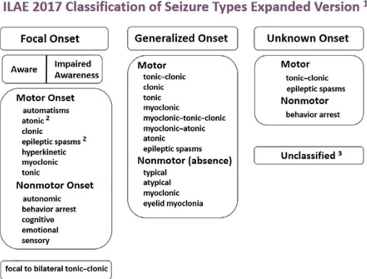
    ILAE Classification: Seizure and Epilepsy Types

    The International League Against Epilepsy (ILAE) classification provides a detailed framework for understanding seizures (the event) and epilepsy (the underlying condition).

    Table 1: Classification of Seizure Types (The Event)
    Seizure Type Category Subtype Key Characteristics & Observable Features Correlation with Previous Notes
    I. Focal Onset Seizures Focal Aware Seizure
    • Originates in one area/hemisphere of the brain.
    • Consciousness preserved throughout.
    • "Aura" is now understood as a Focal Aware Seizure with specific sensory, emotional, cognitive, or autonomic symptoms (e.g., peculiar taste, dizziness, abdominal rising sensation, déjà vu).
    Directly correlates with "Aura phase" from Grand Mal and early symptoms of Temporal Lobe/Jacksonian seizures.
    Focal Impaired Awareness Seizure
    • Originates in one area/hemisphere of the brain.
    • Consciousness is impaired at any point (dazed, confused, unresponsive).
    • Motor Features: Automatisms (e.g., chewing, lip smacking, picking motions), atonic (localized limpness), clonic (localized jerking), epileptic spasms, hyperkinetic (fidgeting, thrashing), myoclonic (localized jerks), tonic (localized stiffening).
    • Non-Motor Features: Autonomic (e.g., heart rate changes), behavioral arrest, cognitive (e.g., difficulty speaking), emotional (e.g., unprovoked fear or joy), sensory experiences (e.g., strange smell/taste).
    Correlates with Temporal Lobe Epilepsy (purposeless repetitive movements, unresponsiveness) and Jacksonian Epilepsy (localized twitching/tremors).
    Focal to Bilateral Tonic-Clonic Seizure
    • A focal seizure (aware or impaired awareness) that then spreads to involve both hemispheres, leading to a generalized tonic-clonic event.
    • Includes the Tonic phase (sustained stiffening, falling, cry), Clonic phase (rhythmic jerking, potential bladder/bowel release, frothy/blood-stained saliva), followed by a Post-ictal phase.
    Corresponds to what was previously often described as "Grand Mal Epilepsy" particularly if it began with an "aura."
    II. Generalized Onset Seizures Tonic-Clonic
    • Originates rapidly in both hemispheres from the outset.
    • Consciousness typically impaired immediately.
    • Classic sequence: Tonic phase (total body stiffening, loss of consciousness, fall, epileptic cry) followed by Clonic phase (repeated, rhythmic jerking of all muscles, potential incontinence, tongue/lip biting), ending in a Post-ictal phase (deep sleep, confusion, headache, amnesia).
    Corresponds to "Grand Mal Epilepsy" (Generalized Tonic-Clonic Epilepsy) when there's no preceding focal onset/aura.
    Tonic
    • Sustained stiffening of muscles throughout the body, without a subsequent clonic phase.
    • Consciousness typically impaired.
    Relates to the stiffening aspect of "Tonic and Clonic Seizures."
    Clonic
    • Rhythmic jerking movements of muscles throughout the body, without a preceding tonic phase.
    • Consciousness typically impaired.
    Relates to the jerking aspect of "Tonic and Clonic Seizures."
    Atonic
    • Sudden, generalized loss of muscle tone; body goes limp, slump or collapse ("drop attacks").
    • Consciousness typically impaired.
    Directly correlates with Atonic Seizures (Drop Attacks).
    Myoclonic
    • Brief, shock-like jerks or increases in muscle tone, affecting muscles or muscle groups.
    • Can occur in bouts.
    Directly correlates with Myoclonic Seizures.
    Epileptic Spasms
    • Sudden flexion or extension of the body (e.g., arms fling outward, knees pull up, body bends forward).
    • Often occur in clusters.
    Directly correlates with Infantile Spasms.
    Typical Absence
    • Brief (seconds) staring spells with unresponsiveness.
    • Often mistaken for daydreaming.
    • May involve subtle automatisms.
    • Consciousness impaired.
    Directly correlates with "Petit Mal Epilepsy" (Absence Seizures).
    Other Absence Types Atypical Absence, Myoclonic Absence, Eyelid Myoclonia (more specific subtypes).
    III. Unknown Onset Seizures -
    • When the beginning of the seizure is not observed or cannot be determined.
    • May later be reclassified once more information is available.
    Applies when the initial moments of an event (e.g., a Tonic-clonic seizure or Epileptic spasm) are unwitnessed.
    Table 2: Pre-Seizure and Post-Seizure Stages
    Stage Characteristics
    Prodrome
    • Non-specific symptoms occurring hours or days before a seizure.
    • Not part of the seizure activity itself.
    • Examples: Mood changes (irritability, depression), talkativeness, restlessness, violence.
    Post-ictal Stage
    • The period immediately after a seizure as the brain recovers.
    • Symptoms vary based on seizure type and intensity.
    • Examples: Confusion, fatigue, headache, amnesia for the event, disorientation, emotional changes (calmness, quietness, isolation, retarded mobility, depression).
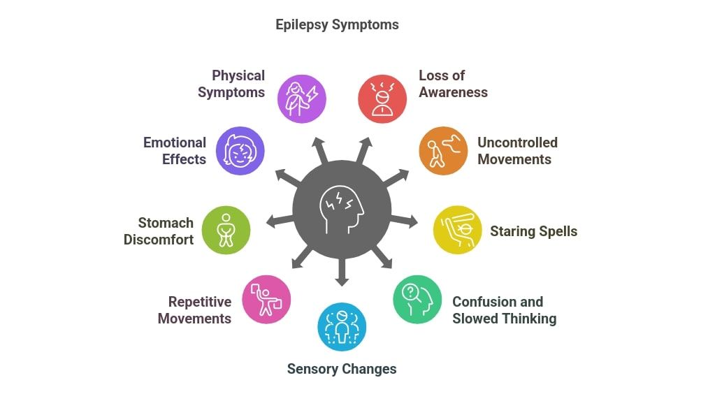
    Clinical Manifestations (What Epilepsy Looks Like)

    Clinical manifestations are the signs and symptoms that occur during a seizure event and can be highly varied, depending on the seizure type and the brain region involved.

    1. Generalized Onset Seizures:
  • Generalized Tonic-Clonic Seizure (formerly Grand Mal):
    • Prodrome (Pre-ictal): Hours or days before the seizure, the person may experience non-specific symptoms like mood changes, irritability, or difficulty concentrating.
    • Aura (often absent or not remembered if truly generalized onset): If present, it would indicate a focal onset that rapidly generalized.
    • Tonic Phase: Sudden loss of consciousness, body stiffens symmetrically, often a cry or groan (as air is forced out). Person falls to the ground. Eyes roll back. Breathing may stop briefly, leading to cyanosis. Lasts usually 10-30 seconds.
    • Clonic Phase: Rhythmic jerking of the limbs and body, typically bilateral. May involve tongue biting (often side of tongue), incontinence (bladder and rarely bowel), frothing at the mouth (which can be blood-stained from biting). Lasts usually 30 seconds to 2 minutes.
    • Post-ictal Phase: Gradual recovery of consciousness. Confusion, drowsiness, headache, muscle aches, and complete amnesia for the event are typical. May enter a deep sleep. Can last minutes to hours.
  • Absence Seizures (formerly Petit Mal):
    • Onset: Typically abrupt, without warning.
    • Manifestations: Brief (seconds, typically <15-20 sec) episodes of staring, blank expression, unresponsiveness. May involve subtle automatisms like eyelid fluttering, lip-smacking, or mild head nodding.
    • Termination: Abrupt. The individual quickly resumes prior activity, often unaware of the seizure or with immediate return of alertness. No post-ictal confusion.
    • Typical Population: Most common in children, often mistaken for daydreaming or inattention.
  • Myoclonic Seizures: Sudden, brief, shock-like jerks or twitches of a muscle or group of muscles (e.g., arms, shoulders, head). Often bilateral but can be unilateral. Consciousness usually preserved.
  • Atonic Seizures (Drop Attacks): Sudden, brief loss of muscle tone, causing the person to fall abruptly to the ground, often without warning. High risk of head and facial injury.
  • Tonic Seizures: Sudden, brief stiffening or tensing of muscles, typically in the trunk and limbs. Can cause falls.
  • Clonic Seizures: Rhythmic jerking movements, usually symmetrical, but without the preceding tonic phase.
  • 2. Focal Onset Seizures:
  • Focal Aware Seizures (formerly Simple Partial):
    • Manifestations: Vary widely depending on the brain region affected, but consciousness is fully preserved. The person is aware of the event.
    • Motor: Twitching, jerking, or stiffening of a specific body part (e.g., face, arm, leg). "Jacksonian March" describes the spread of motor symptoms.
    • Sensory: Tingling, numbness, visual disturbances, auditory hallucinations, olfactory, gustatory, or vertigo.
    • Autonomic: Changes in heart rate, breathing, sweating, epigastric rising sensation, flushing, pallor.
    • Psychic/Cognitive/Emotional: Feelings of fear, anxiety, déjà vu, jamais vu, memory disturbances. Often experienced as an "aura" before evolving to a more complex seizure.
  • Focal Impaired Awareness Seizures (formerly Complex Partial):
    • Manifestations: Consciousness is impaired or lost. The person may appear to be awake but is unresponsive, confused, or has an altered state of awareness.
    • Automatisms: Repetitive, non-purposeful movements are common (e.g., lip-smacking, chewing, swallowing, fumbling with clothes, walking aimlessly, repeating phrases). These are characteristic of temporal lobe seizures.
    • Duration: Typically 30 seconds to 2 minutes.
    • Post-ictal Phase: Common, characterized by confusion, drowsiness, and often amnesia for the seizure event.
  • Focal to Bilateral Tonic-Clonic Seizure: Begins with symptoms of a focal seizure (e.g., an aura, focal motor activity, or impaired awareness), then rapidly progresses to a generalized tonic-clonic seizure with loss of consciousness.
  • Diagnosis of Epilepsy

    Diagnosing epilepsy involves confirming that the events are indeed epileptic seizures, classifying the seizure type, identifying the epilepsy syndrome, and determining the etiology.

    1. Clinical History (The Most Crucial Step):
    • Detailed Seizure Description: A meticulous history from the patient (if possible) and, crucially, from an eyewitness (family member, friend, colleague) is paramount. Questions focus on:
      • Pre-event: Prodrome, triggers, warning signs (aura).
      • During the Event: Onset (sudden vs. gradual), movements (type, location, symmetry), vocalizations, eye movements, head turning, color changes, incontinence, tongue biting, level of awareness/responsiveness.
      • Post-event: Duration of confusion, memory of the event, fatigue, headache, muscle soreness.
    • Medical History: Birth history, developmental milestones, head injuries, CNS infections, fevers, family history of epilepsy, past medical conditions, medications, drug/alcohol use.
    2. Neurological Examination:
    • Usually normal between seizures, but may reveal focal deficits if there is an underlying brain lesion (e.g., hemiparesis, sensory loss). Post-ictally, transient neurological deficits (Todd's paralysis) can be observed.
    3. Electroencephalography (EEG):
    • Purpose: Records the electrical activity of the brain to identify abnormal brain wave patterns (epileptiform discharges).
    • Interictal EEG: Performed between seizures. Can show characteristic patterns (e.g., spikes, sharp waves) that support a diagnosis. A normal interictal EEG does not rule out epilepsy.
    • Ictal EEG: Performed during a seizure (e.g., during video-EEG monitoring). Captures the actual seizure activity and is the most definitive EEG finding for diagnosis and localization.
    • Activation Procedures: Hyperventilation, photic stimulation, and sleep deprivation are used to provoke epileptiform activity.
    4. Neuroimaging:
    • Magnetic Resonance Imaging (MRI) of the Brain:
      • Purpose: To identify structural abnormalities causing seizures (e.g., tumors, strokes, malformations, mesial temporal sclerosis).
      • Importance: Crucial for identifying the etiology, especially in focal epilepsies.
    • Computed Tomography (CT) Scan of the Brain: Less sensitive than MRI but can be used in emergency situations (e.g., to rule out acute hemorrhage).
    5. Blood Tests and Other Laboratory Investigations:
    • To rule out other conditions that can cause seizures (e.g., metabolic derangements, infections, drug/alcohol withdrawal, electrolyte imbalances). Examples: CBC, electrolytes, glucose, liver/kidney function tests, toxicology screen.
    6. Video-EEG Monitoring:
    • Continuous simultaneous recording of EEG and video of the patient over several days. Gold standard for confirming diagnosis, classifying seizure types, and localizing onset zone for surgery.
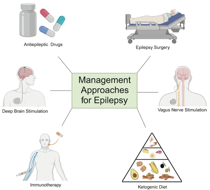
    Management and Treatment Options for Epilepsy

    The management of epilepsy is multifaceted, encompassing immediate care during a seizure, long-term pharmacological and non-pharmacological treatments, addressing complications, and providing comprehensive patient education.

    I. Immediate Management and First Aid During a Seizure (Emergency Management):

    A seizure can be frightening for bystanders, but knowing how to act can prevent injury and ensure patient safety.

    1. General Principles of Emergency Management:
    • Stay Calm: Remain composed and speak calmly.
    • Safety First: Remove the person from immediate danger (e.g., clear sharp objects). If the patient is safe, do not move them.
    • Time the Seizure: Note the exact start time. Crucial for determining if medical help is needed.
    • Loosen Clothing: Around the neck to ease breathing.
    • Protect the Head: Support with a soft, flat material (e.g., folded jacket).
    • Ensure Airflow: Clear space and minimize crowds.
    • Recovery Position: As soon as jerking stops, turn onto side to prevent choking.
    • Check Breathing: If breathing sounds difficult after the seizure, call for an ambulance.
    • Clear Airway: Gently check for blocks (e.g., false teeth) but do not force mouth open.
    • Stay with Patient: Until fully awake and reoriented.
    • Reassurance: Reorient and reassure the patient after recovery.
    2. What NOT to Do During a Seizure:
    • Do not put any hard object (e.g., spoon) in the person's mouth.
    • Do not hold their limbs tightly.
    • Do not give anything to eat or drink until fully alert.
    • Do not attempt mouth-to-mouth resuscitation (unless breathing doesn't resume after seizure).
    3. When to Call for Emergency Medical Help:
    • The person has never had a seizure before.
    • The person has difficulty breathing or waking up after the seizure.
    • The seizure lasts longer than 5 minutes (Potential Status Epilepticus).
    • The person has another seizure soon after the first one without full recovery.
    • The person is hurt during the seizure.
    • The seizure happens in water.
    • The person has a pre-existing health condition like diabetes, heart disease, or is pregnant.
    II. Long-Term Medical Management (Drug Management):

    The cornerstone of long-term epilepsy treatment is typically anti-seizure medications (ASMs).

    1. Principles of Pharmacological Treatment:
    • Goal: Reduce frequency of seizures or eradicate them.
    • Individualized Treatment: Based on seizure type, age, comorbidities.
    • Titration: Start low and gradually increase.
    • Monitoring: Regular follow-ups for progress and side effects.
    • Monotherapy vs. Polytherapy: Start with one drug; add others if needed.
    2. Commonly Used Anti-Seizure Medications:
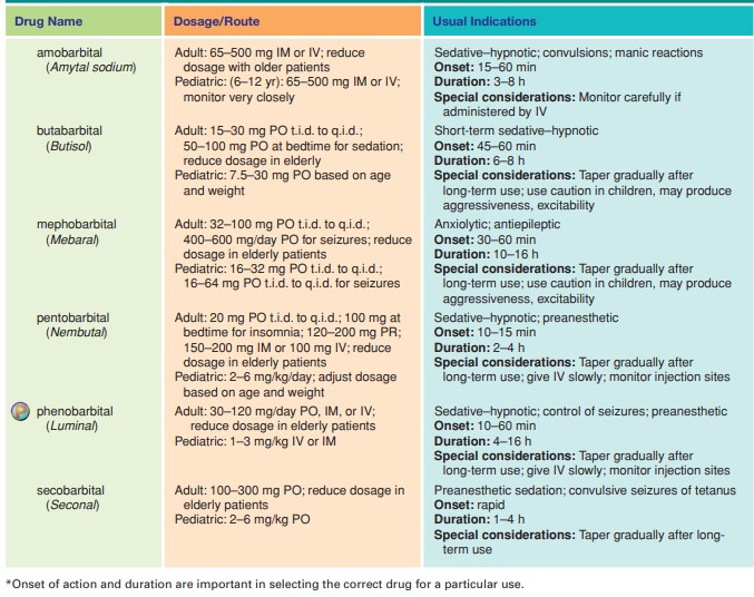
    • Phenobarbitone: Typically 30 to 90 mg two to three times daily (divided doses). An older, broad-spectrum ASM.
    • Phenytoin Sodium: Typically 100-300 mg daily (DDD - once daily or divided doses). Effective for focal and generalized tonic-clonic seizures.
    • Sodium Valproate (Valproic Acid): Typically 200-1200 mg two to three times daily (divided doses). Broad-spectrum, effective for various seizure types.
    • Carbamazepine: Typically 100-1200 mg in 3 divided doses. Primarily used for focal seizures.
    3. General Principles of the Treatment of Epilepsy:
    • Treat Causative Factors: Treat underlying causes like malaria, meningitis, or cerebral growths.
    • Avoidance of Precipitating Factors: Identify and avoid triggers.
    • Anticipation of Natural Variation: Understand seizure timing (e.g., during sleep).
    • Appropriate and Regular Administration: Strict adherence to prescribed regimen.
    Seizure Triggers & Complications
    Seizure Triggers:
    • Physiological Stressors: Fevers, sleep deprivation, fasting.
    • Emotional Stressors: Fear, anger, excitement.
    • Sensory Stimuli: Flickering lights (photosensitivity), specific sounds.
    • Substance Use: Alcohol intoxication or withdrawal.
    • Environmental Factors: Fatigue, boredom, high altitude.
    • Hormonal Changes: Menstrual cycle fluctuations.
    • Medication Non-adherence.
    Complications of Epilepsy:
    1. Status Epilepticus: A medical emergency defined by a seizure lasting longer than 5 minutes, or recurrent seizures without return to baseline consciousness. Requires urgent medical treatment.
    2. Mental Deterioration (Cognitive Impairment): Chronic brain syndrome where repeated seizures can lead to progressive brain damage.
    3. Physical Injuries: Falls, burns, fractures.
    4. Psychosocial Issues: Stigma, anxiety, depression, social isolation.
    5. SUDEP (Sudden Unexpected Death in Epilepsy): The most common cause of epilepsy-related death where no other cause is found.
    Patient and Community Education & Prevention
    For Patients and Caretakers:
    • Epilepsy is an illness like any other; with treatment, a person can lead a full life.
    • Encourage participation in activities safely.
    • Emphasize importance of taking medications exactly as prescribed.
    • Advise against dangerous activities (swimming alone, driving until seizure-free, operating heavy machinery).
    For the Community:
    • Combat Stigma: Educate that labeling patients is traumatizing.
    • Inclusion: Children should attend school; adults should be encouraged to marry.
    • Contagion: Teach that epilepsy is not contagious.
    Prevention of Epilepsy:
    • Prevent Head Injury (seat belts, helmets).
    • Seek Immediate Medical Attention after a first seizure.
    • Good Prenatal Care.
    • Manage Cardiovascular Risk Factors (hypertension).
    • Avoid Excess Alcohol Abuse.
    • Manage Fevers in Children.
    • Treat Infections and Ensure Nutrition.
    Nursing Diagnoses and Specific Nursing Interventions
    Nursing Diagnosis 1: Risk for Injury

    Related to uncontrolled seizure activity, loss of consciousness, uncontrolled muscle movements, or falls during seizures.

    Specific Nursing Interventions Details
    Seizure Precautions Pad side rails, keep bed in lowest position, instruct to avoid sharp objects in environment, recommend medical alert bracelet.
    Seizure First Aid Stay with patient, protect head, loosen clothing, turn to recovery position, move furniture, DO NOT restrain or insert objects in mouth, time the seizure.
    Post-Ictal Monitoring Monitor vital signs, level of consciousness, assess for injuries, allow rest, reorient gently.
    Activity Modification Educate on avoiding swimming alone, driving restrictions, and home modifications (e.g., shower chair).
    Nursing Diagnosis 2: Ineffective Airway Clearance

    Related to neuromuscular impairment during tonic-clonic seizures (tongue biting, increased salivary secretions, aspiration risk).

    Specific Nursing Interventions Details
    Acute Seizure Management Do not attempt to open mouth during seizure. Once movements cease, turn to recovery position to facilitate drainage. Suction secretions as needed.
    Post-Ictal Assessment Monitor respiratory rate/depth, assess breath sounds for aspiration (crackles), monitor for hypoxia.
    Patient Education Educate family on recovery position importance and when to call for emergency help if breathing is compromised.
    Nursing Diagnosis 3: Inadequate Health Knowledge

    Regarding epilepsy (disease process, triggers, medication regimen, first aid, emergency protocols).

    Specific Nursing Interventions Details
    Comprehensive Education Explain disease process, ASM purpose/dose/side effects, importance of daily dosing, triggers, first aid, and emergency protocols (e.g., seizure >5 min).
    Resources Provide written materials and support group info.
    Teach-Back & Reinforcement Ask patient to explain back what they learned; reinforce at every visit.
    Nursing Diagnosis 4: Excessive Anxiety

    Related to unpredictable nature of seizures, fear of public seizures, social stigma.

    Specific Nursing Interventions Details
    Establish Trust Provide non-judgmental environment to express fears.
    Empowerment Address knowledge deficits to reduce anxiety; emphasize productive lives are possible.
    Coping Strategies Encourage relaxation techniques, mindfulness, support group participation.
    Referrals & Stigma Refer to mental health professionals if needed; discuss strategies for talking to employers/friends.
    Nursing Diagnosis 5: Impaired Social Interaction

    Related to fear of seizures in public, stigma, withdrawal, or limitations on activities.

    Specific Nursing Interventions Details
    Address Anxiety/Deficits Educate to dispel myths; help patient develop confidence in disclosing condition.
    Promote Participation Discuss safe activities (e.g., cycling with supervision), public transport options, encourage social groups.
    Support & Advocacy Recommend support groups; advocate for patient in social settings by educating others on first aid.
    Nursing Diagnosis 6: Noncompliance (Medication Adherence)

    Related to perceived side effects, forgetfulness, or lack of understanding.

    Specific Nursing Interventions Details
    Detailed Teaching Explain "why" consistent use prevents complications. Review side effects and strategies to manage them.
    Adherence Strategies Suggest pill organizers, alarms, linking to daily routines. Discuss refilling prescriptions early.
    Address Beliefs Explore beliefs/misconceptions. Involve family in medication management if appropriate.
    Nursing Diagnosis 7: Fatigue

    Related to post-ictal state, sleep disturbance, or side effects of anti-seizure medications.

    Specific Nursing Interventions Details
    Assessment & Sleep Hygiene Assess fatigue severity. Educate on regular sleep schedules, avoiding caffeine/alcohol before bed.
    Medication Review Collaborate with physician if ASMs cause excessive sedation.
    Energy Conservation Teach pacing activities and prioritizing tasks.
    Healthy Lifestyle Encourage regular exercise and balanced diet. Allow adequate rest post-seizure.

    Epilepsy Read More »

    Narcotics

    Narcotics

    Narcotics

    Narcotics or Narcotic drugs are drugs that react with different type of opioid receptors, receptor sites that respond to naturally occurring peptides, enkephalins, and endorphins.

    These are found in the CNS, peripheral nerves, and GI tract cells.

    In the spinal cord, they integrate and relate pain information. Pain relief and side effects depend on the type of receptor site.

    Pain

    Pain is mostly a subjective experience of unpleasant sensation and emotional experience. People respond to pain differently because of cultural differences, learned experiences, and environmental stimuli.

    A-delta and C-fibers are two sensory nerves that respond to stimulation by generating nerve impulses that produce pain sensations.

    Classification of pain

    Pain Classification According to Duration:

    1. Acute Pain – is caused by tissue It is the type of pain which makes the person aware of the injury and leads him to seek for care and education about the injury and how to take care for it.
    2. Chronic Pain – is a constant or intermittent pain that keeps occurring long past the time the area would be expected to This is the type that can interfere with activities of daily living.

    Pain Classification According to Source

    1. Nociceptive Pain – caused by direct pain receptor stimulus
    2. Neuropathic Pain – caused by nerve injury
    3. Psychogenic Pain – associated with emotional, psychological, or behavioral stimuli

    Types of opioid receptors

    1. Mu-receptors – primarily pain-blocking receptors; also account for respiratory depression, euphoria, and development of physical
    2. Beta-receptors – modulate pain transmission by reacting with enkephalins in the periphery
    3. Kappa-receptors – associated with some analgesia, pupillary constriction, sedation, and dysphoria
    4. Sigma-receptors – pupillary dilation, hallucinations, psychoses with narcotic use.

    Types of narcotic drugs

    Narcotics are divided into 3 classes;

    1. Narcotic Agonists – react with opioid receptors in the CNS; cause analgesia, sedation, or They are classified as controlled substances because they have potential for physical dependence.
    2. Narcotic Agonists-Antagonists – stimulate certain opioid receptors but block other such They exert similar analgesic effect with that of morphine but they have less potential for abuse. However, they are associated with more psychotic like reactions.
    3. Narcotic Antagonists – bind strongly to opioid receptors without causing receptor activation. They block opioid receptor effects as well as effects of too much opioids in the system.

    Narcotic Agonists

    These drugs react with opioid receptors in the CNS; cause analgesia, sedation, or euphoria.

    Therapeutic Action

    The desired and beneficial action of narcotic agonist is:

    • Narcotic agonists act as agonist to specific opioid receptors in the CNS to produce analgesia, euphoria, and sedation.

    Indications

    Narcotic agonists are indicated for the following medical conditions:

    1. Relief of moderate to severe acute pain or chronic pain
    2. Preoperative medication
    3. Component of combination therapy for severe chronic pain
    4. Intra-spinal to reduce intractable

    Indication of narcotic agonists in different age groups

    Children

      1. Safety and effectiveness has not been established in
      2. Narcotic agonists that have established pediatric dosage guidelines are codeine, fentanyl (except transdermal), hydrocodone, meperidine, and morphine
      3. Naloxone is the antidote for narcotic overdose and reversal of narcotic

    Adults

    1. They should be informed and reassured that associated abuse with the use of narcotics in acute pain is remote.
    2. They should be educated about the importance of asking for pain medication before the pain becomes acute.
    3. Caution is advised for pregnant and lactating women because of potential adverse effects to the fetus.
    4. Narcotics used in labor include morphine, meperidine, and oxymorphone.
    5. All narcotic agonists are pregnancy category B except oxycodone (category C) so it might be the drug of choice if one is needed during pregnancy.

    Older adults

    1. They are more susceptible to drug adverse effects because of existing medical conditions.
    2. Safety measures should be established (side rails, call light, assistance to ambulate).

    Contraindications and Cautions

    The following are contraindications and cautions for the use of narcotic agonists:

    1. Allergy to narcotic agonists. Prevent hypersensitivity reaction
    2.  Diarrhea caused by toxic poisons. Drug depresses GI activity and this could lead to increased absorption and toxicity
    3.  Respiratory dysfunction. Exacerbated by respiratory depression caused by drugs
    4. Recent GI/GU surgery, acute abdomen, ulcerative colitis. Can be worsen by the GI depressive effects of the narcotics
    5.  Head injuries, alcoholism, delirium tremens, cerebral vascular disease. Can be exacerbated by
      the CNS effects of the drug
    6.  Liver, renal dysfunction. Can interfere with metabolism and excretion of the drug
    7.  Pregnancy, lactation. Potential adverse effects to the fetus and the baby.

    Adverse Effects

    Use of narcotic agonists may result to these adverse effects:

    1. CNS: light-headedness, dizziness, psychoses, anxiety, fear, hallucinations, pupil constriction, impaired mental processes
    2. GI: nausea, vomiting, constipation, biliary spasm
    3. GU: ureteral spasm, urinary retention, hesitancy, loss of libido
    4. Others: sweating, physical and psychological dependence
    5. Narcotic-induced respiratory center depression: respiratory depression with apnea, cardiac arrest, shock

    Interactions

    The following are drug-drug interactions involved in the use of narcotic agonists:

    1. Barbiturates, phenothiazines, MAOIs: increased likelihood of respiratory depression, hypotension, and sedation or coma
    2. SSRI, MAOI, TCA, Johns Wort: increased risk of potentially life-threatening serotonin syndrome if taken with tapentadol, the newest narcotic agonists that blocks norepinephrine reuptake in the CNS
    3. Methylnaltrexone bromide (Relistor) is the treatment for opioid-induced constipation in palliative care patients who are no longer responding to traditional laxatives.

    Drugs used as narcotic agonists

    Drug

    Indications

    Dosage ranges

    Key issues to note

    Codeine

    Analgesic, antitussive

    Relief of pain

    Adult: 30-60mg every 4-6 hours when necessary max dose 240mg daily.

    Children: 1-12years: 0.5-1 mg/kg every 4-6hours

    Diarrhoea

    Adults: 30mg 3-4 times daily

    1.  Increase fluids and fibre intake to avoid constipation

    2.  Avoid alcohol during therapy with codeine

    3.  Avoid abrupt discontinuation after prolonged use

    4.  Codeine is not recommended for treatment of productive cough

    5.  Codeine may be administered with food to minimise nausea

    and GI upset

    Pethidine

    1.  Pre-operative medication

    2.  Acute analgesia

    3.  Post-operative pain

    4.  Moderate to severe acute pain Obstetric analgesia

    Acute pain

    Adult: SC or 1M injection; 50- 150mg repeated after 4 hours Children: O.5-2mg/kg every 4 hours

    Obstetric analgesia: SC or 1M injection, 50-100mg repeated 1- 3 hours later if necessary max dose is 400mg in a day

    Post-operative pain: SC/IM

    injection

    1.  Prolonged use of Pethidine may result in physical dependence

    2.  Lowest effective doses are recommended especially during

    labor


    Adult: 25-100mg repeated every 2-3 hours if necessary

    Children: 0.5-2mg/kg every 2 to

    3 hours


    Oxycodone

    Analgesic

    Oxycodone: 10–20 mg

    PO q12h; 5 mg for break thru pain


    Other narcotic agonist analgesic drugs

    1. Methadone
    2. Oxymorphone
    3. Propoxyphene
    4. Fentanyl

    Short notes about Morphine

    Morphine is the ‘gold standard’ against which other opioid analgesics are measured.

    When used correctly, patients don’t become dependent, tolerance is uncommon and respiratory depression doesn’t usually occur.

    The correct morphine dose is the one that gives pain relief: there is no ‘ceiling’ or maximum dose — the right dose is the one that controls the patient’s pain without side effects, however you need to increase the dose gradually.

    Dosage of morphine

    Morphine has no ceiling effect to the analgesia.

    There is no standard dose of morphine for the treatment of chronic pain in patients with cancer and HIV/AIDS.

    It must be individually titrated for each patient and the correct dose is that which controls the pain whilst causing tolerable side effects.

    The dose required depends on many factors including the severity of pain, the type of pain, individual pharmacokinetic variations, the development of tolerance., and the psychosocial issues that affect the perception of pain.

    Acute pain, postoperative pain:

    • Oral: 5-20mg every 4 hours

    By SIC or 1M injection

    • Adult: 10 mg every 4 hours if necessary
    • Neonate: 150mcg 1kg every 6hours
    • 6-12 years: 5-10mg every 4 hours
    • 1-5years: 2.5-5mg every 4 hours
    • 1 month -12months: 200mcg/kg every 6hours

    Chronic pain: Oral ISC or 1M:

    • Adult: 10-15mg every 4 hours. Dose may be increased according to the response
    • Children: 2-12years: Initially 200-500mcg/kg every 4hours adjusted according to response
    • 1-2years: Initially 200-400mcg/kg every 4hours adjusted according to response
    • 1-12months: Initially 80mcg/kg every 4hours

    Myocardial infarction:

    • By slow IV injection (2mg/ minute), 10mg followed by a further 5-10mg if necessary.
    • Elderly or debilitated patients, give a half a dose

    Acute pulmonary oedema:

    • By slow IV injection (2mg/minute) 5-10mg

    Action of morphine

    Morphine acts on the opioid receptors in the brain and spinal cord to produce analgesia.

    The perception of pain is altered both by a direct effect on the spinal cord, modulating peripheral nociceptive input, arid by activating the descending inhibitory systems from the brain stem and basal ganglia.

    Morphine also acts on the limbic system and on higher centers to modify the emotional response to pain.

    The system effects, including those affecting the gastrointestinal and respiratory tracts, arc partly centrally mediated via the autonomic nervous system and may partly be due to a direct effect on opioid receptors in the peripheral tissues.

    Indications

    1. Post-operative pain
    2. Myocardial infarction
    3. Premedication before surgery
    4. Severe pain
    5. Sickle cell crisis
    6. Acute pulmonary oedema
    7. Chronic pain (cancer)

    Common Side effects

    The common side effects of morphine include:

    1.  Constipation — therefore you should always give a laxative alongside morphine (unless the individual has diarrhoea) e.g. Bisacodyl 5mg at night increasing the dose to l5mg if needed.
    2.  Nausea and vomiting — if this occurs, give anti-emetics e.g. plasil 10mg 8 hourly.
    3.  Drowsiness — may occur in the first few days of taking morphine. If it does not improve after three days reduce the dose of morphine.
    4.  Itching — not very common but if it occurs reduce the dose of morphine

    Contraindication

    1. Morphine should be given with caution to patients with renal impairment, severe hepatic dysfunction, significant pulmonary disease (including acute or severe bronchial asthma), and CNS depression from any cause.
    2. Elderly patients and those who are debilitated or cachectic should initially be treated with reduced doses.

    Dose

    Titrating oral Morphine into other formulations

    • Titrate the regular dose of morphine over several days until the patient is pain free. Either add the total daily dose and the total breakthrough dose given in 24 hours and divide by six to get the new 4hrly dose, or give 30—50% increments, e.g. 5—10—15mg etc., given as 4hrly doses. Increments of less than 30% are ineffective.
    • If the patient cannot swallow, use other routes, e.g. Rectal, subcutaneous, buccal, intravenous, or administer via an alternative enteral route such as a gastrostomy tube.
    • The ratio of morphine PO: SC is 2:1, g. 10mg oral morphine is 5mg SC morphine.
    • The ratio of morphine PO:IV is 2—3:1, g. 30mg oral morphine is 10mg IV morphine
    • Morphine is available in immediate and slow-release oral Use slow- release morphine once pain is controlled, dividing the total 24-hour dose into two to get the twice-daily dosage.

    Useful tips when using morphine

    1.  Oral morphine can be absorbed through the mucosa of the buccal cavity (mouth) or of rectum, so small amounts can be given even for unconscious patients.

    2.  Even though a patient is on regular oral morphine they may have breakthrough pain, an additional dose of oral morphine may be given to control this pain. This may be a one off incidence of pain but if more frequent breakthrough doses are required this may mean the 4hourly dose needs increasing.

    3.  Pain has to be controlled before other problems can be addressed and treated, as it is not possible to have meaningful discussions about psychosocial concerns if a patient has
      uncontrolled pain

    4.  Pain can be caused or aggravated by psychosocial concerns, which must be addressed before good pain control can be achieved. Where psychosocial or spiritual problems are causing or
      aggravating pain, no amount of well-prescribed analgesia will relieve the pain until the responsible psychosocial issues are identified and addressed.

    5.  Oral morphine is effective for chronic severe pain and can be given for many years and the dose can keep increasing, some patients can even take up to several hundred mgs 4 hourly.

    6.  If the pain stimulus is removed, then the dose of morphine should be decreased gradually to minimize the effects of physical dependence.

    7.  Opiates can also be used as a short term analgesia: in AIDS opportunistic infections such as
      cryptococcal meningitis; sickle cell crisis; burns and other painful conditions and does not cause
      addiction

    Narcotic Agonists-Antagonists

    These drugs stimulate certain opioid receptors but block other such receptors.

    Therapeutic Action

    The desired and beneficial action of narcotic agonist-antagonist is:

    • Narcotic agonists-antagonists act on certain opioid receptors but block other such receptors. They have less potential for abuse compared to narcotic agonists but are able to exert similar analgesic effect as morphine.

    Indications

    Narcotic agonists-antagonists are indicated for the following medical conditions:

    1. Relief of moderate to severe pain; pre-anesthetic medication and a supplement to surgical anesthesia
    2.  May be desirable for relieving chronic pain in patients who are susceptible to narcotic dependence.

    Here are some important aspects to remember for indication of narcotic agonist-antagonists in different age groups:

    Children

    • Safety and effectiveness has not been established in children.
    • Narcotic agonist-antagonist of choice for children older than age 13 is buprenorphine.
    • Naloxone is the antidote for narcotic overdose and reversal of narcotic effects..

    Adults

    • They should be informed and reassured that associated abuse with the use of narcotics in acute pain is remote.
    • They should be educated about the importance of asking for pain medication before the pain becomes acute.
    • Caution is advised for pregnant and lactating women because of potential adverse effects to the fetus.

    Older adults

    • They are more susceptible to drug adverse effects because of existing medical conditions.
    • Safety measures should be established (side rails, call light, assistance to ambulate).

    Contraindications and Cautions

    The following are contraindications and cautions for the use of narcotic agonists-antagonists:

    1. Allergy to narcotic agonists-antagonists. Prevent hypersensitivity reaction
    2. Physical dependence on narcotics. Withdrawal symptom may be precipitated
    3. COPD, other respiratory dysfunction. Can be exacerbated by respiratory depression
    4. MI, CAD, hypertension. Can be exacerbated by cardiac stimulatory effects
    5. Renal, hepatic dysfunction. Interfere with drug metabolism and excretion
    6. Pregnancy, lactation. Potential adverse effects to the fetus and the baby.
    7. Nalbuphine is specifically contraindicated to patients who are also allergic to sulfites to prevent cross-hypersensitivity reactions.

    Interactions

    The following are drug-drug interactions involved in the use of narcotic agonist-antagonists:

    1. Barbiturates, phenothiazines,    MAOIs:    increased    likelihood    of    respiratory depression, hypotension, and sedation or coma
    2. Tripelennamine: increased hallucinogenic and euphoric effect with pentazocine (“Ts and Blues”)
    3. Methylnaltrexone bromide (Relistor) is the treatment for opioid-induced constipation in palliative care patients who are no longer responding to traditional laxatives.

    Types of drugs used as narcotic agonists antagonist

    Drug

    Indications

    Dosage ranges

    Nalbuphine

    Analgesia

    10 mg/70 kg SC, IM,

    IV q3–6h PRN

    Pentazocine

    Analgesia

    50–100 mg PO q3–4h PRN; up to

    30 mg IM, SC, IV q3–4h PRN

    Pentazocine

    Analgesia

    1 tablet q4h


    Nursing Considerations when administering narcotic agonists and narcotic agonist-antagonists

    Nursing Assessment

    These are the important things the nurse should include in conducting assessment, history taking, and examination:

    1. Assess for mentioned cautions and contraindications (e.g. drug allergy, respiratory dysfunction, myocardial infarction and CAD, hepatorenal dysfunction, ) to prevent untoward complications.
    2. Conduct pain assessment with patient to establish baseline and evaluate effectiveness of drug therapy.
    3. Perform thorough physical (CNS, vital signs, bowel sounds, urine output) to establish baseline status before beginning therapy, determine drug effectiveness and evaluate for any potential adverse effects.
    4. Monitor laboratory results (liver function, kidney function) to determine need for possible dose adjustment and identify toxic drug effects.

    Nursing Diagnoses

    Here are some of the nursing diagnoses that can be formulated in the use of these drugs for therapy:

    1. Impaired gas exchange related to respiratory depression
    2. Disturbed sensory perception related to CNS effects
    3. Constipation related to GI effects
    4. Risk for injury related to CNS effects

    Implementation with Rationale

    These are vital nursing interventions done in patients who are taking narcotic agonists and narcotic agonists-antagonists:

    1.  Perform baseline and periodic pain assessments with patient to monitor drug effectiveness and provide appropriate changes in pain management protocol as needed.
    2.  Have a narcotic antagonist and equipment for assisted ventilation readily available when administering this drug IV to provide patient support in case of severe reaction.
    3.  Monitor timing of analgesic doses. Prompt administration may provide a more acceptable level of analgesia and lead to a quicker resolution of the pain.
    4.  Provide non-pharmacological pain measures like breathing exercises, back rubs, and stress reduction to increase drug effectiveness and reduce pain.
    5.  Provide comfort measures (e.g. small, frequent meals for GI upset) to help patient tolerate drug effects.
    6.  Provide safety measures (e.g. adequate lighting, raised side rails, etc.) to prevent injuries.
    7.  Educate client on drug therapy to promote understanding and compliance.

    Evaluation

    Here are aspects of care that should be evaluated to determine effectiveness of drug therapy:

    1. Monitor patient response to therapy (relief of pain, sedation).
    2. Monitor for adverse effects (e.g. GI depression, respiratory depression, arrhythmias, etc).
    3. Evaluate patient understanding on drug therapy by asking patient to name the drug, its indication, and adverse effects to watch for.
    4. Monitor patient compliance to drug therapy.

    Narcotic Antagonists

    They bind strongly to opioid receptors without causing receptor activation. They block opioid receptor effects as well as effects of too much opioids in the system

    Therapeutic Action

    The desired and beneficial action of narcotic antagonists is as follows:

    • Narcotic antagonists are drugs that bind strongly to opioid receptors but do not activate them. They block the opioid receptors and reverse the effects of opioids like respiratory depression and sedation.

    Indications

    Narcotic antagonists are indicated for the following medical conditions:

    1. Indicated for complete or partial reversal of narcotic depression; diagnosis of suspected opioid

    Indication of narcotic antagonists in different age groups:

    Children

    1.  Safety and effectiveness has not been established in children.
    2.  Naloxone is the antidote for narcotic overdose and reversal of narcotic effects.

    Adults

    1. They should be informed and reassured that associated abuse with the use of narcotics in acute pain is remote.
    2. They should be educated about the importance of asking for pain medication before the pain becomes acute.
    3. Caution is advised for pregnant and lactating women because of potential adverse effects to the fetus and the baby.

    Older adults

    1. They are more susceptible to drug adverse effects because of existing medical conditions.
    2. Safety measures should be established (side rails, call light, assistance to ambulate).

    Contraindications and Cautions

    The following are contraindications and cautions for the use of narcotic antagonists:

    1.  Allergy to narcotic antagonists. Prevent hypersensitivity reaction
    2.  Pregnancy, lactation. Potential adverse effects to the fetus and the baby.
    3.  Narcotic addiction. Precipitation of a withdrawal symptom
    4.  CV disease. Exacerbated by the reversal of the depressive effects of narcotics

    Adverse Effects

    Use of narcotic antagonists may result to these adverse effects:

    1. CNS: excitement, reversal of analgesia
    2. CV: tachycardia, blood pressure changes, dysrhythmias, pulmonary edema
    3. Acute narcotic abstinence syndrome: nausea, vomiting, sweating, tachycardia, hypertension, tremulousness, feelings of anxiety. A naloxone challenge should be administered before giving naltrexone to help to avoid acute reactions.

    Interactions

    There is no significant drug-drug interactions involved with narcotic antagonists.

    Types of drugs used as narcotic antagonists

    Drug

    Indications

    Dosage ranges

    Nalmefene

    Complete or partial reversal of opioid effects

    Initial dose: 0.5 mg /170 kg IV PRN, second dose of 1 mg170 kg 2-5 min

    later; maximum dose, 1.5 mg /170 kg

    Naltrexone

    Narcotic overdose. postoperative narcotic

    depression

    0.4-2 mg IV initially with additional doses repeated at 2-3 min intervals; smaller doses used for post-

    operative narcotic depression

    Pentazocine

    Narcotic addiction. alcohol dependence

    Maintenance treatment: 50 mg PO daily or 100 mg every other day, or 150 mg PO every third day; 2 mL IV,

    SC


    Nursing Considerations

    Nursing Assessment

    These are the important things the nurse should include in conducting assessment, history taking, and examination:

    1. Assess for mentioned cautions and contraindications (e.g. drug allergy, history of narcotic addiction, myocardial infarction, ) to prevent untoward complications.
    2. Conduct pain assessment with patient to establish baseline and evaluate effectiveness of drug therapy.
    3. Perform thorough physical (neurological status, respiratory rate and rhythm, vital signs) to establish baseline status before beginning therapy, determine drug effectiveness and evaluate for any potential adverse effects.
    4. Obtain an electrocardiogram as appropriate to evaluate for cardiac effects.

    Nursing Diagnoses

    Here are some of the nursing diagnoses that can be formulated in the use of this drug for therapy:

    1. Decreased cardiac output related to CV effects
    2. Acute pain related to withdrawal and CV effects
    3. Risk for injury related to CNS effects

    Implementation with Rationale

    These are vital nursing interventions done in patients who are taking narcotic antagonists:

    1. Maintain open airway and provide artificial ventilation and cardiac massage as needed to support the patient.
    2. Administer vasopressors as ordered and as needed to manage narcotic overdose.
    3. Administer naloxone challenge before giving naltrexone because of the serious risk of acute withdraw.
    4. Provide comfort measures to help patient cope with withdrawal syndrome.
    5. Provide safety measures (e.g. adequate lighting, raised side rails, ) to prevent injuries.
    6. Ensure that patients receiving naltrexone have been narcotic-free for 7-10 days to prevent severe withdrawal syndrome.
    7. Educate client on drug therapy to promote understanding and compliance.

    Evaluation

    Here are aspects of care that should be evaluated to determine effectiveness of drug therapy:

    1. Monitor patient response to therapy (reversal of opioid effects, treatment of alcohol dependence).
    2. Monitor for adverse effects (e.g. CV changes, arrhythmias, hypertension, etc).
    3. Evaluate patient understanding on drug therapy by asking patient to name the drug, its indication, and adverse effects to watch for.
    4. Monitor patient compliance to drug therapy.

    Opioid infusion administration considerations

    1. Unless the patient has received a recent dose of opioid, a loading dose should be administered (according to the EPIC prescription) at the commencement of the infusion to ensure therapeutic plasma levels are quickly reached.
    2. For rapid relief of pain (or anticipated pain), the prescribed bolus dose should be reached.
    3. The infusion rate may be adjusted by the nurse within the dose range specified, according to the patient’s level of pain.
    4. It takes approximately four half-lives (8hrs for morphine/hydromorphone, ~1.5hrs for fentanyl) to reach steady state plasma concentration if given as an infusion, therefore if the rate is to be increased, a bolus should be given as well.
    5. Ideally the infusion rate should not be increased unless 3 boluses are required in a 1 hour period.
    6. The volume infused should be checked every hour and rate verified on the fluid balance flow chart.

    Narcotics Read More »

    Want notes in PDF? Join our classes!!

    Send us a message on WhatsApp
    0726113908

    Scroll to Top
    Enable Notifications OK No thanks