COOPERATIVES
COOPERATIVES
A cooperative is a business organization that is owned and controlled by its members.
Cooperatives are often formed to provide goods or services to their members at a lower cost than they would be able to obtain from a traditional business.
It is an association of persons who have voluntarily joined together to meet their economic, social and cultural needs and aspirations through a jointly and democratically controlled enterprise.
Example:
- A farmers’ cooperative that sells agricultural products to its members.
- A consumer cooperative that sells groceries and other household goods to its members.
- A housing cooperative that provides affordable housing to its members.

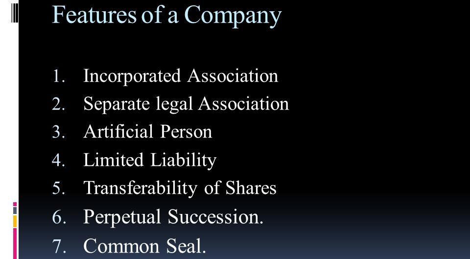
Features of Cooperative Organizations:
- Democratic Management, Election, and Control: Members have equal voting rights and participate in the decision-making process.
- Separate Legal Entity: Cooperatives are legally recognized as separate entities from their members.
- Voluntary Membership: Individuals are free to join or leave a cooperative voluntarily.
- Service Motto or Driven by a Fundamental Objective: Cooperatives are driven by a desire to serve their members and achieve a common goal.
- Government/State Control: Cooperatives are subject to government regulation and supervision.
- Members’ Economic Participation: Members contribute to the cooperative’s capital and share in its profits and losses.
- Disposal of Surplus: Surplus funds are distributed among members based on their patronage or participation.
Types of Cooperatives:
- Market / Sales Cooperatives: These cooperatives help farmers and producers sell their products collectively to get better prices.
- Savings and Credit Cooperatives: These cooperatives provide financial services to their members, such as savings accounts, loans, and insurance.
- Producers / Industrial Cooperatives: These cooperatives are owned and operated by workers who share the profits and losses.
- Consumers Cooperatives: These cooperatives are owned and operated by consumers who pool their resources to buy goods and services at lower prices.
- House Cooperatives: These cooperatives provide housing for their members.
Merits of Cooperative Societies
- Continuity or Long-Term Survival: Cooperatives are often more resilient than other types of businesses due to their democratic structure and member loyalty.
- Democratic Management: Members have a say in the management of the cooperative, which promotes transparency and accountability.
- Limited Liability: Members’ liability is limited to the amount of capital they have contributed to the cooperative.
- Government Assistance: Cooperatives often receive government support and assistance, such as tax breaks and subsidies.
- Reduce Inequalities: Cooperatives can help to reduce income inequality by providing equal opportunities for all members.
- Ease of Formation: Cooperatives are relatively easy to form and operate, especially compared to other types of businesses.
Demerits of Cooperatives
- Lack of Secrecy: Due to the democratic nature of cooperatives, there may be less secrecy compared to other types of businesses.
- Government Interference: Cooperatives are subject to government regulation and supervision, which can sometimes be burdensome.
- Limited Capital: Cooperatives may have limited access to capital compared to other types of businesses.
- Lack of Harmony and Innovation: Decision-making in cooperatives can be slow and bureaucratic, which may stifle innovation.
- Poor Management: Cooperatives may suffer from poor management due to the lack of professional expertise among members.
Reasons for Failure of Cooperative Societies:
- Government Interference: Excessive government interference can affect the autonomy and flexibility of cooperatives.
- Poor Infrastructure: Lack of adequate infrastructure, such as transportation and communication networks, can hinder the operations of cooperatives.
- Price Fluctuations: Cooperatives may be vulnerable to price fluctuations in the market.
- Political Instability: Political instability can create an uncertain and risky environment for cooperatives.
- Liberalization of the Economy: Liberalization of the economy can increase competition and make it difficult for cooperatives to compete with larger, more established businesses.
- Poor Financing: Cooperatives may have difficulty accessing financing, especially in developing countries.
- Lack of Harmony of Members: Disagreements and conflicts among members can weaken the cooperative and hinder its progress.
- Poor Methods of Production: Cooperatives may use outdated or inefficient methods of production, which can lead to lower productivity and profitability.
- Poor Management Systems: Poor management systems and practices can lead to mismanagement and financial problems.
- Natural Calamities: Natural disasters, such as floods, droughts, and earthquakes, can disrupt the operations of cooperatives and cause financial losses.
- Lack of Diversification: Cooperatives that rely on a single product or service may be vulnerable to changes in market demand.
- Substitute Influence: The emergence of substitute products or services can reduce the demand for the products or services offered by cooperatives.
Reasons for Revival of Cooperatives:
- Improves on the Standards of Living: Cooperatives can help to improve the living standards of their members by providing access to essential goods and services at affordable prices.
- Strengthen the Private Sector: Cooperatives can strengthen the private sector by providing employment opportunities and stimulating economic growth.
- Eradicate Poverty: Cooperatives can help to eradicate poverty by providing access to financial services and economic opportunities for marginalized communities.
- Equip Members with Practical and Theoretical Skills: Cooperatives can provide members with practical and theoretical skills through training and education programs.
- Mobilize Special Interest Groups: Cooperatives can mobilize special interest groups, such as the youth, farmers, and women, to work together for their common benefit.
- Provide Employment to Society: Cooperatives can provide employment opportunities for people who may have difficulty finding work in the formal sector.
- Reduce on Income Inequality: Cooperatives can help to reduce income inequality by providing equal opportunities for all members.
- Reduce on Regional Imbalance: Cooperatives can help to reduce regional imbalance by promoting economic development in rural and underserved areas.