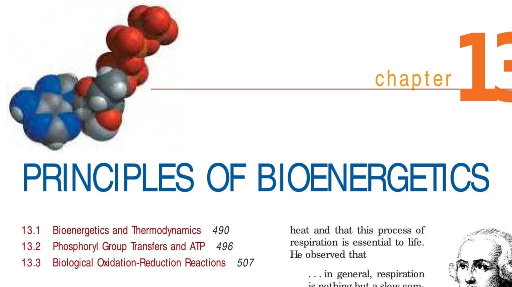
Bioenergetics (Thermodynamics and ATP)
Thermodynamics & ATP Bioenergetics: The Engine of Life
By the conclusion of this exhaustive master guide, you will be deeply conversant with:
- The fundamental definition of Bioenergetics and the specific types of "work" performed by biological systems.
- The intricate molecular structure of ATP (Adenosine Triphosphate) and why its bonds harbor so much accessible energy.
- The integration of Exergonic and Endergonic reactions via energy coupling.
- The unbreakable Laws of Thermodynamics (Zeroeth, First, Second, and Third) and their direct clinical implications.
- The mathematical and physiological breakdown of the Gibbs Free Energy Equation (ΔG = ΔH - TΔS).
- The critical mechanisms of Phosphoryl Group Transfers and Redox Reactions (Oxidation-Reduction) driving cellular respiration.
I. Bioenergetics: How Organisms Manage Energy
Let's shift our focus to the foundational biochemical concept of Bioenergetics. The term itself is highly descriptive:
- "Bio" means life.
- "Energetics" means the study of energy under transformation.
Therefore, Bioenergetics is the rigorous scientific study of how living organisms manage, transfer, and utilize energy in biological systems. It delves into the precise intracellular mechanisms that allow life to exist, thrive, and adapt—from the smallest unicellular bacteria to the largest mammals.
This critical field encompasses several key physiological aspects:
- Acquisition: How organisms obtain initial energy from their environment.
- Transformation: How organisms convert this raw energy from one form to another (e.g., converting food into a usable cellular "currency").
- Utilization: How organisms expend this currency to perform the literal "work" necessary for life.
At its core, Energy is defined as the capacity or ability to do work. In biology, "work" is a massive, overarching concept encompassing all the dynamic processes that sustain life and defy entropy. Just as a mechanical engine requires continuous fuel to operate, all living organisms require a relentless supply of energy to function and survive.
Examples of "Work" in Biological Systems Requiring Energy
Biological work is broadly categorized into three distinct physiological domains:
Gross Motor & Microscopic Movement
Just as a vehicle requires petrol to turn its wheels, our muscles require raw energy to contract. This powers our ability to walk, lift, and breathe.
- Macro-level: The myocardium (heart muscle) continuously contracting to pump blood against systemic resistance; the diaphragm contracting to expand the thoracic cavity for ventilation.
- Micro-level: The beating of microscopic cilia in the respiratory tract to clear mucus; the rapid movement of white blood cells (macrophages) chasing invading bacteria; the transport of intracellular vesicles along microtubule "highways" by motor proteins like kinesin.
Growth, Repair, and Development
Creating complex structures from simple building blocks is an energy-intensive "building" process.
- Cellular Division: A toddler growing into an adult requires massive energy to synthesize new cells and tissues.
- Molecular Synthesis: The replication of DNA during the S-phase of the cell cycle; the transcription of RNA; the translation of thousands of complex amino acid chains into functional proteins by ribosomes.
- Reproduction: The biological cost of forming gametes and sustaining fetal development requires tremendous synthetic work.
Sustaining Homeostasis
Even when a patient is comatose or deep in sleep, their body is performing immense "invisible" work.
- Active Transport: The continuous firing of the Sodium-Potassium pump (Na+/K+ ATPase) in every cell membrane, which consumes up to 30% of all cellular energy just to maintain the resting membrane potential of nerves and muscles.
- Thermoregulation: Generating metabolic heat to maintain a core body temperature of 37°C in freezing environments.
- Waste Removal: The kidneys actively filtering and secreting toxins against concentration gradients into the urine.
II. The Cosmic Source: The Journey of Sunlight Energy
For planet Earth, the ultimate, original, and most abundant source of energy is the nuclear fusion occurring within the Sun. However, human cells cannot directly utilize solar radiation to power a heartbeat. The energy must take a fascinating journey through the global food web.
- Plants (Producers – The Solar Collectors): Organisms containing chlorophyll capture photons of light energy from the sun through a process called photosynthesis. They utilize simple, low-energy molecules like Carbon Dioxide (CO&sub2;) and water (H&sub2;O) to convert solar energy into highly organized, energy-rich chemical bonds in the form of Glucose (C&sub6;H&sub1;&sub2;O&sub6;).
- Animals (Consumers – The Energy Transfer Agents): When you consume a plant-based product, you are directly ingesting the stored solar energy locked within that glucose molecule. When you consume an animal product, you are indirectly acquiring that solar energy, heavily filtered through the trophic levels of the food chain.
Why Bioenergetics Matters at the Bedside
- Nutrition and Energy Intake: Nurses continuously assess patients' nutritional status via Enteral or Parenteral feeding. Processes like wound healing, fighting sepsis, and post-operative recovery demand massive spikes in bioenergetic output. Malnutrition directly starves the cell of the fuel needed to heal.
- Metabolic Disorders: Diseases like Diabetes Mellitus are textbook examples of impaired bioenergetics. The patient has massive amounts of glucose in the blood, but lacking insulin, the cells are essentially "starving in a sea of plenty," unable to bring the fuel inside to make energy.
- Pharmacology: Many life-saving and life-threatening drugs directly manipulate bioenergetic pathways (e.g., Metformin alters cellular energy metabolism in the liver; Cyanide kills by instantly halting cellular respiration).
- Exercise Physiology & Rehabilitation: Understanding the energy demands of physical therapy and cardiac rehabilitation is a direct application of managing patient bioenergetics to rebuild endurance.
III. ATP: The Body's Universal Energy Currency
Regardless of what macros you ingest (carbohydrates, lipids, proteins), your cells do not directly use these large, clunky food molecules to power a single muscle twitch. That would be like trying to pay for a cup of coffee with a solid gold brick.
Instead, the body breaks down these macromolecules through metabolic pathways (Glycolysis, Krebs Cycle) to release their stored chemical energy. This energy is then captured and used to synthesize a highly specialized, highly manageable molecule called ATP (Adenosine Triphosphate). ATP is the exact "cash" your cells demand for almost all microscopic work.
The Anatomy of ATP: Why is it so powerful?
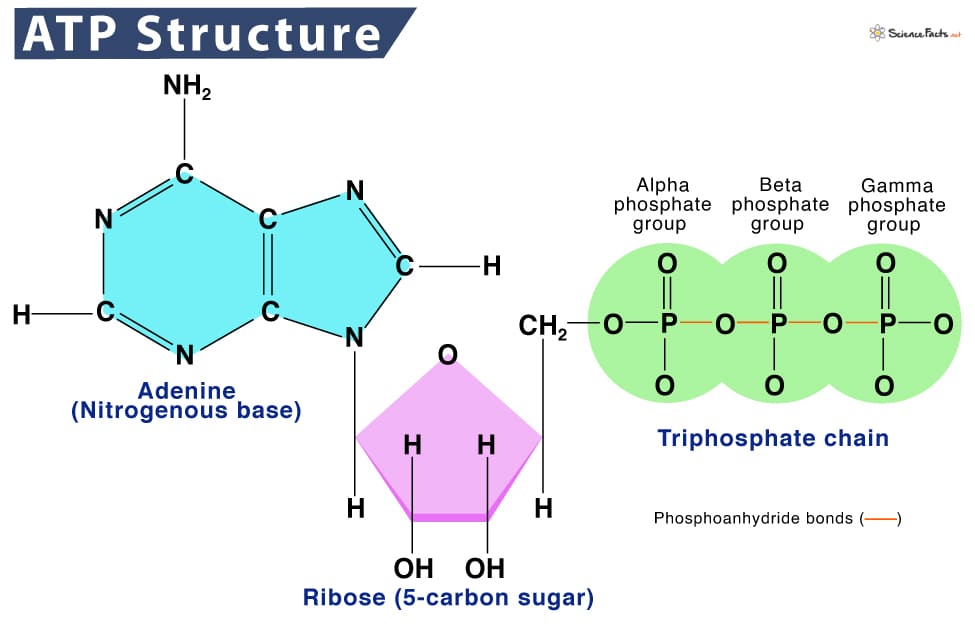
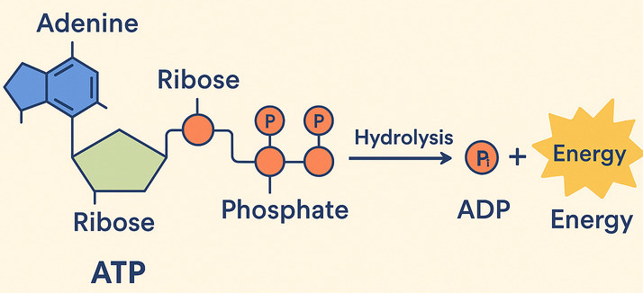
ATP is a nucleotide derivative consisting of three critical components:
- Adenine: A nitrogenous base.
- Ribose: A 5-carbon sugar.
- A Triphosphate Tail: A chain of three phosphate groups (PO&sub4;³&supmin;) attached to the ribose.
The Secret to the "High-Energy" Bond:
The power of ATP lies exclusively in the chemical bonds connecting those three phosphate groups. At physiological pH, each phosphate group carries a heavy negative charge. Because like charges severely repel one another, forcing three negative phosphates to sit right next to each other creates massive electrostatic repulsion (like trying to push the negative ends of three strong magnets together).
This creates a molecule under extreme tension. When the cell needs energy, it breaks off the outermost (terminal) phosphate group. Releasing this tension is like cutting the string on a highly compressed coiled spring—a significant amount of free energy is instantly released for the cell to capture and use.
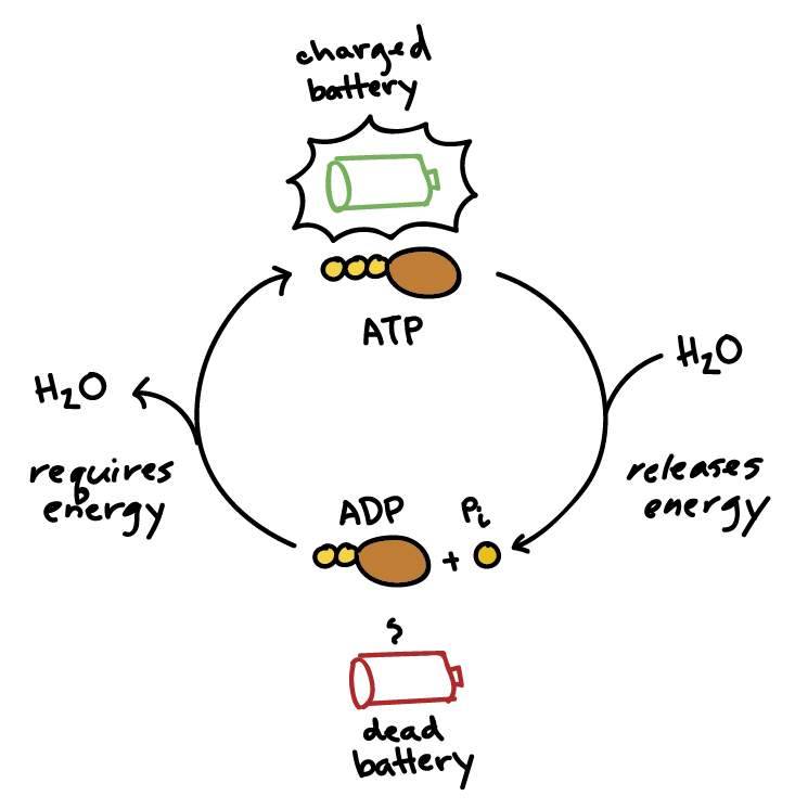
ATP + H&sub2;O → ADP (Adenosine Diphosphate) + Pi (Inorganic Phosphate) + FREE ENERGY
This reaction is infinitely reversible. When your body breaks down a meal (releasing energy), it uses that energy to force the phosphate back onto the ADP, regenerating ATP and "recharging the cellular battery."
IV. Free Energy: Exergonic vs. Endergonic Reactions
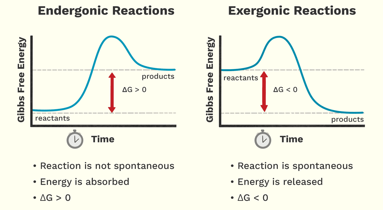
In bioenergetics, we use the concept of Gibbs Free Energy (G). Free energy is the amount of energy available to do actual, useful work within a system. By measuring the change in free energy (ΔG) before and after a reaction, we can predict whether a chemical reaction will happen spontaneously or if we must force it to happen by supplying energy.
| Reaction Type | Characteristics & ΔG | Biological Examples |
|---|---|---|
| Exergonic Reactions (Energy-Releasing) |
|
|
| Endergonic Reactions (Energy-Requiring) |
|
|
The Critical Concept: Energy Coupling
Life thrives by ingeniously linking these two types of reactions together. Cells use the energy released from an exergonic reaction (like ATP breaking down) to directly drive an endergonic reaction that needs energy to happen. This brilliant biological mechanism is called Energy Coupling.
ATP acts as the perfect molecular bridge, carrying the free energy released from your digesting lunch and delivering it directly to the muscle proteins trying to contract.
V. Thermodynamics: The Universal Rules of Energy
The overarching scientific field that dictates all of the aforementioned energy concepts is Thermodynamics. Derived from the Greek words for "heat" (therme) and "power" (dynamis), it is the branch of physics dealing with the transformation and interconversion of different forms of energy.
While "heat" is in the name, in biological systems, thermodynamics seamlessly encompasses light, thermal, chemical, electrical, and mechanical energy.
The Laws of Thermodynamics: Unbreakable Rules of the Universe
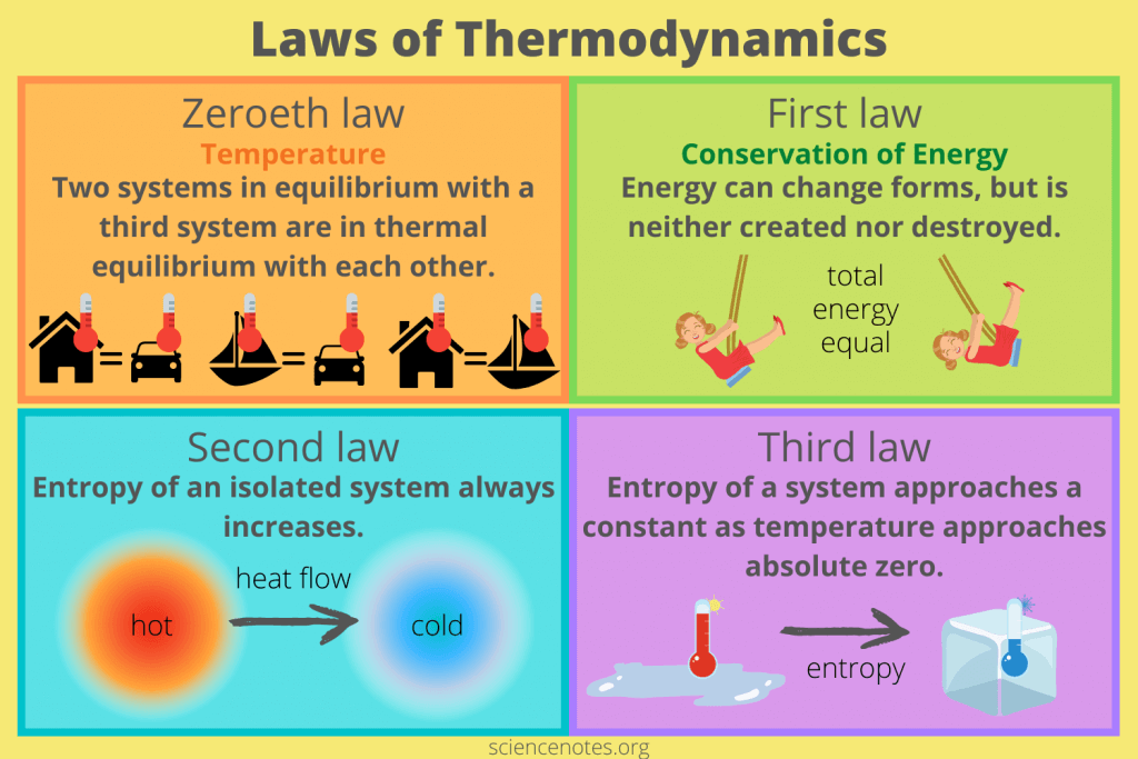
Thermodynamics is built upon four foundational principles. These laws are absolute; they govern every energy transformation in the cosmos, including the metabolic pathways inside the human body.
Defining Temperature & Thermal Equilibrium
"If two thermodynamic systems are each in thermal equilibrium with a third system, then they are in thermal equilibrium with each other."
- Meaning: This law establishes the fundamental definition of temperature and proves that heat will naturally flow from a hot object to a cold object until they are equal. This is the mathematical principle that allows a digital thermometer to accurately measure a patient's core temperature.
- Clinical Implication: Underpins thermoregulation. When a patient is placed on a cooling blanket for severe hyperthermia, heat continuously transfers from the patient's core to the blanket until equilibrium is achieved.
Conservation of Energy
"Energy can neither be created nor destroyed; it can only be transferred or transformed from one form to another."
- Meaning: The total amount of energy in an isolated system (the universe) remains completely constant. You cannot get "something for nothing."
- Biological Implication: Plants do not magically "make" energy; they transform solar photons into chemical glucose. In the human body, chemical energy from food is transformed into mechanical energy (muscle contraction), electrical energy (action potentials in the brain), and thermal energy (body heat).
- Clinical Implication: This is the basis of the Basal Metabolic Rate (BMR) and weight management. If caloric energy intake (eating) exceeds energy expenditure (metabolic transformation and exercise), the First Law dictates that the excess energy cannot be destroyed—it MUST be transformed and stored as adipose tissue (fat).
The Law of Entropy (Disorder)
"In any isolated system, the total entropy (disorder) can only increase over time or remain constant; it will never decrease naturally."
- Meaning: The universe inherently trends toward chaos, randomness, and disorder. Things naturally decay, rot, and fall apart. They do not spontaneously organize themselves into perfectly structured entities without the continuous addition of outside energy.
- Biological Implication: A human being is an incredibly complex, highly ordered structure. To maintain this high degree of order and fight off the relentless pull of entropy (decay/death), organisms must constantly consume massive amounts of energy. Life is a continuous, uphill battle against the Second Law.
- Clinical Implication (Metabolic Inefficiency): Every time energy is transformed in the body (e.g., from glucose to ATP to muscle movement), the transfer is highly inefficient. A large percentage of that energy is permanently "lost" to the environment as unusable, chaotic heat. This specific loss of heat is what keeps our bodies at 37°C. It also explains the physical deterioration of the body as we age—a gradual succumbing to entropy.
Absolute Zero
"The entropy of a perfect crystal approaches a constant minimum (zero) as its temperature approaches absolute zero (-273.15°C or 0 Kelvin)."
- Meaning: Entropy is directly linked to temperature. As a system gets colder, molecular movement slows down, and disorder decreases. At absolute zero, all molecular vibration ceases entirely, creating a state of perfect structural order.
- Clinical Implication: This is the thermodynamic foundation of Medical Cryopreservation. By plunging human tissues (like sperm, embryos, or transport organs) into liquid nitrogen (-196°C), we drastically reduce their temperature. This halts all entropic metabolic decay, essentially freezing biological time and preserving the cells indefinitely without degradation.
VI. The Gibbs Free Energy Equation: The Math of Life
We know the Second Law dictates that the universe trends towards disorder (Entropy). This gives us the ultimate equation to determine if a biological reaction will proceed. The Gibbs Free Energy Equation calculates the exact amount of usable energy (ΔG) left over.
Breaking Down the Variables:
- ΔG (Change in Gibbs Free Energy): The final amount of useful energy available to do cellular work. If negative, the reaction is spontaneous (Exergonic). If positive, the reaction requires energy to be forced (Endergonic).
- ΔH (Change in Enthalpy): The total heat content of the system.
- Exothermic: Releases heat into the body (negative ΔH). Favors a spontaneous reaction.
- Endothermic: Absorbs heat from the body (positive ΔH). Resists spontaneity.
- T (Temperature): The absolute temperature measured in Kelvin. (This acts as an amplifier for entropy).
- ΔS (Change in Entropy): The change in molecular disorder/chaos.
- Breaking a large glycogen molecule into 100 small glucose molecules heavily increases disorder (positive ΔS). This highly favors a spontaneous reaction.
The Golden Rule of Thermodynamics: Biological reactions are most likely to be spontaneous and energy-releasing if they release heat (negative ΔH) AND increase cellular disorder (positive ΔS).
Applying the Equation: Photosynthesis vs. Cellular Respiration
A. Photosynthesis (Highly Endergonic)
6CO&sub2; + 6H&sub2;O + Light Energy → C&sub6;H&sub1;&sub2;O&sub6; (Glucose) + 6O&sub2;
- ΔS is negative: We take simple, highly disordered gases (CO&sub2;) and force them into a highly complex, ordered solid structure (Glucose). We are decreasing entropy.
- ΔH is positive: We must absorb massive amounts of solar energy to build these bonds. It is endothermic.
- Result: Because both variables fight against spontaneity, ΔG is highly positive. Photosynthesis is impossible without continuous forced energy from the sun.
B. Cellular Respiration (Highly Exergonic)
C&sub6;H&sub1;&sub2;O&sub6; (Glucose) + 6O&sub2; → 6CO&sub2; + 6H&sub2;O + ATP Energy
- ΔS is positive: We smash a complex, highly ordered glucose molecule into tiny, chaotic CO&sub2; gas molecules. Entropy massively increases.
- ΔH is negative: Breaking these bonds releases huge amounts of heat into our bodies. It is exothermic.
- Result: Because both variables favor spontaneity, ΔG is highly negative. Cellular respiration explosively releases energy that we capture as ATP.
VII. Phosphoryl Group Transfers: How ATP Actually Works
We know ATP hydrolysis releases energy, but how does that energy physically make a muscle move or a pump work? It rarely happens by just exploding like a microscopic bomb. Instead, the primary mechanism is through Phosphoryl Group Transfer (Phosphorylation).
The Mechanism:
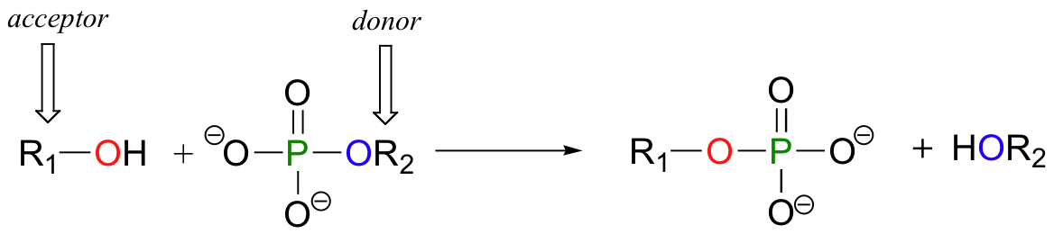
A phosphoryl group transfer is the enzyme-catalyzed physical movement of the terminal phosphate group (Pi) from ATP directly onto another recipient molecule (like a protein or a sugar). ATP becomes ADP, and the recipient molecule becomes phosphorylated.
Why is this the ultimate mechanism for cellular work?
- Raises the Free Energy of the Recipient: Jamming a bulky, highly negatively charged phosphate group onto a stable protein heavily "energizes" or "activates" it. The recipient molecule becomes violently unstable and highly reactive.
- Induces Conformational Changes (Shape-Shifting): Because the phosphate is so negatively charged, when it attaches to a protein, it repels other negative amino acids nearby. This physically forces the entire protein to fold, twist, and change its 3D shape.
The Sodium-Potassium Pump (Na+/K+ ATPase)
This pump must push Na+ out of the cell against its gradient (Endergonic work). How?
- The pump binds 3 Na+ ions from inside the cell.
- ATP transfers its phosphate group to the pump protein (Phosphorylation).
- The negative charge of the phosphate instantly changes the physical shape of the pump, causing it to hinge open towards the outside of the cell, physically dumping the Na+ out.
- The phosphate falls off, the pump returns to its original shape, and the cycle repeats. Shape equals function!
Kinase Enzymes & Pharmacology
Enzymes that transfer phosphate groups are called Kinases. They act as master ON/OFF switches for cell division and metabolism. In many cancers, mutant kinases are stuck in the "ON" position, constantly phosphorylating proteins that tell the cell to divide uncontrollably. Modern targeted chemotherapies (like Imatinib) are designed specifically to block these rogue kinases and halt the phosphoryl transfer.
VIII. Biological Oxidation-Reduction (Redox) Reactions: The Energy Harvest
While phosphoryl group transfers are the mechanism for spending energy, Oxidation-Reduction (Redox) reactions are the mechanism for harvesting energy from the food you eat.
What are Oxidation and Reduction?
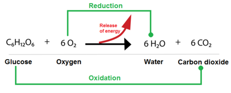
These are coupled chemical reactions involving the transfer of electrons. They never happen alone; if one molecule loses electrons, another must catch them.
- Oxidation: The loss of electrons (and often the loss of hydrogen atoms). A molecule that is oxidized loses energy.
- Reduction: The gain of electrons (and often the gain of hydrogen atoms). A molecule that is reduced gains energy.
Mnemonic: LEO the lion says GER! (Lose Electrons Oxidation, Gain Electrons Reduction).
Electron Carriers: The "Couriers" of Redox Energy
As glucose is ripped apart during digestion and cellular respiration, highly energetic electrons are stripped away. Free electrons are dangerous, so the cell uses specialized "taxi cab" molecules to safely carry them to the mitochondria.
- NAD+ (Nicotinamide Adenine Dinucleotide): Derived from Vitamin B3 (Niacin). Its oxidized, empty form is NAD+. When it picks up 2 electrons and 1 proton from a digested meal, it is reduced into the high-energy passenger NADH.
- FAD (Flavin Adenine Dinucleotide): Derived from Vitamin B2 (Riboflavin). Its empty form is FAD. It gets reduced to FADH&sub2;, carrying 2 electrons and 2 protons.
The Grand Finale: The Electron Transport Chain (ETC)
The ultimate goal of all bioenergetics culminates in the inner membrane of the mitochondria.
- Delivery: NADH and FADH&sub2; travel to the mitochondria and drop off their high-energy electrons into a series of membrane proteins called the Electron Transport Chain.
- Energy Release: As the electrons are passed down the chain from one protein to the next (a series of continuous redox reactions), they step down to lower and lower energy states. The energy they release is used to pump protons (H+) into the intermembrane space, creating a massive, highly pressurized acidic gradient.
- ATP Synthesis: The protons desperately want to flow back across the membrane to achieve equilibrium. They are forced to flow through a microscopic biological turbine called ATP Synthase. The physical spinning of this turbine generates enough energy to slam a phosphate onto ADP, creating massive amounts of ATP (Oxidative Phosphorylation).
- The Final Acceptor: At the very end of the chain, the "spent," low-energy electrons must be removed so the chain doesn't back up. The molecule that catches these final electrons is Oxygen (O&sub2;). The oxygen grabs the electrons and some free protons to safely form Water (H&sub2;O). This is the sole physiological reason human beings must breathe oxygen to survive.
Clinical Implications of the Electron Transport Chain
- Hypoxia/Ischemia: If a patient stops breathing or suffers a heart attack, Oxygen is no longer present to catch the final electrons. The entire ETC immediately backs up. ATP production drops from 36 ATP per glucose down to 2 ATP (anaerobic). The cell rapidly runs out of currency, the Na+/K+ pumps fail, the cells swell, and the tissue undergoes irreversible necrosis.
- Metabolic Poisons (Cyanide & Carbon Monoxide): Cyanide gas is incredibly lethal because it binds irreversibly to Cytochrome c Oxidase (Complex IV) in the ETC. It physically blocks oxygen from catching the electrons. Even if the patient is breathing 100% oxygen, their cells instantly suffocate and die on a molecular level because the electron transport chain is locked shut.
- Nutritional Deficiencies: Severe lack of B-vitamins (Niacin/Riboflavin) means the body cannot build NAD+ or FAD. Without these couriers, electrons cannot be transferred from food to the mitochondria, leading to profound lethargy, neurological issues, and systemic metabolic failure (e.g., Pellagra).
IX. References and Recommended Reading
- Nelson, D. L., & Cox, M. M. (2017). Lehninger Principles of Biochemistry (7th ed.). W.H. Freeman. (Comprehensive coverage of bioenergetics, thermodynamics, and ATP cycles).
- Hall, J. E., & Guyton, A. C. (2015). Guyton and Hall Textbook of Medical Physiology (13th ed.). Saunders. (Detailed physiological applications of metabolic rates and cellular work).
- Harvey, R. A., & Ferrier, D. R. (2011). Lippincott's Illustrated Reviews: Biochemistry (5th ed.). Lippincott Williams & Wilkins. (Excellent clinical correlates regarding redox reactions and the electron transport chain).
- Berg, J. M., Tymoczko, J. L., & Gatto, G. J. (2015). Stryer Biochemistry (8th ed.). W.H. Freeman. (In-depth analysis of the Gibbs free energy equation and phosphoryl group transfers).
Bioenergetics (Thermodynamics and ATP) Read More »