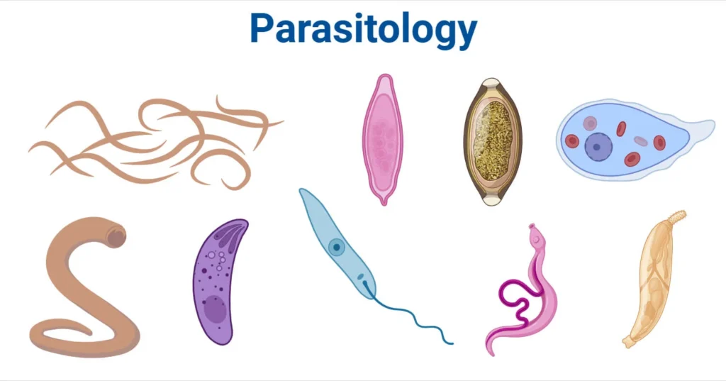
nursesrevision@gmail.com

Growth and Development in Adolescents

Growth and Development in Adolescents

Growth and Development in Adolescents

Growth in adolescence refers to the physical changes that occur during this period of development. 

These changes are influenced by hormones and genetics.

Development in adolescence refers to the psychological, emotional, and social changes that occur during this period

These changes are influenced by a combination of biological, psychological, and social factors

Biological, psychological, and cognitive changes that commence in puberty and persist throughout adolescence significantly impact nutritional status and nutrient needs. Adolescents undergo physical growth and development during puberty, increasing their requirements for energy, protein, vitamins, and minerals.

 Concurrently, they experience notable changes in their ability to assess information, seek independence, and become unique individuals. The heightened need for energy and nutrients, coupled with increasing financial independence, autonomy in food choices, and immature cognitive abilities, places adolescents in potentially distressing and costly situations with long-lasting repercussions.

Changes during Adolescence

Changes during Adolescence

Physical Changes

1. Growth of Reproductive Organs: Primary and secondary sex characteristics mark the physical changes, with adolescence transitioning from childhood to physical maturity.

  • Primary Sex Characteristics:
  1. In girls, the onset of the menstrual period.
  2. In boys, enlargement of the penis and testicles, accompanied by sperm production.
  • Secondary Sex Characteristics:
  1. Girls: Breast development, pubic hair growth, hip fat deposition, voice softening, changes in body image, weight gain, and height increase.
  2. Boys: Voice deepening, facial and body hair growth, penile enlargement, erections, changes in body image, weight gain, and height increase.

2. Physiological Changes:

  • Girls: Menstruation (menarche), acne, smooth skin appearance, and voice softening.
  • Boys: Wet dreams, sperm production, decreased dependence on family, voice changes, and Adam’s apple development.

3. Psychological/Emotional Changes:

  • Self-awareness of sexual needs.
  • Interest in the opposite sex.
  • Potential resistance, disobedience, hostility, and mood swings.

4. Sexual Changes:

  • Awakening of sexual arousal.
  • Masturbation.
  • Attraction to the opposite/same sex, leading to behavioral changes.

5. Behaviour Changes:

  • Formation of peer groups.
  • Independence from family.
  • Habit formation, including peer group associations, with possible involvement in activities like smoking and alcohol ingestion.

Adolescents’ Reactions to Change and Behaviours

Adolescents

Adults

Anxiety

Overprotection

Shyness

Anxiety

Depression

Judgmental attitudes


Misconceptions Myths Associated with Growth and Development (1)

Misconceptions/Myths Associated with Growth and Development

Females:

  • Girls with big buttocks are presumed to be highly sexually active.
  • The notion that smaller breasts are more desirable.
  • Belief that engaging in sex can alleviate menstrual cramps.
  • Incorrect association of menstrual cramps with infertility.
  • Misunderstanding that the vagina is hollow, suggesting things can disappear in it.
  • Girls can supposedly only get pregnant after a year of menstruation.
  • Reliance on safe days as a contraception method.
  • The misconception that girls with thin hips are considered more attractive.
  • False belief that menstrual flow is unhygienic and results in unpleasant odors.
  • Uncircumcised girls are thought to be restless and sexually aroused all the time.
  • Girls starting menstruation are mistakenly assumed to have engaged in sexual intercourse.
  • The belief that hip enlargement is solely due to sexual activity.
  • False notion that menstrual flow leads to the loss of virginity (hymen).
  • Girls with small breasts are incorrectly assumed to lack sufficient milk to feed their babies.
  • Girls are perceived as never children; they are considered mature at any stage.
  • Breasts are believed to enlarge when touched by men.
  • A girl who does not give birth is compared to a mule with an arid womb.
  • During the rainy season, it is wrongly believed that every woman becomes fertile.
  • The idea that one should have children at a very young age.
  • False belief that sexual intercourse helps in treating skin issues such as spots and pimples.

Males:

  • The misconception that a larger penis is better.
  • Belief that an erection implies the necessity for sexual activity.
  • Boys not growing beards are considered not yet men.
  • The misconception that only boys with hairy chests are considered attractive.
  • Girls are believed to be attracted only to tall, masculine boys.
  • Boys are thought to be capable of making girls pregnant only at the age of 18 years.
  • Boys with big toes are falsely believed to have a large penis.
Problems Faced by Adolescents as a Result of Body Changes

Problems Faced by Adolescents as a Result of Body Changes

  • Adolescents are confronted with societal expectations, including the pressure to exhibit adult behaviour, such as specific clothing choices and maintaining a high standard of living.
  • Emancipation from home becomes a challenge, coming from restrictions on activities like going out with members of the opposite sex, handling money, and coming home late. Adolescents may perceive these restrictions as excessive parental control.
  • Relationships with peer groups become crucial for adolescents, and they may continue to value their group even if they face exclusion or rejection at home.
  • Girls encounter various menstrual-related issues, acne problems, excessive sweating, vaginal discharge concerns, and changes in body size and shape during adolescence.
  • Boys experience challenges such as wet dreams, frequent erections, and notable changes in body size and shape.

Solutions to the Problems

  1. Promote Open Communication: Encourage parents and guardians to engage in open and honest communication with adolescents to better understand their concerns and perspectives.
  2. Provide Comprehensive Education: Implement educational programs in schools that address not only the physical changes but also the emotional and psychological aspects of adolescence.
  3. Foster Supportive Peer Environments: Create a supportive atmosphere within peer groups and schools to help adolescents navigate challenges collectively.
  4. Offer Counseling Services: Establish accessible counselling services for adolescents to discuss their concerns, both related to body changes and broader life issues.
  5. Parental Guidance Workshops: Conduct workshops for parents to equip them with effective strategies for guiding adolescents through this transitional phase.
  6. Encourage Healthy Body Image: Promote positive body image through awareness campaigns to help adolescents accept and embrace their changing bodies.
  7. Flexible Parental Boundaries: Advocate for balanced parental boundaries that allow for independence while maintaining necessary guidelines to ensure safety.
  8. Incorporate Life Skills Education: Integrate life skills education into the curriculum, covering topics such as decision-making, communication, and conflict resolution.
  9. Community Involvement: Involve the community in supporting adolescents, fostering understanding, and creating an environment conducive to their healthy development.
  10. Accessible Health Resources: Ensure adolescents have access to health resources, including information about menstrual health, reproductive well-being, and mental health support.
Developmental Stages of Adolescents

Developmental Stages of Adolescents

Early Adolescence (Approximately 10-14 years of age)

Middle Adolescence (Approximately 15-16 years)

Late Adolescence (Approximately 17-19 years)

Movement Toward Independence:

Movement Toward Independence:

Movement Toward Independence:

– Struggle with identity.

– Self-involvement, alternating between high expectations and poor self-concept.

– Firmer identity.

– Moodiness.

– Complaints of parental interference.

– Ability to delay gratification.

– Improved speech expression.

– Strong concern with appearance and body.

– Ability to think ideas through and express them.

– Expression of feelings through action.

– Lowered opinion of parents, withdrawal.

– Developed sense of humor.

– Growing importance of close friendships.

– Effort to make new friends.

– Stable interests.

– Less attention to parents, occasional rudeness.

– Emphasis on the new peer group.

– Greater emotional stability.

– Realization of parents’ imperfections.

– Periods of sadness due to psychological loss of parents.

– Ability to make independent decisions.

– Search for new relationships.

– Examination of inner experiences.

– Ability to compromise.

– Return to childish behavior.


– Pride in one’s work.

– Peer group influences interests and clothing.


– Self-reliance.


– Greater concern for others.

Future Interests and Cognitive Development:

Future Interests and Cognitive Development:

Future Interests and Cognitive Development:

– Increasing career interests.

– Intellectual interests gain importance.

– More defined work habits.

– Mostly interested in present and near future.

– Sexual and aggressive energies directed into creative and career interests.

– Higher concern for the future.

– Greater ability to work.


– Thoughts about one’s role in life.

Sexuality:

Sexuality:

Sexuality:

– Girls ahead in development.

– Concerns about sexual attractiveness.

– Concerned with serious relationships.

– Shyness, blushing, modesty.

– Frequently changing relationships.

– Clear sexual identity.

– Experimentation with body (masturbation).

– Exploration of heterosexuality with fears of homosexuality.

– Capacities for tender and sensual love.

– Worries about normalcy.

– Tenderness and fears toward opposite sex.


– Feelings of love and passion.


Ethics and Self-Direction:

Ethics and Self-Direction:

Ethics and Self-Direction:

– Rule and limit testing.

– Development of ideals and role models.

– Capable of useful insight.

– Occasional experimentation with substances.

– Consistent evidence of conscience.

– Stress on personal dignity and self-esteem.

– Capacity for abstract thought.

– Greater capacity for setting goals.

– Ability to set goals and follow through.


– Interest in moral reasoning.

– Acceptance of social institutions and cultural traditions.


– Self-regulation of self-esteem.

Physical Changes:

Physical Changes:

Physical Changes:

– Height and weight gains.

– Continued height and weight gains.

– Most girls fully developed.

– Growth of pubic and underarm hair.

– Growth of pubic and underarm hair.

– Boys continue to gain height, weight, muscle mass, body hair.

– Increased body sweat.

– Increased body sweat.


– Changes in skin and hair.

– Changes in skin and hair.


– Breast development and menstruation in girls.

– Breast development and menstruation in girls.


– Growth of testicles and penis in boys.

– Growth of testicles and penis in boys.


– Nocturnal emissions (wet dreams).

– Nocturnal emissions (wet dreams).


– Deepening of voice.

– Deepening of voice.


– Growth of facial hair in boys.

– Growth of facial hair in boys.


Growth and Development in Adolescents Read More »

ADOLESCENT REPRODUCTIVE HEALTH

ADOLESCENT REPRODUCTIVE HEALTH

ADOLESCENT REPRODUCTIVE HEALTH 

Adolescent Reproductive Health (ARH) is a state of complete physical, mental and social well being and not merely the absence of disease or infirmity, in all matters relating to the reproductive system of people between the ages of 10 and 19

Adolescent sexual and reproductive health refers to the physical and emotional wellbeing of adolescents and includes their ability to remain free from unwanted pregnancy, unsafe abortion, STIs (including HIV/AIDS), and all forms of sexual violence and coercion.

The rapid increase in the numbers of adolescents/young people points to the potential to contribute positively to the socio-economic and political development of the country. However, if not well directed, it can lead to consequences that may be harmful to the health status of the entire population.


Young people are vulnerable to all kinds of health challenges by virtue of their level of willingness to take risks and limited information. This includes R.H problems such as STIs/HIV/AIDs, early or unwanted pregnancy, unsafe abortion and psychosocial problems such as substance abuse, sexual abuse, delinquency etc.

Factors that predispose them to vulnerability include economic issues such as poverty, over dependence on adults or lack of employment opportunities. The majority of these young people are engaged in subsistence agriculture or petty trade in the informal sector. This situation is worsened by lack of adequate social services, characterized by low access to information, low demand and utilization of R.H services, high school dropouts and an unconducive teaching and learning environment in schools and health facilities.

DEFINITION OF TERMS

  1. Adolescent: This refers to a boy or girl aged between 10-19 years.
  2. Youth: This represents the period between childhood and adulthood or the transition from dependence in childhood to independence and awareness of interdependence as members of a community. According to the Uganda constitution, youths are considered to be between 18-30 years.
  3. Adolescence: This is a gradual process in which a child grows and develops into an adult, beginning at 10 years and continuing until the age of 18-19 years.
  4. Young person: A young person is a boy or girl aged between 10-24 years old.
  5. Puberty: These are reproductive changes that occur during adolescence, this is the period when adolescents reach sexual maturity and become capable of reproduction. In males, it usually starts between 10-14 years and stops between 15-17 years of age, characterized by the enlargement of external genitalia and increased pubic hair growth. In females, puberty can begin as early as 9 years of age, marked by breast enlargement and development at 9-13 years, with menarche (start of periods) occurring later between 11-15 years.
  6. Sexual health: This is a state of physical, mental, and social well-being in relation to sexuality. It goes beyond the mere absence of disease, dysfunctions, or infirmity. Sexual health requires a positive approach to sexuality and sexual relationships, ensuring the possibility of having pleasurable and safe sexual experiences, free of coercion, discrimination, and violence. Achieving and maintaining sexual health necessitates the respect and fulfilment of the sexual rights of all individuals.
  7. Sexuality: This is the sexual knowledge, beliefs, attitudes, values, and behaviours of individuals. It includes identity, orientation, roles, personality, thoughts, feelings, and relationships. The expression of sexuality is influenced by ethical, spiritual, cultural, and moral concerns.

Reasons for sexual and adolescent health

  1. Protecting and Promoting Adolescent Rights: To protect and promote the rights of adolescents of the rights of adolescents to health, education, information and care.
  2. Creating Supportive Legal and Socio-cultural Environments: To create an enabling legal and socio-cultural environment that promotes provision of better health information and services for young people.
  3. Involving Adolescents in Health Program Development: Actively engaging adolescents in the conceptualization, design, implementation, monitoring, and evaluation of adolescent health programs, which ensures programs are relevant and effective.
  4. Developing Responsible Health Behavior: Fostering responsible health-related behavior among adolescents, with a focus on equitable and respectful relationships, helps them navigate gender issues as they transition to adulthood.
  5. Legal and Social Protection: Promotion of legal and social protection of young people especially the girl child against harmful traditional practices and all forms of abuse including sexual abuse, exploitation, trafficking and violence.
  6. Training Providers and Reorienting Health Systems: Adequately training healthcare providers and restructuring health systems at all levels helps meet the unique needs of adolescents.
  7. Advocating for Increased Resources: Advocating for increased resource commitment for the health of adolescents in conformity with their numbers, needs and requirements at all levels.
  8. Enhancing Monitoring and Evaluation: Improvement of the capacity of local constitutions in monitoring and evaluation, research of adolescent health programs and needs and to promote dissemination and utilization of the relevant information to create awareness which influence behaviour change amongst individuals, communities, providers and leaders concerning adolescent health.
  9. Coordinating and Networking: To promote coordination and networking between different sectors and among NGOs/youth serving NGOs working in the field of adolescent health.
  10. Empowering Through Interventions: Encouraging interventions based on the capabilities and resources of youths empowers them to actively participate in their health and well-being.

Challenges Faced by Adolescents Regarding Reproductive Health:

In Uganda, adolescents suffer with severe health risks, including life-threatening challenges related to unwanted pregnancies, HIV/AIDS, and sexually transmitted infections (STIs). Adolescence, marked by experimentation and risk-taking, exposes individuals to various vulnerabilities. Key factors contributing to adolescent susceptibility to sexual and reproductive health problems include:

  1. Substance Abuse: Widespread substance abuse, including marijuana, khat (mairungi), and Cuba, poses significant health and social challenges, with devastating impacts on the well-being of young people.
  2. Mental Health Issues: Adolescents face mental health problems due to societal pressures, impacting their psychosocial development, self-esteem, and academic performance. Psychoactive drug use further contributes to self-inflicted mental health issues.
  3. Injuries from Accidents or Violence: Young people’s proclivity for risk-taking activities makes them prone to accidents, leading to physical injuries and, in severe cases, fatalities. Substance abuse, such as alcohol and smoking, can exacerbate this issue.
  4. Occupational Health Problems: Limited employment opportunities force many young people into harsh working conditions, both in formal and informal sectors.
  5. Nutrition Challenges: Adolescents require proper nutrition for growth, energy, and immunity. Common nutritional issues include poor growth, anaemia, and micronutrient deficiencies, which can have long-term consequences on their health.
  6. Socio-economic Consequences (Poverty): Unhealthy adolescents strain educational systems, limit contributions to national development, and jeopardize the stability of future generations. Investing in the health of young people is crucial for broader societal well-being.
  7. STIs, including HIV/AIDS: Uganda faces a serious health socio-economic problem with high rates of STIs/HIV/AIDS among young people, significantly impacting morbidity and mortality. Early onset of sexual activity contributes to the rising prevalence after 15 years of age.
  8. Unwanted Pregnancies and Unsafe Abortions: Adolescents experience high rates of unwanted pregnancies and unsafe abortions due to low contraceptive use. This leads to maternal mortality and morbidity, affecting both the young mothers and their infants.
  9. Harmful Traditional Practices: Practices like early marriage, female genital mutilation, gender-based violence, and inheritance violations violate adolescent rights, particularly in sexual and reproductive health, leading to adverse health outcomes.
  10. Lack of Awareness and Correct Information: Insufficient awareness and misinformation regarding the risks of unwanted pregnancies and STIs.
  11. Peer and Social Pressures: Influence from peers and societal expectations leading to risky behaviours.
  12. Lack of Skills to Resist Pressures: Inadequate skills to resist societal pressures and engage in safe behaviours.
  13. Absence of Youth-Friendly Services: Limited access to youth-friendly sexual health and counselling services.
  14. Poverty: Economic challenges contributing to vulnerability.
  15. Cultural Norms: Traditional cultural norms, such as young men expected to initiate sexual encounters with prostitutes, fostering risky behaviours.
  16. Limited Power to Resist Coercion: Inability to resist persuasion or coercion into unwanted sexual encounters.
  17. Educational Challenges: Dropping out of school, resulting in unattained goals and loss of opportunities.
  18. Impact on Self-Esteem: Loss of self-esteem due to guilt and damage to reputation.
  19. Fatal Consequences: Potential outcomes include death and life-threatening situations.
  20. Teenage Pregnancy Risks: Higher risk of morbidity and mortality, complications during labor, and associated health risks.
  21. Unsafe Abortion: Adolescent unwanted pregnancies often lead to unsafe abortion, posing significant health dangers.
  22. Female Genital Cutting/Mutilation: Severe reproductive health consequences for affected girls.
  23. Sexual Violence: Physical trauma, unintended pregnancy, STIs, psychological trauma, and increased likelihood of high-risk sexual behavior.
  24. Child Prostitution: Engagement in risky behavior with potential physical and psychological repercussions.
  25. Endemic Diseases: Health challenges associated with diseases like tuberculosis and malaria.

Management and Preventive Measures for Reproductive Health Problems:

  1. Health Education: Providing comprehensive health education to adolescents, addressing major concerns such as HIV/AIDS and pregnancies.
  2. Advocacy for Sex Education: Promoting the inclusion of sex education in primary and secondary school curricula.
  3. Family Planning Advocacy: Encouraging sexually active adolescents to use family planning methods to prevent early pregnancies.
  4. Post-Abortion Care Services: Advocating for accessible post-abortion care services to reduce complications resulting from abortion.
  5. Discouraging Early Marriages: Promoting initiatives to discourage early marriages among teenage adolescents.
  6. Parental Education: Educating parents about proper nutrition practices for their children.
  7. Community Sensitization: Sensitizing community members about the availability of ANC services for adolescents.
  8. Involvement in Preventive Services: Engaging adolescents in HIV/AIDS preventive services.
  9. Advocacy for Mental Health Services: Advocating for the availability of mental health services tailored to the needs of adolescents.
  10. Community Awareness: Sensitizing communities about the importance of adolescent reproductive services.

Focus Areas for Adolescent and Sexual Reproductive Health:

  • Behavior Change Counseling
  • Provision of Adolescent-Friendly Services
  • Provision of Contraceptive Services
  • Screening and Management of STIs

Importance of Sexual and Adolescent Health:

  1. Promotes good health, especially among adolescent girls.
  2. Essential for effective safe motherhood strategies.
  3. Provides a framework for policy development.
  4. Identifies gaps in policy and barriers to services.
  5. Reduces maternal mortality due to early pregnancy.

Factors Influencing Reproductive Health Needs of Adolescents:

  1. Age: The stage of adolescence significantly affects reproductive health needs, considering the diverse physical and emotional changes during this period.
  2. Marital Status: Adolescents who are married may face different reproductive health challenges compared to unmarried peers, including early pregnancies and family planning decisions.
  3. Gender Norms: Societal expectations and norms related to gender can impact the reproductive health needs of adolescents, influencing behaviours and choices.
  4. Sexual Status: Sexual activity or inactivity plays a crucial role in determining the reproductive health needs of adolescents, including concerns related to contraception and sexually transmitted infections (STIs).
  5. School Status: Adolescents in educational settings may have various reproductive health needs, including access to sexual education and resources within schools.
  6. Child Bearing Status: Whether adolescents have experienced childbirth or are in the process of childbearing can significantly influence their reproductive health requirements.
  7. Rural/Urban Residence: The geographical location of adolescents, whether in rural or urban areas, can impact the accessibility of reproductive health services and information.
  8. Peer Pressure: Influence from peers can shape adolescents’ attitudes and behaviours, potentially affecting their reproductive health decisions.
  9. Cultural/Political Conditions: Cultural and political factors within a society can create an environment that either supports or restricts access to reproductive health services and information.
  10. Economic Status: The economic background of adolescents can influence their access to healthcare resources, family planning options, and overall reproductive health services.
  11. Educational Attainment: The level of education achieved by adolescents can impact their understanding of reproductive health issues and their ability to make informed decisions.

Adolescents at High Risk:

  • Pregnant Below 18 Years: Pregnancies at a young age pose specific challenges related to maternal health and childbearing.
  • Post-partum and Post-abortion: Adolescents who have recently given birth or undergone an abortion may require specialized reproductive health care.
  • Adolescents in Labour: Those in the process of childbirth may need immediate and appropriate medical attention to ensure a safe delivery.
  • Adolescents with STIs: Those with sexually transmitted infections require prompt diagnosis, treatment, and preventive education.
  • Married Adolescents: Early marriages can present unique reproductive health challenges, including family planning decisions and maternal health concerns.

Adolescents with Special Needs:

  1. Sexually Abused: Victims of sexual abuse require sensitive and comprehensive reproductive health support.
  2. Drug and Substance Abusers: Substance abuse can impact reproductive health.
  3. Mentally and Physically Challenged: Adolescents facing mental or physical challenges may need specialized care and accessible reproductive health information.
  4. Adolescents Needing Pregnancy Prevention: Those seeking to prevent pregnancy require education on contraception and family planning methods.
  5. Menstrual Problems like Excessive Bleeding: Adolescents experiencing menstrual issues, such as excessive bleeding, may need medical attention and guidance.
  6. Problems Related to Growth and Development: Issues related to overall growth and development may require holistic care, considering both physical and emotional well-being.

ADOLESCENT REPRODUCTIVE HEALTH Read More »

Male Reproductive System

Male Reproductive System

Male Reproductive System

The organs of the male reproductive system include the testes, a system of ducts (including the epididymis, ductus deferens, ejaculatory ducts, and urethra), accessory sex glands (seminal vesicles, prostate, and bulbo-urethral glands), and several supporting structures, including the scrotum and the penis

Functions of Male Reproductive Organs

Male reproductive system functions vital for reproduction and urinary excretion.

Testes:

  • Production of Spermatozoa: Spermatogenesis occurs in the testes, generating mature sperm cells.
  • Maturation and Storage: Epididymis facilitates sperm maturation and serves as a storage site.
  • Epididymal Maturation: Sperm undergo maturation in the epididymis, gaining motility and fertilization potential.
  • Testosterone: Produced by testes, influences sperm production and development of secondary sexual characteristics. Regulated by the hypothalamus-pituitary-gonadal axis.

Ejaculatory Duct:

  • Vas deferens transports mature sperm to the ejaculatory duct, contractions facilitate sperm release during ejaculation.

Seminal Vesicles, Prostate Gland, and Bulbourethral Glands:

  • Contribute secretions to form semen, enhancing sperm viability.
  • Semen nourishes and protects sperm during transport in the female reproductive tract.

Penis and Urethra:

  • Copulation and Sperm Delivery: Penis facilitates copulation and the delivery of sperm into the female reproductive tract.
  • Urethra serves as the passageway for both semen and urine.
Scrotum Anatomy

Scrotum 

The scrotum is a pouch of pigmented skin, fibrous and connective tissue and smooth muscle

  • It is divided into two compartments, each of which contains one testis, one epididymis and the testicular end of a spermatic cord
  • It lies below the symphysis pubis, in front of the upper parts of the thighs and behind the penis. 
  • Externally, the scrotum looks like a single pouch of skin separated into lateral portions by a median ridge called the raphe.  
  • Internally, the scrotum is separated into two sacs by the septum. The septum is made up of a subcutaneous layer and muscle tissue called the dartos muscle, which is composed of bundles of smooth muscle fibers.
  • Associated with each testis in the scrotum is the cremaster muscle, a series of small bands of skeletal muscle that descend as an extension of the internal oblique muscle through the spermatic cord to surround the testes.
  • The location of the scrotum and the contraction of its muscle fibers regulate the temperature of the testes. Normal sperm production requires a temperature about 2 to 3°C below core body temperature. This lowered temperature is maintained within the scrotum because it is outside the pelvic cavity.
  • In response to cold temperatures, the cremaster and dartos muscles contract. Contraction of the cremaster muscles moves the testes closer to the body, where they can absorb body heat. 
  • Contraction of the dartos muscle causes the scrotum to become tight (wrinkled in appearance), which reduces heat loss. Exposure to warmth reverses these actions.
Testes

Testes

The testes are the male reproductive glands and are the equivalent of the ovaries in the female. 

  • They are about 4.5 cm long, 2.5 cm wide and 3 cm thick and are suspended in the scrotum by the spermatic cords. 

They are surrounded by three layers of tissue. 

  1. Tunica vaginalis. This is a double membrane, forming the outer covering of the testes, and is a downgrowth of the abdominal and pelvic peritoneum. During early fetal life, the testes develop in the lumbar region of the abdominal cavity just below the kidneys. They then descend into the scrotum, taking with them coverings of peritoneum, blood and lymph vessels, nerves and the deferent duct. The peritoneum eventually surrounds the testes in the scrotum, and becomes detached from the abdominal peritoneum. Descent of the testes into the scrotum should be complete by the 8th month of fetal life. 
  2. Tunica albuginea. This is a fibrous covering beneath the tunica vaginalis. Ingrowths form septa, dividing the glandular structure of the testes into lobules. Each of the 200–300 lobules contains one to three tightly coiled tubules, the seminiferous tubules where sperm are produced. The process by which the seminiferous tubules of the testes produce sperm is called spermatogenesis. The seminiferous tubules contain two types of cells: spermatogenic cells, the sperm-forming cells, and Sertoli cells, which have several functions in supporting spermatogenesis. Sertoli cells nourish spermatocytes, spermatids, and sperm; phagocytize excess spermatid cytoplasm as development proceeds; and control movements of spermatogenic cells and the release of sperm into the lumen of the seminiferous tubule. They also produce fluid for sperm transport, secrete the hormone inhibin, and regulate the effects of testosterone and FSH (follicle-stimulating hormone).
  3. Tunica vasculosa. . The tunica vasculosa is the vascular layer of the testis, consisting of a plexus of blood vessels, held together by delicate areolar tissue.
spermatic cord

Spermatic cords/Vas Deferens

  • The spermatic cords suspend the testes in the scrotum
  • Each cord contains a testicular artery, testicular veins, lymphatics, a deferent duct and testicular nerves, which come together to form the cord from their various origins in the abdomen. 
  • The cord, which is covered in a sheath of smooth muscle and connective and fibrous tissues, extends through the inguinal canal and is attached to the testis on the posterior wall.
  • The deferent duct is around 45 cm long. It passes upwards from the testis through the inguinal canal and ascends medially towards the posterior wall of the bladder where it is joined by the duct from the seminal vesicle to form the ejaculatory duct.
Seminal Vesicle

Seminal Vesicles

  • The seminal vesicles are two small fibromuscular pouches, 5 cm long, lined with columnar epithelium and lying on the posterior aspect of the bladder 
  • At its lower end each seminal vesicle opens into a short duct, which joins with the corresponding deferent duct to form an ejaculatory duct.

Functions 

  • The seminal vesicles contract and expel their stored contents, seminal fluid, during ejaculation. 

Seminal fluid, which forms 60% of the volume of semen, is alkaline to protect the sperm in the acidic environment of the vagina, and contains fructose to fuel the sperm during their journey through the female reproductive tract.

Ejaculatory Duct

Ejaculatory ducts 


  • The ejaculatory ducts are two tubes about 2 cm long, each formed by the union of the duct from a seminal vesicle and a deferent duct.
  • They pass through the prostate gland and join the prostatic urethra, carrying seminal fluid and spermatozoa to the urethra.
  • The walls of the ejaculatory ducts are composed of the same layers of tissue as the seminal vesicles. 
Prostate

Prostate gland 

  • The prostate gland lies in the pelvic cavity in front of the rectum and behind the symphysis pubis, completely surrounding the urethra as it emerges from the bladder.
  • It has an outer fibrous covering, enclosing glandular tissue wrapped in smooth muscle. The gland weighs about 8 g in youth, but progressively enlarges (hypertrophies) with age and is likely to weigh about 40 g by the age of 50. 
  • The prostate gland secretes a thin, milky fluid(slightly acidic fluid (pH about 6.5) that makes up about 30% of the volume of semen, and gives it its milky appearance.
  • Several proteolytic enzymes, such as prostate-specific antigen (PSA), pepsinogen, lysozyme, amylase, and hyaluronidase, eventually break down the clotting proteins from the seminal vesicles. 
  • The  clotting enzyme thickens the semen in the vagina, increasing the likelihood of semen being retained close to the cervix.

Bulbourethral glands 

  • The paired bulbourethral glands or Cowper’s glands are about the size of peas. 
  •  They are located inferior to the prostate on either side of the membranous urethra within the deep muscles of the perineum, and their ducts open into the spongy urethra
  • During sexual arousal, the bulbourethral glands secrete an alkaline fluid into the urethra that protects the passing sperm by neutralizing acids from urine in the urethra.  
  • They also secrete mucus that lubricates the end of the penis and the lining of the urethra, decreasing the number of sperm damaged during ejaculation. 

 Penis

  • The penis contains the urethra and is a passageway for the ejaculation of semen and the excretion of urine
  • It is cylindrical in shape, consists of a body, glans penis, and a root.  
  • The body of the penis is composed of three cylindrical masses of tissue, each surrounded by fibrous tissue called the tunica albuginea 
  • The two dorso-lateral masses are called the corpora cavernosa, the smaller mid-ventral mass, the corpus spongiosum, contains the spongy urethra and keeps it open during ejaculation.
  • Skin and a subcutaneous layer enclose all three masses, which consist of erectile tissue. Erectile tissue is composed of numerous blood sinuses (vascular spaces) lined by endothelial cells and surrounded by smooth muscle and elastic connective tissue.
  • The distal end of the corpus spongiosum is a slightly enlarged, called the glans penis.. The distal urethra enlarges within the glans penis and forms a terminal slit-like opening, the external urethral orifice.  
  • Covering the glans in an uncircumcised penis is the loosely fitting prepuce, or foreskin.  
  • The bulb of the penis is attached to the inferior surface of the deep muscles of the perineum and is enclosed by the bulbo-spongiosus muscle, a muscle that aids ejaculation.  
  • The weight of the penis is supported by two ligaments that are continuous with the fascia of the penis, The fundiform ligament arises from the inferior part of the linea Alba and The suspensory ligament of the penis arises from the pubic symphysis.

Sperm/Spermatozoa

  • Each day, approximately 300 million sperm complete spermatogenesis. 
  • Spermatogenesis is the process by which haploid(a single set of chromosomes) spermatozoa develop from germ cells in the seminiferous tubules of the testicle. 
  • A sperm is about 60 micrometres long, major parts of a sperm are the head and the tail.
  • The head, about 4–5 micrometres long, contains a nucleus with 23 highly condensed chromosomes.
  • The acrosome, covering the anterior two-thirds of the nucleus, is a cap-like vesicle filled with enzymes, including hyaluronidase and proteases, aiding in penetrating the secondary oocyte during fertilization.
  • The tail is subdivided into neck, middle piece, principal piece, and end piece.
  • The neck contains centrioles, the middle piece has mitochondria for energy (ATP) production, and the principal and end pieces contribute to the tail’s length.
  • Once ejaculated, most sperm do not survive more than 48 hours within the female reproductive tract.

Semen

  • Semen is a mixture of sperm and seminal fluid from the seminiferous tubules, seminal vesicles, prostate, and bulbourethral glands.
  • The typical ejaculation volume is 2.5–5 millilitres, containing 50–150 million sperm per millilitre.
  • Seminal fluid provides sperm with transportation, nutrients, and protection.
  • Coagulation proteins from seminal vesicles cause semen to coagulate within 5 minutes of ejaculation.
  • Prostate-specific antigen (PSA) and other enzymes from the prostate lead to semen re-liquefying after 10 to 20 minutes.
  • The prostate gland secretes a fluid making up about 30% of semen, contributing to its milky appearance.
  • Functions of hormones in spermatogenesis include FSH stimulating sperm production, LH promoting testosterone production, and inhibin decreasing FSH secretion.
  • The process of spermatogenesis starts at puberty, with millions of sperm formed daily under testosterone influence.
  • Erection is initiated by parasympathetic fibres releasing nitric oxide, causing blood flow to erectile tissue.
  • Ejaculation is a sympathetic reflex releasing semen from the urethra; emission may precede it, and muscular contractions are involved.

Process of Erection:

  • Erection refers to the enlargement and stiffening of the penis.
  • Sexual stimulation (visual, tactile, auditory, olfactory, or imagined) triggers parasympathetic fibres from the sacral portion of the spinal cord to release nitric oxide (NO).
  • NO(Nitric Oxide) causes relaxation of smooth muscle in the walls of arterioles supplying erectile tissue, leading to dilation of these blood vessels.
  • This dilation allows a substantial amount of blood to enter the erectile tissue of the penis.
  • NO also induces relaxation of the smooth muscle within the erectile tissue, resulting in the widening of the blood sinuses, contributing to the overall erection.
  • Expansion of the blood sinuses compresses the veins draining the penis, slowing blood outflow and helping to sustain the erection.
Ejaculation

Process of Ejaculation:

  • Ejaculation is the forceful release of semen from the urethra to the exterior.
  • It is a sympathetic reflex coordinated by the lumbar portion of the spinal cord. As part of this reflex, the smooth muscle sphincter at the base of the urinary bladder closes, preventing urine expulsion during ejaculation and semen entry into the urinary bladder.
  • Before ejaculation, peristaltic contractions in the epididymis, ductus (vas) deferens, seminal vesicles, ejaculatory ducts, and prostate propel semen into the penile portion of the urethra (spongy urethra), typically leading to emission—discharge of a small semen volume before ejaculation. Emission may also occur during sleep (nocturnal emission).
  • The musculature of the penis (bulbospongiosus, ischiocavernosus, and superficial transverse perineus muscles), supplied by the pudendal nerve, contracts during ejaculation.
  • After sexual stimulation ends, the arterioles supplying the erectile tissue of the penis constrict, and the smooth muscle within the erectile tissue contracts, reducing the size of the blood sinuses. This relieves pressure on the veins, allowing blood to drain, and the penis returns to its flaccid (relaxed) state.

PUBERTY IN MALES

Puberty in males starts around the ages of 10 to 14 and is marked by a surge in luteinising hormone from the anterior lobe of the pituitary gland. This hormone stimulates the interstitial cells of the testes, leading to an increased production of testosterone. The influence of testosterone triggers various changes associated with sexual maturation, including:

  • Growth: There is a significant increase in height and weight, accompanied by the growth of muscles and bones.
  • Voice Changes: The larynx enlarges, resulting in the deepening of the voice, commonly known as “voice breaking.”
  • Development of Body Hair: Hair growth becomes prominent on the face, axillae (armpits), chest, abdomen, and pubic region.
  • Genital and Prostate Growth: The penis, scrotum, and prostate gland undergo enlargement and maturation.
  • Sperm Production: The seminiferous tubules mature, leading to the production of spermatozoa (sperm).
  • Skin Changes: The skin undergoes thickening and increased oiliness.

Male Reproductive System Read More »

Internal and External Female Reproductive Organs

Internal and External Female Reproductive Organs

Female Reproductive System

The functions of the female reproductive system are: 

  • Formation of ova 
  • Reception of spermatozoa 
  • Provision of suitable environments for fertilisation and fetal development 
  • Parturition (childbirth) 
  • Lactation, the production of breast milk, which provides complete nourishment for the baby in its early life.

 The female reproductive organs, or genitalia, include both external and internal organs

External Genitalia (Vulva)

The external genitalia are known collectively as the vulva, and consist of the labia majora and labia minora, the clitoris, the vaginal orifice, the vestibule, the hymen and the vestibular glands (Bartholin’s glands).

External Genitalia RH

Labia majora 

  • These are the two large folds forming the boundary of the vulva. 
  • They are composed of skin, fibrous tissue and fat and contain large numbers of sebaceous and eccrine sweat glands
  • Anteriorly the folds join in front of the symphysis pubis, and posteriorly they merge with the skin of the perineum. 
  • At puberty, hair grows on the mons pubis and on the lateral surfaces of the labia majora. 

Labia minora 

  • These are two smaller folds of skin between the labia majora, containing numerous sebaceous and eccrine sweat glands. 
  • The cleft between the labia minora is the vestibule. 
  • The vagina, urethra and ducts of the greater vestibular glands open into the vestibule.
Clitoris

Clitoris

  • The clitoris corresponds to the penis in the male and contains sensory nerve endings and erectile tissue.
  • It is a small cylindrical mass composed of two small erectile bodies, the corpora cavernosa, and numerous nerves and blood vessels.
  • The clitoris is located at the anterior junction of the labia minora.
  • A layer of skin called the prepuce of the clitoris is formed at the point where the labia minora unite and covers the body of the clitoris.
  • The exposed portion of the clitoris is the glans clitoris.

Vestibule

  • The region between the labia minora is the vestibule.
  • Within the vestibule are the hymen (if still present), the vaginal orifice, the external urethral orifice, and the openings of the ducts of several glands.
  • Anterior to the vaginal orifice and posterior to the clitoris is the external urethral orifice, the opening of the urethra to the exterior.
  • On either side of the external urethral orifice are the openings of the ducts of the paraurethral (Skene’s) glands. These mucus-secreting glands are embedded in the wall of the urethra.
  • On either side of the vaginal orifice itself are the greater vestibular (Bartholin’s) glands, which open by ducts into a groove between the hymen and labia minora. They produce a small quantity of mucus during sexual arousal and intercourse that adds to cervical mucus and provides lubrication.

Hymen

  • The hymen is a thin layer of mucous membrane that partially occludes the opening of the vagina.
  • It is normally incomplete to allow for passage of menstrual flow.
  • It is partially torn by coitus and completely torn by child birth.
Bartholins glands (1)

Vestibular glands(Bartholin’s glands)

  • The vestibular glands (Bartholin’s glands) are situated one on each side near the vaginal opening.
  • They are about the size of a small pea and have ducts, opening into the vestibule immediately lateral to the attachment of the hymen.
  • They secrete mucus that keeps the vulva moist.

Perineum

  • The perineum is a roughly triangular area extending from the base of the labia minora to the anal canal. 
  • It consists of connective tissue, muscle and fat. It gives attachment to the muscles of the pelvic floor.
Internal Genitalia

Internal Genitalia 

The internal organs of the female reproductive system lie in the pelvic cavity and consist of the vagina, uterus, two uterine tubes and two ovaries

Vagina

  • The vagina is a fibromuscular tube lined with a non keratinized stratified squamous epithelium that extends from the exterior of the body to the uterine cervix, connecting the external and internal organs of reproduction.
  • It runs obliquely upwards and backwards at an angle of about 45° between the bladder in front and rectum and anus behind.
  • It is situated between the urinary bladder and the rectum.
  • In the adult, the anterior wall is about 7.5 cm long and the posterior wall about 9 cm long. The difference is due to the angle of insertion of the cervix through the anterior wall.

Structure

The vagina has three layers:

  • An outer covering of areolar tissue
  • A middle layer of smooth muscle
  • An inner lining of stratified squamous epithelium that forms ridges or rugae.

It has no secretory glands but the surface is kept moist by cervical secretions.

  • Between puberty and the menopause, Lactobacillus acidophilus bacteria are normally present, which secrete lactic acid, maintaining the pH between 4.9 and 3.5. The acidity inhibits the growth of most other microorganisms that may enter the vagina from the perineum.

Arterial supply: An arterial plexus is formed round the vagina, derived from the uterine and vaginal arteries, which are branches of the internal iliac arteries.

Venous drainage: A venous plexus, situated in the muscular wall, drains into the internal iliac veins.

Functions

  • The vagina acts as the receptacle for the penis during sexual intercourse (coitus).
  • Provides an elastic passageway through which the baby passes during childbirth.
  • It acts as the outlet for menstrual flow.

Uterus

The uterus, a hollow, pear-shaped organ with a flattened anteroposterior orientation, is situated in the pelvic cavity between the urinary bladder and the rectum. 

Weighing between 30 to 40 grams, the uterus assumes an almost horizontal position, measuring approximately 7.5 cm in length, 5 cm in width, and possessing walls around 2.5 cm thick.

Uterine Parts

  • Fundus: The dome-shaped upper part of the uterus above the openings of the uterine tubes.
  • Body: The main and widest part, narrowing inferiorly at the internal as it connects with the cervix.
  • Cervix: The neck of the uterus, protruding through the anterior wall of the vagina and opening into it at the external os. 
  • N.B: The isthmus, a constricted region about 1 cm long, lies between the body and the cervix.

Uterine Structure:

The uterus comprises three layers of tissue: perimetrium, myometrium, and endometrium.

  • Perimetrium: Covers the fundus, body, and cervix, forming the vesicouterine pouch anteriorly and the rectouterine pouch (of Douglas) posteriorly. The broad ligament attaches the uterus to the sides of the pelvis at its lateral ends.
  • Myometrium: The thickest layer, consisting of smooth muscle fibers interlaced with areolar tissue, blood vessels, and nerves.
  • Endometrium: Composed of columnar epithelium with mucus-secreting tubular glands. Functionally divided into the upper functional layer, shed during menstruation if the ovum is not fertilized, and the basal layer, regenerating the functional layer during each cycle. The upper two-thirds of the cervical canal is lined with this mucous membrane.

Vascular Supply:

  • Arterial supply is provided by uterine arteries, branches of the internal iliac arteries, passing up the lateral aspects of the uterus between the layers of the broad ligaments. 
  • Venous drainage follows a similar route, eventually draining into the internal iliac veins.

Functions of the Uterus:

  • Menstrual Cycle: Prepares the uterus for receiving, nourishing, and protecting a fertilized ovum.
  • Implantation Site: Serves as the site for the embedment of the fertilized ovum (zygote).
  • Uterine Secretions: Nourish the ovum before and after implantation in the endometrium.
  • Placental Attachment: Acts as a site of attachment for the placenta.
  • Labour: During labour, the uterus expels the baby through powerful, rhythmic contractions.
supporting structures of the uterus

Supporting Structures of the Uterus/The wall of the Uteus

The uterus is intricately supported within the pelvic cavity by surrounding organs, pelvic floor muscles, and an array of ligaments that suspend it from the pelvic walls. The key supporting structures include:

1. Broad Ligaments:

  • Formed by a double fold of peritoneum on each side of the uterus.
  • Extend down from the uterine tubes, appearing draped over them, with lateral ends attached to the sides of the pelvis.
  • Uterine tubes are enclosed in the upper free border, penetrating the posterior wall of the broad ligament near lateral ends, opening into the peritoneal cavity.
  • Ovaries are attached to the posterior wall, one on each side.
  • Blood, lymph vessels, and nerves pass to the uterus and uterine tubes between the layers of the broad ligaments.

2. Round Ligaments:

  • Bands of fibrous tissue situated between the two layers of the broad ligament on either side of the uterus.
  • Extend to the sides of the pelvis and traverse the inguinal canal, concluding by merging with the labia majora.

3. Utero-sacral Ligaments:

  • Originate from the posterior walls of the cervix and vagina.
  • Extend backward, one on each side of the rectum, ultimately attaching to the sacrum.

4. Transverse Cervical (Cardinal) Ligaments:

  • Extend from each side of the cervix and vagina to the side walls of the pelvis.

5. Pubo-cervical Fascia:

  • Extends forward from the transverse cervical ligaments on each side of the bladder.
  • Attaches to the posterior surface of the pubic bones.
fallopian tubes

Fallopian Tubes

  • The uterine (Fallopian) tubes, measuring approximately 10 cm in length, extend laterally from the uterus, positioned between its body and fundus. 
  • Located in the upper free border of the broad ligament, the trumpet-shaped lateral ends of these tubes penetrate the posterior wall, opening into the peritoneal cavity in close proximity to the ovaries. 
  • Each tube terminates with fingerlike projections known as fimbriae, with the ovarian fimbria being the longest and closely associated with the ovary. 
  • These tubes lie within the folds of the broad ligaments of the uterus, providing a crucial pathway for sperm to reach an ovum and facilitating the transportation of secondary oocytes and fertilized ova from the ovaries to the uterus.
Fallopian Tube Parts

Fallopian Tube Parts:

  • Fimbriae End: The extremity of each tube with fingerlike projections.
  • Infundibulum: A funnel-shaped section close to the ovary, open to the pelvic cavity.
  • Uterine Tube Extension: Extends medially and eventually inferiorly, attaching to the superior lateral angle of the uterus.
  • Ampulla: The widest, longest portion, constituting approximately the lateral two-thirds of the tube’s length.
  • Isthmus: A more medial, short, narrow, thick-walled portion connecting to the uterus.

Fallopian Tube Structure:

  • The uterine tubes exhibit an outer covering of peritoneum (broad ligament), a middle layer of smooth muscle (muscularis), and are lined with ciliated epithelium.
  • The epithelium contains ciliated simple columnar cells, acting as a “ciliary conveyor belt” to facilitate the movement of a fertilized ovum (or secondary oocyte) within the uterine tube towards the uterus. Non-ciliated cells, peg cells, possess microvilli and secrete fluid providing nutrition for the ovum.
  • The muscularis layer comprises an inner, thick, circular ring of smooth muscle and an outer, thin region of longitudinal smooth muscle. Peristaltic contractions and the ciliary action of the mucosa work collaboratively to propel the oocyte or fertilized ovum towards the uterus.
  • The outer layer of the uterine tubes is a serous membrane, the serosa (peritoneum).
  • Local currents generated by the movements of the fimbriae during ovulation sweep the ovulated secondary oocyte from the pelvic cavity into the uterine tube. 
  • Sperm encounter and fertilize a secondary oocyte in the ampulla of the uterine tube, although fertilization in the pelvic cavity is not uncommon.

Fallopian Tube Functions:

  • Ovum Movement: Uterine tubes facilitate the movement of the ovum from the ovary to the uterus through peristalsis and ciliary movement.
  • Mucus Secretion: The mucosa secretes mucus, creating ideal conditions for the movement of ova and spermatozoa.
  • Fertilization Site: Fertilization of the ovum usually occurs in the uterine tube, with the zygote propelled into the uterus for implantation.

Ovaries

  • The ovaries, functioning as the female gonads responsible for producing sex hormones and ova, are situated in a shallow fossa on the lateral walls of the pelvis. 
  • They measure between 2.5 to 3.5 cm in length, 2 cm in width, and 1 cm in thickness. 
  • Homologous to the testes, each ovary is attached to the upper part of the uterus by the ovarian ligament and to the back of the broad ligament by a broad band of tissue known as the mesovarium. Blood vessels and nerves access the ovary through the mesovarium.

Ovary Ligaments and Attachment:

  • The broad ligament of the uterus, a part of the parietal peritoneum, connects to the ovaries through a double-layered peritoneal fold named the mesovarium.
  • The ovarian ligament anchors the ovaries to the uterus, while the suspensory ligament attaches them to the pelvic wall.
  • Each ovary contains a hilum, the point of entrance and exit for blood vessels and nerves, along which the mesovarium is attached.

Ovary Structure:

The ovaries consist of two main parts:

Medulla:

  • Located at the centre and comprises fibrous tissue, blood vessels, and nerves.

Cortex:

  • Surrounds the medulla.
  • Features a framework of connective tissue or stroma, covered by germinal epithelium.
  • Contains ovarian follicles in various stages of maturity, each housing an ovum.
  • Before puberty, the ovaries are inactive, but the stroma already contains immature (primordial) follicles, present from birth.
  • Approximately every 28 days during the childbearing years, one ovarian follicle (Graafian follicle) matures, ruptures, and releases its ovum into the peritoneal cavity, a process known as ovulation.
  • Following ovulation, the ruptured follicle transforms into the corpus luteum (meaning ‘yellow body’), leaving a small permanent scar of fibrous tissue called the corpus albicans (meaning ‘white body’) on the ovary’s surface.

Ovary Vascular Supply:

  • Arterial supply is provided by the ovarian arteries branching from the abdominal aorta just below the renal arteries.
  • Venous drainage occurs through a plexus of veins behind the uterus, giving rise to the ovarian veins. The right ovarian vein opens into the inferior vena cava, while the left opens into the left renal vein.

Ovary Hormone Production:

The ovaries play a pivotal role in hormone production, synthesizing oestrogen and progesterone.

Mammary Glands/Breasts.

  • The breasts, or mammary glands, serve as accessory glands in the female reproductive system and exist in a rudimentary form in males. 
  • Each breast is a variable-sized hemispheric projection located anterior to the pectoralis major and serratus anterior muscles, attached to them by a layer of fascia composed of dense irregular connective tissue.

Breast Anatomy:

  • Nipple: Each breast features a pigmented projection known as the nipple, which contains closely spaced openings of ducts called lactiferous ducts, through which milk emerges.
  • Areola: The circular pigmented area of skin surrounding the nipple is called the areola, appearing rough due to the presence of modified sebaceous (oil) glands.
  • Suspensory Ligaments: Strands of connective tissue, known as the suspensory ligaments of the breast (Cooper’s ligaments), run between the skin and fascia, providing support to the breast. These ligaments may loosen with age or due to excessive strain, as seen in long-term jogging or high-impact aerobics. Wearing a supportive bra can slow this process and help maintain the strength of the suspensory ligaments.

Mammary Gland Structure:

  • Within each breast lies a mammary gland responsible for milk production.
  • A mammary gland comprises 15 to 20 lobes, or compartments, separated by variable amounts of adipose tissue.
  • Each lobe contains smaller compartments called lobules, consisting of grapelike clusters of milk-secreting glands known as alveoli, embedded in connective tissue.
  • Contraction of myoepithelial cells surrounding the alveoli facilitates the propulsion of milk toward the nipples.
  • During milk production, it moves from the alveoli into secondary tubules and then into mammary ducts.
  • Near the nipple, mammary ducts expand to form sinuses called lactiferous sinuses, where some milk may be stored before draining into a lactiferous duct.
  • Each lactiferous duct carries milk from one of the lobes to the exterior, allowing for breastfeeding and milk secretion.
female purberty

Female Puberty

Puberty is the age at which the internal reproductive organs reach maturity, usually between the ages of 12 and 14.

This is called the menarche, and marks the beginning of the childbearing period.

The ovarian maturation is stimulated by gonadotropins from the anterior pituitary, namely follicle-stimulating hormone and luteinizing hormone.

Key Changes During Female Puberty:

  1. Reproductive Organs Maturity: The uterus, uterine tubes, and ovaries attain maturity, preparing for their crucial roles in the reproductive system.
  2. Menstrual Cycle and Ovulation Onset (Menarche): Menarche means the commencement of the menstrual cycle and ovulation.
  3. Breast Development and Enlargement: Significant changes occur in the breasts, characterized by development and enlargement.
  4. Growth of Pubic and Axillary Hair: Pubic and axillary hair growth signifies the onset of secondary sexual characteristics.
  5. Height Increase and Pelvic Widening: Puberty is accompanied by a noticeable increase in height and the widening of the pelvis, facilitating the accommodation of potential reproductive processes.
  6. Distribution of Subcutaneous Fat: There is an increase in subcutaneous fat deposition, particularly at the hips and breasts.

The Reproductive Cycle in Females

This is a series of events, occurring regularly in females every 26 to 30 days throughout the childbearing period between menarche and menopause.

The cycle consists of a series of changes taking place concurrently in the ovaries and uterine lining, stimulated by changes in blood concentrations of hormones

Hormonal Regulation:

The hypothalamus secretes luteinizing hormone-releasing hormone (LHRH), stimulating the anterior pituitary to release follicle-stimulating hormone (FSH) and luteinizing hormone (LH).

FSH promotes the maturation of ovarian follicles and the secretion of oestrogen, while LH triggers ovulation and supports the development of the corpus luteum.

Menstrual Cycle nurses revision uganda

Menstrual Phase:

  • In the absence of fertilization, the corpus luteum degenerates, causing a decline in oestrogen and progesterone levels.
  • The functional layer of the endometrium, dependent on these hormones, is shed during menstruation.
  • Menstrual flow consists of endometrial secretions, cells, blood, and the unfertilized ovum.

Proliferative Phase:

  • FSH stimulates the growth of an ovarian follicle, leading to the production of oestrogen.
  • Oestrogen triggers the proliferation of the endometrial functional layer in preparation for potential fertilization.
  • Rising oestrogen levels prompt an LH surge, culminating in ovulation and marking the end of the proliferative phase.

Secretory Phase:

  • After ovulation, LH stimulates the development of the corpus luteum, which produces progesterone, estrogen, and inhibin.
  • Progesterone induces endometrial changes, enhancing the secretory glands and facilitating spermatozoa movement.
  • Cervical mucus increases, aiding sperm passage, and observable changes indicate ovulation.

Fertilization and Pregnancy:

The ovum has a short fertilizable window(probably as little as 8 hours), while sperm can survive longer.

If fertilization occurs, the zygote travels to the uterus, where it embeds, produces hCG, and sustains the corpus luteum.

Hormones from the corpus luteum, placenta, and gonadotrophins support the early stages of pregnancy, inhibiting further follicle maturation.


Internal and External Female Reproductive Organs Read More »

Pelvic Inflammatory Diseases (PID)

Pelvic Inflammatory Diseases (PID)

Pelvic Inflammatory Diseases (PID)

Pelvic inflammatory disease (PID) refers to various inflammatory conditions affecting the upper genital tract in females.

Pelvic inflammatory diseases are diseases of the upper genital tract.

It is a spectrum of infection and inflammation of the upper genital tract organs involving the endometrium, fallopian tubes, ovaries, pelvic peritoneum and surrounding structures.


Infections, often ascending from the vagina, can lead to salpingitis, endometritis, pelvic peritonitis, or the formation of tubo-ovarian abscesses.

Aetiology of Pelvic Inflammatory Diseases

Exact cause is unknown but PID is often attributed to multiple pathogens, including

  • Neisseria Gonorrhoeae: A bacterium that causes the sexually transmitted infection gonorrhoea. If left untreated, gonorrhoea can ascend from the cervix to the upper reproductive organs, leading to PID.
  • Chlamydia Trachomatis: The bacterium responsible for chlamydia, another common sexually transmitted infection. Chlamydia can infect the cervix and ascend to the uterus and fallopian tubes, leading to PID.
  • Mycoplasma: Certain species of Mycoplasma, such as Mycoplasma genitalium, have been implicated in PID. These bacteria can cause inflammation and infection in the reproductive tract.
  • Gardnerella Vaginalis: An overgrowth of Gardnerella vaginalis can lead to bacterial vaginosis, an imbalance of vaginal bacteria that can contribute to the development of PID.
  • Bacteroides: Bacteroides species are anaerobic bacteria that can be involved in the polymicrobial infection associated with PID.
  • Gram-Negative Bacilli like Escherichia Coli: Certain gram-negative bacteria, including Escherichia coli, commonly found in the gastrointestinal tract, can cause infections in the reproductive organs, contributing to the development of PID.

Risk Factors/Other Factors.

The aetiology of pelvic inflammatory diseases (PIDs) can be attributed to several other factors, including:

  1. Sexually Transmitted Infections (STIs): Infections such as chlamydia and gonorrhoea are common causes of PID. These bacteria can travel from the cervix to the upper genital tract, leading to inflammation and infection.
  2. Bacterial Vaginosis: Imbalance of normal vaginal bacteria can increase the risk of developing PID. The overgrowth of harmful bacteria can lead to inflammation and infection of the reproductive organs.
  3. Postpartum or Post-Abortion Infections: Infections following childbirth or abortion can lead to inflammation of the reproductive organs, increasing the risk of PID.
  4. IUD Insertion: Insertion of intrauterine devices (IUDs) for contraception can introduce bacteria into the reproductive tract, potentially leading to PID.
  5. Endometrial Procedures: Certain medical procedures, such as endometrial biopsy or dilation and curettage (D&C), can introduce bacteria into the uterus, increasing the risk of PID.
  6. Unprotected Sexual Activity: Engaging in unprotected sexual activity with multiple partners can increase the risk of acquiring STIs, which can lead to PID.
  7. Douching: Douching is the practice of washing or flushing the vagina with water or other fluids. It can disrupt the natural balance of bacteria in the vagina, increasing the risk of developing PID.
  8. Previous PID Infections: Individuals with a history of pelvic inflammatory disease are at an increased risk of developing recurrent episodes of PID.
  9. Multiple or New Sexual Partners: Engaging in sexual activity with multiple partners or having a new sexual partner can elevate the risk of acquiring sexually transmitted infections (STIs) that can lead to PID.
  10. History of STIs in the Patient or Her Partner: A history of sexually transmitted infections, such as chlamydia or gonorrhea, in either the patient or her partner can increase the likelihood of developing PID.
  11. History of Abortion: Previous induced abortions can be a risk factor for PID, particularly if the procedure leads to infections in the reproductive tract.
  12. Young Age (Less Than 25 Years): Younger individuals, particularly adolescents, are at a higher risk of PID, possibly due to increased sexual activity and immature cervix, which may facilitate the spread of infections.
  13. Postpartum Endometritis: Infections following childbirth, particularly involving the lining of the uterus (endometritis), can increase the risk of developing PID.
Clinical Manifestations of Pelvic Inflammatory Diseases (PID)

Clinical Manifestations of Pelvic Inflammatory Diseases (PID)

  1. Lower Abdominal Pain (usually <2 weeks): Patients with PID commonly experience pain in the lower abdominal region, usually lasting for less than two weeks. This pain is often a result of the inflammation and infection affecting the pelvic organs.  The nature of the pain is bilateral, affecting both sides of the lower abdomen.
  2. Dysuria, Fever: Dysuria (painful or difficult urination) and fever are indicative symptoms of PID. These manifestations result from the inflammatory response and the body’s attempt to combat the infection.
  3. Smelly Vaginal Discharge Mixed with Pus: PID can lead to an alteration in vaginal discharge, which may become malodorous and contain pus. This change is a consequence of the infection affecting the reproductive organs and the discharge’s composition.
  4. Painful Sexual Intercourse (Dyspareunia): Dyspareunia, or pain during sexual intercourse, is a common symptom of PID. Inflammation and infection can make sexual activity uncomfortable or painful.
  5. Cervical Motion Tenderness: Cervical motion tenderness is a clinical sign observed during a pelvic examination. It involves pain or discomfort when the cervix is moved, indicating inflammation in the pelvic region, specifically around the cervix.
  6. Abnormal Uterine Bleeding: PID may cause irregular or abnormal uterine bleeding. The inflammatory processes can disrupt the normal menstrual cycle, leading to unusual bleeding patterns.
  7. Palpable Swellings in Severe Cases: In severe cases of PID, palpable swellings may be detected, indicating the presence of pus in the fallopian tubes or the development of a pelvic abscess. Signs of peritonitis, such as rebound tenderness (pain upon release of pressure), suggest an advanced and serious stage of the disease.
  8. Urinary Symptoms: PID can sometimes affect the nearby urinary structures, leading to symptoms like increased frequency or urgency of urination. This occurs due to the proximity of the reproductive and urinary organs in the pelvic region.
  9. Gastrointestinal Symptoms: PID’s inflammatory processes can extend to the gastrointestinal tract, causing symptoms such as nausea, vomiting, or diarrhoea. These symptoms may result from the proximity of the reproductive and digestive organs in the pelvic cavity.
  10. Painful Bowel Movements: PID can cause inflammation around the pelvic organs, leading to pain during bowel movements. This symptom is a consequence of the infection affecting the nearby structures.
  11. Adnexal Mass:  The presence of an adnexal mass, indicating swelling or enlargement in the region near the uterus and ovaries, can be detected in PID cases. This mass is a clinical finding associated with pelvic inflammation.
  12. Speculum Examination: A speculum examination may reveal a congested cervix with purulent discharge, providing visual evidence of cervical involvement in PID.
  13. Intermenstrual Bleeding: Intermenstrual bleeding, occurring between regular menstrual cycles, is another symptom associated with PID, contributing to the spectrum of abnormal bleeding patterns.
  14. Post-coital Bleeding: Post-coital bleeding, or bleeding following sexual intercourse, is highlighted as a distinctive symptom of PID, reflecting the impact of inflammation on the reproductive organs.
Diagnosis and Investigations for Pelvic Inflammatory Diseases (PID)

Diagnosis and Investigations for Pelvic Inflammatory Diseases (PID)

  1. Gram Staining: Gram staining to detect intracellular diplococci, providing microscopic evidence of bacterial presence. This method aids in identifying pathogens like Neisseria gonorrhoeae.
  2. Cervical Culture and Sensitivity: Collecting pus samples for culture and sensitivity from the cervix helps identify the specific microorganisms causing the infection and their sensitivity to antibiotics.
  3. Abdominal Pelvic Ultrasound Scan: An ultrasound scan assesses the abdominal and pelvic regions. While it may appear normal in some cases, it is crucial for detecting complications such as pelvic tubo-ovarian abscess or hydrosalpinx.
  4. Pelvic Tubo-Ovarian Abscess: Visualization of a pelvic tubo-ovarian abscess is a diagnostic indicator, revealing a localized collection of pus and inflammatory tissue within the pelvic region.
  5. Physical Examination – Must Include:
  • Lower Abdominal Pain (LAP): Assessment of lower abdominal pain,, as PID commonly presents with pelvic discomfort.
  • Cervical Motion Tenderness: Tenderness observed during movement of the cervix is a clinical sign of PID.
  • Adnexal Tenderness: Tenderness in the adnexal region (near the uterus and ovaries).

6. Speculum Examination: A speculum examination assists in assessing the cervix and vaginal canal.

7. Pregnancy Test: Conducting a pregnancy test is essential to rule out pregnancy-related causes of pelvic symptoms.

Management of Pelvic Inflammatory Diseases

Management of Pelvic Inflammatory Diseases

Aims of Management 

  • To eliminate the infection.
  • To relieve symptoms.
  • To prevent complications.

Medical Management:

Outpatient treatment involves a combination of medications covering multiple microorganisms.

  • Ceftriaxone 250 mg IM (or cefixime 400 mg stat if ceftriaxone is not available)
  • Doxycycline 100 mg orally every 12 hours for 14 days
  • Metronidazole 400 mg twice daily orally for 14 days
  • In pregnancy, erythromycin 500 mg every 6 hours for 14 days replaces doxycycline.

  • Do not use doxycycline during pregnancy and breastfeeding.

For severe Cases, Admission is considered.

  • Severe cases or those not improving after 7 days require referral for ultrasound scan and parenteral treatment.
  • Patients with severe PID should be admitted and injectable antibiotics should be given for at least 2 days then switch to the oral antibiotics.
  • IV Clindamycin 900 mg 8 hourly plus gentamycin 2 mg/kg loading dose then 1.5mg/kg 8 hourly. OR Ceftriaxone 1 g IV daily plus metronidazole 500mg IV every 8 hours until clinical improvement, then continue oral regimen.
  • Note: A number of patients have repeated infection resulting from inadequate treatment or re-infection from untreated partners.
  • Therefore: Male sexual partners should be treated with drugs that cover N.gonorrhoeae and C. Trachomatis to avoid reinfection.ie. Cefixime 400 mg stat Plus Doxycycline 100mg 12 hourly for 7 days.

Nursing Interventions for Pelvic Inflammatory Disease (PID):

  1. Assessment (History and Physical Examination): Thorough assessment, including a detailed history and physical examination, helps identify specific symptoms, risk factors, and the extent of pelvic involvement.
  2. Fever Management: Effective management of fever involves monitoring temperature regularly and implementing interventions such as antipyretic medications and cooling measures to ensure patient comfort and prevent complications.
  3. Pain Management: Over-the-counter nonsteroidal anti-inflammatory drugs (NSAIDs), like ibuprofen, can alleviate pelvic pain and inflammation. Prescription pain medications may be considered for severe cases.
  4. Anxiety Alleviation:  Addressing emotional well-being is crucial. Provide support and information to alleviate anxiety related to the diagnosis, treatment, and potential complications of PID.
  5. Health Education: Patient education focuses on understanding PID, its causes, and the importance of compliance to prescribed medications. Information on preventive measures, symptom recognition, and follow-up care is also provided.
  6. Rest and Sleep Promotion: Encouraging adequate rest and sleep aids in the body’s recovery process. Assist in creating a conducive environment for rest, addressing discomfort and promoting relaxation.
  7. Hygiene (Bowel and Bladder Care):  Maintaining proper hygiene, especially regarding bowel and bladder care, is emphasized to prevent infections and promote overall well-being during the recovery phase.
  8. Dietary Guidance: Provide dietary recommendations to support healing. Adequate nutrition is essential for recovery, and guidance may include hydration, balanced meals, and nutritional supplements if necessary.
  9. Discharge Advice: Comprehensive discharge instructions cover post-treatment care, prescribed medications, and potential signs of complications. Patients are educated on when to seek medical attention and the importance of completing the entire course of antibiotics.
  10. Sexual Partners: Educating and treating sexual partners exposed to the same STIs is A MUST. This preventive measure aims to interrupt the cycle of reinfection and reduce the transmission of STIs.
  11. Follow-up Care:  Post-treatment follow-up ensures the effectiveness of antibiotic therapy. Recommend additional tests or visits to confirm resolution and assess for any complications.
  12. Prevention: Emphasize preventive measures, including safe sex practices, consistent condom use, regular STI testing, and limiting sexual partners. Vaccination against specific STIs, such as HPV and hepatitis B, is promoted to reduce the risk of PID. Education on maintaining a healthy sexual lifestyle is also provided.
Complications of Pelvic Inflammatory Disease (PID)

Complications of Pelvic Inflammatory Disease (PID)

  •  
  1. Infertility: PID poses a high risk of infertility by causing scarring and damage to the reproductive organs. This can impair fertility by obstructing the fallopian tubes, disrupting normal ovulation, or affecting the uterus.
  2. Ectopic Pregnancy: The increased likelihood of scarring in the fallopian tubes from PID raises the risk of ectopic pregnancies. An ectopic pregnancy occurs when a fertilized egg implants outside the uterus, usually in the fallopian tubes, posing a serious medical emergency.
  3. Chronic Pelvic Pain: Persistent or recurrent pelvic pain may develop as a long-term consequence of PID. 
  4. Pelvic Abscess: In some cases, untreated or severe PID can lead to the formation of a pelvic abscess—a collection of pus within the pelvic cavity. 
  5. Pelvic Peritonitis: Pelvic peritonitis refers to inflammation of the peritoneum, the lining of the pelvic cavity. It can result from the spread of infection within the pelvis, leading to severe abdominal pain, tenderness, and potential complications.
  6. Tubo-ovarian Abscess (TOA): A tubo-ovarian abscess is a localized collection of infected fluid involving the fallopian tubes and ovaries. This serious complication may necessitate surgical intervention, such as drainage or removal of the abscess.
  7. Adhesions and Scarring: PID can contribute to the formation of adhesions and scarring within the pelvic organs. These adhesions may lead to structural changes, increasing the risk of complications such as bowel obstruction or chronic pain.

Pelvic Inflammatory Diseases (PID) Read More »

CERVICAL ECTROPION (CERVICAL EROSION)

CERVICAL ECTROPION (CERVICAL EROSION)

CERVICAL ECTROPION (CERVICAL EROSION)

Cervical ectropion is a condition where cells from inside the cervix form a red, inflamed patch on the outside the cervix.

Cervical ectropion is a condition in which there is a raw-looking area on the cervix. 

Cervical ectropion happens when cells from inside the cervical canal grow onto the outside of the cervix. These cells are called glandular cells. Glandular cells are red, so the area may look red. Cervical ectropion is sometimes called cervical erosion or cervical ectopy.


This is a benign (non-cancerous) condition and does not lead to cervical cancer.

The cervix is the lower portion of the uterus. It is composed of two regions; the ectocervix and the endocervical canal.

  1. Endocervical canal (endocervix) – the more proximal, and ‘inner’ part of the cervix. It is lined by a mucus-secreting simple columnar epithelium.
  2. Ectocervix – the part of the cervix that projects into the vagina. It is normally lined by stratified squamous non-keratinized epithelium. A cervical ectropion is the presence of everted endocervical columnar epithelium on the ectocervix. This change is thought to be induced by high levels of oestrogen and does not represent metaplasia.
cervical ectropion erosion

Etiology

The most common cause of a Cervical Ectopy is hormonal changes. Women who are taking oral contraceptives often have cervical ectopy. This is thought to be a response to high levels of oestrogen in the body. The cells which line the inside surface of the cervix often travel and sit on the exterior surface of the cervix. This can be seen when examination with a speculum is performed. 

It is thought that cervical ectropion is induced by high levels of oestrogen. Therefore, factors that increase the risk of ectropion are related to those that increase levels of oestrogen:

  • Use of the combined oral contraceptive pill
  • Pregnancy
  • Adolescence
  • Menstruating age (it is uncommon in postmenopausal women)
Clinical Features of Cervical Erosion s

Clinical Features of Cervical Erosion

Cervical ectropion is most commonly asymptomatic. It can occasionally present with post-coital bleeding, intermenstrual bleeding, or excessive discharge (non-purulent). On speculum examination, the everted columnar epithelium has a reddish appearance – usually arranged in a ring around the external os.

  1. Unexpected Vaginal Bleeding: Cervical ectropion can lead to unexpected vaginal bleeding, which may occur spontaneously or be triggered by factors like sexual intercourse. The raw area on the cervix, exposed due to ectopy, is more susceptible to irritation and bleeding.
  2. Spotting or Blood-Streaked Discharge: Women with cervical ectropion may experience spotting or a discharge with streaks of blood. This is a result of the fragile blood vessels in the exposed glandular cells, which can rupture easily, causing small amounts of bleeding.
  3. Common Occurrence During or After Sexual Intercourse: Bleeding is often associated with sexual intercourse. The friction and pressure exerted during penetration can irritate the raw area on the cervix, leading to bleeding. This may not happen every time but can be a recurring issue for some women.
  4. Association with Vaginal Infections (Thrush or Bacterial Vaginosis): Presence of vaginal infections such as thrush or bacterial vaginosis can exacerbate the symptoms of cervical ectropion. These infections cause additional irritation to the already sensitive area, leading to increased risk of bleeding.
  5. Possibility of Asymptomatic Cases: Some women may have cervical ectropion without experiencing noticeable symptoms. The condition may only be identified during routine examinations, such as Pap smears.
Clinical Features of Cervical Erosion

Investigations of Cervical Erosion.

Cervical erosion/ectropion is diagnosed through a clinical examination. The main role of any investigation is to exclude other potential diagnoses:

  • Pregnancy test: To rule out pregnancy as a cause of cervical erosion
  • Triple swabs – if there is any suggestion of infection (such as purulent discharge), endocervical and high vaginal swabs should be taken. (A triple swab refers to the collection of three separate swab samples from different areas of the reproductive system during a medical examination)
  • Cervical smear – to rule out cervical intraepithelial neoplasia. If a frank lesion is observed, a biopsy should be taken (note that biopsies are not performed as routine).
Management of Cervical Erosion

Management of Cervical Erosion

Cervical ectropion is not a harmful condition and does not usually require treatment unless symptomatic.

  1. First-line treatment is to stop any oestrogen-containing medications – most commonly the combined oral contraceptive pill. This is effective in the majority of cases.
  2. If symptoms persist, the columnar epithelium can be ablated, using cryotherapy or electrocautery. This will result in significant vaginal discharge until healing is completed.
  3. Medication to acidify the vaginal pH has been suggested, such as boric acid pessaries.
  4. Mostly cervical erosion is present in women who do not have any symptoms and thus no specific treatment is advised.

There are three different versions of cauterization therapy:

  • Diathermy: This uses heat to cauterize the affected area.
  • Cryotherapy: This uses very cold carbon dioxide to freeze the affected area. A 2016 Study found this to be an effective treatment for women with cervical ectropion who were experiencing a lot of discharge.
  • Silver nitrate: This is another way to cauterize the glandular cells.

After the treatment, the doctor may recommend that a woman avoids sexual activity and using tampons for up to 4 weeks. After this time, her cervix should have healed.

If a woman experiences any of the following after the treatment, she should go back to the doctor:

  • Discharge that smells bad
  • Heavy bleeding (more than a average period)
  • Ongoing bleeding
CERVICAL POLYPS

CERVICAL POLYPS

Cervical polyps are benign growths protruding from the inner surface of the cervix. 

They are benign tumours arising from the endocervical epithelium and may be seen as smooth reddish protrusion in the cervix.

They are usually asymptomatic, but a very small minority can undergo malignant change. They are estimated to be present in 2-5% of women.

Types of Cervical Polyps

Ectocervical Polyps:

  • Location: Ectocervical polyps originate from the outer surface of the cervix, protruding into the vaginal canal.
  • Characteristics: These polyps typically emerge from the stratified squamous non-keratinised epithelium of the ectocervix. Due to their external location, they are readily visible during routine gynecological examinations.
  • Appearance: Ectocervical polyps often present as finger-like or grape-like growths extending from the cervix. Their appearance may vary in size, and they are usually distinguishable by their pedunculated or stalk-like structure.
  • Symptoms: While ectocervical polyps are frequently asymptomatic, they can cause abnormal vaginal bleeding, spotting, or post-coital bleeding when symptoms are present. Their visibility facilitates diagnosis during speculum examinations.

Endocervical Polyps:

  • Location: Endocervical polyps develop within the endocervical canal, the more proximal and inner part of the cervix.
  • Characteristics: These polyps arise from the mucus-secreting simple columnar epithelium lining the endocervical canal. Unlike ectocervical polyps, their location within the canal makes them less visible during routine examinations.
  • Appearance: Endocervical polyps are less noticeable externally but may be detected through imaging techniques like ultrasound or hysteroscopy. They may obstruct the cervical canal, leading to symptoms like infertility or irregular bleeding.
  • Symptoms: Similar to ectocervical polyps, endocervical polyps may cause abnormal bleeding or spotting. However, their impact on fertility or interference with cervical smears may be more pronounced due to their location.

Clinical Features of Cervical Polyps

Often asymptomatic, identified only through routine cervical screening.

Abnormal vaginal bleeding:

  • Menorrhagia (heavy menstrual bleeding)
  • Intermenstrual bleeding (bleeding between periods)
  • Post-coital bleeding (bleeding after sex)
  • Post-menopausal bleeding
  • Increased vaginal discharge
  • Rarely, large polyps can block the cervical canal, causing infertility.
  • On speculum examination, cervical polyps are usually visible as polypoid growths projecting through the external os.

Aetiology

The aetiology of cervical polyps remains unknown. Some of the risk factors are:

  • Premenopausal women
  • Multigravida
  • Sexually transmitted infections
  • Previous history of cervical polyps
  • Chronic cervicitis.
  • Chronic inflammation
  • Abnormal response to oestrogen (cervical polyps are associated with endometrial hyperplasia)
  • Localized congestion of the cervical vasculature

Investigations

The definitive diagnosis for a cervical polyp is histological examination after its removal. Therefore, the main role of any other investigations is to exclude alternative causes of the symptoms:

  • Triple swabs – if there is any suggestion of infection (such as purulent discharge), endocervical and high vaginal swabs should be taken.
  • Cervical smear – to rule out cervical intraepithelial neoplasia (CIN). Sometimes the polyp can prevent the smear being taken, in which case the smear should be repeated after the polyp has been removed.

Management

  1. Cervical polyps have a small (less than 0.5%) risk of malignant transformation – and so it is common practice to remove them whenever they are identified (even if asymptomatic).
  2. Polyps are easily removed in the doctor’s office, without anaesthesia.
  3. They’re simply held and twisted off gently or taken off with polypectomy forceps or  ring forceps. Any resulting bleeding can be cauterized with silver nitrite. They’re then sent to the laboratory to make sure that there’s no sign of cancer. polypectomy forceps or ring forceps.
  4. If the polyps is infected antibiotics may be prescribed.
  5. Larger polyps, or those that are more difficult to access can be removed by diathermy loop excision in the colposcopy clinic, or under general anaesthesia if the base of the polyp is broad.
  6. Any excised polyps should be sent for histological examination to exclude malignancy. They have a recurrence rate of 6-12%.
CERVICAL TRAUMA

CERVICAL TRAUMA

Cervical trauma refers to any injury occurring on the cervix.

Etiology

It is caused by

  • Childbirth: Trauma during childbirth, especially prolonged or difficult deliveries, can result in cervical injuries.
  • Rough Sexual Intercourse: Forceful or rough sexual activities may cause trauma to the cervix.
  • Surgical Procedures: Gynaecological surgeries or procedures involving the vaginal canal can lead to cervical trauma.
  • High Vaginal Fluid Acidity: Elevated acidity levels in vaginal fluids can contribute to irritation and potential trauma.
  • Tampon Usage: Improper or forceful tampon insertion and removal may cause cervical injuries.
  • Criminal Abortion: Unregulated and unsafe abortion practices, including the use of inappropriate instruments, can result in cervical trauma.
  • Gynaecological Procedures: Certain medical interventions, such as dilation and curettage (D&C), may pose a risk of cervical trauma.

Clinical Features

  • Dyspareunia: Pain during sexual intercourse is a common symptom of cervical trauma.
  • Postcoital Bleeding: Bleeding following sexual activity is a notable clinical feature.
  • Vaginal Bleeding: Unexplained or persistent vaginal bleeding may indicate cervical trauma.
  • Lower Abdominal Pain: Discomfort or pain in the lower abdominal region can be associated with cervical injuries.

Investigations

  • Speculum Examination: A thorough examination using a speculum helps visualize any visible signs of trauma.
  • High Vaginal Swab: Swabs may be taken to assess for infections or abnormal discharge.
  • Cryotherapy: In some cases, cryotherapy may be used to evaluate and treat cervical trauma.
  • History Taking: Understanding the patient’s medical history and the context of the symptoms is crucial.

Management

  • Antibiotics: Prescribe broad-spectrum antibiotics to prevent or address potential infections resulting from cervical trauma.
  • Analgesics: Provide analgesic medications to manage pain associated with cervical trauma. Nonsteroidal anti-inflammatory drugs (NSAIDs) or other suitable pain relievers may be recommended.
  • Restrictions for Sexual Intercourse: Emphasize the importance of abstaining from sexual activity until the cervical trauma has adequately healed. Educate the patient on the potential risks of premature resumption of sexual intercourse.
  • Rest and Sleep: Advice on sufficient rest and sleep to support the body’s natural healing processes. Stress the importance of avoiding activities that could strain the pelvic region during the recovery period.
  • Follow-up Examinations: Schedule regular follow-up examinations to monitor the progress of cervical healing. Adjust the management plan based on the findings.
  • Pelvic Floor Exercises: Recommend simple pelvic floor exercises to promote muscle tone and support the healing of cervical tissues. 
  • Hygiene Practices: Emphasize proper hygiene practices to prevent infections in the healing cervix.
  • Avoidance of Vaginal Products: Instruct the patient to refrain from using irritant vaginal products, such as douches, tampons or harsh soaps, during the recovery phase.
  • Psychological Support: Acknowledge the potential psychological impact of cervical trauma and provide emotional support. Encourage open communication about any concerns or anxieties related to the injury.
  • Patient Education: Educate the patient about the causes of cervical trauma and preventive measures. Provide information on recognizing warning signs that may necessitate immediate medical attention.
  • Monitoring for Complications: Monitor for any signs of complications, such as persistent bleeding, worsening pain, or signs of infection.

CERVICAL ECTROPION (CERVICAL EROSION) Read More »

HABITUAL ABORTION (RECURRENT ABORTION)

HABITUAL ABORTION (RECURRENT ABORTION)

HABITUAL ABORTION (RECURRENT ABORTION)

Habitual abortion refers to the occurrence of spontaneous abortion in three or more consecutive pregnancies

  • Recurrent abortion is the consecutive loss of 3 or more fetuses weighing less than 500g.

It usually occurs before 20 weeks of gestation and the risk of further abortion increases with further pregnancy loss.

Approximately 1% of women experience this, with an increased risk of further abortion with each pregnancy loss. The high incidence suggests underlying causes.

Causes of Habitual Abortion

Causes of Habitual Abortion

  1. Genetic causes: Abnormal parental karyotype, commonly translocation.
  2. Immunological factors: Women with a history of pregnancy loss lack immunological G (1gG) locking agent (rhesus incompatibility).
  3. Endocrine factors: Hypersecretion of Luteinizing hormone may affect the oocyte or endometrium, leading to errors in implantation.
  4. Polycystic ovaries in mothers increase the risk of early pregnancy loss.
  5. Infections (TORCHES): Toxoplasmosis, Rubella, Syphilis, Herpes Simplex Virus, and Cytomegalovirus.
  6. Structural abnormalities:
  • Uterine abnormalities like bicornuate uterus.
  • Cervical incompetence.

Management of Habitual Abortion

  1. Mothers should be referred to specialized clinics for screening services.
  2. The treatment of recurrent abortion depends on the cause.
  3. Recurrent abortion due to cervical incompetence is treated with cervical suture/ cerclage at the 14th week of pregnancy and remains in place until the 38th week of pregnancy.
  4.  Specific treatment for any identified cause, e.g., cervical cerclage at 14 weeks using Shirodkar’s or McDonald’s method.
  5. An absorbable suture is inserted at the level of the cervical os, remaining until 38 weeks or the onset of labour when it is removed.

CRIMINAL ABORTIONS

Criminal abortions are intentionally performed to end pregnancy, violating the law. 


Implements like knives, sticks, and oxytocin drugs are used, often leading to septic abortion.

Treatment

  • Treatment follows the protocol for septic abortion.

At the medical Centre.

  • Mother is received and put in bed.
  • Counselling but she must be sent to the hospital
  • She should be started on Antibiotics for example ceftriaxone 1g stat IV or any other antibiotics available but in large doses.
  • Resuscitate the mother depending on her condition
  • Refer to hospital for further management
  • A full report will be received plus the general examination of the mother.
  • Re-assurance is necessary

At the Hospital.

  • The mother is admitted and preferably isolation done due to the fear of infection
  • Doctor is informed, Meanwhile the following should be done:
  • Histories are obtained from the mother.
  • General examination will be done and Vaginal examination too.
  • If sepsis has set in, she will be put on IV drugs immediately (antibiotics) like Gentamycin 160 mg o.d for 5/7 and metronidazole 500 mg 8 hourly for 5/7 then the evacuation of the products.

Dangers of Criminal Abortions:

  • Death due to haemorrhage.
  • Pelvic Sepsis.
  • Pelvic peritonitis.
  • General peritonitis.
  • Sterility.
  • Acute renal failure.

THERAPEUTIC ABORTION

Therapeutic abortion consists of evacuating the uterus and is undertaken as a treatment to save the life of the mother.

 It is performed only by a doctor, with the consent of the woman and her husband.

Indications for Therapeutic Abortion

  • Chronic nephritis.

  • Severe hypertension.

  • Heart defects.


HABITUAL ABORTION (RECURRENT ABORTION) Read More »

SEPTIC ABORTION

SEPTIC ABORTION

SEPTIC ABORTION

Septic abortion is characterized by micro-organisms invading the retained products of conception in the uterus.


 It commonly arises as a complication of induced or incomplete abortion.

Causative Organisms of Septic Abortion

Causative Organisms of Septic Abortion

  • Escherichia coli.
  • Non-hemolytic streptococci.
  • Staphylococcus aureus.
  • Streptococcus pyogenes.
  • Streptococcus pneumoniae.

Clinical Features of Septic Abortion

  • History of amenorrhea and incomplete abortion.
  • Pyrexia (fever).
  • Tender uterus on palpation.
  • Rapid pulse.
  • Patient’s awareness of interference with the pregnancy.
  • Headache.
  • General malaise.
  • Severe lower abdominal pain.
  • Profuse offensive brownish discharge from the vagina.
  • Mental confusion and endotoxic shock.

Management of Septic Abortion

In the Maternity Center:

  • Arrange transport to the hospital.
  • While waiting for transport, keep the patient isolated.
  • Examine the patient and record all particulars.
  • Send the patient to the hospital with a written note.

In the Hospital:

  1. Admit the mother to a gynaecological ward in isolation.
  2. Take a detailed history from the patient.
  3. Nurse the patient in a sitting-up position to aid drainage of liquor or pus from the uterus.
  4. Perform a general examination to rule out anaemia, shock, etc.
  5. Monitor vital observations.
  6. Inform the doctor.
  7. Conduct necessary investigations, e.g., blood for HB, grouping, and cross-match, high vaginal swab for culture and sensitivity.
  8. CBC indicates elevation of white blood cell count.
  9. Vaginal swab may be recommended to identify the causative bacteria 
  10. Administer intravenous fluids for rehydration and electrolyte replacement.
  11. Conduct blood transfusion if the patient is anaemic.
  12. Provide a highly nutritious diet.
  13. Administer broad-spectrum antibiotics based on laboratory results.
  14. Avoid urine retention and oliguria indicating tubular necrosis.
  15. Perform evacuation after the course of antibiotics.
  16. Medical treatment includes injection penicillin, gentamycin, and hydrocortisone to counteract shock.
  17. Treatment should start with IV antibiotics and then switch to oral antibiotics. Amoxicillin 500mg 8 hourly for 7 days plus metronidazole 400mg 8 hourly for 7 days.
  18. Give IV fluids and tetanus toxoid.

Complications of Septic Abortion

  • Septicemia: Septic abortion can lead to the development of septicemia, a condition characterized by the systemic spread of infection, resulting in high fever, rapid heart rate, and altered mental status.
  • Renal Failure: In severe cases of septic abortion, the systemic infection can lead to acute renal failure, a condition marked by the loss of kidney function, resulting in decreased urine output, fluid retention, and electrolyte imbalances.
  • Uterine Perforation: Instrumentation or medical procedures associated with septic abortion can lead to uterine perforation, a serious complication that may result in internal bleeding, infection.
  • Pelvic Thrombophlebitis: Septic abortion can increase the risk of developing pelvic thrombophlebitis, a condition characterized by the formation of blood clots in the pelvic veins, leading to pain, swelling, and the risk of pulmonary embolism.
  • Anaemia: Prolonged or heavy bleeding associated with septic abortion can lead to anaemia, a condition marked by a low red blood cell count, resulting in fatigue, weakness, and shortness of breath.
  • Disseminated Intravascular Coagulation (DIC): In severe cases of septic abortion, the body’s response to infection can lead to disseminated intravascular coagulation, a condition characterized by abnormal blood clotting and bleeding.

MISSED ABORTION

Missed abortion occurs when the embryo dies or fails to develop, and the products of conception are retained in the uterus for weeks or months.

Missed abortion also refers to fetal death without expulsion of products of conception.

Death of the embryo usually occurs before 8 weeks gestation.


Cessation of symptoms of pregnancy usually prompts the mother to seek medical attention from a health facility. Symptoms include vaginal bleeding, abdominal pain, brown vaginal discharge and the cervix is usually closed.

Clinical Features of Missed Abortion

  • History of amenorrhea.
  • Symptoms of threatened abortion occur and cease.
  • Absence of usual signs of pregnancy progress.
  • Reduction in breast size.
  • Fundus loss for dates due to fetal non-growth.
  • Complete separation of products of conception from uterine walls without expulsion.
  • Uterus ceases to enlarge, and the cervix remains tightly closed.
  • After several weeks, brown discharge precedes bleeding, lower abdominal pain, and expulsion of a reddish-brown mass.
Management of Missed Abortion

Management of Missed Abortion

In the Maternity Center:

  1. Prepare transport to the hospital.
  2. While waiting, obtain the patient’s history and conduct an examination.
  3. Send the patient to the hospital with a written note.

In the Hospital:

  • Admit the patient to a gynaecological ward.
  • If spontaneous abortion doesn’t occur, intravenous infusion of prostaglandins or oxytocin may be given.
  • If the mole is not expelled, surgical emptying of the uterus using a suction curette is performed after cervix dilation.
  • Administer analgesics for pain relief.
  • Monitor vital observations: temperature, pulse, respiration, and blood pressure.

SEPTIC ABORTION Read More »

INCOMPLETE ABORTION

INCOMPLETE ABORTION

INCOMPLETE ABORTION

Incomplete abortion occurs when some products of conception, that is the placental tissues (chorionic membranes), are retained within the uterus.
Clinical Features of Incomplete Abortion

Clinical Features of Incomplete Abortion

  • Heavy and Excessive Vaginal Bleeding: Incomplete abortion presents with heavy and profuse vaginal bleeding, which may be accompanied by the passage of blood clots and tissue fragments.
  • Abdominal Pain and Backache: Individuals experiencing incomplete abortion commonly report abdominal pain, which may be crampy or persistent, and backache, indicative of ongoing uterine contractions and tissue expulsion.
  • Cervical Changes: Physical examination may reveal a partially open and soft cervix, reflecting the incomplete nature of the abortion process and the presence of retained products of conception within the uterus.
  • Bulky Uterus: A palpable enlargement of the uterus may be observed, indicating the presence of retained tissue and blood within the uterine cavity.
  • Products of Conception Felt on Abdominal Palpation: May detect the presence of retained products of conception during abdominal palpation.
  • Signs of Anaemia: Symptoms such as fatigue, weakness, and pallor may indicate the development of anaemia due to prolonged or heavy bleeding associated with incomplete abortion.
  • Signs of Shock: In severe cases, incomplete abortion can lead to signs of shock, including rapid heart rate, low blood pressure, and cold, clammy skin, reflecting the body’s response to significant blood loss and compromised circulation.
  • Fever and Chills: The presence of fever and chills may indicate an infection, which can complicate incomplete abortion and necessitate immediate medical attention.
  • Foul-Smelling Vaginal Discharge: In cases of infection, individuals may experience a foul-smelling vaginal discharge, suggestive of uterine or pelvic involvement requiring evaluation and treatment.
  • Emotional Distress: Coping with the physical symptoms and emotional impact of incomplete abortion can lead to psychological distress, including feelings of grief, anxiety, and depression.
Management of Incomplete Abortion

Management of Incomplete Abortion

  1. Admit the mother to a gynaecological ward.
  2. Take the patient’s history.
  3. Reassure the patient and relatives to allay anxiety.
  4. Notify the doctor for investigations such as HB, grouping, and cross-match.
  5. Resuscitate the patient with intravenous fluids.
  6. If in shock, keep the patient warm.
  7. Monitor vital observations.
  8. Obtain informed consent.
  9. Administer oxytocin or misoprostol to contract the uterus and expel retained products, controlling bleeding.
  10. If products are seen in the vagina, perform manual evacuation with sterile gloves.
  11. Monitor airway, breathing, and circulation.
  12. Transfuse according to laboratory HB results.
  13. Shave and dress the patient in a clean theatre gown, informing theatre staff.
  14. Evacuate the uterus under general anaesthesia in theatre when the mother is stable. Manual vacuum aspiration is recommended to remove retained products
  15.  Misoprostol 400-800 mcg every 6 hours orally OR Oxytocin may be recommended to control blood loss.
  16.  Rhesus negative mothers should receive Anti-D immunoglobulin 300 μg IM single dose.
  17. Provide prophylactic antibiotics.
  18. Administer ferrous/folic acid.
  19. Ensure a good nutritious diet.

Provide advice on discharge:

  • Adequate rest at home.
  • Nutritious diet.
  • Report for scheduled reviews.
  • Take prescribed medications.
  • Attend antenatal care clinics as required.
Complications of Incomplete Abortion

Complications of Incomplete Abortion

  • Hemorrhagic Shock: Excessive bleeding associated with incomplete abortion can lead to hemorrhagic shock, a critical condition characterized by inadequate blood flow to vital organs. This can result in symptoms such as rapid heart rate, low blood pressure, and organ dysfunction.
  • Anaemia: Prolonged or heavy bleeding during incomplete abortion can lead to anaemia, a condition characterized by a low red blood cell count, which can result in fatigue, weakness, and shortness of breath.
  • Sepsis: Incomplete abortion can increase the risk of uterine infection, potentially leading to sepsis, a life-threatening condition characterized by the body’s extreme response to an infection. Symptoms of sepsis may include fever, rapid breathing, elevated heart rate, and altered mental status.
  • Uterine Perforation: In rare cases, instrumentation or medical procedures during incomplete abortion can lead to uterine perforation.
  • Retained Tissue: Incomplete abortion may result in the retention of fetal or placental tissue within the uterus, increasing the risk of infection, haemorrhage, and ongoing symptoms.
  • Future Fertility Concerns: In cases of recurrent or severe incomplete abortion, there may be concerns about its impact on future fertility and reproductive health.
  • Emotional and Psychological Impact: Coping with the physical and emotional aspects of incomplete abortion can lead to psychological distress, including feelings of grief, guilt, and anxiety.

COMPLETE ABORTION

Complete abortion occurs when all products of conception have been expelled spontaneously.

Clinical Features of Complete Abortion

  • Resolution of Pain: Following a complete abortion, patients experience a cessation of the previously reported abdominal pain and cramping, indicating the successful expulsion of all products of conception from the uterus.
  • Minimal Blood Loss: Scanty or minimal vaginal bleeding is commonly observed after a complete abortion, reflecting the natural cessation of uterine bleeding as the pregnancy-related tissues are fully expelled.
  • Well-Contracted Uterus: Physical examination may reveal a well-contracted uterus, indicating that the uterine muscles have effectively expelled all fetal and placental tissues, leading to the restoration of its normal size and tone.
  • Regression of Signs of Pregnancy: As the pregnancy-related tissues are expelled during a complete abortion, signs and symptoms of pregnancy, such as breast tenderness, nausea, and fatigue, usually regress, reflecting the resolution of the pregnancy.
  • Emotional Relief: Following a complete abortion, individuals may experience a sense of emotional relief and closure as they no longer experience the physical symptoms and uncertainty associated with an ongoing pregnancy complication.
  • Negative Pregnancy Test: A pregnancy test may return negative following a complete abortion, indicating the absence of pregnancy hormones in the bloodstream.

Management of Complete Abortion.

  1. Evacuation of the uterus is NOT necessary.
  2. Observe for heavy bleeding.
  3. Ensure follow-up of the woman after treatment.
  4. Recommend bed rest
  5. Rhesus negative mothers should receive Anti-D immunoglobulin 300 μg IM single dose within 72 hours.
  6. Advice the mother to come back if bleeding reoccurs or she develops fever which may be a sign of infection.

Note: If no active bleeding and ultrasound shows an empty uterine cavity, no further treatment is required, and hospital admission is unnecessary.

INCOMPLETE ABORTION Read More »

THREATENED ABORTION

THREATENED ABORTION

THREATENED ABORTION

Threatened abortion occurs when products of conception tend to be expelled before 28 weeks of gestation, but the disturbance is minor enough that the fetus can continue to term.

Clinical Features of Threatened Abortion

Clinical Features of Threatened Abortion

Symptoms:

  • History of amenorrhea.
  • Painless vaginal bleeding.
  • Slight or no abdominal pain.
  • Patient may complain of backache and abdominal discomfort.

Examination (Signs):

  • General condition is good; on Vaginal examination, the OS is closed.
  • No uterine contractions.
  • Membranes remain intact.
  • Slight bleeding per vagina.
  • Signs of pregnancy.
  • The size of the uterus corresponds with the weeks of amenorrhea.
  • On abdominal palpation, the height of the fundus usually corresponds to the period of amenorrhea.

NB: No vaginal examinations must be done unless the bleeding is severe with clots.

Management of Threatened Abortion

Management of Threatened Abortion

Maternity:

  • Admit the patient and ensure complete bed rest.
  • Take personal and obstetrical histories, including the last normal menstrual period.
  • Monitor vital signs: temperature, pulse, respiration, and blood pressure.
  • Conduct a general examination to rule out anaemia, dehydration, and jaundice.
  • Investigations include, Blood smear for malaria parasites, Urine for urinalysis
  • Aseptic vulval scrubbing, provide a clean pad, and save used pads.
  • Administer Phenobarbital tablets 30mg – 60 mg 8 hourly.
  • Observe for 4-6 hours.
  • Paracetamol 1 g every 6-8 hours prn for 5 days.
  • Provide lubricants such as liquid paraffin (30 ml) to lubricate the faeces.
  • No enema is provided.
  • Change vulval scrubbing pads and examine used ones for blood loss.
  • Record the amount of blood loss, including clots and membranes.
  • Ensure daily bath and oral hygiene.
  • Pay special attention to the bladder; ensure regular urination and bowel movements.
  • If bleeding stops, avoid strenuous activity and abstain from sex for at least 14 days.
  • Follow up in 2 days in the ANC clinic.
  • If bleeding persists, refer to the hospital.

Hospital:

  • Admit the mother in the gynaecological ward for complete bed rest.
  • Take personal and obstetrical histories, including the last normal menstrual period.
  • Monitor vital signs: temperature, pulse, respiration, and blood pressure.
  • Conduct a general examination to rule out anaemia, dehydration, and jaundice.
  • Order investigations: blood for HB, grouping and cross-match, blood smear for malaria parasites, urine for urinalysis.
  • Reassure and calm the mother.
  • Conduct vaginal inspection, clean the vulva with normal saline, and apply a clean pad.
  • Encourage frequent urination to avoid urine retention.
  • Provide roughages to prevent constipation.
  • Ensure a highly nutritious diet.
  • Administer prescribed mild sedatives if the patient is restless and anxious.
  • Treat the identified cause of abortion (e.g., malaria).
  • Administer Phenobarbital tablets 30mg – 60 mg 8 hourly as prescribed.
  • Observe for 4-6 hours.
  • Paracetamol 1 g every 6-8 hours prn for 5 days as prescribed.
  • Provide lubricants such as liquid paraffin (30 ml) to lubricate the faeces.
  • No enema is provided.
  • Change vulval scrubbing pads and examine used ones for blood loss.
  • Record the amount of blood loss, including clots and membranes.
  • Ensure daily bath and oral hygiene.
  • Pay special attention to the bladder; ensure regular urination and bowel movements.
  • Maintain hygiene by changing soiled linen, carrying out bed baths, and ensuring oral hygiene.
  • Provide clean clothing to the patient.

Advice on discharge:

  • Continue bed rest at home.
  • Avoid sexual intercourse for 3-6 weeks.
  • Avoid heavy work such as lifting heavy things.
  • Report immediately if bleeding reoccurs.
  • Attend antenatal clinics.
  • Take only prescribed drugs.

Note: If threatened abortion is not attended to properly, it may lead to inevitable abortion.

Causes of Inevitable Abortion

INEVITABLE ABORTION

Inevitable abortion occurs when no measures can be taken to stop the abortion, leading to the cessation of pregnancy.

Causes of Inevitable Abortion

  • Maternal Infections: Infections such as syphilis, especially during the mid-trimester, can significantly increase the risk of inevitable abortion. Syphilis can lead to complications that affect the developing fetus and the health of the pregnancy, potentially resulting in unavoidable pregnancy loss.
  • Congenital Abnormalities: Fetal congenital abnormalities, which may arise due to genetic factors or environmental influences, can contribute to inevitable abortion. These abnormalities can impact the fetus’s viability and development, leading to pregnancy complications that cannot be averted.
  • History of Induced Abortion: A previous history of induced abortion can be a risk factor for inevitable abortion in subsequent pregnancies. The uterine scarring or damage resulting from a previous abortion procedure may increase the likelihood of pregnancy loss in future gestations.
  • Incompetent Cervix: Also known as cervical insufficiency, this condition involves the cervix opening too early during pregnancy, potentially leading to inevitable abortion. The weakened cervix is unable to support the growing fetus, resulting in premature dilation and pregnancy loss.
  • Uterine Anomalies: Structural abnormalities of the uterus, such as a septate or bicornuate uterus, can predispose women to inevitable abortion. These anomalies can interfere with the implantation and development of the fetus, increasing the risk of pregnancy loss.
  • Hormonal Imbalances: Fluctuations in hormone levels, particularly progesterone, can impact the maintenance of a healthy pregnancy. Hormonal imbalances may lead to inadequate support for the developing fetus, contributing to inevitable abortion.

Clinical Features of Inevitable Abortion

  • History of amenorrhea.
  • Lower abdominal pain and backache.
  • Heavy vaginal bleeding with clots.
  • Dilated cervix.
  • Painful uterine contractions.
  • Rupture of membranes, with liquor visible, especially after 16 weeks.
  • On speculum examination, membranes and other products of conception may protrude through the cervix or vagina.
  • Signs and symptoms of shock in the mother.
  • Palpable uterus may be smaller than expected.

NB: Inevitable abortion may be either complete or incomplete.

Management of Inevitable Abortion

In the Maternity Center:

  1. Considered a gynaecological emergency requiring swift actions.
  2. Admit the patient and provide reassurance.
  3. Take a history, including presenting complaints.
  4. Perform a physical examination, including vital signs and general examination.
  5. Rule out signs of shock.
  6. Examine to determine the level of the fundus and estimate gestation.
  7. For pregnancies above 12 weeks, conduct a speculum examination to remove blood clots or visible products of conception.
  8. If bleeding is heavy, administer ergometrine 0.5mg IM or oxytocin IV to induce uterine contractions and expel products of conception.
  9. Administer pethidine injection 100 mg if Blood Pressure is 100/80 mm/Hg or more.
  10. Keep the mother on IV fluids to prevent shock.
  11. After complete expulsion of products of conception, check the uterus for adequate contraction.
  12. Measure and record all blood loss and observations accurately.
  13. Allow the mother to rest comfortably.
  14. Assess if abortion is complete or incomplete and manage accordingly.

In the Hospital:

  1. Admit the mother to a well-equipped gynaecological ward.
  2. Take a complete history, focusing on the onset and amount of bleeding and any history of infection or disease.
  3. Reassure the patient and relatives.
  4. Conduct a brief general examination to assess the mother’s condition and rule out anaemia, dehydration, and shock.
  5. Palpate the mother’s abdomen to estimate weeks of gestation.
  6. Take baseline vital observations.
  7. Clean the vulva, prepare for a vaginal examination, and apply a sterile pad.
  8. Attempt to remove parts of the placenta or fetus visible through the cervical os or vagina.
  9. Inform the doctor.
  10. Carry out investigations as requested by the doctor.
  11. Doctor’s treatment includes IV oxytocin for pregnancies 16 weeks and below; blood loss control with oxytocin/ergometrine injection; possible blood transfusion according to lab results; administration of intravenous fluids to prevent shock.
  12. Prescribe analgesics to reduce pain, haematinics such as ferrous, and ensure good hygiene.
  13. Provide a nutritious diet to the patient.

Prevention of Inevitable Abortion

  • Regular Antenatal Care: Attending antenatal clinics is crucial for the early identification and management of potential risk factors for inevitable abortion. Regular prenatal check-ups allow healthcare providers to monitor the pregnancy closely and address any emerging issues promptly.
  • Prompt Reporting of Symptoms: Early detection of symptoms such as bleeding is vital in preventing inevitable abortion. Individuals experiencing any signs of potential pregnancy complications, such as abnormal bleeding, should promptly report to maternity centres for thorough evaluation and appropriate intervention.
  • Timely Medical Consultation: Seeking prompt medical advice and treatment in the event of any concerning symptoms or risk factors can significantly contribute to preventing inevitable abortion. Timely intervention by healthcare professionals can help mitigate potential complications and support the continuation of a healthy pregnancy.
  • Lifestyle Modifications: Adopting a healthy lifestyle, including proper nutrition, regular exercise, and avoiding harmful substances such as alcohol and tobacco, can contribute to reducing the risk of inevitable abortion. Maintaining a healthy weight and managing pre-existing medical conditions can also play a role in preventing pregnancy loss.
  • Addressing Underlying Health Conditions: Managing pre-existing health conditions such as diabetes, hypertension, and thyroid disorders through appropriate medical care and lifestyle modifications can help minimize the risk of inevitable abortion.
  • Genetic Counselling: For individuals with a history of genetic abnormalities or recurrent pregnancy loss, genetic counselling can provide valuable insights and guidance on family planning, prenatal testing, and potential interventions to reduce the risk of inevitable abortion.

Complications of Inevitable Abortion

  • Hemorrhagic Shock: Excessive bleeding associated with inevitable abortion can lead to hemorrhagic shock, a life-threatening condition characterized by inadequate blood flow to vital organs. This can result in symptoms such as rapid heart rate, low blood pressure, and organ dysfunction.
  • Anaemia: Prolonged or heavy bleeding during inevitable abortion can cause anaemia, a condition characterized by a low red blood cell count. Anaemia can lead to fatigue, weakness, and shortness of breath, impacting the overall health and well-being of the individual.
  • Dehydration: Significant blood loss and prolonged bleeding can lead to dehydration, potentially resulting in electrolyte imbalances and compromised organ function. Dehydration can manifest as dizziness, dry mouth, decreased urine output, and in severe cases, may necessitate medical intervention.
  • Infection: Incomplete evacuation of the products of conception during inevitable abortion can increase the risk of uterine infection. This can lead to symptoms such as fever, pelvic pain, and abnormal vaginal discharge, requiring prompt medical attention to prevent complications.
  • Psychological Distress: Coping with the emotional impact of inevitable abortion can lead to psychological distress, including feelings of grief, guilt, and anxiety. Providing appropriate emotional support and counselling is essential to address the mental health implications of pregnancy loss.
  • Uterine Perforation: In rare cases, instrumentation or medical procedures during inevitable abortion can lead to uterine perforation, a serious complication that requires immediate medical evaluation and intervention.
  • Long-term Reproductive Health Implications: In some instances, inevitable abortion may be associated with long-term reproductive health implications, including scarring of the uterus, which can impact future pregnancies.

THREATENED ABORTION Read More »

Contact us to get permission to Copy

We encourage getting a pen and taking notes,

that way, the website will be useful.

Scroll to Top