Nurses Revision

Reproductive Health

POST ABORTION CARE

POST ABORTION CARE

POST ABORTION CARE

Post abortion care (PAC) is an approach aimed at reducing injuries and death resulting from incomplete and unsafe abortions and their related complications. It is a critical component of comprehensive abortion care and includes five essential elements.

components of POST ABORTION CARE

Components of PAC:

1. Emergency Treatment of Incomplete Abortion and Life-Threatening Complications:

  • Immediate medical attention for incomplete abortions and associated complications.
  • Evacuation of the uterus to prevent further health risks.

2. Post-Abortion Counseling:

  • Psychological support to help mothers overcome trauma.
  • Education on recognizing and responding to complications, such as fever, severe hemorrhage, and acute lower abdominal pain (LAP).
  • Guidance on when to seek medical help if complications arise.

3. Initiation of Post-Abortal Family Planning Counseling and Services:

  • Education on the rapid return to fertility post-abortion, typically around 10 days.
  • Information on available family planning methods.
  • Support and provision of chosen contraceptive methods to prevent future unwanted pregnancies.

4. Integration with the Reproductive Health Care System:

  • Continuation of post-abortion emergency services within the broader reproductive health care framework.
  • Access to comprehensive services including STI/HIV screening and cervical cancer prevention.

5. Community Participation in Complication Prevention:

  • Education of community members on the dangers of abortion complications, such as:
  1. Bleeding
  2. Foul-smelling discharge
  3. Abdominal pain
  4. Fever
  • Promotion of measures to prevent complications:
  1. Personal hygiene practices
  2. Treatment of STIs
  3. Use of post-abortion family planning methods

IMPORTANCE OF POST ABORTION CARE

  1. Life-saving services: PAC reduces the risk of maternal mortality and morbidity associated with unsafe abortions.
  2. Reduces fertility problems: Helps mitigate long-term reproductive health issues caused by unsafe abortions.
  3. Prevents unwanted pregnancies: Through effective family planning and contraceptive services.
  4. Accessible quality health services: Ensures women have access to necessary health services.
  5. Improves overall health: Enhances the physical, social, spiritual, and psychological well-being of women.
  6. Better referral management: Streamlines the process for accessing advanced medical care.
  7. Encourages proactive health-seeking behavior: Empowers women to seek timely medical assistance.

COMPREHENSIVE ABORTION CARE

Comprehensive abortion care involves the primary, secondary, and tertiary prevention of unsafe abortions and connects abortion care to other reproductive health services. 

It aims to minimize and prevent the negative outcomes of an abortion.

Components of Comprehensive Abortion Care:

Prevention of unintended pregnancies:

  • Sexuality education
  • Safe sex practices
  • Contraception and family planning
  • Emergency contraception
  • Community involvement

Provision of abortion services to the full extent of the law:

  • Legal and safe abortion procedures
  • Medical and surgical options

Post-abortion care, which includes five elements:

  • Emergency treatment of abortion complications including evacuation of the uterus for incomplete abortion.
  • Provision of post abortion and family planning counselling.
  • Provision of family planning methods.
  • Linkage between abortion care services and other RH services such as STI/HIV prevention and screening for cancer of the cervix
  • Community involvement.

MANAGEMENT OF ABORTION

(Find detailed management for each in Gynaecology by clicking here)

Triage for abortion patients:

  • Monitor vital signs (e.g., BP, pulse)
  • Assess for shock, excessive pain, level of consciousness, general condition, vaginal bleeding, and fever
  • Resuscitate if necessary before taking history

THREATENED ABORTION:

  • Admit to the maternity ward for monitoring
  • Administer medications such as ferrous sulphate, nifedipine, and Nospa
  • Advise on bed rest and avoid strenuous activities
  • Follow up in antenatal clinic if bleeding stops; reassess if it persists

INEVITABLE ABORTION:

  • Hospitalization and medical management
  • Perform MVA for pregnancy <16 weeks; administer oxytocin or misoprostol for pregnancies >16 weeks
  • Schedule follow-up and offer PAC

INCOMPLETE ABORTION:

  • Hospitalization and surgical management
  • Use forceps for minimal bleeding or MVA for profuse bleeding
  • Administer oxytocin or misoprostol for pregnancies >16 weeks
  • Perform PAC before discharge

COMPLETE ABORTION:

  • Usually, no need for uterine evacuation
  • Monitor for bleeding and provide PAC
  • Administer antibiotics before discharge

COMPLICATIONS OF ABORTION

Acute Complications:

  • Incomplete abortion
  • Sepsis
  • Hemorrhage
  • Uterine perforation
  • Bowel injury

Long-term Complications:

  • Chronic pelvic pain
  • Pelvic inflammatory disease
  • Tubal blockage and secondary infertility
  • Ectopic pregnancy
  • Increased risk of spontaneous abortion or premature delivery in subsequent pregnancies
BARRIERS TO post abortion care

BARRIERS TO PAC

  • Knowledge gap among health workers
  • Inadequate infrastructure and facilities
  • Insufficient supportive laws and policies
  • Long distances to health facilities
  • Lack of necessary equipment
  • Mandatory waiting periods
  • High costs of care
  • Social stigma
  • Health workers’ refusal based on personal beliefs.

PREVENTION OF ABORTION

  1. Primary Prevention: Avoiding unwanted pregnancies through education and family planning.
  2. Secondary Prevention: Preventing unsafe abortions through access to safe and legal abortion services.
  3. Tertiary Prevention: Managing post-abortion complications and preventing future unsafe abortions through comprehensive PAC.

Strategies for Prevention:

  • Counseling and universal access to family planning.
  • Increase availability of safe abortion services as per the law.
  • Improve quality and accessibility of PAC.
  • Educate communities about reproductive health and the dangers of unsafe abortion.
  • Advocate for policy changes to protect women’s reproductive health.
  • Promote gender equality and decision-making.
  • Support education for girls and employment for women.
  • Encourage attendance at antenatal service centers.
  • Provide social protection for abandoned women.
  • Offer non-judgmental counseling.
  • Ensure access to emergency contraceptives.

Detailed Post-Abortion Care (PAC)

Post-abortion care involves several essential elements to ensure the health and well-being of women who have undergone an abortion. 

1. Emergency Treatment of Abortion Complications:

Aspiration and Evacuation: For incomplete abortions, the uterus must be evacuated. The method of evacuation depends on the gestational period. 

  • For pregnancies below 12 weeks, Manual Vacuum Aspiration (MVA) is typically used.
  • For pregnancies below 9 weeks, Misoprostol is used to terminate first-trimester intrauterine pregnancies. The standard dose is 800 micrograms administered orally, sublingually, or vaginally.
  • Intravenous Fluids and Resuscitation: In cases of shock, administer normal saline (1 liter in 15-20 minutes) and use plasma expanders if available.
  • Blood Transfusion: Monitor the amount of blood transfused and the patient’s response to treatment.
manual vacuum aspirator pump

Manual Vacuum Aspiration (MVA):

Manual Vacuum Aspiration (MVA) is a method of termination of pregnancy where a healthcare provider uses a handheld device (an aspirator) to remove the contents from the uterus using suction undertaken with the patient awake. A narrow tube attached to a syringe is used to empty the womb using aspiration (gentle suction). Local anaesthetic is injected into the cervix (neck of the womb) to minimize discomfort.

Manual Vacuum Aspiration (MVA) is a preferred, appropriate, and cost-effective procedure for managing abortion in low-resource settings. It is particularly effective up to 12 weeks of pregnancy and has been proven highly efficacious in several randomized controlled trials. MVA has largely replaced dilation and curettage (D&C) in many industrialized and other countries.

Preparation:

  • Prepare the patient, the room, equipment, supplies, and ensure the presence of an assistant.
  • Select the appropriate size of the cannula based on the gestational age and uterine size.

Requirements for MVA Procedure:

Trolley (Top Shelf):

Trolley (Bottom Shelf):

Bedside Setup:

Sterile MVA set

Casco speculum

Vulsellum uterine sound

Receiver

Bowl of cotton swabs

Sponge holding forceps

Cannula

Lidocaine or bupivacaine

Syringe and needle

KY jelly

Antiseptic lotion

Gumboots

Buckets

Screens

Apron


Procedure for Manual Vacuum Aspiration:

1. Review Indications:

  • Inevitable abortion before 16 weeks
  • Incomplete abortion
  • Molar pregnancy
  • Delayed postpartum hemorrhage due to retained placental fragments

2. Provide Emotional Support and Encouragement: Ensure the woman feels supported and reassured throughout the procedure.

3. Offer Pain Relief: Administer paracetamol 30 minutes before the procedure or perform a para-cervical block.

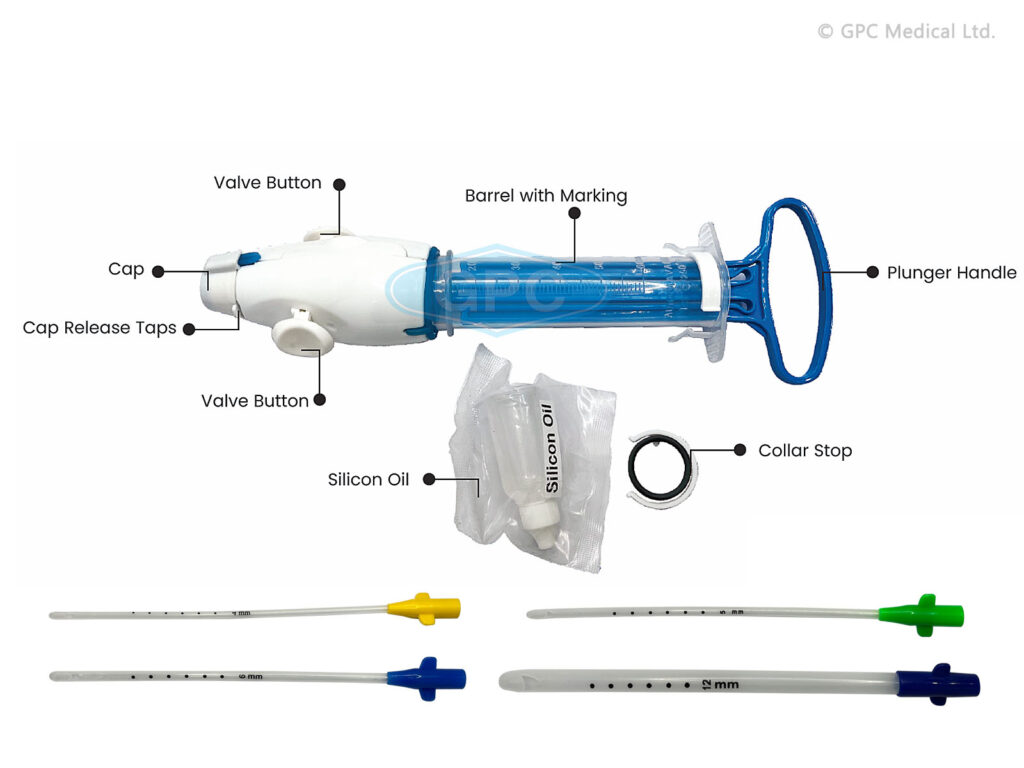
4. Prepare the MVA Syringe:

  • Assemble the syringe, close the pinch valve, and pull back on the plunger until the plunger arms lock.
  • For molar pregnancy, have three syringes ready. For very early pregnancy, insert the cannula without prior cervical dilation.

5. Administer Oxytocin or Ergometrine: Give oxytocin 10 units IM or ergometrine 0.2 mg IM to firm the myometrium and reduce perforation risk.

6. Perform a Bimanual Pelvic Examination: Re-assess the size and position of the uterus and the conditions of the fornices.

7. Insert Sterile Speculum and Visualize the Cervix: Apply antiseptic solution to the vagina and cervix, especially around the os.

8. Check the Cervix for Tears or Protruding POC: Remove any products of conception (POC) with ring (or sponge) forceps.

9. Gently Grasp the Anterior Lip of the Cervix: Use ring forceps or a single-toothed tenaculum. If using a tenaculum, first inject 1 mL of 0.5% lignocaine solution into the cervix.

10. Dilate the Cervix if Needed: For missed abortion or prolonged retention of POC, use mechanical or osmotic dilators, or cervical priming with mifepristone or prostaglandin.

11. Insert the Cannula: While applying gentle traction to the cervix, insert the cannula through the cervix into the uterine cavity just past the internal os.

12. Attach the Prepared MVA Syringe: Hold the ring forceps or tenaculum and the end of the cannula in one hand and the syringe in the other. Release the pinch valve(s) to transfer the vacuum to the uterine cavity.

13. Evacuate Remaining Contents: Gently rotate the syringe from side to side and move the cannula back and forth within the uterine cavity without losing vacuum.

14. Check for Signs of Completion: Look for red or pink foam without tissue, a grating sensation, and the uterus contracting around the cannula.

15. Withdraw the Cannula: Detach the syringe, place the cannula in decontamination solution, and empty the syringe contents into a strainer.

16. Perform a Bimanual Examination: Check the size and firmness of the uterus post-procedure.

17. Inspect the Tissue Removed: Ensure complete evacuation, assess for molar pregnancy, and if necessary, strain and rinse the tissue for examination.

18. Address Absence of POC: If no POC are seen, consider complete abortion, breakthrough bleeding, or possible ectopic pregnancy.

19. Reinsert Speculum and Examine for Bleeding: If persistent bleeding or soft uterus, repeat evacuation.

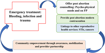
Post-Procedure Care: Administer paracetamol 500 mg as needed, consider antibiotics, encourage the woman to eat, drink, and walk, and offer other health services.

Discharge Uncomplicated Cases: Discharge within 1-2 hours, advising on symptoms that require immediate attention.

Precautions for Performing MVA:

  • Delay the procedure if conditions like shock, severe vaginal bleeding, or intra-abdominal injury are present, and stabilize the patient first.
  • Stabilization involves oxygen, IV fluids, antibiotics for sepsis, and blood transfusion if needed.

Shock Management:

  • Rapid, weak pulse, low blood pressure, pallor, sweatiness, rapid breathing, anxiousness, confusion, or unconsciousness.
  • Treat with oxygen, IV fluids, antibiotics, and blood transfusion if necessary.

Severe Vaginal Bleeding:

  • Heavy bright red bleeding, pallor, and blood-soaked materials.
  • Assess all bleeding sources, stabilize, and evacuate POC.

Intra-Abdominal Injury:

  • Distended abdomen, decreased bowel sounds, rigid abdomen, rebound tenderness, nausea, vomiting, pain, fever, or cramping.
  • Immediate management with IV fluids, antibiotics, and potential surgery. Perform MVA after stabilization.

POST ABORTION CARE Read More »

OPPORTUNISTIC INFECTIONS IN HIV/AIDS

OPPORTUNISTIC INFECTIONS IN HIV/AIDS

OPPORTUNISTIC INFECTIONS IN HIV/AIDS

Opportunistic infections (OIs) are infections affecting HIV patients with weakened immunity, indicated by a white blood cell count below 200 cells/cm³ (14%).

  • Advanced HIV infection makes individuals vulnerable to opportunistic infections or malignancies. These infections exploit the weakened immune system.
  • Childhood acquisition of OIs and HIV often occurs from infected mothers.
  • Women living with HIV are more prone to co-infections with opportunistic pathogens, increasing the risk of transmission to their infants.
  • Adolescents with HIV, including long-time survivors of perinatal infection, are increasingly common. Treatment guidelines also apply to youth living with HIV who have not yet completed pubertal development.

Examples of Opportunistic Infections

Category

Infection

Explanation

Bacterial OIs

Pneumococcal pneumonia

A bacterial infection causing severe respiratory illness, commonly affecting HIV patients due to weakened immunity.

Pulmonary tuberculosis

A serious infectious disease that primarily affects the lungs, prevalent in HIV patients due to compromised immune defenses.

Salmonellosis

An infection caused by Salmonella bacteria, leading to severe gastrointestinal symptoms, more common in immunocompromised individuals.

Extra-pulmonary tuberculosis

Tuberculosis infection occurring outside the lungs, such as in the lymph nodes or bones, often seen in advanced HIV cases.

Viral OIs

Herpes zoster

Also known as shingles, caused by the reactivation of the chickenpox virus, leading to painful skin rashes in HIV patients.

Recurrent/disseminated viral herpes simplex

Chronic or widespread herpes simplex virus infections, more severe and frequent in individuals with HIV.

Parasitic OIs

Pneumocystis carinii pneumonia

A fungal infection (previously classified as parasitic) causing severe lung disease, a common and life-threatening infection in HIV patients.

Toxoplasmosis

An infection caused by the Toxoplasma gondii parasite, leading to severe neurological issues in immunocompromised individuals like those with HIV.

Fungal OIs

Cryptosporidium

A parasitic infection causing severe diarrhea, often found in HIV patients due to their weakened immune systems.

Oro-pharyngeal candida

A fungal infection in the mouth and throat, also known as thrush, common in HIV patients.

Candida Esophagitis

A severe fungal infection of the esophagus, causing difficulty in swallowing and chest pain, prevalent in advanced HIV cases.

Histoplasmosis

A fungal infection caused by inhaling Histoplasma spores, leading to lung disease, more severe in immunocompromised patients.

Coccidioidomycosis

A fungal disease also known as Valley fever, causing respiratory issues, especially severe in those with weakened immune systems.

Cryptococcal meningitis

A life-threatening fungal infection of the brain and spinal cord, common in advanced HIV/AIDS patients.

Opportunistic Cancers

Invasive cervical cancer

Cancer caused by the human papillomavirus (HPV), more prevalent and aggressive in women with HIV.

Kaposi sarcoma

A cancer caused by human herpesvirus 8 (HHV-8), leading to lesions on the skin and other organs, commonly seen in HIV patients.

Non-Hodgkin lymphoma

A type of cancer affecting the lymphatic system, more common and aggressive in individuals with HIV.

Other OIs

Oral hairy leukoplakia

A condition characterized by white patches on the tongue, caused by Epstein-Barr virus, indicating immunosuppression in HIV patients.

Leukoencephalopathy

A rare, progressive viral disease affecting the white matter of the brain, often seen in severe immunocompromised states like advanced HIV.

Progressive multifocal leukoencephalopathy

A demyelinating disease of the central nervous system caused by the JC virus, highly fatal in HIV patients.

Causes of Opportunistic Infections:

  • Poor adherence to treatment
  • Presence of other diseases (e.g., juvenile diabetes mellitus)
  • Delay in identification of the infection
  • High viral load
  • Poor nutrition
  • Exposure to opportunistic infectious agents
  • Ingestion of substances contaminated with opportunistic infectious agents
  • Missing out on immunization programs
  • Poor hygiene
  • Poor sanitation
  • Poor ventilation

Prevention of Opportunistic Infections:

  • Avoidance of contact with the disease agents
  • Proper treatment of other underlying diseases
  • Adherence to HIV drug treatment
  • Immunization of children against killer diseases
  • Ensuring that children consume well-cooked food and boiled water
  • Early identification and treatment of opportunistic diseases
  • Health education of the family and infected child about opportunistic infections

HEPATITIS B

Hepatitis B is a chronic liver infection characterized by inflammation of hepatocytes caused by the hepatitis B virus.

Transmission:

  • High: Blood
  • Moderate: Semen, Urine, Serum, Wound exudate, Vaginal fluid
  • Low/Not Detectable: Saliva, Feces, Sweat, Tears, Breast milk

Stages of Hepatitis B:

  1. Immune Tolerance: Represents the incubation period, lasting approximately 2-4 weeks in healthy adults, and often decades in newborns.
  2. Immune Active/Immune Clearance: Inflammatory reaction occurs with active viral replication. Duration varies; for acute infection, approximately 3-4 weeks; for chronic infection, up to 10 years.
  3. Inactive Chronic Infection: Host targets infected hepatocytes and HBV, with low or no measurable viral replication in serum. Anti-HBe can be detected.
  4. Chronic Disease: Chronic HBeAg-negative disease may emerge.
  5. Recovery: Virus undetectable in blood, antibodies to viral antigens produced.

Clinical Features:

Symptoms can be symptomatic or asymptomatic:

  • Weakness, malaise, low-grade fever
  • Nausea, loss of appetite, vomiting
  • Pain or tenderness over right upper abdomen
  • Jaundice, dark urine, severe pruritus
  • Enlarged liver
  • Complications: liver cirrhosis, hepatocarcinoma

Investigations:

  • Hepatitis B surface antigen positive for >6 months
  • Hepatitis B core antibody: Negative IgM and Positive IgG to exclude acute hepatitis B infection
  • Liver tests, repeated at 6 months
  • HBeAg (can be positive or negative)
  • HBV DNA if available
  • HIV serology
  • APRI (AST to Platelets Ratio Index): a marker for fibrosis
  • Alpha fetoprotein at 6 months
  • Abdominal ultrasound at 4-6 months

Management:

General Principles:

  • Screen for HIV and refer if positive.
  • Refer to a regional hospital for specialist management if HIV is negative.
  • Antiviral treatment is given to prevent complications and usually for life.
  • Patients with chronic hepatitis B need periodic monitoring and follow-up for life.
  • Periodic screening for hepatocarcinoma with alfa fetoprotein and abdominal ultrasound once a year.

Treatment with Antivirals:

  • Treat with antivirals based on specific criteria.

First-line antivirals:

  • Adults and children >12 years or >35 kg: tenofovir 300 mg once a day
  • Child 2-11 years (>10 kg): Entecavir 0.02 mg/kg

Health Education:

  • Management is lifelong.
  • Bed rest is recommended.
  • Avoid alcohol as it worsens the disease.
  • Immunization of household contacts.
  • Do not share items that the patient puts in the mouth (e.g. toothbrushes, cutlery, razor blades).

OPPORTUNISTIC INFECTIONS IN HIV/AIDS Read More »

Post-exposure prophylaxis (PEP)

Post-exposure prophylaxis (PEP)

Post-exposure prophylaxis (PEP)

Post-exposure prophylaxis (PEP) involves the short-term use of antiretroviral medications to reduce the risk of acquiring HIV infection after potential exposure, either occupational or non-occupational.

Types of Exposure:

  • Occupational Exposures: Occur in healthcare or laboratory settings, including needle stick injuries or body fluid splashes.
  • Non-occupational Exposures: Include unprotected sex, assault (e.g., rape), and accidents.

Steps for Providing PEP:

Step 1: Clinical Assessment and First Aid

  • Rapidly assess the client to evaluate exposure and risk, providing immediate care.
  • For needle stick injuries: Avoid squeezing, wash the site with mild disinfectant, and do not use strong antiseptics.
  • For body fluid splashes on intact skin: Wash the area immediately with mild disinfectant.

Step 2: Eligibility Assessment

  • Provide PEP if exposure occurred within 72 hours and the exposed individual is HIV-negative.
  • PEP is not provided if the individual is already HIV-positive, the source is known to be HIV-negative, or if exposure involves bodily fluids posing minimal risk.

Step 3: Counselling and Support

  • Provide comprehensive counseling on HIV risk, PEP benefits and side effects, adherence, and support for further assistance, especially in cases of sexual assault.

Step 4: Prescription

  • PEP should be started as early as possible, not beyond 72 hours of exposure.
  • Recommended regimens include:
  • For pregnant mothers/adults: TDF+3TC+ATV/r.
  • For children: ABC+3TC+LPV/r.
  • A complete course of PEP should run for 28 days.
  • Do not delay the first doses because of a lack of baseline HIV test.
  • Document the event and patient management in the PEP register (ensure confidentiality of patient data).

Step 5: Provide follow-up

  • Discontinue PEP after 28 days.
  • Perform follow-up HIV testing three months after exposure.
  • Counsel and link to HIV clinic for care and treatment if HIV-positive.
  • Provide prevention and education/risk reduction counselling if HIV-negative.


ORAL PRE-EXPOSURE PROPHYLAXIS (PrEP)

PrEP involves using ARV drugs by individuals not infected with HIV to prevent HIV acquisition.

Where will PrEP services be offered?

Initially, PrEP will be available in select accredited ART sites with the capacity and funding for comprehensive services. Further rollout depends on outcomes. PrEP isn’t yet available in all public health facilities.

The process of providing pre-exposure prophylaxis (PrEP)

Step 1: Eligibility for PrEP

PrEP is suitable for HIV-negative individuals at high risk, including those with multiple sexual partners, engaging in transactional sex, injecting drugs or alcohol abuse, multiple STI occurrences, discordant couples, recurrent PEP users, and those engaging in anal sex.

Step 2: Screening for PrEP eligibility

After meeting criteria:

  • Confirm HIV-negative status.
  • Rule out acute HIV infection.
  • Assess hepatitis B status; negative indicates PrEP eligibility, positive requires management.
  • Assess contraindications to TDF/FTC.

Step 3: Steps to initiation of PrEP

Provide risk-reduction and adherence counselling:

  • Distribute condoms and educate on usage.
  • Develop a medication adherence plan.
  • Prescribe TDF (300mg) and FTC (200mg) once daily.
  • Initially, provide a 1-month TDF/FTC prescription with a follow-up in 1 month.
  • Counsel on TDF/FTC side effects.

Follow-up/monitoring clients on PrEP

  • Schedule a two-month follow-up after the initial visit, then quarterly.
  • Conduct HIV antibody tests every three months.
  • Perform pregnancy tests for women based on clinical history.
  • Review understanding of PrEP, barriers to adherence, tolerance, and side effects.
  • Evaluate and support PrEP adherence at each visit.
  • Assess for STI symptoms and treat as needed.

Guidance on discontinuing PrEP

Discontinue PrEP if:

  • HIV infection occurs.
  • Risk of HIV acquisition decreases due to lifestyle changes.
  • Intolerable toxicities or side effects arise.
  • Chronic non-adherence persists despite intervention.
  • Personal choice.
  • In sero-discordant relationships, if the positive partner achieves sustained viral load suppression (condoms should still be used consistently).

MOTHER-TO-CHILD TRANSMISSION OF HIV

Approximately one-third of the women who are infected with HIV can pass it to their babies.

Elements of Elimination of Mother-to-Child Transmission

  • Prong 1: Primary prevention of HIV infection for women and men of reproductive age, including adolescents.
  • Prong 2: Prevention of unintended pregnancies among women living with HIV, including adolescents and their partners.
  • Prong 3: Prevention of HIV transmission from women living with HIV to their infants, including pregnant and breastfeeding women, as well as adolescents living with HIV.
  • Prong 4: Provision of treatment, care, and support to women infected with HIV, their children, and their families, including women living with HIV and their families.

Cause and Time of Transmission

  • During pregnancy: 15-20%
  • During labour and delivery: 60%-70%
  • After delivery through breastfeeding: 15%-20%

Predisposing Factors

  • High maternal viral load
  • Depleted maternal immunity (e.g., very low CD4 count)
  • Prolonged rupture of membranes
  • Intrapartum haemorrhage and invasive obstetrical procedures
  • Increased risk for the first twin compared to the second twin in twin pregnancies
  • Premature birth poses a higher risk compared to full-term birth
  • Mixed feeding carries a higher risk than exclusive breastfeeding or replacement feeding

Investigations

  • Blood: HIV serological test
  • HIV DNA PCR testing of babies

Management

All HIV services for pregnant mothers are offered in the MCH clinic. After delivery, mother and baby will remain in the MCH postnatal clinic until the HIV status of the child is confirmed. Then, they will be transferred to the general ART clinic.

The current policy aims at the elimination of Mother-to-Child Transmission (eMTCT) through a continuum of care.

Management of HIV Positive Pregnant Mother

Key Interventions for eMTCT:

  • Routine HIV Counseling and Testing during ANC (at 1st contact. If negative, repeat HIV test in the third trimester/ labour).
  • Enrolment in HIV care if the mother is positive and not yet on treatment.
  • If the mother is already on ART, perform viral load and continue the current regimen.
  • ART in pregnancy, labour, post-partum, and for life – Option B+.

Recommended ARV for option B+:

One daily Fixed Dose Combination (FDC) pill containing TDF + 3TC + EFV started early in pregnancy irrespective of the CD4 cell count and continued during labor and delivery, and for life.

Alternative regimens for women who may not tolerate the recommended option are:

  • If TDF contraindicated: ABC+3TC+EFV
  • If EFV contraindicated: TDF + 3TC + ATV/r
  • TDF and EFV are safe to use in pregnancy.
  • Those newly diagnosed during labor will begin HAART for life after delivery.

Prophylaxis for Opportunistic Infections

Cotrimoxazole 960 mg 1 tab daily during pregnancy and postpartum –– Mothers on cotrimoxazole DO NOT NEED IPTp with SP for malaria.

Care of HIV Exposed Infant

HIV-exposed infants should receive care at the mother-baby care point together with their mothers until they are 18 months old. The goals of HIV-exposed infant care services are:

  • To prevent the infant from being HIV infected.
  • Among those who get infected: to diagnose HIV infection early and treat it.
  • Offer child survival interventions to prevent early death from preventable childhood illnesses.

The HIV Exposed Infant and the mother should consistently visit the health facility at least nine times during that period i.e  (i.e., at 6, 10 and 14 weeks, then at 5, 6, 9,  12, 15 and 18 months). 

Nevirapine Prophylaxis

Provide NVP syrup from birth for 6 weeks: Give NVP for 12 weeks for babies at high risk, that is breastfeeding infants who mothers: 

  • Have received ART for 4 weeks or less before delivery; or 
  • Have VL >1000 copies in 4 weeks before delivery; or 
  • Diagnosed with HIV during 3rd trimester or breastfeeding period (Postnatal) 

Do PCR at 6 weeks (or at first encounter after this age) and start cotrimoxazole prophylaxis 

  • If PCR positive, start treatment with ARVs and cotrimoxazole and repeat PCR (for confirmation) 
  • If PCR negative and the baby never breastfed, the child is confirmed HIV negative. Stop cotrimoxazole, continue clinical monitoring and do HIV serology test at 18 months. 
  • If PCR is negative but the baby has breastfed/is breast feeding, start/continue cotrimoxazole prophylaxis and repeat PCR 6 weeks after stopping breastfeeding.
  • Follow up any exposed child and do PCR if they develop any clinical symptom suggestive of HIV at any  time and independently of previously negative results.
  • For negative infants, do serology at 18 months before final discharge.

Dosages of Nevirapine

Age Group

Weight Range

Dosage

Syrup Volume (10 mg/ml)

Child 0-6 weeks

2-2.5 Kg

10 mg once daily

1 ml

Child 0-6 weeks

>2.5 Kg

15 mg once daily

1.5 ml

Child 6 weeks – 12 weeks

Any weight

20 mg once daily

2 ml

Cotrimoxazole Prophylaxis: Provide cotrimoxazole prophylaxis to all HIV exposed infants from 6 weeks of age until they are proven to be uninfected.

  • Child <5 kg: 120 mg once daily  
  • Child 5-14.9 kg: 240 mg once daily 

Isoniazid (INH) Preventive Therapy (IPT): 

  • Give INH for six months to HIV-exposed infants who are exposed to TB.
  • Isoniazid 10 mg/kg + pyridoxine 25 mg daily 
  • For newborn infants, if the mother has TB disease and has been on anti-TB drugs for at least two weeks before delivery, INH prophylaxis is not required. 

Immunization

  • Immunise HIV exposed children as per national immunisation schedule.
  • In case of missed BCG at birth, do not give if the child has symptomatic HIV.
  • Avoid yellow fever vaccine in symptomatic HIV.  
  • Measles vaccine can be given even in symptomatic HIV.

Counselling on Infant Feeding Choice

  • Explain the risks of HIV transmission by breastfeeding (15%) and other risks of not breastfeeding (malnutrition, diarrhoea).
  • Mixed feeding may also increase the risk of HIV transmission and diarrhoea.
  • Tell her about options for feeding, advantages, and risks.
  • Help her to assess choices, decide on the best option, and then support her choice.

Feeding Options

  • Recommended option: Exclusive breastfeeding, then complementary feeding after the child is 6 months old.
  • Exclusive breastfeeding stopping at 3-6 months old if replacement feeding is possible after this.
  • If replacement feeding is introduced early, the mother must stop breastfeeding.
  • Replacement feeding with home-prepared formula or commercial formula and then family foods (provided this is acceptable, feasible, safe, and sustainable/affordable).

If Mother Chooses Breastfeeding

The risk may be reduced by keeping the breasts healthy (mastitis and cracked nipples raise HIV infection risk).

Advise exclusive breastfeeding for 3-6 months.

If Mother Chooses Replacement Feeding

  • Counsel and teach her on safe preparation, hygiene, amounts, times to feed the baby, etc.
  • Follow up within a week from birth and at any visit to the health facility.

Post-exposure prophylaxis (PEP) Read More »

Management of HIV/AIDs and hepatitis

Management of HIV/AIDs and Hepatitis

HIV/AIDS 

HIV (Human Immunodeficiency Virus) is a virus that attacks the body’s immune system, specifically the CD4 cells (T cells), which are important for immune defence. 

If untreated, HIV can lead to AIDS (Acquired Immunodeficiency Syndrome), a condition where the immune system is severely weakened. 

HIV is a lenti-virus (slow and long acting) and belongs to the Retroviruses group. HIV invades the helper T cells to replicate itself thereby limiting the body’s ability to fight infection . HIV is the virus that causes AIDS, and it has no cure.

Types of HIV 

  1. HIV-1: This is the most common and widespread type of HIV, accounting for the vast majority of HIV infections globally. It is highly infectious and has several subtypes (or clades), labelled A through K. HIV-1 is the primary cause of the global HIV pandemic and is more aggressive in its progression to AIDS compared to HIV-2.
  2. HIV-2: This type is less common and primarily found in West Africa. It is less transmissible and generally progresses more slowly to AIDS than HIV-1. There are fewer subtypes of HIV-2, labelled A through H. 

Characteristics of HIV/AIDS.

  1. RNA Virus: HIV is an RNA virus that can convert its RNA into DNA using reverse transcription.
  2. Receptor Binding: The virus has specific proteins on its surface that bind to receptors on host cells, allowing entry.
  3. Heat Sensitivity: HIV is easily destroyed by high temperatures (around 600°F).
  4. Human Host: HIV can only survive and replicate in humans; it dies outside the human body and when the host dies.
  5. Immune Attack: The virus primarily targets and destroys white blood cells, especially CD4+ T cells.
  6. Rapid Replication: HIV replicates rapidly, producing billions of new virions each day.
  7. Disease Progression: HIV infection progresses through stages, eventually leading to AIDS if untreated.
  8. Latency: After initial infection, HIV can hide in cells and become dormant before reactivating later.
  9. Genetic Diversity: The virus has many subtypes and mutates quickly, making treatment and vaccine development challenging.
  10. Immune Evasion: HIV evades the immune system by mutating and hiding within cells.
  11. Transmission: HIV is transmitted through contact with infected bodily fluids, such as blood, semen, vaginal fluids, and breast milk.
  12. ART Treatment: Antiretroviral therapy (ART) can control HIV, preventing the progression to AIDS and allowing individuals to live longer, healthier lives.

Epidemiology.

According to the August 2017 Uganda Population-Based HIV Impact Assessment report, the prevalence of HIV among adults aged 15 to 64 in Uganda is 6.2%, with a higher rate among females (7.6%) compared to males (4.7%). This equates to approximately 1.2 million adults living with HIV in this age group. HIV prevalence is notably higher in women living in urban areas (9.8%) than in rural areas (6.7%).

 Among children aged 0-14, the HIV prevalence is 0.5%, corresponding to about 95,000 children living with HIV. The viral load suppression (VLS) rate among HIV-positive adults is 59.6%, with higher rates in females (62.9%) than males (53.6%). For children aged 0-14, the VLS rate is 39.3%. HIV prevalence peaks at 14.0% among men aged 45 to 49 and 12.9% among women aged 35 to 39. There is a significant gender disparity among young adults, with HIV prevalence nearly four times higher in females than males aged 15 to 19 and 20 to 24. Additionally, HIV prevalence is almost three times higher in adults aged 20-24 compared to those aged 15-19.

Modes of HIV Transmission

Sexual Contact:

  • Unprotected Vaginal Sex: HIV can be transmitted through vaginal fluids and semen during unprotected vaginal intercourse..

Blood-to-Blood Contact:

  • Sharing Needles: Using contaminated needles or syringes, common among intravenous drug users, can transmit HIV.
  • Blood Transfusions: Although rare in countries with stringent blood screening, HIV can be transmitted through infected blood transfusions.
  • Exposure to Contaminated Blood: Health care workers can be at risk through needle stick injuries or contact with open wounds.

Mother-to-Child Transmission:

  • During Pregnancy: HIV can cross the placenta from mother to baby.
  • During Childbirth: The baby can be exposed to HIV in the mother’s blood and vaginal fluids during delivery.
  • Breastfeeding: HIV can be transmitted through breast milk from an infected mother to her child.

Other Modes:

  • Contaminated Medical Equipment: Use of non-sterile instruments during medical or dental procedures can transmit HIV.
  • Organ and Tissue Transplants: Transplantation of infected organs or tissues, though rare due to screening practices, can transmit HIV.

Less Common Modes:

  • Tattooing and Piercing: If non-sterile needles are used, there is a risk of HIV transmission.
  • Contact Sports: Although extremely rare, transmission can occur if both participants have open wounds.

Factors That Facilitate Mother-to-Child Transmission of HIV

Maternal Factors:

Viral Load and Immune Status:

  • High Viral Load: Higher levels of HIV in the mother’s blood increase the risk of transmission to the baby.
  • Low CD4 Count: A weakened immune system due to low CD4 counts enhances transmission risk.
  • Maternal Acquisition of HIV: New HIV infections during pregnancy or lactation significantly increase transmission risk.

Infections and Inflammation:

  • Vaginal Infections: Infections such as bacterial vaginosis can elevate the risk of HIV transmission.
  • Chorioamnionitis: Inflammation of the foetal membranes due to infection can facilitate HIV transmission.

Access to Antiretroviral Therapy (ART):

  • Lack of ART: Mothers who do not receive ART are more likely to transmit HIV.
  • Poor Adherence to ART: Inconsistent use of ART reduces its effectiveness in preventing transmission.
  • Timing of ART Initiation: Starting ART late in pregnancy or not at all reduces its preventive benefits.

Socioeconomic Factors:

  • Lack of Healthcare Access: Limited access to prenatal care and HIV testing can lead to missed opportunities for prevention.
  • Education and Awareness: Lack of knowledge about HIV transmission and prevention strategies among pregnant women.

Nutritional Status:

  • Poor Maternal Nutrition: Malnutrition can weaken the mother’s immune system, increasing the risk of transmission.
Labour and Delivery Factors:

Delivery Method:

  • Vaginal Delivery: Higher risk of transmission compared to elective caesarean section, especially if the mother has a high viral load.
  • Prolonged/Difficult Labour: Increased exposure to maternal fluids during extended or complicated labour can raise the risk.

Prematurity:

  • Premature Birth: Prematurity can increase the risk of transmission due to underdeveloped immune systems in infants.

Membrane Rupture:

  • Prolonged Rupture of Membranes (PROM): Rupture lasting more than 4 hours before delivery increases the risk of HIV transmission.

Invasive Monitoring and Procedures:

  • Use of invasive monitoring or procedures during labour can increase the risk of HIV transmission.
Postnatal Feeding Factors:

Breastfeeding Practices:

  • Prolonged Breastfeeding: Longer duration of breastfeeding increases the risk of HIV transmission.
  • Breast Health: Conditions like sore nipples, abscesses, or mastitis can increase the risk.
  • Mixed Feeding: Combining breastfeeding with other foods or fluids increases transmission risk. Exclusive breastfeeding for the first 3-6 months does not show excess transmission compared to formula feeding alone.

Exclusive Breastfeeding:

  •  Exclusive breastfeeding means providing breast milk only, without additional fluids, water, food, teats, or pacifiers, and involves on-demand feeding.

Oral Health in Infants:

  • Oral Thrush: Presence of oral thrush in breastfed infants can increase the risk of HIV transmission.
Life-Cycle-of HIV (1)

Phases of HIV Entry into Host Cells

  1. Binding: The HIV virus first attaches to the CD4 receptors on the surface of the host cell, typically a type of immune cell called a CD4+ T lymphocyte. HIV’s envelope protein, gp120, specifically binds to the CD4 receptor. This interaction triggers a conformational change in gp120 that allows it to also interact with a co-receptor, usually CCR5 or CXCR4, on the host cell surface. This dual receptor binding is essential for the virus to proceed to the next step.
  2. Fusion: After binding, the HIV viral envelope fuses with the host cell membrane, allowing the viral contents to enter the host cell. The conformational change in gp120 caused by CD4 and co-receptor binding exposes another viral protein, gp41. gp41 facilitates the merging of the viral envelope with the host cell membrane, creating a fusion pore through which the viral capsid containing the viral RNA and enzymes can enter the host cell cytoplasm.
  3. Reverse Transcription: Once inside the host cell, the viral RNA genome is reverse transcribed into DNA. The enzyme reverse transcriptase, carried within the viral capsid, converts the single-stranded viral RNA into double-stranded DNA. This process is error-prone, leading to a high mutation rate which contributes to the virus’s ability to evade the immune system and develop drug resistance.
  4. Integration: The newly synthesized viral DNA is integrated into the host cell’s genome. The viral DNA is transported into the host cell nucleus, where the enzyme integrase integrates it into the host cell’s DNA. This integrated viral DNA is known as a provirus and can remain dormant for a period before becoming active.
  5. Replication: Once integrated, the viral DNA can be transcribed and translated to produce new viral RNA and proteins. The host cell’s machinery reads the integrated viral DNA and begins to produce viral RNA. Some of this RNA will serve as genomes for new viral particles, while others will be used to produce viral proteins through the process of translation.
  6. Assembly: New viral particles are assembled within the host cell. The newly made viral RNA and proteins are transported to the host cell’s surface, where they assemble into new immature viral particles. This assembly process involves the gathering of viral components into a budding virion.
  7. Budding: The new viral particles bud off from the host cell, acquiring an envelope from the host cell membrane in the process. The immature viral particles bud off from the host cell, during which they incorporate a portion of the host cell’s membrane as their envelope. The viral enzyme protease then cleaves certain viral precursor proteins into their mature forms, resulting in a fully mature and infectious virus ready to infect other cells.

Clinical Manifestations of HIV/AIDS

The World Health Organization (WHO) has established a staging system to classify HIV infection and disease progression:

Clinical Stage I:

  1. Asymptomatic: No symptoms of HIV-related illness.
  2. Persistent Generalized Lymphadenopathy: Enlargement of lymph nodes lasting more than three months.
  3. Performance Scale 1: Asymptomatic with normal activity level.

Clinical Stage II:

  1. Moderate Weight Loss: Less than 10% of presumed or measured body weight lost.
  2. Minor Muco-cutaneous Manifestations: Skin conditions like seborrheic dermatitis, prurigo, or fungal nail infections.
  3. Herpes Zoster: History of shingles within the last five years.
  4. Recurrent Upper Respiratory Tract Infections: Such as bacterial sinusitis, tonsillitis, or otitis media.
  5. Performance Scale 2: Symptomatic but normal activity level.

Clinical Stage III:

  1. Severe Weight Loss: More than 10% of presumed or measured body weight lost.
  2. Unexplained Chronic Diarrhoea: Lasting more than one month.
  3. Unexplained Prolonged Fever: Constant or intermittent, lasting more than one month.
  4. Oral Candidiasis: Oral thrush, a fungal infection.
  5. Oral Hairy Leukoplakia: White patches on the tongue or mouth.
  6. Pulmonary Tuberculosis: Active TB infection.
  7. Severe Bacterial Infections: Such as pneumonia, pyomyositis, or bacteremia.
  8. Acute Necrotizing Ulcerative Gingivitis: Severe gum disease.
  9. Unexplained Anaemia, Neutropenia, or Thrombocytopenia: Abnormal blood counts.
  10. Performance Scale 3: Bedridden for less than 50% of the day during the last month.

Clinical Stage IV:

  1. HIV Wasting Syndrome: Weight loss of more than 10% with chronic diarrhoea or prolonged fever.
  2. Pneumocystis Pneumonia (PCP): A severe fungal lung infection.
  3. Toxoplasmosis of the Brain: Brain infection caused by the Toxoplasma parasite.
  4. Cryptosporidiosis: Parasitic infection causing prolonged diarrhea.
  5. Cytomegalovirus Infection: A viral infection affecting various organs.
  6. Progressive Multifocal Leukoencephalopathy (PML): Brain infection causing neurological symptoms.
  7. Lymphoma: Cancer of the lymphatic system.
  8. Kaposi’s Sarcoma: Cancerous skin lesions caused by a herpesvirus.
  9. HIV Encephalopathy: Cognitive and/or motor dysfunction due to HIV infection.
  10. Atypical Disseminated Leishmaniasis: Parasitic infection affecting multiple organs.
  11. Symptomatic HIV-Associated Nephropathy or Cardiomyopathy: Kidney or heart disease associated with HIV.
  12. Performance Scale 4: Bedridden for more than 50% of the day during the last month.
Diagnostic Measures for HIV/AIDS

Diagnostic Measures for HIV/AIDS

Pre and Post-Counselling and Consent: Essential for all diagnostic procedures unless in specific circumstances:

  • Testing of very sick, unconscious, symptomatic, or mentally ill individuals by healthcare teams for better patient management.
  • Routine testing for individuals likely to pose a risk of HIV infection to others, such as pregnant and breastfeeding mothers, sexual offenders and survivors, and blood or organ donors. These individuals must still be given the opportunity to know their status.

Criteria for Diagnosis: Diagnosis based on:

  • Clinical Staging Criteria.
  • Positive HIV Blood Test: Confirmation of HIV infection through serological (antibody) testing.

Testing Protocol: Testing for Adults and Children >18 Months:

  • Serological (Antibody) Testing: Most common method. Due to the window period between infection and antibody production, negative individuals should be re-tested after three months if exposed.
  • Reactive Rapid Test: Requires confirmation before diagnosis.

Diagnostic Tests

Screening Tests:

  • ELISA (Enzyme-Linked Immunosorbent Assay) AglAb Tests: Commonly used to screen blood donations to exclude those in the window period.

Molecular Tests:

  • PCR (Polymerase Chain Reaction) Tests: Nucleic-Acid Amplification Testing (NAT) detects genetic material of HIV itself, not antibodies or antigens.

Considerations: Testing should consider:

  • Clinical status, medical history, and risk factors of the individual being tested.
  • Use of tests in conjunction with patient assessment for accurate diagnosis and appropriate care.

Immediate Connection to HIV Care

  • If positive, immediate referral to HIV care services for management and treatment initiation.
HIV Testing Provision Protocol uganda

HIV Testing Provision Protocol

Step 1: Pre-Test Information and Counseling

  • Provide information on HIV transmission, prevention measures, and testing benefits.
  • Discuss potential test results, available services, and ensure consent and confidentiality.
  • Conduct individual risk assessment and complete necessary documentation.

Step 2: HIV Testing

Perform blood-based testing.

  • For infants below 18 months: Use DNA PCR testing.
  • For individuals above 18 months: Conduct antibody testing as per testing algorithms.

Step 3: Post-Test Counseling (Individual/Couple)

  • Assess readiness to receive results and deliver them simply.
  • Address concerns, provide guidance on disclosure, partner testing, and risk reduction.
  • Offer information on basic HIV care, ART, and complete documentation.

Step 4: Linkage to Other Services

  • Provide information on available services and assist in completing referral forms.
  • Upon enrollment in services, record pre-ART enrollment numbers and transfer relevant information to ART registers.

Principles of HIV Testing Services (HTS)

  1. Confidentiality: Ensure privacy and confidentiality of test results.
  2. Consent: Obtain informed consent from individuals before testing.
  3. Counselling: Offer supportive counselling before and after testing.
  4. Correct Test Result: Ensure accuracy of test results through proper testing procedures.
  5. Connection to Other Services: Facilitate access to appropriate services for individuals testing positive.

Linkage from HIV Testing to Prevention, Care, and Treatment

Linkage is the process of connecting individuals who test positive for HIV to the necessary services. 

Successful linkage to care ensures that patients receive the services they need. For HIV-positive clients, linkage should occur promptly, within seven days if within the same facility, and within 30 days for referrals between facilities or from the community. Lay providers are recommended as linkage facilitators. 

Types of Linkages:
  • Internal Facility Linkage: Connecting patients within the same facility.
  • Inter-Facility Linkage: Connecting patients to another facility.
  • Community-Facility Linkage: Connecting clients from the community to a health facility.

Internal Facility Linkage Steps:

  1. Post-Test Counselling: Provide accurate results and information about available care.
  2. Next Steps Discussion: Describe the care and treatment process, emphasizing early treatment benefits.
  3. Address Barriers: Identify and overcome any obstacles to linkage.
  4. Involvement: Involve the patient and family in decision-making.
  5. Documentation: Complete client and referral forms.
  6. Escort to Clinic: A linkage facilitator escorts the client to the ART clinic.
  7. Enrollment: Register the patient, open an ART file, and provide preparatory counselling.
  8. Initiation: Start ART if ready, and continue with counselling support.
  9. Integrated Care: Coordinate other services if needed.
  10. Follow-Up: Ensure the patient attends appointments.

Inter-Facility and Community-Facility Linkages:

  • Inter-Facility Linkage: Refers to connecting patients to another facility. The referring facility should track referred patients and ensure enrollment within 30 days.
  • Community-Facility Linkage: Connects clients from the community to a health facility. Utilize community health systems and mobilize peer leaders for outreach and follow-up. Linkage should occur within 30 days after diagnosis.

Treatment Modalities of HIV/AIDS

Treatment Modality

Description

Antiretroviral Therapy (ART)

Suppresses viral load to undetectable levels, reducing morbidity, mortality, and transmission of HIV.

Treatment of Acute Bacterial Infections

Addresses immediate bacterial infections.

Prophylaxis and Treatment of Opportunistic Infections

Prevents and manages opportunistic infections.

Maintenance of Good Nutrition

Ensures adequate nutrition to support overall health.

Immunization

Administers vaccines to prevent opportunistic infections.

Management of AIDS-Defining Illnesses

Addresses specific illnesses associated with advanced HIV infection.

Psychological Support for the Family

Provides emotional support and guidance for affected families.

Palliative Care for the Terminally Ill

Offers comfort and support for patients nearing the end of life.

Antiretroviral Drug Treatment

Goal of ART: Suppress viral load to undetectable levels, reducing morbidity, mortality, and transmission of HIV.

When to Initiate ARV:

  • All HIV-infected children below 12 months.
  • Clinical AIDS
  • Mild to moderate symptoms and immunosuppression.

Process of Starting ART:

  1. Assess for opportunistic infections, defer ART if TB or cryptococcal meningitis present.
  2. Offer ART on the same day through an opt-out approach.
  3. If not ready for same-day initiation, agree on a timely ART preparation plan.

Available ARVs in Uganda

Drug Class

Examples

Nucleoside Reverse Transcriptase Inhibitors (NRTIs): Incorporate into the DNA of the  virus, thereby stopping the building process. 

Tenofovir (TDF), Zidovudine (AZT), Lamivudine (3TC), Abacavir (ABC)

Non-Nucleoside Reverse Transcriptase Inhibitors (NNRTIs): stop HIV production by binding directly onto the reverse transcriptase enzyme, and prevent the conversion of RNA to DNA.

Efavirenz (EFV), Nevirapine (NVP), Etravirine (ETV)

Integrase Inhibitors: interfere with the HIV DNA’s ability to insert itself into the host DNA and copy  itself.

Dolutegravir (DTG), Raltegravir (RAL)

Protease Inhibitors (PIs): prevent HIV from being successfully assembled and released from the infected CD4 cell.

Atazanavir (ATV), Lopinavir (LPV), Darunavir (DRV)

Entry Inhibitors:  prevent the HIV virus particle from infecting the CD4 cell.

Enfuvirtide (T-20), Maraviroc

Recommended First Line Regimens in Adults, Adolescents, Pregnant Women and Children

HIV management guidelines are constantly being updated according to evidence and public policy decisions. Always refer to the latest official guidelines.

The 2022 guidelines recommend DOLUTEGRAVIR (DTG) an integrase inhibitor as the anchor ARV in the preferred first and second-line treatment regimens for all HIV infected clients; children, adolescents, men, women (including pregnant women, breastfeeding women, adolescent girls and women of child bearing potential).

Patient Category

Preferred Regimens

Alternative Regimens

Adults and Adolescents


Adults (including pregnant women, breastfeeding mothers, and adolescents ≥30Kg)

TDF + 3TC + DTG

– If DTG is contraindicated: TDF + 3TC + EFV400

– If TDF is contraindicated: TAF + FTC + DTG 

– If TDF or TAF is contraindicated: ABC + 3TC + DTG 

– If TDF or TAF and DTG are contraindicated: ABC + 3TC + EFV400 

 – If EFV and DTG are contraindicated: TDF + 3TC + ATV/r or ABC + 3TC + ATV/r

Children


Children ≥20Kg – <30Kg

ABC + 3TC + DTG

– If DTG is contraindicated: ABC + 3TC + LPV/r (tablets) 

 – If ABC is contraindicated: TAF + FTC + DTG (for children >6 years and >25Kg) 

 – If ABC and TAF are contraindicated: AZT + 3TC + DTG

Children <20Kg

ABC + 3TC + DTG

– If intolerant or appropriate DTG formulations are not available: ABC + 3TC + LPV/r granules 

– If intolerant to LPV/r: ABC + 3TC + EFV (in children >3 years and >10Kg) 

 – If ABC is contraindicated: AZT + 3TC + DTG or LPV/r

Notes:

  • Contraindications for DTG include known diabetics, patients on anticonvulsants (carbamazepine, phenytoin, phenobarbital) – use the DTG screening tool prior to DTG initiation.
  • Contraindications for TDF and TAF include renal disease and/or GFR <60ml/min, weight <30Kg.
  • TAF can be used in subpopulations with bone density anomalies.
  • Children will be assessed individually for their ability to correctly take the different formulations of LPV.

Notes from Ministry of Health(Uganda)

  1. For clients on an ABC-3TC-DTG based regimen weighing >25 kg, use the fixed-dose combination of Abacavir/Lamivudine/Dolutegravir 600/300/50 mg instead of the separate pills of Abacavir/Lamivudine 600/300 mg plus Dolutegravir 50 mg.
  2. Use Abacavir/Lamivudine 600/300 mg for patients on the following regimens: ABC-3TC-ATV/r, ABC-3TC-LPV/r, and ABC-3TC-DRV/r.
  3. Use the single pill of Dolutegravir 50 mg for patients on AZT-3TC-DTG based regimens.
  4. For eligible patients on ATV/r and LPV/r, optimize to Dolutegravir.
  5. For PrEP, while the guidelines provide options for the use of either TDF/3TC 300/300 mg or TDF/FTC 300/200 mg, use TDF/FTC 300/200 mg for PrEP in terms of programmatic implementation.

Monitoring of ARV Treatment

The monitoring of patients on antiretroviral therapy (ART) serves several purposes:

  1. Assess Response to ART and Diagnose Treatment Failure
  2. Ensure Safety of Medicines: Identify Side Effects and Toxicity
  3. Evaluate Adherence to ART

Methods of Monitoring ARV Treatment

  1. Clinical Monitoring: Involves medical history and physical examination.
  2. Laboratory Monitoring: Includes various laboratory tests.
  • Viral Load Monitoring: Preferred for assessing response to ART and diagnosing treatment failure.
  • CD4 Monitoring: Recommended in specific scenarios.
  • Other Minor Laboratory Tests: Includes tests for specific indications.

Viral Load Monitoring

  • Preferred method for monitoring ART response. A patient who has been on ART for more than 6 months and is responding to ART should have viral suppression (VL <1000 copies/ml) irrespective of the sample type (either DBS or plasma). 
  • Provides an early and more accurate indication of treatment failure and the need to switch from first line to second-line drugs, hence reducing the accumulation of drug resistance mutations and improving  clinical outcomes. 
  • Early and accurate indication of treatment failure.
  • Differentiates between treatment failure and non-adherence.
  • Recommended frequency: Every six months for children and adolescents under 19 years.

CD4 Monitoring

  • Baseline CD4 count is essential for assessing opportunistic infection risk.
  • Recommended for patients with high viral load or advanced clinical disease.

Other Laboratory Tests

Tests

Indication

CrAg

Screen for cryptococcal infection

Complete Blood Count (CBC)

Assess anaemia risk

TB Tests

Suspected tuberculosis

Serum Creatinine

Assess kidney function

ALT, AST

Evaluate liver function

Lipid Profile, Blood Glucose

Assess metabolic health

Problems Associated with ARV Treatment

Immune Reconstitution Inflammatory Syndrome (IRIS)

IRIS is a spectrum of clinical signs and symptoms linked to immune recovery triggered by ART. It occurs in 10–30% of individuals starting ART, usually within the first 4–8 weeks.

  • Serious Forms: Most severe cases happen in patients co-infected with TB, Cryptococcus, Kaposi’s sarcoma, and herpes zoster.
  • Risk Factors: Include low CD4+ cell count (<50 cells/mm3) at ART initiation and disseminated opportunistic infections.
  • Management: Usually self-limiting; treat co-infections to reduce symptoms and reassure patients to maintain ART adherence.

Steps to Reduce IRIS Development

  1. Early HIV Diagnosis: Initiate ART before CD4 declines to below 200 cells/mm3.
  2. Optimal Management of Opportunistic Infections: Screen and treat infections before starting ART, especially TB and cryptococcus.

ARV Drug Toxicity

  • Range of Toxicities: ARVs can cause mild to life-threatening side effects.
  • Challenges: Differentiating between ARV toxicity and HIV complications can be complex.
  • Management: Assess patients for side effects at every clinic visit and take appropriate actions based on severity.

Management of ARV Side Effects/Toxicities

Category

Action

Severe, Life-threatening Reactions (e.g., SJS/TEN, severe hepatitis)

– Discontinue all ARVs immediately. 

– Manage the medical event and substitute offending drug when stable.

Severe Reactions (e.g., Hepatitis and Anemia)

– Substitute offending drug without stopping ART.

Moderate Reactions (e.g., Gynaecomastia, Lipodystrophy)

– Substitute with a drug in the same class or different class with a different toxicity profile. 

– Do not discontinue ART; continue if feasible.

Mild Reactions (e.g., Headache, Minor Rash, Nausea)

– Do not discontinue or substitute ART. 

– Provide reassurance and support to mitigate adverse reactions. 

– Counseling about the events.

Adherence Preparation, Monitoring, and Support

Sustaining adherence to Antiretroviral Therapy (ART) is essential for achieving HIV viral suppression, reducing drug resistance, and improving overall health outcomes. Conversely, poor adherence is a significant contributor to treatment failure. Regular assessment and reinforcement of adherence by the clinical team are critical components of HIV care.

Adherence Preparation: Preparing people to start antiretroviral therapy (ART) is an important step to achieving ART success.

  • Initiation Discussion: Healthcare providers should engage patients in detailed discussions regarding their readiness and willingness to commence ART. While healthcare providers should provide necessary information and guidance, the ultimate decision to initiate ART rests with the patient or caregiver.
  • 5 As Principles for Chronic Care: The clinical team should employ the “5 As” principles—Assess, Advise, Agree, Assist, and Arrange—to offer pre-ART adherence counselling and psychosocial support.

Steps in Preparation for ART:

  1. Assess: Evaluate patients’ understanding of HIV, ARVs, and potential adherence barriers.
  2. Advise: Provide patients with relevant information to empower them to enrol in treatment.
  3. Agree on: Develop an adherence plan and identify family and community support systems.
  4. Assist: Help patients identify and address potential barriers to adherence.
  5. Arrange for: Schedule appointments for ARV prescription, follow-up counselling sessions, and involvement in psychosocial support groups.
Barriers to Adherence of ART

Barrier

Adolescents

Pregnant or Breastfeeding Women

Adults

Key Populations

Psychosocial issues

Peer pressure, perceived need to conform


Inconsistent daily routine

Yes


Child abuse and neglect

Yes


Stigma and discrimination

Yes

Yes

Yes

Yes

Left out of decisions/limited discussion opportunities

Yes


Limited treatment literacy  or adherence counselling tools

Yes


Challenges during transition from paediatric to adolescent care

Yes


Pregnancy-related conditions (nausea/vomiting)


Yes


Suboptimal understanding of HIV, ART, eMTCT


Yes


Lack of partner disclosure/support


Yes


Non-disclosure


Yes

Yes


Gender-based violence (GBV)


Yes


Yes

Drug sharing


Yes


Service delivery barriers


Yes


Poor-quality clinical practices


Yes


Gaps in provider knowledge/training


Yes


Poor access to services


Yes


Social barriers (work schedules/job nature)


Yes


Forgetfulness


Yes


Lack of trust in providers/medicines


Yes


Lack of social support


Yes


Drug side effects


Yes


Pill burden


Yes


Inadequate information about ARVs


Yes


Alcohol and substance abuse

Yes


Yes

Yes

Provider attitude


Yes

High mobility


Yes

Lack of peer support


Yes

Lack of health worker knowledge about KPs


Yes


Methods of Monitoring Adherence to ART
  • Viral Load Monitoring: Considered the gold standard for assessing adherence and treatment response. It should be conducted six months after initiating treatment and annually thereafter.
  • Self-reporting: Rapid, inexpensive, and easily implemented, but may be subject to bias.
  • Pill Counting: Limited by patients potentially discarding tablets before clinic visits, but can be enhanced when combined with self-reported adherence.
  • Pharmacy Refill/Clinic Records: Provides reliable documentation of medication collection patterns and can indicate potential adherence challenges.
Adherence Support

Adherence support interventions should be provided to people on ART through the following interventions.

  • Peer counsellors: These include peer mothers in the eMTCT program, adolescent peers, expert clients  and other peers as patients and caregivers usually relate better to peers. 
  • Mobile phone calls and text messages: These should be used with the patient or caregiver consent. The patient or caregiver should provide the appropriate phone numbers to avoid accidental disclosure when messages are sent to a wrong person. 
  • Reminder devices like calendars, pill boxes and diaries can be used by clients. 
  • Behavioural skills training and medication adherence training: These include module based interventions  and those designed to improve life skills, attitudes, behavior and knowledge. 
  • Fixed-dose combinations and once-daily regimens: When available, health-care workers should prescribe fixed dose combinations because they reduce the pill burden. If once daily regimens are available and recommended they should be used. 
  • Use of treatment buddies: This is an individual identified by the client to take on the role of a treatment supporter. This person reminds/gives the client their medication whenever it is time and also reminds them of their refill dates. 
  • Peer-led dialogues: These include group discussions among clients. They could discuss the challenges they face and come up with possible solutions. 

Uses of ART (Antiretroviral Therapy)

  1. Treatment of HIV/AIDS: ART is the primary treatment for managing HIV/AIDS, helping to control the viral load and maintain the health of the immune system.
  2. Prevention of Mother-to-Child Transmission (PMTCT): ART is crucial in preventing the transmission of HIV from an infected mother to her baby during pregnancy, childbirth, and breastfeeding.
  3. Post-Exposure Prophylaxis (PEP): ART is used as an emergency intervention for individuals who have been potentially exposed to HIV. It must be started within 72 hours of exposure to be effective.
  4. Pre-Exposure Prophylaxis (PrEP): ART can be taken by HIV-negative individuals at high risk of infection to prevent acquiring HIV. This is particularly useful for people with HIV-positive partners, among others.
  5. Treatment and Support for Children: Ensuring children with HIV receive ART is essential for their growth, development, and long-term health. Adherence to the treatment regimen is crucial for its effectiveness.
  6. Reducing Viral Load to Undetectable Levels: ART helps reduce the viral load in the body to undetectable levels, significantly lowering the risk of HIV transmission and improving overall health.
  7. Improving Quality of Life: Effective ART can improve the quality of life for people living with HIV by reducing the incidence of opportunistic infections and other HIV-related complications.
  8. Increasing Life Expectancy: ART has been shown to increase the life expectancy of people living with HIV, allowing them to live longer, healthier lives.
  9. Preventing Sexual Transmission of HIV: By reducing the viral load to undetectable levels, ART can prevent the sexual transmission of HIV, a strategy known as “treatment as prevention” (TasP).
  10. Reducing HIV-Related Stigma and Discrimination: Successful ART can help reduce stigma and discrimination associated with HIV by enabling individuals to lead healthy, productive lives, thereby changing perceptions about the disease.
  11. Managing Co-Infections: ART can help in managing co-infections such as hepatitis B and C, tuberculosis, and other conditions that are common in people living with HIV.

HIV/AIDS Prevention

In Uganda, the HIV epidemic is driven by multiple behavioural, biomedical and structural factors. There is thus no single HIV prevention intervention that is sufficient to prevent all HIV transmissions.

Behavioral Change and Risk Reduction Interventions

Delaying sexual debut, reducing unsafe sex practices, discouraging cross-generational sex.

Types of Behavioural Change:

  • Service Delivery: Ensuring designated focal persons, staff training, and outreach programs.
  • Risk Assessment: Offering HIV testing, assessing sexual behavior, and providing Socio-Behavioral Change Communication (SBCC).
  • Condom Promotion: Encouraging condom use, addressing misconceptions, and overcoming barriers.

Biomedical Prevention Interventions

Key Interventions:

  • EMTCT: EMTCT programs aim to prevent the transmission of HIV from an HIV-positive mother to her child during pregnancy, labor, delivery, or breastfeeding.
  • Safe Male Circumcision (SMC): Studies have shown that SMC can reduce the risk of heterosexual men acquiring HIV by approximately 60%. SMC also helps in reducing the risk of other sexually transmitted infections (STIs), such as human papillomavirus (HPV) and herpes simplex virus type 2 (HSV-2).
  • ART: Reduces the amount of HIV in the blood to undetectable levels. People with an undetectable viral load have effectively no risk of transmitting HIV sexually.
  • PEP: PEP involves taking antiretroviral medicines after potential exposure to HIV to prevent infection. Must be started within 72 hours after exposure, up to usually 28 days, such as occupational exposure (e.g., needlestick injury) or non-occupational exposure (e.g., unprotected sex, sexual assault).
  • PrEP: PrEP is a daily medication taken by HIV-negative individuals to prevent HIV infection. Includes individuals with HIV-positive partners, people who inject drugs, and those with high-risk sexual behaviors. When taken consistently, PrEP reduces the risk of HIV infection from sexual contact by about 99% and from injection drug use by at least 74%.
  • Blood transfusion safety: Ensuring that blood and blood products are safe from HIV and other infections.Testing blood donors for HIV and other bloodborne pathogens. Implementing strict protocols for the collection, testing, and transfusion of blood.
  • STI screening and treatment: Regular screening and timely treatment of sexually transmitted infections (STIs) to prevent the spread of HIV. STIs can increase the susceptibility to and transmission of HIV. Encouraging routine health check-ups and screenings, particularly for high-risk populations. Providing prompt treatment for any detected STIs to reduce complications and transmission risk.

Management of HIV/AIDs and Hepatitis Read More »

ATOPIC DERMATITIS

ATOPIC DERMATITIS

ATOPIC DERMATITIS.

Atopic dermatitis (AD) also known as atopic eczema is a common type of eczema that causes the skin to become itchy, dry, and cracked.

It  results in itchy, red, swollen, and cracked skin. Clear fluid may come from the affected areas, which often thicken over time.

Other  names  include  “infantile eczema“, “flexural eczema“, “prurigo Besnier“, “allergic eczema“, and “neurodermatitis“.

While the condition may occur at any age, it commonly starts in childhood with changing severity over the years. In children under one year of age much of the body may be affected.

As children get older , the back of the knees and front of the elbows are the most common areas affected.

 In adults the hands and feet are the most commonly affected areas.

Scratching worsens symptoms  and  affected  people  have  an  increased  risk  of  skin infections.

Many people with atopic dermatitis develop hay fever  or asthma.

Causes and Predisposing Factors of Atopic Dermatitis

Causes and Predisposing Factors of Atopic Dermatitis

The exact cause of atopic dermatitis (AD) is unknown, but several factors are believed to contribute to its development, including genetics,  immune system dysfunction, environmental exposures, and difficulties with the permeability of the skin.

  1. Genetics: AD is strongly influenced by genetics. If one identical twin has AD, there is an 85% chance that the other twin will also have the condition. Many people with AD have a family history of atopy. Atopy is an immediate-onset allergic reaction (type 1 hypersensitivity reaction) that manifests as asthma, food allergies, AD or hay fever. 
  2. Immune system dysfunction: People with AD have an overactive immune system that reacts to allergens and irritants in the environment. This leads to the production of inflammatory chemicals that cause the skin to become red, itchy, and inflamed.
  3. Environmental exposures: Exposure to certain environmental factors can trigger or worsen AD symptoms. These factors include: Dust mites, Pollen, Pet dander, Smoke, Chemicals, Dry air, Stress. Exposure to certain chemicals or frequent hand washing makes symptoms worse. Those who live in cities and dry climates are more commonly affected.
  4. Skin barrier defects: People with AD have a defective skin barrier that allows allergens and irritants to penetrate the skin more easily. This leads to inflammation and the development of AD symptoms. Studies have found that abnormalities in the skin barrier of persons with AD are exploited by S. aureus to trigger cytokine expression, thus aggravating the condition.
  5. Staphylococcus aureus (S. aureus) colonization: S. aureus is a type of bacteria that is commonly found on the skin. In people with AD, S. aureus can colonize the skin and produce toxins that worsen the inflammation and symptoms of AD.
  6. Calcium carbonate in household water: Studies have found that children who live in areas with high levels of calcium carbonate in their household water are more likely to develop AD. Atopic dermatitis  in  children may be  linked  to  the  level of  calcium carbonate  or  “hardness”  of  household water.
  7. Hygiene Hypothesis: According to  the hygiene hypothesis , when children are brought up exposed to allergens in  the environment at a young age, their immune system is more likely to tolerate them, while children brought up in a modern “sanitary” environment are less likely to be exposed to  those allergens at a young age, and, when  they are  finally exposed, develop allergies. There is some support for  this hypothesis with respect to AD.

Triggers of Atopic Dermatitis:

There are several factors that can trigger or worsen symptoms, including:

  • Food allergies
  • Stress
  • Heat and humidity
  • Certain fabrics, such as wool and synthetic fibers
  • Harsh soaps and detergents
  • Perfumes and other fragrances

Pathophysiology of Atopic Dermatitis 

 The disorder is not contagious. The pathophysiology may involve a mixture of type I and type IV -like hypersensitivity reactions.

After a Genetic Predisposition or any other predisposing factor, it leads to Skin Barrier Dysfunction hence a compromised skin barrier allowing irritants to penetrate more easily.

The immune system, especially the T-cells, responds exaggeratedly to these triggers leading to the release of inflammatory chemicals like histamines.

Inflammation occurs in the skin layers, causing redness, swelling, and itching. Scratching the itchy skin worsens the inflammation, leading to a cycle of itching and scratching leading to an imbalance of the skin’s normal flora like Staphylococcus aureus (S. aureus) with an overgrowth of S. aureus, further contributing to inflammation.

Chronic inflammation and persistent scratching can lead to a thinning of the skin layers which makes the skin more susceptible to infections and environmental damage.

Immune cells release cytokines like Interleukins, in particular, leading to even more inflammation. The combined effects of inflammation, scratching, and immune response result in characteristic eczematous lesions. These include red, dry, and scaly patches of skin. Triggers like allergens, stress, and environmental factors can exacerbate symptoms, leading to flare ups.

Signs and symptoms of Atopic Dermatitis

  • Dry and scaly skin that spans the entire body, except perhaps the diaper area: Atopic dermatitis can cause dry, scaly skin that affects most of the body, except for areas that are usually covered by a diaper.
  • Intensely itchy red, splotchy, raised lesions in the bends of the arms or legs, face, and neck: These lesions are a hallmark symptom of atopic dermatitis and can be extremely itchy. They often appear in the creases of the elbows, knees, and neck.
  • Dennie-Morgan infraorbital fold, infra-auricular fissure, and periorbital pigmentation on the eyelids: These are subtle signs of atopic dermatitis that can appear on the eyelids. The Dennie-Morgan infraorbital fold is a crease below the lower eyelid, the infra-auricular fissure is a groove in front of the ear, and periorbital pigmentation is darkening of the skin around the eyes.
  • Post-inflammatory hyperpigmentation on the neck, giving it a classic ‘dirty neck’ appearance: This is a darkening of the skin on the neck that can occur after inflammation from atopic dermatitis.
  • Lichenification, excoriation, erosion, or crusting on the trunk, indicating secondary infection: Lichenification is a thickening and hardening of the skin, excoriation is scratching of the skin, erosion is a loss of the top layer of skin, and crusting is a buildup of dried fluid on the skin. These signs can indicate that a secondary infection has developed on the skin.
  • Flexural distribution with ill-defined edges with or without hyperlinearity on the wrist, finger knuckles, ankle, feet, and hand: Flexural distribution means that the rash appears in the creases of the body, such as the elbows, knees, and wrists. Ill-defined edges means that the rash does not have a clear border. Hyperlinearity is an increase in the lines on the palms of the hands and soles of the feet.

Additionally;

  • Dry, itchy skin: Intense itching is a hallmark symptom.
  • Redness and inflammation: Skin appears red and inflamed, often with small bumps or blisters.
  • Eczema: Dry, scaly patches of skin that can become crusty or oozing.
  • Oozing or crusting: Blisters or lesions may break open and release fluid that crusts over.
  • Lichenification: Thickening and hardening of the skin due to chronic scratching.

  • Skin infections: Due to compromised skin barrier, infections such as staph or yeast can occur.
  • Allergic reactions: Atopic dermatitis can be triggered by allergens, leading to flare-ups with symptoms such as hives, swelling, and itching.

Diagnosis of Atopic Dermatitis

Atopic dermatitis is diagnosed clinically, meaning that it is diagnosed based on signs and symptoms alone, without special testing. However, several different forms of criteria developed for research have also been validated to aid in diagnosis.

Assessment: This involves a physical examination and a review of the patient’s medical history. The physical examination will focus on the skin, and the doctor will look for signs of atopic dermatitis, such as:

  • Dry, itchy skin
  • Redness and swelling
  • Scaling and crusting
  • Lichenification (thickening and leathery appearance of the skin)

They will also ask about the patient’s family history of atopic dermatitis and other allergic conditions. A diagnostic criteria such as the UK Diagnostic Criteria can be used.

UK Diagnostic Criteria

The UK diagnostic criteria for atopic dermatitis are as follows:

  • People must have itchy skin, or evidence of rubbing or scratching, plus 3 or more of the following:
  1. Skin creases are involved.
  2. Flexural dermatitis of fronts of ankles, antecubital fossae, popliteal fossae, skin around eyes, or neck, (or cheeks for children under 10 years old).
  3. History of asthma or allergic rhinitis (or family history of these conditions if patient is a child ≤4 years old).
  4. Symptoms began before age 2 (can only be applied to patients ≥4 years old).
  5. History of dry skin (within the past year).
  6. Dermatitis is visible on flexural surfaces (patients ≥age 4) or on the cheeks, forehead, and extensor surfaces (patients<age 4).

Explanation of the Criteria:

  • Itchy skin or evidence of rubbing or scratching: This is a hallmark symptom of atopic dermatitis.
  • Skin creases are involved: Atopic dermatitis often affects the creases of the body, such as the elbows, knees, and neck.
  • Flexural dermatitis: This refers to a rash that appears in the creases of the body.
  • History of asthma or allergic rhinitis: Atopic dermatitis is often associated with other allergic conditions, such as asthma and allergic rhinitis.
  • Symptoms began before age 2: Atopic dermatitis typically begins in childhood.
  • History of dry skin: Dry skin is a common symptom of atopic dermatitis.
  • Dermatitis is visible on flexural surfaces or on the cheeks, forehead, and extensor surfaces: The location of the rash can help to distinguish atopic dermatitis from other skin conditions.

Other Investigations

In some cases, the doctor may order other investigations to help confirm the diagnosis of atopic dermatitis. These investigations may include:

  • Allergy testing: Allergy testing can be used to identify the allergens that are triggering the atopic dermatitis. This testing can be done through skin prick tests or blood tests.
  • Patch testing: Patch testing is a type of allergy test that is used to identify the allergens that are causing contact dermatitis. This test involves applying small amounts of different allergens to the skin and then observing the skin for signs of a reaction.

Differential Diagnosis

Atopic dermatitis can sometimes be confused with other skin conditions, such as:

  • Contact dermatitis
  • Seborrheic dermatitis
  • Psoriasis
  • Eczema

Treatment of Atopic Dermatitis

There is no known cure for atopic dermatitis (AD), but treatments can help to reduce the severity and frequency of flares. Treatment involves both preventive measures and medications.

Preventive Measures

  • Avoiding triggers: Identifying and avoiding triggers that make the condition worse is an important part of managing AD. Common triggers include wool clothing, soaps, perfumes, chlorine, dust, and cigarette smoke.
  • Daily bathing and moisturizing: Bathing daily with lukewarm water and applying a fragrance-free, hypoallergenic moisturizer afterwards can help to keep the skin hydrated and reduce the need for other medications.
  • Using mild soaps and detergents: Harsh soaps and detergents can irritate the skin and worsen AD. It is important to use mild, fragrance-free soaps and detergents that are designed for sensitive skin.
  • Wearing loose, cotton clothing: Loose, cotton clothing allows the skin to breathe and helps to prevent irritation.
  • Managing stress: Stress can trigger AD flares. Finding healthy ways to manage stress, such as exercise, music, or meditation, can help to reduce the frequency and severity of flares.

Medications

  • Topical corticosteroids: Topical corticosteroids, such as hydrocortisone, are effective in reducing inflammation and itching. They are typically applied to the affected areas of skin once or twice daily.
  • Topical calcineurin inhibitors: Topical calcineurin inhibitors, such as tacrolimus and pimecrolimus, are non-steroidal medications that can be used to treat AD. They are typically used for short periods of time, as they can cause side effects such as skin irritation and burning.
  • Systemic immunosuppressants: Systemic immunosuppressants, such as cyclosporine, methotrexate, and azathioprine, are used to suppress the immune system and reduce inflammation. They are typically used for severe AD that does not respond to other treatments.
  • Antidepressants and naltrexone: Antidepressants and naltrexone can be used to control pruritus (itchiness).
  • Antibiotics: Antibiotics may be used to treat bacterial infections that can develop on the skin of people with AD.
  • Phototherapy: Phototherapy involves exposing the skin to ultraviolet (UV) light. This can help to reduce inflammation and improve the skin’s appearance.

Other Treatments

  • Moisturizers: Applying moisturizers regularly can help to keep the skin hydrated and reduce the need for other medications.
  • Salt water baths: Bathing in salt water can help to soothe the skin and reduce inflammation.
  • Dilute bleach baths: Dilute bleach baths have been shown to be effective in managing AD.
  • Vitamin D supplementation: There is some evidence that vitamin D supplementation may improve AD symptoms.
  • Dietary changes: This is only effective if allergens have been identified in certain foods hence avoiding them.
Complications of Atopic Dermatitis

Complications of Atopic Dermatitis

  • Skin infections: People with AD are more likely to develop skin infections, such as eczema herpeticum (a viral infection) and impetigo (a bacterial infection).
  • Allergic contact dermatitis: AD can increase the risk of developing allergic contact dermatitis, which is a type of skin irritation caused by contact with an allergen.
  • Hand eczema: AD can lead to hand eczema, which is a type of eczema that affects the hands. Hand eczema can be difficult to treat and can interfere with daily activities.
  • Sleep problems: The itching and discomfort of AD can make it difficult to sleep.
  • Psychological problems: AD can lead to psychological problems, such as anxiety, depression, and low self-esteem.
  • Increased risk of asthma and hay fever: People with AD are more likely to develop asthma and hay fever.
  • Poor quality of life: AD can have a significant impact on quality of life, affecting work, school, and social activities.
  • Erythroderma: Erythroderma is a rare but serious complication of AD that causes the skin to become red, swollen, and itchy. Erythroderma can be life-threatening if not treated promptly.
  • Lymphoma: People with severe AD are at an increased risk of developing lymphoma, a type of cancer that affects the lymph nodes.

ATOPIC DERMATITIS Read More »

Inguinal Buboes Syndrome

Inguinal Buboes Syndrome

Inguinal Buboes Syndrome

Inguinal buboes refer to localized swellings or enlarged lymph glands in the groin and femoral area. 

They are often locally described as “grenades.” 

Case Definition:

Inguinal Buboes Syndrome is characterized by the clinical manifestation of localized swellings or enlarged lymph glands in the groin and femoral area.

Aetiology

It is crucial to differentiate sexually transmitted causes, specifically LGV and chancroid, from non-sexually transmitted local and systemic infections, such as lower limb or gluteal region infections. Exclusion of these non-STI causes is essential for accurate diagnosis. Other causes inclide;

  • Chlamydia strains: lymphogranuloma venereum (LGV).
  • Haemophilus ducreyi: chancroid.
  • Treponema pallidum: syphilis.

Clinical features

These swellings may present with pain and fluctuation and are commonly associated with Lymphogranuloma venereum (LGV) and chancroid. In the case of chancroid, an observable ulcer may accompany the buboes.

  • Excessively swollen inguinal glands.
  • Pain, tenderness.
  • Swellings may become fluctuant if pus forms.

Treatment Protocol: Inguinal Buboes Syndrome

Examination and Differential Diagnosis to rule out Non-STI Causes:

  • Thorough clinical examination to rule out non-sexually transmitted infections causing inguinal swellings.
  • Rule out infections in the foot, leg, or buttock.
  • Exclude the possibility of an inguinal hernia.
  • Differential diagnosis to consider both STI-related and non-STI-related etiologies.

Follow the Management Flow Chart:

  • Adhere to the syndromic management flow chart for inguinal buboes.
  • Treatment options may include antibiotics and other therapeutic measures based on the underlying cause.
  • If a genital ulcer is present, initiate treatment following the established protocol.
  • Administer doxycycline 100 mg orally every 12 hours for a duration of 14 days.
  • Ensure treatment for both the patient and their partner.
Inguinal Buboes Syndrome flow chart

Pregnant Partner Consideration:

  • In cases involving a pregnant partner, substitute doxycycline with erythromycin.
  • Administer erythromycin 500 mg orally every 6 hours for a period of 14 days.

Aspiration of Fluctuant Swellings:

  • Fluctuant swellings, indicative of fluid accumulation, should be aspirated daily.
  • Do not incise the bubo; instead, aspirate through normal skin using a large bore needle (gauge <20) every 2 days until resolution.
  • As an alternative to doxycycline, azithromycin 1 g as a single dose can be considered.
  • Caution: Never incise fluctuant swellings, as this may lead to sinus formation.

Address Underlying STI:

  • If LGV or chancroid is diagnosed, initiate appropriate antibiotic therapy as per established guidelines.
  • Consider partner notification and treatment to prevent further transmission.

Monitoring and Follow-up:

  • Regular monitoring of the swelling’s progression and response to treatment.
  • Follow-up examinations to assess resolution and ensure the absence of complications.

Persistent Bubo Treatment:

  • If the inguinal bubo persists and the partner was not treated, continue the prescribed treatment for an additional 14 days.

Referral for Specialist Management:

  • In cases where the condition does not show improvement, consider referral for specialist management.

Lower Abdominal Pain Syndrome

Lower abdominal pain syndrome stands out as one of the most prevalent and serious STI syndromes among women, bearing significant reproductive health and socio-economic consequences. 

Its presentation can be acute or chronic, posing diagnostic challenges due to numerous potential differential diagnoses. 

Patients usually present with

  • abdominal pain, 
  • Bleeding,
  • dyspareunia, 
  • meno-metrorrhagia, 
  • fever, and occasional vomiting. 

Comprehensive evaluation involves assessing 

  • abdominal tenderness, 
  • cervical motion, 
  • adnexal tenderness, 
  • uterine tube enlargement, and pelvic masses. 
  • Elevated temperature may be indicative, requiring thorough bimanual vaginal examination.

Case Definition:

Symptoms of lower abdominal pain and pain during sexual intercourse, coupled with examination findings such as vaginal discharge, lower abdominal tenderness on palpation, or a temperature > 38 degrees Celsius.

Aetiology:

This syndrome strongly suggests pelvic inflammatory disease (PID), encompassing salpingitis and/or endometritis. Causative agents may include gonococcal, chlamydial, or anaerobic infections.

Management:

Referral for Surgical Emergencies:

  • Patients presenting with symptoms of other surgical emergencies resembling lower abdominal pain syndrome should be promptly referred for inpatient admission and management.

Syndromic Antibiotic Treatment:

  • Ciprofloxacin, metronidazole, and ceftriaxone are prescribed, targeting the likely causative agents due to the challenge of specific diagnosis.
  • Outpatient treatment is extended due to the chronic nature of the condition.

Intrauterine Contraceptive Devices (IUCD):

  • Patients with IUCDs, predisposing factors for PID, should have the device removed after initiating treatment for at least 2 days.
  • Contraceptive counseling is essential for these patients.

Comprehensive STI Case Management:

  • Include other components such as partner notification and treatment, education on treatment compliance, and promotion of preventive measures.
  • Continuous monitoring and evaluation to address potential complications and ensure treatment effectiveness.

Inguinal Buboes Syndrome Read More »

Genital Ulcer Syndrome

Genital Ulcer Syndrome

Genital Ulcer Syndrome

Genital ulcer disease is a common syndrome affecting both men and women, characterized by single or multiple ulcers with different clinical manifestations. 

Genital ulcer disease refers to breaks in the skin or mucosa and may present as ulcers, sores or vesicles. 

Case Definition:


Non-vesicular Genital Ulcer: Ulcer on the penis, scrotum, or rectum in men, and on the labia, vagina, or rectum in women, with or without inguinal adenopathy. Vesicular ulcers involve the presence or history of vesicles.

Causes:

The aetiology of this syndrome varies across geographical regions and can evolve over time, often presenting challenges due to mixed infections and the influence of HIV.

  1. Non-vesicular Ulcers: Commonly caused by syphilis, chancroid, lymphogranuloma venereum, granuloma inguinale, or atypical cases of genital herpes (HSV infection).
  2. Vesicular Ulcers: Primarily caused by Herpes genitalis, syphilis, and Chancroid in Uganda.

Multiple organisms can cause genital sores, commonly:

  • Treponema pallidum bacteria: syphilis
  • Herpes simplex virus: genital herpes
  • Haemophilus ducreyi: Chancroid
  • Donovania granulomatis: Granuloma inguinale
  • Chlamydia strains: lymphogranuloma venereum (LGV)

Clinical Presentation:

In men, genital ulcer disease occurring under the prepuce may present with a discharge in uncircumcised male patients, therefore, prepuce should be retracted and examined for ulcer lesions. 

Female patients should have the labia separated and inspected, with speculum examination if necessary.

Mixed infections are common

  • Primary syphilis: the ulcer is at first painless and may be between or on the labia or on the penis.
  • Secondary syphilis: multiple, painless ulcers on the penis or vulva.
  • Genital Herpes: small, multiple, usually painful blisters, vesicles, or ulcers. Often recurrent.
  • Granuloma inguinale: an irregular ulcer which increases in size and may cover a large area.
  • Chancroid: multiple, large, irregular ulcers with enlarged painful suppurating lymph nodes.

Management:

Prompt treatment is important due to the increased risk of HIV transmission associated with genital ulcers. 

Treatment strategies for both genders should align with the local epidemiology. In Uganda, the following approach is recommended:

  1. Non-vesicular Ulcers: Treatment according to the provided flow chart, distinguishing between specific etiologies.
  2. Vesicular Ulcers: Given the increased risk of HIV transmission, treatment for genital herpes is strongly recommended.

Genital Ulcer Disease Flow Chart

Management Protocol for Genital Ulcer Syndrome:

Multiple Painful Blisters or Vesicles (Likely Herpes):

  • Administer Aciclovir 400 mg every 6 hours for 7 days.
  • If RPR (Rapid Plasma Reagin) positive, add Benzathine penicillin 2.4 MU IM single dose (half in each buttock).
  • In case of persistent lesions, repeat Acyclovir for an additional 7 days.

All Other Cases:

  • Prescribe Ciprofloxacin 500 mg every 12 hours for 3 days.
  • Add Benzathine penicillin 2.4 MU IM single dose (half in each buttock).
  • For individuals with penicillin allergy, substitute with Erythromycin 500 mg every 6 hours for 14 days.

If Ulcer Persists Beyond 10 Days and Partner Was Treated:

  • Add Erythromycin 500 mg every 6 hours for 7 days.

If Ulcer Still Persists:

  • Refer the individual for specialist management.

Important Notes:

  • A negative RPR does not exclude early syphilis.
  • Genital ulcers may appear with enlarged and
  • fluctuating inguinal lymph nodes (buboes). Do not incise buboes.

Other Components of STI Case Management:

In addition to antimicrobial therapy, comprehensive STI case management includes:

  1. Partner Notification and Treatment: Partners should be notified and treated, irrespective of symptoms.
  2. Preventive Measures: Emphasize preventive measures, including safe sexual practices and condom use.
  3. Health Education: Counsel and educate all clients on:
  • The importance of treatment compliance and regular follow-ups.
  • Condom use and provide condoms.
  • Partner management.
  • Offer or refer for HIV VCT services if necessary.
  • Schedule a return visit if feasible.
  • Abstaining from sex symptoms resolve.

Genital Ulcer Syndrome Read More »

Abnormal Vaginal Discharge Syndrome

Abnormal Vaginal Discharge Syndrome

Abnormal Vaginal Discharge Syndrome

Abnormal vaginal discharge is defined as discharge that is different from usual with respect to colour/odour/consistency (e.g. discoloured or purulent or malodorous).

While all women experience physiological vaginal discharge, concerns arise when it is perceived as abnormal. This syndrome is primarily attributed to endogenous vaginal infections, such as bacterial vaginosis and vaginal candidiasis, which are not exclusively sexually transmitted.

Candida vaginitis and bacterial vaginosis are NOT sexually transmitted diseases, even though sexual activity is a risk factor.

Case Definition:

Abnormal vaginal discharge (indicated by amount, colour, and odour) with or without lower abdominal pain or specific risk factors.

Aetiology:

  • Vaginitis and Vaginosis: Commonly caused by bacterial vaginosis(by Gardnerella vaginalis, Mycoplasma hominis), vulvovaginal candidiasis, and trichomoniasis.
  • Cervicitis: Gonococcal and chlamydial infections contribute to cervicitis, which is often asymptomatic,  and rarely a cause of abnormal vaginal discharge.

Clinical Presentation:

All women with vaginal discharge are treated for trichomoniasis, bacterial vaginosis, and candidiasis. 

Increased Quantity of Discharge, Abnormal Color, and Odor:

  • Lower abdominal pain, itching, and discomfort during sexual intercourse may be reported.
  • Candida Albicans Vaginitis: Characterized by a very itchy, thick, or lumpy white discharge and red, inflamed vulva.
  • Trichomonas Vaginalis: Presents with an itchy, greenish-yellow, frothy discharge accompanied by an offensive smell.
  • Bacterial Vaginosis: Manifests as a thin discharge with a distinct fishy odour.

Specific Discharge Characteristics for Different Infections:

  • Gonorrhoea-Induced Cervicitis: Rarely causes vaginitis. Presents with purulent, thin, mucoid, slightly yellow pus discharge devoid of smell and non-itchy.
  • Chlamydia-Induced Cervicitis: May present with a non-itchy, thin, colourless discharge.

Note: Microscopy and speculum examination are recommended to rule out early lesions of cervical carcinoma.

Management:

Women with vaginal discharge should be managed according to the flow chart. The flow chart differentiates between candidiasis and other vaginal discharges.

Abnormal Vaginal Discharge Syndome Flow Chart

However, all women with abnormal vaginal discharge are treated for bacterial vaginosis and trichomoniasis and candidiasis. At the moment, it is not possible in this country to identify women with cervicitis, and all women with a non- curd like discharge should be treated for cervicitis.

Management Protocol for Abnormal Vaginal Discharge Syndrome:

Initial Assessment:

  • Conduct a thorough history and examine for genital ulcers and abdominal tenderness.
  • Perform speculum examination to check for cervical lesions.
  • Assess the risk for sexually transmitted diseases.

Lower Abdominal Tenderness with Sexual Activity:

  • If lower abdominal tenderness is present and the individual is sexually active, treat as per Pelvic Inflammatory Disease (PID) guidelines.

Thick, Lumpy Discharge with Itching and Erythema/Excoriations (Likely Candida):

  • Administer Clotrimazole pessaries 100 mg: Insert high in the vagina once daily before bedtime for 6 days or twice daily for 3 days.
  • Alternatively, prescribe Fluconazole 200 mg tablets as a single oral dose.
  • Consider Metronidazole 2 g stat dose if indicated.

Abundant/Smelly Discharge (Possible Trichomonas or Vaginosis):

  • Prescribe Metronidazole 2 g stat dose.

Purulent Discharge, High STD Risk, or Previous Ineffective Treatment:

Treat for Gonorrhea, Chlamydia, and Trichomonas:

  • Cefixime 400 mg stat or Ceftriaxone 1g IV stat.
  • Doxycycline 100 mg 12 hourly for 7 days.
  • Metronidazole 2 g stat dose.
  • If pregnant, replace Doxycycline with Erythromycin 500 mg every 6 hours for 7 days or Azithromycin 1 g stat.
  • Ensure partner treatment.

Persistent Discharge or Dysuria Despite Partner Treatment:

  • Refer the individual for further management.

Key management points include:

Treatment for Vaginal Infections:

  • All women are treated for bacterial vaginosis, trichomoniasis, and candidiasis.
  • Identification of cervicitis is challenging; hence, all women with non-curd-like discharge are treated for cervicitis.

Promotion of Syndromic Management Package:

  • Encourage adherence to comprehensive STI management, including partner treatment, preventive measures, and health education.

Communication:

  • Explain the endogenous and recurrent nature of vaginitis to patients to prevent marital discord.
  • Partners with urethral discharge should be treated for cervicitis.

Evaluation and Referral:

  • Persistent abnormal vaginal discharge warrants evaluation to exclude cervical cancer.
  • Speculum examination and referral for specialist management may be necessary.

Counsel and educate all clients on: 

  • Treatment compliance.
  • Condom use and provide condoms.
  • Partner management.
  • Offer or refer for HIV VCT services if necessary.
  • Schedule a return visit.
  • Abstaining from sex till symptoms resolve.

Abnormal Vaginal Discharge Syndrome Read More »

Urethral Discharge Syndrome

Urethral Discharge Syndrome

Urethral Discharge Syndrome

Urethral discharge syndrome is a prevalent sexually transmitted infection (STI) among men, marked by purulent urethral discharge, with or without dysuria. 


The amount of discharge varies depending on the causative pathogens as well as prior antibiotic treatment. 

Clinical Presentation:

  • Chief Complaint: Patients with this syndrome often complain of a discharge from the urethra. Mucus or pus at the tip of the penis; staining underwear
  • Symptoms: They may have symptoms of burning sensation while passing urine and frequency of micturition. 
Physical Examination:
  • Visual Inspection: Examination might reveal a purulent discharge from the urethra. If the discharge is not readily seen, it may be necessary to milk the penis and massage it forwards before the discharge becomes visible. If the discharge is copious, do not milk or squeeze the penis.
  • Prepuce Examination: If the patient is not circumcised, you should examine with the foreskin retracted so that you ascertain whether the discharge is from the urethra or from beneath the prepuce. 
  • Discharge Characteristics: The discharge may range from frank pus to mucopurulent.

Case Definition: Urethral discharge in men with or without dysuria.

Causes (Common and Uncommon):

  • Neisseria Gonorrhoeae and Chlamydia Trachomatis: This syndrome is commonly caused by Neisseria gonorrhoeae and Chlamydia trachomatis in over 98% of cases.
  • Other Infectious Agents: Trichomonas vaginalis, Ureaplasma urealyticum, and Mycoplasma spp.
  • Mixed Infections: Mixed infections especially of Neisseria gonorrhoeae and Chlamydia trachomatis occur

Management:

All male patients with urethral discharge should be managed according to the syndromic chart.

Urethral Discharge Flow Chart Updated

Medicines

  • Ceftriaxone 250 mg IM or Cefixime 400 mg single dose plus Doxycycline 100 mg every 12 hours for 7 days

If partner is pregnant

  • Substitute doxycycline with erythromycin 500 mg every 6 hours for 7 days  or Azithromycin 1 g stat if available

Treatment Procedure:

Clinical Assessment:

  • Obtain a comprehensive medical history and conduct a thorough examination of the client.
  • If urethral discharge is not evident, perform urethral milking.
  • Retract the prepuce (if applicable) and examine for ulcers.

Comprehensive Treatment:

  • Treat both the patient and their sexual partners simultaneously.
  • Provide counselling on abstinence or emphasize condom use to prevent further transmission.

Medication:

  • Administer Ceftriaxone 250 mg intramuscularly (IM) or Cefixime 400 mg as a single dose.
  • Prescribe Doxycycline 100 mg every 12 hours for a duration of 7 days.

For Pregnant Partners:

  • If the partner is pregnant, substitute Doxycycline with Erythromycin 500 mg every 6 hours for 7 days.
  • Alternatively, administer Azithromycin 1 g as a single stat dose if available.

Persistent Symptoms Despite Partner Treatment:

  • Investigate for the presence of ulcers under the prepuce.
  • If discharge or dysuria persists, repeat Doxycycline 100 mg every 12 hours for 7 days.
  • Administer Metronidazole 2 g as a single dose.

If Partners Were Not Treated Initially:

  • Restart the initial treatment regimen and ensure partners are treated simultaneously.

Comprehensive STD Case Management Package:

  • Education: Emphasis on treatment compliance.
  • Condom Promotion: Provision and demonstration of correct usage.
  • Partner Notification: Treatment for partners, whether symptomatic or not.
  • HIV VCT Services: Offer or refer when necessary.

Continued Persistence of Discharge:

  • Administer Ceftriaxone 1 g IM.
  • If symptoms persist, consider referral for specialist management.

Counsel and educate all clients on:

  • Treatment compliance.
  • Condom use and provide condoms.
  • Partner management.
  • Offer or refer for HIV VCT services if necessary.
  • Schedule a return visit.
  • Abstinence from sex till all symptoms have resolved.

Urethral Discharge Syndrome Read More »

Syndromic management of STI

Syndromic management of STI

Syndromic Approach 

Syndromic approach to STI management is a method of diagnosing and treating sexually transmitted infections (STIs) based on the patient’s clinical signs and symptoms, rather than laboratory confirmation.

Instead of targeting a specific pathogen, healthcare providers address the symptoms and syndromes associated with various STIs. 


 It is very useful in settings where laboratory testing is limited or unavailable.

Advantages of using Syndromic Approach.

Advantages of using Syndromic Approach.

Improved Clinical Diagnosis:

  • The syndromic approach can help to improve clinical diagnosis of STIs, as it allows healthcare providers to identify and treat STIs based on the patient’s symptoms and signs, even if laboratory testing is not available. This can help to reduce the number of misdiagnoses and ineffective treatments.

Ease of Learning for Primary Health Care Workers:

  • The syndromic approach is easy for primary healthcare workers to learn, as it does not require specialized equipment or training. This makes it a feasible approach for use in resource-limited settings where laboratory testing may not be available.

Rapid diagnosis and treatment:

  • The syndromic approach allows for rapid diagnosis and treatment of STIs, as it does not require waiting for laboratory results. This can help to prevent complications and reduce transmission of STIs.

Same-Visit Treatment for Symptomatic Patients:

  • Symptomatic patients can receive immediate treatment during a single visit, eliminating the need for multiple visits for laboratory tests. This accelerates care and minimizes patient inconvenience.

Cost-effectiveness:

  • The syndromic approach is more cost-effective than laboratory-based testing, as it does not require expensive equipment or reagents. This makes it a more sustainable approach for use in resource-limited settings.

Accessibility at Lower Health Units:

  • Treatment is available at the initial point of contact with the healthcare system, extending STI management even to lower health units. Referrals are reserved for complex cases.

Preventive Partner Treatment:

  • The syndromic approach includes the treatment of sexual partners, contributing to preventing reinfection and interrupting the transmission cycle.

Resource Efficiency:

  • Resource utilization is optimized as the approach minimizes the demand for laboratory testing, making it suitable for resource-limited settings.

Increased access to treatment: 

  • The syndromic approach can help to increase access to treatment for STIs, as it allows healthcare providers to treat patients without having to wait for laboratory results. This is particularly important in settings where patients may have difficulty accessing laboratory services.

Disadvantages of using Syndromic Approach

Inadequate Care for Asymptomatic Individuals:

  • The syndromic approach may overlook individuals with STDs who exhibit no symptoms, particularly asymptomatic women. This limitation hinders the identification and treatment of silent infections.

Overuse/Wastage of Medications:

  • The syndromic approach can lead to wasting of drugs, as patients may be treated for STIs that they do not actually have. This can be a particular problem in settings where resources are limited.

Poor Predictive Value for Some Infections:

  • Symptoms and signs, especially in women, may have limited predictive value for certain STIs, such as gonococcal and chlamydial infections. This can result in missed diagnoses and delayed appropriate treatment.

Risk of Antibiotic Resistance:

  • Over reliance on syndromic treatment may contribute to antibiotic resistance, as broad-spectrum antibiotics are often used without targeting the specific pathogens causing the infection.

Challenges in Addressing Co-Infections:

  • Co-infections with multiple pathogens may pose challenges as the syndromic approach focuses on a single syndrome, potentially missing the concurrent presence of different STIs such as Chlamydia or Gonorrhea, in individuals with genital ulcers who are also infected with genital herpes or syphilis. This can lead to inadequate treatment and potential complications.

Inefficacy for Viral Infections:

  • The approach may be less effective for viral STIs, as antiviral medications may require specific identification of the viral agent, which the syndromic approach does not provide.

Potential for misdiagnosis: 

  • The syndromic approach may lead to misdiagnosis of STIs, as it is not always possible to accurately identify the specific STI causing the symptoms based on clinical signs and symptoms alone. This can lead to incorrect treatment and potential complications.

Inadequate care for asymptomatic patients: 

  • The syndromic approach does not adequately care for people with STIs who have no symptoms, especially women with STIs, as they are often asymptomatic. This can lead to untreated infections and potential complications.

NOTE:

There are different approaches to STI diagnosis and management.

  1. CLINICAL APPROACH: Identifying and treating a particular STI following signs and symptoms based on clinical experience.
  2. LABORATORY TESTING: Identifying and managing STI by considering causative organisms identified by laboratory tests.
  3. SYNDROMIC APPROACH: Identifying and treating all possible causative organisms for a given group of symptoms and signs (syndrome of STI).

SO, In our current circumstances, the advantages of syndromic approach outweigh the disadvantages. A theoretical comparison of the cost effectiveness of the three approaches to diagnose 500 patients with genital ulcer, 500 patients with urethral discharge, and 500 with vaginal discharge found that the clinical and laboratory approach to diagnosis and management, each cost 2 -3 times as much as syndromic diagnosis. The cost of personnel and consequences of incorrect diagnosis accounted for most of the difference. By treating all STDs that cause a syndrome, syndromic diagnosis avoids many complications. Even in developed countries, many health care providers prefer to use the syndromic approach to avoid delay in treating their patients while waiting for laboratory results. 

RATIONALE OF SYNDROMIC APPROACH

Limited laboratory facilities:

  • In many healthcare settings, particularly in resource-limited areas, laboratory facilities for STI testing may be limited or unavailable.
  • The syndromic approach allows for the diagnosis and treatment of STIs based on clinical symptoms and signs, without the need for laboratory confirmation.

Multiple organisms causing STI syndromes:

  • Most STI syndromes can be caused by more than one organism.
  • For example, urethral discharge in men can be caused by Chlamydia trachomatis, Neisseria gonorrhoeae, or Trichomonas vaginalis.
  • The syndromic approach uses broad-spectrum antibiotics that are effective against a wide range of organisms, ensuring effective treatment even when the specific causative agent is unknown.

Delayed laboratory results:

  • Even where laboratory facilities exist, test results may not be immediately available.
  • Patients may be unlikely to return for their results and treatment, leading to delays in diagnosis and treatment and increased risk of transmission.
  • The syndromic approach allows for immediate treatment, reducing the risk of complications and transmission.

Common occurrence of mixed infections:

  • Mixed infections, where a patient is infected with more than one STI, are common.
  • The syndromic approach uses broad-spectrum antibiotics that are effective against a range of organisms, increasing the likelihood of treating all infections.

Influence of self-medication and immunity on clinical picture:

  • Self-medication and immunity can alter the clinical presentation of STIs, making it difficult to make a specific diagnosis based on symptoms alone.
  • The syndromic approach provides a standardized treatment approach that is effective regardless of the specific causative organism or the influence of self-medication and immunity.

STD Syndromic Treatment Flow Charts (Algorithms)

Diagnosis and treatment flow charts formalizing the Syndromic approach have been developed. They provide health workers with step by step instructions to diagnose and treat STDs with recommended drugs.

STD syndromic treatment flow charts, also known as algorithms, offer several advantages in the diagnosis and management of sexually transmitted infections (STIs):

  1. Problem-oriented and improved clinical diagnosis: Algorithms are designed to focus on the specific symptoms and signs of STIs, guiding healthcare providers in making accurate diagnoses.
  2. Training tool for primary care providers: Algorithms serve as valuable training tools for primary care providers, including those with limited experience in STI management.
  3. Standardization of treatment: Algorithms promote the standardization of STI treatment across different healthcare settings and providers. This consistency ensures that patients receive appropriate and evidence-based treatment, reducing variations in care.
  4. Disease surveillance: Algorithms facilitate the collection of standard data on STI diagnoses and treatments. This information is important for disease surveillance, monitoring trends, and evaluating the effectiveness of STI control programs.
  5. Evaluation of training: Algorithms can be used to assess the effectiveness of STI training programs for healthcare providers. By comparing the diagnostic and treatment practices of providers before and after training, the impact of training interventions can be evaluated.
  6. Treatment in one visit: Algorithms enable the diagnosis and treatment of STIs in a single visit, improving patient convenience and reducing the risk of transmission. This is particularly important in settings where patients may have limited access to healthcare services or may be reluctant to return for multiple visits.
STI SYNDROMES

STI SYNDROMES

Commonest:

  • Urethral discharge
  • Abnormal vaginal/cervical discharge
  • Genital ulcers
  • Lower abdominal pain
  • Enlarged groin lymph nodes (Bubo)

Others:

  • Painful scrotal swelling
  • Bartholin’s abscess
  • Conjunctivitis with pus in newborn (ophthalmia neonatorum)
  • Genital growth
  • Inflammation of glans penis and prepuce (Balanitis)
  • Acquired immunodeficiency syndrome

Syndromic management of STI Read More »

Want notes in PDF? Join our classes!!

Send us a message on WhatsApp
0726113908

Scroll to Top
Enable Notifications OK No thanks