Nurses Revision

Medical Nursing

Medical Nursing related content

Self study questions for nurses and midwives

Self Study Question For Nurses and Midwives

PREPARATORY QUESTIONS FOR END OF SEMESTER EXAMINATIONS

SURGERY

1a) define the term epistaxis

b) What are the causes of epistaxis?

c) Write down the management of a patient presenting with epistaxis

2a) define a sty

b) What are the causes of a sty?

c) Outline the signs and symptoms of a sty

3 An adult has been admitted to a surgical ward with difficulty in breathing, he requires urgent tracheostomy.

a) List the indications of tracheostomy

b) Describe the post-operative management of this patient till discharge

c) Outline the complications that are likely to occur

d) Formulate five actual nursing diagnoses and four potential diagnoses from this patient with tracheostomy

4. Mrs Akello 38years old has presented with nasal polyps and she is to undergo polypectomy

a) List the causes of nasal polyps

b) Outline the signs and symptoms of nasal polyps

c) Give the specific pre and post-operative management of this patient

d) List four complications of nasal polyps

5. a) Define tonsillitis

b) List 6 symptoms and signs of a patient with tonsillitis

c) Give the specific post-operative management for a patient who has undergone tonsillectomy

6. Mrs Nabukeera was admitted on a surgical with a diagnosis of adenitis .She is to undergo adenoidectomy

a) Define adenitis

b) List the signs and symptoms of adenitis

c) Describe the specific post-operative management you would give to her till discharge

7. a) Define burns

b) What are the causes of burns?

c) How can burns be classified

d )Mr. KK has sustained burns on the neck and chest

>calculate the percentage of the area burnt

>what specific management do you give to Mr. KK in the first 72hrs of admission

>give five actual nursing diagnoses Mr KK will have due to the burns

8a) Define the term electrolyte imbalance

b) Give the causes of electrolyte imbalance

c) List the signs and symptoms of electrolyte imbalance

d) Mention the types of electrolyte imbalance in the body

e) How can you manage patient with electrolyte imbalance

9a) Define the term gangrene

b) What are the causes of gangrene?

c) Write down the types of gangrene

d) Mention the signs and symptoms of different types of gangrene

e) Describe the specific management which is given to this patient with gas gangrene

10a) Define the term shock

b) Write down the types/classification of shock

c) State the clinical features of shock

d) Write down all possible complications of shock

e) How can a health worker prevent surgical shock?

11a) Outline the classifications of wounds

b) Give the factors that delay wound healing

c) State five complications of wounds

d) What advice do you give to a patient about wound care at home who is due for discharge?

e) Explain the process of wound healing

12a) Define the term a fracture

b) Mention the different types of fracture

c) Describe the management of a closed fracture of a femur

d) List any 6 complications of a fracture

13a) Define the term inflammation

b) List the signs and symptoms of inflammation

c) Describe the process of inflammation

d) Explain the specific management of a 12yr old patient with inflammation on the lower limb

13A 28year old male was admitted on a surgical ward with a diagnosis of tetanus

a) List five cardinal signs and symptoms this patient would present with

b) Explain the specific nursing management you would give to this from admission to discharge

c) Formulate four actual and two potential nursing diagnoses from this patient’s condition

14a) Define the term immunity

b)Classify immunity

c) Explain the factors that affect an individual’s immune system

15a) Define hemorrhage

b) Explain the different types of hemorrhage

c) Explain the mechanism of hemostasis

d) Outline the specific management of a patient with severe bleeding on the left lower leg

16a)What is blood transfusion?

b) Describe five complications that may occur due to blood transfusion

c) What would cause failure of of a blood drip to run during blood transfusion

d) Explain the nurse’s responsibility before , during, and after blood transfusion

17a) Define a cataract

b) outline the cardinal signs of a cataract

c)Describe the management of Mr Moses a 40yr old presented to your OPD department with a cataract using a nursing process

d)list the likely complications of a cataract

MENTAL HEALTH

18. Define the following terms

a)suicide

b) Suicidal ideation

c) Attempted suicide

d) par suicide

e) paradoxical suicide

19a) outline the common psychiatric conditions associated with suicidal ideation

b) Explain the common factors contributing to suicide in the community

c) Mention the impact of suicide to the family and the community

d) Describe the management of a patient who intends to commit suicide

e) Explain the assessment you would carry out on a patient with suicidal ideation

20a) Define PTSD

b) Outline four signs and symptoms of a patient with PTSD

c) Manage an 11yr old girl who presented with PTSD after rape

21a) Define the term delirium tremens

b) Identify the causes of delirium tremens

C) How can you manage the patient with delirium tremens?

d) Formulate 5 potential nursing diagnoses for a patient with delirium tremens

22. Madam EKEB a 26yr old is very aggressive on the ward that she cares away fellow patients

a) Differentiate between aggression and violence

b) What management do you give to madam EKEB who presents with severe aggression on the ward?

23a) what is a psychiatric emergency?

b) List 10 common psychiatric emergencies

c) Which admission procedure would you follow when admitting a patient presenting with any of the psychiatric emergencies

23a) Explain standards of care in psychiatry

b) Who is a class B criminal lunatic?

c) Mention all the orders used to admit mentally ill patient

d) Write down and explain all the sections used in discharging a mentally ill patient

e) Outline the rights of a mentally ill patient

24. A 30yr old patient has presented in a psychiatric ward with status epilepticus

a) Define status epilepticus

b) Manage the patient who presents with status epilepticus on a ward

c) Formulate four potential and 2actual nursing diagnoses for a patient with status epilepticus

25aDefine mental retardation

b) Classify mental retardation

c) Explain 8 causes of mental retardation

d) What advice do you give to a family with a mentally retarded child?

26. ADHD is one of the common psychiatric conditions in children

a) Outline 6 signs and symptoms of ADHD

b) Manage an 11yr old boy with ADHD

c) What specific advice do you give to a family with a child having ADHD?

27a) Define autism

b) Explain the common features of autism

c) Describe the management of the above condition

28. Depression is one of the common psychiatric conditions

a) Define depression

b) Outline the specific management of a patient with severe depression on a psychiatric ward

c) Make 4 priority nursing diagnoses for a patient with severe depression

COMMUNITY HEALTH

29. a) Define PHC

b) Mention the principles of PHC

c) Outline components /elements of PHC

d) What strategies are used to achieve PHC activities in a given community?

30a) What is community assessment?

b) Explain how you would identify any health problems in a given community

c) Outline 9 important information you would find out in a given home during assessment

31a) Define a home visit

b) Explain how you apply a nursing process during a home visit

c) Outline the merits and demerits of a home visit

32a) Define vital statistics in health

b) Explain the importance of vital statistics in health

c) Outline 6 key vital statistics used to determine the health status of a community or country

33a) Explain the relationship between PHC and CBHC

b) Explain the role of a community nurse/midwife in implementation and achievement of any 4 of the PHC principles

c) Outline the advantages of PHC over other specialized medical services

34a) Define community mobilization

b) Describe how you would mobilize a community towards implementation of a health education program

35a) Define school health

b) Explain the importance of a school health program

c) Explain the role of a nurse in the provision of a school health program

d) Outline the components of school health services

36a) Explain the role of a community in PHC services

b) Give 8 advantages of community participation in PHC services

c) Explain the obstacles to effective community participation in PHC programs

37a) Define community diagnosis

b) Discuss why community diagnosis is important

c) Explain the steps in conducting community diagnosis

38Health promotion are actions related to lifestyles and choices that maintain/enhance population health

a) Outline any 5 health promotion interventions you would implement in a given a community

b) Explain 5major steps in community mobilization

39. Describe the different levels of disease prevention

40. Appropriate technology is one of the elements of PHC

a) How is appropriate technology expressed in implementation of PHC services?

b) Explain the advantages and disadvantages of appropriate technology as an element

41. a) Define the term epidemics

b) Explain the factors that contribute to the causes of epidemics

c) What is the role of a nurse in the management of an epidemic in the community?

42a) Define community health and community based health care

b) State the characteristics of CBHC

c) Describe how you would enter a village in Mityana to implement a community health activity

TROPICAL MEDICINE

43a) Define schistomiasis

b) Explain the different types of schistosomiasis

c )Give the clinical manifestations of schistosoma mansoni

d) Describe the lifecycle of schistosomiasis haematobium using a well labelled diagram

e) Outline the preventive measures of all types of schistosomiasis

44The current disease burden in Uganda is attributed to communicable diseases

a) Describe the modes of transmission of communicable diseases in general

b) Describe the methods/approaches used to prevent and control communicable diseases in the community

c) Explain the types of water diseases and their examples

45a) Define diarrhoea

b) Outline the causes of diarrhoea in Uganda

c) Discuss the drugs used in the management of diarrhoea in children

d) Formulate 5 priority nursing diagnoses of this patient

46a) Define measles

b) Outline the signs and symptoms of measles basing on the stages

c) Describe the management of a12yr old child presenting with measles from admission to discharge

d) List the likely complications of measles

47. Malaria is one of the communicable diseases affecting most communities of Uganda

a) Classify malaria

b) Outline the cardinal signs of complicated malaria

c) Describe the lifecycle of malaria in both man and the mosquito with the aid of diagrams

d) How can different communities prevent the spread of malaria?

e) Make 5 actual and 3 potential diagnoses of malaria

48a) Describe the life cycle of ackylostomiasis with the aid of diagrams

b) Explain the preventive measures of hook worm infestation

c) List the likely complications of neglected worms

49a) Ebola is one of the hemorrhagic fevers devastating some communities and countries due to known and unknown reasons

a) Define hemorrhagic fevers

b) List the different hemorrhagic fevers

c) Outline the different causes and predisposing factors to hemorrhagic fevers

d) Describe the management of Mr. X presented to your hospital suspected to be an Ebola patient

50a) Define rabies

b) Describe the management of rabbis both at home and in the hospital

c) Explain the complications of rabies

51a) Define bacilliary dysentery

b) State the differences between bacilliary dysentery and amoebic dysentery

c) Describe the specific management of a 3yr old child with bacilliary dysentery from admission to discharge

52a) Define typhoid fever

b) Explain the cardinal signs and symptoms of typhoid fever

c) Describe the important information you would give to the community concerning prevention of typhoid fever

53a) Define trachoma

b) Outline the signs and symptoms of trachoma

c) Explain the management of 23yr female presenting with trachoma

d) List the complication

54. Samuel a 30yr old peasant has been presented to the OPD with all the features of tetanus

a) Outline the clinical features of tetanus

b) Describe the management from admission to discharge

c) List the complications of tetanus

MIDWIFERY 1 AND 2 AND OBSTETRIC ANATOMY

55. List the 5 medications used in antenatal and discuss them under

a) Dose

b) Indication

c) Side effects

56a) Outline the obstetrical causes of anemia in pregnancy

b) List the five causes of hemolytic anemia

c) Describe the management of Mrs. mucosal who presents at 36weeks with severe anemia

57a) Define a cervix

b) With the aid of a diagram, describe the structure of the cervix

c) Outline the 6 functions of the cervix

58a) Define the term good antenatal care

b) Give the indications of referring a mother to a doctor during this period

c) How would you manage a mother who comes with lower back pain in antenatal at 32weeks?

59a) Define normal puerperium

b) Describe the management of a mother who has had normal delivery up to discharge

c) List the complication that may occur during this period

60a) Outline the symptoms of pregnancy

61a) Explain the characteristics of normal uterine action during first stage of Labour

b) What is the management of a gravid 3 para 2 mother at term who presents to hospital with history of precipitate Labour on the previous pregnancies?

62a) Describe a vagina

b) What information is got on vaginal examination during labor?

c) Mention four contractions of vaginal examination giving reasons for each

d) List the complications of vaginal examination

63a) Define intrauterine fetal death

b) Outline the causes of IUFD

c) How is the diagnosis of IUFD made?

d) What is the management of IUFD in the hospital?

64a) Describe the pelvic floor

b) Outline injuries that can occur to the pelvic floor during Labour

c) Explain how the knowledge of fetal skull can help you as a midwife prevent perineal tears

65a) Describe the fetal skull

b) How is fetal wellbeing monitored during pregnancy?

C) List the indications of ultrasound scan in late pregnancy

66a) Describe a non-pregnant uterus

b) Describe the changes that take place in this organ during pueperium

c) List the likely complication in the first stage of labor

67a) what is the effect of DM on pregnancy?

68a) how does pregnancy affect DM?

b) How would you care for a diabetic mother who has had a caesarean section in the first 48hours of the operation

69a) Describe the umbilical cord

b) Describe the different abnormalities of the cord

70. Malaria is of the conditions contributing affecting pregnancy and contributing factor to increased maternal mortality and morbidity

a) Explain why pregnant women are more susceptible to malaria

b)Describe the a primigravida who presents to your maternity center at 34 weeks with severe malaria

c) Outline the likely complications of malaria on pregnancy

71. Essential hypertension is one of the hypertensive disorders experienced by pregnant women

a) Define essential hypertension

b) Classify hypertensive disorders in pregnancy

c) Describe the management of Mrs Nangobi a G4P2+1 presenting in antenatal clinic at 32weeks with a diagnosis of essential hypertension

d) How does hypertension affect pregnancy?

72a) outline the signs and symptoms of first stage of Labour

b) Describe the management of a young primigravida in first stage of Labour

c) List the complications likely to occur during this stage of Labour

73a) Define hyperemesis gravidarum

b) Outline the causes of hyperemesis gravidarum

c) Describe the management of G2P1+0 presenting to your maternity center with hyperemesis gravidarum at 28 weeks of gestation

d) Explain the likely complications of this condition

74a) what is preeclampsia

b) Outline the signs and symptoms of preeclampsia

c) What are the predisposing factors of this condition?

d) Outline the nursing of a mother with severe preeclampsia

e) List the complication of severe preeclampsia

75a) Describe the placenta at term

b)Explain the functions of the placenta

c) Outline the abnormalities that may be found on the placenta

76a) With the aid of a diagram, describe the structure of the female breast

b) Explain the physiology of lactation

c) Explain the factors that promote successful lactation

77a) Define labor

b) Explain the physiology of the first stage of Labour

c) Describe the management of a mother in the second stage of Labour admitted in the hospital

78a) Outline the changes in the cervix during the first stage of labor

b) What information is found on the partograph?

c) A G2P1+0 mother came to a health center in normal labor , what may make you refer?

79. Most women find it helpful to get further information and support in their own homes.

a) Give 5 advantages of following up post-partum mothers

b) Explain postpartum maternal assessment you would carry out during domiciliary care

c) List the problems that you would identify during domiciliary care

80a) Describe 6 factors that influence the length of second stage of labor

b) Explain 3 phases used in conducting 2nd stage of labor

c) Give immediate assessment of the baby after 2nd stage of labor

81a) Mention factors that aid in involution of the uterus

b) Explain how you assess and document uterine involution immediately after delivery to 10days postpartum

c) Give five complications of sub involution of the uterus

82a) Explain the antenatal appointment schedules

b) Give 6 barriers to adherence to goal oriented antenatal visits

c) Identify 5 complications a pregnant woman is likely to get if no antenatal is attended

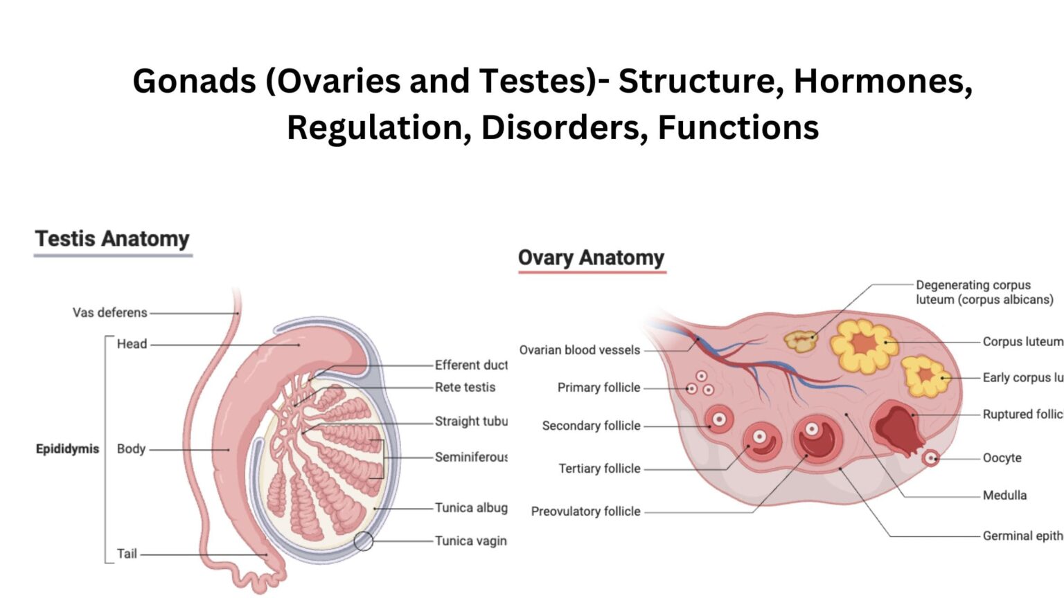
83a) Describe the structure of the ovary

b) List the functions of the ovary

c) Describe the menstrual cycle

MEDICINE I AND 111

84. Mr. KIBULA known hypertensive has been brought to hospital with suggestive features of hypertensive crisis.

a) Mention 8 clinical features of hypertension

b) List 4 causes of HTN and predisposing factors

c) Explain the specific Nursing Care you will give to Mr. KIBULA from the time of admission to discharge.

85. Write short notes on the following (definition, causes, signs and symptoms and complications).

a) Hydrocele

b) Hodgkin’s disease

c) Ankylosing spondylitis

86 a) Define Paget’s disease/Osteitus, deformans?

b) Explain the pathophysiology and etiology of Paget’s disease

c) Describe the specific nursing care you would give to Mr. Muwonge with Paget’s disease

87. Hepatitis B morbidity and mortality is much higher today than before.

a) What are the factors, contributing to the high prevalence of hepatitis B in the communities

b) How does a patient with hep.B present?

c) Give five priority nursing diagnoses for a patient with Hep B infection.

d) Describe the specific nursing management you would give to a patient with hep B.

e) Mention the complications of hep B.

f) Suggest ways how we can prevent hep B infection in the community

88. Define myocardial infarction. List the clinical features of myocardial infarction.

Explain the specific Nursing care given to a patient with myocardial infarction within the first 24Hrs of admission.

89. An adult male patient has presented to OPD with features of pulmonary tuberculosis

a) Outline five cardinal signs and symptoms of pulmonary tuberculosis.

b) List five specific investigations that can be done to confirm pulmonary tuberculosis.

c) Explain the specific nursing care given to this patient from the time of admission until discharge.

90. Mrs. A, a female patient has been admitted on a medical ward with suspected bronchial pneumonia,

a) Outline the clinical features of bronchial pneumonia

b) Describe the specific nursing management you would give to Mrs. X with in the first 72HRS of admission.

c) Explain five likely complications Mrs. X is likely to get following this condition.

91. Mr. Lusoke, a 62 yrs. old male is presented at the OPD with features of congestive cardiac failure

a) Outline the signs and symptoms of congestive cardiac failure.

b) Mention the causes of congestive cardiac failure.

c) Describe the specific nursing care / management you will give to Mr. Lusoke from time of admission to discharge.

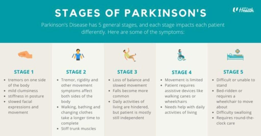
92. Outline the signs and symptoms of Parkinson’s disease.

b) Mention the causes and predisposing factors to Parkinson’s disease.

c) Describe the specific Nursing management given to a patient with Parkinson’s disease.

93. Mr. Okello a 28yrs old male presents at OPD with clinical features of urinary tract infection and was admitted.

a) List 5 causes and 6 signs and symptoms of urinary tract infection.

b) Describe the specific nursing care you would give to Mr.Okello within the first 48 hours of admission.

c) Give the measures that can be taken to prevent urinary tract infections.

94 Define Addison’s disease?

b) Outline the causes and risk factions that leads to Addison’s disease.

c) Using the Nursing process, describe the management of a patient with Addison’s disease.

PEDIATRICS 1 AND 11

95. Define the term Apgar score

a) Outline 10 characteristics of a normal new born baby

b) Describe the care given to the normal new born baby within 72 hours after delivery of the head.

96. Differentiate between SAM and MAM

b) Explain the causes of malnutrition in children under 5 years.

c) Explain the importance of breastfeeding in babies’ up to 2years of age.

97. Define the term congenital abnormalities

a) Classify the congenital abnormalities of the heart

b) Explain ways of preventing congenital abnormalities.

98. Mention the factors that predispose to neonatal infections in new born babies.

b) List 8 clinical features of a child with neonatal tetanus.

c) Describe the specific management of a 3 month old child with tetanus.

99. Outline the factors that predispose to birth injuries

Differentiate between a caput succedaneum and a cephalo hematoma.

c) Describe the specific management you would give to a new born baby who presents with a caput succedaneum.

100. Brandon a five weeks old neonate is admitted on ward with a history of fast breathing, chest in drawing and stridor.

b) Explain the specific nursing care you would offer to Brandon in a hospital within the first eight hours of admission.

101. A five year old child has been bought to OPD in a painful sickle cell crisis.

a) Outline 5 possible causes of sick cell crisis.

b. List 4 diagnostic signs and symptoms of sick cell disease in children.

c) Explain the specific management of this child from admission to discharge.

102. A 4 months old baby has been admitted on a pediatric ward and diagnosed with pneumonia.

a) Outline the clinical presentation of this child.

b) Explain the specific management given to the child with in the first 72 hours.

103. Define the following terms.

1) Fracture

ii)Osteopenia of prematurity

osteogenesis imperfecta

Osteomyelitis

b) Mention 5 signs and symptom of osteomyelitis in children.

c) Describe the nursing management of 3 years old child with osteomyelitis.

104. A 8 month old child has been diagnosed with nephrotic syndrome.

a) List 6 signs and symptoms of nephrotic syndrome in children.

b) Describe the specific nursing management you world give to this child within the first 72 hours of admission on a pediatric ward.

c) Outline five complications of nephrotic syndrome.

105. What are the advantages of breast feeding?

Compare human milk and cow’s milk

Outline problems that are faced by mothers during breastfeeding.

106. List five congenital abnormalities of the G’T and 5 musculoskeletal system

Outline the causes of congenital abnormalities.

How do you cause a mother who has delivered a baby with spinal bifida?

107. List the factors that promote good nutrition in the under-five.

List five pieces of advice you would give to a prime para with a two year old baby suffering from protein calorie malnutrition.

List five problems of birth injuries in Uganda.

Outline the roles of a nurse in prevention of birth injuries in Uganda.

PHARMACOLOGY 1 AND 111

108. Define rational drug use

Outline the medical classification of drugs giving examples of each

Mention the legal classes of drugs with examples of each.

109. Define infertility.

State the common cause of infertility in women

c) State the indications, side effects and contraindications of clomiphene and Bromocriptine.

110. Describe the mechanism of action of non-opioid analgesics.

b) Write briefly about the handling of the class of drugs in a hospital

c) Define the following:-

Chemotherapy

Anti tussive

111. Mention 4 Four sources of drugs

b) Write down all routes which can be used for drug administration giving advantages and disadvantages of each.

c) Write down the factors that affects drugs absorption.

d) What factors affect drug dosage and action?

112. State the clinical uses of oxytocin and mention 6 adverse side effects of the drug.

b) Outline 5(five) contraindications of oxytocin

c) Describe 10 (ten) Nursing considerations while administering oxytocin.

113. Define Narcotic drugs and state the types of narcotics.

b) List down 7 nursing considerations before during and after administrating narcotics on ward.

c) What are the legal implications of Narcotics according to the Uganda narcotic drugs and psychotropic substance control ACT?

114. Define immunity and explain the two major types of immunity.

State the specific side effects, indication and the dosage following drugs:-

  1. Anti D (RHO) Immunoglobulin
  2. B) Rabies vaccine
  3. Pneumococcal Vaccine.

115. Describe the physiology of erection in males

b) State the causes of erectile dysfunction

b) Mention the class, indication, Dosage and side effects of the following drugs.

i) Sildenafil.

ii) Tadalafil

iii) Finesteride.

GYNAECOLOGY

  1. a) Outline signs of breast cancer.

b) Explain post operative care after mastectomy.

c) List possible complications of mastectomy.

  1. . a) Draw a diagram showing possible sites of vaginal fistula.

b) Outline the 5 major causes of vaginal fistula.

c) Explain specific nursing care of a woman after VVF repair.

118. a) Define the different types of Abortion.

b) Outline causes of missed Abortion.

c) Explain different methods used in the management of missed abortion.

d) Outline the 5 elements of PAC.

  1. a) Define ectopic pregnancy.

b) Outline signs and symptoms of tubal pregnancy.

c) A mother presents to the medical facility with a tubal pregnancy, describe her management till discharge.

119. a) List the disorders of menstruation.

b) Explain the advice and treatment given to a 17 year old girl with dysmenorrhea.

120 a) Define Hydatidiform mole.

b) Outline signs and symptoms of hydatidiform mole.

c) Describe the methods of managing the above condition and list complications that may follow.

121. Describe pelvic inflammatory disease.

b) What are the predisposing factors of this condition?

c) Describe management of PID in the hospital.

  1. a) What is infertility?

b) Outline causes of infertility.

c) Explain the different methods that can be used to manage infertility.

  1. a) Draw a diagram of a uterus indicating sites of fibroids.

b) Differentiate between benign and malignant tumor.

c) Give the management of the mother after myomectomy within the first 48 hours.

d) What specific advice would you give this mother on discharge.

REPRODUCTIVE HEALTH

  1. a) Define STDs?

b) Explain ten preventive measures against sexually transmitted infections.

c) Describe the syndromic management of STDs.

  1. a) List 7 components of reproductive health.

b) Outline the advantages and disadvantages of intergrating reproductive health.

c) Outline 10 factors that affect women’s reproductive health.

  1. a) Define sexual abuse?

b) Explain factors that expose adolescent girls to sexual abuse or vulnerability.

c) Outline 5 clinical features of sexual abuse in an adolescent.

  1. a) Define i) Post Abortion Care

ii) Comprehensive abortion care.

b) Explain the Rational for PAC.

  1. a) Who is an adolescent?

b) Describe Tanner’s stage of development in an adolescent.

c) List common health problems faced by adolescents.

  1. a) What is safe motherhood?

b) Outline the 3 delays that can increase maternal mortality.

c) What is your role as a midwife in reduction of maternal mortality in your community?

  1. Describe syndromic approach of managing STIs.
  2. a) Define domestic violence.

b) What are the factors that make you suspect that one is a victim of domestic violence?

c) How would you prevent domestic violence?

  1. Describe manual vacuum aspiration.

FOUNDATIONS OF NURSING.

  1. a) Define wounds.

b) Give 5 types of wounds.

c) Outline the factors that delay wound healing.

d) Give the specific management for a patient with specific wound.

e) What specific advice do you give to a patient with a wound prior to discharge.

f) Describe the process of wound healing.

  1. a) Outline the indications for oxygen administration.

b) Give the rules to follow before, during and after administration of oxygen.

c) Define blood transfusion.

d) Outline the indications of blood transfusion.

e) Outline the appropriate care of the patient before, during and after blood transfusion.

f) Give the complications of blood transfusion.

  1. a) Define drug administration.

b) Outline the different routes of drug administration.

c) Mention the principles of drug administration including the dos and don’ts in drug administration.

  1. a) Define infection prevention and control.

b) Define nosocomial infection.

c) Outline the steps taken to prevent infections of the wound.

d) What are the advantages of oral route drug administration over the parental route.

  1. a) Outline the indications of Tracheostomy.

b) Give the specific pre and post operative nursing care for the patient with tracheostomy.

c) Mention the complications of tracheostomy.

d) Formulate 4 actual nursing diagnoses for a patient with colostomy.

  1. a) Define lumber puncture.

b) Outline the indications of lumber puncture.

c) Explain the specific nursing care given to the patient prior to after the procedure of lumber puncture.

d) List the complications of lumber puncture.

  1. a) Define abdominal paracentesis.

b) Outline the indications of paracentesis.

c) Give the specific care given to the patient before and after abdominal paracentesis.

d) Mention the complications of abdominal paracentesis.

  1. a) Define tractions.

b) Explain the different types of tractions.

c) Outline the specific nursing care given to a patient with tractions.

d) Formulate 5 actual nursing diagnoses for a patient with tractions.

e) Outline the likely complications of the patient on traction.

  1. a) Outline the indications of underwater seal drainage.

b) Give the specific nursing care for a patient on underwater seal drainage.

c) Formulate four nursing diagnoses for a patient on underwater seal drainage.

d) List the complications of underwater seal drainage.

  1. a) Outline 6 indications of gastric lavage.

b) Define colostomy.

c) Formulate 4 actual nursing diagnoses and 4 potential nursing diagnoses for a patient with colostomy.

d) Give the specific nursing care to the patient with colostomy.

  1. a) List the indications of Glasgow coma scale.

b) Describe the Glasgow coma scale.

ANATOMY AND PHYSIOLOGY II

  1. a) With illustration, describe the formation of flow of CSF.

b) List the functions of CSF.

c) Describe the meninges covering the brain and spinal cord.

  1. a) Describe the position and gross structure of the parathyroid glands. Outline the functions of parathyroid hormone and calcitonin.

b) Explain the disorders of the thyroid gland.

  1. a) Describe the structure of a nephron.

b) Explain the processes involved in the formation of urine.

c) Describe how body water and electrolyte balance is maintained.

  1. a) Describe the structure of the ear.

b) Explain the physiology of hearing.

c) Explain the functions of the accessory organs of the eye.

  1. a) Explain the role of lymphatic vessels in the spread of infections and malignant disease.
  2. a) Describe the location of the pharynx and relate it’s structure to it’s function.

b) List the functions of the trachea in respiration.

c) Explain the main mechanisms by which respiration is controlled.

d) Describe the common inflammatory and infectious disorders of the upper respiratory tract.

  1. a) Define a neuron.

b) Outline the 12 cranial nerves of the nervous system.

c) Describe the transmission of an impulse across a synapse.

PALLIATIVE CARE NURSING

150 a) Define palliative care

b) Explain the principles of palliative care

c) Give the challenges faced in implementing in palliative care services in Uganda

151.a) Define pain according to WHO

b) Explain different types of pain in palliative care

c) Describe the principles of pain management in palliative care

d) Describe the steps of breaking bad news

152.a) Explain 6 roles of palliative care in Uganda

b) Outline 6 symptoms commonly experienced by terminary ill patients

153.a) What is grief?

b) Explain 5 stages of grief experienced by palliative care patients

c) Explain the HOPE approach to spiritual pain management

d) Outline the spiritual problems experienced by palliative care patients

Self Study Question For Nurses and Midwives Read More »

Diabetes Mellitus Nursing Management

Diabetes Mellitus Type 2

Diabetes Mellitus

Diabetes Mellitus (DM), commonly referred to as diabetes, is a group of metabolic disorders in which there are high blood sugar levels over a prolonged period.

Insulin is the hormone secreted by β-cells of the pancreas; it helps to incorporate glucose into cells for metabolism.

In insulin deficiency, blood glucose level rises leading to excretion of sugar in  the urine called Glycosuria.

Glucose loss is accompanied by increased loss of water in the urine causing Polyuria; hence Hyperglycemia, Glycosuria and Polyuria are the three cardinal clinical features of diabetes mellitus.

Diabetes mellitus is the most common prevalent endocrine disorder; it affects nearly 2% of the world population


Diabetes can be primary or secondary or idiopathic.

diabetes mellitus pancreas that produces insulin

Image showing the pancreas where Insulin is produced by the body.

Pathophysiology of Diabetes(Simplified)

Diabetes results from two main issues: the pancreas not making enough insulin or the body’s cells not responding properly to the insulin produced.

1. Insufficient Insulin: Pancreas fails to produce adequate insulin.

  • Impaired insulin function disrupts blood sugar regulation, leading to hyperglycemia (elevated blood glucose levels).

2. Consequences of Hyperglycemia:

  • Excess glucose is expelled through urine, causing glycosuria.
  • High glucose in the glomerular filtrate attracts water, resulting in polyuria (excessive urination).
  • Loss of water triggers an intense feeling of thirst (polydipsia).

3. Cellular Deprivation and Compensatory Responses:

  • Despite high blood glucose, cells remain deprived.
  • Body responds with increased appetite, leading to overeating (polyphagia), worsening the condition.

4. Gluconeogenesis:

  • Body initiates gluconeogenesis to create glucose from proteins and fats.

5. Ketone Body Accumulation:

  • Excessive glucose from fats produces abundant ketone bodies, causing ketonemia (increased ketones in blood).

6. Acidosis and Respiratory Response:

  • Accumulated ketones reduce blood pH, leading to acidosis.
  • Body responds with rapid and deep breathing (Kussmaul respirations) to decrease acidity.

7. Potential Life-Threatening Complication:

  • Prolonged acidic state may lead to ketoacidosis, a severe medical/pediatric emergency.
Types of Diabetes Mellitus

Types of Diabetes Mellitus

There are three main types of diabetes mellitus and one unspecified;

Type 1 Diabetes Mellitus

Type 1 Diabetes Mellitus (T1DM) is marked by the pancreas’s failure to produce sufficient insulin, a vital hormone in blood sugar regulation. Formerly known as “insulin-dependent diabetes mellitus” (IDDM) or “juvenile diabetes,” Its cause is unknown.

  •  Insulin Deficiency and Beta Cell Loss: T1DM is characterized by the loss of insulin-producing beta cells in the pancreatic islets. This leads to a deficiency in insulin, disrupting the body’s ability to regulate blood sugar.
  • Immune-Mediated or Idiopathic Classification: T1DM can be classified as immune-mediated or idiopathic. The majority of cases involve immune-mediated processes, where autoimmune attacks by T cells lead to beta cell loss. Onset can occur in children or adults, though historically labeled “juvenile diabetes” due to its prevalence in children.
  • Associated Complications: Complications may include impaired response to low blood sugar, infections, gastroparesis (causing erratic carbohydrate absorption), and endocrinopathies like Addison’s disease.
  • Genetic and Environmental Factors: T1DM has a genetic component, with specific HLA genotypes influencing susceptibility. Environmental triggers, such as viral infections or dietary factors (e.g., gliadin in gluten), can prompt diabetes onset, especially in genetically predisposed individuals.
  • Autoimmune Attack and Viral Influence: An autoimmune attack on pancreatic islets, often triggered by viral infections, is a key contributor. T1DM is more likely to manifest in childhood or early adulthood, with a sudden onset.
  • Management and Risks: Insulin and a comprehensive diet are crucial for managing T1DM. Patients face an increased risk of coma if concurrent infections like pyelonephritis or gastroenteritis are not promptly addressed.

Type 2 Diabetes Mellitus

Type 2 Diabetes Mellitus

The pathophysiology of type 2 diabetes mellitus is characterized by peripheral insulin resistance, impaired regulation of hepatic glucose production, and declining β-cell function, eventually leading to β -cell failure

Reduced insulin secretion and absorption leads to high glucose content in the blood.

  • Insulin Resistance and Reduced Secretion: T2DM is characterized by insulin resistance, where body tissues have a diminished response to insulin. This resistance is sometimes accompanied by a relative reduction in insulin secretion.
  • Insulin Receptor Dysfunction: The defective responsiveness of body tissues to insulin involves the insulin receptor, though, specific defects remain unknown. Diabetes cases with known defects are categorized separately.
  • Prevalence and Early Stage Abnormality: T2DM constitutes the majority, accounting for up to 90% of all diabetes mellitus cases. In the early stage, the primary abnormality is reduced insulin sensitivity, reversible by measures and medications improving insulin sensitivity or reducing liver glucose production.
  • Contributing Factors: Lifestyle factors, genetics, obesity (BMI > 30), lack of physical activity, poor diet, stress, and urbanization contribute to T2DM. Insulin resistance, overeating, inactivity, and obesity play roles in the etiology.
  • Dietary Management and Weight Loss: Management often involves adherence to a low-energy diet to facilitate weight loss. Lifestyle modifications, including dietary changes, exercise, and stress reduction, play roles in controlling T2DM.

Gestational Diabetes

Gestational Diabetes

Gestational diabetes mellitus (GDM) resembles type 2 DM in several aspects, involving a combination of relatively inadequate insulin secretion and responsiveness. It occurs in about 2–10% of all pregnancies and may improve or disappear after delivery.

  • Occurrence and Post-Delivery Transition: GDM shares similarities with type 2 DM, involving inadequate insulin secretion and responsiveness. It affects about 2–10% of pregnancies and may improve or vanish after childbirth.
  • Post-Pregnancy Diabetes Risk: Post-pregnancy, 5–10% of women with a history of gestational diabetes develop diabetes mellitus, often type 2.  However, after pregnancy approximately 5–10% of women with gestational diabetes are found to have diabetes mellitus, most commonly type 2.
  • Temporary Nature and Health Impacts : While temporary during pregnancy, untreated GDM poses risks to both the mother and the fetus. Raised plasma glucose levels during pregnancy may lead to the birth of babies with increased birth weight, skeletal muscle malformations, and increased mortality risk. Risks associated with untreated GDM in newborns include macrosomia (high birth weight), congenital heart and central nervous system abnormalities, and skeletal muscle malformations. Elevated insulin levels in the fetal blood may hinder surfactant production, leading to respiratory distress syndrome.
  • Complications and Perinatal Risks: Complications may arise, such as high blood bilirubin levels due to red blood cell destruction. Severe cases can result in perinatal death, often attributed to poor placental perfusion caused by vascular impairment, leading to macrosomia and shoulder dystocia.
  • Management and Treatment:  Gestational diabetes is fully treatable, but requires careful medical supervision throughout the pregnancy. Management may include dietary changes, blood glucose monitoring, and in some cases, insulin may be required. 
Unspecified Diabetes Mellitus:

Maturity Onset Diabetes of the Young (MODY):

  • Maturity onset diabetes of the young (MODY) is an autosomal dominant inherited form of diabetes, due to one of several single-gene mutations causing defects in insulin production.
  • It is significantly less common than the three main types.
  • The name of this disease refers to early hypotheses as to its nature.
  • Being due to a defective gene, this disease varies in age at presentation and in severity according to the specific gene defect; thus there are at least 13 subtypes of MODY.
  • People with MODY often can control it without using insulin.

Others:

  1. Prediabetes: Prediabetes indicates a condition that occurs when a person’s blood glucose levels are higher than normal but not high enough for a diagnosis of type 2 DM. Many people who later develop type 2 DM spend many years in a state of prediabetes.
  2. “Type 3 Diabetes”: “Type 3 diabetes” has been suggested as a term for Alzheimer’s disease as the underlying processes may involve insulin resistance by the brain.

Aetiological Classification of Diabetes Mellitus:

Primary Diabetes Mellitus (Idiopathic):
  1. Type 1 Diabetes (IDDM): β-cell destruction, usually leading to insulin deficiency.
  2. Type 2 Diabetes (NIDDM): May range from insulin resistance with relative insulin deficiency to a predominantly insulin secretory defect with insulin resistance.
Secondary Diabetes Mellitus: Due to Other Underlying Diseases/Conditions:
  1. Diseases of the pancreas, such as pancreatitis, pancreatic cancer, cystic fibrosis, or hemochromatosis, can destroy the gland leading to reduced insulin production.
  2. Endocrine disorders (insulin antagonism) like Cushing’s syndrome, acromegaly, and hyperthyroidism.
  3. Drug-induced (lactogenic) diabetes, e.g., corticosteroids, phenytoin, thiazide diuretics therapy.
  4. Genetic/chromosomal defects, e.g., Down’s syndrome.
  5. Liver diseases like hepatitis, cirrhosis, are associated with glucose intolerance.
  6. Gestational Diabetes Mellitus (Pregnancy-induced Diabetes Mellitus): Occurs during pregnancy and may resolve after delivery.

Predisposing Causes of Primary Diabetes Mellitus:

  1. Age: 80% of cases occur after 50 years. DM is commonly a disease of middle-aged and elderly people.
  2. Sex: Young males are more affected than females. In middle age, females are more affected.
  3. Heredity: DM follows the family line in occurrence. 5% of patients have a familial history.
  4. Autoimmunity: The body produces cells against insulin production.
  5. Infections: Viral infections and staphylococci are associated with the causation of IDDM.
  6. Obesity: The majority of NIDDM cases are obese.
  7. Lifestyle Factors: Overeating with underactivity is associated with a high risk of incidence.

Other Predisposing Factors:

  • Sedentary lifestyle.
  • Poor dietary habits.
  • Metabolic syndrome.
  • Hypertension.
  • Ethnicity (some ethnic groups are more predisposed).
  • Gestational diabetes (increases the risk later in life).
  • Certain medications (e.g., glucocorticoids).
  • Previous gestational diabetes.
Clinical Features of Diabetes Mellitus

Clinical Features of Diabetes Mellitus

In mild cases, there may be no obvious signs, and the condition is detected accidentally during routine examination. However, in severe cases, especially in young children and young adults, pronounced symptoms may include: 

  1. Polyuria Due to osmotic activity preventing water reabsorption in the renal tubule.
  2. Polydipsia (increased thirst) follows polyuria, leading to dehydration due to constant loss of fluids and electrolytes.
  3. Polyphagia with Weight Loss: Weight loss occurs due to the breakdown of fat and proteins caused by cellular glucose deficiency.
  4. Weakness or Fatigue/Lassitude: Resulting from cells not receiving enough glucose.
  5. Nocturnal Enuresis: Due to renal glucose exceeding the threshold. Nocturnal enuresis is when there is involuntary urination at night while asleep.
  6. Glycosuria: This is when there is excessive amounts of glucose in urine.
  7. Peripheral Neuropathy/Paresthesia: Nerve damage caused by chronically high blood sugar, leading to loss of sensation and numbness in the legs. In severe cases, symptoms include digestive issues, bladder problems, and difficulty controlling heart rate. Paresthesia is a symptom of neuropathy, since neuropathy is an umbrella term for any disease that affects the nerves.
  8. Vulvovaginitis: Irritation of the genitalia caused by the local deposition of glucose. May be severe and disturb sleep.
  9. Ketoacidosis: A serious complication involving excess blood acids (ketones). Symptoms include blurry vision, headache, fatigue, slow healing of cuts, and itchy skin.
  10. Diabetic dermadromes. Skin rashes associated with diabetes with cutaneous eruptions in patients with long standing diabetic disease.
  11. Vision Changes: Prolonged high blood glucose can cause glucose absorption in the lens of the eye, leading to changes in its shape and resulting in vision changes.

Comparison of type 1 and 2 diabetes

Type 1

Type 2

  • Sudden
  • Mostly children
  • Thin or normal
  • Ketoacidosis common
  • Antibodies usually present
  • Insulin low or absent totally
  • In identical twins is approximately 50%
  • Prevalence approximately 10%
  • Gradual
  • Mostly adults
  • Often obese
  • Rare
  • Absent
  • Normal, decreased or increased
  • Is approximately 90%
  • Prevalence approximately 90%
Diagnosis of Diabetes Mellitus:
Diagnosis of Diabetes Mellitus 2

Diagnosis of Diabetes Mellitus:

Diabetes mellitus, characterized by recurrent or persistent high blood sugar, is diagnosed by demonstrating any one of the following criteria:

1. Fasting Plasma Glucose Level:  7.0 mmol/l (126 mg/dl)

  • According to the current definition, two fasting glucose measurements above 126 mg/dl (7.0 mmol/l) are considered diagnostic for diabetes mellitus.

2. Plasma Glucose Two Hours After Oral Glucose Load:  ≥ 11.1 mmol/l (200 mg/dl) two hours after a 75 g oral glucose load, as in a glucose tolerance test.

  • People with plasma glucose at or above 7.8 mmol/l (140 mg/dl) but not exceeding 11.1 mmol/l (200 mg/dl) two hours after the oral glucose load are considered to have impaired glucose tolerance.

3. Symptoms of High Blood Sugar and Casual Plasma Glucose: ≥ 11.1 mmol/l (200 mg/dl)

  • Presence of symptoms along with casual plasma glucose above 11.1 mmol/l (200 mg/dl) indicates diabetes.

Note:

  • According to the World Health Organization, individuals with fasting glucose levels between 6.1 to 6.9 mmol/l (110 to 125 mg/dl) are considered to have impaired fasting glucose.
  • Glycated hemoglobin is considered superior to fasting glucose in determining cardiovascular disease risks and risks of death from any cause.

Important Information:

  • Two fasting glucose measurements above 126 mg/dl (7.0 mmol/l) are diagnostic for diabetes.
  • Impaired glucose tolerance, especially with plasma glucose levels between 7.8 mmol/l (140 mg/dl) and 11.1 mmol/l (200 mg/dl) after oral glucose, is a significant risk factor for progressing to diabetes and cardiovascular disease.

4. Other Diagnostic Investigations for Diabetes Mellitus:

Glucosuria:

  • Method: Detect glucose in urine using a test strip (uristicks).
  • Purpose: To identify the presence of glucose in the urine, indicating possible diabetes.

Ketonuria:

  • Method: Detect ketone bodies in urine.
  • Purpose: To identify the presence of ketones, which may indicate diabetic ketoacidosis.

Fasting Blood Sugar (FBS):

  • Method: Measure glucose concentration in blood samples obtained after at least 8 hours of the last meal.
  • Purpose: Assess baseline blood sugar levels after an overnight fast.

Random Blood Sugar (RBS):

  • Method: Measure glucose concentration in blood samples obtained at any time, regardless of the time of the last meal.
  • Purpose: Provide a snapshot of current blood sugar levels.

Oral Glucose Tolerance Test (OGTT):

  • Method: The patient fasts overnight, then ingests 75gm (5 tablespoons) of glucose with 300 ml of water. Blood samples are drawn at 1, 2, and 3 hours after glucose intake.
  • Purpose: A more accurate test for glucose utilization, especially if fasting glucose is borderline. It helps identify abnormal glucose metabolism over time.

Additional Information:

  • Normally, blood glucose should return to fasting levels (4.5 mmol or 80 mg/100 ml) after 2.5 hours of taking a meal.
  • In diabetes, fasting levels remain elevated above 200 mg/100 ml, indicating impaired glucose metabolism.
Treatment and Nursing Management of Diabetes mellitus

Treatment and Nursing Management of Diabetes mellitus

Diabetes mellitus is a chronic disease, for which there is no known cure except in very specific situations.

Management concentrates on keeping blood sugar levels as close to normal, without causing low blood sugar

This management is dependent on the type of diabetes mellitus and aims to:

  • Control diabetes and prevent complication
  •  To bring down blood sugar levels
  •  To help the patient comply to treatment

Management (non-pharmacological)

  •  Control traditional Cardiovascular risk factors such as smoking, taking alcohol, management  of dyslipidemia, intensive BP control and antiplatelet therapy.
  • Complication monitoring i.e. annual eye examination, annual microalbuminuria detection, feet examination, BP monitoring and lipid profile.
  • Patient’s education: Teach the patient on self-monitoring of blood glucose using glucometer and/or uristicks, moving with a diabetic card, keeping sugary food in the bag, method of insulin administration and consequences neglecting treatment
  •  Patients should also be taught to prevent themselves from injury.
  • Diet
    For type 1 the goal is to regulate insulin administration with a balanced diet
    – In most cases, high carbohydrate, low fat and low cholesterol diet is appropriate
    Diet and insulin must be fixed to avoid fluctuation in blood glucose. Vitamins and minerals must be supplemented
    Small frequent meals should be served to avoid peaks of hyperglycemia and no meal should be delayed. Snacks should be added to the main meals i.e in the middle of morning, early afternoon and before bed time.
    Food should be palatable with high fibre food like legumes, burley, oat. Low salt in diet is advised (6g per day)
    Avoid fried food, sweetened beverage, bakery products, honey and fine sugar
  • Type 2 DM patients need caloric restriction: Diet restriction must be combined with life style modification
    Artificial sweeteners: e.g. Aspartame, saccharin, sucralose, and acesulfame are safe for use in all people with diabetes
    Nutritive sweeteners: e.g. fructose and sorbitol, there use is increasing though they cause acute diarrhea in some patient.
  • Activity:  Exercise improves insulin resistance and achieving glycemic control.
    – Exercise should start slowly for patients with limited activity.
    Patients with Cardiovascular diseases should be evaluated before starting any exercise
    Avoid exercises on an empty stomach, when blood sugar levels are low or high.
    Heavy exercises like mental lifting are dangerous because it triggers hypoglycemia

Pharmacological therapy of diabetes mellitus

(Will be detailed later)

  • Insulin (Type 1 and Type 2 DM)
    Sulfonylurea e.g glibenclamide (Type 2 DM)
    Biguanides e.g metformin (Type 2 DM)
    Meglitinides (Type 2 DM)
    Thiazolidinediones Glitazones e.g Competact(Pioglitazone + Metformin) (Type 2 DM)
    Alpha-Glucosidase inhibitors e.g acarbose (Precose) (Type 2 DM)

Methods of treatment of diabetes
>   Diet
>   Diet + oral hypoglycemic agents
>   Diet and insulin

INSULIN THERAPY

Insulin is indicated for most patients of IDDM and IIDM who do not respond to oral hypoglycemic drugs. Doses are adjusted for individual patient needs to meet target glycemic control

 Administration
• Subcutaneous injections
• Continuous subcutaneous insulin infusion pump
• IV infusion (regular insulin only)

Aim of insulin therapy

  • To maintain blood glucose within normal limits
  •  To relieve hyperglycemia-associated symptoms.
  •  To correct metabolic/biochemical disturbances
  •  To prevent diabetes-associated complications
Types of Insulin

Unmodified/soluble/rapid acting insulin: Dose 40 – 100 IU SC daily in 3 divided doses before meals, 40-80 IU for child.
This is a clear solution and acts in half an hour and reaches peak in 2-6 hours, repeated injections are needed. This insulin can be used to control postprandial hyperglycemia and emergency ketoacidosis i.e,

  1. Ultra short acting-Lispro: (Monomeric) absorbed to the circulation very rapidly and acts in 2-3 hours
  2.  Aspart: (Mono- and dimeric) absorbed to the circulation very rapidly
  3.  Short acting-Regular: (Hexameric) absorbed rapidly but slower than lispro and aspart, includes novolin R, humulin R.

Modified (deport) preparations: these are cloudy preparations/turbid suspensions made by either adding zinc for lente preparations or protamine (protein) for isophane preparations.
They are used for maintenance treatment of type 1DM

  1.  Semi lente/prompt zinc; this is short acting and contains zinc microcrystals in acetate buffer. It is not used for IV because of buffer acetate
  2.  Lente insulin; Intermediate acting and acts in 12 hours e.g. humulin L. Dose: Adults 10-20 IU twice daily SC, Child: 5 – 10 IU twice daily
  3.  Ultralente; Long acting and acts 24-36 hours eg Ultratard.
  4.  Insulin analogues; mixture of modified and unmodified acts in 12 hours i.e,
  • 70% NPH, 30% regular
  • 50% NPH, 50 regular
  • 75% NPH, 25% lispro
  • 70% aspartic, 30% protamine

Insulin mixtures are used for high postprandial hyperglycemia management

Adverse effects of insulin administration
  •  Hypoglycemia: Patients should be aware of symptoms of hypoglycemia.  Oral administration of 10-15 gm glucose.   IV dextrose in patients with lost consciousness or/and 1 gm glucagon IM if IV access is not available
  •  Skin rash at injection site: Use more purified insulin preparation.
  •  Lipodystrophies (increase in fat mass) at injection site: rotate the site of injection
  •  Insulin resistance
  •  Allergy
  • Weight gain
  1. Avoid using propranolol or other B-blockers in diabetics because they mask hypoglycemic symptoms.
  2. Drugs that increase the blood glucose concentration should be avoided e.g Dioxide, Thiazide diuretics, Streptozotocin, Phenytoin. Corticosteroids, Oral contraceptives.
 How to measure the insulin needed.

A good starting dose is 0.6 U/kg/day. The total dose should be divided to;

  • 45% for basal insulin
  • 55% for prandial insulin

The prandial dose is divided to

  • 25% pre-breakfast
  • 15% pre-lunch
  • 15% pre-supper

Example: For a 50 kg patient
The total dose = 0.6 x 50 = 30 U/day
  =  13.5 U for basal insulin (45% of dose)
Administered in one or two doses
  =  16.5 U for prandial insulin (55% of dose)
The 16.5 U are divided to:
    = 7.5 U pre-breakfast (25%)
    = 4.5 U pre-lunch (15%)
    = 4.5 U pre-supper (15%)
The initial regimen should be modified
Most Type 1 patients require 0.5-1.0 IU/kg/day

Medications for Type 2 Diabetes:

Anti-diabetic medications (hypoglycemics) are important for managing diabetes by lowering blood sugar levels. Various classes of these medications exist, some administered orally (e.g., metformin) and others via injection (e.g., GLP-1 agonists). It’s important to note that insulin is the primary treatment for Type 1 diabetes.

Sulphonylureas: Stimulate insulin secretion and release by the pancreas’ beta cells.

  • Examples include glibenclamide and chlorpropamide.

Biguanides: Increase glucose uptake by body cells and decrease glucose production by the liver.

  • Metformin (Glucophage) is a commonly recommended first-line treatment for Type 2 diabetes, showing evidence of decreased mortality.

Alpha-Glucosidase Inhibitors: Inhibit the enzyme hindering glucose uptake by cells.

  • Examples include acarbose and miglitol.

Thiazolidinediones: Decrease insulin resistance.

  • Example: Pioglitazone.

Insulin Injections: Short-acting (e.g., Actrapid), intermediate (e.g., Mixtard), and long-acting (e.g., Insulatard).

  • Primarily used in Type 1 diabetes and in Type 2 when oral medications are ineffective.

Blood Pressure Management:

Given the serious cardiovascular risks associated with diabetes, maintaining blood pressure is crucial.

  • Target blood pressure levels are recommended below 130/80 mmHg, though evidence supports a range between 140/90 mmHg to 160/100 mmHg.
  • Angiotensin-converting enzyme inhibitors (ACEIs) are effective, while angiotensin receptor blockers (ARBs) may not be as beneficial in diabetes.
  • Aspirin is recommended for those with cardiovascular problems; however, routine use hasn’t proven beneficial in uncomplicated diabetes.

Surgery:

  • Weight loss surgery is effective in managing obesity and Type 2 diabetes.
  • Many individuals can maintain normal blood sugar levels with minimal or no medications post-surgery, reducing long-term mortality.
  • Short-term mortality risk from surgery is less than 1%, and eligibility criteria based on body mass index cutoffs are still unclear.
  • Pancreas transplant considerations are rare, usually for individuals with severe complications of Type 1 diabetes, including end-stage kidney disease.

Support:

  • In most healthcare systems, care often occurs outside hospitals unless complications arise.
  • Home telehealth support is an effective management strategy, particularly in cases of complications, challenging blood sugar control, or research projects.
Sites for Insulin Administration
diabetes mellitus sites for insulin administration

Prevention of Diabetes:

Type 1 Diabetes: Unfortunately, there is currently no known preventive measure for Type 1 diabetes. It is primarily considered an autoimmune condition where the body’s immune system mistakenly attacks and destroys the insulin-producing beta cells in the pancreas.

Type 2 Diabetes: Prevention strategies for Type 2 diabetes focus on lifestyle modifications and healthy habits. 

Maintaining a Healthy Diet:

  • Emphasize a balanced and nutritious diet rich in fruits, vegetables, whole grains, and lean proteins.
  • Limit the intake of processed foods, sugary beverages, and foods high in saturated and trans fats.
  • Control portion sizes to avoid overeating.

Regular Physical Exercise:

  • Engage in regular physical activity, such as walking, jogging, swimming, or cycling.
  • Aim for at least 150 minutes of moderate-intensity exercise per week.
  • Include strength training exercises to improve muscle strength and overall fitness.

Maintaining a Normal Body Weight:

  • Achieve and maintain a healthy body weight through a combination of a balanced diet and regular exercise.
  • Weight loss is particularly beneficial for those at risk or diagnosed with prediabetes.

Avoiding Tobacco Use:

  • Quitting or avoiding tobacco products is essential, as smoking is a significant risk factor for Type 2 diabetes.
  • Smoking cessation has numerous health benefits and contributes to overall well-being.

Control of Blood Pressure:

  • Regular monitoring and management of blood pressure are crucial.
  • Adopt a heart-healthy lifestyle, including a low-sodium diet, regular exercise, and stress management.

Proper Foot Care:

  • Individuals with diabetes need to pay special attention to foot care.
  • Regularly inspect feet for any cuts, sores, or signs of infection.
  • Choose comfortable, well-fitting shoes and avoid walking barefoot.

Avoiding Smoking:

  • In addition to its association with Type 2 diabetes, smoking is a risk factor for various cardiovascular and respiratory diseases.
  • Quitting smoking contributes significantly to overall health and reduces diabetes risk.

Additional Measures:

  • Regular health check-ups and screenings for diabetes risk factors.
  • Monitoring and managing stress levels through relaxation techniques and mindfulness.
  • Adequate sleep is essential for overall health and may play a role in diabetes prevention.
  • Limiting alcohol consumption, as excessive drinking can contribute to weight gain and affect blood sugar levels.

Complications of diabetes mellitus

  • Cardiomyopathy: The major long-term complications relate to damage to blood vessels. Diabetes doubles the risk of cardiovascular disease and about 75% of deaths in diabetics are due to coronary artery disease. Other “macrovascular” diseases are stroke, and peripheral artery disease. 
  • Retinopathy: The primary complications of diabetes due to damage in small blood vessels include damage to the eyes, kidneys, and nerves. Damage to the eyes, known as diabetic retinopathy, is caused by damage to the blood vessels in the retina of the eye, and can result in gradual vision loss and blindness. Diabetes also increases the risk of having glaucoma, cataracts, and other eye problems. It is recommended that diabetics visit an eye doctor once a year.
  • Nephropathy:  Damage to the kidneys, known as diabetic nephropathy, can lead to tissue scarring, urine protein loss, and eventually chronic kidney disease, sometimes requiring dialysis or kidney transplantation.
  • Neuropathy: Damage to the nerves of the body, known as diabetic neuropathy, is the most common complication of diabetes. The symptoms can include numbness, tingling, pain, and altered pain sensation, which can lead to damage to the skin.
  • Diabetic foot: Diabetes-related foot problems (such as diabetic foot ulcers) may occur, and can be difficult to treat, occasionally requiring amputation. Additionally, proximal diabetic neuropathy causes painful muscle atrophy and weakness. 
  • Falls: There is a link between cognitive deficit and diabetes. Compared to those without diabetes, those with the disease have a 1.2 to 1.5-fold greater rate of decline in cognitive function.  Being diabetic, especially when on insulin, increases the risk of falls in older people.

Other Complications:

  •  Eye; Retinopathy leading to impaired vision, premature cataract, recurrent styles
  • Urinary system; renal failure, nephritic syndrome and pyelonephritis due to diabetes nephropathy
  • Genital tract; erectile dysfunction, loss of libido in men and menstrual irregularities, recurrent abortion, purulent vaginitis, infertility in females
  •  Nervous system; Neuropathy resulting in tingling and numbness in the feet, stroke.
  •  CVS; Myocardial infarction, peripheral gangrene, hypertension
  •  Skin; Staphylococcal skin infections e.g boils carbuncles, non healing ulcer and mucocutaneous candidiasis
  •  Respiratory system; pneumonia, lung abscess and tuberculosis

Diabetic Emergencies

Hypoglycemia:

Low blood sugar (hypoglycemia) is a common occurrence in individuals with type 1 and type 2 Diabetes Mellitus (DM). While most cases are mild and not deemed medical emergencies, the effects can range from mild to severe.

Symptom Category

Mild Symptoms

Moderate Symptoms

Physical Signs

Common Signs

– Feelings of unease

– Confusion

– Drunkenness

– Sweating

– Changes in behavior (e.g., aggressiveness)

– Rapid breathing

– Trembling

– Seizures

– Sweating

– Increased appetite

– Unconsciousness (rarely, in severe cases)

– Cold and pale skin (although not definitive)

Management

Self-treatment with sugary foods or drinks.

Immediate attention with intravenous glucose or glucagon injections for severe cases

Hyperosmolar Hyperglycemic State:

More common in type 2 DM, hyperosmolar hyperglycemic state is mainly the result of dehydration. This state is characterized by significantly elevated blood sugar levels.

  • Hospitalization is often necessary.
  • Treatment involves fluid replacement, insulin administration, and correction of electrolyte imbalances.
  • Close monitoring of vital signs, blood glucose, and electrolytes.

Diabetic Ketoacidosis (DKA):

Diabetic Ketoacidosis (DKA) stands as a severe acute complication of Diabetes Mellitus where the body produces excess blood acids (ketones), posing a significant risk of death and morbidity, particularly with delayed treatment. The prognosis is notably worse in extreme age groups, with mortality rates ranging from 5-10%, but advancements in therapy have reduced mortality to over 2%.

Pathophysiology:

DKA arises from insulin deficiency and the action of counter-regulatory hormones, leading to hyperglycemia and glycosuria. The absence of insulin forces the body to use fats instead of glucose, resulting in ketosis and metabolic acidosis. Vomiting, insensible water losses, and electrolyte abnormalities further exacerbate the condition, with dehydration potentially leading to acute renal failure.

Precipitating Factors:

  • New onset of type 1 DM: 25%
  • Infections (most common): 40%
  • Drugs (e.g., Steroids, Thiazides, Dobutamine & Turbutaline)
  • Omission of Insulin: 20%

Diagnosis:

Suspect DKA in a diabetic patient presenting with:

  • Dehydration
  • Acidotic (Kussmaul’s) breathing with a fruity smell (acetone)
  • Abdominal pain &\or distension
  • Vomiting
  • Altered mental status ranging from disorientation to coma

Diagnostic Criteria:

  • Hyperglycemia: > 300 mg/dl & glucosuria
  • Ketonemia and ketonuria
  • Metabolic acidosis: pH < 7.25, serum bicarbonate < 15 mmol/l, Anion gap >10.

Management:

Assessment: Evaluate causes & sequele of DKA through history and scan examination.

Quick Diagnosis at the ER: Confirm hyperglycemia, ketonuria, and acidosis promptly.

Baseline Investigations:

  • Plasma & urine levels of glucose & ketones.
  • ABG, U&E (Na, K, Ca, Mg, Cl, PO4, HCO3), & arterial pH.
  • Complete Blood Count with differential.

Treatment Principles:

  • Careful fluid replacement.
  • Correction of acidosis & hyperglycemia via Insulin administration.
  • Correction of electrolyte imbalances.
  • Treatment of underlying causes.
  • Monitoring for complications.

Fluid Replacement:

  • Hypovolemic shock: Administer 0.9% saline, Ringer’s lactate or plasma expander.
  • Dehydration without shock: Administer 0.9% Saline, adjusting to avoid rapid shifts in serum osmolality.

Insulin Therapy:

  • Start infusing regular insulin at 0.1U/kg/hour.
  • Adjust fluid composition as glucose decreases.
  • Continue insulin infusion until acidosis is cleared.

Correction of Electrolyte Balance:

  • Administer potassium supplementation to IV fluid.
  • Adjust based on serum potassium levels.

Monitoring:

  • Use a flow chart for fluid balance & Lab measures.
  • Measure serum glucose and electrolytes regularly.
  • Neurological & mental status examination.

Complications:

  • Cerebral Edema
  • Intracranial thrombosis or infarction.
  • Acute tubular necrosis.
  • Peripheral edema.
Nursing diagnosis for diabetes — 5 sample care plans

These five care plans cover the nursing diagnoses for diabetes you’ll see most often in clinical paperwork. NANDA-I 2026-aligned interventions, and the standard student template (diagnosis ➔ defining characteristics / risk factors ➔ goals ➔ interventions with rationales ➔ evaluation).

Care Plan #1 — Risk for Unstable Blood Glucose Level

This is one of the most frequently used nursing diagnoses for diabetes on the floor, especially for newly diagnosed patients or anyone whose regimen has just changed.

Scenario. A 52-year-old patient is newly diagnosed with type 2 diabetes during a hospital admission for cellulitis. A1C 9.4%. Fingerstick on admission 268 mg/dL. The patient says they haven’t checked sugars at home and don’t know how to use the meter the provider sent home with them.

Diagnosis (NANDA-I 2024–2026)

Risk for Unstable Blood Glucose Level related to new T2DM diagnosis, insufficient diabetes self-management knowledge, and acute illness.

(Risk-for diagnoses use “related to” only — no “as evidenced by.”)

Goals
  • By the end of the shift, the patient verbalizes the steps for checking a fingerstick glucose and demonstrates the technique with the bedside meter.
  • Pre-discharge, the patient identifies their personalized A1C and glucose targets and the three earliest signs of hypoglycemia.
  • Pre-discharge, the patient verbalizes when to call the provider (glucose < 70 or > 300 with symptoms, illness, ketones).
Interventions and rationales
No. Intervention Rationale
1 Monitor fingerstick glucose per facility schedule (typically AC and HS); document trends. Frequent monitoring catches both hyper- and hypoglycemia before clinical decompensation.
2 Administer insulin and oral agents as prescribed; verify dose, type, and timing using the Five Rights and double-check per policy. Insulin is a high-alert medication. Dosing errors are a leading source of inpatient adverse events.
3 Provide a structured 1:1 teach-back session covering meter use, target range, insulin pen technique, and the 15-15 rule. Active demonstration with teach-back is more durable than verbal instruction alone.
4 Coordinate meal trays with mealtime insulin within the facility’s recommended window (often 15 minutes pre / post tray delivery). Mismatched timing increases hypo- and hyperglycemia risk.
5 Refer to inpatient diabetes educator / DSMES program before discharge. Structured DSMES is associated with improved A1C, self-management behaviors, and care engagement.
6 Coordinate post-discharge primary care / endocrinology follow-up within 1–2 weeks. Early follow-up improves medication titration and reduces readmission.
Evaluation
  • Did the patient demonstrate accurate self-monitoring before discharge?
  • Did glucose stay within the individualized target range during hospitalization?
  • Did the patient verbalize hypoglycemia symptoms and rescue steps?
Care Plan #2 — Deficient Knowledge: Diabetes Self-Management

Scenario. A 67-year-old patient with T2DM is being discharged on basal-bolus insulin for the first time after years of metformin alone. They tell you, “I don’t really get when to take the long-acting versus the fast-acting. And I’m scared of needles.” Spouse expresses willingness to help but also has limited knowledge.

Diagnosis (NANDA-I 2024–2026)

Deficient Knowledge: Diabetes Self-Management related to new medication regimen, low prior exposure, and expressed fear of injection, as evidenced by patient stating, “I don’t get when to take which one” and reluctance to handle insulin pen during demonstration.

Goals
  • By the end of teaching session, the patient demonstrates correct insulin pen technique using a saline trainer.
  • Pre-discharge, the patient verbalizes the difference between basal and bolus insulin and when to use each.
  • Pre-discharge, the patient and the spouse together demonstrate fingerstick technique and verbalize what to do for a reading < 70 mg/dL.
Interventions and rationales
No. Intervention Rationale
1 Assess current knowledge, literacy level, cultural / language preferences, and learning style before teaching. Tailored education improves retention and reduces non-adherence.
2 Use teach-back: explain ➔ patient repeats ➔ correct gaps ➔ re-demonstrate. Teach-back is an AHRQ-recommended technique linked to better outcomes in low-health-literacy populations.
3 Provide written materials at a 5th–6th grade reading level with visuals. Avoid medical jargon. Roughly half of U.S. adults have low health literacy; visuals improve comprehension.
4 Demonstrate insulin pen priming, dial-up, injection sites (rotation), and disposal. Use saline pen first if needle anxiety is present. Building competence reduces fear and increases adherence.
5 Include the spouse / caregiver in every teaching encounter. Shared knowledge reduces medication errors at home.
6 Connect to outpatient DSMES program; provide written instructions for follow-up appointment. DSMES is associated with improved A1C and self-management behaviors.
7 Screen for diabetes distress with a validated tool (e.g., Diabetes Distress Scale). High distress predicts non-adherence and depression.
Evaluation
  • Did the patient and spouse demonstrate competence with insulin pen and meter?
  • Did the patient verbalize basal vs. bolus differences in their own words?
  • Was the DSMES referral placed and confirmed before discharge?
Care Plan #3 — Risk for Infection

Scenario. A 58-year-old patient with T2DM is post-op day 2 from a colectomy. Glucose has been running 220–280 mg/dL on sliding scale. WBC trending up from 11 to 14. Surgical site is dry and intact, but the IV site shows mild erythema.

Diagnosis (NANDA-I 2024–2026)

Risk for Infection related to hyperglycemia-impaired immune function, surgical wound, and indwelling vascular and urinary devices.

Goals
  • The patient maintains glucose within the inpatient target range. Per ADA 2026: for most noncritically ill hospitalized patients, 100–180 mg/dL when achievable without significant hypoglycemia; for most critically ill / ICU patients, 140–180 mg/dL. Initiate or intensify therapy for persistent hyperglycemia ≥ 180 mg/dL confirmed on two occasions in 24 hours.
  • The patient is free from signs of new infection (no fever, no purulent drainage, no rising WBC) by hospital day 5.
  • The patient verbalizes three signs of infection to report after discharge.
Interventions and rationales
No. Intervention Rationale
1 Monitor temperature, HR, BP, and WBC each shift; escalate per sepsis screening criteria. People with diabetes have a higher risk of severe sepsis and can present atypically (blunted fever response).
2 Target glucose 100–180 mg/dL for most noncritically ill inpatients when achievable safely; 140–180 mg/dL for critically ill / ICU patients. Notify the provider for persistent values ≥ 180 mg/dL confirmed on two occasions in 24 hours and anticipate regimen intensification — basal +/- prandial + correction insulin. Sliding-scale-only is discouraged for sustained hyperglycemia. Hyperglycemia impairs neutrophil function and wound healing; tighter targets without survival benefit increase hypoglycemia risk.
3 Assess surgical site, IV / central line, and Foley insertion sites every shift. Remove lines as soon as clinically appropriate. Each indwelling device is a portal of entry.
4 Use sterile technique for dressing changes; perform hand hygiene before and after every patient contact. Standard infection-prevention bundle.
5 Encourage incentive spirometry, early mobility, and adequate nutrition / protein intake. Reduces post-op pulmonary infection and supports wound healing.
6 Educate the patient on signs of incision infection — redness, warmth, swelling, drainage, fever, or new pain — and when to call. Empowers early outpatient detection.
Evaluation
  • Were glucose values within the inpatient target range during the shift?
  • No new signs of infection at any monitored site?
  • Did the patient teach back three signs of infection to report after discharge?
Care Plan #4 — Risk for Impaired Skin Integrity (Diabetic Foot)

Scenario. A 71-year-old patient with T2DM x 15 years presents to clinic with bilateral plantar calluses. Reports occasional “numbness like a sock is on.” Monofilament screening: cannot feel the 10-g filament at 6 of 10 plantar sites bilaterally. Pulses palpable but diminished. No active ulcer today.

Diagnosis (NANDA-I 2024–2026)

Risk for Impaired Skin Integrity related to peripheral sensory neuropathy, diminished protective sensation, and chronic hyperglycemia.

(Note: if an actual ulcer or breakdown is present, switch to Impaired Skin Integrity or Impaired Tissue Integrity depending on depth — Wagner grade ≥ 2 typically requires the tissue-integrity label.)

Goals
  • The patient verbalizes a daily foot-inspection routine and demonstrates use of a mirror to view plantar surfaces by the end of the visit.
  • The patient remains free of new foot ulcers, blisters, or calluses requiring intervention at the 3-month follow-up.
  • The patient identifies three specific situations requiring same-day call to the provider (any open lesion, redness, drainage, or sudden change in sensation).
Interventions and rationales
No. Intervention Rationale
1 At routine diabetes visits, remove shoes and socks for visual inspection (skin, nails, deformities, footwear). Perform comprehensive foot exam (visual + 10-g monofilament + 128-Hz tuning fork + pulses + ABI if indicated) at least annually, and more often for high-risk feet. ADA recommends at-least-annual comprehensive foot screening; high-risk patients (neuropathy, prior ulcer, deformity, PAD, loss of protective sensation) need more frequent surveillance.
2 Teach daily self-inspection: top, bottom, between toes; use a mirror or family member if the patient cannot see plantar surfaces. Catches early breakdown before progression.
3 Reinforce footwear teaching: well-fitting, closed-toe shoes; never barefoot; no hot water for soaks; check shoes for foreign objects before wearing. Protective sensation loss means the patient may not feel a pebble, blister, or burn.
4 Refer to podiatry for high-risk feet, deformities, callus debridement, or therapeutic footwear. Specialist care reduces amputation risk.
5 Coordinate glycemic management referral if A1C is uncontrolled. Improved glycemic control slows neuropathy progression.
6 Document the patient’s monofilament map and update at each visit. Allows trending of sensory loss over time.
Evaluation
  • Did the patient demonstrate daily inspection technique?
  • Are the feet free of new lesions, calluses, or open wounds at follow-up?
  • Was a podiatry / vascular referral placed when indicated?
Care Plan #5 — Overweight

Scenario. A 44-year-old patient with T2DM, BMI 34, sedentary office job. A1C 8.2%. Reports that meals are usually fast food on the road; “I know I should eat better but I don’t have time.” Spouse cooks dinner. No prior dietitian referral.

Diagnosis (NANDA-I 2024–2026)

Overweight related to energy intake exceeding energy expenditure, sedentary lifestyle, and reliance on calorie-dense convenience foods, as evidenced by BMI 34, A1C 8.2%, and patient-reported fast-food meal pattern.

(NANDA-I uses Overweight and Obesity as distinct diagnoses by BMI category. Verify the precise label, definition, and defining characteristics against your licensed NANDA-I 2024–2026 text. If your facility’s text uses an alternate label such as “Imbalanced Nutrition” or a health-management diagnosis, swap accordingly.)

Goals
  • The patient identifies two realistic dietary changes they’re willing to try this week (e.g., swap one fast-food meal for a packed lunch, switch sugary drinks to water or unsweetened).
  • The patient verbalizes one weekly activity goal aligned with the ADA recommendation of ≥ 150 minutes / week of moderate activity.
  • A1C decreases by ≥ 0.5% at the 3-month follow-up.
Interventions and rationales
No. Intervention Rationale
1 Refer to a registered dietitian nutritionist for individualized medical nutrition therapy. MNT can meaningfully improve glycemic outcomes and should be individualized by an RDN. Medicare covers MNT for diabetes.
2 Use motivational interviewing — explore what the patient values and is willing to change. Avoid prescriptive advice. Patient-led goal-setting outperforms top-down instruction in sustained behavior change.
3 Teach plate method or carb-counting basics, whichever fits the patient’s literacy and preference. Both approaches are ADA-endorsed and adaptable.
4 Discuss medication options for weight management when lifestyle alone is insufficient (GLP-1 RA, GLP-1 / GIP, SGLT2 inhibitor — provider-led). Newer agents offer cardio-renal protection plus weight reduction.
5 Encourage gradual activity — start with 10-minute walks if the patient is sedentary. Small consistent increases are more sustainable than large abrupt changes.
6 Screen for emotional eating, food insecurity, and barriers (work schedule, neighborhood food access). Social determinants drive nutrition behavior; an MNT plan that ignores them won’t stick.
Evaluation
  • Did the patient identify and attempt the dietary changes?
  • Is the patient meeting the activity goal at follow-up?
  • Has A1C decreased at the 3-month visit?
Hypoglycemia rescue (15-15 rule)
Conscious patient, able to swallow safely:
  1. Give 15 grams of fast-acting carbohydrate — examples: 4 oz juice, 4 oz regular soda, 3–4 glucose tablets, 1 tablespoon honey, 1 tube glucose gel.
  2. Wait 15 minutes and recheck blood glucose.
  3. If still < 70 mg/dL, repeat steps 1–2.
  4. Once back in range, give a complex carb + protein snack if the next meal is more than an hour away.
Severe hypoglycemia (unable to swallow safely or altered mental status):
  • Do not give oral glucose — aspiration risk.
  • Administer glucagon 1 mg IM / SC (Glucagon Emergency Kit) or 3 mg intranasal (Baqsimi) per facility protocol or family / school training.
  • In the hospital: D50W 25 g IV per provider order.
  • Place patient in recovery position, monitor airway, and call rapid response / 911 if outside hospital.

Special note for alpha-glucosidase inhibitors: Patients on acarbose or miglitol must rescue with glucose (dextrose) specifically — sucrose absorption is delayed.

Sick day rules

When a person with diabetes is sick (cold, flu, GI illness, infection), glucose can swing dramatically. Teach:

  • Do not stop insulin — even if not eating. Basal insulin almost always continues. Adjust per provider sick-day plan.
  • Check glucose every 3–4 hours (or per provider).
  • Check ketones (urine or blood) if T1DM and glucose > 250 mg/dL.
  • Hydrate — sip clear fluids; if unable to eat, take regular (non-diet) clear fluids in small amounts to provide some carbohydrate.
  • Know when to call: persistent vomiting, glucose > 300, ketones moderate or large, signs of DKA (Kussmaul respirations, fruity breath, abdominal pain), altered mental status.
  • If the patient takes an SGLT2 inhibitor (Jardiance, Farxiga, Invokana, etc.): follow the provider’s sick-day plan for temporary hold during acute illness, dehydration, or poor oral intake — and before scheduled procedures. Watch for euglycemic DKA: nausea, vomiting, abdominal pain, rapid breathing, or general malaise warrant evaluation even if the glucose isn’t very high. ADA convention is to stop most SGLT2 inhibitors ~3 days before scheduled surgery.

Every patient with diabetes should have a written sick-day plan personalized by their provider.

Diabetic foot care checklist
Daily — by the patient:
  • Wash with warm (not hot) water; pat dry, especially between toes
  • Inspect tops, bottoms, sides, and between toes (mirror or helper if needed)
  • Moisturize tops and bottoms (not between toes — fungal risk)
  • Wear clean, dry socks
  • Inspect shoes for foreign objects, rough seams, or wear before putting them on
  • Never go barefoot, not even at home
Periodically:
  • Trim toenails straight across; file rough edges
  • Have a podiatrist trim if vision, dexterity, or sensation are limited
  • Replace shoes when they show wear
At routine diabetes visits:
  • Remove shoes and socks
  • Visually inspect skin, nails, deformities, footwear, and pulses as clinically indicated
At least annually (and more often for high-risk feet — neuropathy, PAD, deformity, prior ulcer, loss of protective sensation):
  • Comprehensive foot exam with 10-g monofilament, 128-Hz tuning fork (vibration), and full vascular and neurologic assessment
  • Refer to podiatry for: prior ulcer, deformity (hammer toes, Charcot foot), peripheral artery disease, loss of protective sensation on monofilament screening, or any active foot lesion.

Quick Quiz

DM Type 2 Quiz

Medical Nursing - mobile-friendly and focused practice.

Privacy: Your details are used only for quiz tracking and certificates.

Diabetes Mellitus Type 2 Read More »

Hyperaldosteronism

Hyperaldosteronism

Hyperaldosteronism 

Hyperaldosteronism refers to excessive levels of aldosterone.

Aldosteronism refers to an abnormal excess of aldosterone, a hormone produced by the adrenal glands. Aldosterone plays a big role in regulating sodium and water balance in the body, thereby influencing blood pressure.

Aldosterone is a major mineralocorticoid hormone produced by the adrenal gland,  in the zona glomerulosa, which is the outermost layer of the adrenal cortex. Aldosterone plays an important role in the regulation of sodium and water in the body, thereby maintaining and having an effect on blood pressure.

It is a type under ALDOSTERONISM, so therefore, let’s start from the very beginning.

Types of Aldosteronism (1)

Types of Aldosteronism

Aldosteronism is broadly classified into two categories:

1. Primary Hyperaldosteronism (Conn’s Syndrome):

This condition is characterized by excessive aldosterone production due to a problem within the adrenal glands themselves. This leads to sodium retention, potassium loss, and ultimately, a combination of hypokalemia (low potassium) and hypertension.

a) Causes:

  • Adrenal Adenoma (Conn’s Syndrome): This is the most common cause of primary hyperaldosteronism, accounting for approximately 60% of cases. It involves a benign tumor in the adrenal gland, leading to overproduction of aldosterone.

b) Clinical Presentation:

  • Hypertension: This is the most common symptom, often resistant to traditional antihypertensive medications.
  • Hypokalemia (<3.5 mmol/L): This is a characteristic feature, often leading to muscle weakness, fatigue, and even cramps or tetany (involuntary muscle contractions).
  • Nocturia: Frequent urination at night due to increased fluid retention.
  • Metabolic Alkalosis: The excess aldosterone can cause an imbalance in the body’s pH, leading to metabolic alkalosis.
  • Other Symptoms: Headaches, polydipsia (excessive thirst), and muscle weakness.

c) Diagnosis:

  • Elevated Serum Aldosterone: Measurement of aldosterone levels in the blood is the primary diagnostic tool.
  • Low Plasma Renin Activity: As aldosterone secretion is independent of renin in this case, renin levels are typically low.
  • Salt Loading Test: This test involves a high-salt diet followed by measurement of aldosterone levels. In primary aldosteronism, aldosterone levels remain elevated despite salt loading.
  • Renin-Aldosterone Stimulation Test: This test involves stimulating the renin-angiotensin system and assessing the response of aldosterone levels.
  • Imaging Studies: CT scan and MRI can be used to visualize the adrenal glands and identify any tumors.

d) Treatment and Management:

Surgical Removal (Adrenalectomy): This is the definitive treatment for adrenal adenomas, aiming to remove the tumor and restore normal aldosterone levels.

Medical Management:

  • Aldosterone Antagonists: Spironolactone (100-400mg daily) and eplerenone are effective in blocking the action of aldosterone and correcting hypokalemia.
  • Calcium Channel Blockers: Nefidipine can be used to control hypertension.
  • Steroid Replacement (Post-Surgery): Following adrenalectomy, patients may require lifelong steroid replacement therapy to prevent adrenal insufficiency. This may include medications such as:
  1. Hydrocortisone (Cortef)
  2. Cortisone acetate (Cortate)
  3. Prednisone (Deltasone)
  4. Prednisolone (Prelone)
  5. Triamcinolone (Kenalog)
  6. Betamethasone (Celestone)
  7. Fludrocortisone (Florinef)
  • Fluid Management: Maintaining adequate fluid intake is important, especially following surgery.
  • Blood Sugar Monitoring: Regular monitoring of blood sugar is recommended due to potential effects on glucose metabolism.
2. Secondary Hyperaldosteronism:

This condition occurs when there is an increase in aldosterone production as a result of factors outside the adrenal glands. It is essentially a compensatory mechanism triggered by other conditions that lead to increased renin activity.

a) Common Causes:

  • Renovascular Hypertension: Narrowing of the renal arteries, leading to reduced blood flow to the kidneys and activating the renin-angiotensin-aldosterone system.
  • Heart Failure: The heart’s inability to effectively pump blood can lead to reduced blood flow to the kidneys, triggering renin release.
  • Cirrhosis: Liver disease can impair the synthesis of renin, causing a compensatory increase in aldosterone.
  • Nephrotic Syndrome: This condition involves protein loss in urine, which can activate the renin-angiotensin-aldosterone system.
  • Malnutrition: Prolonged malnutrition can lead to a decrease in circulating sodium, triggering the renin-angiotensin-aldosterone system.
  • Pregnancy: During pregnancy, there is a natural increase in aldosterone levels.

b) Treatment:

Treatment for secondary hyperaldosteronism focuses on addressing the underlying cause:

  • Angiotensin-Converting Enzyme (ACE) Inhibitors: Captopril, enalapril, etc., are effective in blocking the production of Angiotensin II, which in turn reduces aldosterone levels.
  • Angiotensin II Receptor Blockers (ARBs): Losartan, etc., block the action of Angiotensin II, lowering blood pressure and aldosterone levels.
  • Spironolactone: Can be used to directly block the action of aldosterone.

Complications of Aldosteronism:

High Blood Pressure Complications: Persistent hypertension can lead to:

  • Heart attack
  • Heart failure
  • Stroke
  • Kidney disease or failure

Hypokalemia (Low Blood Potassium): Can cause:

  • Arrhythmias (irregular heartbeats)
  • Muscle cramps
  • Weakness
  • Fatigue
  • Paralysis

Other Complications:

  • Metabolic alkalosis
  • Kidney stones
  • Bone loss
  • Diabetes

Nursing Care Plan: Hyperaldosteronism

Patient Data: A patient diagnosed with hyperaldosteronism presents with hypertension, muscle weakness, fatigue, polyuria, polydipsia, and hypokalemia. Lab results show elevated aldosterone levels, low potassium levels, and metabolic alkalosis.

Assessment

Nursing Diagnosis

Goals/Expected Outcomes

Nursing Interventions

Rationale

Evaluation

Patient presents with persistent hypertension, headache, blurred vision, and increased blood pressure readings.

Decreased Cardiac Output related to hypertension and electrolyte imbalance as evidenced by elevated BP (e.g., 160/100 mmHg), palpitations, and headache.

– Patient’s blood pressure will be maintained within normal limits. 

– Patient will verbalize understanding of hypertension management. 

– Patient will adhere to prescribed antihypertensive medications.

1. Monitor blood pressure, heart rate, and signs of hypertensive crisis. 

2. Administer prescribed antihypertensive medications (e.g., spironolactone, calcium channel blockers). 

3. Educate the patient on lifestyle modifications (low-sodium diet, weight control). 

4. Monitor for complications like left ventricular hypertrophy and heart failure. 

5. Prepare the patient for surgical adrenalectomy if indicated.

1. Prevents complications from sustained hypertension. 

2. Spironolactone blocks aldosterone effects and helps control BP. 

3. Lifestyle changes enhance BP control and prevent worsening of symptoms. 

4. Early detection prevents cardiac complications. 

5. Surgery may be necessary for aldosterone-secreting tumors (Conn’s syndrome).

– Patient maintains stable BP without complications. 

– Patient verbalizes adherence to lifestyle and medication regimen.

Patient has hypokalemia as evidenced by muscle weakness, fatigue, leg cramps, and ECG changes.

Impaired water- electrolyte Imbalance related to excessive aldosterone secretion as evidenced by serum potassium <3.5 mEq/L and muscle weakness.

– Patient’s potassium levels will return to normal (3.5–5.0 mEq/L). 

– Patient will demonstrate knowledge of potassium-rich dietary sources. 

– Patient will remain free from cardiac arrhythmias.

1. Monitor serum potassium levels and ECG for arrhythmias. 

2. Administer potassium supplements as prescribed. 

3. Encourage potassium-rich foods (bananas, oranges, spinach). 

4. Educate about the importance of medication adherence (spironolactone to conserve potassium). 

5. Monitor urinary output and renal function.

1. Hypokalemia can cause life-threatening arrhythmias. 

2. Corrects potassium deficit and prevents complications. 

3. Helps maintain normal potassium levels naturally. 

4. Spironolactone prevents potassium loss by blocking aldosterone. 

5. Ensures potassium is not lost excessively through urine.

– Patient maintains normal potassium levels. 

– No signs of arrhythmias or muscle weakness. 

– Patient adheres to dietary recommendations.

Patient reports excessive thirst (polydipsia) and frequent urination (polyuria).

Inadequate Fluid Volume related to excessive urinary loss due to aldosterone excess as evidenced by increased urine output and dehydration signs.

– Patient’s fluid balance will be maintained. 

– Patient will report decreased thirst and normal urine output. 

– Patient’s serum sodium and potassium levels will remain within normal limits.

1. Monitor intake and output, daily weights, and signs of dehydration. 

2. Encourage adequate fluid intake unless contraindicated. 

3. Administer IV fluids (e.g., isotonic saline) if severe dehydration occurs. 

4. Educate patient on fluid replacement strategies. 

5. Monitor serum sodium levels to prevent hypernatremia.

1. Early detection of dehydration prevents complications. 

2. Prevents dehydration-related symptoms. 

3. IV fluids help restore intravascular volume. 

4. Prevents excessive thirst and compensatory fluid loss. 

5. Prevents sodium imbalances that can worsen symptoms.

– Patient maintains normal hydration. 

– No signs of excessive thirst or dehydration. 

– Serum sodium remains stable.

Patient expresses anxiety about condition and potential need for surgery.

Excessive Anxiety related to uncertainty about disease and treatment as evidenced by patient verbalizing concerns about long-term health and surgery.

– Patient will verbalize reduced anxiety. 

– Patient will demonstrate understanding of the condition and treatment. 

– Patient will actively participate in care decisions.

1. Assess anxiety level and provide emotional support. 

2. Educate the patient on hyperaldosteronism, treatment options, and expected outcomes. 

3. Encourage expression of fears and concerns. 

4. Provide information on surgical adrenalectomy if indicated. 

5. Offer relaxation techniques (deep breathing, guided imagery).

1. Helps identify the patient’s emotional needs. 

2. Increases understanding and reduces fear of the unknown. 

3. Promotes coping and psychological well-being. 

4. Helps patient make informed treatment decisions. 

5. Helps reduce stress and its physiological effects.

– Patient verbalizes reduced anxiety. 

– Patient demonstrates understanding of condition. 

– Patient actively participates in treatment.

Patient reports difficulty engaging in daily activities due to muscle weakness and fatigue.

Activity Intolerance related to hypokalemia-induced muscle weakness as evidenced by patient reporting fatigue and inability to perform normal activities.

– Patient will report improved energy levels. 

– Patient will tolerate activities of daily living without excessive fatigue. 

– Patient will participate in gradual activity progression.

1. Assess muscle strength, fatigue levels, and ability to perform daily activities. 

2. Encourage rest periods between activities. 

3. Provide a potassium-rich diet and encourage adherence to medications. 

4. Assist with activities as needed but encourage independence. 

5. Monitor for muscle cramps, arrhythmias, and weakness progression.

1. Identifies severity of fatigue and weakness. 

2. Prevents overexertion and worsening of symptoms. 

3. Correcting potassium levels restores muscle function. 

4. Promotes independence while ensuring safety. 

5. Early detection prevents severe complications.

– Patient tolerates daily activities without excessive fatigue.

 – Muscle strength improves.

 – No signs of severe weakness or arrhythmias.

NANDA 2024-26


Considerations

  • Medications: Spironolactone (Aldactone) as first-line treatment; Eplerenone as an alternative.
  • Surgical Treatment: Adrenalectomy for patients with unilateral adrenal adenomas.
  • Dietary Modifications: Potassium-rich, low-sodium diet to counteract aldosterone effects.
  • Monitoring: BP, electrolytes, renal function, and cardiac status.

Quick Quiz

Hyperaldosteronism Quiz

Medical Nursing - mobile-friendly and focused practice.

Privacy: Your details are used only for quiz tracking and certificates.

Hyperaldosteronism Read More »

Cushing’s syndrome

 Cushing’s Syndrome

CUSHING’S SYNDROME

Cushing’s syndrome results from secretion of excessive cortisol either in response to excess ACTH production by the pituitary tumors and adrenal adenoma or nodular hyperplasia.

Cushing’s syndrome is simply defined as a hormonal disorder associated with excessive production of corticosteroids by the adrenal gland or the pituitary gland and/or prolonged use of corticosteroids. 

Hypersecretion of ACTH. (disease)

  • Hypersecretion of ACTH by the anterior pituitary-causes increased release of both cortisol and androgenic hormones

Hypersecretion of Cortisol. (syndrome)

  • … too much cortisol secreted by the adrenal cortex organ itself.
Causes of Cushing’s Syndrome  (1)

Causes of Cushing’s Syndrome 

Cushing’s syndrome arises from excessive cortisol production, which can be caused by;

  1. Pituitary Adenoma (Cushing’s Disease): This is the most common cause, involving a non-cancerous tumor in the pituitary gland. The tumor produces excessive amounts of adrenocorticotropic hormone (ACTH), which in turn stimulates the adrenal glands to produce excess cortisol.
  2. Adrenal Adenoma (Primary Adrenal Hyperplasia): A non-cancerous tumor within the adrenal glands themselves. The tumor directly produces excess cortisol, bypassing the regulation of ACTH.
  3. Adrenal Carcinoma: A cancerous tumor in the adrenal gland. The cancerous cells uncontrollably produce large amounts of cortisol.
  4. Iatrogenic Cushing’s Syndrome: This type is caused by long-term use of corticosteroid medications. Corticosteroids, such as prednisone, are synthetic versions of cortisol, and long-term use can lead to similar symptoms as Cushing’s syndrome.

Classifications of Cushing’s Syndrome:

Cushing’s syndrome can be classified based on the underlying cause of excess cortisol production:

1. ACTH-Dependent Cushing’s Syndrome:

Cause: Excess cortisol production is driven by high levels of ACTH. This can occur due to:

  • Pituitary Adenoma (Cushing’s Disease): Most common cause, with a tumor in the pituitary gland producing ACTH.
  • Ectopic ACTH Syndrome: A tumor outside the pituitary gland produces ACTH, such as in the lungs, pancreas, or thymus.

2. ACTH-Independent Cushing’s Syndrome:

Cause: Excess cortisol production is not driven by ACTH, but rather by the adrenal glands themselves. This can occur due to:

  • Adrenal Adenoma (Primary Adrenal Hyperplasia): A benign tumor in the adrenal gland directly producing cortisol.
  • Adrenal Carcinoma: A malignant tumor in the adrenal gland producing excessive cortisol.
  • Iatrogenic Cushing’s Syndrome: Long-term use of corticosteroid medications.
Cushing’s syndrome

Signs and Symptoms of Cushing’s Syndrome:

Cushing’s syndrome is a condition caused by prolonged exposure to high levels of cortisol, a hormone produced by the adrenal glands. This can be due to an adrenal tumor, pituitary tumor, or external medications. 

  1. Weight Gain: Cortisol promotes fat deposition, especially in the face, abdomen, and upper back.  Increased cortisol levels lead to increased fat storage in these areas.
  2. Moon Face: A round, puffy face due to fat deposition. Cortisol stimulates fat accumulation in the face, resulting in a characteristic rounded appearance.
  3. Buffalo Hump: Fat deposition in the upper back between the shoulders, creating a hump.  Similar to moon face, cortisol leads to fat accumulation in this specific area.
  4. Thinning Skin: The skin becomes thinner and more fragile due to protein breakdown. Cortisol promotes protein breakdown, leading to thinner skin, making it more prone to tearing and bruising.
  5. Easy Bruising: Bruising occurs more easily due to the thinning of the skin and increased fragility of blood vessels. Thin skin and increased fragility of blood vessels make it easier for the capillaries to leak, causing bruising.
  6. Striae (Stretch Marks): Stretch marks appear on the abdomen, thighs, and breasts due to rapid skin stretching and thinning. Cortisol weakens the collagen fibers in the skin, making it more prone to tearing, leading to striae.
  7. Purple Striae (Purple Stretch Marks): Stretch marks appear purple or red due to increased blood vessel fragility and rupture. Similar to regular striae, but the increased vascular fragility leads to discoloration.
  8. Acne: Cortisol stimulates oil production in the skin, leading to acne. Increased oil production clogs pores, promoting bacterial growth and causing acne.
  9. Hirsutism (Excessive Hair Growth): Excessive hair growth on the face, chest, and back, particularly in women. Cortisol can change the way the body processes androgens, leading to increased hair growth in areas typically affected by androgens.
  10. Muscle Weakness and Fatigue: Muscle breakdown and weakness due to protein catabolism. Cortisol promotes protein breakdown, weakening muscles and contributing to fatigue.
  11. High Blood Pressure: Cortisol increases blood pressure by constricting blood vessels and increasing sodium retention. Increased cortisol levels directly affect blood pressure regulation, causing vasoconstriction and increased sodium retention.
  12. High Blood Sugar: Cortisol inhibits insulin’s action, leading to high blood sugar levels.  Cortisol’s interference with insulin function leads to impaired glucose uptake and utilization, resulting in high blood sugar.
  13. Mood Changes and Depression:  Cortisol can affect mood and lead to depression.  Chronic exposure to high cortisol levels can disrupt neurotransmitters involved in mood regulation, leading to mood swings and depression.
  14. Increased Thirst and Frequent Urination:  Increased thirst and urination due to increased fluid loss and sodium excretion. Cortisol’s influence on fluid balance leads to increased sodium excretion and water loss, causing thirst and frequent urination.
  15. Osteoporosis: Increased bone loss and decreased bone density due to protein breakdown and calcium excretion. Cortisol’s effect on protein and calcium metabolism weakens bones, increasing the risk of fractures.
  16. Menstrual Irregularities: Irregular periods or amenorrhea (absence of periods) in women. High cortisol levels can interfere with the hormone regulation of the menstrual cycle.
  17. Impotence: Erectile dysfunction in men due to hormonal imbalances and reduced testosterone levels. Cortisol’s influence on hormone balance can lead to reduced testosterone levels, contributing to impotence.
  18. Delayed Wound Healing: Wounds heal more slowly due to impaired immune function and tissue repair. Cortisol’s immunosuppressive effect inhibits the body’s natural healing response, delaying wound healing.

Diagnosis and Investigations of Cushing’s Syndrome

1. Clinical Evaluation:

  • History and Physical Examination: Detailed medical history focusing on symptoms like weight gain, fatigue, muscle weakness, skin changes, and hypertension. Physical examination to assess for signs of Cushing’s, such as moon face, buffalo hump, purple striae, and high blood pressure.

2. Laboratory Tests:

  • 24-Hour Urine Free Cortisol: Measures the total amount of cortisol excreted in urine over 24 hours. A high level is suggestive of Cushing’s syndrome.
  • Overnight Dexamethasone Suppression Test: A low dose of dexamethasone (a synthetic corticosteroid) is given at bedtime. Ideally, this should suppress cortisol production in a healthy individual. In Cushing’s, cortisol levels remain high, indicating the problem is not responsive to feedback suppression.
  • ACTH Levels: Measured to distinguish between ACTH-dependent and ACTH-independent Cushing’s.
  • Cortisol Levels: Blood tests can measure serum cortisol levels, particularly in the morning when they should be high.

3. Imaging Studies:

  • MRI of the Pituitary Gland: To visualize the pituitary gland and detect any tumors (for Cushing’s disease).
  • CT or MRI of the Adrenal Glands: To detect tumors in the adrenal glands (for primary adrenal hyperplasia or carcinoma).

Management of Cushing’s Syndrome

Treatment is dependent on the site of the disease.

  1. If pituitary source, may need transsphenoidal hypophysectomy(surgery done to remove the pituitary gland) 
  2. Radiation of pituitary also appropriate 
  3. Adrenalectomy may be needed in case of adrenal hypertrophy 
  4. Adrenal enzyme reducers may be indicated if source if ectopic and inoperable. Examples include: ketoconazole, mitotane and metyrapone. 
  5. If cause is related to excessive steroid therapy, tapering slowly to a minimum dosage may be appropriate. 

Assessment:

Patient History: Obtain a detailed medical history focusing on:

  • Symptoms: Weight gain, fatigue, muscle weakness, skin changes (striae, acne, hirsutism), hypertension, menstrual irregularities, mood swings, depression, sleep disturbances, etc.
  • Family History: Any history of Cushing’s or other endocrine disorders.
  • Medication History: Current medications, especially corticosteroid use, and previous treatments.

Physical Examination: Thoroughly assess for signs of Cushing’s, including:

  • Moon Face: Round, puffy face.
  • Buffalo Hump: Fat deposit on the upper back.
  • Purple Striae: Stretch marks on the abdomen, thighs, and breasts.
  • Thinning Skin and Easy Bruising: Due to collagen breakdown.
  • Hypertension: Elevated blood pressure.
  • Proximal Muscle Weakness: Weakness in the arms and legs.

Investigations:

Laboratory Tests:

  • 24-Hour Urine Free Cortisol
  • Overnight Dexamethasone Suppression Test
  • ACTH Levels
  • Serum Cortisol Levels
  • Other Hormonal Tests: (TSH, Thyroid Function, etc.)

Imaging Studies:

  • MRI of the Pituitary Gland: For Cushing’s disease.
  • CT or MRI of the Adrenal Glands: For primary adrenal hyperplasia or carcinoma.

Reassurance and Explanation:

  • Communicate Clearly: Explain the diagnosis and treatment plan in a way that the patient understands.
  • Address Concerns: Answer any questions the patient may have.
  • Empathy and Support: Emphasize that Cushing’s can be effectively managed.
  • Provide Educational Resources: Offer reliable information about Cushing’s and its management.

Medical Management:

Treatment Goals:

  • Control Excess Cortisol: Reduce cortisol levels to a normal range.
  • Manage Symptoms: Address specific symptoms like hypertension, diabetes, and osteoporosis.
  • Prevent Complications: Minimize long-term risks associated with Cushing’s.

Treatment Strategies:

ACTH-Dependent Cushing’s:

  • Surgery: Pituitary tumor removal (transsphenoidal surgery).
  • Radiation Therapy: Used if surgery is not possible or unsuccessful.
  • Medical Therapy: Drugs like ketoconazole or pasireotide to suppress ACTH production.

ACTH-Independent Cushing’s:

  • Surgery: Removal of adrenal tumors.
  • Medical Therapy: Drugs like metyrapone, aminoglutethimide, or mitotane to block cortisol production.

Iatrogenic Cushing’s (Corticosteroid-Induced):

  • Tapering the Corticosteroid: Slowly reducing the dose under close monitoring.
  • Alternatives: Exploring non-corticosteroid treatments if possible.

Nursing Care:

  • Monitoring for Complications: Regularly assess for signs of hyperglycemia, hypertension, infection, electrolyte imbalance, and other potential complications.
  • Education and Support: Provide ongoing education about the disease and treatment plan.
  • Symptom Management: Assist with managing symptoms like weight gain, fatigue, skin problems, and mood changes.
  • Promote Self-Care: Encourage healthy lifestyle practices, including diet, exercise, and stress management.

Follow-up Care:

  • Regular Checkups: Schedule routine visits for monitoring and adjustments to treatment.
  • Laboratory Tests: Monitor cortisol levels and other relevant markers.
  • Imaging Studies: Periodic imaging to assess the tumor status if applicable.
  • Long-Term Management: Focus on controlling symptoms, preventing complications, and maintaining quality of life.
hypophysectomy

Hypophysectomy

Nursing care plan for Cushing’s syndrome:

Assessment

Nursing Diagnosis

Goals/Expected Outcomes

Interventions

Rationale

Evaluation

1. Patient presents with central obesity, moon face, and buffalo hump.

Distrupted Body Image related to changes in physical appearance as evidenced by patient expressing dissatisfaction with appearance.

The patient will verbalize acceptance of body changes and demonstrate positive body image behaviors.

– Provide emotional support and encourage the patient to express feelings about body image changes.

– Involve the patient in grooming and self-care activities to enhance self-esteem.

– Refer to a counselor or support group specializing in chronic illness and body image issues.

Emotional support helps the patient cope with body changes.

Involvement in self-care promotes a sense of control and improves self-esteem.

Counseling and support groups offer a space for shared experiences and coping strategies.

The patient expresses acceptance of body changes and participates in self-care activities.

2. Patient reports fatigue, muscle weakness, and difficulty with physical activities.

Activity Intolerance related to muscle weakness and fatigue as evidenced by patient’s inability to perform daily activities without exhaustion.

The patient will demonstrate improved activity tolerance and participate in daily activities with minimal fatigue.

– Encourage rest periods between activities to conserve energy.

– Assist with activities of daily living (ADLs) as needed.

– Gradually increase physical activity as tolerated.

Rest periods prevent exhaustion and allow for energy conservation.

Assistance with ADLs reduces the physical strain on the patient.

Gradual increase in activity helps build endurance without overwhelming the patient.

The patient reports increased energy and is able to participate in daily activities with minimal fatigue.

3. Patient presents with hypertension, edema, and weight gain.

Excess Fluid Volume related to sodium and water retention as evidenced by edema, hypertension, and rapid weight gain.

The patient will demonstrate reduced edema and stable weight, with blood pressure within normal limits.

– Monitor daily weight, intake and output, and blood pressure regularly.

– Administer diuretics as prescribed and monitor for effectiveness.

– Restrict sodium intake as prescribed and educate the patient on a low-sodium diet.

– Elevate edematous limbs to promote venous return.

Monitoring helps detect fluid retention and assess intervention effectiveness.

Diuretics reduce fluid overload.

Sodium restriction helps prevent further fluid retention.

Elevation of limbs reduces edema and promotes circulation.

The patient shows reduced edema, stable weight, and blood pressure within normal limits.

4. Patient has elevated blood glucose levels and history of diabetes mellitus.

Risk for Unstable Blood Glucose Levels related to increased cortisol production as evidenced by hyperglycemia.

The patient will maintain blood glucose levels within the normal range.

– Monitor blood glucose levels regularly and adjust insulin or oral hypoglycemic agents as prescribed.

– Educate the patient on the importance of adhering to prescribed diabetic diet and medication regimen.

– Teach the patient to recognize signs and symptoms of hyperglycemia and hypoglycemia.

– Collaborate with a dietitian to develop an appropriate meal plan.

Regular monitoring helps manage blood glucose levels.

Adherence to diet and medication prevents blood glucose fluctuations.

Early recognition of symptoms allows for prompt intervention.

A meal plan supports stable blood glucose levels.

5. Patient reports difficulty sleeping, restlessness, and increased stress.

Disrupted Sleep Pattern related to elevated cortisol levels as evidenced by patient verbalizing difficulty sleeping and feeling restless.

The patient will experience improved sleep patterns and report feeling well-rested.

– Establish a regular sleep routine and create a restful environment.

– Encourage relaxation techniques before bedtime, such as deep breathing or meditation.

– Limit caffeine and fluid intake in the evening.

– Administer prescribed sleep aids if needed and monitor their effectiveness.

A regular sleep routine promotes better sleep.

Relaxation techniques help reduce stress and promote sleep.

Limiting caffeine and fluids prevents sleep disturbances.

Sleep aids may be necessary to manage sleep disturbances.

The patient reports improved sleep quality and feels more rested.

6. Patient presents with thin, fragile skin, bruises, and delayed wound healing.

Risk for Impaired Skin Integrity related to thinning of the skin and delayed wound healing as evidenced by bruising and skin tears.

The patient will maintain intact skin with no further breakdown or injury.

– Assess skin condition daily and document any changes.

– Protect skin from injury by using padding on bony prominences and gentle handling.

– Encourage a high-protein diet to promote skin healing.

– Apply prescribed topical treatments to any wounds and monitor for signs of infection.

Daily assessment helps identify early signs of skin breakdown.

Protecting the skin prevents injury and tears.

A high-protein diet supports tissue repair and wound healing.

Topical treatments aid in wound healing and prevent infection.

Quick Quiz

Cushings Syndrome Quiz

Medical Nursing - mobile-friendly and focused practice.

Privacy: Your details are used only for quiz tracking and certificates.

 Cushing’s Syndrome Read More »

Pheochromocytoma

Pheochromocytoma

Pheochromocytoma 

Pheochromocytoma is a tumor that produces excessive amounts of catecholamines, including adrenaline (epinephrine) and noradrenaline (norepinephrine). 

Pheochromocytoma is a type of neuroendocrine tumor that grows from cells called chromaffin cells. These cells produce hormones needed for the body and are found in the adrenal glands.

It is usually benign but can be malignant in some cases.


Pheochromocytomas can occur at any age but are commonly diagnosed in adults between the ages of 30 and 50. 

Pathophysiology 

Pheochromocytomas arise from chromaffin cells, which are specialized cells in the adrenal medulla that produce and release catecholamines into the bloodstream. In pheochromocytoma, there is uncontrolled and excessive secretion of catecholamines, leading to episodic or sustained hypertension(high blood pressure). The excess catecholamines can stimulate adrenergic receptors in various organs and tissues, resulting in a wide range of symptoms. 

Effects on Blood Pressure: 

Catecholamines have potent effects on blood vessels and the heart. They can cause vasoconstriction, leading to elevated blood pressure. They can increase heart rate and cardiac contractility, further contributing to elevated blood pressure.

Clinical Presentation of Pheochromocytoma

Clinical Presentation of Pheochromocytoma

Pheochromocytomas can cause a variety of symptoms, often due to the excessive release of catecholamines (epinephrine and norepinephrine). These symptoms can be sporadic or persistent.

Common Signs and Symptoms:

  • Headaches: Often severe and can be throbbing.
  • Sweating (Hyperhidrosis): Profuse and generalized sweating episodes.
  • Tachycardia: A rapid or racing heartbeat. Palpitations may also be present.
  • Hypertension: High blood pressure, which can be sustained or occur in sudden spikes (paroxysmal hypertension).
  • Pallor: A pale face, often accompanying episodes of high blood pressure.
  • Nausea and Vomiting: Feeling sick to the stomach.
  • Anxiety and Panic: Feelings of intense anxiety, nervousness, and impending doom.
  • Tremor: Shakiness or trembling, often in the hands.
  • Agitation: Feeling restless, irritable, and uneasy.
  • Chest Pain or Discomfort: May mimic angina.

Less Common Symptoms:

  • Visual Disturbances: Blurred vision.
  • Abdominal Pain: Less frequent, but possible.
  • Constipation: Due to the effects of catecholamines on the digestive system.
  • Weight Loss: Unexplained weight loss can occur.
  • Hyperglycemia: High blood sugar.
  • Orthostatic Hypotension: Drop in blood pressure upon standing.
  • Seizures: In rare cases, very high blood pressure can lead to seizures.

Diagnosis and Investigations:

History and Physical Examination: A careful medical history, focusing on symptom onset, duration, and severity, is crucial. Physical examination may reveal signs of hypertension, tachycardia, and tremor.

Biochemical Testing:

  • Plasma and Urine Catecholamine Levels: Measurement of epinephrine, norepinephrine, and metanephrines (breakdown products of catecholamines) in plasma and urine is the primary diagnostic tool.
  • Plasma Free Metanephrines: This test is highly sensitive and specific for pheochromocytoma.

Imaging Studies:

  • Abdominal Computed Tomography (CT) Scan: Used to visualize the adrenal glands and identify any tumors.
  • Magnetic Resonance Imaging (MRI) Scan: Provides detailed anatomical images, particularly helpful in differentiating tumors from other adrenal masses.

Genetic Testing: Recommended in cases with a family history of pheochromocytoma or associated genetic syndromes.

How the tumor affects the adrenal glands
The adrenal glands make the hormones adrenaline and noradrenaline, which are released into the bloodstream when needed. These hormones control heart rate, blood pressure and metabolism (the chemical processes that keep your organs working).

A phaeochromocytoma can cause the adrenal glands to produce too much of these hormones, which often results in problems such as heart palpitations and high blood pressure.

Management of Pheochromocytoma

Aims of management

The primary goals of managing pheochromocytoma are;

  • to control symptoms
  • stabilize blood pressure
  • ultimately remove the tumor.

1. Pre-operative Management (Medical Management)

Alpha-Adrenergic Blockers: These are the cornerstone of pre-operative management.

  • Mechanism: Alpha-blockers (e.g., phenoxybenzamine, doxazosin, prazosin) block the effects of norepinephrine on blood vessels, preventing vasoconstriction and reducing blood pressure.
  • Duration: Typically administered for 1-3 weeks before surgery to allow for adequate blood pressure control and expansion of blood volume.
  • Goal: To achieve adequate blood pressure control (target usually <130/80 mmHg) and minimize the risk of hypertensive crisis during surgery.

Beta-Adrenergic Blockers:

  • Use: Beta-blockers (e.g., propranolol, metoprolol) are only initiated after adequate alpha-blockade has been established.
  • Mechanism: Beta-blockers help control tachycardia (rapid heart rate) and arrhythmias caused by excess catecholamines.
  • Caution: Starting beta-blockers before alpha-blockers can lead to unopposed alpha-adrenergic stimulation, resulting in a dangerous hypertensive crisis.

Calcium Channel Blockers:

  • Use: May be used as adjunctive therapy or in patients who cannot tolerate alpha-blockers.
  • Mechanism: They help relax blood vessels and lower blood pressure.

Metyrosine:

  • Use: An alternative or adjunct to alpha and beta blockers.
  • Mechanism: Inhibits tyrosine hydroxylase, an enzyme involved in catecholamine synthesis.
  • Benefit: Can help reduce catecholamine levels and improve blood pressure control.

High-Sodium Diet and Fluid Intake:

  • Rationale: Pheochromocytomas can cause chronic vasoconstriction and reduced blood volume.
  • Goal: To expand blood volume and prevent hypotension after tumor removal.

Patient Education:

  • Importance: Patients need to understand the importance of medication adherence and monitoring blood pressure regularly.
  • Symptom Management: Educate patients on how to recognize and manage symptoms of catecholamine excess.

2. Surgical Management

Surgical Resection: The definitive treatment for pheochromocytoma.

Laparoscopic Adrenalectomy (“Keyhole” Surgery):

  • Approach: Preferred approach for most pheochromocytomas.
  • Advantages: Smaller incisions, less pain, shorter hospital stay, faster recovery.

Open Adrenalectomy:

  • Indications: Larger tumors, suspicion of malignancy, or when laparoscopic surgery is not feasible.
  • Approach: Requires a larger incision in the abdomen or flank.

Bilateral Adrenalectomy:

  • Indication: For bilateral pheochromocytomas (tumors in both adrenal glands).
  • Considerations: Requires lifelong hormone replacement therapy (glucocorticoids and mineralocorticoids).

Intraoperative Management:

  • Anesthesia: Requires careful monitoring and management by an experienced anesthesiologist.
  • Medications: Anesthesiologists use medications to manage blood pressure fluctuations during surgery.
  • Post-Resection Hypotension: Be prepared for hypotension after tumor removal due to sudden drop in catecholamine levels. Volume expansion and vasopressors may be required.

3. Management of Malignant Pheochromocytoma

Surgery: Resection of primary tumor and any metastases, if feasible.

Radiation Therapy: May be used to control local tumor growth or palliate symptoms.

Chemotherapy:

  • Regimens: Often involves a combination of cyclophosphamide, vincristine, and dacarbazine (CVD).
  • Efficacy: Response rates are variable.

Targeted Therapy:

  • Tyrosine Kinase Inhibitors (TKIs): (e.g., sunitinib) may be used in some cases.

Peptide Receptor Radionuclide Therapy (PRRT):

  • Mechanism: Uses radiolabeled somatostatin analogs to target tumor cells.

Radiofrequency Ablation (RFA) or Cryoablation:

  • Use: To treat liver or bone metastases.

4. Nursing Care

Pre-operative Care:

  • Monitoring: Frequent monitoring of vital signs (blood pressure, heart rate).
  • Medication Administration: Ensure accurate and timely administration of alpha and beta blockers.
  • Patient Education: Provide clear instructions about medications and potential side effects.

Post-operative Care:

  • Monitoring: Continuous monitoring of vital signs.
  • Fluid Management: Careful management of fluid balance to prevent hypotension or fluid overload.
  • Pain Management: Administer pain medication as prescribed.
  • Wound Care: Monitor incision site for signs of infection.
  • Hormone Replacement: If bilateral adrenalectomy was performed, initiate hormone replacement therapy and educate the patient on how to take the medications.

Long-Term Follow-Up:

  • Monitoring: Regular monitoring of blood pressure, catecholamine levels, and imaging studies to detect recurrence.
  • Genetic Counseling: Offer genetic counseling and testing, especially for patients with a family history of pheochromocytoma or associated genetic syndromes.

Nursing Care Plan: Pheochromocytoma

Assessment

Nursing Diagnosis

Goals/Expected Outcomes

Interventions

Rationale

Evaluation

Patient presents with hypertension, palpitations, headaches, excessive sweating, and anxiety. Laboratory results show elevated catecholamines.

Risk for Hypertensive Crisis related to excessive catecholamine secretion as evidenced by severe hypertension, palpitations, and headaches.

– Patient’s blood pressure will be maintained within normal limits. 


– Patient will report reduced episodes of palpitations and headaches. 


– Patient will avoid triggers that exacerbate symptoms.

1. Monitor blood pressure and heart rate frequently. 


2. Administer prescribed antihypertensive medications (alpha-blockers and beta-blockers). 

3. Educate patient on avoiding triggers like stress, caffeine, and strenuous activity. 

4. Prepare patient for surgical removal of the tumor (adrenalectomy) if indicated. 

5. Monitor for signs of hypertensive crisis (severe headache, visual disturbances, seizures).

1. Early detection of hypertensive episodes helps prevent complications. 

2. Controls blood pressure and prevents complications. 

3. Reduces catecholamine surges and symptom exacerbation. 

4. Definitive treatment to remove the source of excessive catecholamine secretion. 

5. Prevents life-threatening complications like stroke or myocardial infarction.

– Patient maintains stable blood pressure. 


– Patient reports reduced palpitations and headaches.


 – Patient adheres to lifestyle modifications.

Patient reports episodes of anxiety, excessive sweating, and restlessness. Patient appears nervous and agitated.

Anxiety related to catecholamine excess as evidenced by restlessness, tachycardia, and diaphoresis.

– Patient will verbalize reduced anxiety and use coping strategies. 


– Patient’s vital signs will remain stable.


 – Patient will participate in relaxation techniques.

1. Assess level of anxiety and provide a calm environment. 

2. Teach relaxation techniques (deep breathing, guided imagery). 

3. Administer prescribed anxiolytics if indicated. 

4. Reassure the patient and provide psychological support. 

5. Educate the patient on the physiological cause of symptoms.

1. Minimizes stress, which can trigger catecholamine release. 

2. Helps the patient manage anxiety episodes. 

3. Controls severe anxiety and autonomic symptoms. 

4. Reduces fear and emotional distress. 

5. Enhances understanding and reduces uncertainty.

– Patient verbalizes reduced anxiety. 


– Patient demonstrates relaxation techniques.


 – Vital signs remain within normal range.

Patient reports headaches, dizziness, and episodes of fainting.

Risk for Decreased Cardiac Output related to excessive catecholamine secretion as evidenced by tachycardia, hypertension, and palpitations.

– Patient will maintain stable cardiac function with normal heart rate and blood pressure.


 – Patient will remain free from syncope and dizziness.


 – Patient will adhere to prescribed medications and treatments.

1. Monitor ECG for arrhythmias and signs of myocardial strain. 

2. Assess for signs of heart failure (dyspnea, edema, chest pain). 

3. Administer beta-blockers or calcium channel blockers as prescribed. 

4. Encourage adequate hydration and sodium intake (if not contraindicated). 

5. Educate the patient about the importance of adherence to treatment.

1. Detects potential cardiac complications early. 

2. Prevents worsening of cardiac function. 

3. Helps regulate heart rate and blood pressure. 

4. Prevents dehydration-related hypotension. 

5. Ensures effective symptom management.

– Patient remains hemodynamically stable. 


– No episodes of dizziness or syncope.


 – Patient follows medication regimen.

Patient is scheduled for surgical tumor removal (adrenalectomy). Patient expresses fear and uncertainty about the procedure.

Deficient Knowledge related to unfamiliarity with pheochromocytoma and its management as evidenced by patient’s questions and concerns.

– Patient will verbalize understanding of the disease and treatment plan. 


– Patient will express reduced fear and anxiety about surgery.


 – Patient will adhere to preoperative and postoperative care instructions.

1. Explain pheochromocytoma, its effects, and treatment options. 

2. Educate patient on preoperative preparation, including medication use (e.g., alpha-blockers). 

3. Inform the patient about potential postoperative complications. 

4. Provide written educational materials for reinforcement. 

5. Encourage patient to ask questions and express concerns.

1. Increases patient understanding and reduces uncertainty. 

2. Ensures safe surgery by preventing hypertensive crisis. 

3. Helps the patient anticipate and manage postoperative recovery. 

4. Supports learning and recall of important information. 

5. Promotes active patient participation in care.

– Patient demonstrates understanding of condition and treatment.


 – Patient verbalizes reduced fear about surgery.


 – Patient follows preoperative and postoperative instructions.

Patient is unable to engage in normal activities due to fatigue, dizziness, and palpitations.

Activity Intolerance related to catecholamine-induced cardiovascular instability as evidenced by fatigue, dizziness, and exertional dyspnea.

– Patient will gradually resume activities without excessive fatigue.


 – Patient will report improved tolerance to physical exertion.


 – Patient will engage in energy-conserving techniques.

1. Assess activity tolerance and monitor for symptoms of intolerance. 

2. Encourage rest periods between activities. 


3. Teach energy conservation strategies. 

4. Gradually reintroduce physical activity as tolerated. 

5. Monitor blood pressure and heart rate during activity.

1. Prevents overexertion and worsening of symptoms. 

2. Conserves energy and prevents fatigue. 

3. Helps the patient manage limited energy levels. 

4. Improves endurance and quality of life. 

5. Ensures hemodynamic stability during exertion.

– Patient engages in activities with minimal fatigue. 


– Patient reports improved energy levels. 


– Vital signs remain stable during exertion.

Quick Quiz

Pheochromocytoma Quiz

Medical Nursing - mobile-friendly and focused practice.

Privacy: Your details are used only for quiz tracking and certificates.

Pheochromocytoma Read More »

Addison’s disease (Adrenal insufficiency)

Addison’s disease (Adrenal insufficiency)

Addison’s Disease (Adrenal Insufficiency) 

Addison’s disease, also known as primary adrenal insufficiency, is a rare disorder characterized by the insufficient production of hormones by the adrenal glands.

Addison’s disease is a clinical condition characterized by adrenocorticotrophic hormone hyposecretion due to primary disease of the adrenal glands or secondary to pituitary gland disorder.

The adrenal glands, located above the kidneys, produce hormones such as cortisol and aldosterone that are essential for maintaining normal bodily functions.

It’s mostly idiopathic or auto immune but can occur in tuberculosis infection of the gland or  obstruction by the adrenal tumor, metastasis / hemorrhage

Hormones Produced by the Adrenal Cortex


Hormone

Examples of the Hormone

Effects of the Hormone

Glucocorticoids

Cortisol, Cortisone, Hydrocortisone

Protein Breakdown: Enhances the breakdown of proteins, especially in muscle cells, to release amino acids.

Glucose Formation: Promotes gluconeogenesis, converting non-carbohydrate sources into glucose, raising blood sugar levels.

Lipolysis: Stimulates the breakdown of stored fats into fatty acids and glycerol.

Resistance to Stress: Increases blood pressure and blood glucose, providing the body with energy to handle stress.

Anti-inflammatory Effects: Inhibits white blood cells that participate in inflammatory responses, reducing inflammation and allergic reactions.

Depression of Immune Responses: Lowers the activity of the immune system, which can decrease the body’s ability to fight infections.

Mineralocorticoids

Aldosterone

Electrolyte Balance: Regulates sodium (Na⁺) and potassium (K⁺) ions, maintaining blood pressure and fluid balance.

Blood Pressure Regulation: Adjusts blood pressure and blood volume by increasing sodium retention and water reabsorption in the kidneys.

Acid-Base Balance: Promotes the excretion of hydrogen ions (H⁺) in the urine, preventing acidosis and helping to maintain blood pH.

Androgens

Dehydroepiandrosterone (DHEA), Androstenedione

Secondary Sexual Characteristics: Influence the development of male secondary sexual characteristics, such as facial hair and deepening of the voice.

Sex Drive: Contribute to libido in both males and females.

Precursor for Estrogen: In postmenopausal women, androgens serve as precursors for estrogen synthesis.

Hormones Produced by the Adrenal Medulla
 

Hormone

Cells that Produce It

Action of the Hormone

Adrenaline (Epinephrine)

Chromaffin Cells

Fight or Flight Response: Increases heart rate, blood pressure, and blood glucose levels; dilates airways to prepare the body for stress.

Energy Mobilization: Stimulates the breakdown of glycogen to glucose in the liver, providing quick energy.

Increased Alertness: Enhances alertness and readiness by stimulating the central nervous system.

Noradrenaline (Norepinephrine)

Chromaffin Cells

Fight or Flight Response: Similar to adrenaline, it constricts blood vessels to increase blood pressure and redirects blood flow to vital organs.

Vasoconstriction: Causes blood vessels to constrict, which increases peripheral resistance and helps maintain blood pressure during stress.


Pathophysiology of Adrenal Insufficiency

Adrenal glands are incapable of producing sufficient cortisol and other steroids . It is distinguished from acute primary adrenocortical insufficiency caused by Waterhouse-Friderichsen syndrome.

Mineralocorticoid deficiency: Because mineralocorticoids stimulate sodium reabsorption and potassium excretion, deficiency results in increased excretion of sodium and decreased excretion of potassium, chiefly in urine but also in sweat, saliva, and the gastrointestinal tract. A low serum concentration of sodium (hyponatremia) and a high concentration of potassium (hyperkalemia) result.

Urinary salt and water loss cause severe dehydration, plasma hypertonicity, acidosis, decreased circulatory volume, hypotension, and, eventually, circulatory collapse. However, when adrenal insufficiency is caused by inadequate adrenocorticotropic hormone (ACTH) production (secondary adrenal insufficiency), electrolyte levels are often normal or only mildly deranged, and the circulatory problems are less severe.

Glucocorticoid deficiency: Glucocorticoid deficiency contributes to hypotension and causes severe insulin sensitivity and disturbances in carbohydrate, fat, and protein metabolism. In the absence of cortisol, insufficient carbohydrate is formed from protein; hypoglycemia and decreased liver glycogen result. Weakness follows, due in part to deficient neuromuscular function. Resistance to infection, trauma, and other stress is decreased. Myocardial weakness and dehydration reduce cardiac output, and circulatory failure can occur.

Decreased blood cortisol results in increased pituitary ACTH production and increased blood beta-lipotropin, which has melanocyte-stimulating activity and, together with ACTH, causes the hyperpigmentation of skin and mucous membranes characteristic of Addison disease. Thus, adrenal insufficiency secondary to pituitary failure does not cause hyperpigmentation.

Addison's

Causes of Addison’s Disease(Can be predisposing Factors too)

  1. Autoimmune Reaction: Addison’s Disease can occur when the body’s immune system mistakenly attacks and damages the adrenal glands. This is known as an autoimmune reaction. In this case, the immune system views the adrenal glands as foreign entities and targets them for destruction, leading to a deficiency in adrenal hormones.
  2. Idiopathic Atrophy of the Adrenal Glands: In some cases, the adrenal glands may undergo atrophy, which means they shrink and lose their function without a clear identifiable cause. This condition is referred to as idiopathic adrenal atrophy.
  3. Surgical Removal of Both Adrenal Glands: Addison’s Disease can result from the surgical removal of both adrenal glands. This usually occurs as a last resort when treating conditions such as Cushing’s syndrome or adrenal tumors. After removal, the individual will need hormone replacement therapy.
  4. Adrenal Carcinoma: Adrenal carcinoma is a rare type of cancer that originates in the adrenal glands. In some instances, the cancerous growth can disrupt the normal functioning of the adrenal glands, leading to adrenal insufficiency and Addison’s Disease.
  5. Infections such as TB: Certain infections, particularly tuberculosis (TB), can infiltrate and damage the adrenal glands. TB-induced damage to the adrenal glands can impair their ability to produce hormones, causing Addison’s Disease.
  6. Abnormal/Malfunction of the Pituitary Gland: The pituitary gland plays a crucial role in regulating adrenal function by secreting adrenocorticotropic hormone (ACTH). If the pituitary gland malfunctions and doesn’t produce an adequate amount of ACTH, the adrenal glands won’t receive the necessary signals to produce hormones, leading to Addison’s Disease.
  7. Prolonged Use of Steroid Medication: Long-term use of corticosteroid medications, which are often prescribed for conditions like autoimmune diseases or inflammation, can suppress the production of ACTH by the pituitary gland. This can lead to adrenal gland atrophy and result in Addison’s Disease.

Additional causes  include:

  • Genetic Factors: While most cases of Addison’s Disease are not inherited, there is a rare genetic form known as familial glucocorticoid deficiency (FGD). In FGD, specific genetic mutations can lead to the inadequate production of adrenal hormones.
  • Hemorrhage into the Adrenal Glands: Severe bleeding into the adrenal glands, often due to injury or other medical conditions, can damage the glands and impair their hormone production.
  • Amyloidosis: Amyloidosis is a rare condition in which abnormal proteins (amyloids) build up in various organs, including the adrenal glands. This accumulation can disrupt adrenal function and cause Addison’s Disease.
Clinical Presentation of Addison's Disease

Clinical Presentation of Addison’s Disease:

Due to Cortisol Deficiency: Addison’s Disease primarily results in the deficiency of cortisol, which is a crucial hormone for various bodily functions.

Common symptoms due to cortisol deficiency include:

  • Weakness: Individuals with Addison’s Disease often experience significant weakness, making even simple tasks challenging.
  • Weight Loss: Unexplained weight loss can occur due to a disruption in metabolism.
  • Fatigue: Profound fatigue and tiredness are typical, even after a full night’s sleep.
  • Nausea and Vomiting: Persistent nausea and vomiting may be present.
  • Diarrhea: Chronic diarrhea can develop as a result of gastrointestinal disturbances.

Due to Increased ACTH Production (If the Cause Is in the Adrenal Gland): When Addison’s Disease is caused by issues within the adrenal glands, it can lead to increased production of adrenocorticotropic hormone (ACTH).

Symptoms related to excess ACTH include:

  • Hyperpigmentation of Skin and Mucous Membranes: A distinctive symptom is the darkening of the skin and mucous membranes, which may appear as tan or bronze patches. This is often referred to as hyperpigmentation.

Due to Mineralocorticoid Deficiency: Addison’s Disease can also lead to the deficiency of mineralocorticoids, particularly aldosterone, which plays a crucial role in regulating electrolyte balance.

Symptoms associated with mineralocorticoid deficiency include:

  • Very Low Blood Pressure (Hypotension): The absence of aldosterone can result in extremely low blood pressure, leading to dizziness and fainting.
  • Serum Potassium High, but Sodium and Chloride Are Low: Electrolyte imbalances can manifest as high levels of potassium and low levels of sodium and chloride.

Due to Androgen Deficiency: In some cases, Addison’s Disease may also cause androgen deficiency, which can lead to specific symptoms:

  • Sparse Hair in Females: Women with Addison’s Disease may experience hair thinning or loss.
  • General Weakness: Overexertion, exposure to cold, or acute infections can exacerbate the overall weakness and fatigue experienced by individuals with Addison’s Disease.

Other symptoms: Dizziness, headache, and menstrual irregularities in women may also occur.

In severe cases, adrenal crisis can occur, which is a life-threatening condition characterized by 

  • extremely low blood pressure, electrolyte imbalances, and shock.

CLASSIC TRIAD/CARDINAL SIGNS

Hyperpigmentation: A darkening of the skin, MOSTLY in areas exposed to the sun and areas of friction or pressure.

The hyperpigmentation is a result of increased production of melanin due to elevated levels of adrenocorticotropic hormone (ACTH).

Weakness and Fatigue: Individuals with Addison’s disease often experience generalized weakness and fatigue.

This is attributed to the deficiency of glucocorticoids, such as cortisol, which play a crucial role in maintaining energy balance.

Low Blood Pressure (Hypotension): Addison’s disease can lead to decreased production of aldosterone, contributing to low blood volume and hypotension (low blood pressure).

Electrolyte imbalances, particularly low sodium levels, also play a role in hypotension.

DIAGNOSTIC EVALUATION

History collection

  • History of recent infection, steroid use, or adrenal or pituitary surgery.
  • History of poor tolerance for stress, weakness, fatigue, and activity intolerance.
  • Anorexia, nausea, vomiting, or diarrhea as a result of altered metabolism.
  • Dizziness due to orthostatic hypotension.
  • History of craving for salt or intolerance to cold.
  • Presence of altered menses in females and impotence in males.

Physical examination

  • Signs of dehydration such as tachycardia, altered level of consciousness, dry skin with poor turgor, dry mucous membranes, weight loss, and weak peripheral pulses.
  • Postural hypotension
  • Inspect the skin for pigmentation changes
  • Inspect the patient’s gums and oral mucous membranes to see if they are bluish-black.
  • Temperature
  • Any loss of axillary and pubic hair that could be caused by decreased androgen levels.

Investigations

1. ACTH stimulation test

  • Short test: compares blood cortisol levels before and after 250 mcg of tetracosactide (IM/IV) is given.
  • Increased ACTH level: Primary insufficiency
  • Decreased ACTH level: Secondary insufficiency

2. Other investigations

  • Complete blood count (CBC): Anaemia
  • Blood urea nitrogen (BUN): Increased
  • Electrocardiography (ECG): Shows low voltage and peaked T waves caused by hyperkalaemia
  • Computed Tomography (CT) scan and Magnetic Resonance Imaging (MRI): To assess the adrenal glands
  • Urine cortisol and aldosterone: Decreased
  • Hypoglycaemia, hyponatremia, hyperkalaemia, leukocytosis.

Complications of Addison’s Disease:

Renal Failure:

  • One of the potential complications of Addison’s Disease is renal failure, which refers to the loss of kidney function.
  • Addison’s Disease can disrupt the balance of electrolytes in the body, particularly causing increased levels of potassium. This imbalance can have a detrimental effect on the kidneys.
  • High levels of potassium can lead to abnormal heart rhythms (arrhythmias) and, in severe cases, impair kidney function.

Adrenal Hemorrhage:

  • Adrenal hemorrhage, though rare, is another complication that can occur in individuals with Addison’s Disease.
  • It involves bleeding into the adrenal glands, typically due to an adrenal crisis or severe stress. This can lead to sudden and severe abdominal or back pain.
  • Adrenal hemorrhage requires immediate medical attention, as it can be life-threatening.

Addisonian Crisis:

  • An Addisonian crisis, also known as an adrenal crisis, is a severe and potentially life-threatening complication of Addison’s Disease.
  • It occurs when the adrenal glands fail to produce enough cortisol to meet the body’s needs, often triggered by stress, illness, trauma, or abrupt cessation of corticosteroid medications.
  • Symptoms of an Addisonian crisis can include extreme weakness, confusion, low blood pressure, rapid heart rate, and even loss of consciousness.
  • Prompt emergency medical treatment is essential to stabilize the patient, typically through intravenous administration of cortisol and fluids.

Depression:

  • Chronic illnesses like Addison’s Disease can lead to emotional and psychological challenges, including depression.
  • Coping with the demands of managing a chronic condition, along with the physical symptoms and potential complications, can take a toll on a person’s mental health.
  • It’s essential for individuals with Addison’s Disease to receive comprehensive care that includes addressing emotional well-being and providing support for mental health issues such as depression.

Management of Addison’s Disease

The management of Addison’s disease involves lifelong hormone replacement therapy to compensate for the deficiency of cortisol and aldosterone. Treatment aims to:

Replace missing hormones:

  • Glucocorticoids: Hydrocortisone is the most commonly used glucocorticoid, administered in divided doses throughout the day to mimic the body’s natural cortisol production. (hydrocortisone-15 mg on waking and 5 mg at 6p.m.)
  • Mineralocorticoids: Fludrocortisone is the primary mineralocorticoid used to replace aldosterone. (fludrocortisone 0.05 to 0.1mg daily).
  • If the adrenal gland does not regain function, the patient needs lifelong replacement of corticosteroids and mineralocorticoids to prevent recurrence of adrenal insufficiency.

Manage complications:

  • Addisonian crisis: A life-threatening emergency caused by severe adrenal insufficiency. It requires immediate medical attention with intravenous fluids, electrolyte replacement, and high doses of hydrocortisone.

Treatment of patient with Addisonian crisis

  • Administration of fluid, glucose, and electrolytes, especially sodium.
  • Replacement of missing steroid hormones; and vasopressors(Vasopressors are drugs used to raise blood pressure in people whose blood pressure is very low.)
  • Large volumes of 0.9% saline solution and 5% dextrose are administered to reverse hypotension and electrolyte imbalances until blood pressure returns to normal.

Electrolyte imbalances: Regular monitoring of electrolytes (sodium, potassium) and prompt correction of imbalances are crucial.

Osteoporosis: Glucocorticoid therapy can increase the risk of osteoporosis. Calcium and vitamin D supplementation, along with weight-bearing exercises, are recommended.

Patient education:

  • Self-management: Patients need to learn about their condition, medication regimen, and how to recognize and manage symptoms.
  • Emergency preparedness: Patients should carry an emergency kit containing injectable hydrocortisone and a medical identification card.
  • Stress management: Patients should avoid excessive stress and learn techniques to manage stress effectively.
  • Dietary modifications: A balanced diet with adequate salt intake is essential.
  • Supplement dietary intake with salt during GI losses of fluids through vomiting and diarrhoea.
  • Regular follow-up: Regular visits with a healthcare provider are necessary to monitor the patient’s condition and adjust medication doses as needed.

Nursing Interventions. 

  1.  Hormone Replacement Therapy: Collaborate with the healthcare team to ensure the patient receives appropriate hormone replacement therapy, with glucocorticoids (such as hydrocortisone) and mineralocorticoids (such as fludrocortisone). Administer medications as prescribed and monitor for the desired therapeutic response. 
  2.  Medication Education: Provide education to the patient and their family regarding the importance of adhering to the prescribed medication regimen. Explain the purpose, dosage, timing, and potential side effects of hormone replacement medications.
  3.  Stress Management: Educate the patient about the need for increased medication during times of physical or emotional stress, such as illness, injury, or surgery. Instruct them to carry an emergency card or wear a medical alert bracelet to inform others about their condition. 
  4. Fluid and Electrolyte Balance: Monitor the patient’s fluid intake and output and assess for signs of dehydration or electrolyte imbalances. Encourage the patient to maintain adequate hydration and offer oral rehydration solutions as needed. 
  5. Blood Pressure Monitoring: Regularly measure the patient’s blood pressure to assess for hypotension. Collaborate with the healthcare team to adjust medication dosages if necessary to maintain appropriate blood pressure levels. 
  6. Dietary Education: Provide dietary education to the patient, emphasizing the importance of a well-balanced diet with adequate sodium intake. Encourage the patient to include foods rich in sodium in their diet or consult with a dietitian for personalized guidance.
  7.  Emotional Support: Provide emotional support and encourage open communication with the patient. Offer a safe space for them to express any concerns, fears, or emotional challenges related to their condition. 
  8. Education on Recognizing and Managing Emergencies: Educate the patient and their family about the signs and symptoms of adrenal crisis, a life-threatening condition that can occur in Addison’s disease. Instruct them to seek immediate medical help if symptoms such as severe weakness, dizziness, abdominal pain, or altered consciousness occur.  Increase dosage in times of stress.
  9. Collaboration and Referrals: Collaborate with the healthcare team to ensure comprehensive care for the patient. This may involve referrals to specialists such as endocrinologists or social workers who can provide additional support and resources. 

Nursing Concerns in Addison’s Disease:

Hypotension and Fluid Balance:

  • Concern for the patient’s risk of hypotension and dehydration.
  • Monitoring blood pressure and fluid status, implementing interventions to address imbalances.

Electrolyte Imbalances:

  • Concern for potential electrolyte imbalances, such as hyponatremia and hyperkalemia.
  • Regular monitoring of electrolyte levels and interventions to maintain balance.

Adrenal Crisis Risk:

  • Concern for the risk of adrenal crisis during stress or illness.
  • Patient education on stress dosing and vigilant monitoring during times of increased stress.

Medication Adherence:

  • Concern for adherence to medication regimens.
  • Assessing the patient’s understanding of the importance of medication compliance.

Skin Integrity:

  • Concern for skin changes and hyperpigmentation.
  • Regular skin assessments and education on skin care to prevent breakdown.

Medical Management

  • Restore blood circulation IV fluids NS and Dextrose.
  • Small dose of fludrocortisones 0.05 – 0.1mg/day is given to maintain BP and electrolytes
  • Hormone replacement with cortisone daily in divided doses i.e prednisolone 20mg in the  morning and 10mg in the evening
  • Vasopressor amines may be required if hypertension persists.
  • Antibiotic therapy if infection has precipitated the adrenal crisis
  • Lifelong replacement of corticosteroids and mineralocorticoids.
  • ORS for salt replacement.
  • May need additional salt intake
  • Treat underlying cause ie TB

Nursing Care

  • Monitor for BP,P, as patient moves from lying, sitting, and standing position to asses for inadequate fluid volume.
  • Assess skin color and turgor
  • Assess history of weight changes, muscle weakness, and fatigue.
  • Ask patient and family about onset of illness or increased stress that may have precipitated the crisis

Nursing diagnosis

  • Electrolyte imbalance related to low sodium level as evidenced by craving for salt, vomiting and diarrhea.
  • Ineffective tissue perfusion related to hyperpigmentation of skin as evidenced by skin tanning.
  • Risk for fluid volume deficit related to vomiting and diarrhea.

.

Nursing care plan for Addison’s disease:

Assessment

Nursing Diagnosis

Goals/Expected Outcomes

Interventions

Rationale

Evaluation

1. Patient reports fatigue, muscle weakness, and dizziness.

Activity Intolerance related to fatigue and muscle weakness as evidenced by the patient reporting the inability to perform daily activities without exhaustion.

The patient will demonstrate increased energy levels and participate in daily activities with minimal fatigue.

– Encourage rest periods between activities to conserve energy.

– Assist with activities of daily living (ADLs) as needed.

– Educate the patient on the importance of balancing activity and rest.

Rest periods prevent exhaustion and allow for energy conservation.

Assistance with ADLs reduces the physical strain on the patient.

Education promotes effective energy management.

The patient reports increased energy and is able to participate in daily activities with minimal fatigue.

2. Patient presents with hypotension, darkened skin, and weight loss.

Deficient Fluid Volume related to adrenal insufficiency as evidenced by hypotension, weight loss, and decreased skin turgor.

The patient will maintain adequate fluid volume as evidenced by stable blood pressure and normal skin turgor.

– Monitor vital signs, especially blood pressure, regularly.

– Administer prescribed corticosteroid therapy (e.g., hydrocortisone).

– Encourage increased oral fluid intake, and administer IV fluids as needed.

– Educate the patient on recognizing signs of dehydration and the importance of fluid intake.

Monitoring vital signs detects changes in fluid status.

Corticosteroid therapy helps manage adrenal insufficiency.

Increased fluid intake and IV fluids help maintain fluid balance.

Education empowers the patient to prevent dehydration.

The patient maintains stable blood pressure and demonstrates normal skin turgor.

3. Patient expresses concern about skin changes and weight loss.

Disturbed Body Image related to hyperpigmentation and weight loss as evidenced by the patient verbalizing distress about appearance.

The patient will verbalize acceptance of their appearance and demonstrate positive body image behaviors.

– Provide emotional support and counseling to address concerns about appearance.

– Encourage participation in self-care and grooming activities.

– Refer to a support group or counselor specializing in chronic illness.

Emotional support helps the patient cope with changes in appearance.

Self-care activities can enhance self-esteem.

Support groups provide a network for shared experiences and coping strategies.

The patient reports acceptance of their appearance and demonstrates positive body image behaviors.

4. Patient reports nausea, vomiting, and decreased appetite.

Imbalanced Nutrition: Less than Body Requirements related to nausea and vomiting as evidenced by weight loss and decreased appetite.

The patient will maintain adequate nutritional intake and demonstrate stable weight.

– Monitor daily weight and nutritional intake.

– Offer small, frequent meals with high-calorie, nutrient-dense foods.

– Administer antiemetics as prescribed to control nausea.

– Collaborate with a dietitian to develop a nutrition plan that meets the patient’s needs.

Monitoring weight and intake helps assess nutritional status.

Small, frequent meals are easier to tolerate and help maintain calorie intake.

Antiemetics reduce nausea and improve appetite.

A dietitian can tailor a nutrition plan to the patient’s needs.

The patient maintains stable weight and reports improved appetite.

5. Patient reports feelings of anxiety about managing the disease and its symptoms.

Anxiety related to chronic illness and potential complications as evidenced by patient verbalizing concerns about managing Addison’s disease.

The patient will verbalize reduced anxiety and demonstrate effective coping strategies.

– Assess the patient’s understanding of Addison’s disease and its management.

– Provide education on the disease, including symptom management and when to seek medical help.

– Teach stress management techniques, such as deep breathing exercises and relaxation techniques.

– Refer the patient to a counselor or support group if needed.

Understanding the disease reduces fear and anxiety.

Education empowers the patient to manage their condition effectively.

Stress management techniques help reduce anxiety levels.

Counseling or support groups provide additional emotional support.

The patient reports reduced anxiety and effectively manages the disease with appropriate coping strategies.

6. Patient presents with a blood glucose level of 60 mg/dL, sweating, and confusion.

Risk for Hypoglycemia related to impaired gluconeogenesis and decreased cortisol levels.

The patient will maintain blood glucose levels within the normal range.

– Monitor blood glucose levels regularly.

– Educate the patient on recognizing early signs of hypoglycemia, such as sweating, shaking, and confusion.

– Administer glucose or dextrose as prescribed in case of hypoglycemia.

– Encourage the patient to carry fast-acting carbohydrates (e.g., glucose tablets) at all times.

Regular monitoring detects hypoglycemia early.

Early recognition allows for prompt intervention.

Glucose administration rapidly corrects hypoglycemia.

Carrying fast-acting carbohydrates ensures the patient can quickly address hypoglycemia.

Quick Quiz

Addisons Disease Quiz

Medical Nursing - mobile-friendly and focused practice.

Privacy: Your details are used only for quiz tracking and certificates.

Addison’s disease (Adrenal insufficiency) Read More »

dwarfism

Dwarfism (Panhypopituitarism)

Dwarfism (Panhypopituitarism) 

Dwarfism is a medical condition characterized by short stature

It is defined as an adult height of 4 feet 10 inches (147 centimeters) or shorter. There are different types of dwarfism, which can be caused by various underlying factors.

It is a condition characterized by the underproduction or deficiency of several hormones produced by the pituitary gland. The primary hormones affected in panhypopituitarism include: 

  1. Growth hormone (GH): GH plays a key role in stimulating growth and development in children. Its deficiency can result in impaired growth and short stature. 
  2. Thyroid-stimulating hormone (TSH): TSH regulates the function of the thyroid gland, which affects metabolism, energy levels, and growth. Deficiency of TSH can lead to thyroid hormone deficiency. 
  3. Adrenocorticotropic hormone (ACTH): ACTH stimulates the production of cortisol by the adrenal glands. Its deficiency can result in adrenal insufficiency. 
  4. Gonadotropins (Luteinizing hormone [LH] and Follicle-stimulating hormone [FSH]): These hormones regulate the function of the gonads (testes in males, ovaries in females) and play a crucial role in reproductive function. Deficiency of gonadotropins can lead to infertility and sexual dysfunction. 
  5. Prolactin: Prolactin is involved in milk production in females. Its deficiency may result in decreased lactation in breastfeeding women.

Types of Dwarfism

There are two main types of dwarfism — disproportionate and proportionate.

Disproportionate dwarfism: Disproportionate dwarfism is characterized by an average-size torso and shorter arms and legs or a shortened trunk with longer limbs. The most common types of dwarfism, known as skeletal dysplasia’s, are genetic. Skeletal dysplasia’s are conditions of abnormal bone growth that cause disproportionate dwarfism.

Skeletal dysplasia’s include:

1.  Achondroplasia: The most common cause of dwarfism which causes disproportionately short stature. This is the most common form of dwarfism, occurs in about one out of 26,000 to 40,000 babies and is evident at birth. People with achondroplasia have a relatively long trunk and shortened upper parts of their arms and legs.

    • This disorder usually results in the following:
    • An average-size trunk
    • Short arms and legs, with particularly short upper arms and upper legs
    • Short fingers, often with a wide separation between the middle and ring fingers
    • Limited mobility at the elbows
    • An adult height around 4 feet (122 cm)
    • a large head with a prominent forehead and a flattened bridge of the nose
    • protruding jaw
    • crowded and misaligned teeth
    • forward curvature of the lower spine
    • Progressive development of bowed legs

2.  Spondyloepiphyseal dysplasia Congenita (SEDC): Another rare cause of disproportionate dwarfism that affects approximately one in 95,000 babies. It refers to a group of conditions characterized by a shortened trunk, which may not become apparent until a child is between ages 5 and 10.

  • A very short trunk
  • A short neck
  • Shortened arms and legs
  • Average-size hands and feet
  • Slightly flattened cheekbones
  • Hip deformities that result in thighbones turning inward
  • Instability of the neck bones.
  • Progressive hunching curvature of the upper spine.
  • Progressive development of swayed lower back
  • Vision and hearing problems.
  • Arthritis and problems with joint movement.
  • Adult height ranging from 3 feet (91 cm) to just over 4 feet (122 cm).
  • club feet (A foot that’s twisted or out of shape).
  • Opening in the roof of the mouth (cleft palate).
  • severe osteoarthritis in the hips
  • weak hands and feet.
  • barrel-chested appearance (Broad, rounded chest)

3.  Diastrophic dysplasia: A rare form of dwarfism, diastrophic dysplasia occurs in about one in 100,000 births. People who have it tend to have shortened forearms and calves (calf muscles-this is known as mesomelic shortening).

achondroplasis

Achondroplasia

SDC

Spondyoepipheseal

Diastrophic

Proportionate dwarfism: In Proportionate dwarfism, the body parts are in proportion but shortened. It usually results from medical conditions present at birth or appearing in early childhood that limit overall growth and development. So the head, trunk and limbs are all small, but they’re proportionate to each other. Because these disorders affect overall growth, many of them result in poor development of one or more body systems. Growth hormone deficiency is a relatively common cause of proportionate dwarfism. It occurs when the pituitary gland fails to produce an adequate supply of growth hormone, which is essential for normal childhood growth.

Signs include:

  • Height below the third percentile on standard pediatric growth charts
  • Growth rate slower than expected for age
  • Delayed or no sexual development during the teen years.

Causes of Dwarfism

  • Most dwarfism-related conditions are genetic disorders, but the causes of some disorders are unknown. Most occurrences of dwarfism result from a random genetic mutation in either the father’s sperm or the mother’s Ovum rather than from either parent’s complete genetic makeup.
  • Dwarfism can be caused by any of more than 200 conditions. Causes of proportionate dwarfism include metabolic and hormonal disorders such as growth hormone deficiency.
  • The most common types of dwarfism, known as skeletal dysplasias, are genetic. Skeletal dysplasias are conditions of abnormal bone growth that cause disproportionate dwarfism.

Other causes include;

  • Deficiency of growth hormone
  • Malnutrition
  • Inherited defect i.e. turners syndrome(Turner syndrome, a condition that affects only girls and women, results when a sex chromosome (the X chromosome) is missing or partially missing. A female inherits an X chromosome from each parent. A girl with Turner syndrome has only one fully functioning copy of the female sex chromosome rather than two)
  • Renal disorders
  • Congenital heart disease
  • Chronic infection in childhood

Diagnosis of Dwarfism

  • Some forms of dwarfism are evident at birth or during infancy and can be diagnosed through X-rays and a physical exam.
  • A diagnosis of achondroplasia, diastrophic dysplasia, or spondyloepiphyseal dysplasia can be confirmed through genetic testing. In some cases, prenatal testing is done if there is concern for specific conditions.
  • Sometimes dwarfism doesn’t become evident until later in a child’s life, when dwarfism signs lead parents to seek a diagnosis. Here are signs and symptoms to look for in children that indicate a potential for dwarfism:
    • Late development of certain motor skills, such as sitting up or walking.
    • Breathing problems
    • Curvature of the spine
    • bowed legs
    • Joint stiffness and arthritis
    • Lower back pain or numbness in the legs
    • Crowding of teeth.
  • Measurements. A regular part of a well-baby medical exam is the measurement of height, weight and head circumference. At each visit, they will be plotted on a chart to show the child’s current percentile ranking for each one. This is important for identifying abnormal growth, such as delayed growth or a disproportionately large head. If any trends in these charts are a concern, the health worker may make more-frequent measurements.
  • Appearance. A child’s appearance may also help to make a diagnosis. Many distinct facial and skeletal features are associated with each of several dwarfism disorders.
  • Imaging technology. Imaging studies, such as X-rays, may be ordered because certain abnormalities of the skull and skeleton can indicate which disorder a child may have. Various imaging devices may also reveal delayed maturation of bones, as is the case in growth hormone deficiency.
  • A magnetic resonance imaging (MRI) scan may reveal abnormalities of the pituitary gland or hypothalamus, both of which play a role in hormone function.
  • Genetic tests. Genetic tests are available for many of the known causal genes of dwarfism-related disorders, but these tests often aren’t necessary to make an accurate diagnosis. If the pediatrician believes the daughter may have Turner syndrome, then a special lab test may be done that assesses the X chromosomes extracted from blood cells.
  • Family history. The pediatrician may take a history of stature in siblings, parents, grandparents or other relatives to help determine whether the average range of height in the family includes short stature.
  • Hormone tests. Tests that assess levels of growth hormone or other hormones that are critical for childhood growth and development may be ordered.

Management of Dwarfism

Treatment for Underlying Cause: Focus on addressing the specific cause of dwarfism, if possible.

Growth Hormone Therapy: GH injections can be administered to stimulate growth in children and adolescents.

Physical Therapy:

  • Improve Mobility: Develop strategies to compensate for mobility limitations.
  • Strengthen Muscles: Improve overall strength and endurance.

Psychological Support:

  • Counseling: Address any emotional issues related to self-esteem, body image, and social integration.
  • Support Groups: Connect with others who have dwarfism to share experiences and build support networks.

Nursing Care:

Education and Support:

  • Provide comprehensive information about dwarfism, its causes, and treatment options.
  • Encourage open communication and emotional support for the individual and their family.

Medication Administration:

  • Administer GH injections accurately and monitor for side effects.
  • Educate patients and families about proper injection techniques and storage.

Physical Care:

  • Assess mobility, and provide assistive devices and adaptive techniques as needed.
  • Promote healthy weight management and encourage regular exercise.

Emotional Support:

  • Empathize with the challenges of living with dwarfism and provide emotional support.
  • Facilitate access to counseling and support groups for the individual and their family.

Advocacy:

  • Advocate for the individual’s needs and rights.
  • Connect them with resources and support services for people with dwarfism.

Nursing Concerns:

  1. Growth Hormone Therapy: Monitor for side effects of GH treatment, such as fluid retention, joint pain, and increased risk of diabetes.
  2. Mobility and Safety: Assess for potential falls and injuries related to mobility limitations. Provide modifications and adaptations to improve safety in the home and community.
  3. Psychological Well-being: Monitor for signs of depression, anxiety, and social isolation. Promote self-esteem and body image through counseling and support groups.
  4. Accessibility: Advocate for accessible environments and accommodations for individuals with dwarfism.
  5. Long-Term Management: Educate individuals and families about the lifelong implications of dwarfism and the need for ongoing care and support.

Surgical Management

  • Surgical procedures that may correct problems in people with disproportionate dwarfism include:
  • Correcting the direction in which bones are growing
  • Stabilizing and correcting the shape of the spine
  • Increasing the size of the opening in bones of the spine (vertebrae) to alleviate pressure on the spinal cord
  • Placing a shunt to remove excess fluid around the brain (hydrocephalus), if it occurs to drain excess fluid and relieve pressure on the brain.

Limb lengthening

  • Some people with dwarfism choose to undergo surgery called extended limb lengthening. This procedure is controversial for many people with dwarfism because, as with all surgeries, there are risks.
  • Because of the emotional and physical stress of multiple procedures, waiting until the person with dwarfism is old enough to participate in the decision to have the surgery is recommended.

Ongoing health care

  • Regular checkups and ongoing care by a doctor familiar with dwarfism can improve quality of life.
  • Because of the range of symptoms and complications, treatments are tailored to address problems as they occur, such as assessment and treatment for ear infections, spinal stenosis or sleep apnea.
  • Adults with dwarfism should continue to be monitored and treated for problems that occur throughout life.
  • In many cases, people with dwarfism have orthopaedic or medical complications. Treatment of those can include:

    • A tracheotomy to improve breathing through small airways.
    • Corrective surgeries for deformities such as cleft palate, club foot, or bowed legs
    • Surgery to remove tonsils or adenoids to improve breathing problems related to large tonsils, small facial structures, and/or a small chest.
    • Surgery to widen the spinal canal to relieve spinal cord compression.

Other treatments may include:

  • Physical therapy to strengthen muscles and increase joint range of motion.
  • Back braces to improve curvature of the spine
  • Placement of draining tubes in the middle ear to help prevent hearing loss due to repeated ear infections.
  • Orthodontic treatment to relieve crowding of teeth caused by a small jaw.
  • Nutritional guidance and exercise to help prevent obesity, which can aggravate skeletal problems.

Life style and home remedies 

  • Talk with the pediatrician or a specialist about home care. Issues particularly critical for children with disproportionate dwarfism include:
  • Car seats. Use an infant car seat with firm back and neck supports. Continue using a car seat in the rear-facing direction to the highest weight and height possible (and beyond the recommended age limit).
  • Infant carriers and play equipment. Avoid infant devices such as swings, umbrella strollers, carrying slings, jumper seats and backpack carriers that don’t support the neck or that curve the back into a C shape.
  • Adequate support. Support the child’s head and neck when he or she is seated.
  • Complications. Monitor the child for signs of complications, such as ear infection or sleep apnea.
  • Posture. Promote good posture by providing a pillow for the lower back and a footstool when the child is sitting.
  • Healthy diet. Begin healthy eating habits early to avoid later problems with weight gain.
  • Healthy activities. Encourage participation in appropriate recreational activities, such as swimming or bicycling, but avoid sports that involve collision or impact, such as football, diving or gymnastics.
  • Coping and support. If a child has dwarfism, a number of steps to help him or her cope with challenges and function independently:
  • Seek help. Organizations provides social support, information about disorders, advocacy opportunities and resources. Many people with dwarfism stay actively involved in this organization throughout their lives.
  • Modify the home. Make changes in the home, such as putting specially designed extensions on light switches, installing lower handrails in stairways and replacing doorknobs with levers.
  • Provide personal adaptive tools. Everyday activities and self-care can be a problem with limited arm reach and problems with dexterity. An occupational therapist also may be able to recommend appropriate tools for home and school use.
  • Talk to educators. Talk to school personnel about what dwarfism is, how it affects the child, what needs the child may have in the classroom and how the school can help meet those needs.
  • Talk about teasing. Encourage the child to talk to you about his or her feelings, and practice responses to insensitive questions and teasing.
  • If the child tells you that bullying occurs in school, seek help from the child’s teacher, principal or the school guidance counselor and ask for a copy of the school’s policy on bullying.

Complications of Dwarfism

Complications of dwarfism-related disorders can vary greatly, but some complications are common to a number of conditions.

Disproportionate dwarfism

  • The characteristic features of the skull, spine and limbs shared by most forms of disproportionate dwarfism result in some common problems.
  • Delays in motor skills development, such as sitting up, crawling and walking.
  • Frequent ear infections and risk of hearing loss.
  • Bowing of the legs.
  • Difficulty breathing during sleep (sleep apnea).
  • Pressure on the spinal cord at the base of the skull.
  • Excess fluid around the brain (hydrocephalus).
  • Crowded teeth
  • Progressive severe hunching or swaying of the back with back pain or problems breathing
  • Narrowing of the channel in the lower spine (spinal stenosis), resulting in pressure on the spinal cord and subsequent pain or numbness in the legs
  • Arthritis
  • Weight gain that can further complicate problems with joints and the spine and place pressure on nerves

Proportionate dwarfism

  • With proportionate dwarfism, problems in growth and development often result in complications with poorly developed organs. For example, heart problems often present in Turner syndrome can have a significant effect on health.
  • An absence of sexual maturation associated with growth hormone deficiency
  • Turner syndrome affects both physical development and social functioning.
  • Pregnancy: Women with disproportionate dwarfism may develop respiratory problems during pregnancy. A C-section (cesarean delivery) is almost always necessary because the size and shape of the pelvis doesn’t allow for successful vaginal delivery.
  • Public perceptions
  • Most people with dwarfism prefer not to be labeled by a condition. However, some people may refer to themselves as dwarfs or little people.
  • People of average height may have misconceptions about people with dwarfism. And the portrayal of people with dwarfism in modern movies often includes stereotypes.
  • Misconceptions can impact a person’s self-esteem and limit opportunities for success in school or employment.
  • Children with dwarfism are particularly vulnerable to teasing and ridicule from classmates. Because dwarfism is relatively uncommon, children may feel isolated from their peers.

Quick Quiz

Dwarfism Quiz

Medical Nursing - mobile-friendly and focused practice.

Privacy: Your details are used only for quiz tracking and certificates.

Dwarfism (Panhypopituitarism) Read More »

acromegaly and gigantism

Acromegaly & Gingatism (Hyperpituitarism)

Acromegaly/Gigantism (Hyperpituitarism) 

Acromegaly and Gigantism are conditions that result from hyperpituitarism, which is the excessive secretion of growth hormone (GH) by the pituitary gland.

Acromegaly and gigantism can also be referred to as hyperpituitarism and the most common cause is prolonged hypersecretion of growth hormone (GH), usually by a hormone-secreting pituitary tumour. As the tumour increases in size, compression of nearby structures may lead to: hyposecretion of other pituitary hormones of both the anterior and posterior lobes damage to the optic nerves, causing visual disturbances.

  • Gigantism occurs when there is an overproduction of GH in children or adolescents before the closure of the growth plates in bones. This leads to excessive growth in height and overall large stature.
  • Acromegaly occurs when GH overproduction happens in adulthood, after the growth plates have closed. Instead of growing taller, individuals with acromegaly experience abnormal growth of the hands, feet, and facial features, leading to a distinct appearance.

The effects of excess GH include:
>  excessive growth of bones
>  enlargement of internal organs
>  formation of excess connective tissue
>  enlargement of the heart and raised blood pressure
>  reduced glucose tolerance and a predisposition to diabetes mellitus.

Growth hormone stimulates skeletal and soft tissue growth. Growth hormone (GH) excess therefore produces gigantism in children and acromegaly in adults.

Gigantism occur before fusion of the diaphysis and an individual increases in height
reaching 7-8 feet.
Acromegaly occur after fusion of the diaphysis with the epiphysis and there is enlargement of the acral parts
Both are caused due to a pituitary tumor in almost all cases.

Pathophysiology and clinical manifestations of acromegaly/gigantism

Pituitary adenomas secrete excessive amounts of GH, which stimulates the production of insulin-like growth factor 1 (IGF-1) in the liver and other tissues. IGF-1 is responsible for the growth-promoting effects of GH. The excessive GH and IGF-1 levels result in tissue overgrowth, primarily affecting bones, cartilage, and soft tissues throughout the body. 

Clinical manifestations Acromegaly and Gigantism:

The clinical manifestations of acromegaly and gigantism are similar and include gradual enlargement and thickening of the bones and tissues. This can lead to changes in facial features, such as;

  • Enlarged hands and feet: The bones and soft tissues in the hands and feet can become enlarged, resulting in larger glove and shoe sizes.
  • Facial changes: This can include a protruding jaw (prognathism), enlarged nose, thickened lips, and a prominent forehead. 
  • Enlarged tongue: The tongue may become larger, potentially causing difficulties with speech and swallowing.
  • Increased size of internal organs: The heart, liver, and other organs may enlarge, leading to various complications.

 The hands and feet may also increase in size. Other manifestations may include 

  • joint pain
  • limited joint mobility
  • increased sweating
  • oily skin
  • sleep apnea, and enlarged nerves.

In children with gigantism, excessive growth can lead to abnormally tall stature. 

Clinical Feature

Acromegaly

Gigantism

Onset

Adulthood (after growth plates have closed)

Childhood or adolescence (before growth plates have closed)

Height

Normal height (since growth plates are closed)

Abnormally increased height (due to prolonged bone growth)

Facial Features

Enlarged jaw, nose, and brow; coarse facial features

Normal facial proportions, but overall larger facial structure

Hand and Foot Size

Enlarged hands and feet, with thickened skin

Large hands and feet relative to body size

Joint Pain

Common due to joint hypertrophy

May occur but less common

Skin Changes

Thick, oily skin; excessive sweating

May have thickened skin

Organ Enlargement (Visceromegaly)

Enlarged organs (heart, liver, kidneys)

Possible, due to overall body enlargement

Cardiovascular Complications

Hypertension, cardiomyopathy

May develop cardiovascular issues due to increased body size

Bone and Soft Tissue Overgrowth

Bone thickening, particularly in the skull and jaw

Generalized overgrowth of bones and soft tissues

Visual Disturbances

Possible due to optic chiasm compression by a pituitary tumor

Possible if tumor compressing optic chiasm

Other Symptoms

Headaches, fatigue, sleep apnea, carpal tunnel syndrome

Headaches, fatigue, may develop other symptoms as they age

Diagnosis and treatment of acromegaly/gigantism

Clinical Evaluation:

History: Look for symptoms like:

  • Enlarged hands, feet, and facial features (jaw, nose, forehead)
  • Headaches, vision problems
  • Joint pain and stiffness
  • Sleep apnea, snoring
  • Excessive sweating, fatigue
  • Menstrual irregularities in women
  • Impotence in men
  • Increased ring size, shoe size, hat size
  • Thickened skin, enlarged tongue

Physical Exam: Assess for signs of:

  • Acral enlargement (hands, feet, jaw)
  • Enlarged tongue
  • Hypertrophy of the soft tissues
  • Carpal tunnel syndrome
  • Enlarged organs (liver, spleen)

Biochemical Tests:

  • IGF-1 (Insulin-like Growth Factor-1): The most sensitive and reliable test. Elevated levels are highly suggestive of acromegaly or gigantism.
  • GH levels: Can be measured, but are less reliable than IGF-1 as GH levels fluctuate throughout the day.

Imaging Studies:

  • MRI (Magnetic Resonance Imaging): The gold standard for visualizing the pituitary gland and identifying a tumor.
  • CT (Computed Tomography) scan: Can also be used to assess the pituitary gland, but MRI is preferred.

Investigations:

Pituitary Function Tests:

  • Hormonal evaluation: To assess the function of other pituitary hormones (TSH, ACTH, FSH, LH, prolactin) as other pituitary hormones may be affected by the tumor.

Cardiovascular Evaluation:

  • Echocardiogram: To assess heart size and function, as acromegaly can lead to cardiomegaly and heart failure.
  • Electrocardiogram (ECG): To assess heart rhythm and electrical activity.

Management and Treatment

The primary treatment for acromegaly/gigantism is the surgical removal or reduction of the pituitary adenoma through transsphenoidal surgery.

In cases where surgery is not possible or does not fully resolve the condition, other treatment modalities may include medication (such as somatostatin analogs or GH receptor antagonists) to lower GH levels, radiation therapy to target the tumor, or a combination of these approaches. 

1. Medical Management:

  • Somatostatin Analogues: (e.g., octreotide, lanreotide) are synthetic versions of the naturally occurring hormone somatostatin, which inhibits GH release. They are effective in controlling GH levels and reducing tumor size in some patients.
  • Dopamine Agonists: (e.g., bromocriptine, cabergoline) can be effective in some patients, especially those with GH-secreting tumors that are sensitive to dopamine.
  • Pegvisomant: A GH receptor antagonist that blocks the action of GH at its target tissues. It is effective in reducing GH levels and improving symptoms, but can be associated with liver toxicity.

2. Surgical Management:

  • Transsphenoidal Surgery: This involves removing the pituitary tumor through the nose and sinuses. It can be very effective in treating acromegaly, but it is a major surgery with potential risks.

3. Radiation Therapy:

  • Stereotactic Radiosurgery: This is a non-invasive treatment that delivers a high dose of radiation to the tumor, destroying it gradually. It can be used as a primary treatment or as an adjunct to surgery.

Nursing Care:

  • Patient Education: Educate the patient about acromegaly, its causes, treatments, and potential complications.
  • Symptom Management: Help patients manage symptoms like headaches, joint pain, sleep apnea, and fatigue.
  • Medication Administration: Administer medications as prescribed and monitor for side effects.
  • Monitoring for Complications: Observe signs and symptoms of complications like cardiovascular disease, diabetes, and vision problems.
  • Support and Emotional Care: Provide emotional support and guidance to patients and their families as they adjust to the diagnosis and treatment.
  • Regular Monitoring: Includes regular IGF-1 and GH level monitoring, as well as monitoring for complications.
  • Lifestyle Modifications: Weight management, exercise, and a healthy diet are important for improving overall health and managing complications.
 Nursing interventions for acromegaly/gigantism
  1. Monitor and assess the patient’s physical and psychological well-being, including symptoms, vital signs, and emotional state. 
  2.  Educate the patient about the condition, its management, and the importance of treatment compliance. 
  3. Assist in the administration of prescribed medications and monitor for potential side effects. 
  4. Monitor and manage pain, including joint pain and headaches, through appropriate pain management strategies. 
  5. Support the patient in maintaining a healthy lifestyle, including regular exercise and a balanced diet. 
  6. Provide emotional support and counseling to address body image concerns and potential psychosocial challenges. 
  7. Assess and monitor the patient’s endocrine function, including hormonal levels, to evaluate treatment effectiveness and detect any complications. 
  8. Monitor the patient’s cardiovascular health by assessing blood pressure, heart rate, and signs of heart enlargement or dysfunction. 
  9.  Assist with the management of comorbidities that may arise, such as diabetes, hypertension, and sleep apnea. 
  10.  Educate the patient on the importance of regular follow-up appointments, including hormone level monitoring, imaging studies, and other necessary investigations. 
  11. Collaborate with the healthcare team to provide coordinated care and ensure continuity of treatment. 
  12.  Help the patient cope with potential psychological and emotional challenges associated with the condition, such as body image changes, anxiety, and depression. 
  13.  Promote a safe environment by assisting with mobility, falls prevention, and management of joint pain and limited mobility. 
  14.  Encourage the patient to engage in activities that promote overall well-being and quality of life.
  15.  Provide nutritional counseling to ensure a balanced diet that supports bone health.
  16. Foster open communication and a therapeutic relationship with the patient, addressing any concerns or questions they may have.

Complications

  • Sterility in females and importance in males
  • Poor learning ability
  • Lack of sexual development
  • Poor concentration
  • Irritability
  • Heart disease
  • Diabetes mellitus
  • Gallstone
  • Enlargement of internal organs like heart, liver
  • Cancer
  • Polyp formation
Nursing Care Plan for Acromegaly and Gigantism

Assessment

Nursing Diagnosis

Goals/Expected Outcomes

Interventions

Rationale

Evaluation

1. Patient verbalizes anxiety over physical appearance changes (thickened skin, enlarged face, hands, and feet).

Disturbed Body Image related to anxiety over thickened skin and enlargement of face, hands, and feet.

The patient will verbalize acceptance of their appearance and demonstrate behaviors to enhance body image.

– Provide emotional support and counseling to address feelings of self-consciousness.

– Encourage patient participation in grooming and self-care activities.

– Involve the patient in support groups with others experiencing similar conditions.

To reduce anxiety and promote positive coping mechanisms.

Engaging in self-care can enhance self-esteem.

Support groups offer emotional support and shared experiences.

The patient reports reduced anxiety and increased acceptance of physical changes.

2. Patient shows signs of emotional distress, and expresses feelings of helplessness due to changes in appearance.

Ineffective Coping related to change in appearance.

The patient will demonstrate effective coping strategies and verbalize reduced distress.

– Assess the patient’s current coping mechanisms and provide education on effective coping strategies.

– Refer the patient to a psychologist or counselor for additional support.

– Encourage participation in activities that the patient enjoys and finds relaxing.

Understanding current coping methods allows for targeted interventions.

Professional counseling can help the patient develop healthy coping strategies.

Participation in enjoyable activities can reduce stress and improve mood.

The patient demonstrates effective coping strategies and verbalizes reduced emotional distress.

3. Patient reports tingling sensations in the hands and feet, and reduced sensitivity to touch.

Disturbed Sensory Perception related to nerve compression from tissue overgrowth.

The patient will report a reduction in tingling and an improvement in sensory perception.

– Monitor and document changes in sensory perception regularly.

– Educate the patient on the importance of avoiding activities that could lead to injury due to decreased sensation.

– Collaborate with physical therapy to enhance sensory perception.

Regular monitoring helps in identifying worsening or improving conditions.

Educating the patient reduces the risk of injury.

Physical therapy can improve sensory function and prevent complications.

The patient reports reduced tingling and improved sensory perception.

4. Patient reports difficulty sleeping due to soft tissue swelling.

Disturbed Sleeping Pattern related to soft tissue swelling.

The patient will report improved sleep quality and reduced nighttime discomfort.

– Elevate the head of the bed to reduce swelling and improve breathing.

– Encourage the patient to maintain a regular sleep schedule.

– Administer prescribed medications to reduce swelling as needed.

Elevation can help reduce fluid accumulation in tissues.

A regular sleep schedule improves sleep quality.

Medications can help manage swelling and discomfort.

The patient reports improved sleep and reduced nighttime discomfort.

5. Patient shows signs of dehydration (dry skin, decreased urine output).

Fluid Volume Deficit related to increased metabolic demands and soft tissue growth.

The patient will maintain adequate hydration, as evidenced by normal skin turgor and urine output.

– Monitor daily fluid intake and output, and encourage adequate fluid consumption.

– Administer intravenous fluids as prescribed if oral intake is insufficient.

– Educate the patient on the importance of hydration and signs of dehydration to watch for.

Monitoring fluid balance helps prevent complications.

IV fluids provide hydration when oral intake is insufficient.

Education empowers the patient to manage their condition effectively.

The patient maintains normal hydration levels, with normal skin turgor and urine output.

6. Patient expresses anxiety about changes in appearance and possible social implications.

Anxiety related to change in appearance.

The patient will report reduced anxiety and demonstrate relaxation techniques.

– Assess the patient’s level of anxiety and provide reassurance.

– Teach relaxation techniques such as deep breathing and progressive muscle relaxation.

– Encourage open communication about concerns and fears.

Assessing anxiety levels allows for appropriate interventions.

Relaxation techniques help reduce anxiety.

Open communication fosters trust and understanding.

The patient reports reduced anxiety and effectively uses relaxation techniques.

7. Patient shows a lack of understanding about their condition and its management.

Knowledge Deficit related to lack of information about acromegaly/gigantism and its management.

The patient will demonstrate an understanding of their condition and engage in appropriate self-care behaviors.

– Provide comprehensive education about acromegaly/gigantism, including causes, symptoms, and treatment options.

– Encourage the patient to ask questions and participate in care decisions.

– Offer written materials and resources for further learning.

Education empowers the patient to manage their condition effectively.

Involving the patient in care decisions increases adherence to the treatment plan.

Written materials provide ongoing reference and support learning.

The patient demonstrates understanding of their condition and actively participates in their care.

Quick Quiz

Acromegaly & Gingatism Quiz

Medical Nursing - mobile-friendly and focused practice.

Privacy: Your details are used only for quiz tracking and certificates.

Acromegaly & Gingatism (Hyperpituitarism) Read More »

endocrine system

Endocrine System

Applied Anatomy and Physiology of the Endocrine System

The endocrine system is a system of ductless glands, which secrete hormones that are poured in the blood stream to be transported to the target cells. The endocrine system comprises glands and tissues that produce hormones for regulating and coordinating vital bodily functions, including growth and development, metabolism, sexual function and reproduction, sleep and mood.

The endocrine system plays a big role in regulating numerous body functions by releasing hormones, which are chemical messengers, directly into the bloodstream. These hormones travel to target tissues, influencing various physiological activities. The endocrine system controls the growth of many tissues, like the bone and muscle, and the degree of metabolism of various tissues, which aids in the maintenance of the normal body temperature and normal mental functions.

1. Fundamental Principles of Endocrinology
Endocrine vs. Exocrine

Endocrine vs. Exocrine: Understanding the differences between ductless (endocrine) and ducted (exocrine) glands.

  • Exocrine vs. Endocrine Glands: Exocrine glands differ from endocrine glands in that they use ducts to transport their secretions (such as sweat, saliva, or digestive enzymes onto an epithelial surface), whereas endocrine glands release hormones directly into the bloodstream. This extensive vascularization allows for rapid distribution of hormones throughout the body.
Hormone Classifications

Hormone Classifications: Examining the structural classes of hormones and their classifications. Hormones secreted by these glands act on the specific target tissue away from their site of secretion. Some hormones are protein in nature while others are not.

  • Lipid-soluble (steroid and thyroid hormones): These hormones can easily cross the lipid bilayer of cell membranes. They typically bind to intracellular receptors and directly influence gene transcription within the nucleus, leading to the synthesis of new proteins.
  • Water-soluble (peptide, protein, and amino acid-derived hormones): Because they cannot freely cross the cell membrane, they bind to receptors on the surface of the target cell, initiating a cascade of secondary messengers inside the cell.
Mechanisms of Action

Mechanisms of Action: How hormones bind to receptors on target cells and alter cellular activities.

They act by interacting with specific cell membrane receptors to stimulate the intra cellular Adenylyl cyclase system (membrane-bound enzyme that catalyzes the conversion of Adenosine triphosphate (ATP)-organic compound that provides energy to drive many processes in living cells, such as muscle contraction, nerve impulse to Cyclic adenosine monophosphate (cAMP) – messenger used for intracellular signal induction, which in turn forms ATP to stimulate protein synthesis.

Physiological Note: This cascade demonstrates the concept of signal amplification. A single hormone molecule binding to a surface receptor can activate multiple G-proteins, which in turn generate thousands of cAMP molecules, resulting in a rapid and massive cellular response.
Control Systems

Control Systems: Understanding negative and positive feedback mechanisms that govern hormone secretion.

  • Negative Feedback: Hormones regulate their own production through a feedback (negative feedback mechanism) system where the increase in concentration of the hormone suppresses its own production. This is the primary mechanism for maintaining homeostasis (e.g., elevated thyroid hormones inhibit further TSH release).
  • Positive Feedback: A less common mechanism where the biological action of the hormone stimulates further release of that same hormone, amplifying the effect (e.g., oxytocin release during childbirth to amplify uterine contractions).
2. Major Endocrine Glands and Organs

Structure and function of major endocrine glands: The endocrine system consists of several major glands that secrete hormones into the bloodstream. These glands include the pituitary gland, thyroid gland, adrenal glands, and pancreas.

The endocrine system is composed of the following:

  • Pituitary gland
  • Parathyroid gland
  • Thyroid gland
  • Adrenal gland
  • Pancreas
  • Tests and ovaries
The Hypothalamus & Pineal Gland
  • Hypothalamus: Functions as the primary control center and neuroendocrine link. Produces releasing/inhibiting hormones as well as oxytocin and antidiuretic hormone (ADH). It bridges the nervous system and the endocrine system, receiving neural signals and translating them into hormonal outputs.
  • Pineal Gland: Secretes melatonin to regulate sleep-wake and circadian cycles. Its activity is highly influenced by light exposure detected by the retinas.
Pituitary Gland: The Master Gland

Pituitary Gland: (The "Master Gland"). The pituitary gland, often termed the “master gland,” is a small structure hanging from the base of the brain. It controls other endocrine glands through hormone production, which is regulated by the hypothalamus.

But, hypothalamus is what controls the pituitary gland, making it the master of the master gland 🤣

Pituitary Gland Structure and Function: The pituitary gland is divided into two parts: the anterior and posterior pituitary, each with distinct functions and hormone production. The pituitary gland is divided into two parts: the anterior pituitary and the posterior pituitary, each producing different hormones with distinct functions.

Anterior Lobe (Adenohypophysis)

Anterior Lobe (Adenohypophysis): Secretes tropic and direct effector hormones (GH, TSH, ACTH, FSH, LH, Prolactin).

Secretion of the anterior lobe is under the control of Hypothalamus which secretes regulatory hormones. Anterior Pituitary: Produces several hormones including thyroid-stimulating hormone (TSH), growth hormone (GH), adrenocorticotropin (ACTH), follicle-stimulating hormone (FSH), luteinizing hormone (LH), and prolactin.

The pituitary gland secretes hormones like; (Anterior lobe):

  • Adrenocorticotrophic hormone (ACTH)
  • Somatotrophic hormone (STH)/(GH)
  • Thyroid stimulating hormone (TSH)
  • Follicle stimulating hormone (FSH)
  • Luteinizing hormone (LH)
  • Melanocyte stimulating hormone (MSH)

Hormones Produced: Anterior Pituitary Hormones

  1. Thyroid-Stimulating Hormone (TSH): Stimulates the thyroid gland to produce thyroid hormones (T3 and T4), which regulate metabolism, energy levels, and overall growth and development.
  2. Growth Hormone (GH): Promotes growth of body tissues, particularly bones and muscles, by increasing protein synthesis, fat metabolism, and cell division. Growth hormone stimulates muscular and skeletal growth either by regulating synthesis of somatomedins by the liver or by directly stimulating incorporation of amino acids into proteins. Clinical Note: Hypoglycemia is a potent stimulant of growth hormone release; obesity blunts its release. Excess secretion of growth hormone after epiphyseal fusion produces acromegaly where as before epiphyseal fusion causes gigantism.
  3. Adrenocorticotropic Hormone (ACTH): Stimulates the adrenal cortex to release cortisol, a hormone that helps the body respond to stress, maintain blood sugar levels, and regulate metabolism.
  4. Follicle-Stimulating Hormone (FSH): In females, it stimulates the growth and maturation of ovarian follicles; in males, it promotes sperm production in the testes.
  5. Luteinizing Hormone (LH): In females, it triggers ovulation and the production of progesterone; in males, it stimulates the production of testosterone in the testes.
  6. Prolactin: Promotes milk production in the mammary glands after childbirth.
  7. Melanocyte stimulating hormone (MSH): Modulates skin pigmentation by stimulating melanin production in melanocytes.
Posterior Lobe (Neurohypophysis)

Posterior Lobe (Neurohypophysis): Stores and releases oxytocin and ADH produced by the hypothalamus. Posterior Pituitary: Produces oxytocin, which stimulates uterine contractions and milk ejection, and antidiuretic hormone (ADH), which helps the kidneys retain water.

The Posterior lobe secretes:

  • Anti diuretic hormone (ADH) / Vasopressin
  • Oxytocin

Hormones Produced: Posterior Pituitary Hormones

  • Oxytocin: Stimulates uterine contractions during childbirth and promotes the ejection of milk during breastfeeding (milk let-down reflex).
  • Antidiuretic Hormone (ADH): Helps the kidneys manage the amount of water in the body by increasing water reabsorption, reducing urine volume, and helping maintain blood pressure. Its primary trigger for release is elevated blood osmolarity.
Thyroid Gland

Thyroid Gland: Regulates cellular metabolism and energy production using T3 and T4, along with calcitonin for calcium regulation.

Located in the neck just below the larynx, the thyroid gland consists of two lobes connected by an isthmus. It produces thyroid hormones (T4 and T3) that regulate metabolism and calcitonin, which lowers blood calcium levels. The production of T3 and T4 is heavily dependent on dietary iodine.

Parathyroid Glands

Parathyroid Glands: Secretes parathyroid hormone (PTH) to closely control blood calcium levels.

Parathyroid Glands: These small glands, usually four in number, are located near the thyroid and regulate calcium levels in the body through the secretion of parathyroid hormone. PTH achieves this by stimulating osteoclast activity (bone breakdown), increasing renal calcium reabsorption, and promoting the activation of Vitamin D in the kidneys.

Adrenal Glands

Adrenal Glands: Divided into the outer cortex and inner medulla. Situated atop the kidneys, the adrenal glands consist of two parts with distinct functions:

  • Adrenal Cortex: Produces mineralocorticoids (aldosterone), glucocorticoids (cortisol), and androgens. Produces steroid hormones including glucocorticoids (which increase blood glucose), mineralocorticoids (which regulate sodium and potassium), and androgenic hormones. The cortex is structurally layered into three zones (zona glomerulosa, zona fasciculata, zona reticularis), each specializing in a different steroid class.
  • Adrenal Medulla: Releases catecholamines (epinephrine and norepinephrine) for fight-or-flight stress responses. Secretes catecholamines (norepinephrine and epinephrine), which are involved in the sympathetic nervous system’s response to stress by increasing heart rate, dilating bronchioles, and shunting blood to skeletal muscles.
Pancreas

Pancreas: An organ with mixed exocrine and endocrine functions. The pancreatic islets (islets of Langerhans) secrete insulin and glucagon to regulate blood glucose.

The pancreas has both endocrine and exocrine functions, for digestion and blood sugar regulation. The endocrine function occurs in the Islets of Langerhans, which contain alpha, beta, and delta cells.

Cell Type Hormone Secreted Physiological Action
Alpha Cells Glucagon Release glucagon to increase blood glucose levels (promotes glycogenolysis and gluconeogenesis in the liver).
Beta Cells Insulin Produce insulin, which lowers blood glucose by facilitating its uptake into cells (promotes glycogenesis and lipogenesis).
Delta Cells Somatostatin Secrete somatostatin, which inhibits both glucagon and insulin, thereby regulating overall islet cell secretory activity.
Gonads

Gonads: Testes (testosterone) and Ovaries (estrogen and progesterone) for secondary sexual characteristics and reproductive functions. The gonads are the reproductive glands with endocrine functions.

  • Ovaries: Located in the female abdomen, they produce estrogen and progesterone under the control of FSH and LH from the anterior pituitary. These hormones are responsible for the menstrual cycle, pregnancy maintenance, and female secondary sexual characteristics.
  • Testes: Situated in the male scrotum, they produce sperm and testosterone, promoting male growth and secondary sexual characteristics. Testosterone is crucial for spermatogenesis and muscle/bone mass development.

Endocrine System Read More »

Parkinson's Disease

Parkinson’s Disease

PARKINSON’S DISEASE

Parkinson’s disease is a neurodegenerative disorder characterized by the progressive loss of dopamine-producing cells in a specific region of the brain called the substantia nigra. 

This loss of dopamine leads to abnormal brain activity and the manifestation of various motor and non-motor symptoms. 

Cause of Parkinson’s Disease

The exact cause of Parkinson’s disease remains unclear, but it is believed to involve a combination of genetic and environmental factors. The exact cause of Parkinson’s Disease (PD) is unknown.

A combination of genetics and environmental factors is believed to trigger PD.

Factors that contribute to PD include:
  • Dopamine deficiency: Parkinson’s disease is characterized by the loss of dopamine-producing neurons in the brain. Dopamine is a neurotransmitter that plays a crucial role in movement control, so a deficiency leads to motor symptoms like tremors and rigidity.
  • Loss of norepinephrine: Norepinephrine is another neurotransmitter affected in Parkinson’s disease. It regulates various functions, including blood pressure, heart rate, and mood. Its loss can contribute to non-motor symptoms such as fatigue and changes in blood pressure.
  • The protein alpha-synuclein: This protein can form abnormal clumps called Lewy bodies in the brains of people with Parkinson’s disease. These clumps are thought to damage nerve cells and contribute to the development of the disease.
  • Genetics: While most cases of Parkinson’s disease are not directly inherited, certain genetic mutations can increase a person’s risk of developing the condition.
  • Environmental factors: Exposure to certain environmental toxins, such as pesticides and heavy metals, has been linked to an increased risk of Parkinson’s disease.
  • Mitochondria: Dysfunction of mitochondria (the energy-producing structures within cells) may play a role in Parkinson’s disease by leading to oxidative stress and cell damage in the brain.
Pathophysiology of Parkinson's disease:

Pathophysiology of Parkinson’s disease: 

Parkinson’s disease is a progressive neurological disorder that affects movement. The disease is believed to be caused by the death of dopamine-producing cells in the substantia nigra, a part of the brain that helps control movement.

This leads to a depletion of dopamine, a neurotransmitter that helps regulate movement. The loss of dopamine causes problems with nerve signaling in the brain, which leads to the characteristic symptoms of Parkinson’s disease, such as tremors, rigidity, bradykinesia (slowness of movement), and postural instability. These symptoms arise due to the imbalance of excitatory and inhibitory neurotransmitters in the corpus striatum.

In details;

  1. Initiating Factors: Antipsychotic drugs, encephalitis, and other causes can initiate the process.
  2. Substantia Nigra Affected: These factors affect the substantia nigra (SN), a brain region critical for motor control.
  3. Destruction of Dopaminergic Neurons: Leads to the destruction of dopaminergic neuronal cells within the substantia nigra, located in the basal ganglia.
  4. Depletion of Dopamine: This destruction causes a depletion of dopamine stores, a key neurotransmitter involved in movement.
  5. Degeneration of Nigrostriatal Pathway: The dopaminergic nigrostriatal pathway, which connects the substantia nigra to the corpus striatum, degenerates.
  6. Neurotransmitter Imbalance: An imbalance occurs between excitatory (acetylcholine, Ach) and inhibitory neurotransmitters in the corpus striatum, disrupting normal signaling.
  7. Motor Control Impairment: Results in difficulty controlling and initiating voluntary movements.
  8. Parkinson’s Disease Manifestation: Ultimately leads to the manifestation of Parkinson’s disease, characterized by:
  • Tremors
  • Rigidity
  • Bradykinesia (slowness of movement)
  • Postural changes
The 5 Stages of Parkinson's Disease

The 5 Stages of Parkinson's Disease

1. Stage One: Mild Symptoms on One Side

In the beginning, symptoms are very mild and only affect one side of the body. You might notice a patient has a slight tremor (shaking) in one hand or leg, or some stiffness. One side of their face may also show less expression. At this stage, the person can usually continue with their daily activities without much trouble.

2. Stage Two: Symptoms on Both Sides

The stiffness and tremors now spread to affect both sides of the body. The person's posture may start to change, and they may walk more slowly. Their face might look more "mask-like" with less blinking, and their speech can become softer or slower. Balance is not yet a major problem, but everyday tasks will take longer to complete.

3. Stage Three: Balance Problems Begin

This is the middle stage where loss of balance becomes the main problem. The person's movements are much slower, and falls become more common. They can still be independent in many ways (like dressing and eating), but activities are now more difficult. They might need some help to stay safe.

4. Stage Four: Needing Help to Stand and Walk

At this stage, the symptoms are severe. The person needs help to stand up and walk, and may use a walker or other assistive device. They are no longer able to live alone safely and will need a lot of help with daily care. While they may still be able to stand or walk for short periods, it is very difficult.

5. Stage Five: Full-Time Care Needed

This is the most advanced stage. Severe stiffness in the legs may make it impossible to stand or walk, so the person may be in a wheelchair or bed-bound. They require 24-hour nursing care for all their needs. Some patients may also experience confusion, hallucinations (seeing things that are not there), or dementia.

Clinical Features of Parkinson’s Disease 

1. Motor Symptoms

  • Tremors: Tremor present at rest but not during sleep characterized by rhythmic movements of 4 – 5 cycles a second and can occur in the head, facial muscles, limbs, jaw and lips. Micrographic (tiny handwriting) pill rolling character due to movement of the thumb across the palm also occurs. Tremors are increased by emotions.
  • Rigidity: Muscles are stiff with pain in severe cases; rigidity may be continuous or intermittent. Fine limb movements are difficult to perform. Stiffness or resistance in muscles, making movements less fluid and causing muscle pain or discomfort. 
  • Akinesia: Loss or impairment in power of voluntary movement. Bradykinesia (slowness in walking) and hypokinesia (loss of movement): rising from a chair
    becomes difficult and takes several attempts of falling back.
  • Imbalance: Change in gait, tendency to walk forward on toes with small steps may be accelerated. Fascination (work with short steep with no arm swinging) propels patient either forward or backward propulsively until falling is inevitable
  • Postural Instability: Changes in balance ie stooped over posture when up right, difficult in entertaining balance when sited erect and semi flexed arms.  Impaired balance and coordination, resulting in a tendency to stoop, shuffle while walking, and increased risk of falls. 
  • Bradykinesia: This means slowness of movement and speed (or progressive hesitations/halts) as movements are continued. It is one of the cardinal symptoms of Parkinson’s disease (PD). You must have bradykinesia plus at least either tremor or rigidity for a Parkinson’s diagnosis to be considered.

2. Non-Motor Symptoms

  • Cognitive Changes: Some individuals with Parkinson’s disease may experience mild cognitive impairment, memory problems, difficulty with executive functions, and in later stages, dementia. 
  • Sleep Disorders: Including insomnia, restless leg syndrome, excessive daytime sleepiness, and rapid eye movement sleep behavior disorder (acting out dreams during sleep). 
  • Autonomic Dysfunction: Symptoms may include orthostatic hypotension (low blood pressure upon standing), constipation, urinary problems, excessive sweating, and sexual dysfunction. 
  • Mood and Behavioral Changes: Depression, anxiety, apathy, and changes in mood or behavior are common in Parkinson’s disease. 
  • Sensory Symptoms: Loss of sense of smell (anosmia) and visual disturbances such as blurred or double vision. 
  • Speech and Swallowing Difficulties: Speaking softly, slurred speech, difficulty swallowing (dysphagia), and drooling may occur. 
  • Pain and Fatigue: Some individuals with Parkinson’s disease may experience pain, muscle cramps, and fatigue.

Diagnosis of Parkinson’s Disease

1. Medical History: The doctor will begin by taking a detailed medical history, including asking about the patient’s symptoms, their duration, and any family history of Parkinson’s disease. 

2. Physical Examination: A thorough physical examination will be conducted to assess motor symptoms such as tremors, rigidity, bradykinesia (slowness of movement), and postural instability. The doctor will also look for other possible causes of these symptoms. 

3. Assessment of Symptoms: The doctor may use standardized rating scales, such as the Unified Parkinson’s Disease Rating Scale (UPDRS), to evaluate the severity of symptoms and track disease progression. 

4. Response to Medication: Parkinson’s disease symptoms often respond positively to dopaminergic medications. The doctor may prescribe a trial of medication, such as levodopa, to observe if there is a significant improvement in symptoms. This can help support the diagnosis of Parkinson’s disease. 

5. Neurological Examination: A neurological examination may be performed to evaluate other neurological signs and rule out alternative diagnoses. 

6. Imaging Studies: While imaging studies are not mandatory for diagnosis, they can help exclude other conditions that mimic Parkinson’s disease. Imaging techniques such as magnetic resonance imaging (MRI) or computed tomography (CT) scans can be used to assess the brain structure and rule out other causes. 

7. Laboratory Tests: There are no specific blood tests to diagnose Parkinson’s disease. However, blood tests may be performed to rule out other medical conditions that can present with similar symptoms. 


Management of Parkinson’s Disease 

Unfortunately, as of 2025, Parkinson’s disease can’t be cured, but medicines can help control the symptoms. Medicines often work very well. When medicine is no longer helping, Surgery may be considered.

Aims of Management

  • The primary goal in the management of PD is to treat the symptomatic motor and nonmotor features of the disorder, with the objective of improving the patient’s overall quality of life.
  • To relieve symptoms and maintain functioning to improve quality of life.
Medical Management

1. Levodopa (L-Dopa) – Class: Dopamine Precursor 

  • Dosage: 50 – 125 mg three times daily immediately after meals. 
  • Side Effects: Nausea, vomiting, orthostatic hypotension (low blood pressure upon standing), dyskinesias (involuntary movements), hallucinations, confusion, and sleep disturbances. – Contraindications: Use with caution in patients with a history of psychosis, glaucoma, or melanoma. Avoid concurrent use with non-selective monoamine oxidase inhibitors (MAOIs).

2. Carbidopa-Levodopa – Class: Dopamine Precursor with Decarboxylase Inhibitor 

  • Dosage: The dosage is based on the ratio of carbidopa to levodopa, such as 25/100 8 hourly or 25/250 8 hourly. 
  • Side Effects: Similar to levodopa alone, but carbidopa helps reduce the peripheral side effects of levodopa, such as nausea and vomiting. 

3. Dopamine Agonists – Class: Dopamine Receptor Agonists 

  • Examples: Pramipexole, Ropinirole, Rotigotine 
  • Dosage: The dosage varies depending on the specific medication and individual needs. It is initiated at a low dose and gradually increased. 
  • Side Effects: Nausea, dizziness, orthostatic hypotension, hallucinations, impulse control disorders (such as gambling or hypersexuality), and sleep disturbances. 

4. MAO-B Inhibitors – Class: Monoamine Oxidase-B Inhibitors 

  • Examples: Selegiline, Rasagiline 
  • Dosage: The dosage varies depending on the specific medication. It is usually taken once or twice daily. 
  • Side Effects: Nausea, headache, insomnia, and potential interactions with certain foods and other medications. 
  • Contraindications: Use with caution in patients with a history of psychosis, cardiovascular disease, or peptic ulcer disease. Avoid concurrent use with non-selective MAOIs. 

Specific Nursing Interventions for a patient with Parkinson’s disease: 

1. Promote Safety

– Assess the patient’s environment for potential hazards and remove obstacles to prevent falls. 

– Encourage the use of assistive devices such as canes or walkers to improve stability and reduce the risk of falls.

– Provide education to the patient and their caregivers about fall prevention strategies and home safety modifications. 

2. Assist with Mobility: 

– Encourage regular physical exercise and activities tailored to the patient’s abilities to improve mobility, balance, and coordination. 

– Collaborate with physical and occupational therapists to develop a personalized exercise and rehabilitation plan. 

– Use appropriate techniques to assist the patient with transfers, ambulation, and maintaining proper body alignment. 

3. Facilitate Communication: 

– Encourage the patient to speak slowly and clearly, taking breaks between phrases to improve speech clarity. 

– Use visual cues, gestures, or written communication aids to supplement verbal communication. 

– Refer the patient to a speech therapist for evaluation and management of speech difficulties. 

4. Support Swallowing: 

– Provide the patient with a modified diet, including texture modifications or swallowing strategies as recommended by a speech therapist. 

– Offer small, frequent meals to minimize fatigue and aid digestion. 

– Encourage the patient to maintain an upright position while eating and drinking to facilitate swallowing. 

5. Optimize Medication Management

– Collaborate with the healthcare team to ensure timely administration of prescribed medications for symptom management. 

– Monitor the patient’s response to medications and report any side effects or changes in symptoms. 

– Educate the patient and caregivers about the importance of medication adherence and the proper administration of medications. 

6. Manage Constipation: 

– Encourage the patient to maintain a high-fiber diet and an adequate fluid intake. 

– Recommend regular exercise and physical activity to promote bowel regularity. 

– Discuss with the healthcare team the use of stool softeners or laxatives if necessary. 

7. Provide Emotional Support: 

– Offer empathetic and compassionate care to address the emotional and psychological impact of the disease. 

– Encourage the patient to express their feelings and concerns, providing a supportive and non-judgmental environment.

– Refer the patient and their caregivers to support groups or counseling services to connect with others facing similar challenges. 

8. Monitor Mental Health

– Assess the patient for signs of depression, anxiety, or cognitive impairment. 

– Collaborate with the healthcare team to manage and treat mental health symptoms. 

– Encourage engagement in activities that promote mental stimulation, such as puzzles, reading, or social interactions. 

9. Promote Sleep Hygiene: 

– Educate the patient about good sleep hygiene practices, such as maintaining a regular sleep schedule, creating a comfortable sleep environment, and avoiding stimulants before bedtime. 

10. Educate the Patient and Caregivers: 

– Provide education on Parkinson’s disease, its symptoms, and expected progression. 

– Teach self-management strategies, including medication management, exercise, and symptom recognition. 

– Inform the patient and caregivers about available community resources and support services. 

Possible Nursing Diagnosis 


1. Impaired Physical Mobility related to bradykinesia and rigidity as evidenced by inability to initiate movement, staying in the same position for long and needing support to carry out voluntary movement. 

– Explanation: The characteristic motor symptoms of Parkinson’s disease, such as bradykinesia, rigidity, and postural instability, can significantly impair the patient’s ability to move, walk, and perform daily activities independently. Observation of the patient’s gait abnormalities, reduced range of motion, and difficulty with motor tasks can provide evidence for this diagnosis. 

2. Risk for Falls related to tremors and orthostatic hypotension

– Explanation: Parkinson’s disease can increase the risk of falls due to postural instability, gait disturbances, and reduced coordination. The patient may exhibit shuffling gait, decreased arm swing, and a stooped posture. History of falls, presence of orthostatic hypotension, and environmental hazards in the patient’s living area can further support this diagnosis. 

3. Impaired Swallowing related to muscle weakness involved in swallowing as evidenced by difficulty in swallowing, choking during eating, coughing and food sticking in the throat(verbalisation/facial grimace) :

– Explanation: Parkinson’s disease can lead to dysphagia (difficulty swallowing) due to muscle weakness, impaired coordination, and decreased mobility of the muscles involved in swallowing. The patient may exhibit prolonged meal times, coughing or choking during meals, or complaints of food sticking in the throat. Evaluation by a speech therapist and observations during meals can provide evidence for this diagnosis. 

4. Risk for Impaired Verbal Communication related to speech muscle involvement:

 – Explanation: Parkinson’s disease can affect the muscles involved in speech production, leading to reduced volume, slurred speech, and monotone voice. The patient may demonstrate difficulty in articulating words and expressing thoughts. Assessment by a speech therapist and observation of the patient’s speech patterns can support this diagnosis. 

5. Risk for Impaired Skin Integrity related to immobility, incontinence

– Explanation: The combination of bradykinesia, rigidity, and postural instability in Parkinson’s disease can lead to decreased mobility and changes in body alignment. These factors, along with sensory disturbances, can increase the risk of pressure ulcers and skin breakdown. Observation of the patient’s skin condition, areas of pressure, and assessment of skin integrity can provide evidence for this diagnosis. 


 Surgical Management of Parkinson’s disease.

Deep Brain Stimulation (D.B.S):

  • Is a surgical procedure in which electrodes are placed in specific areas of the brain, electrodes are connected to a generator which is programed to send electrical pulses to the brain.
  • The procedure may help to alleviate the following symptoms; tremor, rigidity, stiffness & slowed movement

Facial Nerve Decompression Surgery

  • Management of acute facial paralysis may involve facial nerve decompression surgery in cases of virally-induced facial paralysis (Bell’s palsy, Ramsay-Hunt syndrome) or primary facial nerve repair/grafting in cases of resection or transection of the facial nerve
Facial Nerve Decompression Surgery
Nursing care
  • Perform motion exercises to all joints 3 time a day, massage skeletal muscles to relieve stiffness and use a broad base support when ambulating.
  • Advise patient care givers to avoid pyridoxine protein food and alcohol when using levodopa
  •  Modify home environment to remove hazards and alert the patient on effects of stress, heat and excitement
  • Avoid staying in one position for a long time and try walking with hand clasped behind.
  • Motor patient weight weekly, follow plans for small frequent meals and avoid eating high protein meals at medication time. Ensure adequate fiber and fluid intake.
  • Perform exercise voice regulation by singing or reading loud.
  • Monitor sleep pattern, thought disorders and hallucination.
  • Respond promptly to the urge of urination, defecation and ensure emptiness. Use stool softeners if needed, keep urinal at bedside and monitor bowel habits.
  • Avoid carpets and rugs on floor as a patient sticks on them, use walking aids and offer shoes that are easy to put with smooth soles on to the patient.
  • Alternative medicine i.e massage, tai chi , yoga, pet therapy ,meditation
  • Joining support groups

Diagnosis of Parkinson’s Disease

1. Medical History: The doctor will begin by taking a detailed medical history, including asking about the patient’s symptoms, their duration, and any family history of Parkinson’s disease. 

2. Physical Examination: A thorough physical examination will be conducted to assess motor symptoms such as tremors, rigidity, bradykinesia (slowness of movement), and postural instability. The doctor will also look for other possible causes of these symptoms. 

3. Assessment of Symptoms: The doctor may use standardized rating scales, such as the Unified Parkinson’s Disease Rating Scale (UPDRS), to evaluate the severity of symptoms and track disease progression. 

4. Response to Medication: Parkinson’s disease symptoms often respond positively to dopaminergic medications. The doctor may prescribe a trial of medication, such as levodopa, to observe if there is a significant improvement in symptoms. This can help support the diagnosis of Parkinson’s disease. 

5. Neurological Examination: A neurological examination may be performed to evaluate other neurological signs and rule out alternative diagnoses. 

6. Imaging Studies: While imaging studies are not mandatory for diagnosis, they can help exclude other conditions that mimic Parkinson’s disease. Imaging techniques such as magnetic resonance imaging (MRI) or computed tomography (CT) scans can be used to assess the brain structure and rule out other causes. 

7. Laboratory Tests: There are no specific blood tests to diagnose Parkinson’s disease. However, blood tests may be performed to rule out other medical conditions that can present with similar symptoms. 

Complications of Parkinson’s Disease 

1. Falls and Fall-related Injuries:

– Due to impaired balance, postural instability, and motor symptoms, individuals with Parkinson’s disease are at an increased risk of falls. Falls can result in injuries such as fractures, head trauma, and soft tissue damage. 

2. Dysphagia and Aspiration Pneumonia: 

– Parkinson’s disease can lead to difficulty swallowing (dysphagia), which increases the risk of food or liquid entering the airway (aspiration). Aspiration pneumonia, a lung infection caused by inhaling foreign material, is a potential complication of dysphagia. 

3. Psychiatric and Mood Disorders: 

– Depression, anxiety, and apathy are common psychiatric conditions that can occur in Parkinson’s disease. These mood disorders can significantly impact the patient’s quality of life and may require treatment and psychological support. 

4. Cognitive Impairment and Dementia: 

– As Parkinson’s disease progresses, some individuals may develop cognitive impairment, including problems with memory, attention, and executive functions. In some cases, this can progress to Parkinson’s disease dementia, which affects thinking, judgment, and daily functioning. 

5. Sleep Disorders: 

– Parkinson’s disease is associated with various sleep disturbances, such as insomnia, restless leg syndrome, and rapid eye movement (REM) sleep behavior disorder. These sleep disorders can lead to excessive daytime sleepiness, fatigue, and overall reduced quality of sleep. 

6. Medication-related Complications: 

– Long-term use of medications for Parkinson’s disease, such as levodopa, can lead to complications known as motor fluctuations and dyskinesias. Motor fluctuations are changes in the response to medication, resulting in periods of good symptom control (on periods) and periods of poor symptom control (off periods). Dyskinesias are involuntary, abnormal movements that can occur during certain periods.

Nursing Care Plan: Parkinson’s Disease

Assessment

Nursing Diagnosis

Goals/Expected Outcomes

Interventions

Rationale

Evaluation

Patient presents with resting tremors, bradykinesia, rigidity, and postural instability. Difficulty initiating voluntary movements.

Impaired Physical Mobility related to muscle rigidity and bradykinesia as evidenced by difficulty walking, shuffling gait, and tremors.

– Patient will demonstrate improved mobility and engage in physical activities with minimal assistance. 

– Patient will perform range-of-motion (ROM) exercises daily. 

– Patient will maintain safety while ambulating.

1. Encourage regular physical activity, including passive and active ROM exercises. 2. Provide assistive devices (walker, cane) as needed. 

3. Teach the patient to use the “rocking” technique to initiate movement. 

4. Educate on maintaining an upright posture and taking large, deliberate steps. 

5. Collaborate with a physical therapist for mobility training.

1. Helps prevent stiffness and maintain joint flexibility. 

2. Promotes independence and reduces fall risk. 

3. Overcomes movement initiation difficulties. 

4. Improves gait and reduces the risk of falls. 

5. Enhances mobility and functional ability.

– Patient engages in physical activity with minimal assistance. 

– Patient reports improved mobility. 

– Patient remains free from falls and injuries.

Patient reports difficulty holding utensils, dressing, and writing. Increased time required for daily tasks. (Dressing, Eating, Grooming) 

Decreased Self-Care Deficit syndrome related to bradykinesia and tremors as evidenced by inability to button clothes, feed self, or use utensils effectively.

– Patient will demonstrate improved ability to perform self-care activities with minimal assistance. 

– Patient will use adaptive devices to enhance independence. 

– Patient will maintain personal hygiene and grooming.

1. Encourage the use of adaptive utensils and clothing with Velcro fasteners. 

2. Allow extra time for the patient to complete tasks. 

3. Provide cues and step-by-step instructions for self-care activities. 

4. Encourage family involvement in assisting the patient as needed. 

5. Refer to an occupational therapist for fine motor skill training.

1. Facilitates independence despite motor difficulties. 

2. Reduces frustration and promotes dignity. 

3. Supports cognitive function and task completion. 

4. Ensures patient receives necessary support while promoting autonomy. 

5. Helps improve the patient’s ability to perform daily activities.

– Patient demonstrates improved ability to dress, eat, and groom. 

– Patient uses adaptive devices effectively. 

– Patient experiences less frustration with self-care.

Patient exhibits soft, monotone speech, masked facial expression, and difficulty swallowing.

Impaired Verbal Communication related to muscular rigidity and bradykinesia as evidenced by slow speech, decreased voice volume, and difficulty articulating words.

– Patient will use alternative communication methods as needed. 

– Patient will engage in speech therapy exercises. 

– Patient will demonstrate improved ability to express needs.

1. Encourage the patient to speak slowly and exaggerate pronunciation. 

2. Suggest deep breathing exercises to strengthen vocal cords. 

3. Use communication aids such as writing boards or voice amplifiers. 

4. Encourage speech therapy to improve articulation and voice control. 

5. Provide a calm, quiet environment to enhance communication.

1. Helps improve clarity of speech. 

2. Strengthens respiratory muscles and voice projection. 

3. Compensates for verbal communication difficulties. 

4. Enhances the ability to communicate effectively. 

5. Reduces frustration and improves comprehension.

– Patient reports improved ability to communicate. 

– Patient uses communication aids effectively. 

– Patient participates in speech therapy sessions.

Patient has difficulty swallowing, risk of aspiration, and reports choking episodes.

Inadequate nutritional intake related to impaired swallowing (dysphagia) as evidenced by coughing, drooling, and difficulty swallowing food and liquids.

– Patient will swallow safely without signs of aspiration. 

– Patient will maintain adequate nutritional and hydration status. 

– Patient will use modified diet strategies to prevent aspiration.

1. Assess swallowing ability and risk for aspiration. 

2. Position patient upright (90-degree angle) during meals and for at least 30 minutes after eating. 

3. Encourage small, frequent meals with thickened liquids if needed. 

4. Teach patient to use the “chin tuck” technique while swallowing. 

5. Refer to a speech therapist for swallowing evaluation and therapy.

1. Identifies patients at high risk for aspiration. 

2. Promotes safe swallowing and reduces aspiration risk. 

3. Prevents choking and maintains adequate nutrition. 

4. Helps direct food away from the airway. 

5. Enhances swallowing ability and safety.

– Patient swallows safely without choking or aspiration.

 – Patient maintains adequate nutritional intake. 

– Patient follows recommended swallowing techniques.

Patient expresses sadness, frustration, and social withdrawal due to disease progression.

Chronic confusion related to chronic illness and functional decline as evidenced by social isolation, low mood, and frustration with self-care difficulties.

– Patient will verbalize feelings and express emotions appropriately. 

– Patient will participate in social activities as tolerated. 

– Patient will demonstrate coping strategies to manage frustration.

1. Encourage the patient to express emotions and frustrations. 

2. Provide emotional support and active listening. 

3. Encourage participation in support groups or therapy. 

4. Promote enjoyable activities that the patient can engage in despite limitations. 

5. Monitor for signs of severe depression or suicidal thoughts and refer to a mental health professional if needed.

1. Helps the patient process emotions and reduce distress. 

2. Provides reassurance and support. 

3. Promotes socialization and reduces isolation. 

4. Encourages engagement in life despite limitations. 

5. Ensures early intervention for severe depression.

– Patient reports improved mood and emotional well-being.

 – Patient engages in social activities.

 – Patient verbalizes coping strategies effectively.

Nursing Concerns in Parkinson’s Disease:

Risk of Falls:

Concern for the patient’s increased risk of falls due to impaired balance and coordination.

Implementation of fall prevention strategies and regular assessments of gait and stability.

Functional Independence:

Concern for the preservation of the patient’s functional independence.

Promotion of activities that enhance independence in daily living.

Psychosocial Well-being:

Concern for the patient’s mental health and emotional well-being.

Regular assessment of mood, addressing any signs of depression or anxiety.

Communication Difficulties:

Concern for potential communication challenges.

Monitoring the patient’s ability to express needs and facilitating communication support as required.

Nutritional Status:

Concern for maintaining adequate nutrition.

Regular assessments of the patient’s nutritional intake and collaboration with dietitians to address any deficits.


Quick Quiz

Parkinson\'s Disease Quiz

Medical Nursing - mobile-friendly and focused practice.

Privacy: Your details are used only for quiz tracking and certificates.

Parkinson’s Disease Read More »

Want notes in PDF? Join our classes!!

Send us a message on WhatsApp
0726113908

Scroll to Top
Enable Notifications OK No thanks