Stakeholders in Disaster Management
A stakeholder in disaster management is an individual or group who has an interest in the disaster management program and can be influenced by its process or outcomes.
Think of a stakeholder as anyone who cares about what happens during a disaster. They might be affected by the disaster, they might help respond to it, or they might make decisions about it. If the disaster program succeeds or fails, it matters to them.
Stakeholders are those whose interests can be positively or negatively affected by the components or management processes of the disaster.
If a flood hits a village in Kasese:
- The villagers are stakeholders: their homes are destroyed
- The local hospital is a stakeholder: it must treat the injured
- The Red Cross is a stakeholder: it brings aid
- The government is a stakeholder: it must coordinate relief
- The radio station is a stakeholder: it broadcasts warnings
All of these people and groups have a stake (an interest or share) in what happens.
Reasons Stakeholders Matter:
| Reason | Explanation |
|---|---|
| No single group can manage a disaster alone | Disasters are too big and complex for one organization to handle |
| Each stakeholder brings unique resources | Some have money, some have knowledge, some have manpower |
| Local stakeholders know the community best | They understand the culture, language, and geography |
| Coordination prevents duplication | When stakeholders work together, resources are not wasted |
| Accountability | Stakeholders hold each other responsible for doing their jobs |
| Sustainability | When communities are stakeholders, recovery lasts longer |
There are eight main types of stakeholders in disaster management:
- Communities (Individuals and Households)
- Local Governments (Sub-national Government)
- National Governments
- Regional Institutions
- Non-Governmental Organizations (NGOs)
- Media
- Medical Institutions
- Education, Research Institutions & Scientific Community
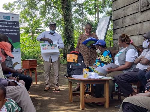
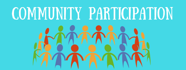
The community includes everyday people: men, women, children, elderly, farmers, market vendors, teachers, and families living in the affected area. They are the first responders and often the most important stakeholders.
- They are always there when disaster strikes before outside help arrives
- They know the local area: where the dangerous places are, where to find help
- They know each other: they can identify missing people and help neighbors
- They have the most to lose: it is their homes, their families, their lives
- Stay Alert to Warning Alerts: Pay attention to warnings on radio, TV, SMS, or community drums. Take warnings seriously and do not ignore them. Share warnings with neighbors who may not have heard. Example: When the Uganda National Meteorological Authority predicts heavy rains in Bududa, community members must listen and prepare to evacuate.
- Understand Risk and Demand Action: Learn what makes the community vulnerable (steep slopes, poor drainage, weak buildings). Demand responsible action from local leaders and businesses. Attend community meetings and speak up about safety concerns. Example: If a factory is polluting the community water source, residents must demand that the local council and the factory owner take action.
- Foster a Culture of Resilience: Resilience means the ability to bounce back after difficulty. Encourage everyone to take responsibility for managing risks. Celebrate community members who prepare for disasters. Make preparedness a normal part of community life. Example: A village in Karamoja where every family stores extra food and water during the dry season is a resilient community.
- Learn Individual Actions to Address Risk: Educate yourself about what YOU can do at home, work, and in the community. Find time for Education (Reading, attending workshops), Training (First aid, fire safety, evacuation procedures), and Capacity-building (Learning new skills to help during disasters). Example: A mother learns how to make oral rehydration solution (ORS) at home so she can treat her child immediately if diarrhea starts after a flood.
- Mobilize Fellow Community Members: Encourage neighbors to participate in disaster prevention programs. Organize community clean-up days to clear drainage channels. Form community disaster committees. Example: A youth group in Kampala organizes weekly drainage clearing in Bwaise to prevent flooding.
- Encourage Family, Friends, and Neighbors: Help others improve their ability to deal with risk factors. Check on elderly neighbors who live alone. Help families make their own emergency plans. Example: A community health worker visits elderly people before the rainy season to make sure their roofs are secure.
- Participate in Strategic Planning: Take part in local and national capacity development planning. Participate in capacity assessments evaluating what the community can and cannot do. Give your opinion as you know your community best. Example: Community members attend a district planning meeting to say which roads flood first and where evacuation routes should be.
- Gather Resources for Disaster Programs: Contribute funds through community savings groups. Contribute supplies like food, blankets, building materials. Contribute volunteer assistance via time and labor. Example: A village savings slope sets aside money each month for emergency supplies.
- Attend Emergency Preparedness Training: Take advantage of training opportunities. Learn skills like first aid, search and rescue, and fire safety. Practice what you learn and teach others. Example: Community members attend a Red Cross first aid training at the local church.
- Stay Actively Engaged: Participate through Schools (Parent-teacher associations), Religious organizations (Churches, mosques, temples), Social networks (Community groups, village teams), and Professional associations (Farmers' cooperatives, traders' associations). Do not wait for disaster to get involved.
- Actively Participate in Decision-Making and Implementation: Do not just listen, speak up in meetings. Vote on community disaster plans. Volunteer to help implement actions (e.g., distributing food, building shelters). Example: Community members help decide where the emergency shelter should be located and then help clean and prepare it.
- Community entry: Establish trust and rapport
- Health education: Teach preparedness in local languages
- Mobilization: Help form community health committees
- Capacity building: Train community health workers
- Advocacy: Help communities voice their needs to leaders
Local government includes:
- District Local Governments: Led by the Resident District Commissioner (RDC) and District Chairperson (LC5)
- Municipal and Town Councils
- Sub-county and Parish Chiefs
- Local Council leaders: LC1 (village), LC2 (parish), LC3 (sub-county)
- Convene Community Stakeholders: Bring together all interested groups in the community. Lead or coordinate local capacity development efforts. Chair disaster management committee meetings. Example: The District Chairperson calls a meeting of health workers, police, religious leaders, and NGOs to plan for the rainy season.
- Perform Risk Assessments: Evaluate the risks faced by the community. Identify capacity needs to see what skills, resources, and systems are missing. Map hazard-prone areas. Example: The district disaster management office maps all flood-prone villages in Kasese district.
- Coordinate and Communicate Assessment Results: Share the findings of community-wide capacity assessments. Make sure everyone knows what the risks are and what is needed. Use local radio, meetings, and bulletins.
- Support the Enabling Environment: Create legislative frameworks like local laws and bylaws. Develop policies that support disaster risk reduction. Establish procedures for response. Allocate budgeting for disaster activities. Do strategic planning for long-term safety. Example: A district council passes a bylaw prohibiting building in wetlands.
- Provide Standards, Accreditation, Technologies, and Resources: Set guidelines for safe construction. Accredit training programs (approve them as meeting standards). Provide technologies like early warning systems. Share resources for planning and communication.
- Integrate Capacity Development: Make sure disaster risk reduction is part of all government offices. Connect disaster management to Sustainable development (Long-term community growth) and Climate change adaptation (Adjusting to changing weather patterns). Do not isolate disaster management, make it everyone's job. Example: The district agricultural office teaches drought-resistant farming as part of disaster risk reduction.
- Increase Awareness of Capacity Needs: Raise awareness in the local community about the importance of building skills and resources. Show the value of reducing capacity gaps.
- Ensure Coordination with Higher Government Levels: Align local activities with sub-national and national government plans. Communicate local capacity needs to national government. Seek support and resources from higher levels. Example: The district requests additional funding from the Office of the Prime Minister for flood mitigation.
- Provide or Support Training and Education: Deliver or help organize training programs. Address capacity needs specific to the local context. Support community-based training.
- Encourage and Empower Leadership and Staff: Help local government leaders and staff understand their role in disaster risk reduction. Establish mechanisms to address their training needs. Make sure civil servants know what to do during disasters.
- Facilitate Community-Based Initiatives: Support capacity development activities led by the community itself. Do not take over, let communities lead. Provide guidance and resources when requested.
- Conduct Monitoring and Evaluation: Regularly check if capacity development efforts are working. Evaluate progress and effectiveness. Make improvements based on findings.
- Identify Capacity Resources in the Community: Find existing skills, equipment, and organizations. Engage with relevant stakeholders to develop these resources further. Example: The district identifies that a local construction company has heavy equipment useful for search and rescue.
- Work with Citizens and Private Sector: Collaborate directly with citizens. Support engagement through NGOs and private companies. Encourage businesses to invest in disaster risk reduction. Example: A local telecom company provides free SMS alerts during disasters as part of corporate social responsibility.
- Participate in district disaster committees
- Report health needs to district health officers
- Advocate for health priorities in district budgets
- Coordinate health responses with local authorities
- Support local training programs
The national government includes:
- The President and Cabinet
- Office of the Prime Minister: Department of Disaster Preparedness and Management
- Ministry of Health
- Ministry of Works and Transport
- Ministry of Water and Environment
- Uganda Police Force and Uganda People's Defence Force (UPDF)
- National Meteorological Authority
- Parliament: Makes laws and approves budgets
- Fund Disaster Management Programs: Allocate financial resources at both national and local levels. Support implementation of disaster management initiatives. Ensure timely release of emergency funds. Example: The government releases funds through the Office of the Prime Minister when Bududa experiences landslides.
- Purchase and Install Disaster Monitoring Systems: Acquire and set up systems that monitor and detect potential disasters. Facilitate timely response and mitigation. Examples: Weather satellites, seismic monitors, river level gauges.
- Develop National Strategy for Capacity Development: Establish a comprehensive strategy that guides planning and implementation. Cover all stakeholder groups. Apply at all levels: national, district, sub-county, community.
- Increase Awareness About Disasters: Raise awareness among the entire population. Educate about different types of disasters. Explain potential impacts. Promote preparedness and risk reduction. Example: National campaigns on radio and TV about landslide warning signs.
- Formulate Policies to Regulate the Environment: Create policies and regulations that manage the environment. Reduce likelihood and severity of disasters. Examples: National Environment Management Policy, Wetlands Policy.
- Provide Relief During Disasters: Food supplies (Distribute emergency food), Shelters (Build or fund emergency housing), Medical services (Deploy national medical teams), Security (Deploy police and army to maintain order). Example: After the 2010 Bududa landslides, the government provided tents, food, and medical teams.
- Fund Research: Support research to understand why disasters occur, what their impacts are, and how to prevent them. Use research to improve policies.
- Coordinate Capacity Assessment Data: Gather and organize data about existing capacity and resources. Enhance awareness across communities, districts, and the nation. Identify where help is needed most.
- Establish National Standards of Operation: Develop standardized protocols and procedures. Ensure effective coordination during disasters so everyone follows the same rules. Example: National guidelines for setting up cholera treatment centers.
- Encourage and Empower Leadership and Staff: Help national leaders and civil servants understand their role in disaster risk reduction. Provide training and mechanisms to address capacity needs.
- Provide Guidance and Documentation: Offer guidance, documentation, and frameworks. Support capacity development at national and local levels. Create manuals, standard operating procedures (SOPs), and training materials.
- Establish Immunization Programs: Implement vaccination programs to prevent epidemics. Especially important after disasters when disease risk is high. Example: Measles vaccination campaigns in refugee camps and displacement sites.
- Emphasize Sanitation in High-Population Areas: Focus on improving sanitation in densely populated areas. Reduce health risks and disease outbreaks. Examples: Slum upgrading in Katanga (Kampala), Bwaise, and similar areas.
- Establish Departments Dedicated to Disaster Management: Create specialized departments or agencies to coordinate disaster-related activities. Example: Office of the Prime Minister: Disaster Preparedness.
- Create Collaborative Platforms: Facilitate forums where government, private sector, academia, and others work together. Promote innovative, practical, affordable, and localized approaches to disaster risk reduction.
- Implement national health policies at the local level
- Report disease outbreaks through the national surveillance system
- Participate in national vaccination campaigns
- Advocate for nursing representation in national disaster planning
- Follow national standards for emergency care
Regional institutions are organizations that cover more than one country in a region. For Uganda, these include:
- East African Community (EAC): Uganda, Kenya, Tanzania, Rwanda, Burundi, South Sudan, DRC
- Intergovernmental Authority on Development (IGAD): Horn of Africa
- African Union (AU): Continental body
- Regional economic communities
- Regional disaster management platforms
- Coordinate Regional Responses: When disasters affect multiple countries, coordinate joint responses. Share resources across borders. Example: During the East African drought, IGAD coordinates food aid across Kenya, Ethiopia, Somalia, and Uganda.
- Share Information and Early Warnings: Share weather data, disease surveillance, and hazard information across borders. A flood in Kenya may warn Uganda of incoming water. A disease outbreak in DRC may warn Uganda to prepare.
- Develop Regional Policies and Frameworks: Create agreements that bind member states to disaster preparedness standards. Harmonize building codes, health standards, and response protocols.
- Pool Resources: Countries contribute to a shared fund or resource pool. Deploy shared equipment (e.g., helicopters, rescue boats) where needed most.
- Capacity Building Across Borders: Train disaster managers from all member countries together. Share best practices between countries. Learn from each other's experiences.
- Cross-Border Disaster Management: Manage disasters that do not respect borders. Coordinate refugee movements. Manage transboundary diseases. Handle regional climate impacts. Example: Lake Victoria water levels affect Uganda, Kenya, and Tanzania simultaneously; regional coordination is needed.
- Participate in regional training programs
- Share nursing best practices with colleagues in neighboring countries
- Support cross-border health initiatives (e.g., disease surveillance)
- Advocate for regional health standards
NGOs are private, non-profit organizations that work to help communities. In Uganda, major disaster-related NGOs include:
- Uganda Red Cross Society: Largest humanitarian NGO in Uganda
- UNICEF: Focuses on children and women
- World Food Programme (WFP): Food assistance
- Médecins Sans Frontières (MSF): Medical care in emergencies
- World Vision: Child-focused development and relief
- Oxfam: Poverty reduction and emergency response
- Save the Children: Child protection and health
- Local NGOs: Community-based organizations (CBOs)
- Provide Emergency Relief: Food distribution (Immediate food aid), Water and sanitation (Clean water, latrines, hygiene kits), Shelter (Tents, tarpaulins, building materials), Medical care (Mobile clinics, emergency surgery). Example: Uganda Red Cross distributes relief items immediately after landslides in Bududa.
- Supplement Government Efforts: Fill gaps where government capacity is insufficient. Reach remote areas government cannot reach quickly. Provide specialized services.
- Community-Based Programming: Work directly with communities. Respect local knowledge and culture. Build local capacity rather than replacing it.
- Advocacy: Speak up for vulnerable populations. Push governments to invest in disaster prevention. Raise awareness about forgotten disasters. Example: NGOs advocating for Karamoja drought to receive national attention and funding.
- Technical Expertise: Bring specialized knowledge in Water engineering (borehole drilling), Health (epidemic response), Agriculture (drought-resistant crops), and Construction (shelter design).
- Resource Mobilization: Raise funds from international donors. Bring in resources that local communities cannot afford. Manage donations transparently.
- Monitoring and Accountability: Monitor if aid reaches intended beneficiaries. Evaluate program effectiveness. Hold all stakeholders accountable.
- Coordinate referrals: Know which NGO provides which service
- Collaborate on health programs: Joint vaccination campaigns, nutrition programs
- Share information: Report health needs to NGOs operating in your area
- Participate in NGO training: Many NGOs offer excellent disaster nursing training
- Maintain professional boundaries: Work together while respecting each organization's role
The media includes:
- Radio stations: The most important media in Uganda (local language reach)
- Television stations: NTV, NBS, Bukedde, UBC
- Newspapers: Daily Monitor, New Vision, Observer
- Social media: Facebook, Twitter, WhatsApp groups
- Community media: Community radios, town criers, notice boards
- Raise Awareness, Advocate, and Motivate Society on DRR: Use all platforms to increase public awareness about Disaster Risk Reduction (DRR). Advocate for preparedness and risk reduction measures. Motivate individuals to take action. Example: Radio talk shows explaining why building in wetlands is dangerous.
- Extend Special Programs for Media Staff: Offer specialized training for journalists and media professionals. Enhance their understanding of disaster management. Improve reporting on DRR-related issues so trained reporters give accurate, helpful information instead of causing panic.
- Strengthen Linkages with Other Stakeholders: Establish strong connections with government, NGOs, and communities. Ensure smooth flow of accurate and timely information. Be the bridge between authorities and the public.
- Introduce Innovative Products for Risk Information: Develop new ways to share risk information. Use various channels and technologies: SMS alerts, Interactive radio programs, Social media campaigns, Mobile apps.
- Collect, Analyze, and Share Information: Gather relevant data about disasters. Analyze it for accuracy before sharing. Share accurate and up-to-date information with the public to help people make informed decisions.
- Encourage All Groups to Provide Information: Encourage diverse groups to share experiences including Government agencies, NGOs, Community leaders, and Affected individuals. Foster comprehensive understanding of disaster situations.
- Maintain a Link to Academia: Connect with universities and research institutions. Access expert insights and research findings. Ensure accurate reporting and promote evidence-based practices. Example: A journalist interviews a Makerere University professor about climate change and drought patterns.
| Positive Role | Negative Role |
|---|---|
| Spreads early warnings quickly | Can spread rumors and cause panic |
| Educates the public | May sensationalize suffering |
| Holds leaders accountable | May report inaccurate information |
| Mobilizes resources for relief | May invade privacy of victims |
- Provide accurate health information to journalists
- Correct misinformation quickly (e.g., false disease outbreak rumors)
- Use media for health education: Participate in radio programs
- Protect patient privacy: Ensure media does not exploit victims
- Be a trusted source: Nurses are often seen as credible voices in the community
Medical institutions include:
- National and regional referral hospitals: Mulago, Butabika, Mbarara, Arua, Gulu, Mbale, Jinja, Fort Portal, Soroti, Hoima
- District hospitals: One per district
- Health Centre IVs, IIIs, and IIs
- Private hospitals and clinics
- Pharmacies and drug shops
- Medical training institutions: Makerere University, Mbarara University, Kampala International University, nursing schools
- Provide Emergency Medical Care: Treat injuries during disasters. Perform emergency surgeries. Manage mass casualties. Stabilize critically ill patients.
- Maintain Surge Capacity: Be able to handle sudden increases in patient numbers. Have flexible space (e.g., tents, empty wards). Maintain emergency supply stockpiles.
- Disease Surveillance and Outbreak Response: Monitor for unusual disease patterns. Report outbreaks immediately. Set up isolation and treatment centers.
- Continuity of Care: Ensure chronic disease patients do not miss treatment. Maintain medication supplies. Keep records secure and accessible.
- Health Worker Training: Train staff in disaster medicine. Conduct regular drills. Maintain certifications (BLS, ACLS, trauma care).
- Coordination with Other Stakeholders: Work with government, NGOs, and community health workers. Share patient information appropriately. Accept referrals and transfer patients when needed.
- Mental Health Services: Provide crisis counseling. Treat PTSD and depression. Support staff who are also traumatized.
- Frontline care: Nurses are often the first to see disaster victims
- Triage: Sort patients by severity
- Coordination: Liaise between emergency department and other units
- Documentation: Maintain accurate records for surveillance and legal purposes
- Staff support: Support colleagues under stress
- Universities: Makerere, Mbarara, Gulu, Kyambogo, Kampala International University
- Research institutes: National Agricultural Research Organisation (NARO), Uganda Virus Research Institute (UVRI), National Forestry Authority research units
- Scientific community: Meteorologists, geologists, epidemiologists, engineers, environmental scientists
- Vocational and technical schools
- Raise Awareness of Capacity Development Needs: Increase awareness within the academic community. Educate both internal and external stakeholders about the importance of building disaster management skills.
- Encourage Research Supporting Disaster Risk Reduction: Encourage faculty and students to conduct research on disaster causes, prevention methods, health impacts, and community resilience. Contribute to the knowledge base. Example: Makerere University researchers study landslide patterns on Mt. Elgon.
- Provide Relevant Curriculum and Courses: Teach the next generation of disaster management experts. Equip students with necessary skills. Ensure skills taught match what the field actually needs. Example: Nursing schools should include disaster nursing in their curriculum.
- Expand the Disaster Risk Reduction Curriculum: Incorporate DRR topics into many disciplines including Finance and financial risk management, Development studies, Urban planning, Public policy, Public health, Engineering, and Agriculture. Do not limit DRR to one department.
- Support Identification of Key Competencies: Work with stakeholders to identify what skills are needed for effective disaster risk reduction. Provide competency-based learning to teach exactly what is needed.
- Coordinate with Other Sectors: Work with Government agencies, Private sector, and Civil society organizations to understand and address training needs that meet job requirements.
- Develop Accessible and Tailored Courses: Create courses for people outside traditional academia: Shorter timeframes, Non-technical terminology, Local context. Increase interest and participation from community members. Example: A short course for community health workers on epidemic detection, taught in the local language over two days.
- Collect and Share Data and Information: Gather research findings. Share best practices. Disseminate relevant resources. Enhance knowledge and inform decision-making. Example: Uganda Virus Research Institute shares data on Ebola and Marburg to help the Ministry of Health prepare.
- Participate in research: Help collect data during disasters
- Use evidence-based practice: Apply research findings to your nursing care
- Contribute case studies: Write about your experiences for journals
- Stay updated: Attend conferences and read new research
- Teach students: Host nursing students and teach them disaster nursing
Without coordination:
- Duplication: Two NGOs build latrines in the same village while another has none
- Gaps: No one provides mental health services
- Confusion: Communities receive conflicting messages
- Waste: Resources are used inefficiently
- Conflict: Stakeholders compete instead of collaborate
| Mechanism | Purpose | Example |
|---|---|---|
| Disaster management committees | Bring stakeholders together regularly | District Disaster Management Committee meets monthly |
| Cluster system | Group stakeholders by sector (health, shelter, water) | Health Cluster coordinates all medical actors |
| Information sharing platforms | Share data and plans | Weekly coordination meetings, shared databases |
| Joint assessments | Evaluate needs together | Multi-agency assessment after floods |
| Memoranda of Understanding (MoUs) | Formal agreements between organizations | Hospital and Red Cross agree on referral procedures |
Nurses often serve as natural coordinators because they:
- Work in communities and hospitals
- Know both patients and administrators
- Are trusted by the public
- Have skills in communication and organization
- Liaison: Connect community health workers with district health office
- Information hub: Collect and share health data with all stakeholders
- Meeting facilitation: Lead community disaster committee meetings
- Resource mapping: Know who has what resources and where
Remembering the 8 Stakeholders:
- Communities
- Local Government
- National Government
- Regional Institutions
- NGOs
- Media
- Medical Institutions
- Education/Research
- Share information
- Help each other
- Act responsibly
- Respect local knowledge
- Evaluate together
- Participate in planning
- Attend training
- Respond to warnings
- Take individual action
- Inform neighbors
- Contribute resources
- Implement decisions
- Prepare your family
- Advocate for safety
- Teach others
- Engage actively
- Q1: Define "stakeholder" in disaster management.
Answer: A stakeholder is an individual or group who has an interest in the disaster management program and can be influenced by its process or outcomes. Their interests can be positively or negatively affected by the disaster management processes. - Q2: List the eight types of stakeholders in disaster management.
Answer: Communities, local governments, national governments, regional institutions, NGOs, media, medical institutions, and education/research institutions. - Q3: What are three responsibilities of local government in disaster management?
Answer: Convene community stakeholders and coordinate local efforts; perform risk assessments; support the enabling environment through policies and budgeting; provide standards and resources; integrate capacity development across government offices. (Any three) - Q4: Why is the media an important stakeholder in disaster management?
Answer: The media raises awareness, advocates for preparedness, disseminates early warnings, shares accurate information, motivates the public to act, and holds leaders accountable. - Q5: How can communities participate in disaster management?
Answer: By staying alert to warnings, understanding local risks, attending training, mobilizing neighbors, participating in planning, gathering resources, and taking individual actions to prepare their families. - Q6: What is the role of education and research institutions in disaster management?
Answer: They raise awareness of capacity needs, encourage research on disaster risk reduction, provide relevant curriculum, expand DRR education across disciplines, identify key competencies, coordinate with other sectors, develop accessible courses, and collect and share data. - Q7: Why is coordination among stakeholders essential?
Answer: Coordination prevents duplication of efforts, fills gaps in services, reduces confusion, ensures efficient use of resources, and prevents conflict among responding agencies.
Heavy rains have caused flooding in Mbale. Multiple stakeholders are responding.
- Which stakeholders should be involved? Local government, national government/OPM, Uganda Red Cross, media, medical institutions, communities.
- What is the community's role? Evacuate, help neighbors, share information, participate in cleanup.
- What is the nurse's coordination role? Liaise between health center and district; report disease cases; coordinate with NGOs for supplies.
Karamoja is experiencing severe drought. Food insecurity is widespread.
- Which stakeholders are most critical? National government for funding, WFP for food aid, NGOs for water and nutrition programs, research institutions for drought-resistant crops, communities for adapting practices.
- What should regional institutions do? IGAD should coordinate cross-border drought response, share early warning data.
- What is the nursing role? Malnutrition screening, ORS education, referral to feeding centers, reporting to district health office.
A suspected Ebola case has been identified. Rumors are spreading on social media.
- What is the media's responsibility? Report accurately, avoid sensationalism, share official Ministry of Health messages.
- What is the medical institution's role? Isolate patient, activate outbreak response, coordinate with national level.
- What is the community's role? Cooperate with contact tracing, report symptoms, avoid spreading rumors.
- What is the nurse's role? Use PPE, follow protocols, correct misinformation, support frightened community members.
- Stakeholders are anyone with an interest in disaster management outcomes.
- There are 8 main types of stakeholders.
- Communities are the most important as they are first responders and have the most to lose.
- Local government coordinates at the district and sub-county level.
- National government sets policy, funds programs, and provides relief.
- Regional institutions handle cross-border issues.
- NGOs supplement government efforts with specialized services.
- Media informs, educates, and advocates but must be accurate.
- Medical institutions provide emergency care and surveillance.
- Education/research institutions generate knowledge and train future experts.
- Coordination among all stakeholders is essential to avoid gaps and duplication.
- Nurses serve as bridges between stakeholders, especially between the health system and community.
- International Council of Nurses (ICN). (2019). Core Competencies in Disaster Nursing.
- World Health Organization (WHO). (2020). Health Emergency and Disaster Risk Management Framework.
- Veenema, T. G. (2018). Disaster Nursing and Emergency Preparedness (4th ed.). Springer Publishing Company.
- Office of the Prime Minister, Republic of Uganda. (2010). National Policy for Disaster Preparedness and Management.
Quick Quiz
Stakeholders Quiz
Disaster Management - mobile-friendly and focused practice.
Privacy: Your details are used only for quiz tracking and certificates.
Stakeholders Quiz
Disaster Management
Preparing questions...
Choose your answer and keep your streak alive.
Great effort.
Here is your quick performance summary.
Stakeholders in Disaster Management Read More »