A comprehensive guide on how to interpret and answer UNMEB question types: EXPLAIN, OUTLINE, DESCRIBE, MENTION, IDENTIFY, STATE, LIST, WHAT, and GIVE.
This guide explores specific nursing interventions, considerations, concerns, and issues frequently tested in professional medical exams.
EX
The EXPLAIN Approach
In Simple Terms:
"Explain" means to give details and reasons. You need to show *how* or *why* something happens, not just what it is.
1
Understand the question: Carefully read and identify the main concept. Pay attention to specific instructions.
2
Organize your response: Create a mental map. Start with a concise introduction, context, and clear thesis.
3
Provide thorough explanation: Elaborate using clear language. Use nursing terminology and case studies.
Simulated Examination Sheet
Qn: Explain the pathophysiology of diabetes mellitus and its effects on the body.
Diabetes mellitus is a chronic metabolic disorder characterized by high blood
glucose levels due to impaired insulin secretion, insulin action, or both. The
pathophysiology of diabetes involves multiple factors that contribute to the
development and progression of the disease.
Firstly, in type 1 diabetes, an autoimmune process leads to the destruction of
insulin-producing beta cells in the pancreas. This results in a deficiency of
insulin and requires external insulin administration. On the other hand, type 2
diabetes is primarily characterized by insulin resistance, where the body’s
cells become less responsive to insulin.
Insulin is a hormone produced by the beta cells of the pancreas, and its main
function is to regulate glucose metabolism. In diabetes, the lack of insulin or
the body’s inability to use it effectively leads to hyperglycemia. Persistently
high blood glucose levels can have detrimental effects on various organs and
systems in the body.
The effects of diabetes on the body are many. It can lead to macrovascular
complications, such as cardiovascular disease, stroke, and peripheral vascular
disease. Also, microvascular complications may arise, affecting small blood
vessels in the eyes, kidneys, and nerves. Diabetes can also increase the risk of
infections, slow wound healing, and cause diabetic neuropathy and
nephropathy
OU
The OUTLINE Approach
In Simple Terms:
"Outline" means to create a structured summary. Use main headings and sub-points to show parts in an organized way.
1
Analyze the question: Identify main components that need to be outlined.
2
Organize your response: Identify main headings and arrange them in a coherent order.
3
Provide detailed information: Use concise and informative language under each heading. JUMP A LINE, UHPAB HAS VERY MANY BOOKLETS
Simulated Examination Sheet
Qn: Outline the steps involved in the nursing process.
1. Assessment:
Gather relevant patient data, including physical, psychological, social, and
cultural aspects. Perform a comprehensive health history and physical
examination. Utilize assessment tools and techniques to collect objective and
subjective data. Document and organize the collected data systematically.
2. Diagnosis:
Analyze the assessment data to identify health problems, risks, or potential
complications. Formulate nursing diagnoses based on the identified issues.
Ensure that the diagnoses are accurate, concise, and specific.
Collaborate with other healthcare professionals when necessary.
3. Planning:
Establish patient-centered goals and outcomes in collaboration with the patient.
Develop a nursing care plan that includes evidence-based interventions and
strategies. Prioritize nursing actions based on the urgency and importance of each
goal. Ensure that the care plan is feasible, realistic, and adaptable.
4. Implementation:
Execute the planned nursing interventions effectively and efficiently.
Provide safe and compassionate care while considering the patient's preferences.
Document the implementation process and any modifications made. Collaborate with
the interdisciplinary healthcare team to deliver comprehensive care.
5. Evaluation:
Assess the patient's response to the nursing interventions and the achievement
of goals. Compare the actual outcomes with the expected outcomes. Modify the
care plan if needed based on the evaluation findings. Document the evaluation
results and communicate them to the healthcare team.
DE
The DESCRIBE Approach
In Simple Terms:
"Describe" means to paint a picture with words. Give a detailed account of characteristics or features. Describe usually likes STEPS in order, even using IMAGES where applicable!
1
Understand the question: Identify the main topic that needs characterization.
2
Provide comprehensive description: Offer thorough details, features, or components.
3
Use terminology: Accurately describe concepts to demonstrate knowledge.
Simulated Examination Sheet
Qn: Describe the stages of wound healing.
1. Hemostasis:
This initial stage begins immediately after the injury occurs.Blood vessels constrict to reduce blood flow and prevent excessive bleeding.
Platelets aggregate to form a temporary clot. The clotting process releases various
growth factors and cytokines, initiating the subsequent stages of healing.
2. Inflammatory phase:
This phase typically lasts for 2-3 days.Inflammation occurs as a response to tissue injury. Vasodilation and increased
vascular permeability allow immune cells to migrate to the wound site. Neutrophils
arrive first to eliminate debris and prevent infection. Macrophages then remove dead
tissue and release additional growth factors to stimulate healing.
3. Proliferative phase:
This phase generally occurs between days 3 and 20.New blood vessels form to supply oxygen and nutrients to the wound. Fibroblasts
produce collagen, which provides structural support for wound healing. Epithelial
cells migrate from the wound edges to resurface the wound. Granulation tissue
forms, consisting of new blood vessels, fibroblasts, and extracellular matrix.
4. Maturation phase:
This final phase can last for several months to years.Collagen fibers reorganize and remodel, increasing the wound's tensile strength. Scar
tissue forms, but it may not possess the same strength and flexibility as the original
tissue. The scar gradually becomes more refined and fades over time.
ST
MENTION / IDENTIFY / STATE
In Simple Terms:
These words mean "give a short, direct answer." Just name the facts without extra explanation.
1
Identify facts: Read and identify the specific information required.
2
Direct response: Offer a concise response. Avoid unnecessary elaboration.
Simulated Examination Sheet
Qn: State the types of delusions.
Grandiose delusions; the patient believes s/he is somebody great /important
,knowledgeable or powerful contrary to the social cultural ,religious background
and experiences.
Delusion of guilty and worthlessness; the patient believes s/he is not worth to
live even though there’s nothing to justify this belief.
Delusions of jealousy; the patient believes that spouse/partner is being
unfaithful even when there is no evidence to suggest so.
Delusion of persecution: the patient believes they’re being deliberately wronged,
conspired or harmed by another person or agency even when there’s no evidence
to suggest so.
Religious delusions; the individual believes he or she has a special link with God
that is out keeping with people of the same religious belief.
Delusions of control, influence or phenomenon; these are three types; belief
that the person performs activities as a result of an extreme force.
LI
The LIST Approach
In Simple Terms:
"List" means to present points one after another, usually with a short description for each.
1
Identify factors: Carefully identify the elements that need to be listed.
2
Organize: Present items in a logical order using bullet points.
Simulated Examination Sheet
Qn: List the risk factors for cardiovascular disease.
- Hypertension: increases strain on heart.
- Smoking: damages blood vessels.
- Obesity: increases risk of diabetes.
- Sedentary lifestyle: contributes to obesity.
WH
The WHAT Approach
In Simple Terms:
"What" asks for a definition. Give a clear, simple explanation of the term or concept.
1
Identify term: Pinpoint the specific procedure or concept to define.
2
Clear explanation: Offer a concise definition using simple language.
Simulated Examination Sheet
Qn: What is sepsis?
Sepsis is a potentially life-threatening condition that occurs when the body’s response to an infection becomes unregulated, leading to widespread inflammation and organ dysfunction.
Mood stabilizers are a class of psychotropic medications primarily used to treat bipolar affective disorder (BAD) and other conditions involving severe mood swings or instability. They help control the frequent fluctuation of mood at short intervals and are utilized as maintenance drugs to prevent relapses.
Mood stabilizers are psychotropic drugs which are used in controlling mood disorders. They are essential for managing the extreme highs and lows associated with bipolar disorders and related psychiatric conditions.
I. Introduction & History
Definition
No consensus definition: Officially, the FDA states, "There is no such thing as a mood stabilizer."
Clinical Definition (Sachs, 1996): Any medication that is able to decrease vulnerability to subsequent episodes of mania or depression; and does not exacerbate the current episode or maintenance phase of treatment.
Important Rule: Patients with acute-phases of mania or depression should generally be treated first with anti-psychotics or anti-depressants respectively, alongside or before transitioning to maintenance mood stabilizers.
Brief History & Trivia
1817: Lithium was discovered as a chemical element.
1871: First recorded use as a treatment of mania.
1876: Li₂CO₃ used in the prevention of depression.
Early 20th Century: Use of Lithium largely abandoned due to its severe toxicity.
1949: Use of Lithium for mania rediscovered by Australian psychiatrist John Cade.
1970: FDA approved the use of Lithium for mania.
1995: Sodium valproate approved for acute mania.
Trivia: The popular soft drink "7 Up" originally contained Lithium citrate from 1929 to 1950 and was marketed as a good remedy for relieving alcoholic hangovers!
II. Pathophysiology & General Mode of Action
The exact mode of action of mood stabilizers is not fully understood, but three primary pharmacological theories explain their efficacy:
Neurotransmitter Regulation: Mood stabilizers regulate the levels of monoamine neurotransmitters (serotonin, dopamine, and norepinephrine). Imbalances lead to mood swings. Mood stabilizers normalize these imbalances, reducing bipolar symptoms.
Ion Channel Modulation (Voltage-Sensitive Channels): They modulate the activity of voltage-gated ion channels, particularly sodium (Na⁺) and calcium (Ca²⁺) channels. These channels regulate neuronal excitability. Abnormal activity leads to mania or depression; stabilization reduces this aberrant firing.
Structural and Signal Transduction Modulation: They affect the structure and function of brain regions regulating mood (prefrontal cortex, amygdala) by acting on downstream signal transduction cascades (e.g., G-proteins, second messengers like inositol monophosphate, and enzymes like GSK-3 and PKC), promoting neuroprotection and long-term neuroplasticity.
III. Classification of Drugs Used as Mood Stabilizers
Lithium is a naturally occurring element. It is the classic mood stabilizer, possessing profound anti-manic and anti-suicidal properties.
Lithium carbonate is a medication that is commonly used as a mood stabilizer to treat bipolar disorder. It is a naturally occurring element that is found in small amounts in the body, and it works by affecting the levels of certain neurotransmitters in the brain, particularly serotonin and norepinephrine.
1. Indications for Use
Acute treatment of mania.
Prophylaxis of bipolar affective disorder (More effective in preventing manic relapses than depressive relapses).
Augmentation of antidepressants in treatment-resistant unipolar depression.
Prevention of aggressive behavior in patients with learning difficulties or personality disorders.
Schizoaffective disorder and Alcoholism.
Lithium and Suicide: The estimated suicide rate in bipolar patients is 10-15%. Lithium is unique among psychiatric drugs as it reduces both attempted and completed suicide by a staggering 80%.
2. Mechanism of Action
Exact mechanism is uncertain, but it works by affecting signal transduction:
Through the inhibition of 2nd messenger enzymes (e.g., inositol monophosphate).
By modulation of G proteins.
By interaction at various sites within downstream signal transduction cascades (e.g., inhibition of Glycogen Synthase Kinase-3 [GSK3] and Protein Kinase C [PKC]).
Cerebral sodium concentration is reduced, which helps control mania and reduces excitements.
3. Pharmacokinetics & Dosing
Dosage
For adults:
The typical starting dose of lithium carbonate for the treatment of bipolar disorder is 300-600 mg per day, divided into two or three doses.
The dosage is then gradually increased, usually by 300-600 mg per week, until a therapeutic blood level is achieved.
Maintenance doses of lithium carbonate can range from 600-2400 mg per day, depending on the individual’s needs and the therapeutic blood level.
For children:
Lithium carbonate is not typically prescribed for children under the age of 12, as there is limited research on its safety and efficacy in this population.
For adolescents aged 12-18, the starting dose of lithium carbonate is 300 mg per day, divided into two or three doses.
The dosage is then gradually increased as needed, usually by 150-300 mg per week, until a therapeutic blood level is achieved.
4. Pre-treatment Tests & Monitoring
Lithium has a very narrow therapeutic index. Monitoring is critical for patient survival.
Note: When one is on lithium carbonate, the following base line investigations should be done:
Blood serum lithium level:
The normal range 0.6-1.2 mEq/l (for prevention of relapse in BAD).
Therapeutic levels 0.8-1.2mEq/l (for treatment of acute mania).
Toxic lithium levels greater than 2.0 mEq/l.
Blood tests: A complete blood count (CBC) and blood chemistry panel should be done to assess the person’s overall health and to check for any underlying medical conditions that could affect their response to lithium. The blood chemistry panel should include tests for electrolyte levels (including sodium and potassium), kidney function, liver function, and thyroid function.
Urinalysis: A urinalysis should be done to assess kidney function and to check for any signs of kidney damage.
ECG: An electrocardiogram (ECG) should be done to assess the person’s heart function and to check for any underlying cardiac conditions that could affect their response to lithium.
Pregnancy test: Women of childbearing age should have a pregnancy test before starting lithium, as this medication can be harmful to a developing fetus.
Medical history: A thorough medical history should be taken to assess the person’s overall health, including any past or current medical conditions, medications, or allergies.
Psychiatric evaluation: A comprehensive psychiatric evaluation should be done to assess the person’s symptoms and to establish a diagnosis of bipolar disorder.
Baseline mood assessment: A baseline assessment of the person’s mood and behavior should be done to establish a baseline for monitoring the effects of the medication.
N.B: A patient on lithium carbonate should also be given a thyroxin tablet (also known as levothyroxine), a medication that is used to treat an underactive thyroid gland (hypothyroidism). This is because lithium can affect the functioning of the thyroid gland.
5. Side Effects
Lithium carbonate is generally well-tolerated when used as a mood stabilizer for the treatment of bipolar disorder. However, like any medication, it can cause side effects, both in the short term and the long term.
Short-term side effects of lithium can include:
Nausea, vomiting, and diarrhea
Dry mouth and thirst
Increased urination
Muscle weakness and tremors
Fatigue and drowsiness
Headaches
Increased appetite and weight gain
Mild cognitive impairment (difficulty with attention, memory, or problem-solving)
Skin rash or acne
Vertigo
Dysarthria (impaired articulation of speech)
Cardiac arrhythmias
In some patients they may have oedema
Nystagmus
Many of these side effects are usually mild and may resolve on their own as the body adjusts to the medication. Some people may be able to manage these side effects by adjusting the dosage or timing of their medication, or by taking it with food or milk.
Long-term use of lithium can cause more serious side effects, including:
Kidney damage or kidney failure
Hypothyroidism (underactive thyroid gland) i.e. reduced thyroxin in the body as lithium carbonate interferes with metabolism
Increased risk of diabetes mellitus
Cardiovascular disease (heart disease)
Neurological effects (such as hand tremors, slurred speech, and impaired coordination)
Teratogenicity (birth defects in fetuses exposed to lithium during pregnancy)
Nephrogenic diabetes insipidus
Depletion of calcium in bones
Memory impairment affects memory centers of the brain.
NB: Big doses of neuroleptics e.g. haloperidol when used together with lithium for a long time may cause irreversible toxic encephalopathy.
Mnemonic: LITHIUM Side Effects
L - Leukocytosis (Reversible increase in WBCs)
I - Insipidus (Nephrogenic Diabetes Insipidus - polyuria/polydipsia due to blocking of ADH)
T - Tremor (Fine hand tremors) & Teratogenicity (Ebstein's anomaly in 1st trimester)
H - Hypothyroidism & Hyperparathyroidism (Hypercalcemia)
I - Increased weight (especially in women) / Interstitial nephritis (Rare)
U - Urine output increased (Polyuria)
M - Metallic taste / Mouth dry / Memory impairment
Contraindications of Lithium Carbonate
Allergies: People who are allergic to lithium carbonate or any of its ingredients should not take this medication.
Kidney disease: Lithium is primarily excreted by the kidneys, and people with kidney disease may be at risk of toxicity if they take lithium. Lithium is contraindicated in people with severe kidney disease or end-stage renal disease.
Dehydration: Lithium can affect the body’s electrolyte balance, particularly sodium, and dehydration can increase the risk of toxicity. People who are dehydrated or at risk of dehydration should not take lithium.
Cardiovascular disease: Lithium can affect the cardiovascular system, and people with a history of heart disease or other cardiovascular conditions may be at increased risk of complications if they take lithium.
Pregnancy and breastfeeding: Lithium can cross the placenta and pass into breast milk, and it may be harmful to a developing fetus or nursing infant. Lithium is contraindicated during pregnancy and breastfeeding, unless the benefits outweigh the risks (Note: known to cause Ebstein's anomaly).
Low sodium levels: Lithium can cause or worsen hyponatremia (low sodium levels in the blood), which can be life-threatening. Lithium is contraindicated in people with severe or uncontrolled hyponatremia.
Seizures: Lithium can lower the seizure threshold, and it is contraindicated in people with a history of seizures or epilepsy.
Thyroid disease: Lithium can affect thyroid function and may exacerbate hypothyroidism (an underactive thyroid). Lithium is contraindicated in people with severe hypothyroidism.
> 2.0 mmol/L (Serious toxic effects): Disorientation, seizures, coma, death. Irreversible toxic encephalopathy can occur, especially if combined with high-dose neuroleptics (e.g., haloperidol).
Risk Factors for Toxicity:
Mostly involve reduced plasma Na (Sodium) levels. The kidney cannot easily differentiate Lithium from Sodium. If Sodium drops, the kidney reabsorbs Lithium, causing fatal toxicity.
Low salt diet or Dehydration (heavy sweating, outdoor labor).
Drug interactions: Diuretics (esp. Thiazides), NSAIDs, ACE inhibitors, Angiotensin II receptor blockers (ARBs), and some antibiotics like metronidazole.
Management of Toxicity:
Stop taking lithium: The person should stop taking lithium immediately to prevent further toxicity.
Fluid and electrolyte replacement: Intravenous fluids may be given to help flush out the excess lithium from the body and to restore electrolyte balance.
Supportive care: The person may need to be hospitalized for close monitoring of their vital signs and mental status. In severe cases, mechanical ventilation and dialysis may be necessary.
Extra NaCl to stimulate osmotic diuresis and compete with lithium reabsorption.
Pharmacological treatment: Depending on the severity of the symptoms, the person may be given medications to help control nausea, vomiting, seizures, or other symptoms.
Gastric lavage, emesis, or activated charcoal for acute short-time ingestion.
If level > 3.0 mmol/L (or severe symptoms): Peritoneal dialysis or Haemodialysis is indicated to wash the lithium out of the blood.
Lithium Discontinuation:
There is a high risk of manic relapse (even in patients symptomless for 5 years). It is recommended not to start Lithium without an intention to continue for at least 3 years. Discontinuation should be done slowly over at least 1 month.
NURSE’S RESPONSIBILITIES FOR PATIENT RECEIVING LITHIUM
Assessment: Nurses should perform a thorough assessment of the patient before starting lithium treatment, including a physical examination, medical history, and laboratory tests to establish baseline values. Nurses should also monitor the patient regularly for potential side effects or adverse reactions.
Education: Nurses should educate the patient and their family members about the proper use of lithium, including the dosage, timing, and potential side effects. They should also instruct the patient to avoid dehydration and to maintain a consistent level of sodium intake.
Medication administration: Nurses should administer lithium carbonate according to the prescribed dosage and timing. They should also monitor the patient’s compliance with the medication regimen and report any missed doses or concerns to the healthcare provider.
Monitoring: Nurses should monitor the patient regularly for potential side effects or adverse reactions, such as tremors, confusion, or kidney dysfunction. They should also monitor the patient’s blood levels of lithium, electrolytes, and kidney function regularly and report any abnormalities to the healthcare provider.
Collaboration: Nurses should collaborate with the healthcare provider and other members of the healthcare team to ensure the safe and effective use of lithium carbonate. They should also communicate any concerns or changes in the patient’s condition to the healthcare provider in a timely manner.
Precautions to achieve therapeutic effect and prevent lithium toxicity:
Lithium must be taken on regular basis, preferably at the same time daily. For example, a client taking lithium on TID schedule, who forgets a dose, should wait until the next scheduled time to take lithium and not take twice the amount at one time, because lithium toxicity can occur.
When lithium therapy is initiated, mild side effects such as fine tremors, increased thirst and urination, nausea, anorexia etc may develop.
Serious side effects of lithium that necessitate its discontinuance include vomiting, extreme hand tremors, sedation, muscle weakness and vertigo. The psychiatrist should be notified immediately if any of these effects occur.
Since polyurea can lead to dehydration with risk of lithium intoxication, patients should be advised to drink water to compensate for the fluid loss.
Various situations can require an adjustment in the amount of lithium administered to a client, such as the addition of a new medicine to the client’s drug regimen, a new diet or an illness with fever or excessive sweating. People involved in heavy outdoor labor are prone to excessive sweating and sodium loss through sweating. Must be advised to consume large quantities of water with salt, to prevent lithium toxicity due to decreased sodium levels.
Frequent serum lithium level evaluation is important. Blood for determination of lithium levels should be drawn in the morning approximately 12-14 hours after the last dose was taken.
The patient should be told about the importance of regular follow up. In every six months, a blood sample should be taken for estimation of electrolytes, urea, creatinine, a full blood count, and thyroid function test.
V. Anticonvulsant Mood Stabilizers
1. Sodium Valproate (Epilim / Depakote)
An anticonvulsant that acts as a highly effective mood stabilizer. Believed to be more effective than Lithium in treating rapid cycling and mixed episodes of mania.
Sodium valproate is a medication used to treat a variety of neurological and psychiatric conditions. It belongs to a class of drugs called anticonvulsants, which are typically used to treat epilepsy, but sodium valproate has also been found to be effective in treating bipolar disorder, migraine headaches, and certain types of seizures.
Indications of Sodium Valproate
Epilepsy: Used to prevent and control seizures in patients with epilepsy, including generalized and partial seizures, absence seizures, and myoclonic seizures.
Bipolar disorder: Used as a mood stabilizer in the treatment of bipolar disorder, which is characterized by episodes of mania and depression.
Migraine prophylaxis: Sometimes used to prevent migraines, particularly in patients who do not respond to other treatments.
Neuropathic pain: May be used to treat certain types of neuropathic pain, such as trigeminal neuralgia and diabetic neuropathy.
Agitation and aggression: May be used to treat agitation and aggression in patients with dementia, autism, or other psychiatric conditions.
Alcohol withdrawal: May be used to treat alcohol withdrawal symptoms, such as seizures and delirium tremens.
Treatment of acute mania, prophylaxis of bipolar disorder, Epilepsy (all forms, drug of choice in myoclonic seizures), migraine prophylaxis, neuropathic pain, agitation in dementia, alcohol withdrawal. Shown to be useful for patients unresponsive to Lithium and Carbamazepine.
Mode of Action
Sodium valproate works by increasing the levels of a neurotransmitter called gamma-aminobutyric acid (GABA) in the brain (by inhibiting GABA transaminase), which helps to calm overactive neurons and prevent seizures. It also blocks voltage-gated sodium channels. It has mood-stabilizing properties that make it effective in treating bipolar disorder.
Enhancing actions of GABA – Promotes inhibitory neurotransmission by inhibiting GABA transaminase (the enzyme that breaks down GABA).
Regulating downstream signal transduction cascades – Promotes neuroprotection and long-term plasticity.
Dosage
For Epilepsy:
Adults: Usual starting dose is 600-1000 mg per day, divided. Maintenance up to 2500-3000 mg per day.
Children: Starting dose is usually 10-15 mg/kg/day. Maintenance up to 30-60 mg/kg/day.
Elderly: Starting dose may be lower, around 250-500 mg per day. Maintenance up to 2000 mg per day.
For Bipolar Disorder:
Adults: Starting dose is usually 500-750 mg per day, divided. Maintenance up to 2000-2500 mg per day.
Children: Starting dose is usually 10-15 mg/kg/day. Maintenance up to 60 mg/kg/day.
Elderly: Starting around 250-500 mg per day. Maintenance up to 2000 mg per day.
Side Effects
Gastrointestinal effects: Nausea, vomiting, diarrhea, and abdominal pain are common.
Weight gain: Can cause weight gain and changes in appetite.
Sedation and drowsiness: Can affect the ability to operate machinery or drive.
Tremor: Involuntary movements of the hands, arms, or other body parts.
Hair loss (Alopecia): Can cause hair loss, although this side effect is usually reversible.
Liver toxicity (Hepatotoxicity): Can cause severe liver toxicity, especially those taking other medications affecting the liver. Requires LFT monitoring.
Blood disorders: Can affect blood cells, leading to anemia, low platelet counts (thrombocytopenia), and increased risk of bleeding.
Pancreatitis: In rare cases, can cause severe inflammation of the pancreas, a potentially life-threatening condition.
Contraindications
Hypersensitivity: Should not be used in patients who have had an allergic reaction.
Liver disease: Can cause liver toxicity, so it should be avoided in patients with pre-existing liver disease or abnormal LFTs.
Pancreatitis: Should not be used in patients with a history of pancreatitis.
Pregnancy: Can cause severe birth defects (e.g., neural tube defects like spina bifida) and developmental problems. Avoided in pregnant women.
Breastfeeding: Passes into breast milk and can harm a nursing baby.
Urea cycle disorders: Can cause fatal hyperammonemia in patients with urea cycle disorders.
Adverse Effects
GI disturbances, tremor, sedation, tiredness (common).
Weight gain & Transient hair loss (alopecia).
Hepatotoxicity: Elevation of liver enzymes.
Hematologic: Thrombocytopenia, inhibition of platelet aggregation, anemia.
Carbamazepine is a medication used primarily to treat seizures and nerve pain, such as trigeminal neuralgia. It works by reducing the excessive electrical activity in the brain that can cause seizures and by reducing the sensitivity of nerve fibers, which can help to relieve pain. Carbamazepine belongs to a class of medications called anticonvulsants, which are also used to treat bipolar disorder and mood disorders.
Mechanism of Action:
Blocks voltage-sensitive Na+ channels (VSSC) at a site within the channel on the alpha subunit.
Reduces glutamate release.
Decreases turnover of norepinephrine and dopamine.
Facilitates 5HT (serotonin) neurotransmission.
Pharmacological Note: It acts by prolonging the inactivated state of voltage-gated sodium channels. It also exhibits auto-induction of hepatic CYP450 enzymes.
Indications
In epilepsy especially in complex partial seizure (drug of choice).
Rapid cycling bipolar disorder.
Acute mania.
Trigeminal neuralgia i.e. inflammation of the trigeminal nerve (absolute drug of choice).
Herpes zoster (post-herpetic neuralgia).
Schizoid affective disorder.
Dosage
Children: Starting dose for epilepsy is usually 10-20 mg/kg/day, divided. Max 1000 mg/day. For trigeminal neuralgia, 100 mg/day divided.
Adults: Starting dose for epilepsy is usually 200-400 mg/day, divided into two or three doses. Max 1200 mg/day. For trigeminal neuralgia, 100-200 mg/day.
Elderly: Starting dose may be lower due to age-related changes in metabolism and potential for side effects. Carefully monitor and adjust.
Side Effects
Dizziness or drowsiness.
Nausea or vomiting.
Headache.
Blurred vision or double vision (diplopia).
Skin rash or itching.
Dry mouth, constipation, or diarrhea.
Swelling or fluid retention (edema).
Unsteadiness (ataxia) or loss of coordination, fatigue, weakness.
Severe Adverse Effects
More serious adverse effects may occur with carbamazepine, and require immediate medical attention. These can include:
Severe skin reactions: such as Stevens-Johnson syndrome (SJS) or toxic epidermal necrolysis (TEN). (HLA-B*1502 genetic testing is recommended for at-risk Asian populations).
Blood disorders: such as agranulocytosis or aplastic anemia.
Liver damage or hepatitis.
Allergic reactions, including anaphylaxis.
Increased risk of suicidal thoughts or behaviors.
Drug Interactions: It induces liver enzymes, causing birth control pills (oral contraceptives) to be less effective, leading to unintended pregnancies.
Drowsiness, dizziness, ataxia, diplopia (double vision), nausea – common at beginning.
Hepatic & Endocrine: Elevation of LFTs (Hepatitis - rare), reduces plasma thyroxin level (clinical hypothyroidism is rare).
Disturbance of cardiac conduction.
Teratogenicity: Neural tube defects. Causes birth control pills to be less effective (CYP450 inducer).
Contraindications
Hypersensitivity or allergy: to carbamazepine or tricyclic antidepressants.
Bone marrow suppression: Can cause a decrease in blood cell production. Do not use in individuals with blood disorders.
History of agranulocytosis: A severe decrease in white blood cells.
Use of MAO inhibitors: Should not be used in combination with monoamine oxidase (MAO) inhibitors (can cause a life-threatening crisis).
Pregnancy: Can cause harm to a developing fetus (neural tube defects).
Breastfeeding: Present in breast milk and may cause harm to a nursing infant.
Monitoring: Baseline FBC, BU, SE, LFT, Weight. Repeat in 6 months. Use adequate contraception in women of childbearing age + Folic acid 5mg/daily. Avoid MAOIs.
3. Oxcarbazepine
Structurally related to carbamazepine (it is a prodrug).
Active form:eslicarbazepine.
Mechanism of action is similar to carbamazepine.
Less sedating, less bone marrow toxicity, and less hepatic enzyme inducing than carbamazepine.
Mood stabilizer effects are not definitively proven, but it is used off-label due to better tolerability than carbamazepine (especially for mania).
4. Lamotrigine
Indications: Acute treatment of bipolar depression and prophylaxis for bipolar depression. Used alone, it does not have significant acute or prophylactic anti-manic actions.
Mechanism of Action: Similar to carbamazepine. Blocks VSSC (alpha subunit) and reduces excitatory glutamate neurotransmission.
Skin Rashes (3%): Usually maculopapular. However, it carries a severe Black Box Warning for life-threatening rashes: Stevens-Johnson Syndrome (SJS) and Toxic Epidermal Necrolysis (TEN). Requires very slow dose titration.
Angioedema.
Risk of cleft palate in women of childbearing age (low risk).
5. Gabapentin & Pregabalin
Structural analogues of GABA.
Mechanism of action: Inhibition of the alpha-2 delta subunit of voltage-gated Ca++ channels.
Not considered to be effective primary mood stabilizers.
They possess anxiolytic, sedative, and analgesic properties, making them useful as adjunctive treatments to other mood stabilizers to control anxiety or sleep disturbances.
6. Topiramate
Anticonvulsant and antimigraine drug. Not clearly effective as a standalone mood stabilizer.
Mechanism of action: Enhance GABA function and reduce glutamate function by interfering with Na+ and Ca++ channels.
Useful as an adjunctive treatment in bipolar disorder primarily by reducing weight gain, insomnia, and anxiety (often counteracting the weight gain caused by Lithium or Valproate). Cognitive dulling is a common side effect.
VI. Atypical Antipsychotics as Mood Stabilizers
Agents such as Olanzapine, Quetiapine, Risperidone, and Aripiprazole.
Atypical antipsychotics are proven to be highly effective in treating acute mania and preventing the recurrence of mania.
Newer data suggests certain atypicals (e.g., Quetiapine, Lurasidone) are effective in treating bipolar depression and preventing the recurrence of depression.
Proposed Mechanisms:
5HT2A antagonism: Reduces glutamate hyperactivity (an action shared by several anticonvulsant mood stabilizers).
Increasing monoamine neurotransmitters (5HT, NA, DA): Important in improving mood in the depressive phases.
VII. Mood Stabilizer Combinations
The majority of bipolar patients need treatment with several medications. Combining agents allows doses of each agent to be lowered to tolerable levels, and synergy provides greater efficacy than a single agent in high doses.
Widely Used (Safe) Combinations:
Atypical Antipsychotic + Lithium
Atypical Antipsychotic + Valproate
Lithium + Valproate
Lamotrigine + Valproate (Note: Valproate inhibits the metabolism of Lamotrigine, doubling its levels. Lamotrigine dose must be drastically reduced to avoid SJS).
Lamotrigine + Lithium
Lamotrigine + Lithium + Valproate
Dangerous Combinations:
Lithium + Carbamazepine: High risk of severe Neurotoxicity.
Nurse's Responsibilities for Patients Receiving Mood Stabilizers (Focus: Lithium)
No.
Nursing Responsibility / Diagnosis
Interventions & Rationale
1
Pre-Treatment Workup & Assessment
Perform a thorough assessment before starting treatment (Physical exam, history).
Ensure baseline labs are drawn: ECG, FBS, Creatinine, Electrolytes, Urinalysis, and TFTs. Rationale: Renal side effects are common; drug is dangerous in compromised kidneys. It also depresses the thyroid gland.
2
Medication Administration & Dosing Adherence
Administer on a regular basis, preferably at the same time daily.
Missed Dose Protocol: If a client on a TID schedule forgets a dose, they should wait until the next scheduled time. Never double the dose.Rationale: Doubling the dose easily triggers acute Lithium toxicity due to the narrow therapeutic index.
3
Risk for Imbalanced Fluid Volume (The Sodium Connection)
Massive Clinical Point: Maintain a consistent, adequate sodium and fluid intake (2-3 liters/day). Rationale: If the patient sweats heavily (losing sodium), the kidney reabsorbs Lithium instead of Sodium, leading to rapid, fatal toxicity.
Never put a Lithium patient on a low-sodium diet or administer loop/thiazide diuretics without extreme caution.
4
Monitoring for Toxicity & Adverse Effects
Monitor for mild expected side effects (fine tremors, polyuria, thirst, nausea) vs. severe toxic effects (vomiting, extreme hand tremors, sedation, muscle weakness, vertigo, ataxia).
If toxic signs appear, hold the medication and notify the psychiatrist immediately.
Monitor Lithium blood levels: Draw blood in the morning, approximately 12-14 hours after the last dose.
5
Patient Education & Collaboration
Educate the patient/family on proper dosage, recognizing toxicity signs, and avoiding dehydration.
Warn against taking OTC NSAIDs (like Ibuprofen) which can increase Lithium levels.
Ensure regular follow-up every 6 months for electrolytes, urea, creatinine, CBC, and TFTs.
References
American Psychiatric Association. (2020). Practice Guideline for the Treatment of Patients With Bipolar Disorder.
Brunton, L. L., Hilal-Dandan, R., & Knollmann, B. C. (2017). Goodman & Gilman's The Pharmacological Basis of Therapeutics (13th ed.). McGraw-Hill Education.
Katzung, B. G. (2017). Basic & Clinical Pharmacology (14th ed.). McGraw-Hill Education.
Sachs, G. S. (1996). Bipolar mood disorder: practical strategies for acute and maintenance phase treatment. Journal of Clinical Psychopharmacology.
Provided Presentation Slides and Lecture Notes on Mood Stabilizers (Source Material).
Quick Quiz
Mood Stabilizers Quiz
Pharmacology
Privacy: Your details are used only for quiz tracking and certificates.
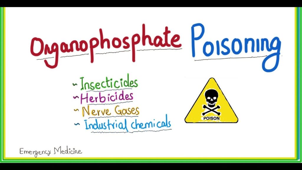
Organophosphates Poisoning is a serious medical emergency that occurs when a person develops an illness as a result of exposure to organophosphates (OPs). Organophosphates are chemicals extensively used in agriculture as insecticides. When humans—particularly agricultural workers—are exposed to large quantities, these chemicals can be severely harmful or fatal.
Other Uses: They are also used as nerve agents in chemical warfare (e.g., Sarin gas, Soman, Tabun, VX) and occasionally as therapeutic agents (e.g., ecothiopate for glaucoma).
Global Impact: There are no rules and regulations governing the purchase of these products in many regions, making them readily available "over the counter." This leads to an estimated 3 million exposures and 300,000 mortalities globally each year.
Exposure Context: Exposure in an attempt to commit suicide is a key problem, particularly in developing countries, and is a more common cause of severe poisoning than the chronic exposure experienced by farmers or sprayers.
Key Definitions
Poison: A foreign chemical that is capable of producing a harmful effect on a biologic system (xenobiotic).
Poisoning: The development of harmful effects on normal body functions following exposure to chemicals after it is swallowed, inhaled, injected, or absorbed cutaneously.
Classification by Toxicity
There are more than a hundred organophosphorus compounds in common use, classified according to their toxicity and clinical use:
Toxicity Level
Common Examples
Primary Use
1. Highly Toxic
Tetra-ethyl pyrophosphates (TEPP), Parathion
Agricultural insecticides
2. Intermediately Toxic
Coumaphos, Chlorpyrifos, Trichlorfon
Animal insecticides, antihelminthic agents
3. Low Toxicity
Diazinon, Malathion, Dichlorvos
Household applications and field sprays
Common Organophosphates include:
Parathion
Fenthion
Malathion
Diazinon
Dursban
Quinalphos
Prothoate
Fast Facts on Organophosphate Poisoning
Nearly 25 million cases of unintentional pesticide poisoning occur in the agricultural industry across the world each year.
Globally, it is reported that 3 million or more people are exposed to OPs every year, accounting for approximately 300,000 mortalities.
In the United States, there are around 8,000 exposures per year, with fewer deaths.
Poisoning leads to significant morbidity and mortality each year in India. According to the National Crime Records Bureau of India, there were 27,657 deaths and suicides by poisoning in 2015.
Cases are most common in regions where workers do not use or do not have access to protective gear, such as specialized suits or masks.
Symptoms and complications vary wildly but can quickly escalate to death.
II. Pathophysiology of Organophosphate Poisoning
Organophosphates exert their acute toxic effects by causing massive overstimulation at cholinergic nerve terminals.
Normal Function: Acetylcholine (ACh) is a vital neurotransmitter found in the central and peripheral nervous systems, neuromuscular junctions, and red blood cells (RBCs). Normally, the enzyme acetylcholinesterase (AChE) catalyzes the rapid degradation of ACh into choline and acetic acid in the nerve synapse, ending the nerve signal.
The Toxic Mechanism: OP pesticides act by binding irreversibly to the AChE enzyme.
The Consequence: This binding severely reduces the ability of the enzyme to break down the neurotransmitter. This produces a massive, uncontrolled accumulation of ACh in the central and peripheral nervous systems.
The Result: This accumulation results in an acute cholinergic syndrome via continuous, unrelenting neurotransmission.
The clinical onset of cholinergic overstimulation can vary from almost instantaneous to several hours after exposure. Although most patients rapidly become symptomatic, the onset and severity of symptoms depend heavily on the specific compound, the amount, the route of exposure, and the rate of metabolic degradation in the patient's body.
Routes of Absorption during OP Poisoning:
Ingestion: Through the Gastrointestinal (GI) tract (can be accidental or deliberate/suicidal).
Cutaneous: Absorption directly through the skin (common in occupational exposure).
Inhalation: Absorption through the lungs.
III. Signs and Symptoms of Organophosphate Poisoning
The length and strength of the exposure will determine the nature of the symptoms, which can range from mild to severe emergency-level toxicity.
A. Symptoms by Severity
1. Mild Exposure:
Blurry or impaired vision, watery/stinging eyes, narrowed pupils
Nausea
Runny nose, extra saliva
Headache
Muscle fatigue, weakness, or minor twitching
Agitation and glassy eyes
2. Moderate Exposure:
Dizziness, disorientation
Very narrow pupils (miosis)
Muscle tremors, pronounced twitching, and weakness
Drooling, excessive phlegm
Wheezing, coughing, and difficulty breathing
Severe vomiting and severe diarrhea
Sneezing and uncontrolled urination or bowel movements
Breathing that becomes ineffective or stops completely (respiratory arrest)
B. Signs and Symptoms according to Receptor Stimulation
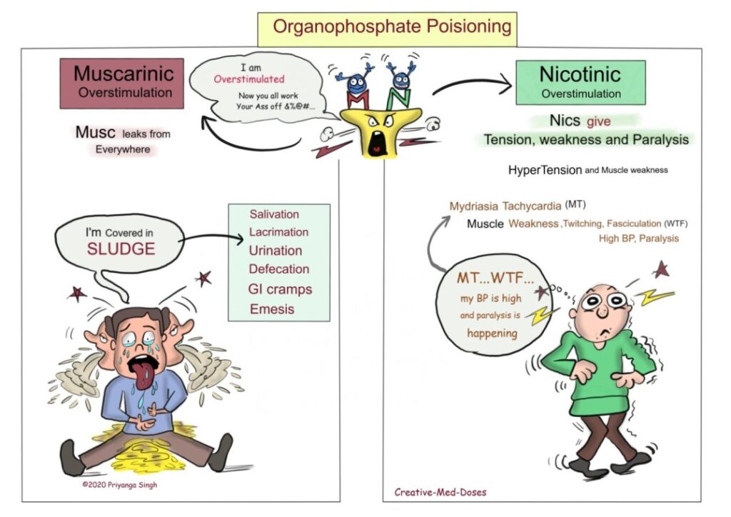
The accumulation of acetylcholine overstimulates two main types of receptors: Muscarinic and Nicotinic.
Muscarinic Signs and Symptoms
"Musc leaks from everywhere" - There are excessive secretions from everywhere in muscarinic overstimulation. Remember the mnemonic SLUDGE:
S - Salivation
L - Lacrimation (tears)
U - Urination
D - Defecation
G - GI cramps
E - Emesis (vomiting)
Nicotinic Signs and Symptoms
"Nics give tension, weakness, and paralysis" - Remember the mnemonic MT WTF:
M - Mydriasis (Note: while miosis is the classic muscarinic sign, sympathetic override can occasionally cause mydriasis)
T - Tachycardia
W - Muscle Weakness
T - Muscle Twitching
F - Muscle Fasciculation
BP is high - Hypertension
Paralysis is happening - Muscle paralysis
C. Long-Term Complications
In addition to immediate symptoms, OP exposure can cause long-term complications depending on the extent of exposure:
Paralysis
Fertility issues
Cancer
Metabolic disorders (e.g., high blood sugar levels)
Inflammation of the pancreas (Pancreatitis)
Excess acid in the blood (Acidosis)
Brain and nerve problems (Organophosphate-induced delayed polyneuropathy - OPIDN)
IV. Diagnosis of Organophosphate Poisoning
History: Determine the type of exposure (occupational, accidental, deliberate ingestion), time of exposure, and specific chemical if known.
Physical Examination: Look for classic signs (SLUDGE, miosis, garlic-like odor on breath).
Vital Signs: Depressed respirations, bradycardia, and hypotension are common, life-threatening findings.
Laboratory Investigations:
Plasma pseudocholinesterase levels: Normal is 3000–8000 U/L. In poisoning, serum levels may drop to < 1000 U/L.
RBC AChE level: A more accurate marker of nervous system AChE inhibition.
White blood cells (WBC): Leucocytosis is frequently seen.
ABG values: To rule out or confirm metabolic and/or respiratory acidosis.
Electrolytes: Potassium and magnesium levels are often decreased.
Imaging Studies:
Chest X-ray for aspiration pneumonia or pulmonary edema.
Electrocardiogram (ECG) for ventricular arrhythmias, prolonged QTc, or heart block.
V. Medical Management of Organophosphate Poisoning
Step I: Identification
Identify the specific nature of the poison (e.g., OP, carbamate, chloride, pyrethroid) to guide specific antidote therapy.
Step II: Decontamination & Safety
Staff Safety: Staff must put on protective equipment before commencing treatment, including masks, gowns, eye protection, and specialized gloves (usually nitrile/rubber, as OPs penetrate latex). Staff involved in direct contact with the patient’s bodily secretions should immediately and thoroughly wash affected areas with soap and water.
Physical Decontamination: Remove all clothing. Wash the patient thoroughly with soap and copious amounts of water.
Gastric Decontamination: Gastric lavage should be done only after stabilizing the airway. It is generally given within 1 hour of ingestion. Activated charcoal (0.5–1 g/kg) can be given within 1 hour of ingestion to bind remaining poison in the gut, though some studies show limited benefit. Do not induce emesis.
Step III: Maintaining Airway, Breathing, and Circulation (ABC)
Airway: Maintain a clear airway via frequent suctioning of excessive secretions. Check the gag reflex. If absent, intubate before any stomach wash is performed to prevent aspiration.
Breathing: Administer oxygen at 6 L/min. Intubation and mechanical ventilation are required if breathing is inadequate, oximetry is <90%, or the Glasgow Coma Scale (GCS) is <8.
Circulation: Administer adequate intravenous (IV) fluids through a wide bore cannula to replace volume loss from severe vomiting and diarrhea.
Step IV: Cardiac Monitoring
Continuously monitor for fatal arrhythmias (e.g., ventricular tachycardia, Torsades de pointes) using an ECG.
Step V: Specific Therapy & Antidotes
1. Atropine (To reverse Muscarinic effects)
Protocol for Atropinisation: Atropinisation must be initiated as soon as the diagnosis is suspected. The aim is to keep the patient's airway dry. Administer Injection Atropine 2 mg IV bolus (or 0.05 mg/kg). The dose is then doubled every 5 minutes until adequate atropinisation is achieved.
Signs of Atropinisation (Target End-Points):
Heart rate about 100/min
Pupils return to mid-position
Bowel sounds just heard (not hyperactive)
Clear lung sounds (secretions dry up)
Dry skin
Signs of Atropine Toxicity (Anticholinergic Toxidrome): Care must be taken not to over-atropinise. Signs include: Dry mucus membranes ("dry as a bone"), mental status changes/delirium ("mad as a hatter"), flushed skin ("red as a beet"), severe mydriasis ("blind as a bat"), fever ("hot as hell"), severe tachycardia, hypertension, decreased bowel sounds, and urinary retention.
Treatment for Toxicity: Atropine toxicity is treated with injection haloperidol (5 mg IM or IV) for agitation, and by immediately reducing or pausing the dose of atropine.
2. Supportive Medications
Oximes (e.g., Pralidoxime/PAM): Given to reactivate the AChE enzyme and reverse nicotinic effects (muscle weakness/paralysis). Must be given early before the enzyme "ages."
Antibiotics: Not usually indicated for OP poisoning itself. However, gastric lavage with an unprotected airway and/or a low GCS creates a massive risk for aspiration. If aspiration pneumonia is suspected (fever, leucocytosis, pulmonary infiltrates), broad-spectrum antibiotics (e.g., Ceftriaxone, Piperacillin/Tazobactam [Piptaz]) are indicated.
Sedation: Agitation may indicate over-atropinisation, hypoxaemia, or distress. Intubated patients need a combination of an analgesic and a sedative (e.g., morphine + lorazepam infusion). Haloperidol may increase the seizure threshold and is not recommended unless patients are unresponsive to other drugs.
Diuretics:Lasix (Furosemide) is the drug of choice if pulmonary edema persists even after full atropinisation has been achieved.
NURSES ROLES DURING MANAGEMENT OF OP POISONING
No.
Nursing Responsibility
Clinical Rationale
A. Airway & Respiratory Management
1
Assess the airway for bilateral equal air entry, respiratory rate, and breath sounds. Check for bronchospasms.
Respiratory failure is the primary cause of death; continuous assessment is vital.
2
Position the patient in semi-Fowler’s at 45° and change position every 2 hours.
Promotes diaphragmatic descent, maximal lung expansion, mobilizes secretions, and actively prevents aspiration of vomit.
Bronchorrhea (massive lung secretions) can rapidly occlude the airway.
4
Assess for cough and gag reflex; assist immediately with intubation if absent.
An absent gag reflex means the patient cannot protect their airway from massive secretions or vomit.
5
Provide humidification to airways and check ventilator settings closely if mechanically ventilated.
Thins out thick secretions and ensures optimal mechanical oxygenation.
B. Hemodynamic & Systemic Monitoring
6
Assess heart rate, rhythm (via ECG) for arrhythmias, BP, capillary refill time, skin turgor, and vital signs every hour.
OP poisoning and Atropine therapy both cause massive, rapidly shifting hemodynamic instability.
7
Administer IV fluids and Atropine infusions accurately as prescribed.
Fluids combat hypovolemia from GI losses; Atropine must be titrated meticulously to maintain the heart rate above 90/min without causing severe toxicity.
8
Monitor urine output every hour via a Foley catheter.
Assesses renal perfusion and checks for urinary retention (a sign of atropine toxicity).
9
Check for neck muscle weakness and the use of accessory muscles for breathing. Assess "single breath count."
These are critical early warning signs of impending nicotinic respiratory muscle paralysis.
C. Infection Control & Supportive Care
10
Follow strict aseptic technique while handling invasive lines and performing suctioning.
Prevents secondary healthcare-associated infections in a critically ill patient.
11
Provide rigorous oral care with chlorhexidine solution.
Prevents Ventilator-Associated Pneumonia (VAP) and manages excessive oral secretions.
12
Administer prophylactic or therapeutic antibiotics (e.g., Injection Piptaz 4.5 g IV q 8 hourly) as per order.
Treats suspected or confirmed aspiration pneumonia.
13
Monitor ABG values, WBC counts, culture and sensitivity results, and chest X-rays.
Provides objective data on respiratory acidosis, infection status, and pulmonary edema.
14
Evaluate the patient's feelings and perception of their lack of power; involve them in care when conscious.
Addresses the severe psychological trauma of a suicide attempt or a terrifying accidental poisoning, promoting mental recovery.
Nurses roles during management of organophosphate poisoning
Assessing the airway for bilateral equal air entry, respiratory rate and breath sounds
Assessing for cough and gag reflex and for bronchospasms.
Changed position every 2 hourly to mobilize secretions.
Positioning him in semi-fowlers at 45° to promote lung expansion and to prevent aspiration.
Maintaining adequate hydration by administering IV fluids.
Providing humidification to airways to thin secretions.
Checking for neck muscle weakness, use of accessory muscles for breathing.
Assessing single breath count.
Assisting for intubation.
Checking the ventilator settings of the patient.
Positioning patient in semi-fowler's position to promote diaphragmatic descent and maximal inhalation.
Performing suctioning whenever necessary.
Assessing heart rate, rhythm for arrhythmias, BP, capillary refill time, skin turgor, vital signs every hour.
Assessing peripheral sites for perfusion.
Monitoring urine output every hour.
Administered atropine infusion to maintain the heart rate above 90/min.
Monitoring vital signs.
Following strict aseptic technique while handling invasive lines and while performing suctioning.
Providing oral care with chlorhexidine solution.
Checking for the colour, consistency and volume of secretions.
Storage, Dispensing, and Legal Implications of Narcotics
Narcotics (Class A / Schedule II controlled drugs) are highly regulated pharmacological agents associated with addiction, tolerance, and a high potential for abuse. Because of these risks, governments and healthcare institutions must enforce strict prevention protocols, ensuring these medications are properly stored in such a way that limits access strictly to authorized personnel.
I. Storage and Dispensing of Narcotics
The following are the specific responsibilities and protocols in regard to the storage of narcotics across different hospital departments.
A. Storage in the Pharmacy
Secure Location: The drugs must be kept in a separate, dedicated locked cupboard.
Key Management: The key to the narcotic cupboard must be handled exclusively by the registered pharmacist.
Documentation: A dedicated register book must be kept up-to-date, indicating the total quantity of each drug, the date of transaction, and the required signatures.
Dispensing Protocol: During the issuing of drugs, the FEFO Method (First Expiry, First Out) must be strictly utilized to prevent drug expiration on the shelves.
Record Retention: The narcotic register book must be kept securely for a minimum of 2 years from the date of the last entry.
B. Storage on the Ward
Double-Lock System: All narcotics must be stored in a double-locked compartment or an automated dispensing cabinet (with the exception of refrigerated narcotic infusion bags, which have specific secure protocols).
Key Management: The keys for the locked compartment/cupboard must be carried on the person of the nursing unit personnel (especially the Ward In-Charge) or stored in an approved lock-box at all times. A spare key must be kept with the pharmacist.
Shift Handovers: In areas with more than one narcotic key, staff must account for all keys at the end of each shift and document this in the Narcotic Controlled Drug (NCD) administration record books.
Organization: Ampoules must be clearly labeled and physically separated by drug type and dosage to prevent medication errors.
Lost Keys: Keys that are lost or removed from the hospital premises require an immediate lock/key replacement by physical plant personnel.
Ward Register: There must be a dedicated register book for stock-in and stock-out on the ward.
Ampoule Retention: Empty ampoules must be kept and returned to the pharmacy for replacement validation.
Stock Rotation & Records: Use the FEFO method and keep all ward narcotic records for two years.
Narcotic Documentation and Discrepancy Management on the Ward:
All narcotics received and issued out on nursing units must be documented in the NCD administration record book or within the automated dispensing system's electronic record. Issues must explicitly include the patient's name, the physician's name, and the exact dose.
Wastage: All wastage of NCDs must be signed by a witness after directly observing the wastage being disposed of into the sharps container on the unit.
Shift Counts: Narcotic counts must be performed once per shift by two nurses. An incident report must be completed for any discrepancies not resolved prior to the shift change.
Variances: A count variance of less than 5% for oral narcotic solutions can be corrected without the completion of an incident report.
Management Oversight: The Patient Services Manager is responsible for ensuring discrepancies are resolved and that all required signatures are obtained in the NCD administration book, which must be returned to the pharmacy within 2 weeks of completion.
II. Key Considerations in Narcotic Storage
The responsibility for the storage of narcotics typically falls on the Pharmacy staff, while on the wards, storage falls on the nursing staff. They must ensure that the drugs are stored in a secure and controlled environment and that access is restricted only to authorized personnel. Nurses must be intimately aware of the regulations and guidelines governing the storage and handling of narcotics, strictly following protocols to prevent diversion, abuse, and misuse.
1. Secure Storage:
Narcotics must be stored in a secure and locked cabinet or safe, accessible only to authorized personnel. For ward storage, the storage area should be located in a secure, well-lit area away from public access, preferably near the nursing station for continuous, easy monitoring.
2. Proper Labeling:
All narcotics must be labeled with their generic name, strength, quantity, lot number, and expiration date. The labels must be highly legible and firmly affixed to the container. Any outdated or damaged labels must be replaced immediately.
3. Accurate Inventory:
An accurate inventory of all narcotics must be maintained at all times, with regular checks and reconciliations between the actual physical stock and the recorded inventory. The nursing staff must meticulously document any discrepancies, losses, or incidents related to the use or storage of narcotics.
4. Temperature Control:
Certain narcotics, such as fentanyl and hydromorphone, are particularly sensitive to temperature and humidity. They must be stored in a cool and dry environment to prevent degradation or loss of potency. The storage area must be monitored regularly for temperature and humidity levels, and any deviations from the recommended range must be promptly reported and addressed.
5. Access Control:
Access to the narcotics storage area must be strictly controlled and limited to authorized personnel who have been trained and approved to handle and administer narcotics. The nursing staff must follow strict protocols for accessing and dispensing narcotics, including checking the patient’s identity, verifying the prescription and dosage, and documenting the administration.
6. Disposal:
Narcotics that are expired, damaged, or no longer needed must be disposed of properly, in strict accordance with government regulations. The nursing staff must follow the legally prescribed procedures for disposing of narcotics.
III. Expired, Rejected, or Returned Class A Drugs
Unused drugs must be returned directly to the prescriber or dispenser.
If a narcotic is expired or rejected for any reason, it must be returned to the pharmacy In-Charge, who will then contact the national Drug Inspector.
Expired drugs should be destroyed by the pharmacy In-Charge ONLY when witnessed by the Drug Inspector.
Destruction must follow the World Health Organization (WHO) guidelines for the disposal of controlled substances.
The precise details of the quantity destroyed and the reason for destruction must be written in the Class A register.
IV. Importation of Class A Drugs
The manufacture and wholesale of Class A drugs require an annual import license.
Currently, the National Drug Authority (NDA) allows only the National Medical Stores (NMS - Government) and the Joint Medical Stores (JMS - NGO) to import narcotics directly.
Private retail pharmacies and private hospitals must access their narcotic stock through the above-authorized agencies.
V. Prescription Practices and Ordering
Prescribing narcotics is the process of sending a highly regulated written document from a licensed prescriber to the dispenser ordering specific controlled substances.
A. Ordering and Dispensing Workflow
Ordering in the Pharmacy to the Wards:
In the pharmacy, the responsible person obtains the drugs from the registered body. As far as ordering is concerned, the pharmacist keeps absolute records of all entries of drugs.
Narcotics must be dispensed by a registered pharmacist or authorized medical practitioner.
Ordering on the Ward:
Being a group of drugs that can easily be abused, the prescription of narcotics on the ward has been limited to registered medical practitioners (doctors) who should prescribe them only after evaluating that other NSAIDs cannot relieve the pain (e.g., severe post-surgical pain, cancer treatment, palliative care).
The doctor must make 2 copies of the prescription: one is retained in the stores/pharmacy, and the other is kept in the patient's clinical file.
The order has to be written clearly with the full names of the prescriber and their signature, the drug name, the patient's name, route of administration, and duration of therapy.
If the Ward In-Charge orders the drug for unit stock, he or she must sign the orders properly.
On collection from the pharmacy, the drugs must be cross-checked.
After checking, the nurse who receives the drugs signs for them to confirm receipt and assumes custody.
The drug given to a patient must subsequently be accounted for by handing over the empty ampoules.
B. Prescription Requirements
Prescription forms for Class A drugs must contain all required details because they are binding legal documents.
Allowed Prescribers for Class A Drugs:
Registered Medical Doctor
Registered Dentist
Registered Veterinary Surgeon
Specialized Palliative Care Nurse or Clinical Officer
Validity and Limitations:
The prescription is valid for 14 days from the date of issue.
The total supply prescribed must not exceed 1 month.
The prescription must be written in duplicate.
Mandatory Inclusions on the Prescription:
Patient's full name, age, sex, and address.
Total dose of drugs prescribed written in both words and figures.
Stipulated form of the drug (e.g., tablets, oral solution, injection).
Specific strength where possible (e.g., 5mg/5mls or 50mg/5mls oral morphine).
VI. Administration of Narcotics on the Ward
The drug to be administered must be explicitly prescribed by the doctor.
The drug must be administered by a qualified nursing staff member or a 3rd-year nursing student under the direct supervision of a qualified staff member.
Both individuals (the administrator and the witness/supervisor) must sign in the narcotic register immediately after administration.
The drug must be administered strictly according to the Five Rights (5 R's) of Medication Administration: Right patient, Right drug, Right dose, Right route, Right time.
Empty ampoules must be physically handed over to the Ward In-Charge.
In case of any remainder (partial dose used), the remaining drug should be taken back to the pharmacy or wasted according to protocol.
Any drug wasted must be recorded and signed for by a witness.
VII. Precautions on Narcotics
Must only be dispensed by a registered pharmacist or medical practitioner.
Medical practitioners are strictly prohibited from obtaining or prescribing the drug for personal use.
Always keep the drug in proximity with its antidote (e.g., Naloxone/Narcan) in case of an overdose or respiratory depression.
The order must be from a doctor/medical practitioner using a legally prescribed form.
Transportation must be legal (should be transported securely by legal means between facilities).
Facilities must fully comply with all rules from the National Drug Authority (NDA).
The Health Inspector must be allowed to check on records and obtain samples at any given time.
Individuals and private entities are not allowed to export or import narcotics. Trade is strictly restricted to licensed pharmacists and authorized drug shops/agencies.
VIII. Legal Implications and Penalties
Narcotic drugs are highly controlled under the law. In Uganda, the legal framework is primarily guided by the National Drug Policy and Authority (NDA) Act and the rigorous Narcotic Drugs and Psychotropic Substances (Control) Act (formerly Act No. 3 of 2016, recently revised/consolidated as the 2024 Act).
A. Regulatory and Institutional Framework
The National Drug Authority (NDA): Establishes the regulatory framework for the importation, exportation, and distribution of controlled substances. The Authority has the power to issue licenses and permits for the manufacture, distribution, and sale of narcotics, and to conduct unannounced inspections to ensure compliance with the Act's provisions. (Note: The NDA statute undergoes periodic review, and guidelines for handling Class A drugs established in 2001 work in tandem with the Pharmacists’ Council).
Treatment and Rehabilitation: The Act creates a legal framework for the treatment and rehabilitation of individuals with substance use disorders. It mandates the establishment of a National Drug Policy and a National Drug Abuse Prevention and Control Program designed to prevent drug abuse, promote public awareness, and rehabilitate struggling individuals under the guidance of the Uganda Mental Health Advisory Board.
B. Criminal Offenses and Severe Penalties
The Act heavily criminalizes the unlawful possession, sale, manufacture, and trafficking of narcotics (including cocaine, heroin, cannabis, etc.). Those found guilty face exceptionally severe penalties, which may include lengthy imprisonment, exorbitant fines, and total forfeiture of property.
Offense
Description
Legal Penalty (per NDPS Act Guidelines)
Unlawful Possession
Possession of narcotic drugs or psychotropic substances without lawful medical/pharmacological authority.
A fine not exceeding 50,000 currency points (approx. 1 Billion UGX) or three times the market value of the drug (whichever is greater), OR imprisonment not exceeding 20 years, or both.
Trafficking
Importation, exportation, manufacture, buying, selling, or distributing narcotics without a license.
A fine not exceeding 50,000 currency points or three times the market value (whichever is greater), OR Life Imprisonment, or both.
Malicious Entry / Seizure
A police officer or authorized person executing a search/seizure without reasonable grounds of suspicion.
A fine not exceeding 24 currency points or imprisonment not exceeding 10 years, or both.
Supply to a Child
Knowingly supplying a narcotic to a child when it is not required for medical treatment.
A fine not exceeding 50,000 currency points or Life Imprisonment, or both.
General Misconduct
Failure to comply with regulations, poor record-keeping, or general unlawful possession of classified drugs.
Historically applied fines not exceeding 2 million UGX or imprisonment for a term not exceeding 2 years, up to the maximums defined by specific schedules.
Important Exemption: Penalties for possession do not apply to a licensed medical practitioner, dentist, veterinary surgeon, registered pharmacist, clinical officer, or specialized palliative care nurse who is in possession of a narcotic drug strictly for a legitimate medical purpose or under a valid NDA license.
Overall, it is critical for healthcare professionals to understand the profound legal implications of narcotics and to comply meticulously with the provisions of these Acts. Proper storage, dispensing, and administration records protect the nurse and the facility from facing these serious legal consequences.
IX. References
The Narcotic Drugs and Psychotropic Substances (Control) Act, Uganda (Consolidated 2024 / Act No. 3 of 2016).
The National Drug Policy and Authority (NDA) Act and 2001 Guidelines for Handling Class A Drugs, Uganda.
World Health Organization (WHO) Guidelines for the Safe Disposal of Unwanted Pharmaceuticals in and after Emergencies.
Standard Operating Procedures for Narcotic Storage and Dispensing in Clinical Settings.
Quick Quiz
Narcotic Storage Quiz
Pharmacology
Privacy: Your details are used only for quiz tracking and certificates.
Anxiolytic agents are drugs used to depress the central nervous system (CNS) to prevent the signs and symptoms of anxiety. Hypnotic agents are drugs used to depress the CNS to cause sleep. In general, these drugs will induce sleep when given in high doses at night and will provide sedation and reduce anxiety when given in low divided doses during the day.
I. Common Terms and Definitions
Anxiety: An unpleasant feeling of tension, fear, apprehension, or nervousness in response to an environmental stimulus, whether real or imaginary. The physical symptoms of severe anxiety are similar to those of fear (tachycardia, sweating, trembling, palpitations) and involve sympathetic activation.
Sedative: A drug that depresses the CNS; produces a calming effect, reduces excitement, and produces a loss of awareness of and reaction to the environment. It may induce drowsiness without necessarily inducing sleep.
Sedation: The loss of awareness of and reaction to environmental stimuli.
Hypnotic: A drug that induces sleep resembling natural sleep. Both sedation and hypnosis may be considered as different grades of CNS depression.
Hypnosis: Extreme sedation resulting in CNS depression and sleep.
Benzodiazepine: A class of drugs that acts in the limbic system and the reticular activating system to make gamma-aminobutyric acid (GABA), an inhibitory neurotransmitter, more effective, causing interference with neuron firing; depresses CNS to block the signs and symptoms of anxiety, and may cause sedation and hypnosis in higher doses.
Barbiturate: A former mainstay drug class used for the treatment of anxiety and for sedation and sleep induction; associated with potentially severe adverse effects and many drug–drug interactions, which makes it less desirable than some of the newer agents.
II. Physiology of Sleep and Sleep Disorders
Sleep is a naturally periodic state of mind and body, characterized by altered consciousness. It is vital for heart health, reducing stress and inflammation, improving memory, weight loss, and maximizing athletic and intellectual performance.
A. The Sleep Cycle
A normal sleep cycle consists of non-rapid eye movement (NREM) and rapid eye movement (REM) sleep.
NREM Sleep (Non-Rapid Eye Movement): Comprises four stages.
Stage 1 (Lightest, 1-7 min): Heartbeat and breathing slow down, muscles relax with occasional twitches, brain waves begin to slow.
Stage 2 (Light, 10-25 min): Heartbeat and breathing slow further, body temperature drops, eye movements stop.
REM Sleep (20-40 min): Eyes move rapidly from side to side behind closed eyelids. Breathing speeds up and becomes irregular, heart rate and blood pressure increase. Dreams typically happen during REM sleep.
B. Common Sleep Disorders
Insomnia: A sleep disorder in which you have trouble falling and/or staying asleep. Acute insomnia lasts from 1 night to a few weeks. Chronic insomnia happens at least 3 nights a week for 3 months or more.
Hyposomnia: Decreased sleep duration.
Parasomnia: Abnormal behaviors during sleep (e.g., Sleep Terrors, Sleep Walking).
Sleep Apnea: Breathing repeatedly stops and starts during sleep.
Narcolepsy: Overwhelming daytime drowsiness and sudden attacks of sleep.
Other disorders: Restless leg syndrome, Bruxism (teeth grinding), REM sleep behavior disorder.
IV. Benzodiazepines (Used as Anxiolytics and Hypnotics)
Benzodiazepines (BDZs) are the most frequently used anxiolytic drugs. They prevent anxiety without causing much associated sedation. In addition, they are less likely to cause physical dependence than many of the older sedatives/hypnotics (like barbiturates) that are used to relieve anxiety.
A. Mechanism of Action (Pharmacodynamics)
GABA is the most potent inhibitory neurotransmitter in the CNS and controls the state of neuronal excitability.
BDZs act by potentiating the action of the neurotransmitter GABA (gamma-aminobutyric acid).
They bind selectively to specific subunits of the GABA-A receptors, at a site distinct from where GABA or barbiturates bind, designated as the benzodiazepine binding site.
Key Feature: Binding of BDZs enhances GABA binding and increases the frequency of chloride channel openings.
This increases the chloride ion concentration inside the neuron, causing hyperpolarization of the neuronal membrane, making it more difficult for excitatory neurotransmitters to depolarize the cell.
The primary sites of action appear to be the limbic system (governing emotions) and the reticular activating system (RAS) (which maintains consciousness, sleep, and alertness).
B. Pharmacological Actions
Antianxiety Effect: They exert a specific effect on the limbic system, reducing anxiety and producing a calming effect. Alprazolam has additional antidepressant properties.
Sedative-Hypnotic Effect: In smaller doses, they cause sedation. In higher doses, they induce sleep (hypnosis). They shorten the time spent in Stage 4 NREM and REM sleep but increase total sleep time. Sleep produced is refreshing with fewer hangover symptoms compared to barbiturates.
Anticonvulsant Effect: Long-acting BDZs (Clonazepam) raise the seizure threshold by potentiating GABA. Lorazepam and Diazepam prevent the spread of seizures in the brain.
Skeletal Muscle Relaxant: Produce relaxation by facilitating GABAergic transmission in the brainstem and spinal cord. Used in acute spasms caused by trauma or upper motor neuron disorders.
Amnesia: BDZs produce anterograde amnesia (loss of memory for events happening after administration). This is highly advantageous during unpleasant surgical or endoscopic procedures.
C. Pharmacokinetics
Absorption: Well absorbed from the gastrointestinal (GI) tract, with peak levels achieved in 30 minutes to 2 hours.
Distribution: Highly lipid-soluble; widely distributed throughout the body. They easily cross the blood-brain barrier, cross the placenta, and enter breast milk.
Metabolism: Metabolized extensively in the liver. Patients with liver disease must receive a smaller dose and be monitored closely.
Excretion: Primarily excreted through the urine.
D. Indications
Anxiety Disorders: Panic disorder, generalized anxiety disorder (GAD), social anxiety disorder, performance anxiety, PTSD, OCD, and extreme phobias.
Sleep Disorders: Used as hypnotic agents for the short-term treatment of insomnia.
Seizures: Status epilepticus (IV Diazepam or Lorazepam), and in the treatment of alcohol withdrawal to reduce the risk of withdrawal-related seizures (Chlordiazepoxide, Diazepam).
Muscular Disorders: Skeletal muscle spasms, degenerative disorders like multiple sclerosis and cerebral palsy.
Preanesthetic / Amnesia: Preoperative relief of anxiety and tension to aid in balanced anesthesia (Midazolam given IM or IV).
E. Contraindications and Cautions
Allergy to any benzodiazepine.
Psychosis: Could be exacerbated by sedation.
Acute conditions: Acute narrow-angle glaucoma, shock, coma, or acute alcoholic intoxication (could be lethally exacerbated by CNS depressant effects).
Pregnancy: Strictly contraindicated. A predictable syndrome of cleft lip/palate, inguinal hernia, cardiac defects, microcephaly, or pyloric stenosis occurs when taken in the first trimester. Neonatal withdrawal syndrome or "floppy infant syndrome" may also result.
Lactation: Contraindicated because of potential adverse effects on the neonate (e.g., severe sedation).
Elderly or Debilitated Patients: Use with extreme caution because of the possibility of unpredictable reactions (paradoxical agitation), delayed metabolism leading to accumulation, and increased fall risk. Dose adjustments are required.
Renal or Hepatic Dysfunction: Alters metabolism and excretion, resulting in direct toxicity.
Genitourinary (GU): Urinary retention, incontinence, hesitancy, loss of libido, and changes in sexual functioning.
Tolerance and Dependence: While present, liability is significantly less than with barbiturates. Abrupt cessation may lead to withdrawal syndrome (nausea, headache, vertigo, malaise, nightmares, and potentially seizures).
G. Drug Interactions & Antidote
The risk of CNS depression increases significantly if taken with alcohol, barbiturates, opioids, or other CNS depressants. Avoid combinations.
Effects of BDZs increase (due to inhibited metabolism) if taken with cimetidine, oral contraceptives, or disulfiram.
Impact may be decreased if given with theophyllines or ranitidine.
Antidote:Flumazenil (0.1 mg/ml). A competitive benzodiazepine receptor antagonist used for the treatment of overdose.
V. Barbiturates (Used as Anxiolytic-Hypnotics)
The barbiturates were once the sedative/hypnotic drugs of choice. However, the likelihood of profound sedation, severe adverse effects, lack of a specific antidote, a narrow therapeutic index, and the high risk of addiction/dependence has caused them to be largely replaced by newer anxiolytic drugs (BDZs).
A. Mechanism of Action
Barbiturates depress the sensory and motor activity in the cerebral cortex.
They act on the reticular activating system (RAS), elevating the firing threshold and depressing the firing rate of neurons.
They bind selectively to subunits of the GABA-A receptors, at a site distinct from the GABA or BDZ binding site (designated as the Barbiturate binding site).
Key Feature: Barbiturates potentiate GABA action by prolonging the duration of the chloride channel openings. (At high doses, they can directly open the channel even without GABA).
This causes profound hyperpolarization of the neuronal membrane and deep CNS depression.
B. Pharmacological Actions
Sedation: In low doses, they show drowsiness, calmness, and a sense of well-being (euphoria). Due to depression of the RAS.
Hypnosis: Induce sleep in a dose 3 to 4 times higher than the sedative dose. They relieve insomnia by inducing stage 2 of NREM sleep (but suppress REM sleep).
General Anesthesia: Ultra-short acting barbiturates (Methohexital, Thiopental) are administered IV for the rapid induction of general anesthesia. They produce reversible loss of consciousness.
Anticonvulsant Action: Long-acting barbiturates (Phenobarbital, Mephobarbital) block excess neuronal firing. Indicated in grand mal epilepsy and cortical focal seizures.
Respiratory Depression: In large doses, they depress the respiratory center in the medulla oblongata, leading to hypoventilation or fatal apnea.
Enzyme Induction: Barbiturates (especially Phenobarbital) powerfully stimulate the functioning of hepatic microsomal enzymes (CYP450 system). This increases the metabolism and decreases the effectiveness of many concomitant drugs.
C. Pharmacokinetics
Absorbed well, reaching peak levels in 20 to 60 minutes.
Metabolized heavily in the liver.
Excreted in the urine. The longer-acting barbiturates (e.g., Phenobarbital) tend to be metabolized slower and excreted to a greater degree unchanged in the urine (making alkalinization of urine an effective treatment for overdose).
D. Contraindications
Allergy to any barbiturate.
Addiction: Previous history of addiction to sedative/hypnotic drugs (barbiturates are highly addictive).
Porphyria: Strictly contraindicated; barbiturates induce enzymes that synthesize porphyrins, which can trigger a severe, life-threatening acute porphyria attack.
Hepatic impairment or nephritis: Alters metabolism and excretion, leading to rapid toxicity.
Severe respiratory dysfunction: Conditions like COPD or severe asthma could be lethally exacerbated by the respiratory depression.
Pregnancy: Contraindicated because of potential adverse effects on the fetus (depression of fetal respiration during labor) and reported congenital abnormalities.
E. Adverse Effects
Hangover: Due to residual depression of the CNS. Accompanied by headache, nausea, vomiting, vertigo, diarrhea, distortions of mood, and impaired judgment.
Drug Automatism (Memory Loss): When used as a hypnotic, confusion and amnesia may cause a patient to forget they took the pill, repeatedly take more doses at night, and accidentally poison themselves.
Tolerance & Dependence: Repeated administration quickly causes tolerance to sedative/hypnotic actions (but NOT to the lethal respiratory depression threshold). High risk of physical and psychological dependence.
CVS Effects: Bradycardia, hypotension (particularly with IV administration), and syncope.
F. Drug Interactions
Profound, synergistic CNS depression if taken with alcohol, antihistamines, or other tranquilizers.
Altered response to phenytoin when combined with barbiturates.
MAO Inhibitors increase serum levels and effects of barbiturates.
Enzyme Induction: Barbiturates accelerate the metabolism of oral anticoagulants (Warfarin), digoxin, TCAs, corticosteroids, oral contraceptives, estrogens, acetaminophen, metronidazole, beta-blockers, griseofulvin, theophyllines, and doxycycline, severely reducing their clinical effectiveness.
G. Acute Barbiturate Poisoning and Management
Often caused by suicidal attempts or drug automatism. Symptoms include deep CNS depression, respiratory depression, peripheral circulatory collapse, wheezing, hypotension, hypopyrexia, absent reflexes, coma, and potentially death. Fatal complications include atelectasis, pulmonary edema, and pneumonia.
Adequate Tissue Oxygenation: Endotracheal intubation is performed when spontaneous respiration is inadequate and to prevent aspiration/remove secretions. If assisted ventilation is required >24 hours, tracheostomy may be performed.
Gastric Lavage: If the patient is conscious and < 4 hours have elapsed since ingestion, induce vomiting or use lavage. In comatose patients, endotracheal intubation must precede gastric lavage to prevent aspiration pneumonia.
Intravenous Fluids: Given in sufficient quantity to prevent dehydration, maintain blood volume, and support diuresis. Vasopressors (dopamine) used if hypotension persists.
Alkalinisation of the Urine: Sodium bicarbonate (50ml of 7.5% solution) is added to IV fluids. Maintaining urinary pH between 7.5 and 8.5 ionizes the barbiturate (a weak acid) in the urine, preventing reabsorption and drastically increasing the excretion of long-acting agents like phenobarbital.
Forced Diuresis: Osmotic diuretics (Mannitol 25%) or loop diuretics (Furosemide 20 mg) are administered to aggressively flush the drug through the kidneys.
Hemodialysis: About 40 times more effective than forced diuresis in promoting elimination. Highly indicated in severe cardiac/renal impairment where massive fluid loading is contraindicated.
Prophylactic Antibiotics: May be necessary for intubated/catheterized patients to prevent secondary infections.
VI. Comprehensive Dosage Table
Drug Name
Dosage / Route
Indications & Considerations
BENZODIAZEPINES
Alprazolam (Xanax)
0.25–0.5 mg PO t.i.d. up to 1–10 mg/day; reduce dose in elderly.
Anxiety, panic attacks. Onset: 30 min. Taper after long-term therapy.
Chlordiazepoxide (Librium)
5–25 mg PO t.i.d. to q.i.d.; or 50–100 mg IV or IM.
Lacks sedative, anticonvulsant, and muscle-relaxant properties. No dependence. May cause dry mouth/headache. Takes weeks to reach full effect.
Zolpidem (Ambien)
Oral drug for short-term insomnia.
Z-Drug. Binds selectively to BDZ receptor subtype (Alpha-1). No anticonvulsant/muscle relaxing properties. Minimal rebound insomnia. Withdraw gradually.
Zaleplon (Sonata)
Oral for short-term insomnia.
Similar to Zolpidem. Take right before bed (devote 4-8 hours to sleep).
Eszopiclone (Lunesta)
Oral for insomnia.
Tablet must be swallowed whole. Allow 8 h for sleep.
Diphenhydramine (Benadryl) / Promethazine
PO, IM, or IV.
Antihistamines. Highly sedating. Used as sleep aids or pre-op. Monitor for thickened respiratory secretions (anticholinergic drying effect).
Chloral Hydrate
PO or PR.
Nocturnal sedation, preoperative sedation. Withdraw gradually over 2 weeks.
Ramelteon (Rozerem)
Oral.
Melatonin receptor agonist. For difficulty falling asleep. Take 30 min before bed.
Meprobamate (Miltown)
Oral.
Short-term management of anxiety. High risk of addiction.
Dexmedetomidine (Precedex)
IV.
Used for newly intubated/ventilated patients in ICU. Alpha-2 agonist. Do not use longer than 24h.
NURSING CARE PLAN & SPECIAL CONSIDERATIONS
General Nursing Interventions for Anxiolytics & Hypnotics
Parenteral Administration Rules: Do not administer intra-arterially (causes severe arteriospasm and gangrene). Monitor injection sites carefully for phlebitis. Give IV drugs very slowly because rapid administration is associated with hypotension, bradycardia, and cardiac arrest. Do not mix IV drugs in solution with other drugs to avoid precipitation and interactions.
Transitioning Therapy: Give parenteral forms only if oral forms are not feasible, and switch to oral forms (which are safer and have fewer adverse effects) as soon as possible.
Narcotic Adjustments: Arrange to reduce the dose of narcotic analgesics in patients receiving a benzodiazepine or barbiturate to decrease potentiated CNS and respiratory depression.
Mobility and Safety: Maintain patients who receive parenteral agents in bed for at least 3 hours. Do not permit ambulatory patients to operate a motor vehicle or heavy machinery. Institute fall precautions (side rails, assistance with ambulation).
Long-Term Monitoring: Monitor hepatic and renal function, as well as CBC, during long-term therapy to detect dysfunction. If noted, arrange to taper and discontinue the drug.
Withdrawal Prevention:Never abruptly stop long-term therapy. Taper dose gradually, especially in epileptic patients. Acute withdrawal could precipitate fatal status epilepticus or severe withdrawal syndrome.
Comfort Measures: Have patients void before dosing. Institute a bowel program as needed. Give food with the drug if GI upset is severe. Provide environmental control (dim lighting, quiet room, temperature regulation) to enhance sleep hygiene and drug efficacy.
Patient Education: Provide thorough teaching including the drug name, prescribed dose, avoidance of alcohol and OTC depressants, warning signs of toxicity, and the need for periodic lab monitoring to promote compliance.
Emergency Preparedness: Always provide standby life-support facilities (resuscitation equipment, intubation tray) in case of severe respiratory depression or hypersensitivity reactions. Keep the antidote (Flumazenil) readily available for BDZ overdoses.
Common Nursing Diagnoses
No.
Nursing Diagnosis
Intervention / Rationale
1
Risk for Injury / Falls related to central nervous system depression, drowsiness, ataxia, and confusion.
Implement rigorous fall precautions. Instruct the patient to call for assistance before getting out of bed. Keep the bed in the lowest position with side rails up. CNS depressants impair motor coordination and judgment, dramatically increasing fall risk, especially in the elderly.
2
Impaired Gas Exchange / Ineffective Breathing Pattern related to dose-dependent respiratory depression (particularly with IV barbiturates/BDZs).
Continuously monitor respiratory rate, depth, and oxygen saturation. Maintain an open airway. Have emergency airway equipment nearby. These agents depress the medullary respiratory center. Hypoxia and hypercapnia can occur rapidly, leading to respiratory arrest.
3
Deficient Knowledge regarding safe drug usage, avoidance of interactions (alcohol), and withdrawal risks.
Educate the patient to strictly avoid alcohol and unprescribed antihistamines. Teach the importance of not abruptly stopping the medication. Combining CNS depressants is synergistic and potentially fatal. Abrupt cessation leads to rebound nervous system hyperactivity and seizures.
4
Disturbed Sleep Pattern related to underlying anxiety, medication "hangover" effect, or REM sleep rebound.
Teach proper sleep hygiene (avoiding caffeine/screens at night). Assess the effectiveness of the hypnotic agent. Reassure the patient that vivid dreams may occur upon discontinuation. While these drugs induce sleep, they alter the natural sleep architecture. REM rebound causes intense nightmares when the drug is stopped.
VIII. References
Deore, A. B. (n.d.). Sedative, Hypnotic and Anxiolytic Drugs [PDF Presentation slides]. MVP’s Institute of Pharmaceutical Sciences, Nashik.
Katzung, B. G. (2017). Basic & Clinical Pharmacology (14th ed.). McGraw-Hill Education.
Brunton, L. L., Hilal-Dandan, R., & Knollmann, B. C. (2017). Goodman & Gilman's The Pharmacological Basis of Therapeutics (13th ed.). McGraw-Hill Education.
Karch, A. M. (2019). Focus on Nursing Pharmacology (8th ed.). Wolters Kluwer.
Quick Quiz
Anxiolytics & Hypnotics Quiz
Pharmacology
Privacy: Your details are used only for quiz tracking and certificates.
Antineoplastic agents are a class of drugs designed to combat cancer by inhibiting the growth and proliferation of neoplastic cells (cancer cells). Also called Anticancer drugs.
The action of antineoplastic agents can be broadly categorized into two main mechanisms: affecting cell survival and enhancing the immune system's ability to fight abnormal cells.
Common Terminology
Alopecia: Hair loss, a common side effect due to the drug's action on rapidly dividing cells.
Angiogenesis: Formation of new blood vessels, which cancer cells induce to supply themselves with nutrients.
Carcinoma: A type of cancer that starts in epithelial cells.
Metastasis: The spread of cancer cells from the original site to other parts of the body.
Neoplasm: An abnormal growth of tissue, which can be benign or malignant (cancerous).
Sarcoma: A type of cancer that arises from connective tissues such as bone, muscle, and fat.
Anaplasia: loss of organization and structure; property of cancer cells.
Cancer: refers to a malignant neoplasm or new growth
Cancer can be divided into;
Solid tumors
Hematological
Solid Tumors; can further be differentiated into carcinomas, or tumors that originate in epithelial cells, and sarcomas, or tumors that originate in the mesenchyme and are made up of embryonic connective tissue cells.
Haematological Malignancies; involve blood forming organs of the body, the bone marrow, the lymphatic system. These malignancies alter the body's ability to produce and regulate the cells found in the blood.
Classification of Antineoplastic Agents:
Alkylating Agents: Interfere with DNA replication, most effective against slow-growing cancers.
Antimetabolites: Resemble natural substances within the cell, disrupting DNA and RNA synthesis.
Antineoplastic Antibiotics: Bind to DNA and prevent RNA synthesis, primarily affecting rapidly growing cells.
Mitotic Inhibitors: Block cell division (mitosis), preventing the replication of cancer cells.
Hormones and Hormone Modulators: Block or mimic hormones to inhibit the growth of hormone-sensitive tumors.
Cancer Cell-Specific Agents: Target specific molecules involved in cancer cell growth and survival, minimizing damage to normal cells.
Miscellaneous Antineoplastics: A diverse group with varying mechanisms of action, often used when other treatments are ineffective.
Starts with G1 Phase: where the cell grows in size and releases enzymes for DNA replication. Cells may not progress hence remain at G0 phase.
S Phase: Synthetic Phase: Where DNA replication occurs, cells make identical copies of their own chromosomes.
G2 Phase: Check Point, When passed, they continue to grow and prepare themselves to divide.
Mitosis Phase: where cell division occurs
Prophase: Chromosomes appear condensed and the nuclear envelope breaks down.
Metaphase: Where microtubules in the center align,
Anaphase: Chromosomes are separated
Telophase: 2 daughter cells formed
Cytokinesis: Either they become dormant(Gap 0) , or continue to G1 phase to create another cell division.
Types of Antineoplastic Drugs
Alkylating Agents/DNA Replication Inhibitors
Alkylating agents work by adding an alkyl group to the DNA, thereby preventing the DNA strands from uncoiling and replicating. This is particularly effective in treating slow-growing cancers.
Indications:
Lymphomas
Leukemias
Myelomas
Ovarian, testicular, and breast cancers
Pancreatic cancer
Pulmonary carcinoma (lung cancer)
Rheumatoid arthritis
Contraindications:
Pregnancy and lactation (due to severe effects on the fetus and neonate)
0.1–0.2 mg/kg per day Orally for 3–6 weeks; Maintenance: 0.03–0.1 mg/kg per day Orally
Antimetabolites
Antimetabolites mimic natural substances within the cell, interfering with DNA and RNA synthesis. These drugs are most effective against rapidly proliferating cells.
Adult: 3.7 mg/m2 IV once weekly; Pediatric: 2.5 mg/m2 IV once weekly
Hormones and Hormone Modulators
Some cancers, particularly those involving the breast tissue, ovaries, uterus, prostate, and testes, are sensitive to estrogen stimulation. Estrogen-receptor sites on the tumor react with circulating estrogen, and this reaction stimulates the tumor cells to grow and divide
Hormones and hormone modulators block or interfere with these receptor sites to prevent growth of the cancer and cause cell death.
Some hormones are used to block the release of gonadotropic hormones in breast or prostate cancer if the tumors are responsive to gonadotropic hormones. Others may block androgen-receptor sites directly.
Indications:
Breast cancer in postmenopausal women
Prostate cancer
Contraindications and Cautions
Known allergy to drug: Prevent hypersensitivity reactions
Hypercalcemia: Contraindication to the use of toremifene because the drug can increase serum calcium
Pregnancy and lactation: Severe effects on the fetus and neonate
Bone marrow suppression: Index of re-dosing and dosing levels
Renal and hepatic dysfunction: Interfere with drug metabolism and excretion
Known GI ulceration or ulcerative diseases: Can be exacerbated by the effects of the drug.
Adverse Effects:
Menopausal symptoms: Hot flashes, vaginal dryness, mood changes
Hematological: Bone marrow suppression
GI: Hepatic toxicity
GU: Renal toxicity
Hypercalcemia
Examples of Hormones and Hormone Modulators:
Drug
Indications
Dosage
Tamoxifen (Nolvadex)
Breast cancer
20–40 mg Orally per day
Anastrozole (Arimidex)
Breast cancer in postmenopausal women
1 mg Orally per day
Incase You Needd Detailed, 👇👇👇👇👇
Antineoplastic agents are a class of drugs designed to combat cancer by inhibiting the growth and proliferation of neoplastic cells (cancer cells). They are also commonly referred to as Anticancer drugs or Chemotherapy.
The action of antineoplastic agents can be broadly categorized into two main mechanisms: affecting cell survival (cytotoxicity) and enhancing the immune system’s ability to fight abnormal cells (immunotherapy/targeted therapy).
I. Historical Timeline & Treatment Options
Before 1940: No effective treatment available for systemic cancers.
Before 1955: Surgery was the primary and only definitive treatment.
1955–1965: Radiotherapy became widely established.
After 1965: The advent and widespread use of systemic Chemotherapy.
Modern Era: Introduction of Immunotherapy and Gene Therapy, revolutionizing targeted cancer treatment.
Cell Cycle Effects of Anticancer Drugs
Cell Cycle Phase
Cell Cycle Specific (CCS) Drugs
Cell Cycle Non-Specific (CCNS) Drugs
G1 - S Phase
Etoposide
These agents affect cells across all phases:
➔ Platinum compounds (Cisplatin, Carboplatin)
➔ Alkylating agents (Cyclophosphamide, Busulfan, Nitrosoureas)
➔ Antibiotics (Anthracyclines, Dactinomycin, Mitomycin)
➔ Camptothecins
Cure or induce prolonged "remission": Macroscopic and microscopic features of the cancer disappear entirely. Highly achievable in: Acute Lymphoblastic Leukaemia (ALL), Wilm's tumor, Ewing's sarcoma, Hodgkin's lymphoma (in children), testicular teratoma, and choriocarcinoma.
Palliation: Aimed at the shrinkage of an evident tumor, alleviation of symptoms, and prolongation of life rather than absolute cure. Targeted for: Breast cancer, ovarian cancer, endometrial carcinoma, CLL, CML, Small cell lung cancer (SCLC), and Non-Hodgkin lymphoma.
Insensitive Tumors: These are less sensitive to chemotherapy, but life may still be prolonged. Includes: Cancer of the esophagus, cancer of the stomach, squamous cell carcinoma of the lung, melanoma, pancreatic cancer, myeloma, and colorectal cancer.
Adjuvant Therapy: Used routinely now for "mopping up" residual cancer cells, including micrometastases, after surgery or radiation. Mainly utilized in solid tumors.
B. General Principles of Chemotherapy
It is analogous to Bacterial chemotherapy but with critical differences:
Selectivity of drugs is highly limited (they attack normal host cells as well).
No or less defense mechanism (the host immune system does not recognize cancer as readily as bacteria; cytokines are used as adjuvants now to boost this).
The goal is to kill all malignant cells to stop progeny.
Tumors contain subpopulations of cells that differ in their rate of proliferation and susceptibility to chemotherapy.
Total Tumor Cell Kill via COMBINATION CHEMOTHERAPY: Formerly, single drugs were used; now, 2-5 drugs are used in intermittent pulses.
Why Combination Chemotherapy is Synergistic:
Utilizes drugs which are effective when used alone.
Combines drugs with different mechanisms of action.
Combines drugs with differing toxicities (and different mechanisms of toxicities) to prevent overlapping fatal side effects (e.g., combining bone marrow-sparing Bleomycin with a myelosuppressive agent).
Utilizes synergistic biochemical interactions.
Applies an optimal schedule determined by trial and error, relying heavily on cell cycle specificity.
C. Drug Resistance
Intrinsic Resistance: Tumors naturally possess primary resistance without prior exposure. Examples: Malignant melanoma, renal cell cancer, and brain cancer.
Acquired Resistance: Develops after initial exposure.
Single drug resistance: Change in the genetic apparatus, amplification, or increased expression of one or more specific genes targeted by the drug.
Multidrug resistance (MDR): Resistance to a wide variety of drugs after exposure to a single variety. Often due to increased expression of a normal gene (the MDR1 gene) which codes for a cell surface glycoprotein (P-glycoprotein) involved in active drug efflux (pumping the drug out of the cell).
III. Toxicities and Countering Agents
Chemotherapy is harmful to normal tissues because it has a steep dose-response curve and a low therapeutic index. It is particularly harmful to rapidly multiplying normal tissues (GI mucosa, Bone Marrow, Reticuloendothelial [RE] system, gonads, and hair cells). Effects occur in a dose-dependent manner.
General Toxicities:
Bone Marrow Depression (BMD): Leukopenia, thrombocytopenia, anemia. This is the primary dose-limiting toxicity for most treatments.
Buccal Mucosa Erosion: High epithelial turnover leads to stomatitis, severe mucositis, and bleeding gums.
Gastrointestinal (GIT): Diarrhea, shedding of mucosa, hemorrhage. Nausea and vomiting are severe due to direct stimulation of the Chemoreceptor Trigger Zone (CTZ).
Skin: Alopecia (hair loss).
Gonads: Oligospermia, impotence, amenorrhoea, and permanent infertility.
Lymphoreticular System: Lymphocytopenia and inhibition of lymphocyte function, leading to a loss of host defense mechanisms and extreme susceptibility to infections.
Other systemic: Carcinogenicity (secondary tumors), Teratogenicity, and Hyperuricemia (due to rapid tumor cell lysis releasing purines).
Distinctive Toxicities of Alkylating Agents & Others:
Drug
Distinctive Toxicity
Cyclophosphamide
Alopecia, Hemorrhagic cystitis, SIADH (Syndrome of Inappropriate Antidiuretic Hormone).
Prevents hemorrhagic cystitis due to Ifosfamide and high-dose Cyclophosphamide.
Leucovorin (Folinic Acid)
Repletes Tetrahydrofolic acid directly.
"Leucovorin Rescue" after high-dose Methotrexate to save bone marrow and GIT mucosa.
Amifostine
Scavenges free radicals.
Prevents radiation-induced xerostomia and Cisplatin-induced nephrotoxicity.
Dexrazoxane
Iron chelator.
Prevents cardiotoxicity due to Anthracyclines (Doxorubicin, Daunorubicin).
Palifermin
Keratinocyte growth factor.
Prevents severe mucositis following heavy chemotherapy.
Pilocarpine
Cholinergic agonist.
Treats radiation-induced xerostomia (dry mouth).
Pamidronate / Zoledronate
Bisphosphonates.
Treats hypercalcemia of malignancy and bone metastases.
Epoetin alpha / Darbepoetin
Recombinant Erythropoietin.
Treats chemotherapy-induced anemia.
Filgrastim / Peg-filgrastim
G-CSF (Granulocyte Colony-Stimulating Factor).
Febrile neutropenia prophylaxis; speeds recovery of granulocytopenia.
Sargramostim
GM-CSF (Granulocyte-Macrophage CSF).
Myeloid reconstitution.
Oprelvekin
IL-11 (Interleukin-11).
Treats severe chemotherapy-induced thrombocytopenia.
Ondansetron / Granisetron / Palonosetron
5-HT3 antagonists.
Prevents acute nausea and vomiting.
Aprepitant
NK-1 (Neurokinin-1) receptor antagonist.
Prevents Cisplatin-induced delayed vomiting.
IV. Detailed Classification & Pharmacology of Antineoplastic Agents
Classification is based on chemical structure, biochemical mechanism of action (blocking nucleic acid synthesis, interfering with DNA structure, blocking RNA/protein synthesis, influencing hormones), and cell cycle phase specificity.
1. Alkylating Agents & Platinum Compounds (CCNS)
Mechanism of Action: They act by binding irreversibly to nucleic acids (DNA). Nitrogen mustards inhibit cell reproduction by adding an alkyl group. Bifunctional alkylating agents cause intrastrand linking and cross-linking of the DNA double helix (sugar-phosphate backbone).
After alkylation, DNA is unable to replicate and cannot synthesize proteins or essential cell metabolites. Consequently, cell reproduction is inhibited, metabolic functions fail, and the cell eventually dies.
Mechlorethamine: The first alkylating agent employed clinically. Bifunctional (crosslinks DNA). Extremely unstable and is inactivated within a few minutes following administration, thus given IV. Used in MOPP regimen for Hodgkin's disease. Highly vesicant (severe local toxicity on extravasation). ADRs: Severe vomiting, myelosuppression.
Cyclophosphamide: A prodrug transformed into active aldophosphamide and phosphoramide mustard in the liver. Given orally or IV. Used for ALL, NHL, Polycythemia Vera, Hodgkin's lymphoma, breast and ovary cancers. Causes hemorrhagic cystitis (due to acrolein).
Ifosfamide: Has a longer half-life, used mainly for testicular tumors. Also causes hemorrhagic cystitis.
Chlorambucil: Orally active against lymphoid tissues. Preferred for CLL, Polycythemia Vera, and NHL.
Melphalan: Oral agent used for Multiple Myeloma.
Alkyl Sulfonates:
Busulfan: Orally active. Great effect for Chronic Granulocytic/Myelogenous Leukemia (CML). Delayed toxicities: Pulmonary infiltrates/fibrosis ("Busulfan lung"), tonic-clonic seizures in epileptics, skin pigmentation, adrenal insufficiency.
Nitrosoureas:
Carmustine (BCNU) & Lomustine (CCNU): Bifunctional; active against broad spectrums of neoplastic disease. They inhibit synthesis of both DNA and RNA, as well as proteins. These drugs are highly lipophilic, easily crossing the blood-brain-barrier (BBB), making them excellent for primary and metastatic brain tumors. Also used for Hodgkin's, melanoma, and GI adenocarcinomas.
Major Toxicity: Highly mutagenic/carcinogenic. Delayed bone marrow depression (prolonged leukopenia/thrombocytopenia), and pulmonary fibrosis.
Platinum Coordination Compounds:
Cisplatin: Forms crosslinks within DNA strands. Very powerful IV agent against Testicular cancer, ovary, bladder, head, and neck carcinomas. Toxicity is severe: Renal tubular damage (minimized via massive hydration + amifostine), Ototoxicity, peripheral neuropathy, and VERY SEVERE vomiting (treated with Ondansetron/Aprepitant).
Carboplatin: A derivative of cisplatin with significantly less nephrotoxicity, neurotoxicity, and ototoxicity (but higher myelosuppression).
2. Antimetabolites (CCS - S Phase)
These are structural analogues of essential metabolites that interfere with DNA/RNA synthesis (block nucleic acid biosynthesis). Myelosuppression is the primary dose-limiting toxicity.
A. Folic Acid Antagonists:
Methotrexate (MTX): Structure is similar to folic acid. It actively transports into mammalian cells and competitively inhibits dihydrofolate reductase (DHFR), the enzyme that normally converts dietary folate to the tetrahydrofolate required for thymidine and purine synthesis.
Kinetics & Indications: Orally, IM, IV, and intrathecally (for CSF entry). Used for ALL, Breast cancer, Tumors of head/neck, Meningeal metastases. It was the first demonstration of curative chemotherapy for Choriocarcinoma.
Leucovorin (Folinic acid) Rescue: Administered as part of high-dose MTX therapy. Leucovorin is directly converted to tetrahydrofolic acid (producing DNA/cellular protein) in spite of the presence of MTX, rescuing bone marrow and GIT mucosal cells from lethal toxicity.
B. Pyrimidine Antagonists:
5-Fluorouracil (5-FU): Analogue of thymine. Converted to 5-fluoro-2-deoxy-uridine monophosphate (5-FdUMP), which irreversibly inhibits thymidylate synthase. This blocks the conversion of deoxyuridylic acid to deoxythymidylic acid, causing failure of DNA synthesis. Used for solid tumors (breast, colorectal, gastric, head/neck). Toxicity: Hand and foot syndrome, severe oral/GI ulceration.
Cytarabine: Inhibits DNA polymerase. Used IV for Acute Myeloid Leukemia (AML). Delayed toxicity includes cerebellar ataxia (neurotoxicity).
C. Purine Antagonists:
6-Mercaptopurine (6-MP) & 6-Thioguanine (6-TG): Inhibit purine ring biosynthesis and nucleotide interconversions. 6-MP is used for childhood ALL maintenance and remission, and in combination with MTX for choriocarcinoma.
Important Interaction: 6-MP is metabolized by xanthine oxidase (which is inhibited by Allopurinol). If a patient is taking Allopurinol for hyperuricemia, the dose of 6-MP must be adjusted to ½ or ¼ to prevent fatal bone marrow toxicity. Well tolerated long term but causes mild hepatotoxicity.
D. Ribonucleoside Diphosphate Reductase Antagonist:
Hydroxyurea: Inhibits ribonucleotide reductase. Used orally for CML, AML (blast crisis), and Polycythemia vera.
3. Antineoplastic Antibiotics
Anthracyclines (Doxorubicin, Daunorubicin): Insert themselves into DNA causing breaks. They activate Topoisomerase II, causing DNA strand breaks, and generate excess free radicals (superoxide) causing DNA damage.
Unique Toxicity: Known to irreversibly damage cardiac cells (Cardiomyopathy). Prevented with Dexrazoxane.
Docetaxel: Advanced breast cancer. Toxicity: Fluid retention, neurotoxicity, neutropenia.
Topoisomerase Inhibitors:
Etoposide: A podophyllotoxin (toxin found in the mandrake root). Inhibits the enzyme Topoisomerase II, causing breaks in DNA inside cancer cells, preventing them from dividing. Useful for Testicular cancer and Small Cell Lung Cancer (SCLC). Side effects: Vomiting, alopecia, bone marrow suppression.
Camptothecin Analogues (Irinotecan, Topotecan): Inhibit Topoisomerase I.
Influence Amino Acid Supply / Miscellaneous:
L-Asparaginase: Depletes the amino acid asparagine, inhibiting protein synthesis. Used IV for ALL in children. Toxicity: Hepatotoxicity, mental depression, pancreatitis.
Mitotane: Used for Adrenocortical carcinoma. Toxicity: Adrenal insufficiency, lethargy, diarrhea.
5. Hormones, Antagonists, and Related Agents
Influence hormone homeostasis in hormone-sensitive tumors.
Agent
Cancers Where Preferred
Delayed Toxicity / Comments
Corticosteroids (Hydrocortisone, Prednisone)
ALL, CLL, NHL, Hodgkin's, Multiple myeloma
Fluid retention, hypertension, diabetes mellitus, susceptibility to infection, moon face.
Androgens (Testosterone)
Premenopausal breast cancer (estrogen receptor positive)
GnRH agonist OR Oestrogen + Androgen antagonist (Flutamide).
Melanoma
Dacarbazine, Cisplatin, Interferon.
Carcinoma adrenal gland
Mitotane.
Carcinoid tumour
Doxorubicin + Cyclophosphamide OR 5-FU + Octreotide.
NURSING CARE PLAN & CONSIDERATIONS FOR CHEMOTHERAPY
I. Nursing Assessment & Monitoring
Contraindications Check: Assess for drug allergies, severe hepatorenal impairment, pre-existing bone marrow suppression, and verify pregnancy/lactation status (ensure barrier contraception is utilized by women of childbearing age).
Baseline Physical Assessment: Check orientation, reflexes, vital signs, bowel sounds, and oral mucosa to establish baseline data before therapy begins.
Laboratory Monitoring:
CBC with differential: Crucial to identify bone marrow suppression (leukopenia, neutropenia, thrombocytopenia, anemia). Calculates the Absolute Neutrophil Count (ANC).
Renal/Hepatic panels (BUN, Creatinine, AST, ALT, Bilirubin): To determine the need for dose adjustments and identify toxicities.
Uric Acid: Monitor for Tumor Lysis Syndrome (prophylactic Allopurinol or Rasburicase may be needed).
II. Nursing Care Plan
No.
Nursing Diagnosis
Interventions & Rationale
1
Risk for Infection related to chemotherapy-induced leukopenia and immunosuppression.
Institute strict neutropenic precautions: Hand hygiene, avoid fresh flowers/plants, and raw unpeeled fruits/vegetables to prevent exposure to environmental pathogens.
Monitor temperature daily: A fever >38°C (100.4°F) in a neutropenic patient is a medical emergency requiring immediate broad-spectrum antibiotics.
Administer Colony-Stimulating Factors (e.g., Filgrastim/Peg-filgrastim) as ordered: Stimulates the bone marrow to accelerate neutrophil recovery.
2
Imbalanced Nutrition: Less than body requirements related to severe nausea, vomiting (CTZ stimulation), and stomatitis/mucositis.
Administer antiemetics prophylactically: Give agents like Ondansetron (5-HT3 antagonists) 30-60 minutes before chemotherapy administration. For delayed emesis (like with Cisplatin), administer Aprepitant.
Provide frequent, small, high-calorie, bland meals: Easier to digest and less likely to trigger nausea than large, spicy, or hot meals.
Implement meticulous oral hygiene: Use soft-bristled toothbrushes and non-alcoholic mouthwashes (or administer Palifermin) to soothe stomatitis and prevent bleeding gums or candidiasis.
3
Risk for Bleeding related to chemotherapy-induced thrombocytopenia.
Implement bleeding precautions: Avoid IM injections, use electric razors, and prevent hard nose-blowing to avoid inducing uncontrollable hemorrhage.
Monitor for petechiae, ecchymosis, or melena: Early clinical indicators of dangerously low platelet counts.
Administer Platelet/Granulocyte transfusions or Oprelvekin (IL-11): As indicated to manage severe thrombocytopenia.
4
Disturbed Body Image related to alopecia and physical changes from targeted therapies (e.g., severe acneiform rash).
Educate prior to hair loss: Inform the patient that hair loss is usually temporary and will regrow after therapy cessation. Suggest exploring wigs, hats, or scarves beforehand.
Provide emotional support: Validate their feelings regarding their changing appearance; refer to oncological support groups if appropriate.
5
Risk for Impaired Tissue Integrity (Vesicant Toxicity) related to extravasation of alkylating agents or antibiotics (e.g., Mechlorethamine, Doxorubicin, Vincristine).
Ensure patent IV access via central line or port: Peripheral lines are highly risky for vesicants. Check for blood return prior to administration.
Monitor IV site continuously during infusion: If swelling, pain, or redness occurs, stop the infusion immediately, aspirate residual drug, and follow hospital extravasation protocols.
References
Presentation Slides: Treatment options of cancer / Anticancer drugs (Slides 57 pages detailing Cell Cycle, Alkylating Agents, Antimetabolites, Antibiotics, Targeted Therapies, Toxicities, and Therapy of Choice).
Brunton, L. L., Hilal-Dandan, R., & Knollmann, B. C. (2017). Goodman & Gilman's The Pharmacological Basis of Therapeutics (13th ed.). McGraw-Hill Education.
Katzung, B. G. (2017). Basic & Clinical Pharmacology (14th ed.). McGraw-Hill Education.
Nursing considerations adapted from Oncology Nursing Society (ONS) guidelines for safe handling and administration of chemotherapy.
Complete Antineoplastic Pharmacology Tables
High-alert medicines: antineoplastic regimens are diagnosis-, stage-, body-surface-area-, organ-function- and protocol-specific. The examples below teach mechanisms and safety; they are not stand-alone prescribing instructions. Independent verification of patient, protocol, cycle/day, height, weight/BSA, laboratory results, dose, route, diluent, rate and cumulative exposure is mandatory.
Leukaemia, lymphoma, osteosarcoma and other cancers; dose varies from low weekly to high-dose infusion.
Pregnancy, severe marrow/liver/renal disease, active severe infection; high-dose use only with rescue/monitoring capacity.
NSAIDs, trimethoprim-sulfamethoxazole, penicillins and PPIs may reduce elimination or add antifolate toxicity; avoid live vaccines.
Mucositis, nausea, fatigue, myelosuppression.
Nephrotoxicity, hepatotoxicity, pneumonitis, severe skin reaction and pancytopenia.
Verify once-weekly versus oncology schedule; monitor CBC, creatinine, LFTs, urine pH/output and methotrexate levels for high dose. Hydration/alkalinization and leucovorin rescue must be precisely timed.
Fluoropyrimidine: 5-fluorouracil
Inhibits thymidylate synthase and incorporates into RNA/DNA; S-phase predominant.
Rapid hepatic catabolism by DPD; continuous infusion and bolus have different toxicity patterns.
Colorectal, gastric, breast, head/neck cancers; mg/m² by regimen, often modulated by leucovorin.
Severe DPD deficiency, profound marrow suppression; caution coronary disease.
Warfarin effect may rise; brivudine/sorivudine cause potentially fatal interaction; additive myelosuppression.
Severe diarrhoea/dehydration, neutropenic sepsis, cardiotoxicity/coronary spasm, encephalopathy.
Baseline/serial CBC, renal/liver tests; inspect mouth/skin, count stools and urgently stop/escalate chest pain, severe diarrhoea or early disproportionate toxicity.
Anthracycline: doxorubicin
DNA intercalation, topoisomerase-II inhibition and free-radical damage.
Extensive tissue distribution and hepatic/biliary clearance; cumulative lifetime cardiotoxicity.
Breast cancer, lymphomas, sarcomas and leukaemias; mg/m² by combination protocol.
Severe cardiac disease, severe hepatic impairment, profound marrow suppression; previous anthracycline/radiation exposure matters.
Other cardiotoxic agents and CYP/P-gp modifiers increase risk.
Alopecia, mucositis, nausea, myelosuppression; harmless red urine for 1–2 days.
Dilated cardiomyopathy/heart failure, severe vesicant extravasation, secondary leukaemia.
Baseline and periodic LVEF, cumulative dose, CBC/LFTs; confirm brisk blood return. Treat pain/burning/swelling as extravasation immediately—stop infusion, leave cannula for aspiration and follow antidote/cold-pack protocol.
Vinca alkaloid: vincristine
Binds tubulin and blocks microtubule formation, arresting mitosis in M phase.
Hepatic CYP3A metabolism and biliary elimination; long tissue persistence.
Leukaemias, lymphomas and paediatric tumours; capped mg/m² dose by protocol.
Severe neuropathy, Charcot-Marie-Tooth disease, hepatic dysfunction.
Azole antifungals/macrolides and other CYP3A inhibitors markedly increase neurotoxicity; additive neuropathy.
Constipation, jaw pain, peripheral neuropathy, alopecia; relatively little myelosuppression.
Paralytic ileus, SIADH, severe neuropathy. Intrathecal vincristine is fatal.
Label and supply in a minibag for IV use only; never have vincristine near intrathecal medicines. Assess bowel sounds/stool, motor/sensory function and reflexes.
Platinum: cisplatin
Forms DNA cross-links causing apoptosis.
Highly protein bound; renal elimination with prolonged tissue retention.
Testicular, ovarian, bladder, lung and head/neck cancers; mg/m² by protocol with hydration.
Significant renal impairment, hearing loss, neuropathy or severe marrow suppression.
Aminoglycosides/other nephrotoxins or ototoxins increase injury; avoid live vaccines.
Severe nausea/vomiting, fatigue, electrolyte loss.
Nephrotoxicity, ototoxicity, neuropathy, hypomagnesaemia/hypokalaemia and anaphylaxis.
Selective estrogen-receptor modulator for ER-positive breast cancer and risk reduction.
Oral prodrug activated by CYP2D6; some SSRIs (e.g., strong CYP2D6 inhibitors) may reduce active metabolite.
Hot flushes, vaginal discharge; VTE, stroke and endometrial cancer risk.
Assess leg/chest symptoms and abnormal uterine bleeding; discuss pregnancy avoidance and adherence.
Immune-checkpoint inhibitors
PD-1/PD-L1 or CTLA-4 blockade restores antitumour T-cell activity across multiple cancers.
Monoclonal antibodies with long half-lives; immune toxicity may occur during or after treatment.
Immune-mediated dermatitis, colitis, hepatitis, pneumonitis, nephritis, thyroid/adrenal/pituitary disease and myocarditis.
New diarrhoea, cough, jaundice, weakness, severe headache or endocrine symptoms need urgent immune-toxicity pathway—not routine symptomatic treatment alone.
Supportive medicines and rescue agents
Agent
Purpose
Safety and monitoring
Mesna
Binds toxic urothelial metabolites of cyclophosphamide/ifosfamide.
Time doses exactly relative to chemotherapy; mesna does not prevent other toxicities. Continue hydration and haematuria monitoring.
Leucovorin (folinic acid)
Rescues normal cells after high-dose methotrexate; also enhances 5-FU action in other regimens.
Timing is critical and guided by methotrexate level/renal function. Confusing rescue with 5-FU modulation can be dangerous.
G-CSF: filgrastim/pegfilgrastim
Stimulates neutrophil production to reduce duration of selected chemotherapy-associated neutropenia.
Bone pain and leukocytosis occur; rare splenic rupture/ARDS. Observe timing relative to cytotoxic dose and never assume it replaces urgent antibiotics for febrile neutropenia.
Antiemetic combination
5-HT3 antagonist + dexamethasone with NK1 antagonist and/or olanzapine according to emetogenic risk.
Give prophylactically, not only after vomiting; monitor QT, constipation, glucose, sedation and interactions.
Allopurinol/rasburicase
Prevent/treat uric-acid burden in tumour lysis syndrome (TLS).
Allopurinol prevents new uric acid but does not remove existing urate; rasburicase rapidly degrades uric acid and is contraindicated in G6PD deficiency. Combine with protocol hydration and frequent electrolytes.
Oncology emergencies: recognition and first nursing actions
Emergency
Red flags
Immediate nursing actions
Febrile neutropenia
Single temperature ≥38.3°C or sustained ≥38.0°C with low/expected-low neutrophils; rigors, hypotension or subtle deterioration.
Activate emergency pathway, cultures/lactate/labs without delaying treatment, administer broad-spectrum IV antibiotics within the local target (commonly 1 hour), fluids and sepsis monitoring. Avoid rectal procedures.
Tumour lysis syndrome
Rising potassium/phosphate/uric acid/creatinine, falling calcium, arrhythmia, seizures or oliguria.
Continuous ECG, urgent labs and IV access, hydration as ordered, strict urine output, administer urate-lowering therapy and escalate for renal replacement therapy.
Extravasation
Burning, pain, swelling, erythema, resistance or absent blood return at infusion site.
STOP infusion; leave cannula in place; aspirate residual drug, disconnect tubing, notify oncology team, mark/photograph area, elevate and use drug-specific warm/cold compress and antidote protocol. Do not flush.
Spinal cord compression
New severe back pain, weakness, sensory level, gait change or bladder/bowel dysfunction.
Urgent neurological observations, spinal precautions as indicated, immediate oncology/neurosurgical pathway and prescribed corticosteroid/imaging.
Superior vena cava syndrome
Facial/upper-limb swelling, venous distension, dyspnoea, stridor or confusion.
Sit up, oxygen if hypoxaemic, urgent senior/oncology review and airway planning; avoid unnecessary upper-limb lines.
Safe chemotherapy administration checklist
Before: verify consent, performance status, allergies, protocol/cycle/day, pregnancy status, BSA from current accurate height/weight, dose modifications, CBC, renal/liver/electrolyte results, cumulative anthracycline exposure and required premedication/hydration.
Independent double-check: patient identifiers, generic drug, calculation, final dose, route, diluent, concentration, rate, sequence, expiry and line suitability.
During: PPE and closed-system precautions where available, patency/blood return, infusion-site checks, vital signs and observation for hypersensitivity. Know spill and extravasation kits.
After: flush/dispose as cytotoxic policy requires; document full administration and response; handle urine, stool, vomit and contaminated linen with PPE for the locally specified excreta-risk period.
Teach: urgent contact for fever, bleeding, breathlessness, chest pain, uncontrolled vomiting/diarrhoea, inability to drink, severe mouth ulcers, reduced urine or neurological change; reinforce oral-drug adherence and safe storage.
Core nursing diagnoses and care
Risk of infection: hand hygiene, daily assessment, line care, neutropenic precautions based on policy, food safety and rapid fever pathway.
Risk of bleeding: monitor platelets/skin/stool/urine, avoid IM injections and traumatic procedures when thrombocytopenic, apply prolonged pressure after venepuncture.
Imbalanced nutrition/mucositis: oral assessment and bland rinses, analgesia, dietitian input, small nutrient-dense meals and hydration; avoid alcohol mouthwash.
Fatigue/activity intolerance: screen anaemia/infection, pace activity, sleep support and thrombosis-prevention mobility.
Body-image/fertility distress: anticipatory counselling, fertility-preservation referral before gonadotoxic therapy when possible and psychosocial support.
Fatal-error prevention: vincristine is for intravenous use only and must never be administered intrathecally. Oral methotrexate schedules must state the exact day and indication—daily administration of a weekly regimen can cause fatal marrow and mucosal toxicity.
Quick Quiz
Anticancer Drugs Quiz
Pharmacology
Privacy: Your details are used only for quiz tracking and certificates.
Spinal cord compression (SCC) results from processes that compress or displace arterial, venous, and cerebrospinal fluid spaces, as well as the cord itself.
Spinal cord compression (SCC) refers to the mechanical or pathological compression of the spinal cord, resulting in the displacement or obstruction of arterial, venous, and cerebrospinal fluid spaces, as well as direct cord involvement.
This compression can arise due to intrinsic or extrinsic causes, leading to varying degrees of neurological dysfunction.
Etiology of Spinal Cord Compression
SCC can result from multiple conditions, broadly categorized into traumatic, neoplastic, degenerative, inflammatory, infectious, vascular, or iatrogenic causes.
1. Traumatic Causes
Trauma is a leading cause of SCC, often resulting from accidents, falls, or sports injuries. The injury can lead to:
Vertebral fractures, commonly affecting the cervical spine.
Facet joint dislocation, which can lead to spinal instability and compression.
Complete transection of the spinal cord, resulting in irreversible neurological deficits.
Brown-Séquard syndrome, a condition caused by spinal hemisection, often due to penetrating injuries. This results in ipsilateral motor weakness and contralateral loss of pain and temperature sensation.
2. Neoplastic Causes
Tumors, whether benign or malignant, can lead to SCC. These include:
Metastatic tumors, commonly from lung, breast, prostate, and renal cancers.
Hematologic malignancies such as lymphoma, multiple myeloma, and leukemia.
Paraneoplastic syndromes leading to acute myelopathy.
Meningeal carcinomatosis, in which cancer spreads to the meninges, causing extensive spinal cord involvement.
3. Degenerative Causes
Age-related degeneration of the spine can lead to compression via:
Intervertebral disc herniation, commonly at L4-L5 and L5-S1, potentially causing cauda equina syndrome.
Cervical disc herniation, which can result in myelopathy.
Cervical spondylotic myelopathy, a progressive condition due to osteophyte formation, disc herniation, and ligamentum flavum hypertrophy.
4. Vascular Causes
Epidural or subdural hematomas, typically occurring after trauma, spinal procedures, or in patients on anticoagulation therapy.
Spinal cord infarction, which may occur due to atherosclerosis, embolism, or systemic hypotension.
Arteriovenous malformations (AVMs), which can rupture and cause compression.
5. Inflammatory and Autoimmune Disorders
Rheumatoid arthritis (RA): Weakening of the ligamentous structures around the odontoid peg can result in atlantoaxial subluxation and high cervical cord compression.
Ankylosing spondylitis: Can cause severe kyphotic deformities leading to compression.
Multiple sclerosis (MS): Can lead to spinal cord demyelination and secondary compression.
6. Infectious Causes
Bacterial infections, such as vertebral osteomyelitis and discitis, can result in spinal cord compression.
Tuberculosis (Pott’s disease), a chronic infection that can cause vertebral collapse and epidural abscess formation.
Fungal infections, such as aspergillosis or cryptococcosis, are more common in immunocompromised patients.
7. Iatrogenic and Miscellaneous Causes
Complications from spinal surgery, including epidural fibrosis and post-operative hematomas.
Spinal manipulation: Though rare, chiropractic or osteopathic manipulation can lead to spinal injury.
Ossification of the posterior longitudinal ligament (OPLL): A condition seen in some Asian populations, leading to spinal canal narrowing.
Clinical Presentation of Spinal Cord Compression
The symptoms of SCC vary depending on the location, severity, and rate of onset.
Advertisements
Neurological Symptoms
Motor dysfunction: Progressive weakness, difficulty with fine motor tasks, clumsiness, and gait disturbances.
Sensory deficits: Loss of pain, temperature, proprioception, and vibration sensation, often in a dermatomal pattern.
Autonomic dysfunction: Loss of bladder, bowel, and sexual function.
Lhermitte’s sign: An electric shock-like sensation radiating down the spine and limbs upon neck flexion.
Neurological Signs
Upper motor neuron (UMN) signs (seen in spinal cord compression above the conus medullaris):
Hyperreflexia
Clonus
Spasticity
Positive Babinski sign (upgoing plantar reflex)
Lower motor neuron (LMN) signs (seen in cauda equina syndrome or nerve root compression):
Muscle atrophy
Hyporeflexia
Flaccid paralysis
Regional Effects of Compression
Cervical spine involvement: Can cause quadriplegia. Lesions at C3-C5 affect the phrenic nerve, leading to respiratory failure.
Thoracic spine involvement: Can cause paraplegia.
Lumbar spine involvement: Can affect the L4-S1 nerve roots, leading to radicular pain and cauda equina syndrome.
Autonomic Dysfunction
Neurogenic shock: Loss of sympathetic tone leading to hypotension and bradycardia.
Paralytic ileus: Gastrointestinal stasis due to autonomic dysfunction.
Urinary retention: Loss of bladder control, leading to overflow incontinence.
Priapism: A sustained, painful erection due to autonomic dysfunction.
Loss of thermoregulation: Impaired ability to control body temperature below the lesion level.
Diagnosis and Investigations
A thorough diagnostic workup is necessary to determine the underlying cause of SCC.
Laboratory Tests
Complete blood count (CBC): To assess for anemia, infection, or malignancy.
Inflammatory markers: ESR and CRP can be elevated in infections and inflammatory conditions.
Coagulation profile: Important if a hematoma is suspected.
Renal function and electrolytes: To assess for dehydration and metabolic abnormalities.
Imaging
MRI of the entire spine (gold standard): Provides detailed visualization of the spinal cord, nerve roots, and soft tissues.
CT scan with myelography: Useful when MRI is contraindicated (e.g., pacemakers, certain implants).
X-rays: Can detect fractures, vertebral instability, and degenerative changes.
Electrophysiological Studies
Somatosensory evoked potentials (SSEP): Can assess functional impairment of the spinal cord.
Electromyography (EMG) and nerve conduction studies (NCS): Useful in distinguishing SCC from peripheral neuropathy.
Management of Spinal Cord Compression (SCC)
Aims of Management
Effective management of spinal cord compression (SCC) requires a multidisciplinary approach aimed at;
stabilizing the spine,
preserving neurological function,
alleviating pain, and addressing the underlying cause.
1. Immediate Management and Supportive Care
Spinal Stability and Nursing Care
Keep the patient flat with the spine in a neutral alignment using logrolling techniques or specialized turning beds. This prevents further injury until spinal and neurological stability are confirmed.
Use rigid cervical collars or spinal orthoses for immobilization in cases of suspected instability.
Corticosteroid Therapy
Dexamethasone is recommended to reduce spinal cord edema and inflammation.
A typical regimen includes a loading dose (e.g., 16 mg IV) followed by gradual tapering over days to weeks depending on the underlying condition.
Contraindications: Active infections, uncontrolled diabetes, gastrointestinal ulcers.
Management of Hemodynamic Instability
Postural hypotension should be managed with gradual position changes, compression garments (e.g., abdominal binders, elastic stockings), and devices to enhance venous return.
Avoid overhydration, as fluid overload can exacerbate pulmonary complications.
Advertisements
Bladder and Bowel Management
Urinary catheterization is often required for neurogenic bladder dysfunction to prevent urinary retention and infections.
Bowel management includes laxatives and scheduled bowel programs to prevent constipation or incontinence.
Respiratory Support
Patients with high cervical cord injuries (above C3-C5) may require mechanical ventilation due to diaphragm paralysis.
Breathing exercises, assisted coughing, suctioning, and chest physiotherapy help prevent aspiration pneumonia and secretion retention.
Psychosocial and Emotional Support
Patients may experience anxiety, depression, or distress due to functional limitations.
Counseling, psychiatric support, and spiritual care should be integrated into the treatment plan.
2. Pain Management
Pain control is essential for improving the patient’s quality of life and may involve a combination of pharmacologic and non-pharmacologic approaches.
Pharmacologic Pain Management
First-line therapy: NSAIDs, acetaminophen.
Moderate to severe pain: Opioids (e.g., morphine, oxycodone, fentanyl patches).
Neuropathic pain: Gabapentin, pregabalin, or tricyclic antidepressants (e.g., amitriptyline).
Bisphosphonates (e.g., zoledronic acid, pamidronate) for pain relief in cases of vertebral involvement from myeloma or metastatic breast/prostate cancer.
Corticosteroids also have analgesic effects, particularly in malignancy-related SCC.
Advanced Pain Control Strategies
For intractable pain, specialized pain procedures may be required:
Epidural analgesia or spinal nerve blocks.
Palliative radiotherapy for pain relief in metastatic SCC.
Vertebroplasty or kyphoplasty for vertebral compression fractures causing severe pain.
3. Definitive Treatment
Timing of Intervention
Early intervention is crucial—treatment should ideally begin before the patient loses ambulation or experiences severe neurological deterioration.
In malignant SCC, interventions should commence within 24 hours of diagnosis.
Surgical Intervention
Surgery is often indicated for mechanical instability, progressive neurological deficits, or refractory pain. Common procedures include:
Depression and anxiety due to loss of independence.
Reduced participation in daily activities and social isolation.
Prognosis of SCC
Spinal cord regeneration is limited, so prognosis depends largely on the severity of the initial injury and timeliness of treatment.
Ambulatory status at the time of diagnosis is a key predictor of recovery—patients who are ambulatory at diagnosis have a significantly better prognosis.
Preventing complications (e.g., infections, pressure sores) is crucial for long-term outcomes.
Underlying etiology (e.g., trauma vs. malignancy) determines overall survival.
In cases of malignant SCC, prognosis depends on:
Primary tumor type and response to treatment.
Presence of metastases elsewhere.
Effectiveness of pain and symptom management.
Nursing care
Nurse the patient flat with the spine in neutral alignment (eg, using logrolling or turning beds) until spinal stability and neurological stability are ensured.
Give a course of dexamethasone unless contra-indicated until a definitive treatment plan is made.
Manage postural hypotension with positioning and devices to improve venous return; avoid overhydration.
Insert a catheter to manage bladder dysfunction.
Use breathing exercises, assisted coughing, and suctioning to clear airway secretions.
Offer and provide psychological and spiritual support as needed (including after discharge).
Analgesia, palliative radiotherapy, spinal orthoses, vertebroplasty or kyphoplasty, or spinal stabilization surgery may be required for pain control.
Bisphosphonates should be offered to all patients with vertebral involvement from myeloma and breast cancer and to patients with prostate cancer in whom conventional analgesia is inadequate.
Specialized pain control procedures may be needed for intractable pain (eg, epidural analgesia).
If definitive treatment of the cord compression is appropriate, it should be started before patients lose the ability to walk or before other neurological deterioration occurs, and ideally within 24 hours.
Definitive treatment may be using surgery (eg, laminectomy, posterior decompression ± internal fixation) or using radiotherapy.
Discharge should be fully planned and community-based rehabilitation and support should be available when the patient returns home. This includes support and any necessary training of carers and families.
Anticonvulsants / antiepileptic drugs are a type of drugs that are used to prevent or treat seizures or convulsions by controlling abnormal electrical activity in the central nervous system (CNS).
I. Common Terms and Definitions
Epilepsy: A disorder of brain function (collection of various syndromes) characterized by recurrent seizures that have a sudden onset.
Seizure: Refers to an uncontrollable physiological response to a sudden discharge of excessive, abnormal electrical energy from nerve cells in the brain.
Convulsion: A specific type of seizure characterized by tonic–clonic muscular reactions to excessive electrical energy.
Generalized seizure: A seizure that begins in one area of the brain and rapidly spreads throughout both hemispheres. Almost always causes loss of consciousness.
Partial seizures: Also called focal seizures; seizures involving one area of the brain typically one hemisphere that do not spread throughout the entire body. May occur with or without impairment of awareness.
Absence seizure: Type of generalized seizure that is characterized by sudden, temporary loss of consciousness, sometimes with staring or blinking for 3 to 5 seconds; formerly known as a petit mal seizure.
Tonic–clonic seizure: Type of generalized seizure that is characterized by serious clonic–tonic muscular reactions and loss of consciousness, with exhaustion and little memory of the event on awakening; formerly known as a grand mal seizure.
Status epilepticus: State in which seizures rapidly recur; the most severe form of generalized seizure.
Antiepileptic: Drug used to treat the abnormal and excessive energy bursts in the brain that are characteristic of epilepsy.
Note: These responses are usually sporadic and self-limiting. Seizure disorders known as epilepsy are indicative of neuronal hyper-excitability but don’t indicate the underlying cause for the condition. AEDs are used to eliminate or reduce seizure activity in patients subject to epilepsy.
II. Pathophysiology of Seizures
Seizures can be viewed as the result of an imbalance between inhibitory and excitatory processes in the brain that produces either too little inhibition or too much excitation.
Inhibition and Excitation Neurotransmitters
Excitatory: Excitatory neurotransmitters “excite” the neuron and cause it to “fire off the message,” meaning, the message continues to be passed along to the next cell.
Examples: Glutamate, Epinephrine, and Norepinephrine.
Inhibitory: Inhibitory neurotransmitters block or prevent the chemical message from being passed along any farther.
Examples: Gamma-aminobutyric acid (GABA), Glycine, and Serotonin.
Modulatory: Influence the effects of other chemical messengers. They “tweak” or adjust how cells communicate at the synapse and affect a larger number of neurons at the same time.
Targets for Anti-Epilepsy-Drugs (Mechanism of Action)
Increase the inhibitory neurotransmitter GABA (Gamma-Amino-butyric Acid).
Decrease the excitatory neurotransmitter system (Glutamate).
Block voltage-gated inward positive currents of Na+ or Ca2+.
These drugs affect the entire brain and reduce the chance of sudden electrical outbursts. They stabilize nerve membranes by blocking channels or altering receptor sites. Because they work generally on the CNS, sedation often results.
These drugs stabilize nerve membranes either directly (altering Na+ and Ca2+ channels) or indirectly (increasing GABA activity). Carbamazepine and oxcarbazepine are often used as monotherapy, while others are used as adjunctive therapy.
1. Carboxamides: Carbamazepine, Oxcarbazepine.
2. GABA Analogs: Gabapentin, Pregabalin.
3. Triazines: Lamotrigine.
4. Fructose derivatives: Topiramate.
C. Miscellaneous Agents
Carbamazepine (also listed above for partial), Magnesium Sulphate (especially for eclampsia).
IV. Detailed Pharmacology of Specific AED Groups
1. Hydantoins (e.g., Phenytoin)
Because hydantoins are generally less sedating than many other antiepileptics, they may be the drugs of choice for patients unwilling to tolerate heavy sedation. However, due to significant adverse effects and a narrow therapeutic index, benzodiazepines have replaced them in many acute situations, and newer anticonvulsants are often preferred for long-term maintenance. Phenytoin is unique for its non-linear (Michaelis-Menten) kinetics, meaning small dose increases can lead to disproportionately large increases in serum levels and toxicity.
Mechanism of Action:
Primary action: Inhibits voltage-gated Na+ channels in the "inactive" state, which prevents the influx of sodium into the neuron.
This action limits the sustained high-frequency repetitive firing of action potentials, thereby stabilizing nerve membranes and preventing the spread of seizure activity.
At high concentrations, it may also influence calcium channels and neurotransmitter release (GABA and Glutamate).
Pharmacokinetics:
Absorbed slowly and somewhat variably from the small intestine; absorption is highly dependent on the formulation (capsule vs. suspension).
Distributed widely and is highly protein-bound (approx. 90% to albumin), meaning patients with low protein levels (malnutrition, renal failure) are at higher risk of toxicity despite "normal" total drug levels.
Metabolized by the liver (CYP2C9/2C19) to inactive metabolites; it saturates its own metabolic enzymes at therapeutic doses.
Excreted in the urine as metabolites.
Indications:
Generalized tonic–clonic (Grand Mal) seizures and complex partial seizures.
Short-term control of status epilepticus (usually administered as a loading dose following a benzodiazepine).
Trigeminal neuralgia (as a second-line agent behind carbamazepine).
Cardiac arrhythmias, specifically those induced by digitalis toxicity (Class 1b antiarrhythmic).
Prevention of early post-traumatic seizures after neurosurgery or severe head trauma.
Dose (Phenytoin):
Adult (Oral): 100 mg TDS (150-300mg daily) as a single or 2 divided doses. Up to 300–400 mg/day; doses must be adjusted based on serum trough levels (target: 10–20 mcg/mL).
Children (Oral): Initially 5 mg/kg daily in 2 divided doses, adjusted to a maximum of 300mg daily.
Status Epilepticus/Arrhythmias: Loading dose 250mg QID for 1 day, then 250mg TDS for two days. Maintenance: 300-400mg/day. Or 10–15 mg/kg IV (must not exceed 50mg/min in adults to avoid cardiovascular collapse).
Contraindications:
Hypersensitivity to hydantoins.
Cardiovascular: Sinus bradycardia, sino-atrial block, 2nd and 3rd-degree heart block (due to effects on myocardial conduction).
Systemic: Stroke, active hepatitis, or severe hepatic impairment.
Pregnancy: Known teratogen (Fetal Hydantoin Syndrome), though used if seizure risks to the mother outweigh fetal risks (Category D).
Adverse Effects:
Gum hyperplasia (Gingival hyperplasia): Overgrowth of gum tissue, occurring in about 20-50% of long-term users.
CNS Toxicity: Nystagmus (earliest sign of toxicity), ataxia (staggering gait), slurred speech, confusion, and drowsiness.
Dermatological/Systemic: Hirsutism (excessive hair growth), skin rash (risk of Stevens-Johnson Syndrome), and interference with vitamin D metabolism (osteomalacia).
Hematological: Megaloblastic anemia (due to interference with folate metabolism).
Drug Interactions:
Potent enzyme inducer: Decreases effects of oral contraceptives, corticosteroids, warfarin, and furosemide.
CNS interactions: Alcohol and other CNS depressants increase respiratory depression and sedation.
Metabolic inhibitors: Phenytoin's effect is decreased by barbiturates, carbamazepine, and folic acid; conversely, its levels are increased by valproate and cimetidine.
Nursing Considerations:
Administration: Should be taken with food to decrease GI upset; when giving IV, only flush with Normal Saline (dextrose causes precipitation).
Patient Education: Advise maintaining excellent oral hygiene and regular dental checkups to manage gum hyperplasia.
Safety: Do not withdraw abruptly as this may precipitate status epilepticus. Monitor for "purple glove syndrome" at the IV site.
Barbiturates are among the oldest classes of anticonvulsants. They act as non-selective CNS depressants that inhibit impulse conduction in the ascending reticular activating system (RAS), depress the cerebral cortex, alter cerebellar function, and depress motor nerve output. While effective, their use is often limited by their significant sedative properties and potential for dependence.
Mechanism of Action:
Bind to the GABA-A receptor at a specific barbiturate binding site.
Mechanism: Prolong the duration of GABA-mediated chloride channel openings (unlike benzodiazepines which increase frequency).
This increases chloride influx, hyperpolarizing the neuron and stabilizing nerve membranes against excitability.
Pharmacokinetics:
Well absorbed orally with high bioavailability.
Widely distributed throughout body tissues and crosses the blood-brain barrier easily.
Metabolized extensively by the liver (induces its own metabolism over time).
Excreted in the urine; alkalinization of urine can speed up excretion in toxicity cases.
Indications:
Generalized tonic–clonic seizures and partial seizures.
First-line agent for neonatal seizures.
Febrile convulsions in children (prophylaxis and acute treatment).
Emergency control of status epilepticus and acute seizures in eclampsia/tetanus.
Non-epileptic uses: Short-term treatment of anxiety (anxiolytic) or insomnia (hypnotic).
Dose (Phenobarbitone):
Adult: 60–100 mg/day Orally at night to capitalize on sedative effects. 200–320 mg IM or IV for acute episodes.
Children: 3–8 mg/kg per day Orally (maintenance). For status epilepticus: 15–20 mg/kg IV infused slowly over 10–15 min.
Febrile convulsion: 8mg/kg daily.
Contraindications:
Hypersensitivity to barbiturates.
Absence seizures: Can actually worsen absence (petit mal) seizures.
Porphyria: Absolute contraindication as it induces enzymes that increase porphyrin production.
Respiratory: Severe respiratory depression or severe COPD.
Organ Failure: Severe liver impairment (risk of hepatic coma).
Adverse Effects:
CNS: Heavy sedation and somnolence; ataxia and nystagmus (signs of intoxication).
Respiratory: Potent respiratory depression, especially when given IV.
Pediatric/Geriatric: Paradoxical excitement or behavioral disturbances (hyperactivity in children; confusion in the elderly).
Mood: Mental depression and cognitive impairment with long-term use.
Dermatological: Allergic skin reactions (can range from simple rash to exfoliative dermatitis).
Hematological: Megaloblastic anemia and Vitamin K deficiency in newborns of treated mothers.
Drug Interactions:
Enzyme Induction: Increases the metabolism (reducing the effect) of carbamazepine, digoxin, oral contraceptives, and anticoagulants (Warfarin).
Toxicity: Sodium valproate inhibits the metabolism of phenobarbitone, leading to a dangerous rise in barbiturate levels and toxicity.
Additive Depression: Alcohol and antihistamines significantly potentiate CNS and respiratory depression.
3. Benzodiazepines (e.g., Diazepam, Clonazepam)
Diazepam (Valium)
Mechanism of Action: Potentiate the effects of GABA by increasing the frequency of GABA-mediated chloride channel openings. Causes muscle relaxation and relieves anxiety.
Indications:Drug of choice for Status epilepticus (IV). Severe convulsions, febrile convulsions, anxiety disorders, insomnia, control of muscle spasms, alcohol withdrawal, convulsions due to poisoning.
Dose:
Status Epilepticus: 5mg/minute IV (10mg IV slowly, repeat if necessary).
Key Issues: Administer with food/water but not grapefruit juice. Heavy smoking accelerates metabolism. Decreases dose slowly over 8-12 weeks to prevent withdrawal status epilepticus.
Drug Interactions: Clarithromycin, erythromycin, cimetidine inhibit its metabolism (leading to toxicity). It induces enzymes that decrease the effectiveness of oral contraceptives. Avoid grapefruit juice!
NURSING CARE PLAN & MANAGEMENT
I. Nursing Assessment
Assess for contraindications: Known allergies, history of bone marrow suppression, renal stones, or hepatic dysfunction that might interfere with drug metabolism.
Pregnancy/Lactation Status: Many AEDs (Carbamazepine, valproate, gabapentin) are dangerous to a fetus. Counsel women of childbearing age on barrier contraceptives.
Physical Exam: Inspect skin for color/lesions (rashes). Assess pulse and BP. Assess level of orientation, affect, reflexes, and bilateral grip strength.
Laboratory Monitoring: Monitor renal/liver function tests (LFTs). Monitor CBC with differential closely to identify changes in bone marrow function (agranulocytosis/thrombocytopenia).
II. Nursing Diagnoses & Interventions
No.
Nursing Diagnosis
Interventions & Rationale
1
Risk for Injury related to CNS effects (drowsiness, ataxia, vertigo) and sudden seizure activity.
Provide safety measures: Keep bed in low position, use padded side rails if actively seizing, clear pathways.
Instruct to avoid hazardous activities: Advise patient to avoid driving or operating machinery until the degree of sedation is determined.
Do NOT withdraw drugs abruptly: Rapid withdrawal is the most common cause of status epilepticus. Taper over weeks.
2
Acute Pain / Imbalanced Nutrition related to GI irritation (nausea, vomiting).
Administer with food/milk: Helps to alleviate GI irritation. Note specific exceptions (e.g., avoid grapefruit with carbamazepine; space out antacids with gabapentin).
Monitor Bowel sounds & I/O: Assesses for severe constipation or vomiting.
3
Risk for Infection / Bleeding related to bone marrow suppression (agranulocytosis, thrombocytopenia) secondary to drug therapy (e.g., Carbamazepine, Valproate).
Monitor CBC regularly: Detects early signs of bone marrow failure.
Protect from infection exposure: Institute reverse isolation if neutropenic.
Discontinue drug (per MD order): If unusual bleeding, severe rash, or extreme personality changes occur.
4
Deficient Knowledge regarding long-term drug therapy, adverse effects, and lifestyle modifications.
Educate on strict adherence: AEDs require strict compliance to maintain therapeutic blood levels.
Discuss lifestyle factors: Strongly advise avoiding all alcoholic beverages as they exacerbate CNS depression.
Provide comprehensive teaching: Ensure the patient can name the drug, dosage, warning signs of toxicity (rash, fever, bleeding), and the critical need to carry medical ID.
III. Evaluation
Seizure Control: Monitor patient response to the drug (decrease in incidence or complete absence of seizures).
Adverse Effects: Continually evaluate for CNS changes, GI depression, bone marrow suppression, severe dermatological reactions, or liver toxicity.
Patient Compliance: Ensure the patient understands the long-term nature of therapy, restrictions, and the absolute necessity of not stopping the drug abruptly.
Quick Quiz
Anticonvulsants Quiz
Pharmacology
Privacy: Your details are used only for quiz tracking and certificates.
A seizure is an occurrence of signs and/or symptoms due to abnormal excessive or synchronous neuronal activity in the brain. Epilepsy, however, is a chronic disorder characterized by recurrent, unprovoked seizures.
Epilepsy is a neurological disorder in which the brain activity becomes abnormal, causing seizures or periods of unusual behaviour, sensations, and sometimes loss of awareness.
The modern definition, established by the International League Against Epilepsy (ILAE), provides clear criteria for diagnosis.
Epilepsy Definition (ILAE)
Epilepsy is defined by the International League Against Epilepsy (ILAE) as a disease of the brain defined by any of the following conditions:
At least two unprovoked (or reflex) seizures occurring more than 24 hours apart.
Unprovoked Seizures: These are seizures that occur without any immediate identifiable cause. This differentiates them from "provoked" seizures, which are acute symptomatic seizures triggered by a temporary or reversible systemic or brain insult (e.g., severe electrolyte imbalance, acute stroke, drug intoxication/withdrawal, high fever in children). A single provoked seizure does not typically lead to a diagnosis of epilepsy.
Reflex Seizures: These are seizures reliably induced by a specific afferent stimulus or specific cognitive activity (e.g., photosensitive epilepsy where seizures are triggered by flashing lights). While provoked, if they recur, they fall under the definition of epilepsy.
One unprovoked (or reflex) seizure and a probability of further seizures similar to the general recurrence risk (at least 60%) after two unprovoked seizures, occurring over the next 10 years.
This criterion acknowledges that some individuals, after a single unprovoked seizure, have underlying conditions (e.g., an epileptogenic lesion on MRI, certain abnormal EEG findings) that confer a high risk of recurrence, essentially making the epilepsy diagnosis certain even after one event. Examples include:
An individual with a clear structural lesion in the brain (e.g., old stroke, tumor, malformation).
Specific epileptiform abnormalities on EEG.
Certain genetic syndromes.
Differentiating Epilepsy from a Single Seizure:
Single Seizure: A person can have one seizure without having epilepsy. This could be a provoked seizure (e.g., due to acute illness, drug overdose, high fever) or a single unprovoked seizure where the risk of recurrence is low (less than 60%). Many individuals will never have another seizure after a first unprovoked event.
Epilepsy: Implies a predisposition to generate seizures due to an underlying chronic brain disorder, requiring ongoing management.
Resolution of Epilepsy:
The ILAE also provides criteria for when epilepsy can be considered "resolved" for practical clinical and epidemiological purposes:
Individuals who have been seizure-free for 10 years, with no anti-seizure medication for the last 5 years.
Individuals who have reached the age-dependent remission criteria for an epilepsy syndrome that is known to resolve with age (e.g., benign epilepsy with centrotemporal spikes, childhood absence epilepsy).
Etiology of Epilepsy (The Cause)
The cause of epilepsy can be identified in many cases, though for some, the cause remains unknown. The International League Against Epilepsy (ILAE) classifies the etiologies of epilepsy into six main categories:
Structural: Epilepsy caused by a visible abnormality in the brain structure. These abnormalities can be seen on imaging scans (like MRI).
Examples:
Brain tumors: Abnormal growths in the brain.
Stroke: Damage to the brain due to interruption of its blood supply.
Traumatic Brain Injury (TBI): Head trauma from accidents, falls, domestic violence, or other impacts. This includes both acute injury and the resulting scar tissue.
Brain malformations: Abnormal development of the brain before birth (e.g., cortical dysplasia).
Scar tissue: Specifically, scar tissue in areas like the temporal lobe (often from previous injury, infection, or stroke) can create an epileptic focus.
Prior hypoxia/anoxia: Brain damage due to lack of oxygen (e.g., at birth, or from other medical events).
Genetic: Epilepsy caused by a known or presumed genetic mutation. These can be inherited or occur spontaneously.
Examples:
Familial epilepsy: Conditions that clearly run in families, suggesting an inherited genetic predisposition.
Specific genetic syndromes: Many syndromes are now known to involve epilepsy as a symptom.
Infectious: Epilepsy resulting from a central nervous system (CNS) infection that causes brain inflammation or damage.
Examples:
Meningitis: Inflammation of the membranes surrounding the brain and spinal cord.
Encephalitis: Inflammation of the brain itself.
AIDS/HIV: The virus or opportunistic infections associated with it can damage the brain.
Neurocysticercosis: Parasitic infection affecting the brain.
Metabolic: Epilepsy due to an underlying metabolic disorder that disrupts the brain's normal chemical balance and function.
Examples:
Inborn errors of metabolism: Genetic disorders that affect the body's ability to process nutrients (e.g., Phenylketonuria, mitochondrial disorders).
Electrolyte imbalances: Severe disturbances in sodium, calcium, magnesium levels.
Hypoglycemia/Hyperglycemia: Critically low or high blood sugar levels.
Immune: Epilepsy caused by an autoimmune process where the body's immune system mistakenly attacks healthy brain cells.
Examples:
Autoimmune encephalitis: Inflammation of the brain caused by antibodies attacking brain proteins (e.g., anti-LGI1, anti-NMDA receptor encephalitis).
Systemic autoimmune diseases: Lupus, celiac disease, etc., can sometimes be associated with epilepsy.
Unknown: When, despite thorough investigation, the cause of the epilepsy cannot be identified. This category applies when there's insufficient evidence to place it in one of the other categories.
Major Types of Epilepsy and Seizures
Epilepsy and seizures are broadly categorized based on where the seizure activity begins in the brain. Here, we'll explore some common types, including generalized seizures (affecting both sides of the brain) and focal seizures (starting in one area).
1. Generalized Tonic-Clonic Seizures (Formerly "Grand Mal Epilepsy")
Generalized tonic-clonic seizures are a major form of epilepsy characterized by a total loss of consciousness and a dramatic, convulsive event. These seizures typically last between 3 to 5 minutes. Following the seizure, the individual spontaneously regains consciousness but may experience confusion or injury sustained during the episode.
A generalized tonic-clonic seizure occurs in four distinct phases:
Aura Phase (Pre-seizure Warning):
Occurs in approximately 50% of patients and lasts less than 10 seconds.
This is a brief warning sensation that can include: Unusual sounds or flashes of light, a peculiar taste in the mouth, feelings of weakness, dizziness, or numbness in a limb, or a brief stomach pain.
Tonic Phase (Stiffening):
If an aura is present, this phase follows immediately.
Characterized by a complete loss of consciousness and falling.
All muscles contract, causing the body to become rigid and hyperextended.
Often accompanied by a cry as air is forcefully expelled through tightened vocal cords.
This phase typically lasts for about 20 seconds.
Clonic Phase (Jerking):
Follows the tonic phase.
Involves repeated, rhythmic contractions and relaxations of all body muscles.
Results in gross motor activity, including jerking of the limbs.
During this phase, bladder control may be lost, and in rare cases, bowel control.
Frothy saliva may come from the mouth; it can be blood-stained if the tongue or lips were bitten.
This phase lasts between 30 to 90 seconds.
Deep Sleep / Post-convulsive Phase (Post-ictal Period):
The individual enters a deep sleep that can last for up to two hours.
Upon waking, confusion and disorientation are common for several minutes.
Headache is a frequent complaint.
Amnesia for the entire seizure event is typical.
2. Absence Seizures (Formerly "Petit Mal Epilepsy")
Absence seizures are a minor form of epilepsy, commonly occurring in children. They are often mistaken for daydreaming due to their subtle nature and lack of a dramatic convulsion or fall.
Brief Loss of Awareness: A sudden, brief interruption of consciousness, typically lasting 15 seconds or less.
Staring Spells: The person will typically stop moving and stare blankly in one direction.
No Fall: The individual usually does not fall down and is often unaware that a seizure has occurred.
Subtle Movements: Slight muscle contractions may occur, but bladder control is rarely lost.
Rapid Recovery: Normal alertness returns immediately after the episode, though the person may not recall the event.
Childhood Onset: Often begins in childhood and may resolve during adolescence, or in some cases, evolve into other seizure types.
Impact on Daily Life: Frequent episodes can lead to poor academic performance or appear as a child dropping objects unknowingly.
3. Atonic Seizures (Drop Attacks)
Atonic seizures are characterized by a sudden and complete loss of muscle tone.
Sudden Muscle Relaxation: The body goes limp, causing the person to slump or collapse.
Risk of Injury: The sudden fall can lead to significant injury.
Associated Syndromes: Atonic seizures are a hallmark of certain epilepsy syndromes, such as Lennox-Gastaut syndrome.
4. Myoclonic Seizures
Myoclonic seizures involve sudden, brief, shock-like muscle jerks or increases in muscle tone.
Sudden "Jolts": The person experiences abrupt, involuntary jerks, similar to those sometimes felt when falling asleep (sleep myoclonus).
Repetitive Nature: Myoclonic seizures can occur in bouts, potentially causing harm if they lead to falls or dropped objects.
Infantile Spasms (A Subtype of Myoclonic Epilepsy)
Onset: Typically begins between 3 and 12 months of age and can persist for several years.
Presentation: Consist of a sudden jerk followed by stiffening. Often, the child's arms fling outward as the knees pull up and the body bends forward.
Duration: Each spasm lasts only a second or two but usually occurs in a series, close together.
Misdiagnosis: Sometimes mistaken for colic, but colic cramps do not typically occur in a series.
Timing: Most common just after waking up or falling asleep.
Severity: This is a particularly severe form of epilepsy that requires prompt evaluation and treatment due to its potential lasting effects on child development.
5. Focal Seizures (Starting in One Area)
Focal seizures, previously known as partial seizures, originate in a specific area of the brain. The symptoms depend entirely on the brain region affected.
a. Jacksonian Epilepsy (Motor Focal Seizures)
Jacksonian epilepsy refers to a type of focal seizure that begins in the motor sensory area of the cerebral cortex.
Localized Onset: Disrupts the function of a particular body part due to excessive electrical discharges from a focal point in the brain.
"March" of Symptoms: Symptoms may begin in a small area (e.g., twitching in a thumb or finger) and then gradually spread to involve an entire limb or even the whole side of the body.
Secondary Generalization: The seizure can insidiously or gradually spread to become a generalized tonic-clonic seizure.
b. Temporal Lobe Epilepsy (Focal Seizures with Impaired Awareness)
Temporal lobe seizures begin in the temporal lobes, which are critical for processing emotions, memory, and language. These lobes are vulnerable to conditions like anoxia at birth, anatomical defects, or scarring.
Variable Awareness: The patient may remain partially aware during some temporal lobe seizures. However, in more intense seizures, the individual might appear awake but be unresponsive, displaying repetitive, purposeless movements.
Emotional and Sensory Symptoms: Symptoms can be related to the temporal lobe's functions, leading to:
Odd feelings like euphoria, déjà vu (a feeling of having experienced something before), or fear.
A sudden, strange odor or taste.
A rising sensation in the abdomen.
Aura (Warning Sensation):
An unusual sensation, or aura, often precedes a temporal lobe seizure, acting as a warning. Not everyone experiences or remembers auras.
The aura is the initial part of the focal seizure before consciousness is significantly impaired.
Examples include: a sudden sense of unprovoked fear or joy, déjà vu, or a strange smell/taste.
Duration: Typically lasts 30 seconds to two minutes for seizures with impaired awareness.
Post-seizure (Post-ictal) Period: After a temporal lobe seizure, the patient may experience: A period of confusion and difficulty speaking, Inability to recall what occurred during the seizure, Unawareness of having had a seizure, Extreme sleepiness.
Potential for Generalization: In some cases, a temporal lobe seizure can evolve into a generalized tonic-clonic seizure.
Treatment: Primarily treated with medication. For individuals unresponsive to medication, surgery may be an option.
Key Terminology:
Tonic: Refers to stiffening of the muscles.
Clonic: Refers to jerking of the muscles.
Tonic-Clonic: Involves both stiffening followed by jerking.
Atonic: Characterized by a loss of muscle tone, causing the body to go limp.
Myoclonic: Involves recurrent, brief jerks of a body part.
ILAE Classification: Seizure and Epilepsy Types
The International League Against Epilepsy (ILAE) classification provides a detailed framework for understanding seizures (the event) and epilepsy (the underlying condition).
Table 1: Classification of Seizure Types (The Event)
Seizure Type Category
Subtype
Key Characteristics & Observable Features
Correlation with Previous Notes
I. Focal Onset Seizures
Focal Aware Seizure
Originates in one area/hemisphere of the brain.
Consciousness preserved throughout.
"Aura" is now understood as a Focal Aware Seizure with specific sensory, emotional, cognitive, or autonomic symptoms (e.g., peculiar taste, dizziness, abdominal rising sensation, déjà vu).
Directly correlates with "Aura phase" from Grand Mal and early symptoms of Temporal Lobe/Jacksonian seizures.
Focal Impaired Awareness Seizure
Originates in one area/hemisphere of the brain.
Consciousness is impaired at any point (dazed, confused, unresponsive).
Correlates with Temporal Lobe Epilepsy (purposeless repetitive movements, unresponsiveness) and Jacksonian Epilepsy (localized twitching/tremors).
Focal to Bilateral Tonic-Clonic Seizure
A focal seizure (aware or impaired awareness) that then spreads to involve both hemispheres, leading to a generalized tonic-clonic event.
Includes the Tonic phase (sustained stiffening, falling, cry), Clonic phase (rhythmic jerking, potential bladder/bowel release, frothy/blood-stained saliva), followed by a Post-ictal phase.
Corresponds to what was previously often described as "Grand Mal Epilepsy" particularly if it began with an "aura."
II. Generalized Onset Seizures
Tonic-Clonic
Originates rapidly in both hemispheres from the outset.
Consciousness typically impaired immediately.
Classic sequence: Tonic phase (total body stiffening, loss of consciousness, fall, epileptic cry) followed by Clonic phase (repeated, rhythmic jerking of all muscles, potential incontinence, tongue/lip biting), ending in a Post-ictal phase (deep sleep, confusion, headache, amnesia).
Corresponds to "Grand Mal Epilepsy" (Generalized Tonic-Clonic Epilepsy) when there's no preceding focal onset/aura.
Tonic
Sustained stiffening of muscles throughout the body, without a subsequent clonic phase.
Consciousness typically impaired.
Relates to the stiffening aspect of "Tonic and Clonic Seizures."
Clonic
Rhythmic jerking movements of muscles throughout the body, without a preceding tonic phase.
Consciousness typically impaired.
Relates to the jerking aspect of "Tonic and Clonic Seizures."
Atonic
Sudden, generalized loss of muscle tone; body goes limp, slump or collapse ("drop attacks").
Consciousness typically impaired.
Directly correlates with Atonic Seizures (Drop Attacks).
Myoclonic
Brief, shock-like jerks or increases in muscle tone, affecting muscles or muscle groups.
Can occur in bouts.
Directly correlates with Myoclonic Seizures.
Epileptic Spasms
Sudden flexion or extension of the body (e.g., arms fling outward, knees pull up, body bends forward).
Often occur in clusters.
Directly correlates with Infantile Spasms.
Typical Absence
Brief (seconds) staring spells with unresponsiveness.
Often mistaken for daydreaming.
May involve subtle automatisms.
Consciousness impaired.
Directly correlates with "Petit Mal Epilepsy" (Absence Seizures).
Other Absence Types
Atypical Absence, Myoclonic Absence, Eyelid Myoclonia (more specific subtypes).
III. Unknown Onset Seizures
-
When the beginning of the seizure is not observed or cannot be determined.
May later be reclassified once more information is available.
Applies when the initial moments of an event (e.g., a Tonic-clonic seizure or Epileptic spasm) are unwitnessed.
Table 2: Pre-Seizure and Post-Seizure Stages
Stage
Characteristics
Prodrome
Non-specific symptoms occurring hours or days before a seizure.
Clinical manifestations are the signs and symptoms that occur during a seizure event and can be highly varied, depending on the seizure type and the brain region involved.
1. Generalized Onset Seizures:
Generalized Tonic-Clonic Seizure (formerly Grand Mal):
Prodrome (Pre-ictal): Hours or days before the seizure, the person may experience non-specific symptoms like mood changes, irritability, or difficulty concentrating.
Aura (often absent or not remembered if truly generalized onset): If present, it would indicate a focal onset that rapidly generalized.
Tonic Phase: Sudden loss of consciousness, body stiffens symmetrically, often a cry or groan (as air is forced out). Person falls to the ground. Eyes roll back. Breathing may stop briefly, leading to cyanosis. Lasts usually 10-30 seconds.
Clonic Phase: Rhythmic jerking of the limbs and body, typically bilateral. May involve tongue biting (often side of tongue), incontinence (bladder and rarely bowel), frothing at the mouth (which can be blood-stained from biting). Lasts usually 30 seconds to 2 minutes.
Post-ictal Phase: Gradual recovery of consciousness. Confusion, drowsiness, headache, muscle aches, and complete amnesia for the event are typical. May enter a deep sleep. Can last minutes to hours.
Absence Seizures (formerly Petit Mal):
Onset: Typically abrupt, without warning.
Manifestations: Brief (seconds, typically <15-20 sec) episodes of staring, blank expression, unresponsiveness. May involve subtle automatisms like eyelid fluttering, lip-smacking, or mild head nodding.
Termination: Abrupt. The individual quickly resumes prior activity, often unaware of the seizure or with immediate return of alertness. No post-ictal confusion.
Typical Population: Most common in children, often mistaken for daydreaming or inattention.
Myoclonic Seizures: Sudden, brief, shock-like jerks or twitches of a muscle or group of muscles (e.g., arms, shoulders, head). Often bilateral but can be unilateral. Consciousness usually preserved.
Atonic Seizures (Drop Attacks): Sudden, brief loss of muscle tone, causing the person to fall abruptly to the ground, often without warning. High risk of head and facial injury.
Tonic Seizures: Sudden, brief stiffening or tensing of muscles, typically in the trunk and limbs. Can cause falls.
Clonic Seizures: Rhythmic jerking movements, usually symmetrical, but without the preceding tonic phase.
2. Focal Onset Seizures:
Focal Aware Seizures (formerly Simple Partial):
Manifestations: Vary widely depending on the brain region affected, but consciousness is fully preserved. The person is aware of the event.
Motor: Twitching, jerking, or stiffening of a specific body part (e.g., face, arm, leg). "Jacksonian March" describes the spread of motor symptoms.
Psychic/Cognitive/Emotional: Feelings of fear, anxiety, déjà vu, jamais vu, memory disturbances. Often experienced as an "aura" before evolving to a more complex seizure.
Manifestations: Consciousness is impaired or lost. The person may appear to be awake but is unresponsive, confused, or has an altered state of awareness.
Automatisms: Repetitive, non-purposeful movements are common (e.g., lip-smacking, chewing, swallowing, fumbling with clothes, walking aimlessly, repeating phrases). These are characteristic of temporal lobe seizures.
Duration: Typically 30 seconds to 2 minutes.
Post-ictal Phase: Common, characterized by confusion, drowsiness, and often amnesia for the seizure event.
Focal to Bilateral Tonic-Clonic Seizure: Begins with symptoms of a focal seizure (e.g., an aura, focal motor activity, or impaired awareness), then rapidly progresses to a generalized tonic-clonic seizure with loss of consciousness.
Diagnosis of Epilepsy
Diagnosing epilepsy involves confirming that the events are indeed epileptic seizures, classifying the seizure type, identifying the epilepsy syndrome, and determining the etiology.
1. Clinical History (The Most Crucial Step):
Detailed Seizure Description: A meticulous history from the patient (if possible) and, crucially, from an eyewitness (family member, friend, colleague) is paramount. Questions focus on:
During the Event: Onset (sudden vs. gradual), movements (type, location, symmetry), vocalizations, eye movements, head turning, color changes, incontinence, tongue biting, level of awareness/responsiveness.
Post-event: Duration of confusion, memory of the event, fatigue, headache, muscle soreness.
Medical History: Birth history, developmental milestones, head injuries, CNS infections, fevers, family history of epilepsy, past medical conditions, medications, drug/alcohol use.
2. Neurological Examination:
Usually normal between seizures, but may reveal focal deficits if there is an underlying brain lesion (e.g., hemiparesis, sensory loss). Post-ictally, transient neurological deficits (Todd's paralysis) can be observed.
3. Electroencephalography (EEG):
Purpose: Records the electrical activity of the brain to identify abnormal brain wave patterns (epileptiform discharges).
Interictal EEG: Performed between seizures. Can show characteristic patterns (e.g., spikes, sharp waves) that support a diagnosis. A normal interictal EEG does not rule out epilepsy.
Ictal EEG: Performed during a seizure (e.g., during video-EEG monitoring). Captures the actual seizure activity and is the most definitive EEG finding for diagnosis and localization.
Activation Procedures: Hyperventilation, photic stimulation, and sleep deprivation are used to provoke epileptiform activity.
Importance: Crucial for identifying the etiology, especially in focal epilepsies.
Computed Tomography (CT) Scan of the Brain: Less sensitive than MRI but can be used in emergency situations (e.g., to rule out acute hemorrhage).
5. Blood Tests and Other Laboratory Investigations:
To rule out other conditions that can cause seizures (e.g., metabolic derangements, infections, drug/alcohol withdrawal, electrolyte imbalances). Examples: CBC, electrolytes, glucose, liver/kidney function tests, toxicology screen.
6. Video-EEG Monitoring:
Continuous simultaneous recording of EEG and video of the patient over several days. Gold standard for confirming diagnosis, classifying seizure types, and localizing onset zone for surgery.
Management and Treatment Options for Epilepsy
The management of epilepsy is multifaceted, encompassing immediate care during a seizure, long-term pharmacological and non-pharmacological treatments, addressing complications, and providing comprehensive patient education.
I. Immediate Management and First Aid During a Seizure (Emergency Management):
A seizure can be frightening for bystanders, but knowing how to act can prevent injury and ensure patient safety.
1. General Principles of Emergency Management:
Stay Calm: Remain composed and speak calmly.
Safety First: Remove the person from immediate danger (e.g., clear sharp objects). If the patient is safe, do not move them.
Time the Seizure: Note the exact start time. Crucial for determining if medical help is needed.
Loosen Clothing: Around the neck to ease breathing.
Protect the Head: Support with a soft, flat material (e.g., folded jacket).
Ensure Airflow: Clear space and minimize crowds.
Recovery Position: As soon as jerking stops, turn onto side to prevent choking.
Check Breathing: If breathing sounds difficult after the seizure, call for an ambulance.
Clear Airway: Gently check for blocks (e.g., false teeth) but do not force mouth open.
Stay with Patient: Until fully awake and reoriented.
Reassurance: Reorient and reassure the patient after recovery.
2. What NOT to Do During a Seizure:
Do not put any hard object (e.g., spoon) in the person's mouth.
Do not hold their limbs tightly.
Do not give anything to eat or drink until fully alert.
Do not attempt mouth-to-mouth resuscitation (unless breathing doesn't resume after seizure).
3. When to Call for Emergency Medical Help:
The person has never had a seizure before.
The person has difficulty breathing or waking up after the seizure.
The seizure lasts longer than 5 minutes (Potential Status Epilepticus).
The person has another seizure soon after the first one without full recovery.
The person is hurt during the seizure.
The seizure happens in water.
The person has a pre-existing health condition like diabetes, heart disease, or is pregnant.
II. Long-Term Medical Management (Drug Management):
The cornerstone of long-term epilepsy treatment is typically anti-seizure medications (ASMs).
1. Principles of Pharmacological Treatment:
Goal: Reduce frequency of seizures or eradicate them.
Individualized Treatment: Based on seizure type, age, comorbidities.
Titration: Start low and gradually increase.
Monitoring: Regular follow-ups for progress and side effects.
Monotherapy vs. Polytherapy: Start with one drug; add others if needed.
2. Commonly Used Anti-Seizure Medications:
Phenobarbitone: Typically 30 to 90 mg two to three times daily (divided doses). An older, broad-spectrum ASM.
Phenytoin Sodium: Typically 100-300 mg daily (DDD - once daily or divided doses). Effective for focal and generalized tonic-clonic seizures.
Sodium Valproate (Valproic Acid): Typically 200-1200 mg two to three times daily (divided doses). Broad-spectrum, effective for various seizure types.
Carbamazepine: Typically 100-1200 mg in 3 divided doses. Primarily used for focal seizures.
3. General Principles of the Treatment of Epilepsy:
Treat Causative Factors: Treat underlying causes like malaria, meningitis, or cerebral growths.
Avoidance of Precipitating Factors: Identify and avoid triggers.
Anticipation of Natural Variation: Understand seizure timing (e.g., during sleep).
Appropriate and Regular Administration: Strict adherence to prescribed regimen.
Sensory Stimuli: Flickering lights (photosensitivity), specific sounds.
Substance Use: Alcohol intoxication or withdrawal.
Environmental Factors: Fatigue, boredom, high altitude.
Hormonal Changes: Menstrual cycle fluctuations.
Medication Non-adherence.
Complications of Epilepsy:
Status Epilepticus: A medical emergency defined by a seizure lasting longer than 5 minutes, or recurrent seizures without return to baseline consciousness. Requires urgent medical treatment.
Mental Deterioration (Cognitive Impairment): Chronic brain syndrome where repeated seizures can lead to progressive brain damage.
Physical Injuries: Falls, burns, fractures.
Psychosocial Issues: Stigma, anxiety, depression, social isolation.
SUDEP (Sudden Unexpected Death in Epilepsy): The most common cause of epilepsy-related death where no other cause is found.
Patient and Community Education & Prevention
For Patients and Caretakers:
Epilepsy is an illness like any other; with treatment, a person can lead a full life.
Encourage participation in activities safely.
Emphasize importance of taking medications exactly as prescribed.
Advise against dangerous activities (swimming alone, driving until seizure-free, operating heavy machinery).
For the Community:
Combat Stigma: Educate that labeling patients is traumatizing.
Inclusion: Children should attend school; adults should be encouraged to marry.
Contagion: Teach that epilepsy is not contagious.
Prevention of Epilepsy:
Prevent Head Injury (seat belts, helmets).
Seek Immediate Medical Attention after a first seizure.
Nursing Diagnoses and Specific Nursing Interventions
Nursing Diagnosis 1: Risk for Injury
Related to uncontrolled seizure activity, loss of consciousness, uncontrolled muscle movements, or falls during seizures.
Specific Nursing Interventions
Details
Seizure Precautions
Pad side rails, keep bed in lowest position, instruct to avoid sharp objects in environment, recommend medical alert bracelet.
Seizure First Aid
Stay with patient, protect head, loosen clothing, turn to recovery position, move furniture, DO NOT restrain or insert objects in mouth, time the seizure.
Post-Ictal Monitoring
Monitor vital signs, level of consciousness, assess for injuries, allow rest, reorient gently.
Activity Modification
Educate on avoiding swimming alone, driving restrictions, and home modifications (e.g., shower chair).
Nursing Diagnosis 2: Ineffective Airway Clearance
Related to neuromuscular impairment during tonic-clonic seizures (tongue biting, increased salivary secretions, aspiration risk).
Specific Nursing Interventions
Details
Acute Seizure Management
Do not attempt to open mouth during seizure. Once movements cease, turn to recovery position to facilitate drainage. Suction secretions as needed.
Post-Ictal Assessment
Monitor respiratory rate/depth, assess breath sounds for aspiration (crackles), monitor for hypoxia.
Patient Education
Educate family on recovery position importance and when to call for emergency help if breathing is compromised.
Narcotics or Narcotic drugs (also known as "opioids") are drugs that react with different types of opioid receptors—receptor sites that respond to naturally occurring peptides, enkephalins, and endorphins. The term "narcotic" comes from the Greek word "to be numb" (narcosis), and originally referred to a variety of substances that dulled the senses and relieved pain.
I. History and International Classification
A. History of Narcotics
The term "narcotic" was coined by the Greek physician Galen, who referred to agents that numb or deaden, causing loss of feeling or paralysis.
It is based on the Greek word narcosis, a term originally used by Hippocrates for the process of numbing or the numbed state.
Galen listed seeds of poppy juice (opium) as the chief examples.
Narcotics in the form of opium were first abused by China.
The leaf of the poppy was chewed beginning in the 1700s for relaxation.
Beginning in 1806, morphine began to be extracted from the poppy flower.
B. The Opium Plant
Opium is the dried latex obtained from the opium poppy plant.
Scientific name: Papaver somniferum.
Approximately 12 percent of the opium latex is made up of the analgesic alkaloid morphine.
The latex is processed chemically to produce heroin and other synthetic opioids for medicinal use and for the illegal drug trade.
The latex also contains closely related opiates like codeine and thebaine, as well as non-analgesic alkaloids such as papaverine and noscapine.
Derivatives of Opiates:
Morphine
Heroin (illicit drug)
Codeine
Thebaine
Oxycodone
Methadone
Hydrocodone
Hydromorphone
C. Single Convention on Narcotic Drugs, 1961
The Single Convention on Narcotic Drugs of 1961 is an international treaty.
It prohibits the production and supply of specific (nominally narcotic) drugs except under license for specific purposes, such as medical treatment and research.
The principal objectives of the Convention are to limit the possession, use, trade, distribution, import, export, manufacture, and production of drugs exclusively to medical and scientific purposes, and to address drug trafficking through international cooperation to deter and discourage drug traffickers.
The adoption of this Convention is regarded as a milestone in the history of international drug law.
The 1961 Convention seeks to control more than 116 drugs that it classifies as narcotic. These include:
Plant-based products such as opium and its derivatives morphine, codeine, and heroin (the primary category of drugs listed).
Synthetic narcotics such as methadone and pethidine.
Cannabis, coca, and cocaine.
D. Schedule Drug Classification
Drugs and substances are classified depending upon the drug's acceptable medical use and the drug's abuse or dependency potential.
Schedule I: Drugs, substances, or chemicals defined as drugs with no currently accepted medical use and a high potential for abuse. Example: Heroin.
Schedule II: Drugs, substances, or chemicals defined as drugs with a high potential for abuse, with use potentially leading to severe psychological or physical dependence. These drugs are considered dangerous. Examples: Combination products with less than 15 milligrams of hydrocodone per dosage unit (Vicodin), cocaine, methadone, hydromorphone (Dilaudid), meperidine (Demerol), oxycodone (OxyContin), fentanyl.
Schedule III: Drugs with a moderate to low potential for physical and psychological dependence. Examples: Products containing less than 90 milligrams of codeine per dosage unit (Tylenol with codeine).
Schedule IV: Drugs with a low potential for abuse and low risk of dependence.
Schedule V: Drugs, substances, or chemicals defined as drugs with lower potential for abuse. Generally used for antidiarrheal, antitussive, and analgesic purposes. Examples: Cough preparations with less than 200 milligrams of codeine or per 100 milliliters.
II. Physiology of Pain and Opioid Receptors
A. Pain and Pain Fibers
Pain is mostly a subjective experience of unpleasant sensation and emotional experience. People respond to pain differently because of cultural differences, learned experiences, and environmental stimuli.
A-delta and C-fibers are two sensory nerves that respond to stimulation by generating nerve impulses that produce pain sensations.
Classification of Pain
According to Duration:
Acute Pain: Caused by tissue damage. It is the type of pain which makes the person aware of the injury and leads him to seek care and education about the injury and how to take care of it.
Chronic Pain: A constant or intermittent pain that keeps occurring long past the time the area would be expected to heal. This is the type that can interfere with activities of daily living.
According to Source:
Nociceptive Pain: Caused by direct pain receptor stimulus.
Neuropathic Pain: Caused by nerve injury.
Psychogenic Pain: Associated with emotional, psychological, or behavioral stimuli.
B. How Narcotics Work & Opioid Receptors
Narcotic drugs work as central nervous system (CNS) depressants by slowing down neural activity in both the brain and the body. Since narcotics effects specifically target the CNS, once drugs enter the brain, their effects can spread quickly throughout the body and slow down overall communications between the brain and the body. These receptors are found in the CNS, peripheral nerves, and GI tract cells. In the spinal cord, they integrate and relate pain information. Pain relief and side effects depend on the type of receptor site.
Types of Opioid Receptors:
Mu-receptors: Primarily pain-blocking receptors; also account for respiratory depression, euphoria, and development of physical dependence.
Beta-receptors: Modulate pain transmission by reacting with enkephalins in the periphery.
Kappa-receptors: Associated with some analgesia, pupillary constriction, sedation, and dysphoria.
Sigma-receptors: Associated with pupillary dilation, hallucinations, and psychoses with narcotic use.
III. Classes of Narcotic Drugs
Narcotics are divided into 3 major classes based on their interaction with opioid receptors:
Narcotic Agonists: React with opioid receptors in the CNS to cause analgesia, sedation, or euphoria. They are classified as controlled substances because they have a high potential for physical dependence.
Narcotic Agonists-Antagonists: Stimulate certain opioid receptors but block others. They exert a similar analgesic effect to that of morphine but have less potential for abuse. However, they are associated with more psychotic-like reactions.
Narcotic Antagonists: Bind strongly to opioid receptors without causing receptor activation. They block opioid receptor effects as well as reverse the effects of too many opioids in the system.
Categorization of Narcotic Drugs
Classification
Drug Name
Key Characteristics & Indications
Narcotic Agonists
React with opioid receptors (primarily Mu-receptors) in the CNS to cause analgesia, sedation, and euphoria. Have a high potential for physical dependence and abuse.
Morphine
The "gold standard" opioid analgesic. Has no ceiling effect. Used for severe acute/chronic pain, myocardial infarction, and acute pulmonary edema.
Heroin (Diamorphine)
Obtained from the acetylation of morphine. Rapid onset, 2-3 times more potent than morphine. Primarily a Schedule I illicit drug (recreational abuse), but used medically in some countries for severe pain.
Codeine
Used for mild to moderate pain, as an antitussive (cough medicine), and for diarrhea. Broken down by the liver into morphine. Not recommended for children under 12.
Pethidine (Meperidine / Demerol)
Used for pre-operative medication, acute analgesia, and obstetric analgesia. Prolonged use can result in accumulation of a toxic metabolite (normeperidine) causing neurotoxicity.
Oxycodone (OxyContin, Roxicodone)
Highly potent semi-synthetic opioid. Used for moderate to severe acute or chronic pain. Pregnancy Category C. High profile for addiction and abuse.
Methadone (Dolophine)
Highly potent synthetic opioid. Used for severe, persistent pain and extensively as maintenance therapy or detoxification for opioid dependence. Carries risk of prolonged QT interval (abnormal heart rhythms).
Fentanyl
Synthetic opioid 50-100 times more potent than morphine. Used as an adjunct to general anesthesia and for severe chronic pain in opioid-tolerant patients (transdermal patches).
Hydrocodone (Zohydro ER)
Often formulated in combination with acetaminophen or ibuprofen. Used for moderate to moderately severe pain. Schedule II drug.
Hydromorphone (Dilaudid)
A hydrogenated ketone of morphine. Significantly more potent (5-7 times) than morphine. Used for severe pain.
Oxymorphone
Highly potent mu-agonist used for severe pain and during labor.
Propoxyphene
A weak opioid agonist historically used for mild pain but largely withdrawn from the market due to fatal cardiac arrhythmias.
Tapentadol
A newer narcotic agonist that also blocks norepinephrine reuptake in the CNS. High risk of serotonin syndrome if taken with SSRIs, MAOIs, or TCAs.
Narcotic Agonists-Antagonists
Stimulate certain opioid receptors (e.g., Kappa) but block others (e.g., Mu). Less abuse potential than pure agonists but can precipitate withdrawal in opioid-dependent patients. Have a ceiling effect for respiratory depression.
Pentazocine
The prototype agonist-antagonist. Can cause hallucinations and psychosis at high doses. Abuse with Tripelennamine ("T's and Blues") is fatal.
Nalbuphine
Provides analgesia without significant changes to blood pressure. Has a ceiling effect on respiratory depression. Specifically contraindicated in patients with sulfite allergy.
Buprenorphine
A partial mu-agonist. The drug of choice for children older than age 13. Widely used for opioid use disorder and severe chronic pain.
Butorphanol
Often used as a nasal spray for acute migraine headaches and for pain relief during labor and delivery.
Narcotic Antagonists
Bind strongly to opioid receptors without causing receptor activation. Used to block opioid effects and reverse narcotic depression or overdose.
Naloxone (Narcan)
The primary antidote for acute narcotic overdose. Reverses respiratory depression rapidly via IV. Shorter half-life than many opioids, so repeat dosing may be required.
Naltrexone
Used for maintaining opioid-free states in narcotic addiction and for alcohol dependence. Patients must be narcotic-free for 7-10 days before starting to avoid severe withdrawal.
Nalmefene
A longer-acting IV antagonist used for complete or partial reversal of opioid effects.
IV. Narcotic Agonists
These drugs react with specific opioid receptors in the CNS to cause analgesia, sedation, or euphoria. They are primarily derived from the opium poppy or synthesized as chemicals with similar properties to natural opiates.
A. General Pharmacology
Therapeutic Action:
Narcotic agonists act as agonists to specific opioid receptors (primarily Mu, Kappa, and Delta) in the CNS to produce analgesia, euphoria, and sedation.
Mu-receptors: Primarily responsible for pain relief, respiratory depression, euphoria, and physical dependence.
Kappa-receptors: Associated with spinal analgesia, sedation, and miosis (pupil constriction).
Delta-receptors: Play a role in modulating emotional response and potentially cardiovascular function.
Indications:
Relief of moderate to severe acute pain or chronic pain. While historically overprescribed, modern clinical guidelines emphasize their role for short-term, intense pain, such as post-operative recovery or acute trauma.
Preoperative medication to induce sedation, provide preemptive analgesia, and reduce the dose of anesthetic required.
Component of combination therapy for severe chronic pain, particularly in palliative care, oncology, or end-of-life care, where the focus shifts from long-term dependency concerns to quality of life and comfort.
Intra-spinal (epidural or intrathecal) administration to manage intractable pain while minimizing systemic side effects.
Treatment of severe cough (antitussives like codeine) and severe diarrhea (antidiarrheals like loperamide).
Indication in Different Age Groups:
Children:
Safety and effectiveness have not been universally established for all agents.
Narcotic agonists with established pediatric dosage guidelines include codeine, fentanyl (injection), hydrocodone, meperidine, and morphine.
Naloxone is the essential antidote for pediatric narcotic overdose or the reversal of accidental ingestion.
Strict monitoring for respiratory depression is vital, as children may be more sensitive to the ventilatory effects of opioids.
Adults:
Education is key; patients should be reassured that the risk of addiction is minimal when narcotics are used for short-term, acute pain in a hospital setting.
Proactive pain management: Patients should be taught to request medication when pain begins rather than waiting for it to become unbearable (the "pain ladder" approach).
Pregnancy/Lactation: Caution is advised due to the risk of Neonatal Opioid Withdrawal Syndrome (NOWS), characterized by irritability, high-pitched crying, and poor feeding.
Most narcotic agonists are pregnancy category B except oxycodone (category C). In labor, morphine, meperidine, and oxymorphone are commonly used, though they can cause respiratory depression in the neonate.
Older adults:
Susceptibility: Increased risk for toxic effects due to decreased hepatic metabolism and renal clearance.
Safety Protocol: Use "start low, go slow" dosing. Establish fall precautions, including side rails, call lights within reach, and mandatory assistance for ambulation to prevent hip fractures or head injuries.
Cognitive Risk: Higher incidence of "sundowning" or delirium when treated with narcotics.
B. Contraindications, Cautions, and Adverse Effects
Contraindications and Cautions:
Allergy to narcotic agonists: Prevent hypersensitivity reaction.
Diarrhea caused by toxic poisons: Drug depresses GI activity and this could lead to increased absorption of the toxin and severe toxicity.
Respiratory dysfunction: Exacerbated by respiratory depression caused by drugs.
Recent GI/GU surgery, acute abdomen, ulcerative colitis: Can be worsened by the GI depressive effects of the narcotics.
Head injuries, alcoholism, delirium tremens, cerebral vascular disease: Can be exacerbated by the CNS effects of the drug.
Liver, renal dysfunction: Can interfere with the metabolism and excretion of the drug.
Pregnancy, lactation: Potential adverse effects to the fetus and the baby, including respiratory depression and withdrawal.
General Adverse Effects:
CNS: Light-headedness, dizziness, psychoses, anxiety, fear, hallucinations, pupil constriction (miosis), impaired mental processes, extreme drowsiness, feeling like you might pass out. Low cortisol levels can cause nausea, vomiting, loss of appetite, dizziness, worsening tiredness, or weakness.
Respiratory: Narcotic-induced respiratory center depression resulting in labored breathing, decreased respiratory effort, apnea, cardiac arrest, and shock. Narcotics overdose can lead to complications including coma, brain damage, and death.
GU: Ureteral spasm, urinary retention, hesitancy, loss of libido.
Others: Sweating, reduced heart rate, physical and psychological dependence, withdrawal upon abrupt dose reduction after long-term use.
Drug-Drug Interactions:
Barbiturates, phenothiazines, MAOIs, Benzodiazepines: Increased likelihood of profound sedation, respiratory depression, hypotension, coma, and death. The FDA warns that concomitant prescribing should be reserved for patients for whom alternative treatment options are inadequate.
SSRI, MAOI, TCA, St. John's Wort: Increased risk of potentially life-threatening serotonin syndrome, especially if taken with tapentadol (a newer narcotic agonist that also blocks norepinephrine reuptake in the CNS).
Note:Methylnaltrexone bromide (Relistor) is an essential adjunct used as a treatment for opioid-induced constipation in palliative care patients who are no longer responding to traditional laxatives.
C. Expanded Profiles of Drugs Used as Narcotic Agonists
1. Morphine
Morphine is a natural phenanthrene alkaloid present in opium (about 10% of the poppy capsule Papaver somniferum). It acts directly on the CNS to decrease the feeling of pain. Morphine is the ‘gold standard’ against which other opioid analgesics are measured.
Trade Names: Astramorph PF, Roxanol, Roxanol-T, Infumorph, MorphaBond ER, MS Contin, Kadian.
Key Features:
When used correctly for acute/severe pain, patients don’t become dependent, tolerance is uncommon, and respiratory depression doesn’t usually occur because the pain stimulus acts as a physiological antagonist to respiratory depression.
Morphine has no ceiling effect to the analgesia: Unlike NSAIDs, increasing the dose will continue to increase pain relief until limited by side effects.
The correct dose is the one that gives pain relief without intolerable side effects; it must be individually titrated due to significant inter-individual variation in metabolism.
It causes peripheral vasodilation, which can result in orthostatic hypotension.
Severe hepatic dysfunction and biliary tract surgery (may cause spasm of the Sphincter of Oddi).
Significant pulmonary disease: Severe bronchial asthma or COPD where respiratory drive is already compromised.
CNS depression, head injuries, or increased intracranial pressure (CO2 retention from respiratory depression further increases ICP).
Paralytic ileus or acute abdomen conditions.
Side Effects:
Constipation: Occurs in nearly all patients; tolerance to this effect does not develop. (Always give a laxative alongside, e.g., Bisacodyl 5mg at night or stimulant/softener combinations).
Nausea/vomiting: Stimulates the chemoreceptor trigger zone (CTZ). (Give anti-emetics e.g., Plasil 10mg 8 hourly).
Drowsiness and sedation: Usually improves after 3-5 days as tolerance develops.
Itching (pruritus): Caused by non-immunological histamine release, not necessarily a true allergy.
Urinary retention: Increases tone of the bladder sphincter, especially in elderly males.
Miosis (pinpoint pupils): A classic sign of opioid effect and overdose.
Dosage Ranges:
Acute/Post-op Pain: Oral 5-20mg every 4 hours. Adult SC/IM 10mg every 4 hours. Neonate 150mcg/kg every 6 hours. 6-12 yrs 5-10mg every 4 hours.
Chronic Pain: Adult Oral/SC/IM 10-15mg every 4 hours; for long-term use, converted to sustained-release formulations every 12 or 24 hours.
Myocardial Infarction: Slow IV injection (2mg/min), 10mg initial dose, followed by 5-10mg if necessary.
Acute Pulmonary Oedema: Slow IV injection (2mg/min) 5-10mg to relieve dyspnea.
Useful Tips for Morphine Administration:
Titration & Dosage Adjustment: Titrate the regular dose over several days until the patient is pain-free. To calculate a new dose, either add the total daily dose and the total breakthrough dose given in 24 hours and divide by six to get the new 4-hourly dose, OR give 30-50% increments (e.g., 5mg—10mg—15mg given as 4-hourly doses). Increments of less than 30% are ineffective.
Alternative Routes: If the patient cannot swallow, use other routes such as subcutaneous (SC), intravenous (IV), or an alternative enteral route (e.g., gastrostomy tube). Oral morphine can also be absorbed through the mucosa of the buccal cavity (mouth) or rectum, allowing administration in unconscious patients or those with severe dysphagia.
Conversion Ratios: The ratio of PO to SC is 2:1 (e.g., 10mg oral = 5mg SC). The ratio of PO to IV is 2-3:1 (e.g., 30mg oral = 10mg IV).
Immediate vs. Slow-Release: Morphine is available in immediate and slow-release oral forms. Use slow-release morphine once the pain is controlled by dividing the total 24-hour dose in half to get the twice-daily dosage.
Breakthrough Pain: Always allow additional doses (usually 1/6th of the total daily dose) for breakthrough pain. While this may be a one-off incidence, if frequent breakthrough doses are required, it means the regular 4-hourly dose needs increasing.
Addressing "Total Pain": Pain must be controlled before other problems can be addressed—it is impossible to have meaningful discussions about psychosocial concerns if a patient has uncontrolled pain. Conversely, psychosocial or spiritual problems can cause or aggravate pain; no amount of well-prescribed analgesia will relieve this pain until the responsible issues are identified and addressed.
Chronic vs. Short-Term Use: Oral morphine is effective for chronic severe pain and can be given for many years safely, with doses occasionally reaching several hundred mgs 4-hourly. Opiates can also be used as short-term analgesia for painful conditions like AIDS opportunistic infections (e.g., cryptococcal meningitis), sickle cell crisis, or burns, and does not cause addiction.
Tapering Doses: If the pain stimulus is removed (e.g., via a nerve block or healing), the dose of morphine should be decreased gradually (tapered) to minimize the effects of physical dependence withdrawal.
2. Heroin (Diamorphine)
Pharmacology:
Heroin is the diacetylated derivative of morphine, making it more lipid-soluble.
Because of its lipophilicity, it crosses the blood-brain barrier much faster than morphine, resulting in a rapid "rush."
Once in the brain, it is rapidly metabolized back into 6-monoacetylmorphine and then morphine.
Effects:
Onset of effects is usually rapid (seconds via IV, minutes via smoking) and lasts for 3-5 hours.
When given by injection into a vein, heroin has two to three times the analgesic potency as a similar dose of morphine.
Causes intense CNS depression and profound miosis.
Clinical Status:
It is a Schedule I illicit drug in the US with no accepted medical use.
In the UK and other nations, it is used medically (Diamorphine) for severe pain, MI, and acute pulmonary edema because its high solubility allows for smaller injection volumes.
Used in supervised injectable opioid treatment (SIOT) for refractory addiction.
Adverse Effects:
Extreme respiratory depression, which is the primary cause of death in overdose.
Intense physical and psychological addiction (dependence).
Infected heart valves (endocarditis), skin abscesses, and blood-borne infections (HIV, Hepatitis B/C) due to IV drug abuse practices.
Risk of "Cotton Fever" and talc granulomas from impurities in street-grade heroin.
3. Codeine
Pharmacology:
A natural phenanthrene alkaloid present in opium (about 0.2%). It is a prodrug with low affinity for opioid receptors itself.
Codeine works by being converted by the liver into morphine via the CYP2D6 enzyme.
Genetics play a massive role: "Ultra-rapid metabolizers" can face toxicity from normal doses, while "Poor metabolizers" get no pain relief.
Indications:
Mild to moderate pain relief (Step 2 of the WHO Pain Ladder).
Antitussive (cough suppressant) for dry, non-productive coughs.
Symptomatic treatment of acute diarrhea.
Greater benefit occurs when combined with paracetamol (Co-codamol) or NSAIDs due to synergistic effects.
Dosage Ranges:
Relief of pain (Adult): 30-60mg PO every 4-6 hours PRN; max dose 240mg daily.
Children (1-12 yrs): 0.5-1 mg/kg every 4-6 hours. Note: Use is strictly discouraged in children post-surgery (tonsillectomy) due to fatal cases in ultra-rapid metabolizers.
Diarrhea (Adult): 30mg up to 4 times daily.
Key Issues to Note:
Has a "ceiling effect" for analgesia; doses above 60mg often increase side effects without increasing pain relief significantly.
Significant risk of constipation; increase fluids and fiber intake.
Avoid alcohol during therapy as it potentiates CNS depression.
Codeine is not recommended for treatment of productive cough because suppressing the cough reflex prevents the clearance of secretions.
4. Pethidine (Meperidine / Demerol)
Indications:
Short-term management of acute analgesia, post-operative pain, and moderate to severe acute pain.
Obstetric analgesia (traditionally favored as it was thought to cause less respiratory depression in the neonate, though this is debated).
Treatment of post-operative shivering (rigors).
Dosage Ranges:
Acute pain (Adult): 50-150mg SC/IM repeated after 4 hours.
Obstetric analgesia: 50-100mg repeated 1-3 hours later if necessary (max 400mg/24h).
Post-operative pain: 25-100mg repeated every 2-3 hours if necessary.
Key Issues:
Metabolizes into normeperidine, a CNS stimulant with a long half-life (15-20 hours).
Normeperidine accumulation causes neurotoxicity: tremors, muscle twitches, hyperreflexia, and grand mal seizures.
Avoid in patients with renal failure (high risk of normeperidine accumulation).
Strictly contraindicated with MAOIs (risk of serotonin syndrome and hyperpyrexic crisis).
Not recommended for chronic pain due to toxicity and short duration of action (2-3 hours).
Relief of moderate to severe acute or chronic pain.
Often used as a second-line opioid if morphine is not tolerated (e.g., due to hallucinations or severe itching).
Dosage:
Immediate release: 5-15mg every 4-6 hours.
Extended-release: 10–20 mg PO q12h (never crush or chew ER tablets).
Approximately 1.5 to 2 times as potent as oral morphine.
Caution:
Extremely high potential for abuse and diverted use.
Metabolized by CYP3A4 and CYP2D6; watch for interactions with grapefruit juice or macrolide antibiotics.
Pregnancy Category C; sometimes preferred if opioids are mandatory as it has a slightly different safety profile than morphine.
6. Methadone
Pharmacology:
A synthetic mu-opioid agonist and NMDA receptor antagonist.
The NMDA antagonism may help in treating neuropathic pain and preventing opioid tolerance.
Has a very long and unpredictable half-life (8 to 59 hours), leading to high risk of accumulation and overdose during the first week of treatment.
Indications:
Maintenance therapy for opioid-use disorder to prevent withdrawal and reduce "drug-seeking" behavior.
Chronic, severe pain management, especially neuropathic pain or cancer pain.
Detoxification protocols.
Adverse Effects & Risks:
QT Prolongation: Can lead to Torsades de Pointes; baseline and follow-up ECGs are recommended.
Severe respiratory depression that lasts longer than the analgesic effect.
Sweating (diaphoresis) is more common with methadone than other opioids.
Fatal overdoses frequently involve concomitant use of benzodiazepines or other CNS depressants.
7. Fentanyl
Pharmacology: A synthetic phenylpiperidine opioid that is 50-100 times more potent than morphine. Highly lipophilic with a rapid onset and short duration of action when given IV.
Indications:
Induction and maintenance of general anesthesia.
Transdermal patches (Duragesic) for stable, chronic pain in opioid-tolerant patients.
Actiq (lozenge) or Fentora (buccal) for breakthrough cancer pain.
Warning: The patch is not for acute pain or opioid-naive patients; heat (fever, heating pads) can dangerously increase the rate of absorption from the patch.
8. Hydrocodone
Pharmacology: A semi-synthetic opioid derived from codeine or thebaine. It is roughly equal in potency to oral morphine.
Formulations: Most commonly found combined with acetaminophen (e.g., Vicodin, Norco) or ibuprofen. Pure hydrocodone ER (Zohydro) exists for severe chronic pain.
Key Note: It is a Schedule II drug. Often used as an antitussive in syrup form (Tussionex). High risk of liver toxicity if taken in excess due to the acetaminophen component.
9. Hydromorphone, Oxymorphone, and Propoxyphene
Hydromorphone (Dilaudid):
5-7 times more potent than morphine.
Highly soluble, allowing for low-volume injections; useful for cachectic patients.
Has fewer active metabolites than morphine, making it potentially safer in renal failure.
Oxymorphone (Opana):
Potent mu-agonist; should be taken on an empty stomach (food significantly increases absorption, increasing overdose risk).
Propoxyphene (Darvon):
Relatively weak analgesia (similar to aspirin).
Withdrawn in many markets (including US/UK) because of cardiotoxicity and narrow therapeutic index.
V. Narcotic Agonists-Antagonists
These drugs stimulate certain opioid receptors (like kappa) but block or partially block others (like mu). They have less potential for abuse compared to pure narcotic agonists but are able to exert a similar analgesic effect as morphine.
A. General Pharmacology
Therapeutic Action: Produce analgesia, sedation, and sometimes euphoria or hallucinations. They have a "ceiling effect" for respiratory depression.
Indications: Relief of moderate to severe pain; pre-anesthetic medication and a supplement to surgical anesthesia. Desirable for relieving chronic pain in patients who are susceptible to narcotic dependence. Often used for pain relief during labor and delivery.
Contraindications and Cautions:
Physical dependence on pure narcotics: Can precipitate a severe withdrawal symptom in opioid-dependent patients
COPD, other respiratory dysfunction.
MI, CAD, hypertension (can be exacerbated by cardiac stimulatory effects).
Renal, hepatic dysfunction.
Pregnancy, lactation.
Nalbuphine is specifically contraindicated in patients who are allergic to sulfites to prevent cross-hypersensitivity reactions.
Interactions: Barbiturates, phenothiazines, MAOIs increase the likelihood of respiratory depression and coma. Interaction with Tripelennamine causes increased hallucinogenic and euphoric effects (notably with Pentazocine, known as “Ts and Blues” on the street).
B. Expanded Profiles of Agonist-Antagonists
Drug
Indications & Characteristics
Dosage Ranges
Pentazocine
The prototype agonist-antagonist. Can cause hallucinations and psychosis at high doses. Abuse with Tripelennamine ("T's and Blues") is fatal.
50–100 mg PO q3–4h PRN; up to 30 mg IM, SC, IV q3–4h PRN (or 1 tablet q4h).
Nalbuphine
Provides analgesia without significant changes to blood pressure. Ceiling effect on respiratory depression. Avoid in sulfite allergy.
10 mg/70 kg SC, IM, IV q3–6h PRN.
Buprenorphine
A partial mu-agonist. The drug of choice for children older than age 13. Widely used for opioid use disorder (often combined with naloxone) and severe chronic pain (transdermal patches).
Varies widely by formulation (SL, Transdermal, IM, IV).
Butorphanol
Often used as a nasal spray for acute migraine headaches and for pain during labor.
1-2 mg IM or IV q3-4h; 1 spray in one nostril q3-4h.
VI. Narcotic Antagonists
Narcotic antagonists bind strongly to opioid receptors without causing receptor activation. They block opioid receptor effects as well as reverse the effects of too many opioids in the system.
A. General Pharmacology
Indications: Complete or partial reversal of narcotic depression (respiratory depression, sedation); diagnosis of suspected opioid overdose.
Contraindications and Cautions: Allergy, pregnancy/lactation, CV disease (exacerbated by rapid reversal of depressive effects). Narcotic addiction: Precipitation of violent withdrawal symptoms.
Acute narcotic abstinence syndrome: nausea, vomiting, sweating, tachycardia, hypertension, tremulousness, feelings of anxiety. A naloxone challenge should be administered before giving long-acting naltrexone to avoid acute reactions.
B. Expanded Profiles of Narcotic Antagonists
Drug
Indications & Characteristics
Dosage Ranges
Naloxone (Narcan)
The primary antidote for acute narcotic overdose. Acts immediately via IV. Rapidly reverses respiratory depression. Has a shorter half-life than many opioids, so repeat dosing may be required.
0.4-2 mg IV initially with additional doses repeated at 2-3 min intervals; smaller doses used for post-operative narcotic depression.
Naltrexone
Used for maintaining opioid-free states in narcotic addiction and for alcohol dependence. Must be narcotic-free for 7-10 days before starting to avoid severe withdrawal.
Maintenance treatment: 50 mg PO daily or 100 mg every other day, or 150 mg PO every third day.
Nalmefene
Longer-acting IV antagonist. Complete or partial reversal of opioid effects.
Initial dose: 0.5 mg/70 kg IV PRN, second dose of 1 mg/70 kg 2-5 min later; maximum dose, 1.5 mg/70 kg.
NURSING CARE PLAN & MANAGEMENT
I. Nursing Assessment
Assess for mentioned cautions and contraindications (e.g. drug allergy, respiratory dysfunction, myocardial infarction, CAD, hepatorenal dysfunction, history of narcotic addiction) to prevent untoward complications.
Conduct pain assessment with the patient to establish a baseline and evaluate the effectiveness of drug therapy.
Perform thorough physical (CNS, vital signs, bowel sounds, urine output, neurological status, respiratory rate and rhythm) to establish baseline status before beginning therapy, determine drug effectiveness, and evaluate for any potential adverse effects.
Obtain an electrocardiogram as appropriate to evaluate for cardiac effects (especially with Methadone).
Monitor laboratory results (liver function, kidney function) to determine the need for possible dose adjustment and identify toxic drug effects.
II. Nursing Diagnoses
Impaired Gas Exchange related to respiratory depression.
Disturbed Sensory Perception related to CNS effects.
Constipation related to GI effects.
Risk for Injury related to CNS effects (dizziness, sedation).
Decreased Cardiac Output related to CV effects (with antagonists).
Acute Pain related to withdrawal and CV effects.
III. Implementation with Rationale
No.
Intervention
Rationale
1
Perform baseline and periodic pain assessments: Monitor drug effectiveness and provide appropriate changes in pain management protocol as needed.
Prompt administration may provide a more acceptable level of analgesia and lead to a quicker resolution of the pain.
2
Have a narcotic antagonist (Naloxone) and resuscitation equipment readily available: Especially when administering IV narcotics.
Provides immediate patient support and reversal in case of severe reaction or respiratory arrest.
3
Provide non-pharmacological pain measures: Breathing exercises, back rubs, and stress reduction.
Increases overall drug effectiveness and reduces perception of pain.
4
Provide comfort measures and safety protocols: Offer small, frequent meals for GI upset. Ensure adequate lighting and raised side rails.
Helps the patient tolerate drug effects and prevents injuries from sedation or dizziness.
5
Manage withdrawal safely (for antagonists): Administer a naloxone challenge before giving naltrexone. Ensure patients receiving naltrexone have been narcotic-free for 7-10 days.
Prevents precipitation of a severe, acute withdrawal syndrome.
IV. Opioid Infusion Administration Considerations
Unless the patient has received a recent dose of opioid, a loading dose should be administered (according to the prescription) at the commencement of the infusion to ensure therapeutic plasma levels are quickly reached.
For rapid relief of pain (or anticipated pain), the prescribed bolus dose should be reached.
The infusion rate may be adjusted by the nurse within the dose range specified, according to the patient’s level of pain.
It takes approximately four half-lives (8 hrs for morphine/hydromorphone, ~1.5 hrs for fentanyl) to reach steady state plasma concentration if given as an infusion. Therefore, if the rate is to be increased, a bolus should be given as well.
Ideally, the infusion rate should not be increased unless 3 boluses are required in a 1 hour period.
The volume infused should be checked every hour and the rate verified on the fluid balance flow chart.
V. Evaluation
Monitor patient response to therapy (relief of pain, appropriate sedation vs. over-sedation).
Monitor for adverse effects (e.g., GI depression, respiratory depression, arrhythmias, hypertension during reversal).
Evaluate patient understanding of drug therapy by asking the patient to name the drug, its indication, and adverse effects to watch for.
Monitor patient compliance to drug therapy.
References
Food and Drug Administration (FDA) Post-Market Drug Safety Information (Guidelines on Opioid Analgesic REMS, Neonatal Opioid Withdrawal Syndrome, and CNS Depressant Interactions).
Single Convention on Narcotic Drugs, 1961 (United Nations Treaty Series).
Standard Pharmacological Guidelines for Opioid Agonists, Partial Agonists, and Antagonists.
Clinical Management of Acute and Chronic Pain (Textbook Reference Material provided via user documentation).